Embed Size (px)
Citation preview
Ch
MMQa
b
c
a
ARRAA
KHgNMV
1
ioirta[Ahaibai
(
R
0d
Vaccine 29 (2011) 1258–1269
Contents lists available at ScienceDirect
Vaccine
journa l homepage: www.e lsev ier .com/ locate /vacc ine
arnauba wax nanoparticles enhance strong systemic and mucosal cellular andumoral immune responses to HIV-gp140 antigen
auricio A. Ariasa,∗, Andrew Loxleyb, Christy Eatmonb, Griet Van Roeya, David Fairhurstb,ark Mitchnickb, Philip Dasha,1, Tom Colea, Frank Wegmannc,uentin Sattentauc, Robin Shattocka,∗∗
Division of Clinical Sciences, St. George’s University of London, London SW17 0RE, UKParticle Sciences, Inc., 3894 Courtney Street, Suite 180, Bethlehem, Pennsylvania 18017, USAThe Sir William Dunn School of Pathology, University of Oxford, South Park Road, Oxford OX1 3RE, UK
r t i c l e i n f o
rticle history:eceived 20 July 2010eceived in revised form 21 October 2010ccepted 24 November 2010
a b s t r a c t
Induction of humoral responses to HIV at mucosal compartments without inflammation is important forvaccine design. We developed charged wax nanoparticles that efficiently adsorb protein antigens and areinternalized by DC in the absence of inflammation. HIV-gp140-adsorbed nanoparticles induced strongerin vitro T-cell proliferation responses than antigen alone. Such responses were greatly enhanced when
vailable online 9 December 2010
eywords:IVp140 antigenanoparticles
antigen was co-adsorbed with TLR ligands. Immunogenicity studies in mice showed that intradermalvaccination with HIV-gp140 antigen-adsorbed nanoparticles induced high levels of specific IgG. Impor-tantly, intranasal immunization with HIV-gp140-adsorbed nanoparticles greatly enhanced serum andvaginal IgG and IgA responses. Our results show that HIV-gp140-carrying wax nanoparticles can inducestrong cellular/humoral immune responses without inflammation and may be of potential use as effective
V vac
ucosal immunityaccinesmucosal adjuvants for HI
. Introduction
The HIV pandemic continues to be a major global health prior-ty, and while there has been good progress in the developmentf antiretroviral drugs that have contributed to longer survival ofnfected individuals, prospects of an effective vaccine against HIVemain largely elusive [1,2]. Different strategies to induce effec-ive immune responses to HIV have been attempted in both animalnd human models but with little success and controversial results3,4], although some protective responses have been reported [5,6].
critical goal of HIV vaccination is the induction of mucosalumoral immune responses. This is predicated on the production ofntibodies (Abs) with capacity of hindering the entrance of HIV and
ts subsequent interaction with target cells at mucosal sites eithery viral neutralization, aggregation, or Fc receptor mediated mech-nisms [7]. Because HIV antigens (Ags) alone induce very low if anymmune responses, the use of adjuvants is of paramount impor-∗ Corresponding author. Tel.: +44 0208725 0449; fax: +44 0208725 3487.∗∗ Corresponding author. Tel.: +44 0208725 5855; fax: +44 0208725 3487.
E-mail addresses: [email protected] (M.A. Arias), [email protected]. Shattock).
1 Present address: School of Biological Sciences, Hopkins Building, University ofeading, Reading RG6 6AH, UK.
264-410X/$ – see front matter © 2010 Elsevier Ltd. All rights reserved.oi:10.1016/j.vaccine.2010.11.084
cine candidates.© 2010 Elsevier Ltd. All rights reserved.
tance. Adjuvants being molecules, compounds or macromolecularcomplexes that boost the potency and longevity of specific immuneresponses to Ag with little toxicity and long-lasting immune effects[8]. Biodegradable nanoparticles (NP, <700 nm) have been stud-ied extensively as vehicles for delivery of Ag to antigen presentingcells (APCs) making them good adjuvant candidates [9–14]. NP canenhance the effectiveness of Ag uptake, which then increases Agdelivery to intracellular compartments of APC such as dendriticcells (DCs) and macrophages [15]. Hence, NP may increase Ag pre-sentation capacity, thus boosting cellular and humoral immuneresponses.
The Ag delivery capacity of NP has been shown both in vitro andin vivo for a wide array of Ags such as tetanus toxoid [16], Neisseriameningitides [17], Bacillus anthracis [18], and HIV Ags [19–22]. Thesestudies provide evidence that NP may be an important tool for Agdelivery and subsequent induction of cellular and humoral immuneresponses, critical for development of vaccines. However, successin the development of NP as delivery systems of vaccines has beenpreviously hampered by their low level of colloidal stability andwide limitations in manufacturing scale-up.
We have developed NP made of yellow carnauba (YC) wax withhigh colloidal stability, low cost and scalable manufacture thatwould provide a rapid product development pathway. These YC-wax NP can efficiently adsorb Ags such as tetanus toxoid (TT)and the trimeric form of HIV Ag CN54-gp140 (gp140), and are

cine 2
rlpigtv
2
2
e(oyfl5ecm
tHtp(
S4isam
dpa1tvSt
2
fa(gmASC
2
aa3otb
M.A. Arias et al. / Vac
eadily internalized by APC with subsequent induction of cellu-ar and humoral immune responses both in vitro and in vivo. Inarticular, Ag-adsorbed NP enhanced T-cell proliferation responses
n human PBMC (TT) and mouse splenocytes (HIV gp140). Also,p140-adsorbed NP greatly enhanced serum IgG and IgA after sys-emic immunization and, more importantly, induced high levels ofaginal IgG and IgA after intranasal immunization.
. Materials and methods
.1. Particle production
Solid lipid NP were prepared using a low pressure melt-mulsify-chill (MEC) process. A molten yellow carnauba (YC) waxKoster Keunen, Watertown, CT) was dispersed into a hot aque-us emulsifier solution under control shear and then cooled toield a stable dispersion of solid lipid NP. For the preparation ofuorescence NP, the oil-soluble fluorescent dye Pyrromethene-67A (emission wavelength 546 nm, Exciton, Dayton, OH) wasncapsulated in the NP. Cationic, anionic and non-ionic emulsifiersomprised of long carbon chains were used to stabilize and alsoodify the surface charge of the NP.Particle size was determined by photon correlation spec-
roscopy using a Brookhaven BI90 Plus (Brookhaven Instruments,oltsville, NY). The zeta (Z) potential (a measure of the surface elec-
rical charge) of the NP and Ags was measured in 1 mM KCl byhase analysis light scattering using a Malvern Zetasizer NanoZS90Malvern Instruments, Malvern, UK).
Particle morphology was analyzed by electron microscopy.erial dilutions of the NP in nanopure water were dispensed in00 nl drops onto a silicon chip, and left to dry. Samples were kept
n the sputtering chamber at 5 × 10² mbar for about 4 h, and thenputter-coated with 15 nM gold. All images were taken at 20 kV, andt various magnifications using a Hitachi S3500N scanning electronicroscope.NP colloidal stability was determined by storing 10% solid NP
ispersions in glass vials at 5 ◦C and 25 ◦C. Particle size and zetaotential were measured over a 12 month period as describedbove. For viscosity assessment, NP suspensions were stored in25 ml plastic bottles for the length of the stability studies andhe viscosity measured at different time points using a Brookfieldiscometer LVT (Brookfield Engineering Labs, Middleboro, MA).pindle #4 (low viscosity sample spindle) was placed directly inhe sample, and speed setting 6 was used for all measurements.
.2. Antigens
A clade C HIV-1 envelope clone p97CN54 was originally isolatedrom a Chinese patient [23] and was made available by H. Wolfnd R. Wagner, University of Regensburg, Germany. Trimeric gp140gp120 plus the external domain (ED) of gp41), designated CN54p140, was produced as a recombinant product in CHO cells andanufactured to GMP specification by Polymun Scientific, Vienna,ustria. Bovine serum albumin (BSA) and TT were obtained fromigma–Aldrich, Ayrshire, UK and Statens Serum Institute, Denmark,openhagen, respectively.
.3. Antigen adsorption to nanoparticles
Antigens were adsorbed to NP by means of electrostatic inter-ction after mixing 750 �g of Ag in 1 ml solution with 56.2 �l of
1% solids NP solution, and incubated at room temperature for0 min. To determine binding of Ag to the NP, the Z potentialf NP was tested before and after protein adsorption, since pro-eins modify the NP surface charge. Adsorption was also testedy Bradford assay, and for gp140 a specific anti-gp140 ELISA was
9 (2011) 1258–1269 1259
performed. Because the NP are made of wax material, it was notpossible to spin down the NP for further testing of unbound Agpresent in the supernatant, therefore a different protocol had to beused. After incubation of NP with Ag, the mix was spun at 4000 × gfor 10 min using a 1,000,000 MW cut-off Vivaspin filter (SartoriusStedim Biotech, Goettingen, Germany). Antigen alone was spun inparallel to control for the amount of Ag retained in the filter. Aftercentrifugation, the NP were retained in the filter and the amountof Ag present in the filtrate was then tested by Bradford and ELISAassays. To determine the amount of Ag adsorbed to NP, the amountof Ag detected in the colorimetric assays was calculated as a per-centage of the amount of Ag alone recovered after filtration.
Co-adsorption of Ag with the TLR-9 ligand CpGB or PolyI:Cwas performed using the YC-Brij700-chitosan NP, which are posi-tively charged. CpGB (Eurofins MWG Operon, Ebersberg, Germany)and Poly (I:C) (Invivogen, San Diego, CA) was added to the NP-Agcomplex at 4.25 �g/ml final concentration and incubated for anadditional 30 min. CpGB and Poly (I:C) binding was assessed usingthe PicoGreen dsDNA and RiboGreen dsRNA quantitation reagents(Invitrogen Ltd., Paisley, UK).
2.4. Isolation and culture of human dendritic cells
Buffy coats obtained from healthy volunteers were used forseparation of mononuclear cells (MNC) by density gradient cen-trifugation using ficoll-hypaque (Histopaque, Sigma). Monocyteswere separated from non-adherent cells by adherence to plas-tic using complete medium (CM: RPMI-1640 plus 10 mM HEPES,2 mM l-glutamine, 100 IU/ml penicillin, 100 �g/ml streptomycin,all from Sigma) supplemented with 0.5% AB pooled human serum(PHS, Dynal Biotech, Ullernchausseen, Norway). Adherent cellswere then cultured for 4 days with 15 ml CM supplemented with5% PHS that contained 25 ng/ml GM-CSF and 30 ng/ml IL-4 (R&DSystems, Inc., Minneapolis, MN). Complete medium with cytokineswas replaced and after 3 days the cells were recovered and testedfor cell morphology by optical microscopy, and for DC phenotypeby immunofluorescence and flow cytometry.
2.5. Optical microscopy
Cells were placed in glass-bottom culture dishes (MatTek, Co.,Ashland, MA) in CM plus GM-CSF and IL-4. The microscope andstage were enclosed within a heated (37 ◦C) chamber (Solent Sci-entific, UK) and cells were cultured in 5% CO2 in air. Images werecaptured using an Olympus IX71 inverted fluorescence microscopeequipped with a Hamamatsu C4742-95 digital camera, using a 20×objective with an additional 1.6× adaptor. Captured images wereanalyzed using Image Pro Plus software (Media Cybernetics, USA).
2.6. Immunofluorescence and flow cytometry
Assessment of DC differentiation/enrichment and modulation ofDC phenotype before and after treatment with gp140-adsorbed NPwas determined by single cell surface immunostaining using mon-oclonal Abs against lineage markers and co-stimulatory molecules.Cells were stained with FITC-labeled anti-CD14, -CD3, -CD19, -CD56, and -DC-SIGN; PE-labeled anti-CD11c, -CD40, -CD80, -CD83,-CD86 and CCR7, and PE-Cy5-labeled-HLA-DR mAb. Ten thousandevents were acquired in a FACSort Becton-Dickinson cytometer(San Jose, CA), and the samples were analyzed using the CellQuestsoftware version 3.3 (Becton Dickinson, PaloAlto, CA).
2.7. Nanoparticle cell internalization and intracellular tracking
Nanoparticle-Ag cell internalization was tested by flow cytom-etry and confocal microscopy using Pyrromethene-567A-labeled
1 cine 2
Nildo
ttd
caw1bmcaG
a(Dagt5ftc
2n
GAtsIcmcp
2
2
www(ttcauiwccat
260 M.A. Arias et al. / Vac
P. Cells (DC or THP-1 cells) were cultured at 5 × 10⁵/welln a 24-well plate with CM plus 5% PHS. Pyrromethen-567A-abeled Ag-adsorbed NP were added to the cells at a finalilution in CM corresponding to 5 �g/ml gp140 and incubatedvernight.
For flow cytometry analysis, the cells were recovered after cul-ure, were washed with PBS, and fixed with 1.5% formaldehyde. Tenhousand events were acquired and analyzed by flow cytometry asescribed above.
For confocal analysis, DC were resuspended in 50 �l of PBSontaining 5.0 �g/ml red fluorescent Alexa Fluor-594 wheat germgglutinin (WGA, Invitrogen) to stain the cell membrane. Cellsere incubated for 10 min at 37 ◦C, then washed and fixed for
0 min. After fixation, the fixing buffer was completely removedy centrifugation, and the cells counterstained with Vectashieldounting medium (Vector Laboratories, Peterborough, UK) that
ontained DAPI. Cells were analyzed by confocal microscopy usingLSM 510 laser scanning microscope (Carl Zeiss MicroImaging,ermany).
Tracking of NP-Ag within DC endolysosomes was assessed usinglysosome specific dye on DC cultured on Lab-tek chamber slides
Nalge Nunc International, Naperville, IL) pre-coated with gelatin.endritic cells were cultured overnight in CM containing IL-4nd GM-CSF. The CM was replaced with serum-free medium, andp140-adsorbed NP at 5 �g/ml Ag, final concentration were addedo the wells together with 100 �M Lysotracker Red (DND-99, Abs77 nm; Em 590 nm, Invitrogen) prewarmed at 37 ◦C in serum-ree medium. The cells were incubated for 2 h at 37 ◦C after whichhe serum-free medium was replaced with CM, and analyzed byonfocal microscopy.
.8. In vitro activation of human DC by antigen-adsorbedanoparticles
Differentiated immature DC were cultured in the presence ofM-CSF + IL-4, with or without gp140-adsorbed NP (5 �g/ml finalg concentration). Modulation of DC activation/maturation was
ested after 24, 48, and 72 h by determining cell surface expres-ion of CD40, CD54, CD80, CD83, CD86, CCR7, and HLA-classI using immunostaining and flow cytometry, and by assessingytokine/chemokine release in the cell culture supernatants byultiplex assay. DC cultured in CM only were used as a negative
ontrol of stimulation, and in the presence of 25 ng/ml TNF-� as aositive control.
.9. Cell proliferation assays
.9.1. Human cellsHuman MNC (2 × 10⁵ cells/200 �l) were stimulated for 5 days
ith 12.5 �g/ml TT in CM plus 5% PHS. Because nearly 100% of the TTas adsorbed to the NP (see Section 3.1), an amount of 12.5 �g/mlas used for both NP-adsorbed and free Ag. Free CpGB and Poly
I:C) were used at a final concentration of 4.25 �g/ml, which washe same amount used for co-adsorption with Ag onto NP. Phy-ohaemaglutinin (PHA, 5 �g/ml, SIGMA) was used as a positiveontrol of stimulation, and CM alone as a negative control. BSA-dsorbed NP, TT plus CpGB without NP, or chitosan alone were alsosed as controls. Cell proliferation was assessed by incorporation
nto DNA of [3H]Td (GE Healthcare, Buckinghamshire, UK). The cells
ere pulsed with 0.5 �Ci [3H]Td/well 18 h before harvesting, andounts per minute (c.p.m.) determined in a liquid scintillation �ounter (1450 Microbeta Plus, Wallac Oy, Turku, Finland). Prolifer-tion response was calculated as the mean ± SD of the c.p.m. fromhree replicates.
9 (2011) 1258–1269
2.9.2. Murine cellsSplenocytes from gp140-immune Balb/c mice were cultured for
3 days in the presence of 5 �g/ml gp140, either free or adsorbed toNP. Concanavalin-A (5 �g/ml, Sigma) was used as a positive con-trol of stimulation. After 48 h, the cells were pulsed as for humancells, and 18 h later the cells were harvested and the c.p.m. counted.Proliferation response was expressed as stimulation index (PI), cal-culated by dividing the mean of the c.p.m. from three replicates ofthe experimental by the mean c.p.m. of the not-stimulated cells.
2.10. ELISA
2.10.1. Detection of antigen-specific antibody responsesDetermination of specific TT serum IgG, specific gp140 serum
IgG, IgG1, IgG2a, and IgA, as well as specific gp140 IgG and IgA invaginal and nasal lavages, and in feces was performed by ELISA.ELISA plates (MaxiSorp, Nalge-Nunc International, Rochester, NY)were coated overnight at room temperature with 4 �g/ml TT or5 �g/ml gp140 in PBS. Blocking was performed for 1 h at 37 ◦Cwith PBS containing 1% BSA. Serially diluted samples were incu-bated for 1 h at 37 ◦C. Bound IgG, IgG1, and IgG2a were detectedby incubation for 1 h at 37 ◦C with goat anti-mouse Ig-HRP (AbDSerotec, Kidlington, Oxford, UK), or with biotinylated goat anti-mouse IgA Ab (SouthernBiotech, Birmingham, AL) to detect boundIgA. An amplification step was performed to detect IgA by incubat-ing the plates with HRP-streptavidin conjugate (R&D Systems) for1 h at 37 ◦C. Plates were developed by adding tetramethylbenzidine(TMB, Pierce-Endogen, Woburn, MA) and incubating the plates inthe dark. The reaction was stopped using 1.0 N H2SO4, and opticaldensities (O.D.) read at 450 nm. A mix of pre-immune samples wasrun in 6-replicates per plate and the cut-off calculated (after sub-tracting the blank) as the mean of these 6 values plus 3 SD, exceptfor that of feces where 5 SD were used.
2.10.2. Detection of gp140ELISA plates were coated with 1 �g/ml in PBS of affinity purified
sheep anti-HIV-1-gp120 polyclonal antibody (AAlto Bio Reagents,Dublin, Ireland) and incubated overnight at room temperature.Blocking was performed for 1 h at 37 ◦C with Buffer 1 (PBS plus 2%skimmed milk powder). gp140 standards and samples were addedto the wells and incubated for 2 h at 37 ◦C. Detection of gp140 wasperformed by incubation for 1 h at 37 ◦C with 2 �g/ml 5F3 anti-gp140 human mAb in Buffer 2 (PBS supplemented with 2% skimmedpowder milk, 5% porcine serum and 0.5% Tween-20), followed byincubation for 1 h at 37 ◦C with goat anti-human IgG-HRP (South-ernBiotech) in Buffer 2. Plates were developed with TMB for 20 minin the dark. The reaction was stopped with 1.0 N H2SO4 and O.D.read at 450 nm.
2.11. Detection of cytokines/chemokines by multiplex assay
Human cytokines/chemokines in cell culture supernatants weredetected using an in-house multiplex assay following a protocolrecommended by the manufacturers (R&D) as previously described[24].
2.12. In vivo immunizations with tetanus toxoid and gp140
2.12.1. AnimalsFemale Balb/c mice, 6–8 week old, were obtained from Harlan
Olac Ltd., UK. Mice were kept at the Biological Research Facility,St. George’s University of London. All procedures were performedin accordance with the United Kingdom’s Home Office standardsunder the Animals Scientific Procedures Act, 1986, and approvedby the School’s Ethical Review Committee.
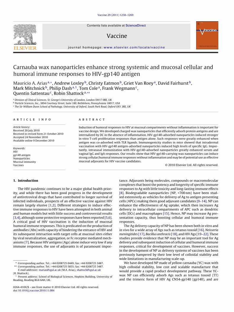
M.A. Arias et al. / Vaccine 29 (2011) 1258–1269 1261
Fig. 1. Physicochemical characterization of nanoparticles and binding to antigen. (A) Scanning electron microscopy of YC-wax NP: a, YC-SDS; b, YC-NaMA; c, YC-B r byp ncy ofp of Ag at
2
alianNnti
rij700chitosan. (B) Colloidal stability of YC-NaMA NP determined up to one yeaotential of naked NP and after adsorption with gp140 Ag. (D) Attachment efficieercentage of Ag bound to NP after subtracting the amount detected on the filtratehe percentage of Ag bound to NP with respect to Ag alone taken as 100%.
.12.2. Immunization protocol and sample collectionMice were inoculated i.d. with 12.5 �g (TT) or 20 �g (gp140) in
total volume of 100 �l in sterile saline on both dorsal flanks fol-owing a prime-boost-boost protocol at 4 (TT) and 3 (gp140) weekntervals. For i.n. immunization, 20 �g gp140 with or without NP inmaximum volume of 25 �l were gently dispensed in the animal’s
ostrils after isofluorane-induced anaesthesia. Antigen-adsorbedP were prepared the same day of immunization. Fresh compo-ents of the formulations were used in these experiments becausehey were performed in parallel with the NP colloidal stability stud-es (see Fig. 1B). These studies suggested nonetheless that similara, particle size distribution (PSD); b, zeta potential (Z); and c, viscosity. (C) Zetagp140 to NP measured by Bradford assay. Numerical values in the figure are thelone. (E) Detection by ELISA of gp140 adsorbed to NP. Values inside bars represent
results would be obtained using the same formulation over time.Alum-Ag complex was prepared by mixing equal volumes of Agand Alum solution (Imject Alum, Pierce, Rockford, IL), and mixedby rotation for 30 min at room temperature.
Blood samples were collected before priming, 1–3 days beforeboosting, and at 4 (TT) and 3 (gp140) weeks after the last boost.
◦
Serum was separated from clotted blood and stored at −80 C untilfurther use. Vaginal samples were collected by flushing 30 �l of PBSthree times into the vagina of anaesthetized animals, pooled andsupplemented with 8 �l of a 25× protease inhibitor cocktail (RocheDiagnostics, Manheim, Germany). Samples were incubated for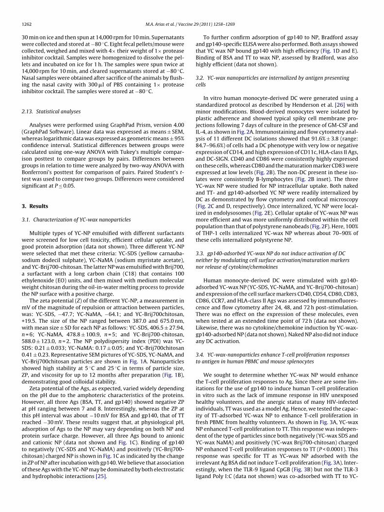
1 cine 2
3wcil1Nii
2
(wccigBts
3
3
wgwsaaewt
mw+wn5S0YsZd
oHatrapatcioa
262 M.A. Arias et al. / Vac
0 min on ice and then spun at 14,000 rpm for 10 min. Supernatantsere collected and stored at −80 ◦C. Eight fecal pellets/mouse were
ollected, weighed and mixed with 4× their weight of 1× proteasenhibitor cocktail. Samples were homogenized to dissolve the pel-ets and incubated on ice for 1 h. The samples were spun twice at4,000 rpm for 10 min, and cleared supernatants stored at −80 ◦C.asal samples were obtained after sacrifice of the animals by flush-
ng the nasal cavity with 300 �l of PBS containing 1× proteasenhibitor cocktail. The samples were stored at −80 ◦C.
.13. Statistical analyses
Analyses were performed using GraphPad Prism, version 4.00GraphPad Software). Linear data was expressed as means ± SEM,hereas logarithmic data was expressed as geometric means ± 95%
onfidence interval. Statistical differences between groups werealculated using one-way ANOVA with Tukey’s multiple compar-son posttest to compare groups by pairs. Differences betweenroups in relation to time were analyzed by two-way ANOVA withonferroni’s posttest for comparison of pairs. Paired Student’s t-est was used to compare two groups. Differences were consideredignificant at P ≤ 0.05.
. Results
.1. Characterization of YC-wax nanoparticles
Multiple types of YC-NP emulsified with different surfactantsere screened for low cell toxicity, efficient cellular uptake, and
ood protein adsorption (data not shown). Three different YC-NPere selected that met these criteria: YC-SDS (yellow carnauba-
odium dodecil sulphate), YC-NaMA (sodium myristate acetate),nd YC-Brij700-chitosan. The latter NP was emulsified with Brij700,surfactant with a long carbon chain (C18) that contains 100
thylenoxide (EO) units, and then mixed with medium moleculareight chitosan during the oil-in-water melting process to provide
he NP surface with a positive charge.The zeta potential (Z) of the different YC-NP, a measurement in
V of the magnitude of repulsion or attraction between particles,as: YC-SDS, −47.7; YC-NaMA, −64.1; and YC-Brij700chitosan,
19.5. The size of the NP ranged between 387.0 and 675.0 nm,ith mean size ± SD for each NP as follows: YC-SDS, 406.5 ± 27.94,= 6; YC-NaMA, 478.8 ± 100.9, n = 5; and YC-Brij700-chitosan,88.0 ± 123.0, n = 2. The NP polydispersity index (PDI) was YC-DS: 0.21 ± 0.033; YC-NaMA: 0.17 ± 0.05; and YC-Brij700chitosan.41 ± 0.23. Representative SEM pictures of YC-SDS, YC-NaMA, andC-Brij700chitosan particles are shown in Fig. 1A. Nanoparticleshowed high stability at 5 ◦C and 25 ◦C in terms of particle size,P, and viscosity for up to 12 months after preparation (Fig. 1B),emonstrating good colloidal stability.
Zeta potential of the Ags, as expected, varied widely dependingn the pH due to the amphoteric characteristics of the proteins.owever, all three Ags (BSA, TT, and gp140) showed negative ZPt pH ranging between 7 and 8. Interestingly, whereas the ZP athis pH interval was about −10 mV for BSA and gp140, that of TTeached −30 mV. These results suggest that, at physiological pH,dsorption of Ags to the NP may vary depending on both NP androtein surface charge. However, all three Ags bound to anionicnd cationic NP (data not shown and Fig. 1C). Binding of gp140
o negatively (YC-SDS and YC-NaMA) and positively (YC-Brij700-hitosan) charged NP is shown in Fig. 1C as indicated by the changen ZP of NP after incubation with gp140. We believe that associationf these Ags with the YC-NP may be dominated by both electrostaticnd hydrophobic interactions [25].9 (2011) 1258–1269
To further confirm adsorption of gp140 to NP, Bradford assayand gp140-specific ELISA were also performed. Both assays showedthat YC wax NP bound gp140 with high efficiency (Fig. 1D and E).Binding of BSA and TT to wax NP, assessed by Bradford, was alsohighly efficient (data not shown).
3.2. YC-wax nanoparticles are internalized by antigen presentingcells
In vitro human monocyte-derived DC were generated using astandardized protocol as described by Henderson et al. [26] withminor modifications. Blood-derived monocytes were isolated byplastic adherence and showed typical spiky cell membrane pro-jections following 7 days of culture in the presence of GM-CSF andIL-4, as shown in Fig. 2A. Immunostaining and flow cytometry anal-ysis of 11 different DC isolations showed that 91.6% ± 3.8 (range:84.7–96.6%) of cells had a DC phenotype with very low or negativeexpression of CD14, and high expression of CD11c, HLA-class II Ags,and DC-SIGN. CD40 and CD86 were consistently highly expressedon these cells, whereas CD80 and the maturation marker CD83 wereexpressed at low levels (Fig. 2B). The non-DC present in these iso-lates were consistently B-lymphocytes (Fig. 2B inset). The threeYC-wax NP were studied for NP intracellular uptake. Both nakedand TT- and gp140-adsorbed YC NP were readily internalized byDC as demonstrated by flow cytometry and confocal microscopy(Fig. 2C and D, respectively). Once internalized, YC NP were local-ized in endolysosomes (Fig. 2E). Cellular uptake of YC-wax NP wasmore efficient and was more uniformly distributed within the cellpopulation than that of polystyrene nanobeads (Fig. 2F). Here, 100%of THP-1 cells internalized YC-wax NP whereas about 70–90% ofthese cells internalized polystyrene NP.
3.3. gp140-adsorbed YC-wax NP do not induce activation of DCneither by modulating cell surface activation/maturation markersnor release of cytokine/chemokines
Human monocyte-derived DC were stimulated with gp140-adsorbed YC-wax NP (YC-SDS, YC-NaMA, and YC-Brij700-chitosan)and expression of the cell surface markers CD40, CD54, CD80, CD83,CD86, CCR7, and HLA-class II Ags was assessed by immunofluores-cence and flow cytometry after 24, 48, and 72 h post-stimulation.There was no effect on the expression of these molecules, evenwhen tested at an extended time point of 72 h (data not shown).Likewise, there was no cytokine/chemokine induction by YC-wax-gp140-adsorbed NP (data not shown). Naked NP also did not induceany DC activation.
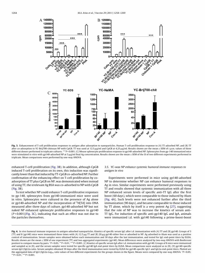
3.4. YC-wax-nanoparticles enhance T-cell proliferation responsesto antigen in human PBMC and mouse splenocytes
We sought to determine whether YC-wax NP would enhancethe T-cell proliferation responses to Ag. Since there are some lim-itations for the use of gp140 to induce human T-cell proliferationin vitro such as the lack of immune response in HIV unexposedhealthy volunteers, and the anergic status of many HIV-infectedindividuals, TT was used as a model Ag. Hence, we tested the capac-ity of TT-adsorbed YC-wax NP to enhance T-cell proliferation infresh PBMC from healthy volunteers. As shown in Fig. 3A, YC-waxNP enhanced T-cell proliferation to TT. This response was indepen-dent of the type of particles since both negatively (YC-wax SDS andYC-wax NaMA) and positively (YC-wax Brij700-chitosan) charged
NP enhanced T-cell proliferation responses to TT (P < 0.0001). Thisresponse was specific for TT as YC-wax NP adsorbed with theirrelevant Ag BSA did not induce T-cell proliferation (Fig. 3A). Inter-estingly, when the TLR-9 ligand CpGB (Fig. 3B) but not the TLR-3ligand Poly I:C (data not shown) was co-adsorbed with TT to YC-
M.A. Arias et al. / Vaccine 29 (2011) 1258–1269 1263
Fig. 2. Analysis of gp140-adsorbed nanoparticle internalization by antigen presenting cells. (A) Phase contrast microscopy of DC. (B) Phenotype of in vitro differentiatedhuman DC. Dot plot shows the gated DC population analyzed. Left histograms represent the isotype control. Inset depicts the staining for CD19+ showing about 10% of Bcells. n represents the number of blood donors with the mean ± SD plus range of cells with a DC phenotype. (C) Flow cytometry analysis of gp140-adsorbed wax NP uptakeb aMA sN rnere C. YelC rene (fl
Bece
y DC. Left histograms correspond to cells without NP. (D) DC gp140-wax NP (YC-Nucleus; Green: fluorescent gp140-adsorbed YC-NaMA NP. Values at the top left covery section. (E) Lysosomal localization of gp140-adsorbed YC-NaMA NP within Domparison of wax NP uptake by THP-1 cells of two different types of NP: polystyuorescent NP.
rij700-chitosan NP, the T-cell proliferation response was furthernhanced (P < 0.0001). To confirm that this effect was due to theo-adsorption of both TT Ag and CpGB to the YC-wax NP, sev-ral controls were performed (Fig. 3B). Specifically, to test that
hown) uptake as demonstrated by confocal microscopy. Red: cell membrane; Blue:of each quadrant represent the distance in �m from top to bottom of the cell afterlow color represents the overlapping of green (NP) plus red (lysosomes) colors. (F)left) vs YC-wax NaMA (right). Top right panel represents the MFI of cells without
the enhancing effect was not due to cell activation induced bythe chitosan present on the YC-wax Brij700-chitosan NP, both chi-tosan alone and together with TT (in the absence of NP) werealso assessed. Results show that neither chitosan nor TT+chitosan
1264 M.A. Arias et al. / Vaccine 29 (2011) 1258–1269
Fig. 3. Enhancement of T-cell proliferation responses to antigen after adsorption to nanoparticles. Human T-cell proliferation response to (A) TT-adsorbed NP, and (B) TTafter co-adsorption to YC-Brij700-chitosan NP with CpGB. TT was used at 12.5 �g/ml and CpGB at 4.25 �g/ml. Results shown are the mean ± SEM of c.p.m. values of threed prolifw . Resut
eiccao(
tiomn(t
F(cRpast*
ifferent donors performed in triplicate cultures. ***P < 0.001. (C) Mouse splenocyteere stimulated in vitro with gp140-adsorbed NP at 5 �g/ml final Ag concentration
riplicate. Mean comparisons were performed by one-way ANOVA.
nhanced T-cell proliferation (Fig. 3B). In addition, although CpGBnduced T-cell proliferation on its own, this induction was signifi-antly lower than that induced by TT-CpGB co-adsorbed NP. Furtheronfirmation of the enhancing effect on T-cell proliferation by co-dsorption of TT plus CpGB on NP, was demonstrated when insteadf using TT, the irrelevant Ag BSA was co-adsorbed to NP with CpGBFig. 3B).
To test whether NP could enhance T-cell proliferative responseso gp-140, splenocytes from gp140-immunized mice were usedn vitro. Splenocytes were cultured in the presence of Ag alone
3
r gp140-adsorbed NP and the incorporation of H[Td] into DNAeasured after three days of culture. gp140-adsorbed NP but notaked NP enhanced splenocyte proliferative responses to gp140P < 0.001)(Fig. 3C), indicating that such an effect was not due tohe particles themselves.
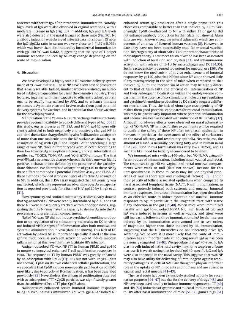
ig. 4. In vivo humoral immune responses to antigen-adsorbed nanoparticles. Kinetics oTT) and 8 (gp140) mice were immunized three times with (A) 12.5 �g TT and (B) 20 �gontrol of immunization. Serum samples were obtained before each immunization andesults shown are the mean ± SEM of one experiment (TT) and two aggregated experimeosttest to compare means by pairs. *P < 0.05; **P < 0.01; ***P < 0.001. (C) Kinetics of specifind sampled as in (B), and the serum samples were tested for specific-gp140 IgA end-poerum IgG1/IgG2a ratio. Serum samples obtained 30 days after the third immunization whe aggregated data of IgG1/IgG2a Log10 ratio values of two different experiments for the*P < 0.01; ***P < 0.001.
eration response to gp140-adsorbed NP. Splenocytes from gp-140 immunized micelts shown are the mean ± SEM of the SI of two different experiments performed in
3.5. YC-wax NP enhance systemic humoral immune responses toantigen in vivo
Experiments were performed in mice using gp140-adsorbedNP to determine whether NP can enhance humoral responses toAg in vivo. Similar experiments were performed previously usingTT and results showed that systemic immunization with all threeNP enhanced serum levels of specific anti-TT IgG after the firstboost (60 days), which were comparable to those induced by Alum(Fig. 4A). Such levels were not enhanced further after the third
immunization (90 days), and became comparable to those inducedby TT alone, which by itself is a very potent Ag [27], suggestingthat the role of NP was to increase the kinetics of serum anti-TT IgG. For induction of specific anti-gp140 IgG and IgA, animalswere immunized i.d. with gp140 following a prime-boost-boostf specific serum IgG after i.d. immunization with (A) TT and (B) gp140. Groups of 3gp140 either free or adsorbed to NP. Ag adsorbed to Alum was used as a positive30 days after the last immunization, and tested for IgG end-point titers by ELISA.nts (gp140). Mean differences were analyzed by two-way ANOVA with Bonferronic serum IgA after i.d. immunization with gp140. Groups of 8 mice were immunizedint titers by ELISA. Mean comparisons were analyzed as in (B). (D) gp140-specificere tested by ELISA for gp140-specific IgG1 and IgG2a end-point titers. Plotted aregroups shown in the figure. Means were compared by one-way ANOVA. *P < 0.05;

M.A. Arias et al. / Vaccine 29 (2011) 1258–1269 1265

1266 M.A. Arias et al. / Vaccine 29 (2011) 1258–1269
Fig. 5. Systemic and mucosal humoral immune responses to gp140-adsorbed nanoparticles after intranasal immunization. (A) and (B) Kinetics of serum and vaginal IgGand IgA levels. Groups of 5 animals were immunized i.n. with 20 �g of gp140 either free or adsorbed to YC-NaMA NP. Serum and vaginal lavages were obtained before eachi 140 Ig* re imms specifit
peeahwePF
otiYgtoNwrs
tOt
mmunization and 21 days after the last immunization, and tested for specific-gp**P < 0.001. (C) Levels of IgG and IgA in nasal lavage after second boost. Animals weacrificed and the nasal cavity washed with saline. Samples were tested for gp140--test.
rotocol at 30 day intervals. Serum samples were taken beforeach immunization and 30 days after the last boost, and the lev-ls of IgG and IgA were tested by gp140-specific ELISA. gp140lone induced significant levels of IgG but these levels were muchigher when the Ag was adsorbed to NP (Fig. 4B). Such IgG levelsere comparable to those induced by Alum (day 60), and differ-
nces were already observable following a single prime (day 30).lateau IgG levels were already observed after first boost (day 60,ig. 4B).
Serum specific anti-gp140 IgA was only detected after the sec-nd immunization (60 days) but the titers were not high becausehe maximum end-point titer levels of IgA detected under thesemmunization conditions were 282.4 ± 88.4 (log10 2.45 ± 1.95).C-Brij700chitosan-gp140 but not YC-SDS-gp140 nor YC-NaMA-p140 promoted significant specific-gp140 IgA titers (P < 0.05) afterhree immunizations (90 days). Such effect was comparable to thatf Alum at the same time point (P < 0.05). However, the effect ofP as a whole on serum specific-gp140 IgA after i.d. immunizationas low because the kinetics and magnitude of specific-gp140 IgA
esponses promoted by Alum after the first boost (60 days) was
ignificantly superior to those of NP (Fig. 4C).To test whether YC-wax NP modulated T-helper cell responses,he gp140 specific IgG1/IgG2a ratio was also determined by ELISA.f note, gp140 alone induced an IgG response that was biased
owards a Th2 phenotype. Such a response did not appear to be
G and IgA end-point titers by ELISA. Means were compared by two-way ANOVA.unized as in (A) and (B), and 21 days after the last immunization the animals werec IgG and IgA end-point titers as for (A) and (B). Means were compared by paired
modulated by Alum, YC-wax NaMA or YC-wax Brij700-chitosan(Fig. 4D). However, YC-wax SDS appeared to induce a more bal-anced Th1/Th2 response (Fig. 4D).
3.6. YC-wax-nanoparticles enhance mucosal humoral responsesto gp140
To test whether NP were also capable of enhancing mucosalhumoral responses to gp140, mice were immunized nasally witheither Ag alone or adsorbed to YC-wax-NaMA NP, and the levelsof IgG and IgA were determined in serum and mucosal fluids. Wechose YC-NaMA NP for i.n. immunization first because, these NPshowed a significant enhancement of systemic humoral immuneresponses to both TT and gp140 across the i.d. immunizations (seeFig. 4A and B). Second, NaMA is a naturally occurring surfactant,present in many natural oils and, more importantly, in human nasalfluid [28]. Alum was not used as a positive control of adjuvanticityfor i.n immunization due to the intrinsic inflammatory role of Alumsalts, since part of their mechanism of action is to induce necroticand damaged cells at the site of injection [29], an effect that would
be incompatible with nasal immunization.Antigen alone failed to induce any response (Fig. 5). In contrast,there was a steady increase over time in both serum IgG and IgA inresponse to gp140 adsorbed to YC-NaMA NP (Fig. 5A). These levelsdid not seem to reach a plateau after the second boost, as it was
cine 2
ohmwatwwir
4
mttfArdf
ptcaoartptpdtttut[
ttgp
tnsagi
ivbnwmpwt
t
M.A. Arias et al. / Vac
bserved with serum IgG after intradermal immunization. Notably,igh levels of IgA were also observed in vaginal secretions, with aoderate increase in IgG (Fig. 5B). In addition, IgG and IgA levelsere also detected in the nasal lavages of these mice (Fig. 5C). No
ntibody induction was observed in feces (data not shown). Of note,he IgG1/IgG2a ratio in serum was very close to 1 (1.57 ± 0.079),hich was lower than that induced by intradermal immunizationith gp-140-YC-wax NaMA, suggesting that the type of T-helper
mmune response induced by NP may change depending on theoute of immunization.
. Discussion
We have developed a highly stable NP vaccine delivery systemade of YC-wax material. These NP have a low cost of production
hat is easily scalable. Indeed, similar particles are already manufac-ured in kilogram quantities for use in the cosmetics industry. Theseeatures, together with their capacity to efficiently adsorb proteings, to be readily internalized by APC, and to enhance immuneesponses to Ag both in vitro and in vivo, make them good potentialelivery systems for vaccines, and in particular that of HIV vaccinesor the developing world.
Manipulation of the YC-wax NP surface charge with surfactants,rovides optimal flexibility to adsorb different types of Ag [30]. Inhis study, Ags as diverse as TT, BSA, and HIV-1 gp140 were effi-iently adsorbed to both negatively and positively charged NP. Inddition, the surface charge flexibility also facilitated co-adsorptionf more than one molecule onto the NP surface as shown by co-dsorption of Ag with CpGB and PolyI:C. After screening a largeange of wax NP, three different types were selected according toheir low toxicity, Ag adsorption efficiency, and cell internalizationrofile, i.e., YC-SDS, YC-NaMA, and YC-Brij700-chitosan. The firstwo NP had a net negative charge, whereas the third one was highlyositive, a characteristic defined by the presence of the carbohy-rate chitosan. We determined adsorption of gp140 to these NP byhree different methods: Z potential, Bradford assay, and ELISA. Allhree methods provided strong evidence of effective Ag adsorptiono NP. In addition, the ELISA assay suggested that antigenicity wasnaffected, which may represent an advantage over Ag encapsula-ion as reported previously for a form of HIV-gp120 by Singh et al.31].
Flow cytometry and confocal microscopy studies clearly showedhat Ag-adsorbed YC NP were readily internalized by APC, and thathese NP were subsequently tracked within endolysosomes, sug-esting that the NP may have the capacity to deliver Ag into the Agrocessing and presentation compartment.
Naked YC-wax NP did not induce cytokine/chemokine produc-ion or up-regulation of co-stimulatory molecules on DC in vitro,or induced visible signs of inflammation after both mucosal andystemic administration in vivo (data not shown). This lack of DCctivation by naked NP is important especially if used at the uro-enital tract, because such cell activation would induce mucosalnflammation at this level that may facilitate HIV infection.
Antigen-adsorbed YC-wax NP (TT in human PBMC and gp140n mouse splenocytes) enhanced T-cell proliferation responses initro. The response to TT by human PBMC was greatly enhancedy co-adsorption with CpGB (Fig. 3B) but not with PolyI:C (dataot shown). CpGB on its own enhanced cellular proliferation, ande speculate that CpGB induces non-specific proliferation of PBMCost likely due to polyclonal B cell activation, as has been described
reviously [32]. Nevertheless, the enhanced proliferation observedith co-adsorption of TT + CpGB particles was significantly greater
han the additive effect of TT plus CpGB alone.Nanoparticles enhanced serum humoral immune responses
o Ag in vivo. Animals immunized i.d. with gp140-adsorbed NP
9 (2011) 1258–1269 1267
enhanced serum IgG production after a single prime, and thiseffect was comparable or better than that induced by Alum. Sur-prisingly, CpGB co-adsorbed to NP with either TT or gp140 didnot enhance antibody production further (data not shown). Alumsalts are well known strong parenteral adjuvants which are com-ponents of an array of licensed human vaccines [8]. However, todate they have not been successfully used for mucosal vaccina-tion. Reactogenicity of Alum salts is an important characteristic oftheir adjuvanticity. Their mechanism of action has been associatedwith induction of local uric acid crystals [33] and inflammasomeactivation with release of IL-1� by macrophages and DC [34,35].Such reactogenicity is deemed too potent for mucosal use [36]. Wedo not know the mechanism of in vivo enhancement of humoralresponses by gp140-adsorbed NP but since NP alone showed littleif any reactogenicity in the skin of mice when compared to thatinduced by Alum, the mechanism of action may be highly differ-ent to that of Alum salts. The efficient cell internalization of NPand their subsequent localization within the endolysosome com-partment in the absence of co-stimulatory molecule up-regulationand cytokine/chemokine production by DC clearly suggest a differ-ent mechanism. Thus, the lack of Alum-type reactogenicity of NPmakes them good potential candidates for mucosal immunization.This may be particularly important where potential inflammationand edema have been associated with induction of Bell’s palsy [37].Although no adverse effects were observed on nasal administra-tion of YC-NaMA NP in mice, further experiments will be requiredto confirm the safety of these NP after intranasal application inhumans, in particular the assessment of the effect of surfactantson the nasal olfactory and respiratory epithelia. Nevertheless, theamount of NaMA, a naturally occurring fatty acid in human nasalfluid [28], used in this formulation was very low (0.025%), and assuch the likelihood for toxicity is considered to be small.
We immunized mice with gp140-adsorbed YC-NaMA using dif-ferent routes of immunization, including nasal, vaginal and rectal.The responses to gp140 via vaginal and rectal mucosal compart-ments were weak or null (data not shown). Reasons for thisunresponsiveness in these mucosas may include physical prop-erties of mucus (pore size and rheological factors) [38], and/ortheir paucity of follicle associated epithelium when compared tonasal associated lymphoid tissue (NALT). Nasal immunization, incontrast, potently induced both systemic and mucosal humoralimmune responses. Intranasal immunization has been describedas an effective route to induce systemic and mucosal immuneresponses to Ag, in particular in the urogenital tract, with scarceif any induction in the gut [39,40]. When mice were immunizednasally with gp140-adsorbed NaMA NP, high levels of IgG andIgA were induced in serum as well as vagina, and titers werestill increasing following three immunizations. IgA levels in seruminduced by i.n. immunization were around one to two ordersof magnitude higher than those induced by i.d. immunization,suggesting that the NP themselves do not inherently drive IgAswitching. We believe it is more likely that the route of immu-nization has an important role at inducing serum IgA as has beenpreviously suggested [39,40]. We speculate that gp140-specific IgAplasma cells induced in the nasal cavity may home to spleen or bonemarrow. It is worth noting that levels of gp140-specific IgG and IgAwere also enhanced in the nasal cavity. This suggests that wax NPmay also have utility for delivering of immunogens against respi-ratory pathogens. M-cells of NALT are thought to play an importantrole in the uptake of NP in rodents and humans and are absent invaginal and rectal mucosa [41–43].
The nasal route has been extensively studied not only for vacci-nation purposes [44–47] but also for the delivery of drugs [48], andNP have been used nasally to induce immune responses to TT [49]and HIV [50]. Induction of systemic and mucosal immune responsesto HIV after nasal immunization of mice [51,52], guinea pigs [51]
1 cine 2
aoamcemewmTabt
D
c
A
Gl
mWAVsbE
R
[
[
[
[
[
[
[
[
[
[
[
[
[
[
[
[
[
[
[
[
[
[
[
[
[
[
[
[
[
[
268 M.A. Arias et al. / Vac
nd macaques [5] with HIV-gp120 Ag has been described previ-usly. In the latter, serum and vaginal Ab responses were inducedfter nasal immunization only when followed by one or two intra-uscular boosts. These levels were highly enhanced in vagina after
hallenge with SHIV, suggesting that the nasal priming inducedffective memory responses at mucosal level [5]. In our mouseodel, three nasal immunizations were enough to induce high lev-
ls of IgG and IgA in serum and vagina. It remains to be confirmedhether this immunization protocol with NP will work similarly inacaques or humans, or whether these Abs would be neutralizing.
herefore, further studies are warranted that assess homologousnd heterologous immunization protocols to determine the feasi-ility of using these NP, as effective delivery systems of HIV Ags, inhe development of mucosal vaccination in humans.
isclosure
Particle Science Inc has IP rights and economical interests inarnauba wax based nanoparticles mentioned in this article.
cknowledgements
This work was funded by a grant to SGUL by the Bill & Melindaates Foundation and the Wellcome Trust, under the Grand Chal-
enges in Global Health Initiative.We are indebted to the Fondation Dormeur for funding of equip-
ent used in the course of this study. We thank Professors Ralfagner and Hans Wolf, University of Regensburg and GENEART
G for the CN54-expressing plasmid. We thank Simon Jeffs, Sueliieira and Saba Hussein for work on gp140 cloning and expres-ion. CN54-gp140 used in this study was produced under contracty Polymun Scientific GmbH. Griet Van Roey is supported by aUROPRISE studentship funded by the European Union.
eferences
[1] McMichael AJ, Borrow P, Tomaras GD, Goonetilleke N, Haynes BF. The immuneresponse during acute HIV-1 infection: clues for vaccine development 1. NatRev Immunol 2010;10(January (1)):11–23.
[2] Letvin NL. Progress and obstacles in the development of an AIDS vaccine. NatRev Immunol 2006;6(December (2)):930–9.
[3] HIV gp120 vaccine. VaxGen: AIDSVAX, AIDSVAX B/B, AIDSVAX B/E, HIV gp120vaccine – Genentech, HIV gp120 vaccine AIDS. Drugs R D 2003;4(4):249–53.
[4] Bradac J, Dieffenbach CW. HIV vaccine development: lessons from the past,informing the future. IDrugs 2009 Jul;12(7):435–9.
[5] Barnett SW, Srivastava IK, Kan E, Zhou F, Goodsell A, Cristillo AD, et al. Protectionof macaques against vaginal SHIV challenge by systemic or mucosal and sys-temic vaccinations with HIV-envelope. AIDS 2008;22(January 30 (3)):339–48.
[6] Rerks-Ngarm S, Pitisuttithum P, Nitayaphan S, Kaewkungwal J, Chiu J, ParisR, et al. Vaccination with ALVAC and AIDSVAX to prevent HIV-1 infection inThailand. N Engl J Med 2009;361(December 3 (23)):2209–20.
[7] Shattock RJ, Haynes BF, Pulendran B, Flores J, Esparza J. Improving defences atthe portal of HIV entry: mucosal and innate immunity. PLoS Med 2008;5(April1 (4)):e81.
[8] Reed SG, Bertholet S, Coler RN, Friede M. New horizons in adjuvants for vaccinedevelopment. Trends Immunol 2009;30(January (1)):23–32.
[9] Chellat F, Merhi Y, Moreau A, Yahia L. Therapeutic potential of nanopar-ticulate systems for macrophage targeting. Biomaterials 2005;26(December(35)):7260–75.
10] Copland MJ, Baird MA, Rades T, McKenzie JL, Becker B, Reck F, et al. Liposo-mal delivery of antigen to human dendritic cells. Vaccine 2003;21(February 14(9–10)):883–90.
11] Kempf M, Mandal B, Jilek S, Thiele L, Voros J, Textor M, et al. Improved stimu-lation of human dendritic cells by receptor engagement with surface-modifiedmicroparticles. J Drug Target 2003;11(January (1)):11–8.
12] Singh M, Kazzaz J, Ugozzoli M, Chesko J, O’Hagan DT. Charged polylactide co-glycolide microparticles as antigen delivery systems. Expert Opin Biol Ther2004;4(April (4)):483–91.
13] Uto T, Wang X, Sato K, Haraguchi M, Akagi T, Akashi M, et al. Targeting of anti-
gen to dendritic cells with poly(gamma-glutamic acid) nanoparticles inducesantigen-specific humoral and cellular immunity. J Immunol 2007;178(March1 (5)):2979–86.14] Wischke C, Borchert HH, Zimmermann J, Siebenbrodt I, Lorenzen DR. Stablecationic microparticles for enhanced model antigen delivery to dendritic cells.J Control Release 2006;114(September 12 (3)):359–68.
[
[
[
9 (2011) 1258–1269
15] Shen H, Ackerman AL, Cody V, Giodini A, Hinson ER, Cresswell P, et al.Enhanced and prolonged cross-presentation following endosomal escape ofexogenous antigens encapsulated in biodegradable nanoparticles. Immunology2006;117(January (1)):78–88.
16] Tobio M, Nolley J, Guo Y, McIver J, Alonso MJ. A novel system based on a polox-amer/PLGA blend as a tetanus toxoid delivery vehicle. Pharm Res 1999;16(May(5)):682–8.
17] Singh M, Kazzaz J, Chesko J, Soenawan E, Ugozzoli M, Giuliani M, et al. Anionicmicroparticles are a potent delivery system for recombinant antigens fromNeisseria meningitidis serotype B. J Pharm Sci 2004;93(February (2)):273–82.
18] Westwood A, Healey GD, Williamson ED, Eyles JE. Activation of dendritic cellsby microparticles containing Bacillus anthracis protective antigen. Vaccine2005;23(May 31 (29)):3857–63.
19] Akagi T, Kawamura M, Ueno M, Hiraishi K, Adachi M, Serizawa T, et al.Mucosal immunization with inactivated HIV-1-capturing nanospheres inducesa significant HIV-1-specific vaginal antibody response in mice. J Med Virol2003;69(February (2)):163–72.
20] Aline F, Brand D, Pierre J, Roingeard P, Severine M, Verrier B, et al. Dendriticcells loaded with HIV-1 p24 proteins adsorbed on surfactant-free anionic PLAnanoparticles induce enhanced cellular immune responses against HIV-1 aftervaccination. Vaccine 2009;27(August 20 (38)):5284–91.
21] Kazzaz J, Neidleman J, Singh M, Ott G, O’Hagan DT. Novel anionic micropar-ticles are a potent adjuvant for the induction of cytotoxic T lymphocytesagainst recombinant p55 gag from HIV-1. J Control Release 2000;67(July 3(2–3)):347–56.
22] Wang X, Uto T, Akagi T, Akashi M, Baba M. Induction of potent CD8+ T-cellresponses by novel biodegradable nanoparticles carrying human immunode-ficiency virus type 1 gp120. J Virol 2007;81(September (18)):10009–16.
23] Su L, Graf M, Zhang Y, von BH, Xing H, Kostler J, et al. Characterization of avirtually full-length human immunodeficiency virus type 1 genome of a preva-lent intersubtype (C/B’) recombinant strain in China. J Virol 2000;74(December(23)):11367–76.
24] Biancotto A, Grivel JC, Iglehart SJ, Vanpouille C, Lisco A, Sieg SF, et al. Abnormalactivation and cytokine spectra in lymph nodes of people chronically infectedwith HIV-1 2. Blood 2007;109(May 15 (10)):4272–9.
25] Chesko J, Kazzaz J, Ugozzoli M, O’Hagan DT, Singh M. An investigation of thefactors controlling the adsorption of protein antigens to anionic PLG micropar-ticles. J Pharm Sci 2005;94(November (11)):2510–9.
26] Henderson RA, Watkins SC, Flynn JL. Activation of human dendritic cells fol-lowing infection with Mycobacterium tuberculosis. J Immunol 1997;159(July15 (2)):635–43.
27] Gupta RK, Siber GR. Adjuvants for human vaccines – current status, problemsand future prospects 2. Vaccine 1995;13(October (14)):1263–76.
28] Do TQ, Moshkani S, Castillo P, Anunta S, Pogosyan A, Cheung A, et al.Lipids including cholesteryl linoleate and cholesteryl arachidonate contributeto the inherent antibacterial activity of human nasal fluid 1. J Immunol2008;181(September 15 (6)):4177–87.
29] Lambrecht BN, Kool M, Willart MA, Hammad H. Mechanism of action ofclinically approved adjuvants 1. Curr Opin Immunol 2009;21(February (1)):23–9.
30] Singh M, Kazzaz J, Ugozzoli M, Malyala P, Chesko J, O’Hagan DT. Polylactide-co-glycolide microparticles with surface adsorbed antigens as vaccine deliverysystems. Curr Drug Deliv 2006;3(January (1)):115–20.
31] Singh M, Chesko J, Kazzaz J, Ugozzoli M, Kan E, Srivastava I, et al. Adsorption ofa novel recombinant glycoprotein from HIV (Env gp120dV2 SF162) to anionicPLG microparticles retains the structural integrity of the protein, whereasencapsulation in PLG microparticles does not. Pharm Res 2004;21(December(12)):2148–52.
32] Bekeredjian-Ding I, Jego G. Toll-like receptors – sentries in the B-cell response.Immunology 2009;128(November (3)):311–23.
33] Kool M, Soullie T, van NM, Willart MA, Muskens F, Jung S, et al. Alum adjuvantboosts adaptive immunity by inducing uric acid and activating inflammatorydendritic cells. J Exp Med 2008;205(April 14 (4)):869–82.
34] Li H, Willingham SB, Ting JP, Re F. Cutting edge: inflammasome activa-tion by alum and alum’s adjuvant effect are mediated by NLRP3. J Immunol2008;181(July 1 (1)):17–21.
35] Sharp FA, Ruane D, Claass B, Creagh E, Harris J, Malyala P, et al. Uptake of partic-ulate vaccine adjuvants by dendritic cells activates the NALP3 inflammasome.Proc Natl Acad Sci USA 2009;106(January 20 (3)):870–5.
36] Harandi AM, Medaglini D, Shattock RJ. Vaccine adjuvants: a priority for vaccineresearch. Vaccine 2010;28(March 11 (12)):2363–6.
37] Lewis DJ, Huo Z, Barnett S, Kromann I, Giemza R, Galiza E, et al. Tran-sient facial nerve paralysis (Bell’s palsy) following intranasal delivery of agenetically detoxified mutant of Escherichia coli heat labile toxin. PLoS One2009;4(9):e6999.
38] Lai SK, Wang YY, Hanes J. Mucus-penetrating nanoparticles for drug andgene delivery to mucosal tissues. Adv Drug Deliv Rev 2009;61(February 27(2)):158–71.
39] Holmgren J, Czerkinsky C. Mucosal immunity and vaccines. Nat Med2005;11(April (4 Suppl.)):S45–53.
40] Kunkel EJ, Butcher EC. Plasma-cell homing. Nat Rev Immunol 2003;3(October(10)):822–9.
41] Brooking J, Davis SS, Illum L. Transport of nanoparticles across the rat nasalmucosa. J Drug Target 2001;9(4):267–79.
42] Davis IC, Owen RL. The immunopathology of M cells. Springer SeminImmunopathol 1997;18(4):421–48.

cine 2
[
[
[
[
[
[
[
[
[
M.A. Arias et al. / Vac
43] Fujimura Y. Evidence of M cells as portals of entry for antigens in the nasopha-ryngeal lymphoid tissue of humans. Virchows Arch 2000;436(June (6)):560–6.
44] Asahi Y, Yoshikawa T, Watanabe I, Iwasaki T, Hasegawa H, Sato Y, et al. Pro-tection against influenza virus infection in polymeric Ig receptor knockoutmice immunized intranasally with adjuvant-combined vaccines. J Immunol2002;168(March 15 (6)):2930–8.
45] Bradney CP, Sempowski GD, Liao HX, Haynes BF, Staats HF. Cytokinesas adjuvants for the induction of anti-human immunodeficiency viruspeptide immunoglobulin G (IgG) and IgA antibodies in serum andmucosal secretions after nasal immunization. J Virol 2002;76(January (2)):517–24.
46] Hirano T, Jiao X, Chen Z, Van WC, Gu XX. Kinetics of mouse anti-body and lymphocyte responses during intranasal vaccination with a
lipooligosaccharide-based conjugate vaccine. Immunol Lett 2006;107(Novem-ber 15 (2)):131–9.47] Sakaue G, Hiroi T, Nakagawa Y, Someya K, Iwatani K, Sawa Y, et al. HIV mucosalvaccine: nasal immunization with gp160-encapsulated hemagglutinating virusof Japan-liposome induces antigen-specific CTLs and neutralizing antibodyresponses. J Immunol 2003;170(January 1 (1)):495–502.
[
9 (2011) 1258–1269 1269
48] Illum L. Transport of drugs from the nasal cavity to the central nervous system.Eur J Pharm Sci 2000;11(July (1)):1–18.
49] Vila A, Sanchez A, Evora C, Soriano I, McCallion O, Alonso MJ. PLA-PEG par-ticles as nasal protein carriers: the influence of the particle size. Int J Pharm2005;292(March 23 (1–2)):43–52.
50] Caputo A, Castaldello A, Brocca-Cofano E, Voltan R, Bortolazzi F, Altavilla G,et al. Induction of humoral and enhanced cellular immune responses by novelcore-shell nanosphere- and microsphere-based vaccine formulations follow-ing systemic and mucosal administration. Vaccine 2009;27(June 2 (27)):3605–15.
51] Bielinska AU, Janczak KW, Landers JJ, Markovitz DM, Montefiori DC, Baker Jr JR.Nasal immunization with a recombinant HIV gp120 and nanoemulsion adju-vant produces Th1 polarized responses and neutralizing antibodies to primaryHIV type 1 isolates. AIDS Res Hum Retroviruses 2008;24(February (2)):271–
81.52] Buonaguro L, Visciano ML, Tornesello ML, Tagliamonte M, Biryahwaho B,Buonaguro FM. Induction of systemic and mucosal cross-clade neutralizingantibodies in BALB/c mice immunized with human immunodeficiency virustype 1 clade A virus-like particles administered by different routes of inocula-tion. J Virol 2005;79(June (11)):7059–67.


![merged document 4 (1) - Amazon S3 · 40 Lake, Yellow 6 Lake, Carnauba Wax, Yellow 5 Lake, Blue 1 Lake, Red 3], Tapioca Starch, Calcium Carbonate and Sucralose. ALLERGY ALERT: This](https://img.pdfslide.us/doc/110x75/5e1beb75cccff911cc232e32/merged-document-4-1-amazon-s3-40-lake-yellow-6-lake-carnauba-wax-yellow-5.jpg)


![Index [rd.springer.com]978-94-011-1482-0/1.pdf · Cantharides 274 Capsicum 274 Carbomers 33 Carnauba wax 220 Carrageenan 75 Castor oil in lipstick 216-17 in nail enamel 251-2 Cationics,](https://img.pdfslide.us/doc/110x75/5d269df488c993e5378dcf9b/index-rd-978-94-011-1482-01pdf-cantharides-274-capsicum-274-carbomers.jpg)