Embed Size (px)
Citation preview

Cardiovascular system I
Congenital heart disease
• Congenital heart diseases are abnormalities ofthe heart or great vessels that are present atbirth.
• Most such disorders arise from faultyembryogenesis during gestational weeks 3through 8, when major cardiovascularstructures develop.

Congenital heart diseases can be subdivided into three major groups:
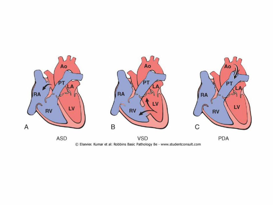
1. Malformations causing a left-to-right shunt
2. Malformations causing a right-to-left shunt(cyanotic congenital heart diseases)
3. Malformations causing obstruction

Left-to-Right Shunts
• left-to-right shunts increase pulmonary bloodflow and are not associated (at least initially)with cyanosis.
• They expose the low-pressure, low-resistancepulmonary circulation to increased pressureand volume, resulting in right ventricularhypertrophy and-eventually-right-sidedfailure.
• represent the most common type of congenital cardiac malformation
• They include
1. atrial septal defects
2. ventricular septal defects
3. patent ductus arteriosus.

• Cyanosis is not an early feature of thesedefects, but it can occur late, after prolongedleft-to-right shunting has produced pulmonaryhypertension sufficient to yield right-sidedpressures that exceed those on the left andthus result in a reversal of blood flow throughthe shunt.
• Such reversal of flow and shunting ofunoxygenated blood to the systemiccirculation is called Eisenmenger syndrome.

• Once significant pulmonary hypertension develops, the structural defects of congenital heart disease are considered irreversible.
• This is the rationale for early intervention, either surgical or nonsurgical.
• Atrial Septal Defects


• three types of ASD are recognized. • ostium secundum ASD:• The most common (90%)• occurs when the septum secundum does not enlarge
sufficiently to cover the ostium secundum.
• Ostium primum ASDs• less common (5% of cases); • occur if the septum primum and endocardial cushion
fail to fuse• are often associated with abnormalities in other
structures derived from the endocardial cushion (e.g., mitral and tricuspid valves).

• The sinus venosus ASDs (5% of cases) are located near the entrance of the superior vena cava and have been associated with frameshiftmutations in the NKX2.5 transcription factor.

Clinical Features
• ASD are less common than VSD, but they are the most common defects to be first diagnosed in adults (which are less likely to spontaneously close).
• ASDs initially cause asymptomatic left-to-right shunts. Later cause pulmonary hypertension (less than 10% of patients with uncorrected ASD).

• Ostium primum defects are more likely to be associated with evidence of CHF, in part because of the high frequency of associated mitral insufficiency.
• The objective of surgical closure is to prevent complications:
heart failure
paradoxical embolization
pulmonary hypertension

• Ventricular Septal Defects
• The ventricular septum is normally formed by the fusion of an intraventricular muscular ridge that grows upward from the apex of the heart with a thinner membranous partition that grows downward from the endocardialcushion.

• The basal (membranous) region is the last part of the septum to develop and is the site of approximately 90% of VSDs.
• Although more common at birth than ASDs, most VSDs close spontaneously in childhood, so that the overall incidence in adults is lower than that of ASDs.

• 30% of VSDs occur in isolation; more commonly, they are associated with other cardiac malformations.
Clinical Features
• Small VSDs may be asymptomatic, and those in the muscular portion of the septum may close spontaneously during infancy or childhood.

• Larger defects, however, cause a severe left-to-right shunt, often complicated by pulmonary hypertension and CHF. Progressive pulmonary hypertension, with resultant reversal of the shunt and cyanosis, occurs earlier and more frequently in patients with VSDs than in those with ASDs; hence, early surgical correction is indicated for such lesions.

• Small- or medium-sized defects that produce jet lesions in the right ventricle are also prone to superimposed infective endocarditis.

• Patent Ductus Arteriosus
• During intrauterine life, the ductus arteriosus permits blood flow from the pulmonary artery to the aorta, thereby bypassing the unoxygenated lungs.
• Shortly after birth, however, the ductus constricts; this occurs in response to increased arterial oxygenation, decreased pulmonary vascular resistance, and declining local levels of prostaglandin E2.
• In healthy term infants, the ductus is functionally nonpatent within 1 to 2 days after birth; complete, structural obliteration occurs within the first few months of extrauterine life to form the ligamentumarteriosum.

• Ductal closure is often delayed (or even absent) in infants with hypoxia (resulting from respiratory distress or heart disease).
• PDAs account for about 7% of cases of congenital heart lesions; 90% of these are isolated defects.
• The remaining occur with other congenital defects, most commonly VSDs.
Clinical Features
• PDAs are high-pressure left-to-right shunts, audible as harsh "machinery-like" murmurs.
• A small PDA generally causes no symptoms, although larger bore defects can eventually lead to the Eisenmenger syndrome with cyanosis and CHF.
• The high-pressure shunt also predisposes affected individuals to infective endocarditis.

• preservation of ductal patency (by administering prostaglandin E) may be critically important for infants with various forms of congenital heart disease wherein the PDA is the only means to provide systemic or pulmonary blood flow (e.g., aortic or pulmonicatresia).

Right-to-Left Shunts
• poorly oxygenated blood from the right side of the heart is introduced directly into the arterial circulation.
• Cardiac malformations associated with right-to-left shunts are distinguished by cyanosis at or near the time of birth.
• Two of the most important conditions associated with cyanotic congenital heart disease are:
tetralogy of Fallot
transposition of the great vessels
• Clinical findings associated with severe, long-standing cyanosis include:
• clubbing of the fingertips (hypertrophic osteoarthropathy) and polycythemia.
• In addition, right-to-left shunts permit venous emboli to bypass the lungs and directly enter the systemic circulation (paradoxical embolism).
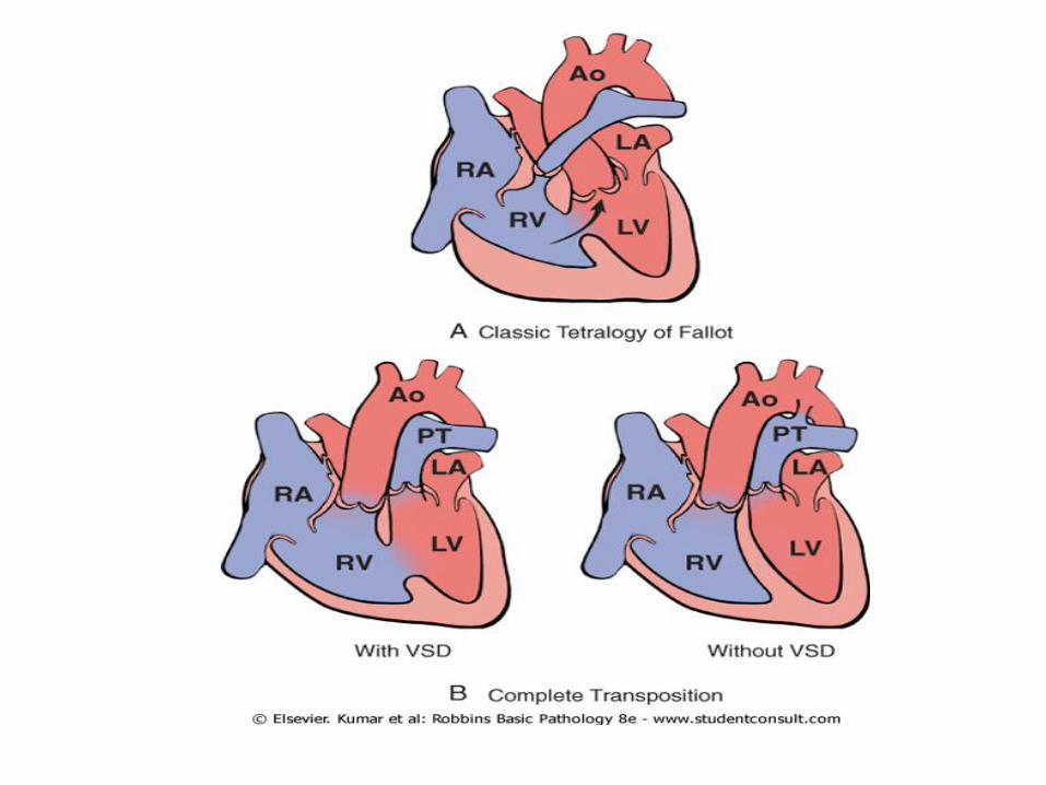

• Tetralogy of Fallot
• Accounting for about 5% of all congenital cardiac malformations,
• is the most common cause of cyanotic congenital heart disease

• The four features of the tetralogy are
1. VSD,
2. obstruction to the right ventricular outflow tract (subpulmonic stenosis),
3. an aorta that overrides the VSD
4. right ventricular hypertrophy.

• the clinical severity largely depends on the degree of the pulmonary outflow obstruction.

Clinical Features
• The extent of shunting (and the clinical severity) is determined by the amount of right ventricular outflow obstruction.

• If the pulmonic obstruction is mild, the condition resembles an isolated VSD, because the high left-sided pressures on the left side cause a left-to-right shunt with no cyanosis.
• More commonly, marked stenosis causes significant right-to-left shunting and consequent cyanosis early in life.

• As patients with tetralogy grow, the pulmonicorifice does not enlarge, despite an overall increase in the size of the heart.
• Hence, the degree of stenosis typically worsens with time resulting in increasing cyanosis.
• The lungs are protected from hemodynamic overload by the pulmonic stenosis, so that pulmonary hypertension does not develop.

• As with any cyanotic heart disease, patients develop erythrocytosis with attendant hyperviscosity, and hypertrophic osteoarthropathy.
• the right-to-left shunting also increases the risk for infective endocarditis, systemic emboli, and brain abscesses.

• Surgical correction of this defect is now possible in most instances.

• Transposition of the Great Arteries
• the aorta arises from the right ventricle and the pulmonary artery emanates from the left ventricle
• The functional outcome is separation of the systemic and pulmonary circulations, a condition incompatible with postnatal life unless a shunt exists for adequate mixing of blood and delivery of oxygenated blood to the aorta.

• Patients with TGA and a VSD (∼35%) tend to have a relatively stable shunt.
• Those individuals with only a patent foramen ovale or PDA (∼65%) tend to have unstable shunts that can close and often require surgical intervention within the first few days of life.

Obstructive Lesions
• Congenital obstruction to blood flow can occur at the level of the heart valves or within a great vessel.
• Obstruction can also occur within a chamber, as with subpulmonic stenosis in tetralogy of Fallot.
• Common examples of congenital obstruction include pulmonic valve stenosis, aortic valve stenosis or atresia, and coarctation of the aorta.

Aortic Coarctation• Males are affected twice
as often as females• Two classic forms have
been described: • an "infantile" form with
hypoplasia of the aortic arch proximal to a PDA,
• an "adult" form in which there is a discrete ridgelike infolding of the aorta, just opposite the ligamentum arteriosumdistal to the arch vessels.

• Coarctation of the aorta may occur as a solitary defect, but in more than 50% of cases, it is accompanied by a bicuspid aortic valve.

Clinical Features
• Clinical manifestations depend almost entirely on the severity of the narrowing and the patency of the ductus arteriosus.

• Preductal coarctation of the aorta with a PDAusually leads to manifestations early in life, hence the older designation of infantile coarctation;
• it may cause signs and symptoms immediately after birth.
• In such cases, the delivery of poorly oxygenated blood through the ductus arteriosus produces cyanosis localized to the lower half of the body.
• Femoral pulses are almost always weaker than those of the upper extremeties.
• Many such infants do not survive the neonatal period without intervention.

• Postductal coarctation of the aorta without a PDA is usually asymptomatic, and the disease may go unrecognized until well into adult life.
• Typically, there is upper extremity hypertension, due to poor perfusion of the kidneys, but weak pulses and a lower blood pressure in the lower extremities.

• Claudication and coldness of the lower extremeties result from arterial insufficiency.
• Adults tend to show exuberant collateral circulation "around" the coarctation involving markedly enlarged intercostal and internal mammary arteries; expansion of the flow through these vessels leads to radiographicallyvisible "notching" of the ribs.