Embed Size (px)
Citation preview
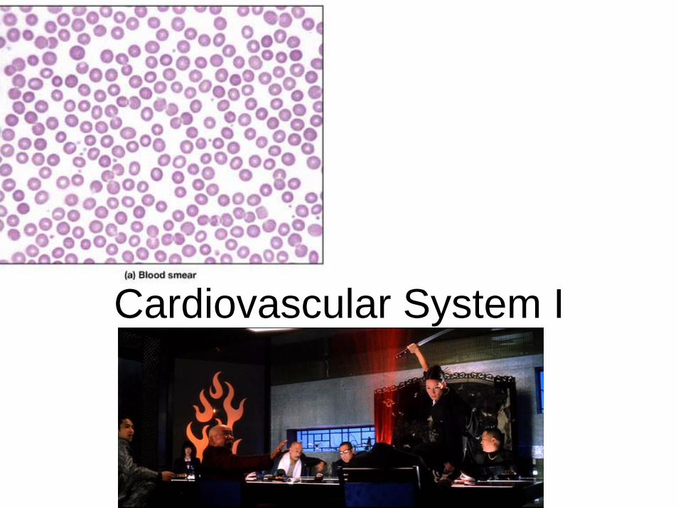
Cardiovascular System I
Functions of the cardiovascular system
• Transport system for:– Gases-O2 & CO2
– Hormones– Nutrients– Wastes
• Temperature regulation• pH regulation of blood• Immune function
Components of the cardiovascular system
• Blood-fluid, carries gases, nutrients, hormones, heat, etc
• Heart-pump, push the blood thru the vessels
• Vessels-pipes, carries blood through out the body
Function of blood
• Transportation of : Nutrients, oxygen and hormones ; wastes and secretion
• Protection: – against microorganisms – against dehydration
• Regulation of vital fluid balances: – pH balance– body temperature.
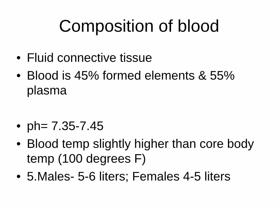
Composition of blood
• Fluid connective tissue• Blood is 45% formed elements & 55%
plasma
• ph= 7.35-7.45• Blood temp slightly higher than core body
temp (100 degrees F)• 5.Males- 5-6 liters; Females 4-5 liters
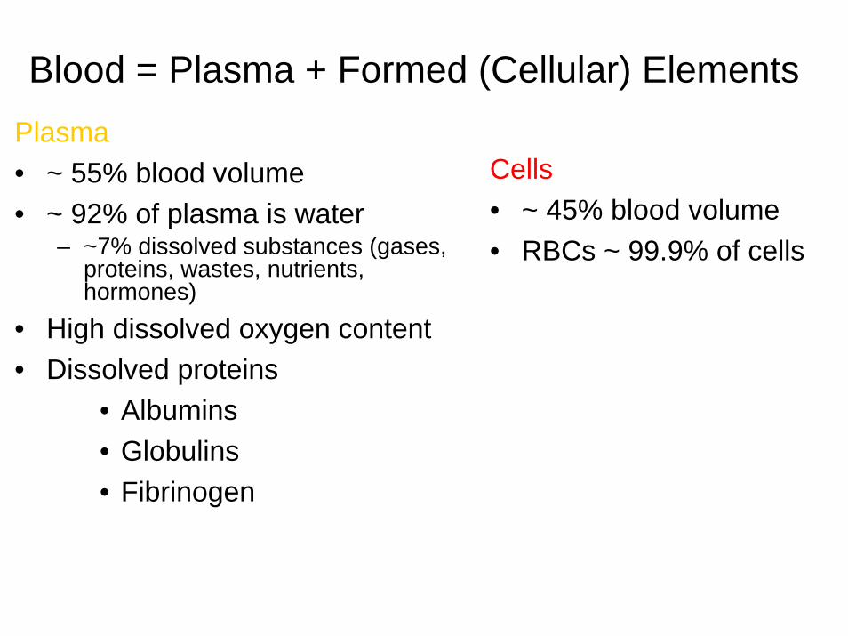
Blood = Plasma + Formed (Cellular) ElementsPlasma• ~ 55% blood volume• ~ 92% of plasma is water
– ~7% dissolved substances (gases, proteins, wastes, nutrients, hormones)
• High dissolved oxygen content• Dissolved proteins
• Albumins• Globulins• Fibrinogen
Cells• ~ 45% blood volume• RBCs ~ 99.9% of cells
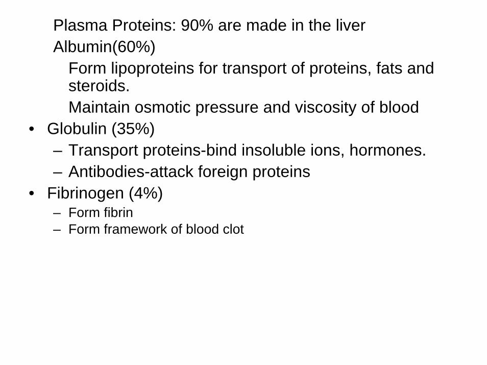
Plasma Proteins: 90% are made in the liverAlbumin(60%)
Form lipoproteins for transport of proteins, fats and steroids.Maintain osmotic pressure and viscosity of blood
• Globulin (35%)– Transport proteins-bind insoluble ions, hormones.– Antibodies-attack foreign proteins
• Fibrinogen (4%)– Form fibrin – Form framework of blood clot
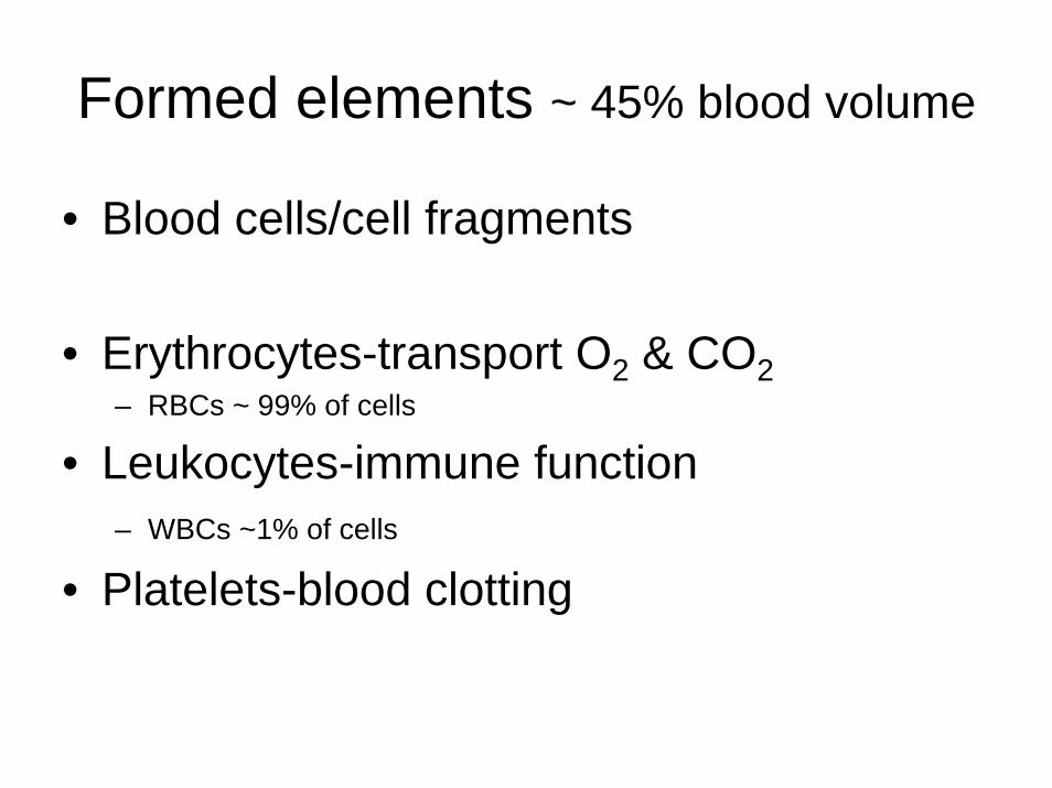
Formed elements ~ 45% blood volume
• Blood cells/cell fragments
• Erythrocytes-transport O2 & CO2– RBCs ~ 99% of cells
• Leukocytes-immune function– WBCs ~1% of cells
• Platelets-blood clotting
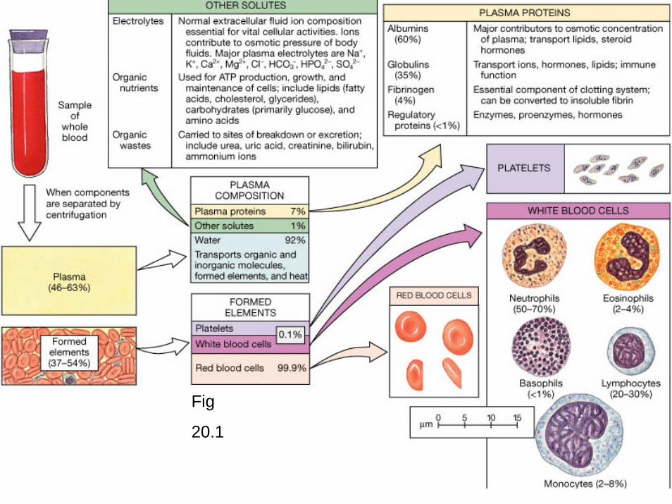
Fig
20.1
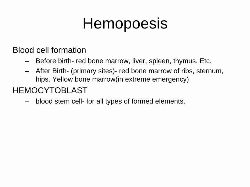
HemopoesisBlood cell formation
– Before birth- red bone marrow, liver, spleen, thymus. Etc.– After Birth- (primary sites)- red bone marrow of ribs, sternum,
hips. Yellow bone marrow(in extreme emergency)
HEMOCYTOBLAST– blood stem cell- for all types of formed elements.
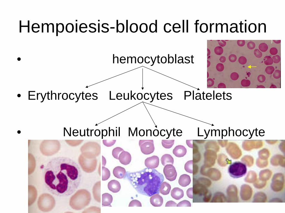
Hempoiesis-blood cell formation
• hemocytoblast
• Erythrocytes Leukocytes Platelets
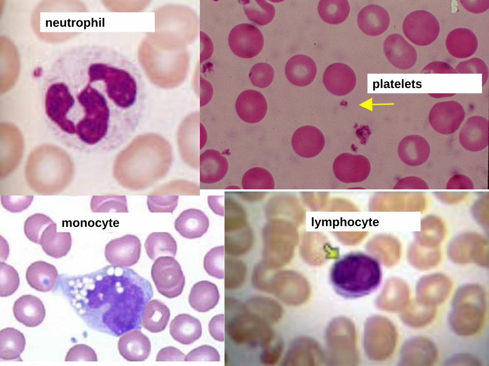
• Neutrophil Monocyte Lymphocyte
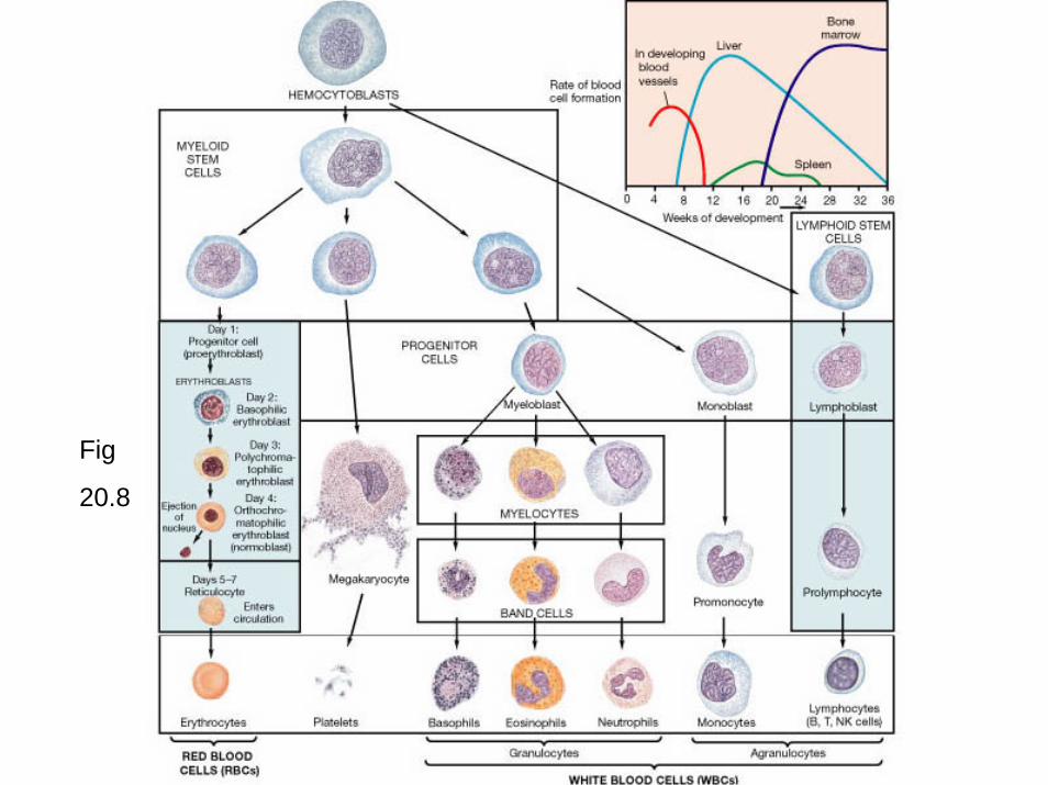
Fig
20.8
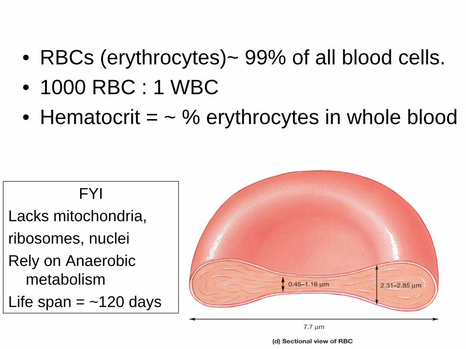
• RBCs (erythrocytes)~ 99% of all blood cells.• 1000 RBC : 1 WBC• Hematocrit = ~ % erythrocytes in whole blood
FYILacks mitochondria, ribosomes, nucleiRely on Anaerobic
metabolism Life span = ~120 days
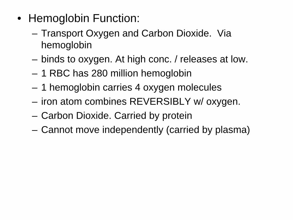
• Hemoglobin Function:– Transport Oxygen and Carbon Dioxide. Via
hemoglobin– binds to oxygen. At high conc. / releases at low.– 1 RBC has 280 million hemoglobin– 1 hemoglobin carries 4 oxygen molecules– iron atom combines REVERSIBLY w/ oxygen.– Carbon Dioxide. Carried by protein– Cannot move independently (carried by plasma)
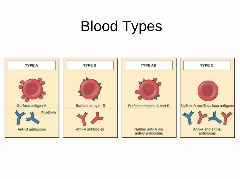
Blood Types
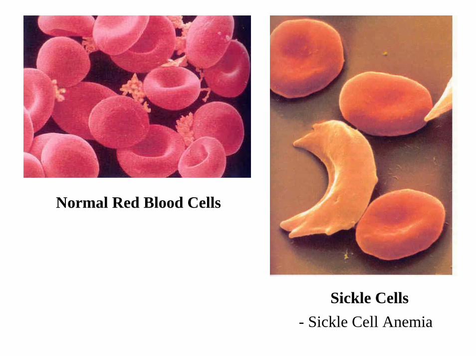
Normal Red Blood Cells
Sickle Cells- Sickle Cell Anemia
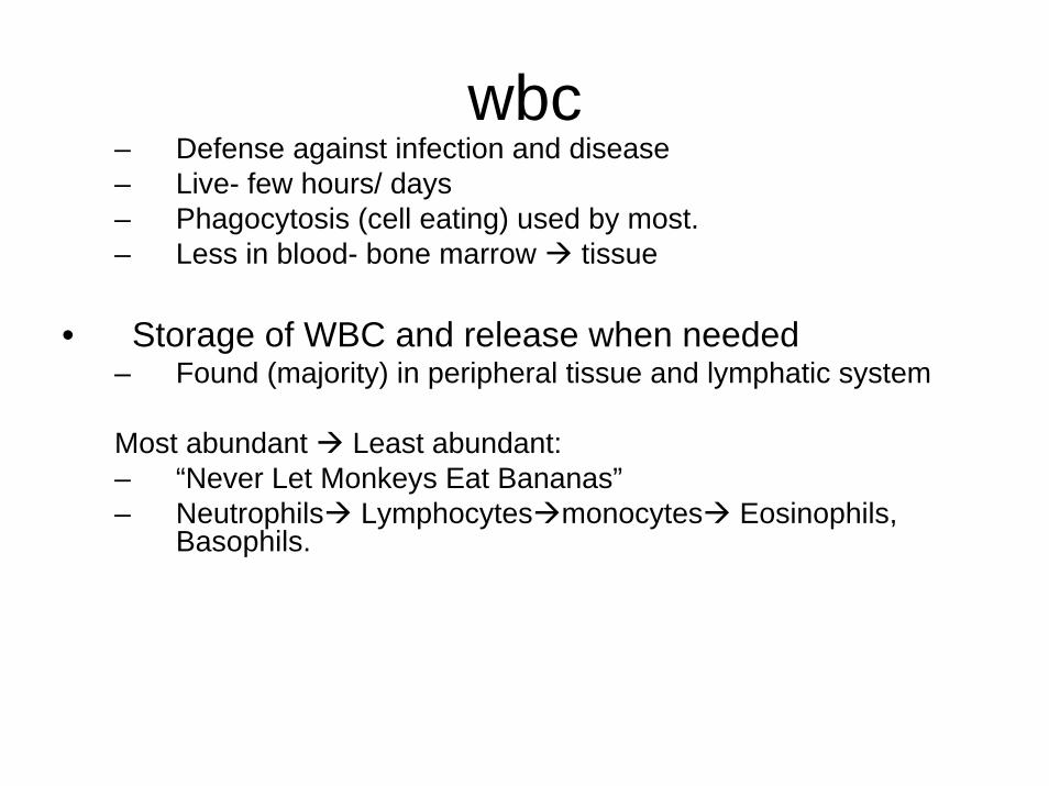
wbc– Defense against infection and disease– Live- few hours/ days– Phagocytosis (cell eating) used by most.– Less in blood- bone marrow tissue
• Storage of WBC and release when needed– Found (majority) in peripheral tissue and lymphatic system
Most abundant Least abundant: – “Never Let Monkeys Eat Bananas”– Neutrophils Lymphocytes monocytes Eosinophils,
Basophils.
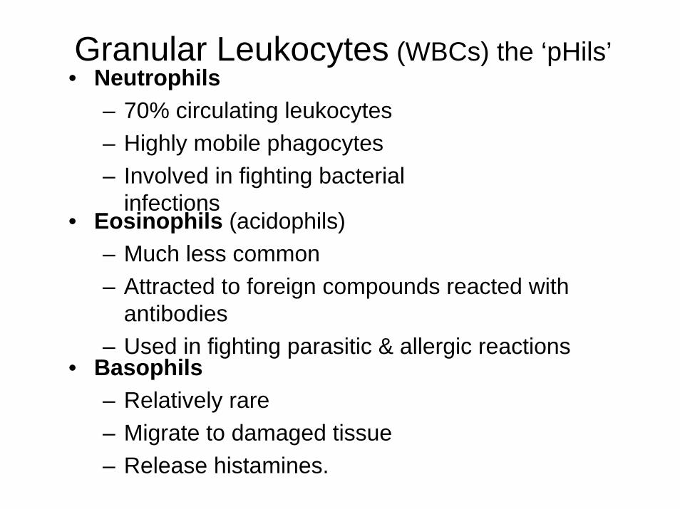
• Neutrophils– 70% circulating leukocytes– Highly mobile phagocytes– Involved in fighting bacterial
infections• Eosinophils (acidophils)
– Much less common– Attracted to foreign compounds reacted with
antibodies– Used in fighting parasitic & allergic reactions
• Basophils– Relatively rare– Migrate to damaged tissue– Release histamines.
Granular Leukocytes (WBCs) the ‘pHils’
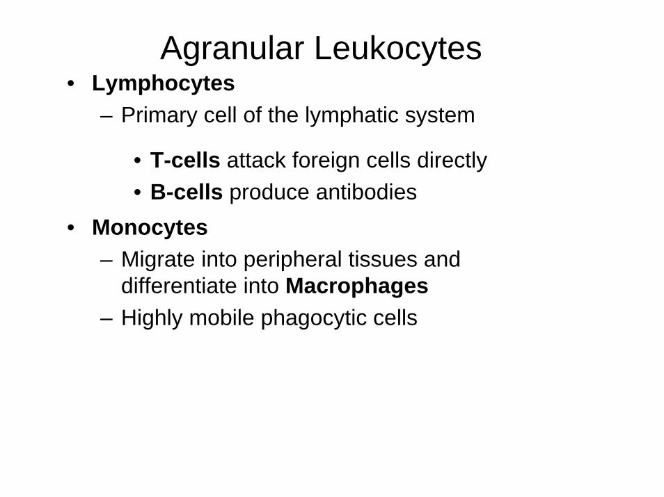
• Monocytes– Migrate into peripheral tissues and
differentiate into Macrophages– Highly mobile phagocytic cells
• Lymphocytes– Primary cell of the lymphatic system
• T-cells attack foreign cells directly• B-cells produce antibodies
Agranular Leukocytes
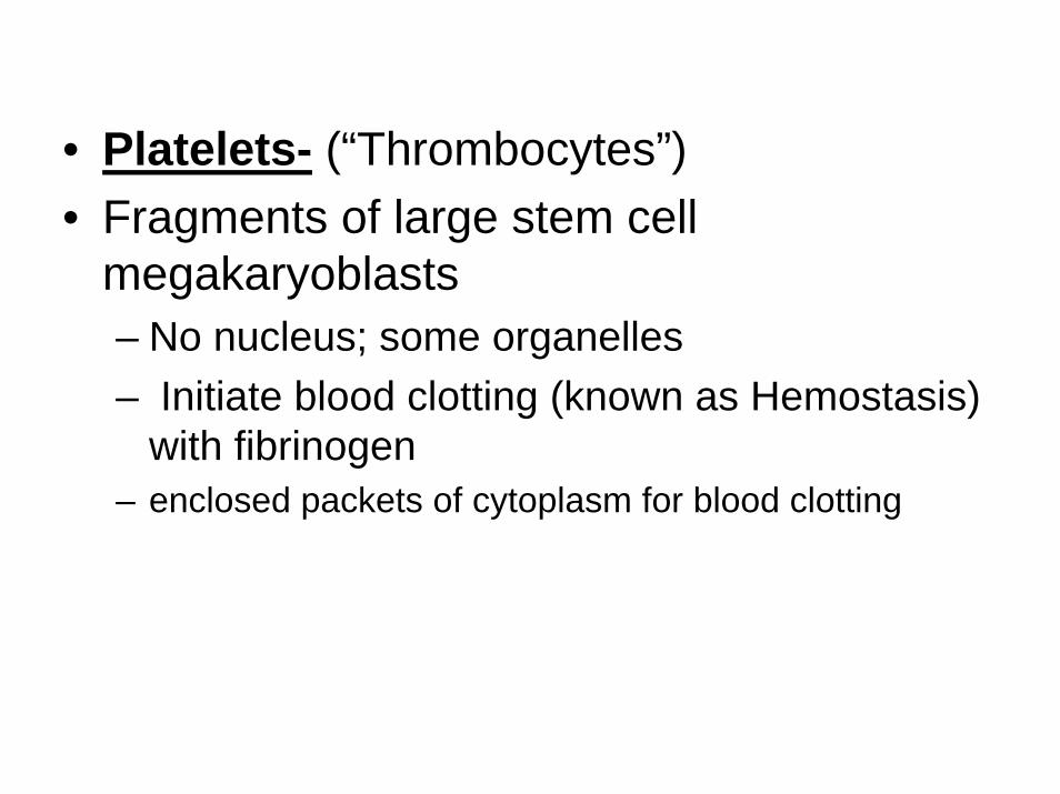
• Platelets- (“Thrombocytes”)• Fragments of large stem cell
megakaryoblasts– No nucleus; some organelles– Initiate blood clotting (known as Hemostasis)
with fibrinogen– enclosed packets of cytoplasm for blood clotting
The heart
• 4 muscular chambers• 2 superior chambers-atria• 2 inferior chambers-ventricles
• The heart pumps blood thru two circuits• Pulmonary circuit-lungs• Systemic circuit-everywhere except the
lungs
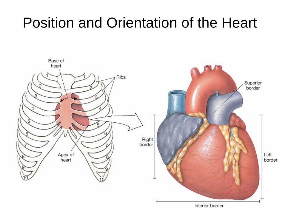
Position and Orientation of the Heart
• Size– 5 in x 3 in (fist)
• Location– Mediastinum (within Pericardial cavity)– Right chambers slightly anterior– Apex points left– Lays on diaphragm
• Base-deep to sternum at 3rd intercostal space• Apex- Deep to 5th left intercostal space
• 3 layers of Heart:• Endocardium• Myocardium• Epicardium- AKA visceral pericardium
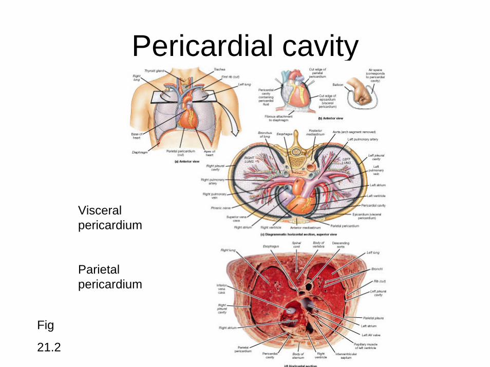
Pericardial cavity
Visceral pericardium
Parietal pericardium
Fig
21.2
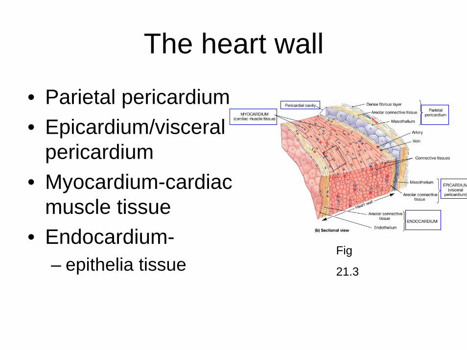
The heart wall
• Parietal pericardium• Epicardium/visceral
pericardium• Myocardium-cardiac
muscle tissue• Endocardium-
– epithelia tissueFig
21.3
The Fibrous Skeleton
Is an internal connective tissue of the heart
1. Provides attachment for heart’s valves
2. Support muscle cells, blood vessels, nerves
3. Evenly distribute the force of contraction
4. Physically isolates muscle cells of the atria from those of the ventricles
• Bands of Fibrous Connective Tissue• -Found within Myocardium• -Shapes Chambers• -prevents overfilling of chambers• -Electrically separates atria from ventricles
Myocardium
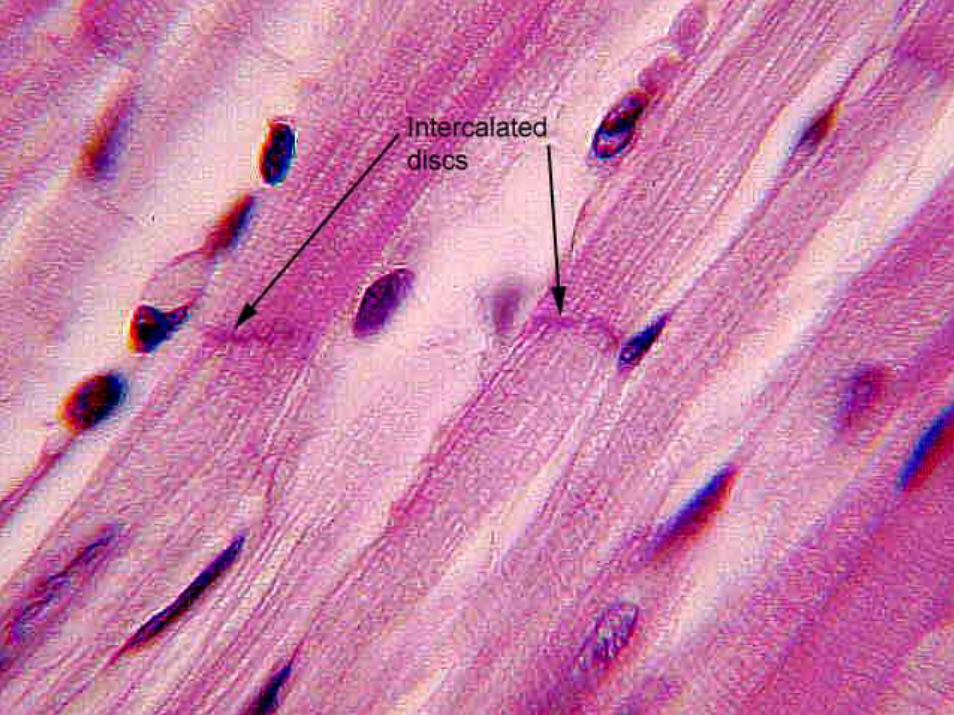
• FYI• Cardiac muscle:• similar to skeletal muscle• much more dependant on oxygen• contracts w/o signaling from the nervous
system• contains cells junctions called intercalated
discs and gap junctions
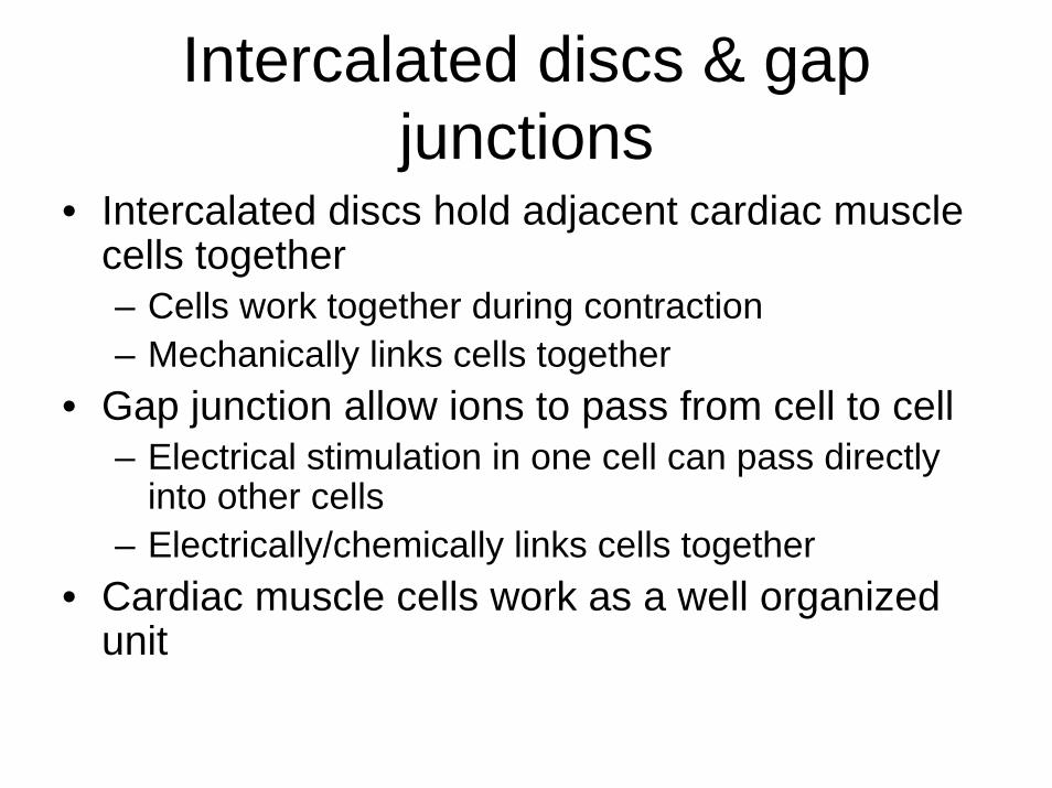
Intercalated discs & gap junctions
• Intercalated discs hold adjacent cardiac muscle cells together– Cells work together during contraction– Mechanically links cells together
• Gap junction allow ions to pass from cell to cell– Electrical stimulation in one cell can pass directly
into other cells– Electrically/chemically links cells together
• Cardiac muscle cells work as a well organized unit
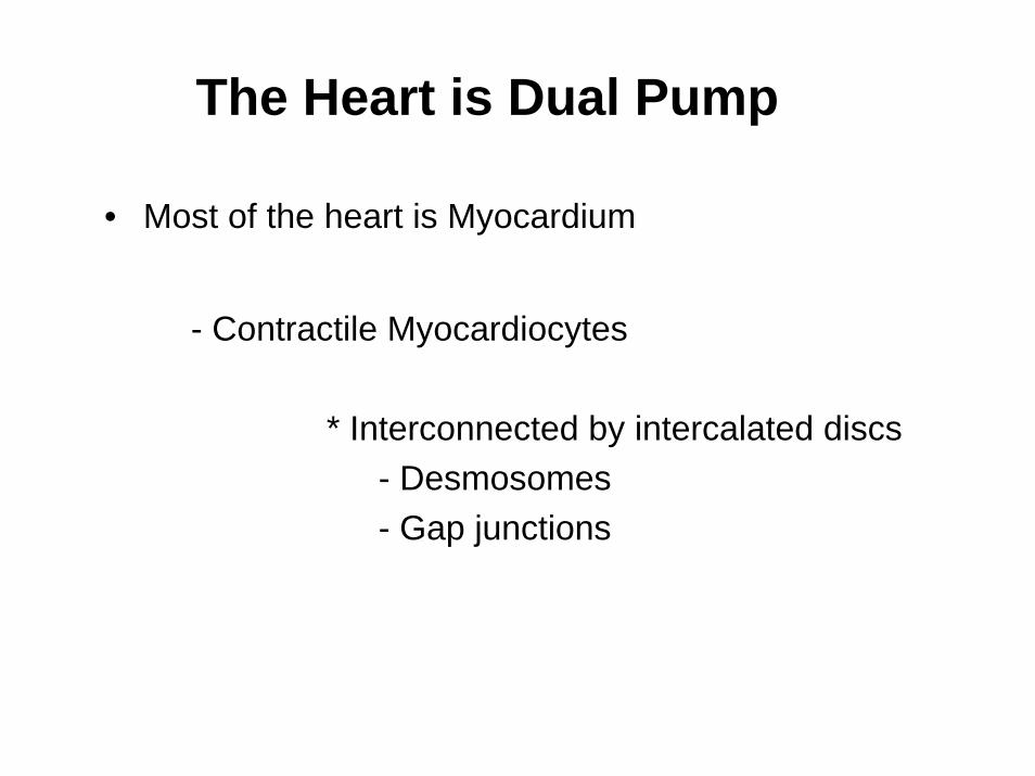
• Most of the heart is Myocardium
- Contractile Myocardiocytes
* Interconnected by intercalated discs- Desmosomes- Gap junctions
The Heart is Dual Pump
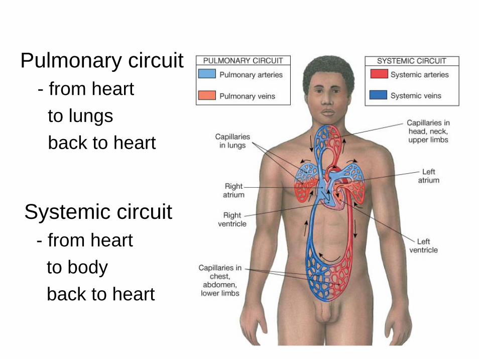
Pulmonary circuit- from heart to lungs back to heart
Systemic circuit- from heart to body back to heart
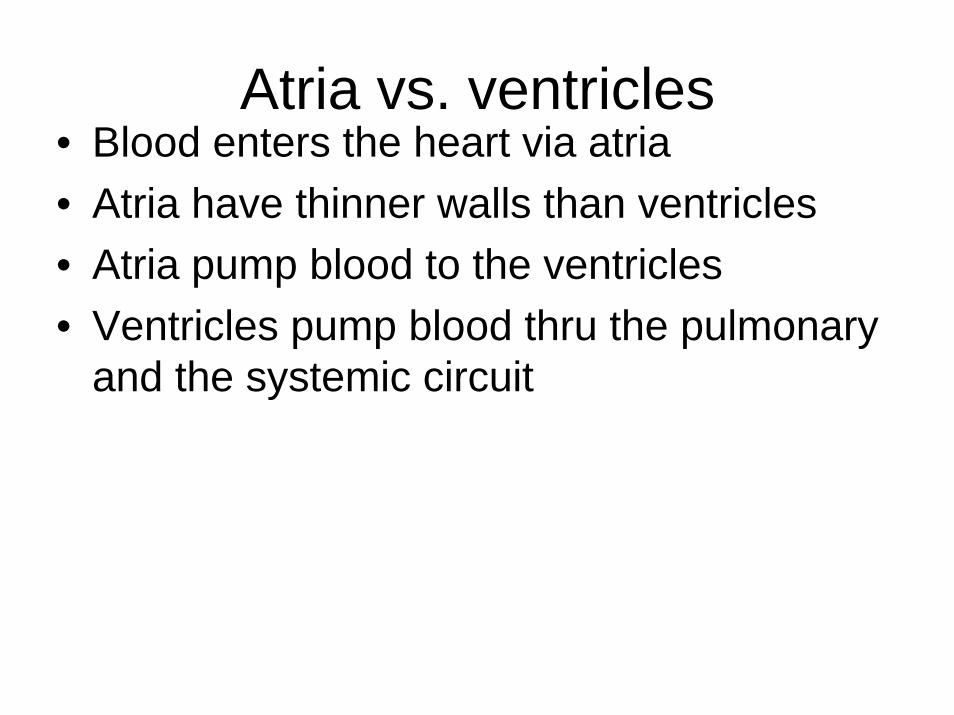
Atria vs. ventricles• Blood enters the heart via atria• Atria have thinner walls than ventricles• Atria pump blood to the ventricles• Ventricles pump blood thru the pulmonary
and the systemic circuit
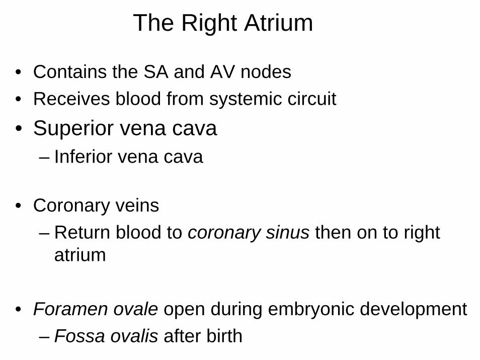
The Right Atrium
• Contains the SA and AV nodes• Receives blood from systemic circuit• Superior vena cava
– Inferior vena cava
• Coronary veins– Return blood to coronary sinus then on to right
atrium
• Foramen ovale open during embryonic development– Fossa ovalis after birth
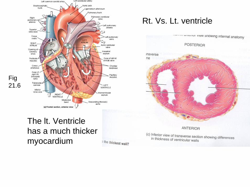
Rt. Vs. Lt. ventricle
The lt. Ventricle has a much thicker myocardium
Fig 21.6
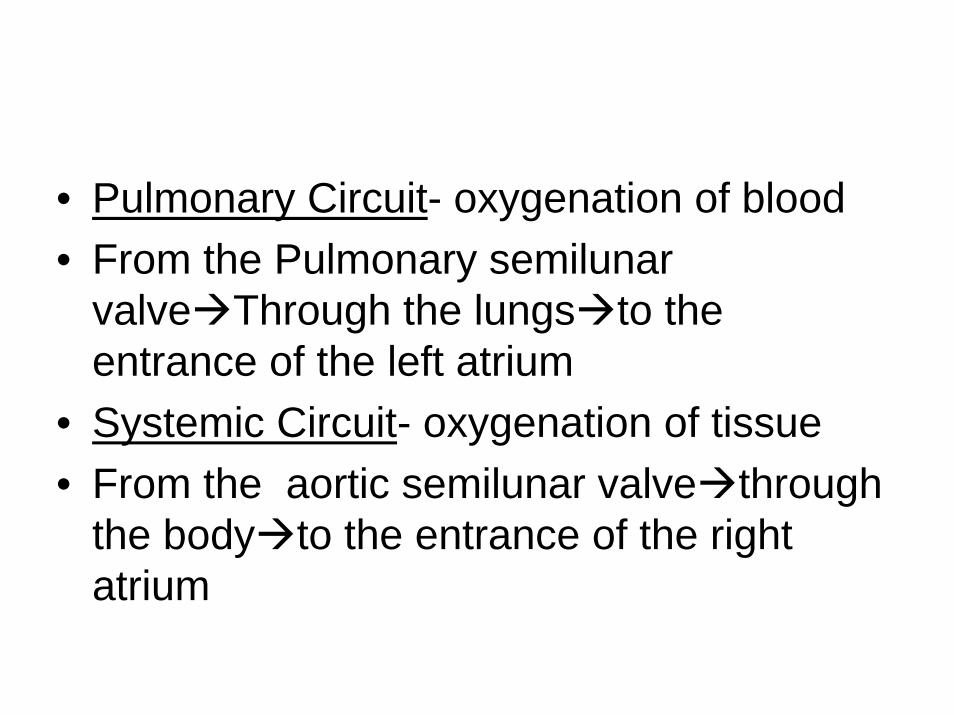
• Pulmonary Circuit- oxygenation of blood• From the Pulmonary semilunar
valve Through the lungs to the entrance of the left atrium
• Systemic Circuit- oxygenation of tissue• From the aortic semilunar valve through
the body to the entrance of the right atrium
Interactive physiology CDHistology CD
neutrophil
monocyte
platelets
lymphocytemonocyte