Embed Size (px)
Citation preview

CARDIAC SYNCOPE IN ATRIAL SEPTAL DEFECT
BY
CORNELIO PAPPFrom the Cardiac Department of the London Chest Hospital
Received March 1, 1957
Atrial septal defect (A.S.D.) in adults is often asymptomatic and may be a chance discovery onclinical examination or at mass radiography. Symptoms arise late in the course of the disease andare those commonly seen in other cardiac conditions with right heart failure. Cardiac syncope hasnot been described as a leading symptom of A.S.D. and must be regarded as a rarity. It may beaccidental that within six months of each other two patients came under observation on accountof syncope and both had A.S.D. One knew of a cardiac murmur he had since childhood; theother was unaware of any heart disease.
'At a time when surgical repair is becoming a routine procedure in A.S.D., any new symptomhas an added interest. This and the different mechanism of syncope in the two cases, caused bycoexistent arrhythmia in both, justifies this report.
CASE 1. A man, aged 22, a fitter, had known of his cardiac murmur since childhood and was rejectedon account of it from military service when aged 18. He never complained of any shortness of breath,and had been able to run for short distances and climb stairs. His effort tolerance thus has been good ifnot normal.
In May 1955 he cycled hurriedly to work because he was late. He wanted to make an excuse for hislate arrival when he suddenly lost consciousness. The first thing he remembered was lying on the couchin the first-aid room some time later; he did not appear to have had a fit, was not incontinent, and did notinjure himself. He went back to work and felt perfectly well. One week later he had a sudden attack ofgiddiness and fell forward on his outstretched hands, injuring himself; he did not lose consciousness butit took him about ten minutes to recover fully. During the next fortnight he had three similar episodes;during the last one he again lost consciousness and became fully conscious only two hours later at theCasualty Department of a nearby hospital.
From May up to his admission to Charing Cross Hospital in October, 1955, he felt perfectly well.On examination he was a well-built man with no cyanosis or clubbing. The pulse varied from 80 to 90;
at times it was regular, at others there were irregularities suggesting extrasystoles. The blood pressure was120/85.
Clinical examination of the heart showed a right ventricular type of diffuse and tapping cardiac impulseat its normal location. The main finding was a systolic thrill over the pulmonary area; corresponding tothis there was a grade III systolic murmur widely heard over the prxcordium but loudest over the pulmonaryarea. The pulmonary second sound was of normal intensity; it was split with delay of the pulmonary com-ponent. There were no signs of congestive heart failure; the rest of the examination was normal.
Radioscopy and chest film showed a bulky heart without obvious enlargement; the apex was rounded,the right ventricular conus full, and the main pulmonary artery slightly prominent. In the left obliqueposition the right ventricle had an increased convexity and the aortic window was obscured by the enlargedleft pulmonary branch. The pulmonary vascularity was above average; the pulmonary stem showedincreased pulsation but there was no hilar dance; the aorta was small. The cardiogram showed partialright bundle-branch block (Fig. 1.) The diagnosis was that of A.S.D.
After his discharge he took ephedrine gr. 4 twice a day. From November, 1955 to July, 1956 when9
on 7 July 2018 by guest. Protected by copyright.
http://heart.bmj.com
/B
r Heart J: first published as 10.1136/hrt.20.1.9 on 1 January 1958. D
ownloaded from

CORNELIO PAPP
ft
.I;:
o~ ~~~~~~~~~~~~~~~~~~~~~~~~~~~~~~~~.*I :1
FIG. 1.-Case 1. " M " complex in VI the only sign of right ventricular hypertrophy;P-R interval=0 26 sec.
he was admitted to the London Chest Hospital he had only two attacks, the latter on June 12. Thesewere shorter and less severe than those in May, 1955. Though they came suddenly he had a short warning;he felt giddy and had a sickly taste in his mouth. He fell to the ground, not completely unconscious, butunable to move.The clinical, radiological, and electrocardiographic findings were unchanged. Cardiac catheterization
confirmed the clinical diagnosis of A.S.D. with a left-to-right shunt of 7 2 1./min. (see Table 1). No spon-taneous syncopal attacks were observed during the stay in hospital, nor could these be provoked by effort,vagal compression, Valsalva manceuvre, or atropine.
TABLE I
MAIN CARDIAC CATHETER DATA
Pressure: mm. of Hg Oxygen saturation
Case Right Pulmonary (%) A- Pu.mo Syst. L-Rventricle artery 02 diff, flow flow shuntventricle_______ artery_______l_ Vol. (I./min.) (I./min.) (I./min.)
S/D Mean S/D Mean SVC RA PA BA (°)
1 26/2 10 16/5 9 73 84 84 93: 42 130 58 72
2 28/2 11 28/7 15 62 79 79 91 60 84 35 4.9
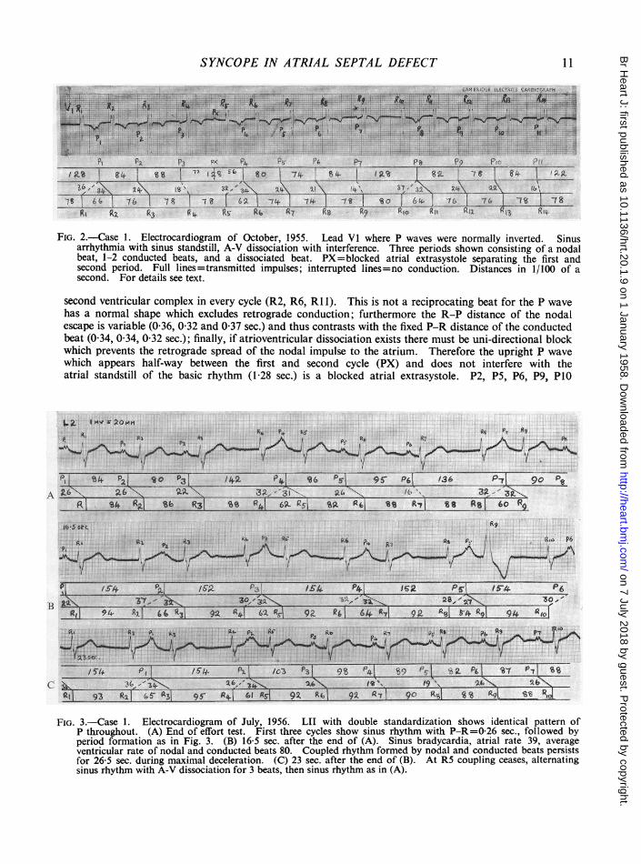
The Arrhythmia. The cardiograms recorded during the first hospital admission in October 1955,while he was free from syncopal attacks for four-and-a-half months, were all similar. They consisted ofperiods containing 3-4 P waves and 4-5 R waves; three of these periods are shown in Fig. 2; the leadrecorded is VI, where the P waves were normally inverted. The periods start with a QRS complex whichis not preceded by a P wave and is therefore a nodal beat (Rl, R5, RlO); this is followed after a varyinginterval of 0 32-037 sec. by a P wave of normal appearance which rides around the peak of the T wave(P1, P4, P8); the next ventricular complex follows after a fixed distance of 0 34-0 32 sec. (R2, R6, Rl I).The P-R distance then gradually shortens from 0-24 to 0 18 and to 0 16 and 0-14 and a nodal escapeintroduces the next period. The P waves follow each other at irregular intervals; after every third or fourthP wave a longer pause follows which in this record is fairly constant (1-28, 1 28, 1 28, and 122 sec.)without being a multiple of the previous P-P intervals. Thus there is no sinu-atrial block in the strictsense, but a sinus arrhythmia with periodical sinus standstill. The R-R distances on the other hand arefairly equal with the exception of the one which follows the nodal escape; the average ventricular rate at80 a minute is faster than the atrial rate at 62 a minute. This suggests atrio-ventricular dissociation.Ventricular captures occur after every nodal escape and this explains the premature appearance of the
:E:AVI? AVL /AVF'.. , . .
V.S5 .::.
Jr1.........w
10
ve,
on 7 July 2018 by guest. Protected by copyright.
http://heart.bmj.com
/B
r Heart J: first published as 10.1136/hrt.20.1.9 on 1 January 1958. D
ownloaded from
SYNCOPE IN A TRIAL SEPTAL DEFECT 1
LAMEPI1kVe tLLC''ITi' CA'OI r.PAPH
A~~~~~~
P, P: P3 ~' P4 Ps- Pi, P P% P9 o Pul(Z,t4 SB " ~~'j7 80 7 4- 6- '76 84/ P.P't C4
_____ 8132D 71
Ii3h3 __2 2
7$64 16 ~18 iS 49 -.& m- to so 6w4 G 1 -7 laRI Rz R3 RyS R16 A1 Re R? Re,14u
FIG. 2.-Case 1. Electrocardiogram of October, 1955. Lead VI where P waves were normally inverted. Sinusarrhythmia with sinus standstill, A-V dissociation with interference. Three periods shown consisting of a nodalbeat, 1-2 conducted beats, and a dissociated beat. PX=blocked atrial extrasystole separating the first andsecond period. Full lines =transmitted impulses; interrupted lines =no conduction. Distances in 1/100 of asecond. For details see text.
second ventricular complex in every cycle (R2, R6, RI 1). This is not a reciprocating beat for the P wavehas a normal shape which excludes retrograde conduction; furthermore the R-P distance of the nodalescape is variable (0-36, 0-32 and 0-37 sec.) and thus contrasts with the fixed P-R distance of the conductedbeat (0-34, 0-34, 0-32 sec.); finally, if atrioventricular dissociation exists there must be uni-directional blockwhich prevents the retrograde spread of the nodal impulse to the atrium. Therefore the upright P wavewhich appears half-way between the first and second cycle (PX) and does not interfere with theatrial standstill of the basic rhythm (1 -28 sec.) is a blocked atrial extrasystole. P2, P5, P6, P9, PlO
1.2Ivr2Pnit,P*~~~~~~~~~~~~~, 15
pi 54 pi to ~ /1+ P4- 56 Psi 9S' P1 /36 P-7 go0PA 16N ZGt.-L4 ..~ 4 ~ ' g 9
P, 84i RaI Sb R31 so g641O695As taL Ptl st R',I is Rsi bo%P.
B
C'
j6~~~~~~~5Sec 1%9~~~~~~~~~~~~~~~~RA'At At h R6 A, A; ~~~~~~~~~~~~~~~~~~~~s Pc / RuP6~~~~~~~~~I
/3-4 P~~~L 16 as. A62 41
FIG. 3.-Case 1. Electrocardiogram of July, 1956. LII with double standardization shows identical pattern ofP throughout. (A) End of effort test. First three cycles show sinus rhythm with P-R=0-26 sec., followed byperiod formation as in Fig. 3. (B) 16-5 sec. after the end of (A). Sinus bradycardia, atrial rate 39, average.ventricular rate of nodal and conducted beats 80. Coupled rhythm formed by nodal and conducted beats persistsfor 26-5 sec. during maximal deceleration. (C) 23 sec. after the end of (B). At R5 coupling ceases, alternating,sinus rhythm with A-V dissociation for 3 beats, then sinus rhythm as in (A).
on 7 July 2018 by guest. Protected by copyright.
http://heart.bmj.com
/B
r Heart J: first published as 10.1136/hrt.20.1.9 on 1 January 1958. D
ownloaded from

CORNELIO PAPP
are also conducted to the ventricle for R3, R7, R8, R12, and R13 are slightly premature when comparedwith R4, R5, R9, RIO, R14 which appear at the genuine nodal distances of0-78-0-80 sec., representing aninherent nodal rate of 76 a minute. The diagnosis is therefore that of sinus arrhythmia with sinus brady-cardia, and atrioventricular dissociation with interference. However, the reason for the syncopal attacksand the prolonged P-R interval of the conducted beats remained obscure until, during an effort test inJuly, 1956 (when he was admitted for cardiac catheter studies), regular sinus rhythm appeared.
Fig. 3 is from a long strip recorded during an effort test; it shows leadII with double standardization(Im. volt=20 mm.) to enlarge the P waves. The first two beats in (A) are sinus beats with a P-R intervalof 0-26 sec.; this was the constant A-V conduction time during the effort test and no faster conduction wasseen at a rate of 90 a minute. Thus during sinus rhythm latent heart block is present. As soon as the rateslows, period formation starts again as in Fig. 2, with the usual sequence of nodal escape, two conductedbeats, A-V dissociation, and sinus standstill. (B) is part of the same record 16 5 seconds after the end of(A); suddenly the atrial rate diminishes to 39 a minute. Were it not for the nodal escapes the ventricularrate would be just as slow but the nodal escapes added to the conducted beats keep the ventricles at an averagerate of 80, double the atrial rate. This rhythm persists for 26-5 seconds. R9 shows aberrant interven-tricular conduction due to its early appearance in diastole. The magnification of the P waves with doublestandardization clearly proves that all are of sinus and not of nodal (e.g. retrograde) origin, since theirshape remains always the same. At the end of strip (C) recorded 23 seconds after the end of (B) sinusrhythm, with the usual P-R interval of 0-26 sec., reappears.
Further electrocardiograms after effort were recorded five months later in November, 1956, when he feltwell and had been without cardiac syncope for six months. The records still showed the period formationas seen in Fig. 2, but the periods were longer, contained more conducted beats, and auricular standstill wasshorter and variable. During maximal deceleration the atrial rate averaged 60 and the ventricular rate85 a minute.
Cardiac syncope in this patient was due to periodical sinus bradycardia which still could be produced18 months after the first syncopal attack by effort, at a time when the nodal automatism, "the ectopicrhythm by default" was fully effective. It has to be remembered that he had the first faint after an unusualeffort, though subsequently he had them at rest as well. Whether prolonged sinus standstill occurred duringthese attacks or not is open to speculation. Though retrograde conduction from the nodal beat to thesinus was excluded by the normal P wave the reactivation of the sinus 030-0 40 sec. after the nodal beatsuggests that the latter may have been instrumental in this. The ventricular contraction caused by the nodalimpulse may have acted as a mechanical stimulus to the flagging sinus which then continued to functionfor some beats. Since the length of diastole preceding the nodal beat was almost constant in the singlerecords, it can be presumed that the nodal pacemaker was active all the time, though it was submergedduring sinus activity (Lewis, 1925). It is thus conceivable that before nodal automatism was fully developedperiods of sinus standstill were responsible for the syncopal attacks, which were then followed by sinusbradycardia and thus produced a state of prolonged semi-consciousness.
CASE 2. A man, aged 58, a painter-decorator, was admitted as an emergency to the Charing CrossHospital in April, 1956. While washing a wall he became giddy, lost consciousness, fell to the ground, andinjured his head. The next thing he remembered was lying on the pavement; he regained consciousnessfully about half an hour later in the Casualty Department. On admission he looked pale and wassweating profusely; he had an irregular pulse of 120-140 and a systolic blood pressure of 100. Theprovisional diagnosis was that of a silent cardiac infarction.
There had been no significant previous illnesses. He had been active always, though he had noticedsome shortness of breath during the past year.
He improved quickly and when transferred to the ward a few hours later the blood pressure rose to140/95; the pulse rate was 110, irregular. There was a loud systolic murmur over the lower end of thesternum, a widely split pulmonary second sound, and a short diastolic murmur. There were no signs ofcongestive heart failure. A chest film showed moderate cardiac enlargement involving the right ventricleand atrium, and enlargement of the pulmonary artery, with overfilling of the pulmonary branches whichshowed hilar dance on screening. The aorta was small. The cardiogram (Fig. 4) showed 2: 1 flutter withright bundle-branch block; there were no signs of cardiac infarction. The diagnosis was atrial septal defect.
Digitalis in high dosage did not restore sinus rhythm nor did it transform the flutter into fibrillation.He was discharged after six weeks. with a 3:1 flutter (A.R.210-V.R.70).
12
on 7 July 2018 by guest. Protected by copyright.
http://heart.bmj.com
/B
r Heart J: first published as 10.1136/hrt.20.1.9 on 1 January 1958. D
ownloaded from

SYNCOPE IN ATRIAL SEPTAL DEFECT 13
FIG. 4.-Case 2. Electrocardiogram of April, 1956, two days after syncope, showing2: 1 auricular flutter, right bundle-branch block, and right ventricular hypertrophy.
Cardiac catheter studies at the London Chest Hospital three months later confirmed the clinical diag-nosis; there was a left-to-right shunt of 5 1./min. with normal pressures in the right chambers and.pulmonaryartery (see Table I). Attempts to restore normal rhythm were again unsuccessful and he was dischargedwith a 4:1 flutter (A.R.232-V.R.58).
When seen in November, 1956, as an outpatient he felt well and had been working in a lighter occupation.There had been no further syncopal attacks, and the 3: 1 flutter was kept in control with digitalis.
The syncopal attack in this patient was produced by sudden change of rhythm; the low cardiac outputduring I: 1 or 2:1 flutter caused the shock-like state present on admission.
DISCUSSIONThe cause of syncope in both cases was the same, the arrhythmia associated with A.S.D., but
the mechanism was different. In Case 1 it was due to impulse inhibition as shown by non-phasicsinus standstill; in Case 2 it was due to rapid impulse formation possibly to 1: 1 flutter. Since allforms of arrhythmia in A.S.D. are thought to be common (Taussig, 1947) one wonders why syncopeis so exceptional. The incidence of arrhythmias in A.S.D. varies in the published series; in theolder ones before cardiac catheterization became a common diagnostic procedure they were con-sidered to be frequent. Out of 10 patients coming to necropsy (Bedford et. al., 1941) four hadauricular fibrillation and in all these there was associated mitral stenosis. Out of 43 clinical casesonly two had auricular fibrillation and one of these had mitral stenosis. In a recent series ofA.S.D. comprising 95 cases by Walker et. al. (1956), all proved by cardiac catheter, and of agesranging from 5 to 40 years, arrhythmias were exceptional and consisted of a few extrasystolesin one and alternating sinus and nodal rhythm in another.
Cardiac arrhythmias in A.S.D. may arise under two different circumstances. Commonly theyarise when the disease is progressing, when the atria dilate, and when other predisposing factorssuch as advancing age and mitral stenosis co-exist. These were the arrhythmias of the older serieswhere the diagnosis was either made at post-mortem examination or when the disease was advanced.The type of arrhythmia was that found in any heart disease, whether congenital or acquired, thatinvolves the atria, i.e. auricular fibrillation or flutter. Case 2, aged 58, is an example.
Rarely auricular arrhythmias are associated with A.S.D. in young people where there is little orno cardiac enlargement, and the cardiogram shows slight right ventricular hypertrophy or onlythe partial right bundle-branch block. The arrhythmia here is of a complex pattern as in Case 1,or may be of the type of partial heart block (Campbell and Thorne, 1956). These are examplesof genuine congenital arrhythmias. Whether they are coincidental with or caused by A.S.D.cannot be decided though the frequent presence of latent heart block in A.S.D. suggests a causalrelationship.
Surgical correction was not indicated in either of these patients. The defect was small and the
on 7 July 2018 by guest. Protected by copyright.
http://heart.bmj.com
/B
r Heart J: first published as 10.1136/hrt.20.1.9 on 1 January 1958. D
ownloaded from

shunt amounted to only 30 per cent more than the systemic blood flow. Case 1, a young adult,had no cardiac enlargement and the cardiogram with an M complex in VI suggested only a slightdegree of right ventricular hypertrophy. Case 2, aged 58, had some cardiac enlargement and signsof incipient heart failure which were easily corrected by digitalis; in view of his age and the per-sistent flutter, operation was considered too risky. Moreover, the symptoms in both patientswere not due to A.S.D. but to the co-existent arrhythmia. The physiological compensatorymechanism of nodal automatism worked satisfactorily in Case 1 and he has not had syncopalattacks for eight months; in Case 2, still in flutter, digitalis maintains a 3:1 block and assures a slowventricular rate.
SUMMARYCardiac syncope was the only symptom in two cases of atrial septal defect, both with a small
shunt. It was caused in the first patient, aged 22, by impulse inhibition through non-phasicatrial standstill, thought to be due to congenital arrhythmia. Compensatory nodal automatism,giving rise to A-V dissociation abolished the attacks. In the second patient, aged 58, syncope wascaused by rapid impulse formation through sudden onset of flutter; the arrhythmia here was relatedto the evolution of the disease and syncope was abolished by reduction of the ventricular rate.
I am greatly indebted to Dr. K. Shirley Smith, Physician to the Cardiac Department for permission to publishthese cases under his care. I also wish to thank Dr. Elizabeth A. Priest and Dr. Ronald Gibson for the cardiaccatheter data.
REFERENCESBedford, D. E., Papp, C., and Parkinson, J. (1941). Brit. Heart J., 3, 37.Campbell, M., and Thorne, M. G. (1956). Brit. Heart J., 18, 90.Lewis, Sir Thomas (1925). The Mechanism and Graphic Registration of the Heart Beat. 3rd ed., Shaw & Sons,
London.Taussig, H. B. (1947). Congenital Malformations of the Heart. Commonwealth Fund, New York.Walker, W. J., Mattingly, T. W., Pollock, B. E., Carmichael, D. B., Inmon, T. W., and Forrester, R. H. (1956).
Amner. Heart J., 52, 547.
14 CORNELIO PAPP
on 7 July 2018 by guest. Protected by copyright.
http://heart.bmj.com
/B
r Heart J: first published as 10.1136/hrt.20.1.9 on 1 January 1958. D
ownloaded from