Embed Size (px)
Citation preview
Retina
Carcinine Has 4-Hydroxynonenal Scavenging Propertyand Neuroprotective Effect in Mouse Retina
Lea D. Marchette,1 Huaiwen Wang,2 Feng Li,1 Mark A. Babizhayev,3 and Anne Kasus-Jacobi4
PURPOSE. Oxidative stress induces retinal damage and contrib-utes to vision loss in progressive retinopathies. Carcinine (b-alanyl-histamine) is a natural imidazole-containing peptidederivative with antioxidant activity. It is predicted to scavenge4-hydroxynonenal (4-HNE), a toxic product of lipid oxidation.The aim of this study was to confirm the 4-HNE scavengingeffect and evaluate the neuroprotective effect of carcinine inmouse retina subjected to oxidative stress.
METHODS. HPLC coupled with mass spectrometry was used toanalyze carcinine and 4-HNE-carcinine adduct. Protection ofretinal proteins from modification by 4-HNE was tested byincubating carcinine with retinal protein extract and 4-HNE.Modified retinal proteins were quantified by dot-blot analysis.Mice were treated with carcinine (intravitreal injection andgavage) and exposed to bright light to induce oxidative damagein the retina. Photoreceptor degeneration was measured byhistology and electroretinography. Retinal levels of retinoldehydrogenase 12 (RDH12) were measured by immunoblotanalysis, after exposure to bright light and in retinal explantsafter exposure to 4-HNE.
RESULTS. The ability of carcinine to form an adduct with 4-HNE,as well as to prevent and even reverse the adduction of retinalproteins by the toxic aldehyde was demonstrated in vitro.Carcinine, administered by intravitreal injection or gavage,strongly protected mouse retina against light-induced photo-receptor degeneration and had a protective effect on RHD12, aprotein found specifically in photoreceptor cells.
CONCLUSIONS. This study suggests that carcinine can beadministered noninvasively to efficiently protect photorecep-tor cells from oxidative damage. Carcinine could be adminis-tered daily to prevent vision loss in progressive retinopathies.(Invest Ophthalmol Vis Sci. 2012;53:3572–3583) DOI:10.1167/iovs.11-9042
Of all the tissues in the body, the retina is the mostsusceptible to oxidative damage because (1) retinal
photoreceptor cells consume more oxygen than any othercell in the body, and this high oxygen metabolism leads to theproduction of damaging reactive oxygen species (ROS); (2)photoreceptor cells are exposed daily to bright light and areloaded with photosensitizers that contribute to the formationof ROS; and (3) photoreceptor cells have the largest percentageof long-chain polyunsaturated fatty acids (PUFAs) that aredirectly oxidized by ROS.1 Oxidation of PUFAs results inproduction of a complex mixture of toxic end products,including malondialdehyde and a,b-unsaturated aldehydes.Endogenous and dietary antioxidants scavenge ROS, maintain-ing the balance between production and inactivation of ROS.When this equilibrium is perturbed in diseases such as AMD,diabetic retinopathy (DR), or RP, oxidative damage overcomescellular defenses and precipitates the progression of vision lossand photoreceptor cell death.2–4 Therefore, a good therapeuticstrategy for these prevalent retinopathies is to scavenge theexcess of ROS and/or enhance endogenous cellular defenses inorder to slow down the irreversible loss of visual function.
The Age-Related Eye Disease Study (AREDS) showed thatdaily administration of exogenous antioxidants through dietsupplementation reduces the risk of progression to advancedAMD and to severe loss of vision.5,6 A prescription of a specificAREDS formulation is now considered part of the standard carefor AMD patients. However, despite the use of this formulation,many patients still develop advanced AMD.5,6 More efficientantioxidant formulations are needed to provide better neuro-protection in AMD and other degenerative retinopathies.
The imidazole-containing peptide derivatives carnosine (b-alanyl-histidine) and carcinine (b-alanyl-histamine) are found inseveral tissues such as muscle, liver, intestine, and brain inmammals, at high (millimolar) concentrations.7–9 These naturalproducts have antioxidant properties, scavenging ROS incells.10–19 They also have lipid-peroxidase activities, reducinglipid hydroperoxides produced by oxidation of membranePUFAs.11,20 If not reduced, lipid hydroperoxides act as auto-amplificatory agents of the lipid peroxidation reaction, andtheir decomposition in the presence of ferrous ions leads tothe formation of a,b-unsaturated aldehydes and other toxic endproducts.10,11,13 These reactive aldehydes are subject toMichael addition reactions with side chains of lysine, histidine,and cysteine residues, forming adduct with proteins that inmost cases inactivates their functions and targets them todegradation.21 Of interest, carnosine has been shown toscavenge 4-hydroxynonenal (4-HNE), one of the most toxicand abundant end products of lipid peroxidation.12,22,23 Thus,multifunctional imidazole-containing peptide derivatives havehigh therapeutic potential because they can scavenge not onlyROS but also the secondary toxic products that mediate andamplify the oxidative damage in cells.
Carcinine would be a better choice than carnosine fortherapeutic applications because it is significantly moreresistant to enzymatic hydrolysis than carnosine.24 Since
From the Departments of 1Ophthalmology, 2Molecular BiologyProteomics Facility, and 4Pharmaceutical Sciences, University ofOklahoma Health Sciences Center, Oklahoma City, Oklahoma; and3Innovative Vision Products Inc., Wilmington, Delaware, andMoscow, Russia.
Supported by the National Center for Research Resources GrantP20RR017703, the National Eye Institute of the National Institutes ofHealth Grants R21EY018907 and P30EY012190, the OklahomaCenter for the Advancement of Science and Technology, and aResearch to Prevent Blindness, Inc. unrestricted grant to theUniversity of Oklahoma Health Sciences Center, Department ofOphthalmology.
Submitted for publication November 8, 2011; revised March 8and April 24, 2012; accepted April 27, 2012.
Disclosure: L.D. Marchette, None; H. Wang, None; F. Li,None; M.A. Babizhayev, P; A. Kasus-Jacobi, None
Corresponding author: Anne Kasus-Jacobi, Department ofPharmaceutical Sciences, University of Oklahoma Health SciencesCenter, 1110 North Stonewall Avenue, Oklahoma City, OK 73117-1223; [email protected].
Investigative Ophthalmology & Visual Science, June 2012, Vol. 53, No. 7
3572 Copyright 2012 The Association for Research in Vision and Ophthalmology, Inc.
Downloaded from iovs.arvojournals.org on 04/24/2019
carcinine contains an imidazole group, it is predicted to have a4-HNE-scavenging activity similar to that of carnosine. In thisstudy, we investigated the 4-HNE-scavenging activity ofcarcinine and its possible neuroprotective effect in mouseretina.
METHODS
Materials
Goat polyclonal anti-HNE and mouse monoclonal anti-b-actin antibod-
ies were obtained from Abcam (Cambridge, MA). Rabbit polyclonal
anti-mouse RDH12 antibody was generated as previously described.25
The 4-HNE was obtained from Cayman Chemical (Ann Arbor, MI).
Carcinine was a generous gift from Exsymol (Monaco, France). All
other chemicals were from Sigma (St. Louis, MO) unless otherwise
noted.
HPLC/Mass Spectrometry (MS) Analysis
To determine if carcinine can form an adduct with 4-HNE, 0.5 mM
carcinine was incubated with 0 or 0.64 mM 4-HNE for 16 hours at room
temperature. A 20-lL aliquot was dried under vacuum, resuspended in
15 lL reagent alcohol-water-triethylamine (TEA) (2:2:1), and dried
again. Twenty microliters derivatization reagent alcohol-TEA-water-
phenylisothiocyanate (PITC) (7:1:1:1) was added to the dried sample
and sealed in vacuum vials for 20 minutes at room temperature.
Reagent was removed with nitrogen for 10 minutes, and dried under
vacuum. Samples were dissolved with 500 lL solvent A (0.09% formic
acid, 0.01% trifluoroacetic acid (TFA), 2% acetonitrile, 97.9% water).
Twenty microliters of 1:100 diluted sample was injected into HPLC/MS.
To measure carcinine levels in the plasma and retina, four mouse
retinas or 200 lL plasma were homogenized in 1 mL of cold 0.01 M HCl
with a Polytron homogenizer (Kinematica AG, Lucerne, Switzerland).
One microgram of the internal standard anserine was added to the
samples. Samples were mixed with 1 mL acetonitrile and centrifuged at
1500g for 5 minutes to remove any solid debris. Supernatants were
dried under vacuum. Samples were dissolved in 100 lL coupling
solution (acetonitrile-pyridine-TEA-water; 10:5:2:3) and dried again.
Samples were redissolved in 100 lL coupling solution, and 5 lL PITC
was added for derivatization. After 5 minutes of incubation at room
temperature, samples were evaporated and dissolved in 200 lL of 10%
acetonitrile. Samples were filtered in microcentrifuge filter tubes
(Corning Inc., Corning, NY) and diluted 2-fold with solvent A. Samples
were analyzed on HPLC/MS (Paradigm MSRB capillary HPLC; Michrom
Bioresources, Auburn, CA; HCT Ultra Ion trap MS; Bruker Daltonics,
Billerica, MA). HPLC was run on column Magic MS C18, 5m, 200 A, 0.5
· 150 mm with solvent A and solvent B (0.09% formic acid, 0.0085%
TFA, 95% acetonitrile, 4.9% water). A gradient of 2% to 18% of solvent B
in 10 minutes, 18% to 60% in 6 minutes, and 60% for 2 minutes was
used. The flow rate was 20 mL/min, and the detection wavelength was
215 nm. The ion trap MS was equipped with an electrospray ion
source, where the spray voltage was set at 4 kV in the positive mode.
The heater temperature was maintained at 3008C. The dry gas pressure
was 10 L/min, and the nebulizer gas pressure was 30 psi. Results were
analyzed with an Esquire data analysis system (Esquire Innovation, Inc.,
Temecula, CA).
Effect of Carcinine on Retinal Protein Modificationby 4-HNE In Vitro
Mouse retinas were homogenized in Tissue Protein Extraction Reagent
(T-PER) reagent from Pierce (Rockford, IL), and proteins were
extracted as described25 according to the manufacturer’s instructions.
Protein concentration was measured using Coomassie reagent (Pierce).
To measure the kinetics of 4-HNE modification, 2.5 lg retinal protein
was diluted in 10 lL of PBS and incubated with 0.64-mM 4-HNE at
room temperature for indicated times. To measure the inhibition of this
reaction by carcinine, increasing amounts of carcinine (from 1 lg to 2
mg) were added to the 10 lL reaction before addition of 4-HNE. To
measure the reversion of this reaction, retinal protein and 4-HNE were
incubated overnight to allow complete modification of retinal proteins
by 4-HNE. Then, 1-mg carcinine was added to the 10-lL reaction and
incubated for indicated times. At the end of each reaction, 4-HNE-
protein adducts were quantified by dot-blot analysis.
Dot-Blot Quantification of 4-HNE Protein Adduct
Dot-blot quantification was carried out as described.26 Each reaction
containing 2.5 lg retinal protein was applied to a 96-well dot-blot
apparatus (Bio-Rad, Hercules, CA) and then transferred to a nitrocel-
lulose membrane (Bio-Rad). Sample loading was monitored by staining
the membrane with Ponceau red. Membranes were blotted with anti-
HNE antibody coupled with horseradish peroxidase (HRP; Abcam).
Signals were quantified using SuperSignal Chemiluminescent Substrate
(Pierce), and the digital Carestream Image Station 4000R (Kodak,
Rochester, NY). Care was taken to ensure that the intensities of
detected signals were within the linear range of the camera and that no
pixels were saturated.
Treatment of Mice with Carcinine and Exposure toBright Light
Seven-week-old BALB/c mice (The Jackson Laboratory, Bar Harbor, ME)
were housed in a 12-hour dim light (10 lux)–dark cycle for 1 week
before treatment. Carcinine was administered to 8-week-old mice by
intravitreal injection or by gavage. Mice were injected intravitreally
using a 36-gauge needle (World Precision Instruments, Sarasota, FL)
through the temporal limbus of the eye. One eye received 1 lL PBS,
and the other eye received 1 lL of 2 M carcinine diluted in PBS. Mice
were returned to dim cyclic light for 48 hours for recovery before light
damage was initiated. Light damage was then induced in mice by 5
hours exposure to 4000 lux of diffuse, cool, white fluorescent light, as
previously described.25 Another group of 8-week-old mice was gavaged
once a day for 5 days with 0, 0.2, 2, or 20 mg carcinine dissolved in 100
lL water. To measure carcinine levels in the plasma and retina, one
group of mice was euthanized 4 hours after the last daily gavage on the
fifth day of treatment. Immediately after CO2 inhalation, blood was
collected from the left ventricle, centrifuged at low speed, and the
plasma was frozen in liquid nitrogen for subsequent analysis. To avoid
any blood contamination of retinal samples, mice were perfused with
saline before dissection of the retinas. Retinas were subsequently
dissected and frozen in liquid nitrogen. Another group of mice was
exposed to 3000 lux of bright white fluorescent light for 4 hours,
immediately after the daily gavage on the fifth day of treatment.25
Conditions of light exposure were different for mice that received
intravitreal injection compared with those that received carcinine
through gavage because we empirically determined that a stronger
light exposure was needed after intravitreal injection to induce retinal
damage. Mice were either euthanized immediately after light exposure
for preparation of retinal protein or returned to dim cyclic light for 7
days to allow the retina to clear all dead cells and restore a well-
organized morphology before ERG testing and histology. During this
recovery period, carcinine gavage was continued. All mice were
euthanized by CO2 inhalation, a method approved by the Panel of
Euthanasia of the American Veterinary Medical Association. All
procedures were performed according to the ARVO Statement for
the Use of Animals in Ophthalmic and Vision Research.
Measurement of Photoreceptor Survival
Whole eyes were enucleated after orientation of the superior half with
a permanent dye. Eyes were embedded in paraffin, and sections were
cut along the vertical meridian, through the optic nerve head (ONH).
Paraffin sections stained with hematoxylin and eosin were prepared
IOVS, June 2012, Vol. 53, No. 7 Carcine Neuroprotection in Mouse Retina 3573
Downloaded from iovs.arvojournals.org on 04/24/2019
from each eye, and the number of rows of photoreceptor nuclei within
the outer nuclear layer (ONL) were counted at a distance of 0.5 mm
from the ONH to the inferior and superior retina as an assessment of
photoreceptor survival.27
Electroretinography
To assess photoreceptor function, mice were dark adapted overnight,
anesthetized by intraperitoneal injection of ketamine (90 mg/kg) and
xylazine (10 mg/kg), and after administration of one drop of local
anesthetic to the cornea (0.5% proparacaine), the pupils were dilated
with tropicamide (1%) and phenylephrine (2.5%). ERGs were
performed as previously described.28 Briefly, mice were placed on a
378C heating pad under a Ganzfield dome stimulator (LKC Technolo-
gies, Gaithersburg, MD). Solid gold electrodes were placed on the
cornea to measure responses to light stimuli. A thin coating of
hypromellose solution was used to keep the eyes moist. Flash
intensities ranging from 0.001 to 1000 cd.s/m2 were administered
and responses were recorded using the Espion E2 ERG System
(Diagnosys, LLC, Lowell, MA). Amplitudes of the a-wave were
determined as the minimum voltage recorded between 0 and 25
milliseconds after the flash. The b-waves were calculated as the
difference between the a-wave and the maximum amplitude recorded
around 110 milliseconds after the flash. Values from both eyes were
averaged for each mouse. Cone b-waves were recorded after 5 minutes
of light adaptation by averaging the responses to five 3700 cd.s/m2
flashes.
Retinal Explants
Retinas were dissected from 6- to 8-week-old pigmented wild-type
mice. Retinas were individually incubated in 200 lL Dulbecco’s
modified Eagle’s medium (DMEM) containing 0 or 5 mM carcinine and
0 or 150 lM 4-HNE, at 378C under 5% CO2 for indicated times. Retinas
(four per group) were removed from incubation, immediately washed
in PBS, and frozen in liquid nitrogen for subsequent protein
preparation.
Protein Preparation and Immunoblot
Frozen retinas were homogenized in T-PER buffer (Pierce), according to
the manufacturer’s instructions. Protein concentrations were mea-
sured, and immunoblot analysis using anti-RDH12 and anti-b-actin
antibodies was performed using 30 lg protein. Signals were detected
using SuperSignal Chemiluminescent Substrate and quantified using
Kodak Image Station 4000R and Kodak Molecular Imaging Software as
previously described.25 The levels of RDH12 were normalized to the
levels of b-actin (used as loading control) and then expressed relative to
those of RDH12 in control retinas, arbitrarily defined as 100%.
Statistical Analyses
Statistical analyses were performed using GraphPad Prism 5.0 software
(GraphPad Software, Inc., La Jolla, CA). The quantitative data are
expressed as averages and SEM for each group. One or 2-way ANOVAs
were performed to assess differences between the means. From the
ANOVAs, Bonferroni’s Multiple Comparison post hoc test was
performed with a 95% confidence interval to determine statistical
significance. Three to five independent samples were used per analysis
as indicated.
RESULTS
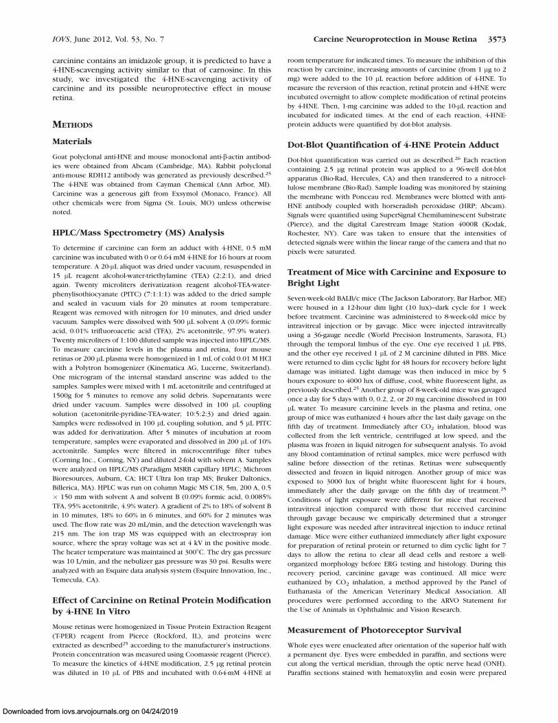
Carcinine Forms an Adduct with 4-HNE In Vitro
To confirm that carcinine can form an adduct with 4-HNE, theywere combined and incubated overnight in water. The productof the reaction was analyzed by HPLC/MS (Fig. 1). Under these
FIGURE 1. Carcinine forms an adduct with 4-HNE in vitro. Carcinine(0.5 mM) was incubated with 0 or 0.64 mM 4-HNE for 16 hours at roomtemperature, and samples were analyzed by HPLC/MS. (A) Represen-tative chromatogram and corresponding mass spectrum obtained fromcarcinine incubated with 0 mM 4-HNE. (B) Representative chromato-gram and corresponding mass spectrum obtained from carcinineincubated with 0.64 mM 4-HNE. Carcinine gives a peak at 318.1 m/zand 4-HNE-carcinine adduct at 474.2 m/z. This experiment wasrepeated three times with similar results.
3574 Marchette et al. IOVS, June 2012, Vol. 53, No. 7
Downloaded from iovs.arvojournals.org on 04/24/2019
HPLC conditions, the carcinine-only control had a retentiontime of 10 minutes, and a mass of 318.1 m/z (Fig. 1A). Afterincubation with 4-HNE, the retention time of the newly formedmolecule was 17.5 minutes, and the mass was increased to thepredicted 474.2 m/z, corresponding to the adduct formedbetween carcinine and 4-HNE (Fig. 1B). This result demon-strated that carcinine reacted with 4-HNE to form an adductdetectable by HPLC/MS.
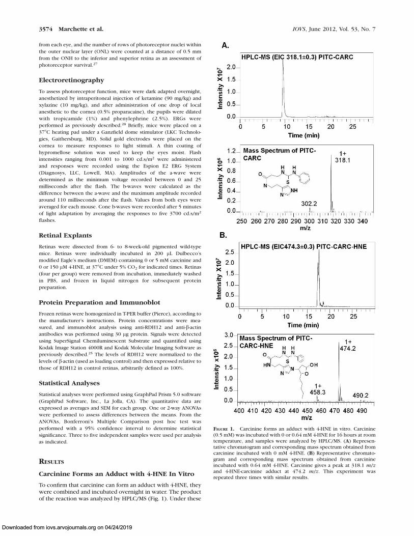
Carcinine’s Effects on 4-HNE–Modified RetinalProtein
Anti-HNE antibody specifically binds to 4-HNE-modified pro-teins but not to the 4-HNE-carcinine adduct (Fig. 2A).Therefore, this antibody was used for subsequent experimentsfor quantification of the 4-HNE-protein adduct. Note thatcarcinine is not detectable by Ponceau staining even though itis present on the membrane (Fig. 2A, lower panel). Todetermine the kinetics of 4-HNE-adduct formation, retinal
proteins were incubated with a molar excess of 4-HNE in PBSat room temperature for indicated times. Adduct formationincreased over time and reached its maximum level at 60minutes of incubation and remained stable until 2 hours ofincubation (Fig. 2B). In the next experiment, retinal proteinsand increasing amounts of carcinine were incubated with 4-HNE for 90 minutes at room temperature. Carcinine inhibitedthe formation of adduct between 4-HNE and retinal proteins ina dose-dependent manner (Fig. 2C). Under these conditions,the inhibition of adduct formation reached 50% (IC50) with acarcinine concentration of 33.2 6 0.6 lg/lL. Completeinhibition was achieved with a carcinine concentration of 0.1mg/lL (1 mg per reaction).
To determine if carcinine could reverse 4-HNE modificationof retinal proteins after adduct formation had taken place,preformed adducts were incubated with 0 or 1 mg carcininefor increasing times (0 to 72 hours). Carcinine reversed 4-HNEmodification of retinal proteins in a time-dependent manner(Fig. 3). This was a slow process that reached complete adduct
FIGURE 2. Carcinine prevents 4-HNE modification of retinal proteins. (A) Carcinine (Car, 2.5 lg) or retinal proteins (Prot, 2.5 lg) were incubatedwith 0 (�) or 0.64 mM (þ) 4-HNE for 16 hours at room temperature before dot-blot analysis. Protein loading is verified by staining the membranewith Ponceau red (lower blot). Signal is detected with anti-HNE antibody coupled with HRP (upper blot). Only retinal proteins modified by 4-HNEwere detected by this method. (B) Representative dot-blot showing time-course of adduct formation between 4-HNE and retinal proteins. Ponceaustaining (lower blot) and 4-HNE-protein adduct detected using anti-HNE antibody (upper blot) are shown. This experiment was repeated three timeswith similar results. (C) Representative dot-blot showing dose-response inhibition of adduct formation between 4-HNE and retinal proteins bycarcinine. Results from three experiments, each done in triplicate, were quantified and plotted as percentage of adduct formed after normalizing toprotein amount. Error bars correspond to SEM.
IOVS, June 2012, Vol. 53, No. 7 Carcine Neuroprotection in Mouse Retina 3575
Downloaded from iovs.arvojournals.org on 04/24/2019
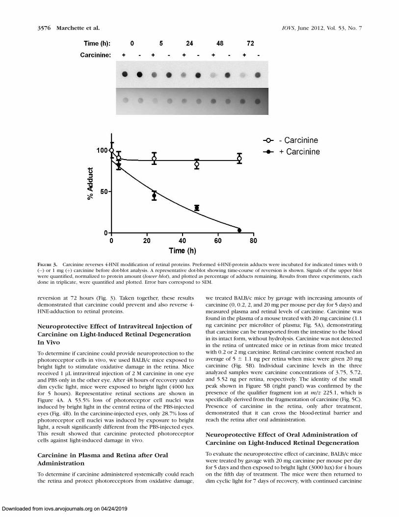
reversion at 72 hours (Fig. 3). Taken together, these resultsdemonstrated that carcinine could prevent and also reverse 4-HNE-adduction to retinal proteins.
Neuroprotective Effect of Intravitreal Injection ofCarcinine on Light-Induced Retinal DegenerationIn Vivo
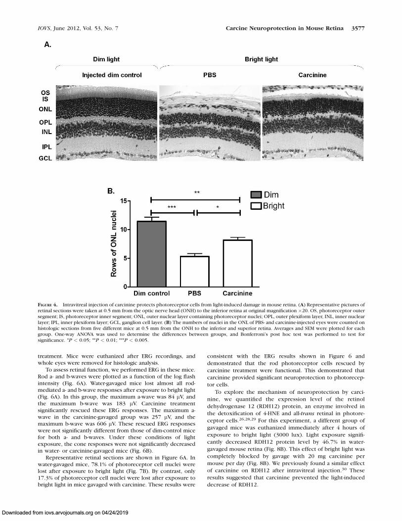
To determine if carcinine could provide neuroprotection to thephotoreceptor cells in vivo, we used BALB/c mice exposed tobright light to stimulate oxidative damage in the retina. Micereceived 1 lL intravitreal injection of 2 M carcinine in one eyeand PBS only in the other eye. After 48 hours of recovery underdim cyclic light, mice were exposed to bright light (4000 luxfor 5 hours). Representative retinal sections are shown inFigure 4A. A 53.5% loss of photoreceptor cell nuclei wasinduced by bright light in the central retina of the PBS-injectedeyes (Fig. 4B). In the carcinine-injected eyes, only 28.7% loss ofphotoreceptor cell nuclei was induced by exposure to brightlight, a result significantly different from the PBS-injected eyes.This result showed that carcinine protected photoreceptorcells against light-induced damage in vivo.
Carcinine in Plasma and Retina after OralAdministration
To determine if carcinine administered systemically could reachthe retina and protect photoreceptors from oxidative damage,
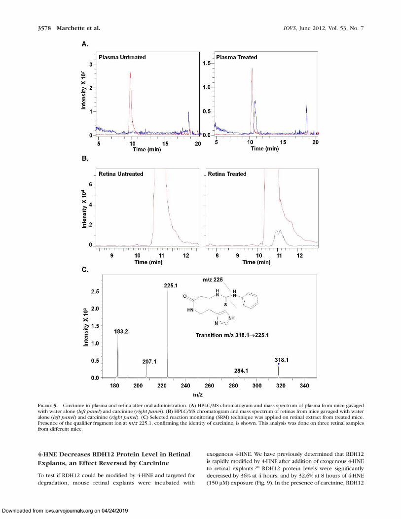
we treated BALB/c mice by gavage with increasing amounts ofcarcinine (0, 0.2, 2, and 20 mg per mouse per day for 5 days) andmeasured plasma and retinal levels of carcinine. Carcinine wasfound in the plasma of a mouse treated with 20 mg carcinine (1.1ng carcinine per microliter of plasma; Fig. 5A), demonstratingthat carcinine can be transported from the intestine to the bloodin its intact form, without hydrolysis. Carcinine was not detectedin the retina of untreated mice or in retinas from mice treatedwith 0.2 or 2 mg carcinine. Retinal carcinine content reached anaverage of 5 6 1.1 ng per retina when mice were given 20 mgcarcinine (Fig. 5B). Individual carcinine levels in the threeanalyzed samples were carcinine concentrations of 3.75, 5.72,and 5.52 ng per retina, respectively. The identity of the smallpeak shown in Figure 5B (right panel) was confirmed by thepresence of the qualifier fragment ion at m/z 225.1, which isspecifically derived from the fragmentation of carcinine (Fig. 5C).Presence of carcinine in the retina, only after treatment,demonstrated that it can cross the blood-retinal barrier andreach the retina after oral administration.
Neuroprotective Effect of Oral Administration of
Carcinine on Light-Induced Retinal Degeneration
To evaluate the neuroprotective effect of carcinine, BALB/c micewere treated by gavage with 20 mg carcinine per mouse per dayfor 5 days and then exposed to bright light (3000 lux) for 4 hourson the fifth day of treatment. The mice were then returned todim cyclic light for 7 days of recovery, with continued carcinine
FIGURE 3. Carcinine reverses 4-HNE modification of retinal proteins. Preformed 4-HNE-protein adducts were incubated for indicated times with 0(�) or 1 mg (þ) carcinine before dot-blot analysis. A representative dot-blot showing time-course of reversion is shown. Signals of the upper blotwere quantified, normalized to protein amount (lower blot), and plotted as percentage of adducts remaining. Results from three experiments, eachdone in triplicate, were quantified and plotted. Error bars correspond to SEM.
3576 Marchette et al. IOVS, June 2012, Vol. 53, No. 7
Downloaded from iovs.arvojournals.org on 04/24/2019
treatment. Mice were euthanized after ERG recordings, andwhole eyes were removed for histologic analysis.
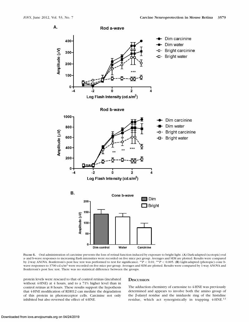
To assess retinal function, we performed ERG in these mice.Rod a- and b-waves were plotted as a function of the log flashintensity (Fig. 6A). Water-gavaged mice lost almost all rod-mediated a- and b-wave responses after exposure to bright light(Fig. 6A). In this group, the maximum a-wave was 84 lV, andthe maximum b-wave was 183 lV. Carcinine treatmentsignificantly rescued these ERG responses. The maximum a-wave in the carcinine-gavaged group was 257 lV, and themaximum b-wave was 606 lV. These rescued ERG responseswere not significantly different from those of dim-control micefor both a- and b-waves. Under these conditions of lightexposure, the cone responses were not significantly decreasedin water- or carcinine-gavaged mice (Fig. 6B).
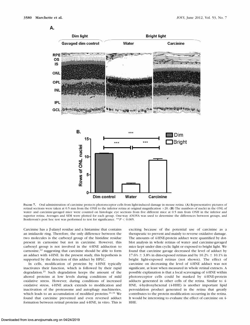
Representative retinal sections are shown in Figure 6A. Inwater-gavaged mice, 78.1% of photoreceptor cell nuclei werelost after exposure to bright light (Fig. 7B). By contrast, only17.3% of photoreceptor cell nuclei were lost after exposure tobright light in mice gavaged with carcinine. These results were
consistent with the ERG results shown in Figure 6 and
demonstrated that the rod photoreceptor cells rescued by
carcinine treatment were functional. This demonstrated that
carcinine provided significant neuroprotection to photorecep-
tor cells.
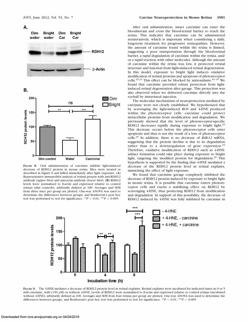
To explore the mechanism of neuroprotection by carci-
nine, we quantified the expression level of the retinol
dehydrogenase 12 (RDH12) protein, an enzyme involved in
the detoxification of 4-HNE and all-trans retinal in photore-
ceptor cells.26,28,29 For this experiment, a different group of
gavaged mice was euthanized immediately after 4 hours of
exposure to bright light (3000 lux). Light exposure signifi-
cantly decreased RDH12 protein level by 46.7% in water-
gavaged mouse retina (Fig. 8B). This effect of bright light was
completely blocked by gavage with 20 mg carcinine per
mouse per day (Fig. 8B). We previously found a similar effect
of carcinine on RDH12 after intravitreal injection.30 These
results suggested that carcinine prevented the light-induced
decrease of RDH12.
FIGURE 4. Intravitreal injection of carcinine protects photoreceptor cells from light-induced damage in mouse retina. (A) Representative pictures ofretinal sections were taken at 0.5 mm from the optic nerve head (ONH) to the inferior retina at original magnification ·20. OS, photoreceptor outersegment; IS, photoreceptor inner segment; ONL, outer nuclear layer containing photoreceptor nuclei; OPL, outer plexiform layer, INL, inner nuclearlayer; IPL, inner plexiform layer; GCL, ganglion cell layer. (B) The numbers of nuclei in the ONL of PBS- and carcinine-injected eyes were counted onhistologic sections from five different mice at 0.5 mm from the ONH to the inferior and superior retina. Averages and SEM were plotted for eachgroup. One-way ANOVA was used to determine the differences between groups, and Bonferroni’s post hoc test was performed to test forsignificance. *P < 0.05; **P < 0.01; ***P < 0.005.
IOVS, June 2012, Vol. 53, No. 7 Carcine Neuroprotection in Mouse Retina 3577
Downloaded from iovs.arvojournals.org on 04/24/2019
4-HNE Decreases RDH12 Protein Level in Retinal
Explants, an Effect Reversed by Carcinine
To test if RDH12 could be modified by 4-HNE and targeted for
degradation, mouse retinal explants were incubated with
exogenous 4-HNE. We have previously determined that RDH12
is rapidly modified by 4-HNE after addition of exogenous 4-HNE
to retinal explants.30 RDH12 protein levels were significantly
decreased by 36% at 4 hours, and by 32.6% at 8 hours of 4-HNE
(150 lM) exposure (Fig. 9). In the presence of carcinine, RDH12
FIGURE 5. Carcinine in plasma and retina after oral administration. (A) HPLC/MS chromatogram and mass spectrum of plasma from mice gavagedwith water alone (left panel) and carcinine (right panel). (B) HPLC/MS chromatogram and mass spectrum of retinas from mice gavaged with wateralone (left panel) and carcinine (right panel). (C) Selected reaction monitoring (SRM) technique was applied on retinal extract from treated mice.Presence of the qualifier fragment ion at m/z 225.1, confirming the identity of carcinine, is shown. This analysis was done on three retinal samplesfrom different mice.
3578 Marchette et al. IOVS, June 2012, Vol. 53, No. 7
Downloaded from iovs.arvojournals.org on 04/24/2019
protein levels were rescued to that of control retinas (incubatedwithout 4-HNE) at 4 hours, and to a 71% higher level than incontrol retinas at 8 hours. These results support the hypothesisthat 4-HNE modification of RDH12 can mediate the degradationof this protein in photoreceptor cells. Carcinine not onlyinhibited but also reversed the effect of 4-HNE.
DISCUSSION
The adduction chemistry of carnosine to 4-HNE was previouslydetermined and appears to involve both the amino group ofthe b-alanyl residue and the imidazole ring of the histidineresidue, which act synergistically in trapping 4-HNE.12
FIGURE 6. Oral administration of carcinine prevents the loss of retinal function induced by exposure to bright light. (A) Dark-adapted (scotopic) roda- and b-wave responses to increasing flash intensities were recorded on five mice per group. Averages and SEM are plotted. Results were comparedby 2-way ANOVA. Bonferroni’s post hoc test was performed to test for significance. **P < 0.01; ***P < 0.005. (B) Light-adapted (photopic) cone b-wave responses to 3700 cd.s/m2 were recorded on five mice per group. Averages and SEM are plotted. Results were compared by 1-way ANOVA andBonferroni’s post hoc test. There was no statistical difference between the groups.
IOVS, June 2012, Vol. 53, No. 7 Carcine Neuroprotection in Mouse Retina 3579
Downloaded from iovs.arvojournals.org on 04/24/2019
Carcinine has a b-alanyl residue and a histamine that containsan imidazole ring. Therefore, the only difference between thetwo molecules is the carboxyl group of the histidine residuepresent in carnosine but not in carcinine. However, thiscarboxyl group is not involved in the 4-HNE adduction tocarnosine,12 suggesting that carcinine should be able to forman adduct with 4-HNE. In the present study, this hypothesis issupported by the detection of this adduct by HPLC.
In cells, modification of proteins by 4-HNE typicallyinactivates their function, which is followed by their rapiddegradation.21 Such degradation keeps the amount of thealtered proteins at low levels during conditions of mildoxidative stress. However, during conditions of increasedoxidative stress, 4-HNE attack extends to modification andinactivation of the proteasome and autophagy machineries,which leads to an accumulation of modified proteins.21,31 Wefound that carcinine prevented and even reversed adductformation between retinal proteins and 4-HNE, in vitro. This is
exciting because of the potential use of carcinine as atherapeutic to prevent and mainly to reverse oxidative damage.The amounts of 4-HNE-protein adduct were quantified by dot-blot analysis in whole retinas of water- and carcinine-gavagedmice kept under dim cyclic light or exposed to bright light. Wefound that carcinine gavage decreased the level of adduct by17.6% 6 3.8% in dim-exposed retinas and by 10.2% 6 10.1% inbright light-exposed retinas (not shown). The effect ofcarcinine on decreasing the level of 4-HNE adduct was notsignificant, at least when measured in whole retinal extracts. Apossible explanation is that a local scavenging of 4-HNE withinphotoreceptor cells could be masked by 4-HNE-proteinadducts generated in other cells of the retina. Similar to 4-HNE, 4-hydroxyhexenal (4-HHE) is another important lipidperoxidation product generated in the retina that greatlycontributes to the protein modification occurring in the retina.It would be interesting to evaluate the effect of carcinine on 4-HHE.
FIGURE 7. Oral administration of carcinine protects photoreceptor cells from light-induced damage in mouse retina. (A) Representative pictures ofretinal sections were taken at 0.5 mm from the ONH to the inferior retina at original magnification ·20. (B) The numbers of nuclei in the ONL ofwater- and carcinine-gavaged mice were counted on histologic eye sections from five different mice at 0.5 mm from ONH in the inferior andsuperior retina. Averages and SEM were plotted for each group. One-way ANOVA was used to determine the differences between groups, andBonferroni’s post hoc test was performed to test for significance. ***P < 0.005.
3580 Marchette et al. IOVS, June 2012, Vol. 53, No. 7
Downloaded from iovs.arvojournals.org on 04/24/2019
After oral administration, intact carcinine can enter thebloodstream and cross the blood-retinal barrier to reach theretina. This indicates that carcinine can be administerednoninvasively, which is important when considering a daily,long-term treatment for progressive retinopathies. However,the amount of carcinine found within the retina is limited,suggesting a poor transportation through the blood-retinalbarrier, a rapid degradation of carcinine within the retina, and/or a rapid reaction with other molecules. Although the amountof carcinine within the retina was low, it protected retinalstructure and function from light-induced retinal degeneration.In this model, exposure to bright light induces oxidativemodification of retinal proteins and apoptosis of photoreceptorcells.25,32 This effect can be blocked by antioxidants.33–35 Wefound that carcinine provided robust protection from light-induced retinal degeneration after gavage. This protection wasalso observed when we delivered carcinine directly into theeyeball by intravitreal injection.
The molecular mechanisms of neuroprotection mediated bycarcinine were not clearly established. We hypothesized thatby scavenging the light-induced ROS and 4-HNE producedwithin the photoreceptor cells, carcinine could protectintracellular proteins from modification and degradation. Wepreviously showed that the level of photoreceptor-specificRDH12 decreases rapidly during exposure to bright light.25
This decrease occurs before the photoreceptor cells enterapoptosis and thus is not the result of a loss of photoreceptorcells.25 In addition, there is no decrease of Rdh12 mRNA,suggesting that the protein decline is due to its degradationrather than to a downregulation of gene expression.25
Therefore, oxidative modification of RDH12 such as 4-HNE-adduct formation could take place during exposure to brightlight, targeting the modified protein for degradation.25 Thishypothesis is supported by the finding that 4-HNE mediated adecrease of the RDH12 protein level in retinal explants,mimicking the effect of light exposure.
We found that carcinine gavage completely inhibited thedecrease of RDH12 protein induced by exposure to bright lightin mouse retina. It is possible that carcinine enters photore-ceptor cells and exerts a stabilizing effect on RDH12 byscavenging 4-HNE, thus protecting RDH12 from modificationand degradation. In support of this possibility, the decrease ofRDH12 induced by 4-HNE was fully inhibited by carcinine in
FIGURE 8. Oral administration of carcinine inhibits light-induceddecrease of RDH12 protein in mouse retina. Mice were treated asdescribed in Figure 6 and killed immediately after light exposure. (A)Representative immunoblot analysis of retinal protein with anti-RDH12antibody (upper blot) and anti-actin antibody (lower blot). (B) RDH12levels were normalized to b-actin and expressed relative to controlretinas (dim controls), arbitrarily defined as 100. Averages and SEMfrom three mice per group are plotted. One-way ANOVA was used todetermine the differences between groups, and Bonferroni’s post hoctest was performed to test for significance. **P < 0.01; ***P < 0.005.
FIGURE 9. The 4-HNE mediates a decrease of RDH12 protein level in retinal explants. Retinal explants were incubated for indicated times in 0 or 5mM carcinine, with (150 lM) or without 4-HNE. Levels of RDH12 were normalized to b-actin and expressed relative to control retinas (incubatedwithout 4-HNE), arbitrarily defined as 100. Averages and SEM from four retinas per group are plotted. One-way ANOVA was used to determine thedifferences between groups, and Bonferroni’s post hoc test was performed to test for significance. **P < 0.01; ***P < 0.005.
IOVS, June 2012, Vol. 53, No. 7 Carcine Neuroprotection in Mouse Retina 3581
Downloaded from iovs.arvojournals.org on 04/24/2019
retinal explants. Alternatively, carcinine could bind to aspecific receptor on the photoreceptor cells and exert itsbiological effect through intracellular neuroprotective signal-ing pathway(s). Carcinine is a natural compound that is welltolerated; even at the high doses administered in our study (upto 1g carcinine per kilogram of body weight), there were noadverse effects observed. However, we are currently investi-gating other routes of administration providing better bioavail-ability of carcinine in the retina. Because it is a multifunctionalcompound, attacking oxidative damage from different anglesby combining antioxidant, lipid peroxidase, 4-HNE-scavengingactivities, and reversion of 4-HNE-adduction, carcinine has thepotential to be more powerful and more effective than classicantioxidants for progressive retinopathies by reversing thedamage that has already occurred as well as preventing furtherdamage. However, a side-by-side comparison with classicantioxidants will have to be performed in future studies.Another crucial step towards the utilization of carcinine inpatients with progressive retinopathies will be to betterunderstand, at the molecular level, the mechanism(s) ofneuroprotection mediated by carcinine.
Acknowledgments
The authors thank Robert E. Anderson and John D. Ash for theirhelpful discussions. The authors also thank the personnel at theAnimal, Imaging, and Molecular Modules of the Vision ResearchCore Facility at the University of Oklahoma Health SciencesCenter.
References
1. Fliesler SJ, Anderson RE. Chemistry and metabolism of lipids inthe vertebrate retina. Prog Lipid Res. 1983;22:79–131.
2. Zarbin MA, Rosenfeld PJ. Pathway-based therapies for age-related macular degeneration: an integrated survey of emerg-ing treatment alternatives. Retina. 2010;30:1350–1367.
3. Jarrett SG, Lewin AS, Boulton ME. The importance ofmitochondria in age-related and inherited eye disorders.Ophthalm Res. 2010;44:179–190.
4. Komeima K, Rogers BS, Campochiaro PA. Antioxidants slowphotoreceptor cell death in mouse models of retinitispigmentosa. J Cell Physiol. 2007;213:809–815.
5. Age-Related Eye Disease Study Research Group. A randomized,placebo-controlled, clinical trial of high-dose supplementationwith vitamins C and E and beta carotene for age-relatedcataract and vision loss: AREDS Report No. 9. Arch Oph-
thalmol. 2001;119:1439–1452.
6. Krishnadev N, Meleth AD, Chew EY. Nutritional supplementsfor age-related macular degeneration. Curr Opin Ophthalmol.2010;21:184–189.
7. Flancbaum L, Brotman DN, Fitzpatrick JC, Van Es T, Kasziba E,Fisher H. Existence of carcinine, a histamine-related com-pound, in mammalian tissues. Life Sci. 1990;47:1587–1593.
8. Boldyrev AA, Koldobski A, Kurella E, Maltseva V, Stvolinski S.Natural histidine-containing dipeptide carnosine as a potenthydrophilic antioxidant with membrane stabilizing function: abiomedical aspect. Mol Chem Neuropathol. 1993;19:185–192.
9. Kohen R, Yamamoto Y, Cundy KC, Ames BN. Antioxidantactivity of carnosine, homocarnosine, and anserine present inmuscle and brain. Proc Natl Acad Sci U S A. 1988;85:3175–3179.
10. Babizhayev MA, Lozovskaya EL, Makareyeva EN, Lul’kin YA,Sapezhinskii II. Photoprotector and antioxidant properties ofhistamine-containing peptidomimetics in the photooxidationof glycyltryptophan. Biochemistry (Mosc). 1998;63:523–528.
11. Babizhayev MA, Seguin MC, Gueyne J, Evstigneeva RP, AgeyevaEA, Zheltukhina GA. L-carnosine (beta-alanyl-L-histidine) andcarcinine (beta-alanylhistamine) act as natural antioxidantswith hydroxyl-radical-scavenging and lipid-peroxidase activi-ties. Biochem J. 1994;304(pt 2);509–516.
12. Aldini G, Carini M, Beretta G, Bradamante S, Facino RM.Carnosine is a quencher of 4-hydroxy-nonenal: through whatmechanism of reaction? Biochem Biophys Res Commun.2002;298:699–706.
13. Babizhayev MA. Biological activities of the natural imidazole-containing peptidomimetics n-acetylcarnosine, carcinine andL-carnosine in ophthalmic and skin care products. Life Sci.2006;78:2343–2357.
14. Babizhayev MA. Antioxidant activity of L-carnosine, a naturalhistidine-containing dipeptide in crystalline lens. Biochim
Biophys Acta. 1989;1004:363–371.
15. Bellia F, Amorini AM, La Mendola D, et al. New glycosidicderivatives of histidine-containing dipeptides with antioxidantproperties and resistant to carnosinase activity. Eur J Med
Chem. 2008;43:373–380.
16. Boldyrev AA. Does carnosine possess direct antioxidantactivity? Int J Biochem. 1993;25:1101–1107.
17. Decker EA, Livisay SA, Zhou S. A re-evaluation of theantioxidant activity of purified carnosine. Biochemistry
(Mosc). 2000;65:766–770.
18. Mozdzan M, Szemraj J, Rysz J, Nowak D. Antioxidantproperties of carnosine re-evaluated with oxidizing systemsinvolving iron and copper ions. Basic Clin Pharmacol
Toxicol. 2005;96:352–360.
19. Reddy VP, Garrett MR, Perry G, Smith MA. Carnosine: aversatile antioxidant and antiglycating agent. Sci Aging
Knowledge Environ. 2005;2005: pe12.
20. Evstigneeva RP, Zheltukhina GA, Ageeva EA, Babizhaev MA.Lipoperoxidase activity of carnosine and carcinine [inRussian]. Dokl Akad Nauk. 1993;333:104–106.
21. Grimsrud PA, Xie H, Griffin TJ, Bernlohr DA. Oxidative stressand covalent modification of protein with bioactive aldehydes.J Biol Chem. 2008;283:21837–21841.
22. Orioli M, Aldini G, Benfatto MC, Facino RM, Carini MHNE.Michael adducts to histidine and histidine-containing peptidesas biomarkers of lipid-derived carbonyl stress in urines: LC-MS/MS profiling in Zucker obese rats. Anal Chem. 2007;79:9174–9184.
23. Aldini G, Granata P, Carini M. Detoxification of cytotoxicalpha,beta-unsaturated aldehydes by carnosine: characteriza-tion of conjugated adducts by electrospray ionization tandemmass spectrometry and detection by liquid chromatography/mass spectrometry in rat skeletal muscle. J Mass Spectrom.2002;37:1219–1228.
24. Pegova A, Abe H, Boldyrev A. Hydrolysis of carnosine andrelated compounds by mammalian carnosinases. Comp Bio-
chem Physiol B Biochem Mol Biol. 2000;127:443–446.
25. Kanan Y, Wicker LD, Al-Ubaidi MR, Mandal NA, Kasus-Jacobi A.Retinol dehydrogenases RDH11 and RDH12 in the mouseretina: expression levels during development and regulationby oxidative stress. Invest Ophthalmol Vis Sci. 2008;49:1071–1078.
26. Marchette LD, Thompson DA, Kravtsova M, Ngansop TN,Mandal NA, Kasus-Jacobi A. Retinol dehydrogenase 12detoxifies 4-hydroxynonenal in photoreceptor cells. Free
Radic Biol Med. 2010;48:16–25.
27. Nachman-Clewner M, Giblin FJ, Dorey CK, et al. Selectivedegeneration of central photoreceptors after hyperbaricoxygen in normal and metallothionein-knockout mice. Invest
Ophthalmol Vis Sci. 2008;49:3207–3215.
28. Maeda A, Maeda T, Imanishi Y, et al. Retinol dehydrogenase(RDH12) protects photoreceptors from light-induced degen-eration in mice. J Biol Chem. 2006;281:37697–37704.
3582 Marchette et al. IOVS, June 2012, Vol. 53, No. 7
Downloaded from iovs.arvojournals.org on 04/24/2019
29. Kurth I, Thompson DA, Reuther K, et al. Targeted disruption
of the murine retinal dehydrogenase gene Rdh12 does not
limit visual cycle function. Mol Cell Biol. 2007;27:1370–
1379.
30. Kasus-Jacobi A, Marchette LD, Xu C, Li F, Wang H, Babizhayev
MA. Mechanisms of RDH12-induced Leber congenital amau-
rosis and therapeutic approaches. Adv Ophthalmol. 2012;1:
473–496.
31. Krohne TU, Stratmann NK, Kopitz J, Holz FG. Effects of lipid
peroxidation products on lipofuscinogenesis and autophagy in
human retinal pigment epithelial cells. Exp Eye Res. 2010;90:
465–471.
32. Tanito M, Elliott MH, Kotake Y, Anderson RE. Proteinmodifications by 4-hydroxynonenal and 4-hydroxyhexenal inlight-exposed rat retina. Invest Ophthalmol Vis Sci. 2005;46:3859–3868.
33. Organisciak DT, Darrow RM, Jiang YI, Marak GE, Blanks JC.Protection by dimethylthiourea against retinal light damage inrats. Invest Ophthalmol Vis Sci. 1992;33:1599–1609.
34. Tanito M, Masutani H, Nakamura H, Ohira A, Yodoi J.Cytoprotective effect of thioredoxin against retinal photic injuryin mice. Invest Ophthalmol Vis Sci. 2002;43:1162–1167.
35. Tanito M, Masutani H, Nakamura H, Oka S, Ohira A, Yodoi J.Attenuation of retinal photooxidative damage in thioredoxintransgenic mice. Neurosci Lett. 2002;326:142–146.
IOVS, June 2012, Vol. 53, No. 7 Carcine Neuroprotection in Mouse Retina 3583
Downloaded from iovs.arvojournals.org on 04/24/2019