Embed Size (px)
Citation preview

1980
Carbon nano-onions (multi-layer fullerenes):chemistry and applicationsJuergen Bartelmess and Silvia Giordani*
Review Open Access
Address:Istituto Italiano di Tecnologia, Nano Carbon Materials, Via Morego 30,16163 Genova, Italy
Email:Silvia Giordani* - [email protected]
* Corresponding author
Keywords:carbon nanomaterials; carbon nano-onions; fullerenes;functionalization
Beilstein J. Nanotechnol. 2014, 5, 1980–1998.doi:10.3762/bjnano.5.207
Received: 13 May 2014Accepted: 10 October 2014Published: 04 November 2014
This article is part of the Thematic Series "Atomic scale interface designand characterisation: Theory – Structure and dynamics".
Guest Editor: C. Ewels
© 2014 Bartelmess and Giordani; licensee Beilstein-Institut.License and terms: see end of document.
AbstractThis review focuses on the development of multi-layer fullerenes, known as carbon nano-onions (CNOs). First, it briefly summa-
rizes the most important synthetic pathways for their preparation and their properties and it gives the reader an update over new
developments in the recent years. This is followed by a discussion of the published synthetic procedures for CNO functionalization,
which are of major importance when elucidating future applications and addressing drawbacks for possible applications, such as
poor solubility in common solvents. Finally, it gives an overview over the fields of application, in which CNO materials were
successfully implemented.
1980
ReviewIntroductionSince the discovery of the fullerene C60 in 1985 by Curl,
Kroto and Smalley [1], carbon nanomaterials have been
the focus of interdisciplinary chemical research. In the
following years, several other carbon based nanomaterials
were discovered, namely carbon nanotubes (CNTs) [2-4],
carbon nanohorns [5], nanodiamonds [6] and graphene [7].
Multi-shell fullerenes, known as carbon nano-onions (CNOs)
and discovered by Ugarte in 1992 [8], are structured by concen-
tric shells of carbon atoms. Over the last years, different
methods for their synthesis have been developed and their prop-
erties have been widely studied. In addition, the chemical func-
tionalization of CNOs has been investigated and several syn-
thetic pathways were found to be applicable for the introduc-
tion of a variety of functional groups. Chemically modified
CNOs were probed in different fields of application and have
revealed to be a promising nanomaterial that attracts a growing
interest among researchers and opens new avenues for investi-
gation.

Beilstein J. Nanotechnol. 2014, 5, 1980–1998.
1981
Figure 1: HRTEM images of (a) diamond nanoparticles, (b) spherical carbon onions, and (c) polyhedral carbon onions. Diamond nanoparticles aretransformed into spherical onions at about 1700 °C. Polyhedral onions are dominant in the sample annealed above 1900 °C. Reprinted with permis-sion from [12]. Copyright 2001 AIP Publishing LLC.
Preparation and structural properties ofcarbon nano-onionsCarbon nano-onions were first discovered by Ugarte in 1992,
who obtained them by intense electron irradiation of carbon
soot [8]. CNOs were later found to be part of detonation soot
[9], and a variety of different methods for their synthesis were
reported. For a detailed review of the different methods and
related mechanisms of CNO formation, we refer to the book
chapter of Luis Echegoyen and co-workers [10]. A common
method, for the preparation of small CNOs consisting of
approx. 5–8 carbon shells, uses nanodiamonds as starting ma-
terial. Nanodiamonds can be converted to graphitic CNOs by
heat treatment (Figure 1) [11,12] or by electron radiation [13].
Another method is the formation of CNOs by arc discharge of
graphite in liquids such as liquid nitrogen or water [14,15]. A
recent novel method for the preparation of large CNOs (30 nm
diameter) includes the use of inorganic starting material, such as
CuCl2·2H2O and CaC [16]. Large CNOs with distinct fluores-
cence emission were produced from wood wool, a natural
resource, which was pyrolized and then subsequently treated
with concentrated nitric acid [17]. Nowadays, CNOs can be
produced in gram-scale quantities by treatment of commer-
cially available nanodiamonds, or by the combustion of naph-
thalene [18]. This good availability of different CNO materials,
grants the future investigation of the applications of CNOs in a
variety of fields.
The characteristic properties of CNOs render them of great
interest for a large number of applications, as we will elucidate
in the corresponding section of this review article. The diam-
eter of the CNO nanomaterial depends on the synthetic
protocol, but nevertheless, CNOs exhibit in general a high
surface area to volume ratio. In his initial studies, Ugarte
reported distances between the carbon layers of 0.34 nm, which
is in good accordance to the distance of the layers in bulk
graphite [8]. In a report from 1995, Daniel Ugarte refers to
CNOs as onion-like graphitic particles, which display a wide
range of structures, explicitly including polyhedral to nearly
spherical morphologies in his definition of CNOs [19]. It is
worth to mention, that in some reports the authors utilize the
term onion-like carbons (OLCs), when referring to CNOs. In
this review article, we have usually included the diameter of the
utilized CNO nanomaterial, together with their fabrication
method. If there are any divergent structural properties from the
common definition of CNOs observed, we have included this
information as well.
High-resolution transmission electron microscopy (HRTEM)
has been widely employed to visualize CNOs and to study the
mechanisms of CNO formation and their structural properties.
Raman spectroscopy is another useful technique for the struc-
tural characterization of CNOs and corroborates the basic
graphitic structure of carbon nano-onions [10,12,20,21]. Typi-
cally, two broad Raman bands can be readily observed in the
area between 1300 and 1600 cm−1 (Figure 2). The D-band at
around 1350 cm−1 resembles structural disorder due to the pres-
ence of sp3 carbons, while the G-band at around 1580 cm−1
corresponds to the E2g mode of sp2-hybridized carbon frame-
works. Covalent CNO functionalization usually leads to an
increase of the D-band intensity, due to the increase of sp3-
hybridized carbon atoms.
Functionalization of carbon nano-onionsThe access to soluble CNOs is important for different applica-
tions. Analogous to carbon nanotubes, CNOs display poor solu-
bility in both aqueous and organic solvents. This is due to
aggregation, promoted by strong intermolecular interactions
such as van-der-Waals forces. To overcome this tendency to

Beilstein J. Nanotechnol. 2014, 5, 1980–1998.
1982
Scheme 1: Covalent functionalization pathways for CNOs.
Figure 2: Typical Raman spectra of pristine CNOs. Reprinted withpermission from [21]. Copyright 2013 Elsevier.
aggregate, functionalization of the surface of the carbon ma-
terials is the method of choice. The covalent as well as the non-
covalent functionalization of CNTs [22-24] have been widely
studied in the past decades and can serve as inspiration for
possible synthetic strategies to decorate CNOs with a variety of
functional groups and also to increase the solubility of CNO
materials.
The following chapter summarizes the published literature
regarding the reported methods for the covalent functionaliza-
tion of CNOs (Scheme 1 and Table 1). In addition, we will give
an overview over some CNO-containing composite materials.
Except for some of these composites, the non-covalent func-
tionalization of CNOs, especially with small molecules or
surfactants, which is widely described for CNTs [23], has not
been reported so far.
Covalent functionalizationSynthetic procedures for the covalent functionalization of
CNOs are largely based on previously described strategies for
the functionalization of CNTs [22,24]. The first study reporting
covalently functionalized CNOs was published in 2003, in
which Prato et al. described an azomethine ylide addition reac-
tion on CNOs [25] (Scheme 2). The CNO material was part of
raw arc-discharge soot, which was processed as produced. The
crude material was heated with an amino acid and
paraformaldehyde. Following filtration and washing with
toluene, the solid was suspended in chloroform. After one week,
the formed precipitate was removed and the functionalized
CNO material was obtained by evaporation of the chloroform.
The authors reported that the final product was well soluble in

Beilstein J. Nanotechnol. 2014, 5, 1980–1998.
1983
Table 1: Overview of covalent functionalization of CNOs.
reaction description and added functional groups reference
1,3-dipolar cycloaddition [25]
[26]
and subsequent amidation with ferrocene
[34]
oxidation [26,27,36,59]
amidation of oxidized CNOs [26]
[26]
and subsequently Zn-tetraphenylporphyrin (ZnTPP)
[32]
further CNO functionalization with biotinamidohexanoic acid andsubsequently avidin
[36]
[83]
and subsequent polymerization with p-phenylenediamine
[45]
cyclopropanation [27]
radical addition and subsequentsulfonation
[27]
fluorination and subsequentderivatization with sucrose
[28]
[29]

Beilstein J. Nanotechnol. 2014, 5, 1980–1998.
1984
Table 1: Overview of covalent functionalization of CNOs. (continued)
radical addition bis-o-diynyl arene (BODA) co-polymers (Figure 2) [30][2 + 1] cycloaddition of nitrenes
and subsequent polymerizations (Scheme 6)
[33]
radical addition of diazoniumcompounds
and subsequent Huisgen click-addition of Zn-porphyrin
[37]
reduction and alkylation reduction with Na-K alloy followed by alkylation [42]
functionalization with4-aminobenzoic acid
and subsequent polymerization with p-phenylenediamine
[45,46]
Scheme 2: Covalent functionalization of CNOs by an azomethine ylide addition [25].
common organic solvents such as ethanol, chloroform and
dichloromethane. Evidence for a successful CNO functionaliza-
tion was derived from 1H NMR spectroscopy, MALDI mass
spectrometry and elemental analysis. TEM suggested that the
individual CNOs had diameters between 60 and 300 nm. In ad-
dition, steady-state absorption and fluorescence spectroscopy
was performed. It was found that the functionalized CNOs ex-
hibit a distinct CNO-centered, excitation-wavelength-depen-
dent fluorescence with a fluorescence quantum yield of 0.08.
The fluorescence lifetimes depended on the emission wave-
length and were determined to be 3.13 ns (at 550 nm) and
1.85 ns (at 400 nm) for the major component of a multiexpo-
nential fit. Transient absorption measurements indicated a
strong difference of the absorption coefficients in the ground
and excited state. However, some concerns were risen later,
whether the investigated carbon nanomaterial in the present
study was comparable to the CNOs described by Ugarte [26].
Especially since the authors report the distance between the
internal shells of the CNOs to be ca. 4 nm, while Ugarte
reported much smaller values of 0.34 nm.
Further functionalization strategies for CNOs were reported
three years later by Echegoyen et al. [26], who described two
novel methods for the functionalization of CNOs and, in addi-
tion, a variation of the mentioned 1,3-dipolar cycloaddition of
an azomethine ylide (Scheme 3). The CNOs were prepared by
arc discharge of graphite in water and had diameters of about
20 nm. For the 1,3-dipolar cycloaddition, the CNOs were puri-
fied by thermal annealing followed by heating under reflux in
3 N HNO3 and subsequent thermal annealing to remove poten-
tial organic functionalities. The reaction mixture, containing the
purified CNOs, N-ethylglycine and dodecanal (or in another ap-
proach tridecanal) was refluxed, yielding the covalently func-
tionalized CNO materials (Scheme 3A). The other two reac-
Beilstein J. Nanotechnol. 2014, 5, 1980–1998.
1985
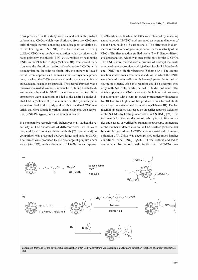
Scheme 3: Methods for the covalent functionalization of CNOs by azomethine ylide addition on CNOs and amidation reactions of carboxylated CNOs[26].
tions presented in this study were carried out with purified
carboxylated CNOs, which were fabricated from raw CNO ma-
terial through thermal annealing and subsequent oxidation by
reflux heating in 3 N HNO3. The first reaction utilizing
oxidized CNOs was the functionalization with a diamine-termi-
nated poly(ethylene glycol) (PEG1500N), realized by heating the
CNOs in the PEG for 19 days (Scheme 3B). The second reac-
tion was the functionalization of carboxylated CNOs with
octadecylamine. In order to obtain this, the authors followed
two different approaches. One was a solid state synthetic proce-
dure, in which the CNOs were heated with 1-octadecylamine in
an evacuated, sealed glass ampoule. The second approach was a
microwave-assisted synthesis, in which CNOs and 1-octadecyl-
amine were heated in DMF in a microwave reactor. Both
approaches were successful and led to the desired octadecyl-
ated CNOs (Scheme 3C). To summarize, the synthetic path-
ways described in this study yielded functionalized CNO ma-
terials that were soluble in various organic solvents. One deriva-
tive, (CNO-PEG1500N), was also soluble in water.
In a comparative research work, Echegoyen et al. studied the re-
activity of CNO materials of different sizes, which were
prepared by different synthetic methods [27] (Scheme 4). A
comparison was presented between larger and smaller CNOs.
The former were produced by arc discharge of graphite under
water (A-CNO), with a diameter of 15–20 nm and approx.
20–30 carbon shells while the latter were obtained by annealing
nanodiamonds (N-CNO) and presented an average diameter of
about 5 nm, having 6–8 carbon shells. The difference in diam-
eter was found to be of great importance for the reactivity of the
CNOs. The first reaction studied was a [2 + 1] Bingel–Hirsch
cyclopropanation, which was successful only for the N-CNOs.
The CNOs were reacted with a mixture of dodecyl malonate
ester, carbon tetrabromide, and 1,8-diazabicyclo[5.4.0]undec-7-
ene (DBU) in o-dichlorobenzene (Scheme 4A). The second
reaction studied was a free-radical addition, in which the CNOs
were heated under reflux with benzoyl peroxide as radical
source in toluene. Also this reaction could be accomplished
only with N-CNOs, while the A-CNOs did not react. The
obtained phenylated CNOs were not soluble in organic solvents,
but sulfonation with oleum, followed by treatment with aqueous
NaOH lead to a highly soluble product, which formed stable
dispersions in water as well as in ethanol (Scheme 4B). The last
reaction investigated was based on an earlier reported oxidation
of the N-CNOs by heating under reflux in 3 N HNO3 [26]. This
treatment led to the introduction of carboxylic acid functionali-
ties and caused, as verified by Raman spectroscopy, an increase
of the number of defect sites on the CNO surface (Scheme 4C).
In a similar procedure, A-CNOs were not oxidized. However,
oxidation of A-CNOs was accomplished under much harsher
conditions (conc. HNO3/H2SO4, 1:1 v/v, reflux) and led to
comparable observations made for the oxidized N-CNO ma-

Beilstein J. Nanotechnol. 2014, 5, 1980–1998.
1986
Scheme 4: Comparison of the reactivity of small N-CNOs and larger A-CNOs, prepared by different methods [27].
terial. It is important to note that N-CNOs were completely
destroyed under the harsh reaction conditions mentioned above
(Scheme 4D). In conclusion, the authors found that the smaller
N-CNO are much more accessible for covalent functionaliza-
tion, while the functionalization of A-CNO requires an aggres-
sive treatment. This observation is led back to a higher surface-
to-volume ratio in the N-CNOs, compared to A-CNOs, as well
as to a larger curvature increasing the degree of surface strain.
Another example for the functionalization of CNOs in a highly
reactive environment was published by Khabashesku et al., who
directly fluorinated CNOs under a stream of F2 and H2 [28].
CNO fluorination was carried out in a custom-built reactor with
different fluorination temperatures. The CNOs used in this
study were prepared from carbon black by heating and the
formed individual CNOs had diameters between 50 and
100 nm. HF was generated in situ under the given reaction
conditions in a continuous flow of a F2 gas mixture (10% F2/
90% He) and H2. The successful fluorination led to an increase
of the mass of the CNO material and the fluorinated CNOs
(F-CNO) were well soluble in ethanol and other alcohols as
well as in DMF. Characterization of the F-CNOs was carried
out by a multitude of techniques such as FTIR, Raman and
UV–vis spectroscopy, SEM/EDX, XRD, XPS, TGA and TEM.
In addition, the authors reported that the fluorination of the
F-CNOs was reversible upon treatment with hydrazine, which
interestingly led also to a regeneration of the “broken” graphene
layers of the CNO.
Some years later, based on the F-CNO material, the same group
reported the preparation of water-soluble sucrose-functional-
ized CNOs [29]. In this case, F-CNOs were reacted with a
lithium monosucrate derivative, which was previously synthe-
sized from sucrose and lithium hydroxide. The sucrose-deco-
rated CNOs showed an improved solubility of up to 200 mg·L−1
in water and 400 mg·L−1 in DMF.
Smith et al. reported the first radical addition of a polymer to
CNOs in 2007 [30]. The CNO starting material was prepared
and purified following an earlier reported procedure [26], and
then further functionalized with bis-o-diynyl arene (BODA),
which is known to thermally form reactive bis-radicals [31].
After ultrasonication and heating of the CNOs and BODA in
N-methyl-2-pyrrolidone (NMP) in a pressure vessel, a

Beilstein J. Nanotechnol. 2014, 5, 1980–1998.
1987
Figure 3: Structure of a CNO–BODA copolymer. (A) CNO starting material (left) and BODA-functionalized CNOs (right) suspended in NMP(0.67 mg·mL−1) immediately after sonication. (B) CNO starting material (left) and BODA-functionalized CNOs (right) suspended in NMP(0.67 mg·mL−1) after 2 h and 48 h of centrifugation. Reprinted with permission from [30]. Copyright 2007 American Chemical Society.
Scheme 5: Preparation of pyridyl-CNOs and an illustration of their supramolecular interaction with Zn-tetraphenylporphyrin (ZnTPP) [32].
CNO–BODA copolymer was obtained. NMP suspensions of the
CNO–BODA copolymer were found to be very stable, even at
high concentrations of up to 0.67 mg·mL−1 (Figure 3).
Characterization was carried out by a multitude of different
techniques, such as GPC, TEM, TGA, XPS and Raman spec-
troscopy.
In 2008, Echegoyen et al. reported a first supramolecular CNO/
Zn-porphyrin complex [32] (Scheme 5). In this set up, acid-
treated CNOs bearing carboxylic acid functionalities, which
were prepared by the oxidation of CNOs (diameter
approx. 6 nm) with conc. HNO3/H2SO4 (3:1, v/v), were reacted
with 4-aminopyridine in an amidation reaction. The synthe-
sized water-soluble CNO material was characterized by using
TEM, NMR, UV–vis and Raman spectroscopy. Based on TGA
studies, the authors estimated that approximately one pyridine
functionality per 120 CNO surface carbon atoms was present.
The pyridine groups were then decorated with Zn-tetraphenyl-
porphyrin (ZnTPP) what was confirmed by NMR spectroscopy
and electrochemistry. No further spectroscopic studies were
carried out with the presented CNO–ZnTPP supramolecular
complex.
A further example of a [2 + 1] cycloaddition on CNOs was
reported by Kong et al., who used different nitrene derivatives
to covalently functionalize pristine CNOs [33] (Scheme 6). The

Beilstein J. Nanotechnol. 2014, 5, 1980–1998.
1988
Scheme 6: Illustration of polymerization reactions on CNOs following initial [2 + 1] cycloaddition reaction of nitrenes [33].
CNO starting material was reacted with either 2-azidoethanol,
leading to OH-functionalized CNOs (CNO-OH) or azidoethyl
2-bromo-2-methylpropanoate, yielding Br-functionalized CNOs
(CNO-Br). The functionalized CNOs showed an increased solu-
bility in organic solvents and water and were characterized by
various techniques, such as TGA, XPS, TEM and Raman spec-
troscopy. Interestingly, the CNO-OH showed distinct fluores-
cence emission with an emission maximum at 453 nm in
aqueous solution, while CNO-Br did not fluoresce. Based on
these two CNO derivatives, the authors reported different poly-
merization reactions, where the CNOs served as macroinitia-
tors. Firstly, CNO-OHs were used for a ring opening polymer-
ization with ε-caprolactone in the presence of stannous octoate.
In a second approach, CNO-Brs were decorated with poly-
styrene in an atom transfer radical polymerization reaction.
Both polymer-functionalized CNO materials showed a good
solubility in common organic solvents and give rise to potential
future applications in various fields of technology.
The synthesis of ferrocene (Fc)-decorated CNOs was reported
by Prato et al. [34]. CNOs were functionalized in a 1,3-dipolar
cycloaddition reaction with a BOC-protected amin/amino acid
[35] and paraformaldehyde. Subsequent deprotection of the
amino functionality and reaction with Fc-carboxylic acid chlo-
ride lead to Fc-functionalized CNOs. By utilizing a TGA-based
method for estimating the number of functionalizations per
surface carbon atom of a CNO, the authors reported that the
CNOs presented in this study contained one functional group
per 36 surface carbon atoms. The properties of the Fc-CNO
conjugates and the electronic interactions between the Fc and
the CNO were investigated by electrochemical and spectro-
scopic methods, and supported by quantum chemical calcula-
tions.
A covalent functionalization of CNOs with biomolecules was
reported by the groups of Plonska-Brzezinska, Simionescu and
Echegoyen in 2010 [36]. In the first step, small CNOs
(6–8 shells) were oxidized by using conc. H2SO4/HNO3 and
subsequently functionalized with PEG to study their cytotoxi-
city on rat dermal fibroblasts. The result was that no significant
cytotoxicity was observable, which renders this CNO material
ideal for future biological applications. Toward the fabrication
of CNO-biosensors, gold electrodes were initially decorated
with a self-assembled monolayer of cysteamine on which the
oxidized CNOs were deposited by an amidation reaction. In
another amidation step, some of the unreacted carboxylic acid
groups on the CNO surface were functionalized with biotin,
which allows the attachment of biomolecules such as avidin.
This first covalent functionalization of CNOs with biomole-
cules, promoted by biotin–avidin interactions, gives rise to
future biological applications such as bio-sensors, especially
since the authors of this study reported that the attached protein
retains its biological activity.
A novel strategy for the surface functionalization of CNOs was
published by our group in 2010 [37] (Scheme 7).

Beilstein J. Nanotechnol. 2014, 5, 1980–1998.
1989
Scheme 7: “Tour” functionalization of CNOs and subsequent “click”-addition of a ZnTPP-derivative [37].
The so-called Tour reaction is well studied for the covalent
functionalization of carbon nanotubes by reacting them with in
situ generated diazonium compounds [38]. This versatile reac-
tion was used in the present study to attach different aniline
derivatives to the surface of CNOs and thus introducing a
variety of functional groups, such as bromides, benzoic acids,
tert-butyl groups, nitro groups, methyl esters, and trimethylsilyl
(TMS) acetylenes. For all reactions, the CNO starting material
was suspended in DMF by ultrasonication and then the aniline
derivative and isoamyl nitrite were subsequently added. After
stirring, the different functionalized CNOs were obtained and
characterized by Raman and TGA analysis. The Raman spec-
troscopy showed an increase of the D-band at 1354 cm−1, a
clear confirmation of a successful covalent functionalization. In
addition, TGA showed a significant weight loss at temperatures
below 450 °C upon functionalization. Based on the method of
Prato et al. [34], the number of surface carbon atoms per func-
tionality was calculated to be between 22 and 60, depending on
the different aniline derivatives used for functionalization.
Multiple repetitions of the reaction lead to a further increase of
the degree of functionalization (Figure 4). All kinds of function-
alization led to an increased dispersibility of the CNOs. In a
next step, the CNO–TMS acetylide material was first depro-
tected and then the free acetylene group was coupled with zinc
triphenyl azidophenyl porphyrin in a copper-mediated “click”
reaction. The successful functionalization was verified by TGA
analysis as well as by Raman, absorption and fluorescence spec-
troscopy.
Following the aforementioned functionalization of CNOs with
benzoic acid [37], fluoresceinamine-based fluorophores [39] as
well as NIR-emitting aza-borondipyrromethenes (azaBODIPYs)
were attached to the CNOs through an amidation reaction [40].
In another very recent study, we attached a meso-phenol-substi-
tuted borondipyrromethene (BODIPY) fluorophore on the same
benzoic acid functionalized CNO nanomaterial through an
esterification reaction [41]. These fluorescent-tagged CNOs
(Scheme 8) were then used for in vitro fluorescence imaging,
which will be discussed in the corresponding section of this
review article.
Figure 4: First derivative TGA weight-loss curves of pristine CNO(black), treated once (light gray) and treated three times with4-bromoaniline (dark gray). Inset shows the enhanced CNOdispersibility in THF upon functionalization. Reprinted with permissionfrom [37]. Copyright 2010 American Chemical Society.
Recently, Echegoyen et al. reported for the first time the alkyl-
ation of CNOs [42], which was achieved by a reductive process
utilizing a Na–K alloy. CNOs were added to a previously
prepared, deep-blue solution of the Na–K alloy in 1,2-
dimethoxyethane under inert atmosphere affording a brownish
dispersion. Then an excess of 1-bromohexadecane was added as
electrophile and the alkylated CNO material (CNO-C16) could
be recovered. TGA analysis and IR as well as Raman spec-
troscopy were used to verify the successful alkylation of the
CNOs. It was reported that the CNO-C16 exhibits an
outstanding solubility in a multitude of organic solvents, even in
high concentrations of up to 0.1 mg·mL−1. This high solubility
enabled the use of 1H NMR spectroscopy, which corroborated
the presence of alkyl groups on the CNO surface. Additional
HRTEM and SEM experiments were carried out to further
support the successful functionalization and excellent solubility
of CNO-C16. The authors also studied the reversibility of this
alkylation reaction, which could be accomplished by annealing
the CNO-C16 material at 415 °C, which was supported by
Raman spectroscopy.

Beilstein J. Nanotechnol. 2014, 5, 1980–1998.
1990
Scheme 8: Fluorophore–CNO conjugates derived from benzoic acid-functionalized CNOs [39-41].
CNO compositesIn addition to the previously mentioned BODA-based CNO
nanocomposites [30] and polymer-functionalized CNOs
prepared by a [2 + 1] cycloaddition of nitrenes [33], several
other CNO-containing composites have been reported
(Scheme 9). In one study, composites consisting of CNOs and
poly(diallyldimethylammonium chloride) (PDDA) or chitosan
(Chit) were prepared and their electrochemical properties were
studied [43]. In another study, CNO–PDDA composite films
were used for the detection of dopamine in the presence of
ascorbic acid and uric acid in solution [44]. The concentration
of dopamine could be determined in a range between 5 × 10⁻⁵
and 4 × 10⁻³ mol·L−1. They also reported the in situ polymer-
ization of aniline on phenylene amine-terminated CNO deriva-
tives [45]. This polyaniline (PANI)-functionalized CNOs were
characterized by a multitude of techniques and showed an
excellent solubility in protic solvents, which gives rise to
several future applications of this material. In a follow-up study,
the properties of CNO–PANI composites were compared to
other PANI-decorated carbon nanostructures [46]. In general,
PANI films containing carbon nanostructures showed impro-
ved properties like a lower resistance and higher mechanical
stability than pure PANI films. Another CNO-containing
composite material was prepared from unmodified or oxidized
CNOs and poly(3,4-ethylenedioxythiophene):poly(styrenesul-
fonate) (PEDOT:PSS) [47]. In a very recent study, they
reported the non-covalent functionalization of CNOs with
poly(4-vinylpyridine-co-styrene) (PVPS) and poly(ethylene
glycol)/Polysorbate 20 (PEG/P20) [48]. The PVPS polymers
were then further functionalized with thiols. Both CNO-
containing polymers, could be modified with flavonoid com-
pounds, for the example quercitin, a compound known for its
anti-inflammatory potential [49], giving rise to future applica-
tions in nanomedicine.
Some additional metal oxide-containing composite materials
were studied for applications as electrode materials in capaci-
tors and lithium-ion batteries and will be discussed in the
corresponding part of this review article.
Toxicological aspectsIn the context of applications in biology and medicine, newly
employed nanomaterials should be carefully evaluated with
regard to biocompatibility, environmental health and safety,
secure processing and sustainable engineering.
In the case of CNOs, limited data regarding their biocompati-
bility has been published. An initial report, investigating the
toxicity of large CNOs with a diameter of about 30 nm was
published in 2005 by Chen et al. [50]. They probed the effects
of large CNOs produced by an underwater carbon-arc
discharge, as well as of multi-wall carbon nanotubes
(MWCTNs) on human skin fibroblasts and found more adverse
effects upon exposure to MWCNTs as compared to CNOs.
However, CNOs were also found to cause negative effects on
the studied cell cultures. The first report investigating the toxi-
city of small CNOs dates back to 2010, when Echegoyen et al.
investigated the biocompatibility of PEGylated CNOs by
exposing rat dermal fibroblasts to different CNO concentra-
tions [37]. The authors could show almost 100% viability of

Beilstein J. Nanotechnol. 2014, 5, 1980–1998.
1991
Scheme 9: Schematic overview over the different polymeric structures utilized to functionalize CNOs [43-48].
cells for concentrations of 30 and 300 μg·mL−1, and a minor
reduction of approx. 15% for 3,000 μg·mL−1. Thus, it follows
that small CNOs are not cytotoxic and can be used safely for
biological studies. In two other reports, highly oxidized CNOs,
derived from pyrolized wood wool, were used for in vivo
imaging of Drosophila melanogaster, Escherichia coli and
Caenorhabditis elegans [17,51]. In both studies, no toxic effects
of the water-soluble CNOs on the investigated organisms were
observed.
We recently reported the weak inflammatory potential and low
cytotoxicity in vitro and in vivo of CNOs and their ability to be
up-taken by antigen-displaying cells. In our work, small benzoic
acid functionalized CNOs were compared with similar function-
alized single wall carbon nanotubes [39]. CNOs showed a lower
inflammatory potential than CNTs and we demonstrated that
chemical functionalization attenuates their inflammatory prop-
erties. This was evidenced by a reduced secretion of the inflam-
matory cytokine IL-1β, and a pronounced decrease in the
recruitment of neutrophils and monocytes following injection
into mice. Subsequently, in two recent studies, we investigated
the effects of two different, fluorophore functionalized CNOs
on HeLa Kyoto [40] and MCF-7 cells [41] and did not observe
any significant cytotoxicity. Our results let us believe that
CNOs are promising materials for biological and medicinal
applications.
ApplicationsIn the following chapter we give an overview over the different
applications of CNOs. We have selected representative studies
from all areas of materials science, nano- and biotechnology
and chemistry where CNOs have been successfully applied. The
presented functionalization pathways usually led to a largely
improved solubility of the CNO materials and thus to an
enhanced processability and applicability. In addition, the intro-
duction of functional groups and functionalities for specific
applications further improved the usability of CNO materials.
However, while other carbon nanostructures have drawn large
attention in a variety of fields of applications, CNOs can still be
considered as being a niche of the research on carbon nano-
structures.
Biological and environmental applicationsBiological imaging: In contrast to other carbon nanomaterials
such as CNTs [52] or carbon quantum dots [53], CNOs have not
been widely employed in biological marking, yet. A first report

Beilstein J. Nanotechnol. 2014, 5, 1980–1998.
1992
Figure 5: a) Autofluorescence images of different developmental stages of Drosophila melanogaster from larva to adult. b) D. melanogaster fed withwater-soluble CNO, under 488 and 561 nm filters. Reprinted with permission from [17]. Copyright 2011 John Wiley and Sons.
Figure 6: High-resolution TEM images of pristine CNOs (left). AFM topographs of pristine CNOs deposited on mica (center). Confocal microscopy ofC57BL/6 BMDCs incubated in the presence of fluorescein-labelled-CNOs and stained with wheat germ agglutinin-Alexa Fluor594 (red), fluorescein(green) and nuclei stain Hoechst (blue) (right). Reprinted with permission from [39]. Copyright 2013 John Wiley and Sons.
was published in 2011 by Sarkar et al. by using large, defect
rich CNOs, synthesized from wood waste, for imaging the life
cycle of D. melanogaster (Figure 5) [17]. The authors claim that
solubility in water was achieved by the presence of a large
number of carboxylate groups on the CNO surface that origi-
nate from the production process. These carboxylate functional
groups, together with the defective nature of the CNOs, also led
to the observed fluorescence emission in the visible and NIR,
which was imposed by spontaneous surface passivation and
quantum confinement and allowed for multicolor biological
imaging. The specimens were fed with fluorescent CNO nano-
material that accumulated in the organisms and could be
observed by fluorescence microscopy. Control experiments
with non-CNO fed specimens were performed, excluding auto-
fluorescence as reason for the observed luminescence of the
organisms. Following their initial work describing in vivo bio-
logical marking of D. melanogaster, they used fluorescent
CNOs also as imaging agents to study E. coli and C. elegans in
vivo [51].
In a recent report from our group [40], we used fluorescein-
functionalized CNOs in a comparative toxicological study in
vitro and in vivo, including biological marking (Figure 6). The
cytotoxicity and immunomodulatory properties of the synthe-
sized fluorescein-CNO derivative were elucidated and
compared with similarly functionalized CNTs. We could show
that CNOs exhibit efficient cellular uptake, weak inflammatory
potential, and low cytotoxicity and are therefore promising ma-
terial for biomedical applications.
Recently we demonstrated the cellular imaging of HeLa Kyoto
[40] and MCF-7 cells [41] after incubating them with
azaBODIPY- or BODIPY-functionalized CNOs (Scheme 8 and
Figure 7). In both cases the CNO conjugates were readily inter-

Beilstein J. Nanotechnol. 2014, 5, 1980–1998.
1993
nalized by the cells. In the earlier study, one of the
azaBODIPY-CNO derivatives showed a pH-dependent
switching (on–off) of the fluorescence, a feature that could also
be observed inside the cells. The latter CNO nanomaterial was
subject to co-localization experiments with Lysotracker Red
dye and it was confirmed by high-resolution imaging that the
CNOs were deposited in the lysosomes of the cells.
Figure 7: Confocal images of azaBODIPY-CNOs in HeLa Kyoto cells(left) and BODIPY-CNOs in MCF-7 cells (right). The blue lumines-cence is due to Hoechst 33342 nuclear stain. Reproduced with permis-sion from [40] and [41]. Copyright 2014 The Royal Society of Chem-istry.
Biological sensing: In the aforementioned study of Luszczyn et
al. [36], CNOs were covalently functionalized with biomole-
cules and studied for the first time as biosensors by using
avitin–biotin interactions. The CNO served as linking layers
between the biomolecules and the gold surface of the sensor and
led to an amplified signal of the biosensor, as determined by
surface plasmon resonance spectroscopy. In addition, the
biocompatibility of CNOs was investigated and found to be
excellent.
Environmental remediation: An application of CNO in envi-
ronmental remediation was studied by Li group [54], who
revealed that surface-oxidized CNO in aqueous suspensions
have a high sorption capacity for heavy metal ions such as Pb2+,
Cu2+, Cd2+, Ni2+ and Zn2+. The sorption capacity of oxidized
CNOs was found to be up to ten times higher than the one of
fullerene C60. These encouraging results could be a first step
toward in situ remediation of heavy metal contaminants.
Electronic applicationsCapacitors: Carbon materials are commonly used as electrode
materials in capacitors, but the first study probing CNOs as
electrode materials in electrical double layer capacitors (EDLC)
with an organic electrolyte was published only in 2007. The
electrochemical performance of CNO electrodes was compared
with electrodes made with nanodiamonds, multi-wall carbon
nanotubes and carbon black [55]. Following this initial work,
several groups studied CNO materials in supercapacitor elec-
trodes. Bushueva et al. for example, found capacitance values of
the investigated CNO material of 20–40 F·g−1 and 70–100 F·g−1
with acidic or basic electrolyte solutions, respectively [56]. In
2010, Pech et al. published the preparation and characterization
of ultrahigh-power micrometer-sized supercapacitors based on
CNOs [57]. In an extensive electrochemical study in different
aqueous and organic electrolytes, McDonough et al. investi-
gated the influence of the CNO structure on their electrochem-
ical performance in supercapacitor electrodes [58]. The increase
of the capacitance of CNO materials was the subject of two
further studies. Borgohain et al. firstly oxidized the CNOs and
subsequently functionalized the surface with polar carboxylic
acid groups, which enabled them to precipitate RuO2 [59]. This
functionalization led to an increase of the capacitance from
45 F·g−1 (for the pure CNO material) to 334 F·g−1 (for the
RuO2·H2O–CNO composite material). Another strategy to
increase the CNO capacitance is the activation of the CNO
surface by treatment with 6 M KOH, creating porosity in the
outer shells of the CNOs (Figure 8) [60]. The activated CNOs
show largely improved properties compared to pristine
CNOs with a maximum specific capacitance of 122 F·g−1
(vs 25.8 F·g−1) , a power densi ty of 153 kW·kg−1
(vs 123 kW·kg−1) and an energy density of 8.5 Wh·kg−1
(vs 1.5 Wh·kg−1).
Composite materials were studied for application in capacitors
as well. The group of Echegoyen found that incorporation of
CNOs in a Chit- or PDDA-polymer matrix leads to a large
increase of the capacitance of the composite films (from
4–10 F·g−1 for the pure Chit or PDDA films to 20–30 F·g−1 for
the corresponding CNO composite material) [43]. Also the
specific electrochemical capacitance of a CNO–PANI
composite (206.64 F·g−1) was much larger than for pure
oxidized CNOs (12.15 F·g−1) [45]. After optimization in a
follow-up study, the highest specific capacitance for a
CNO–PANI composite was measured to be 525 F·g−1, which
renders this composite material interesting for applications in
supercapacitor electrodes [46]. Also the CNO/PEDOT:PSS
composites, which were previously discussed, showed
promising properties for the application as electrode material in
supercapacitors, such as a specific capacity of 96 F·g−1, good
cation-exchange properties and a simple synthesis [47]. In a
recent study, the same group decorated the surface of CNO with
Ni(OH)2 or NiO as pseudocapacitive redox material and
showed that these composites can be promising materials for
the development of supercapacitors [61]. In order to achieve
this, the CNO surface was modified with nickel particles, which
were synthesized in situ from nickel nitrate hexahydrate and
ammoniumhydroxide in ethanol in the presence of (4-dimethyl-
amino)pyridine (4-DMAP) as modifier in a one-pot multi-step
reaction. Calcination of the CNO/4-DMAP/Ni(OH)2 composite
led to the CNO/4-DMAP/NiO composite material. The electro-
chemical properties were promising, especially the specific

Beilstein J. Nanotechnol. 2014, 5, 1980–1998.
1994
Figure 8: (a) A schematic showing the chemical activation of CNOs in KOH. TEM images of pristine CNO (b), ACNO-4M (c), ACNO-6M (d), andACNO-7M (e). ‘‘ACNO-nM’’ denotes the activated CNO prepared using n mol·L−1 KOH solution. Reprinted with permission from [60]. Copyright 2013Elsevier.
electrochemical capacitance could be increased largely to
290.6 F·g−1 for the CNO/4-DMAP/NiO and 1225.2 F·g−1 for
the CNO/4-DMAP/Ni(OH)2 composite, compared to pristine
CNOs with 30.6 F·g−1.
Another example for CNO composite-based capacitors was
reported by the group of H. Y. Yang [62]. The composite was
prepared from KMnO4 and CNOs in different weight ratios in
deionized water by heating in an autoclave. The formed
CNO–MnO2 composite was then implemented in an asym-
metric pseudocapacitor with the CNO–MnO2 composite as
working electrode and nickel foam as counter electrode. The
capacitance of pure MnO2 (40 F·g−1) could be increased by the
incorporation of CNO up to 177.5 F·g−1. In addition, the
authors report an excellent cycling stability with 99–101%
retention of the specific capacitance after 1000 cycles.
Lithium-Ion batteries: Carbon nanotubes are widely studied
for a use in lithium ion batteries [63]. However, also CNOs
were studied for a potential application as anode materials. Han
et al., for example, reported the large scale synthesis of CNOs
starting from CuCl2·2 H2O and CaC2 and found that they ex-
hibit a high capacity in combination with a promising cycling
performance, which renders these as-prepared CNOs as poten-
tial anode materials for lithium-ion batteries [16]. However, no
prototype batteries were prepared by the authors of this report.
In two recent studies, H. Y. Yang and co-workers reported
lithium-ion batteries incorporating CNOs in combination with
Co3O4 [64] and MnO2 [65] as electrode material. In the earlier
study, the CNO-containing anode material was prepared by a
solvo-thermal method from cobalt acetate and CNOs and the
authors found that the novel composite material showed impro-
ved electrochemical properties, compared to pristine Co3O4
electrodes. They observed, for example, an increase of the
specific capacity from 190 mA·h·g−1 to 632 mA·h·g−1 at a
current density of 200 mA·g−1 and also an increased rate capa-
bility [64]. In the latter report, the CNO hybrid material was
prepared from KMnO4 and CNOs by a hydrothermal method
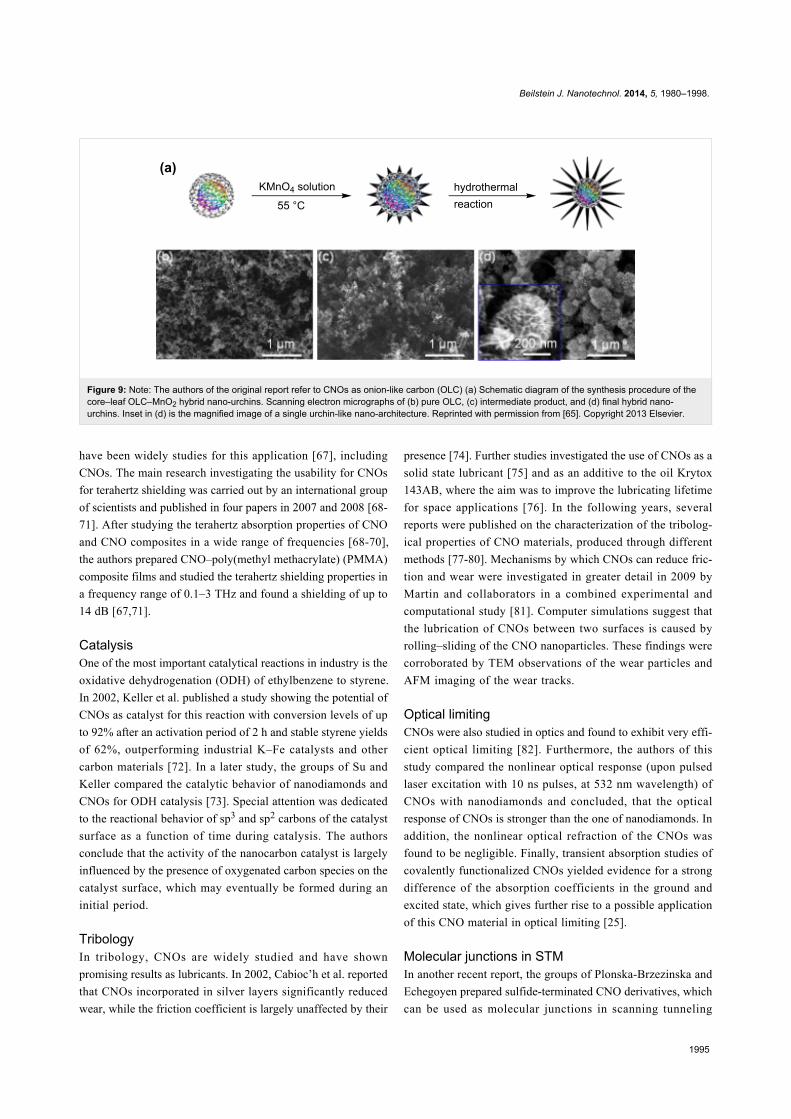
(Figure 9) similar to the one the group reported earlier
for the preparation of electrode materials for pseudocapacitors
[62]. This material was then probed as anode material in
lithium-ion batteries. It was found that the performance of
pure MnO2 anodes could be significantly enhanced by the
incorporation of CNOs. The specific capacity increased from
260 mA·h·g−1 to 630 mA·h·g−1, at a current density of
50 mA·g−1. In addition, the authors report an increased rate
capability, stable cycling performance, and coulomb efficiency
of nearly 100% [65].
Fuel cells: CNOs were also investigated as catalyst support for
application in direct methanol fuel cells. For this, Xu et al.
prepared CNOs decorated with Pt nanoparticles (Pt-CNO) and
compared the performance of this novel catalyst material with
common Pt/Vulcan XC-72 with encouraging results [66]. The
novel Pt-CNO catalyst showed a higher surface area and
smaller Pt particle size (3.05 nm vs 4.10 nm) than the reference
system and the catalytic activity for the electro oxidation of
methanol was increased by about 20%, rendering CNOs as a
promising catalyst support for fuel cells.
Terahertz-shielding: In recent years, terahertz devices, circuits
and terahertz-based communication systems have become
important in many fields. This makes the development of ma-
terials for terahertz shielding essential, to limit electromagnetic
interferences and thus reduce for example noise in cables and
communication systems and signal coupling. Carbon materials
Beilstein J. Nanotechnol. 2014, 5, 1980–1998.
1995
Figure 9: Note: The authors of the original report refer to CNOs as onion-like carbon (OLC) (a) Schematic diagram of the synthesis procedure of thecore–leaf OLC–MnO2 hybrid nano-urchins. Scanning electron micrographs of (b) pure OLC, (c) intermediate product, and (d) final hybrid nano-urchins. Inset in (d) is the magnified image of a single urchin-like nano-architecture. Reprinted with permission from [65]. Copyright 2013 Elsevier.
have been widely studies for this application [67], including
CNOs. The main research investigating the usability for CNOs
for terahertz shielding was carried out by an international group
of scientists and published in four papers in 2007 and 2008 [68-
71]. After studying the terahertz absorption properties of CNO
and CNO composites in a wide range of frequencies [68-70],
the authors prepared CNO–poly(methyl methacrylate) (PMMA)
composite films and studied the terahertz shielding properties in
a frequency range of 0.1–3 THz and found a shielding of up to
14 dB [67,71].
CatalysisOne of the most important catalytical reactions in industry is the
oxidative dehydrogenation (ODH) of ethylbenzene to styrene.
In 2002, Keller et al. published a study showing the potential of
CNOs as catalyst for this reaction with conversion levels of up
to 92% after an activation period of 2 h and stable styrene yields
of 62%, outperforming industrial K–Fe catalysts and other
carbon materials [72]. In a later study, the groups of Su and
Keller compared the catalytic behavior of nanodiamonds and
CNOs for ODH catalysis [73]. Special attention was dedicated
to the reactional behavior of sp3 and sp2 carbons of the catalyst
surface as a function of time during catalysis. The authors
conclude that the activity of the nanocarbon catalyst is largely
influenced by the presence of oxygenated carbon species on the
catalyst surface, which may eventually be formed during an
initial period.
TribologyIn tribology, CNOs are widely studied and have shown
promising results as lubricants. In 2002, Cabioc’h et al. reported
that CNOs incorporated in silver layers significantly reduced
wear, while the friction coefficient is largely unaffected by their
presence [74]. Further studies investigated the use of CNOs as a
solid state lubricant [75] and as an additive to the oil Krytox
143AB, where the aim was to improve the lubricating lifetime
for space applications [76]. In the following years, several
reports were published on the characterization of the tribolog-
ical properties of CNO materials, produced through different
methods [77-80]. Mechanisms by which CNOs can reduce fric-
tion and wear were investigated in greater detail in 2009 by
Martin and collaborators in a combined experimental and
computational study [81]. Computer simulations suggest that
the lubrication of CNOs between two surfaces is caused by
rolling–sliding of the CNO nanoparticles. These findings were
corroborated by TEM observations of the wear particles and
AFM imaging of the wear tracks.
Optical limitingCNOs were also studied in optics and found to exhibit very effi-
cient optical limiting [82]. Furthermore, the authors of this
study compared the nonlinear optical response (upon pulsed
laser excitation with 10 ns pulses, at 532 nm wavelength) of
CNOs with nanodiamonds and concluded, that the optical
response of CNOs is stronger than the one of nanodiamonds. In
addition, the nonlinear optical refraction of the CNOs was
found to be negligible. Finally, transient absorption studies of
covalently functionalized CNOs yielded evidence for a strong
difference of the absorption coefficients in the ground and
excited state, which gives further rise to a possible application
of this CNO material in optical limiting [25].
Molecular junctions in STMIn another recent report, the groups of Plonska-Brzezinska and
Echegoyen prepared sulfide-terminated CNO derivatives, which
can be used as molecular junctions in scanning tunneling

Beilstein J. Nanotechnol. 2014, 5, 1980–1998.
1996
microscopy (STM) [83]. This enabled the authors to study the
conductivity of CNOs and compare their properties with
comparable fullerene-C60 derivatives. The measured data
suggests that the intrinsic conductivity of CNOs and C60 is
within the same order of magnitude.
ConclusionMulti-shell fullerenes, known as carbon nano-onions (CNOs),
were discovered in 1992 and are structured by concentric shells
of carbon atoms in a graphitic interlayer distance. Analogous to
carbon nanotubes, CNOs display poor solubility in both
aqueous and organic solvents as well as a high surface area,
compared to their volume. In order for their full potential to be
realized, the solubility of the CNO nanomaterial has to be
increased. To achieve this, a multitude of different synthetic
pathways for the covalent surface functionalization has been
reported. An alternative to the covalent functionalization is the
surface decoration of CNOs with polymers or their incorpor-
ation into composites. CNOs have been implemented in
different electronic applications, as electrode materials in capac-
itors, as anode materials in lithium-ion batteries, as catalyst
support in fuel cells. They have even attracted the interest of
NASA researchers for their tribological properties as additives
for aerospace applications. Despite much interest in different
carbon-based nano-materials, CNOs as functional constructs for
intracellular transport have not been widely explored. However,
given their size, homogeneity and purity (compared with carbon
nanotubes) they could in principle add an important new avenue
for the transport of imaging and therapeutic agents. These
carbon particles have demonstrated high cellular uptake, low
cytotoxicity and lower inflammatory potential than CNTs and a
very promising future for biomedical applications.
AcknowledgementsFinancial support from the Istituto Italiano di Tecnologia (IIT)
is greatly appreciated. We also thank the Cost Action MP0901:
“Designing novel materials for nanodevices: From Theory to
Practice”.
References1. Kroto, H. W.; Heath, J. R.; O’Brien, S. C.; Curl, R. F.; Smalley, R. E.
Nature 1985, 318, 162–163. doi:10.1038/318162a02. Bollmann, W.; Spreadborough, J. Nature 1960, 186, 29–30.
doi:10.1038/186029a03. Iijima, S. Nature 1991, 354, 56–58. doi:10.1038/354056a04. Iijima, S.; Ichihashi, T. Nature 1993, 363, 603–605.
doi:10.1038/363603a05. Iijima, S.; Yudasaka, M.; Yamada, R.; Bandow, S.; Suenaga, K.;
Kokai, F.; Takahashi, K. Chem. Phys. Lett. 1999, 309, 165–170.doi:10.1016/S0009-2614(99)00642-9
6. Danilenko, V. V. Phys. Solid State 2004, 46, 595–599.doi:10.1134/1.1711431
7. Novoselov, K. S.; Geim, A. K.; Morozov, S. V.; Jiang, D.; Zhang, Y.;Grigorieva, I. V.; Firsov, A. A. Science 2004, 306, 666–669.doi:10.1126/science.1102896
8. Ugarte, D. Nature 1992, 359, 707–709. doi:10.1038/359707a09. Kuznetsov, V. L.; Chuvilin, A. L.; Moroz, E. M.; Kolomiichuk, V. N.;
Shaikhudtdinov, S. K.; Butenko, Y. V.; Mal’kov, I. Y. Carbon 1994, 32,873–882. doi:10.1016/0008-6223(94)90044-2
10. Echegoyen, L.; Ortiz, A.; Chaur, M. N.; Palkar, A. J. Carbon NanoOnions. In Chemistry of Nanocarbons; Akasaka, T.; Wudl, F.;Nagase, S., Eds.; John Wiley & Sons: Chichester, UK, 2010;pp 463–483. doi:10.1002/9780470660188.ch19
11. Kuznetsov, V. L.; Chuvilin, A. L.; Butenko, Y. V.; Mal’kov, I. Y.;Titov, V. M. Chem. Phys. Lett. 1994, 222, 343–348.doi:10.1016/0009-2614(94)87072-1
12. Tomita, S.; Sakurai, T.; Ohta, H.; Fujii, M.; Hayashi, S. J. Chem. Phys.2001, 114, 7477–7482. doi:10.1063/1.1360197
13. Qin, L.-C.; Iijima, S. Chem. Phys. Lett. 1996, 262, 252–258.doi:10.1016/0009-2614(96)01037-8
14. Sano, N.; Wang, H.; Chhowalla, M.; Alexandrou, I.;Amaratunga, G. A. J. Nature 2001, 414, 506. doi:10.1038/35107141
15. Alexandrou, I.; Wang, H.; Sano, N.; Amaratunga, G. A. J.J. Chem. Phys. 2004, 120, 1055–1058. doi:10.1063/1.1629274
16. Han, F.-D.; Yao, B.; Bai, Y.-J. J. Phys. Chem. C 2011, 115,8923–8927. doi:10.1021/jp2007599
17. Ghosh, M.; Sonkar, S. K.; Saxena, M.; Sarkar, S. Small 2011, 7,3170–3177. doi:10.1002/smll.201101158
18. Choucair, M.; Stride, J. A. Carbon 2012, 50, 1109–1115.doi:10.1016/j.carbon.2011.10.023
19. Ugarte, D. Carbon 1995, 33, 989–993.doi:10.1016/0008-6223(95)00027-B
20. Roy, D.; Chhowalla, M.; Wang, H.; Sano, N.; Alexandrou, I.;Clyne, T. W.; Amaratunga, G. A. J. Chem. Phys. Lett. 2003, 373,52–56. doi:10.1016/S0009-2614(03)00523-2
21. Krishnamurthy, S.; Butenko, Y. V.; Dhanak, V. R.; Hunt, M. R. C.;Šiller, L. Carbon 2013, 52, 145–149. doi:10.1016/j.carbon.2012.09.015
22. Singh, P.; Campidelli, S.; Giordani, S.; Bonifazi, D.; Bianco, A.;Prato, M. Chem. Soc. Rev. 2009, 38, 2214–2230.doi:10.1039/b518111a
23. Herranz, M. A.; Martin, N. Noncovalent Functionalization of CarbonNanotubes. In Carbon Nanotubes and Related Structures: Synthesis,Characterization, Functionalization, and Applications; Guldi, D. M.;Martin, N., Eds.; Wiley-VCH Verlag GmbH & Co. KGaA: Weinheim,Germany, 2010; pp 103–134. doi:10.1002/9783527629930.ch5
24. Hauke, F.; Hirsch, A. Covalent Functionalization of Carbon Nanotubes.In Carbon Nanotubes and Related Structures: Synthesis,Characterization, Functionalization, and Applications; Guldi, D. M.;Martin, N., Eds.; Wiley-VCH Verlag GmbH & Co. KGaA: Weinheim,Germany, 2010; pp 135–198. doi:10.1002/9783527629930.ch6
25. Georgakilas, N.; Guldi, D. M.; Signorini, R.; Bozio, R.; Prato, M.J. Am. Chem. Soc. 2003, 125, 14268–14269. doi:10.1021/ja0342805
26. Rettenbacher, A. S.; Elliott, B.; Hudson, J. S.; Amirkhanian, A.;Echegoyen, L. Chem. – Eur. J. 2006, 12, 376–387.doi:10.1002/chem.200500517
27. Palkar, A.; Melin, F.; Cardona, C. M.; Elliott, B.; Naskar, A. K.;Edie, D. D.; Kumbhar, A.; Echegoyen, L. Chem. – Asian J. 2007, 2,625–633. doi:10.1002/asia.200600426
28. Liu, Y.; Vander Wal, R. L.; Khabashesku, V. N. Chem. Mater. 2007, 19,778–786. doi:10.1021/cm062177j

Beilstein J. Nanotechnol. 2014, 5, 1980–1998.
1997
29. Kuznetsov, O. V.; Pulikkathara, M. X.; Lobo, R. F. M.;Khabashesku, V. N. Russ. Chem. Bull. 2010, 59, 1495–1505.doi:10.1007/s11172-010-0269-y
30. Rettenbacher, A. S.; Perpall, M. W.; Echegoyen, L.; Hudson, J.;Smith, D. W., Jr. Chem. Mater. 2007, 19, 1411–1417.doi:10.1021/cm0626132
31. Bergman, R. G. Acc. Chem. Res. 1973, 6, 25–31.doi:10.1021/ar50061a004
32. Palkar, A.; Kumbhar, A.; Athans, A. J.; Echegoyen, L. Chem. Mater.2008, 20, 1685–1687. doi:10.1021/cm7035508
33. Zhou, L.; Gao, C.; Zhu, D.; Xu, W.; Chen, F. F.; Palkar, A.;Echegoyen, L.; Kong, E. S.-W. Chem. – Eur. J. 2009, 15, 1389–1396.doi:10.1002/chem.200801642
34. Cioffi, C. T.; Palkar, A.; Melin, F.; Kumbhar, A.; Echegoyen, L.;Melle-Franco, M.; Zerbetto, F.; Rahman, G. M. A.; Ehli, C.; Sgobba, V.;Guldi, D. M.; Prato, M. Chem. – Eur. J. 2009, 15, 4419–4427.doi:10.1002/chem.200801818
35. Kordatos, K.; Da Ros, T.; Bosi, S.; Vázquez, E.; Bergamin, M.;Cusan, C.; Pellarini, F.; Tomberli, V.; Baiti, B.; Pantarotte, D.;Georgakilas, V.; Spalluto, G.; Prato, M. J. Org. Chem. 2001, 66,4915–4920. doi:10.1021/jo015608k
36. Luszczyn, J.; Plonska-Brzezinska, M. E.; Palkar, A.; Dubis, A. T.;Simionescu, A.; Simionescu, D. T.; Kalska-Szostko, B.; Winkler, K.;Echegoyen, L. Chem. – Eur. J. 2010, 16, 4870–4880.doi:10.1002/chem.200903277
37. Flavin, K.; Chaur, M. N.; Echegoyen, L.; Giordani, S. Org. Lett. 2010,12, 840–843. doi:10.1021/ol902939f
38. Bahr, J. L.; Yang, J.; Kosynkin, D. V.; Bronikowski, M. J.;Smalley, R. E.; Tour, J. M. J. Am. Chem. Soc. 2001, 123, 6536–6542.doi:10.1021/ja010462s
39. Yang, M.; Flavin, K.; Kopf, I.; Radics, G.; Hearnden, C. H. A.;McManus, G. J.; Moran, B.; Villalta-Cerdas, A.; Echegoyen, L. A.;Giordani, S.; Lavelle, E. C. Small 2013, 9, 4194–4206.doi:10.1002/smll.201300481
40. Giordani, S.; Bartelmess, J.; Frasconi, M.; Biondi, I.; Cheung, S.;Grossi, M.; Wu, D.; Echegoyen, L.; O’Shea, D. F. J. Mater. Chem. B2014, 2, 7459–7463. doi:10.1039/C4TB01087F
41. Bartelmess, J.; De Luca, E.; Signorelli, A.; Baldrighi, M.; Becce, M.;Brescia, R.; Nardone, V.; Parisini, E.; Echegoyen, L.; Pompa, P. P.;Giordani, S. Nanoscale 2014, 6, 13761–13769.doi:10.1039/C4NR04533E
42. Molina-Ontaria, A.; Chaur, M. N.; Plonska-Brzezinska, M. E.;Echegoyen, L. Chem. Commun. 2013, 49, 2406–2408.doi:10.1039/c3cc39077b
43. Breczko, J.; Winkler, K.; Plonska-Brzezinska, M. E.; Villalta-Cerdas, A.;Echegoyen, L. J. Mater. Chem. 2010, 20, 7761–7768.doi:10.1039/c0jm01213k
44. Breczko, J.; Plonska-Brzezinska, M. E.; Echegoyen, L.Electrochim. Acta 2012, 72, 61–67.doi:10.1016/j.electacta.2012.03.177
45. Plonska-Brzezinska, M. E.; Mazurczyk, J.; Palys, B.; Breczko, J.;Lapinski, A.; Dubis, A. T.; Echegoyen, L. Chem. – Eur. J. 2012, 18,2600–2608. doi:10.1002/chem.201102175
46. Plonska-Brzezinska, M. E.; Breczko, J.; Palys, B.; Echegoyen, L.ChemPhysChem 2013, 14, 116–124. doi:10.1002/cphc.201200759
47. Plonska-Brzezinska, M. E.; Lewandowski, M.; Blaszyk, M.;Molina-Ontario, A.; Luciński, T.; Echegoyen, L. ChemPhysChem 2012,13, 4134–4141. doi:10.1002/cphc.201200789
48. Plonska-Brzezinska, M. E.; Brus, D. M.; Breczko, J.; Echegoyen, L.Chem. – Eur. J. 2013, 19, 5019–5024. doi:10.1002/chem.201300009
49. Lei, H.; Luo, J.; Tong, L.; Peng, L.-q.; Qi, Y.; Jia, Z.-g.; Wei, Q.Food Chem. 2011, 127, 1169–1174.doi:10.1016/j.foodchem.2011.01.119
50. Ding, L.; Stilwell, J.; Zhang, T.; Elboudwarej, O.; Jiang, H.;Selegue, J. P.; Cooke, P. A.; Gray, J. W.; Chen, F. F. Nano Lett. 2005,5, 2448–2464. doi:10.1021/nl051748o
51. Sonkar, S. K.; Ghosh, M.; Roy, M.; Begum, A.; Sarkar, S.Mater. Express 2012, 2, 105–114. doi:10.1166/mex.2012.1064
52. Gong, H.; Peng, R.; Liu, Z. Adv. Drug Delivery Rev. 2013, 65,1951–1963. doi:10.1016/j.addr.2013.10.002
53. Shen, J.; Zhu, Y.; Yang, X.; Li, C. Chem. Commun. 2012, 48,3686–3699. doi:10.1039/c2cc00110a
54. Seymour, M. B.; Su, C.; Gao, Y.; Lu, Y.; Li, Y. J. Nanopart. Res. 2012,14, 1087. doi:10.1007/s11051-012-1087-y
55. Portet, C.; Yushin, G.; Gogotsi, Y. Carbon 2007, 45, 2511–2518.doi:10.1016/j.carbon.2007.08.024
56. Bushueva, E. G.; Galkin, P. S.; Okotrub, A. V.; Bulusheva, L. G.;Gavrilov, N. N.; Kuznetsov, V. L.; Moiseekov, S. I. Phys. Status Solidi B2008, 245, 2296–2299. doi:10.1002/pssb.200879608
57. Pech, D.; Brunet, M.; Durou, H.; Huang, P.; Mochalin, V.; Gogotsi, Y.;Taberna, T.-L.; Simon, P. Nat. Nanotechnol. 2010, 5, 651–654.doi:10.1038/nnano.2010.162
58. McDonough, J. K.; Frolov, A. I.; Presser, V.; Niu, J.; Miller, C. H.;Ubieto, T.; Fedorov, M. V.; Gogotsi, Y. Carbon 2012, 50, 3298–3309.doi:10.1016/j.carbon.2011.12.022
59. Borgohain, R.; Li, J.; Selegue, J. P.; Cheng, Y.-T. J. Phys. Chem. C2012, 116, 15068–15075. doi:10.1021/jp301642s
60. Gao, Y.; Zhou, Y. S.; Qian, M.; He, X. N.; Redepenning, J.;Goodman, P.; Li, H. M.; Jiang, L.; Lu, Y. F. Carbon 2013, 51, 52–58.doi:10.1016/j.carbon.2012.08.009
61. Plonska-Brzezinska, M. E.; Brus, D. M.; Molina-Ontaria, A.;Echegoyen, L. RSC Adv. 2013, 3, 25891–25901.doi:10.1039/c3ra44249g
62. Wang, Y.; Yu, S. F.; Sun, C. Y.; Zhu, T. J.; Yang, H. Y. J. Mater. Chem.2012, 22, 17584–17588. doi:10.1039/c2jm33558a
63. Landi, B. J.; Ganter, M. J.; Cress, C. D.; DiLeo, R. A.; Raffaelle, R. P.Energy Environ. Sci. 2009, 2, 638–654. doi:10.1039/b904116h
64. Wang, Y.; Yan, F.; Liu, S. W.; Tan, A. Y. S.; Song, H.; Sun, X. W.;Yang, H. Y. J. Mater. Chem. A 2013, 1, 5212–5216.doi:10.1039/c3ta10559h
65. Wang, Y.; Han, Z. J.; Yu, S. F.; Song, R. R.; Song, H. H.; Ostrikov, K.;Yang, H. Y. Carbon 2013, 64, 230–236.doi:10.1016/j.carbon.2013.07.057
66. Xu, B.; Yang, X.; Wang, X.; Guo, J.; Liu, X. J. Power Sources 2006,162, 160–164. doi:10.1016/j.jpowsour.2006.06.063
67. Liu, L.; Das, A.; Megaridis, C. M. Carbon 2014, 69, 1–16.doi:10.1016/j.carbon.2013.12.021
68. Shenderova, O.; Tyler, T.; Cunningham, G.; Ray, M.; Walsh, J.;Casulli, M.; Hens, S.; McGuire, G.; Kuznetsov, V.; Lipa, S.Diamond Relat. Mater. 2007, 16, 1213–1217.doi:10.1016/j.diamond.2006.11.086
69. Maksimenko, S. A.; Rodionova, V. N.; Slepyan, G. Y.; Karpovich, V. A.;Shenderova, O.; Walsh, J.; Kuznetsov, V. L.; Mazov, I. N.;Moseenkov, S. I.; Okotrub, A. V.; Lambin, P. Diamond Relat. Mater.2007, 16, 1231–1235. doi:10.1016/j.diamond.2006.11.025
70. Shenderova, O.; Grishko, V.; Cunningham, G.; Moseekov, S.;McGuire, G.; Kuznetsov, V. Diamond Relat. Mater. 2008, 17, 462–466.doi:10.1016/j.diamond.2007.08.023

Beilstein J. Nanotechnol. 2014, 5, 1980–1998.
1998
71. Macutkevic, J.; Adomavicius, R.; Krotkus, A.; Seliuta, D.; Valusis, G.;Maksimenko, S.; Kuzhir, P.; Batrakov, K.; Kuznetsov, V.;Moseenkov, S.; Shenderova, O.; Okotrub, A. V.; Langlet, R.;Lambin, P. Diamond Relat. Mater. 2008, 17, 1608–1612.doi:10.1016/j.diamond.2007.11.018
72. Keller, N.; Maksimova, N. I.; Roddatis, V. V.; Schur, M.; Mestl, G.;Butenko, Y. V.; Kuznetsov, V. L.; Schlögl, R. Angew. Chem., Int. Ed.2002, 41, 1885–1888.doi:10.1002/1521-3773(20020603)41:11<1885::AID-ANIE1885>3.0.CO;2-5
73. Su, D.; Maksimova, N. I.; Mestl, G.; Kuznetsov, V. L.; Keller, V.;Schlögl, R.; Keller, N. Carbon 2007, 45, 2145–2151.doi:10.1016/j.carbon.2007.07.005
74. Cabioc’h, T.; Thune, E.; Rivière, J. P.; Camelio, S.; Girard, J. C.;Guérin, P.; Jaouen, M.; Henrard, L.; Lambin, P. J. Appl. Phys. 2002,91, 1560–1567. doi:10.1063/1.1421222
75. Hirata, A.; Igarashi, M.; Kaito, T. Tribol. Int. 2004, 31, 899–905.doi:10.1016/j.triboint.2004.07.006
76. Street, K. W.; Marchetti, M.; Vander Wal, R. L.; Tomasek, A. J.Tribol. Lett. 2004, 16, 143–149.doi:10.1023/B:TRIL.0000009724.01711.f4
77. Matsumoto, N.; Joly-Pottuz, L.; Kinoshita, H.; Ohmae, N.Diamond Relat. Mater. 2007, 16, 1227–1230.doi:10.1016/j.diamond.2007.01.031
78. Joly-Pottuz, L.; Vacher, B.; Ohmae, N.; Martin, J. M.; Epicier, T.Tribol. Lett. 2008, 30, 69–80. doi:10.1007/s11249-008-9316-3
79. Yao, Y.; Wang, X.; Guo, J.; Yang, X.; Xu, B. Mater. Lett. 2008, 62,2524–2527. doi:10.1016/j.matlet.2007.12.056
80. Joly-Pottuz, L.; Matsumoto, N.; Kinoshita, H.; Vacher, B.; Belin, M.;Montagnac, G.; Martin, J. M.; Ohmae, N. Tribol. Int. 2008, 41, 69–78.doi:10.1016/j.triboint.2007.05.001
81. Joly-Pottuz, L.; Bucholz, E. W.; Matsumoto, N.; Phillpot, S. R.;Sinnott, S. B.; Ohmae, N.; Martin, J. M. Tribol. Lett. 2010, 37, 75–81.doi:10.1007/s11249-009-9492-9
82. Koudoumas, E.; Kokkinaki, O.; Konstantaki, M.; Couris, S.; Korovin, S.;Detkov, P.; Kuznetsov, V.; Pimenov, S.; Pustovoi, V. Chem. Phys. Lett.2002, 357, 336–340. doi:10.1016/S0009-2614(02)00557-2
83. Sek, S.; Breczko, J.; Plonska-Brzezinska, M. E.; Wilczewska, A. Z.;Echegoyen, L. ChemPhysChem 2013, 14, 96–100.doi:10.1002/cphc.201200624
License and TermsThis is an Open Access article under the terms of the
Creative Commons Attribution License
(http://creativecommons.org/licenses/by/2.0), which
permits unrestricted use, distribution, and reproduction in
any medium, provided the original work is properly cited.
The license is subject to the Beilstein Journal of
Nanotechnology terms and conditions:
(http://www.beilstein-journals.org/bjnano)
The definitive version of this article is the electronic one
which can be found at:
doi:10.3762/bjnano.5.207
![Novel Flame-Gradient Method for Synthesis of Metal Oxide ...€¦ · a variety of nano- and micro-materials such as fullerenes [1-3], carbon nanotubes [4-6], carbon whiskers, diamond](https://img.pdfslide.us/doc/110x75/5f051eb77e708231d4115d81/novel-flame-gradient-method-for-synthesis-of-metal-oxide-a-variety-of-nano-.jpg)


![University of Bath · well-defined in comparison to carbon nano-tubes,[4] nano-onions,[5] or graphene materials,[6,7] they offer many opportunities for new devices and technologies,](https://img.pdfslide.us/doc/110x75/5f109b167e708231d449ee67/university-of-bath-well-defined-in-comparison-to-carbon-nano-tubes4-nano-onions5.jpg)