Embed Size (px)
Citation preview
391
Veterinarni Medicina, 60, 2015 (7): 391–398 Case Report
doi: 10.17221/8388-VETMED
Canine mammary anaplastic carcinoma with concurrent aorto-iliac thrombosis in a dog: a case report
J.H. Kim1, W.J. Kim2, J. Park2, J.I. Shin2, H.Y. Yoon2
1Konkuk University Veterinary Medical Teaching Hospital, Seoul, Republic of Korea2College of Veterinary Medicine, Konkuk University, Seoul, Republic of Korea
ABSTRACT: An 11-year-old, 6.75 kg, spayed female Maltese dog was referred for evaluation of a recurrent mam-mary gland tumour (MGT) after recent lumpectomies. We performed a regional mastectomy, and the mammary gland tumour was diagnosed as a mammary anaplastic carcinoma. On the 11th postoperative day, the dog presented with a one-day history of lethargy and left hind limb weakness. Increased D-dimer level and two-dimensional and Doppler ultrasonography revealed a unilateral aorto-iliac thrombus. Although prompt thrombolytic drug administration by intravenous infusion was recommended, the owner did not consent to further examination and treatment due to the side effects, and the patient died 24 h after it was diagnosed with arterial thrombosis (AT). This is the first report of a canine mammary anaplastic carcinoma with concurrent arterial thrombosis in a dog. These results suggest that cancer malignancy-induced hypercoagulability should be considered in the differential diagnosis of arterial thrombosis.
Keywords: arterial thrombosis; D-dimer; hypercoagulability; malignant; mammary gland tumour; dog
Mammary gland tumours (MGT) are among the most commonly diagnosed tumours in female dogs (Philibert et al. 2003). Epithelial tumours, which are the most commonly diagnosed histologic type of neoplasia affecting the mammary gland, constitute more than 90% of all tumours of the mammary gland (Gilbertson et al. 1983). Approximately 50% of all of these tumours are malignant, and, of these, 50% express the metastatic phenotype (Brodey et al. 1983; Gilbertson et al. 1983). Anaplastic carcinoma is the most malignant tumour of the epithelial type, and it is not classifiable on predominance as an adenocarcinoma, solid carcinoma, squamous cell carcinoma, or mucinous carcinoma (Misdorp et al. 1973; Hampe and Misdorp 1974). Anaplastic carcinoma in dogs is characterised by diffuse in-filtration and extensive metastasis (Misdorp et al. 1973). In cancer patients, the correct prognostic information is important as it increases the pos-sibility of adequate treatment. Generally, prognos-tic factors of canine mammary tumours have been limited to histopathological information, including
the tumour type, stage, and metastatic lymph node size (Hellmén et al. 1993; Benjamin et al. 1999). However, several reports have demonstrated that hypercoagulability is also considered a prognostic factor (Madewall et al. 1980; Auger et al. 1987). Coagulation abnormalities in humans and dogs with mammary carcinoma are well-recognised paraneoplastic syndromes that are frequently as-sociated with a short survival time (Belt et al. 1978; Goodnough et al. 1984; Madewall et al. 1980; Auger et al. 1987). In human patients, there is evidence that these subclinical haemostatic abnormalities can progress into clinically apparent thrombotic or haemorrhagic complications (Colman and Rubin 1990), and patients with adenocarcinoma have a higher incidence of thrombosis (Blom et al. 2005; Blom et al. 2006). In veterinary medicine, a retrospective study reported that canines with mammary carcinoma have a higher frequency and intensity risk of coagulatory abnormalities as the tumour progressed. However, evidence of throm-bosis was not demonstrated (Stockhaus et al. 1999).
392
Case Report Veterinarni Medicina, 60, 2015 (7): 391–398
doi: 10.17221/8388-VETMED
Recently, a single case report demonstrated canine arterial thrombosis (AT) that was complicated by mammary gland adenocarcinoma (Kim and Park 2012). However, no reports have described AT in mammary anaplastic carcinoma, which is the most malignant canine mammary carcinoma. In this report, we describe for the first time the pres-entation, diagnosis, management, and outcome of mammary anaplastic carcinoma with concurrent AT in a Maltese dog.
Case description
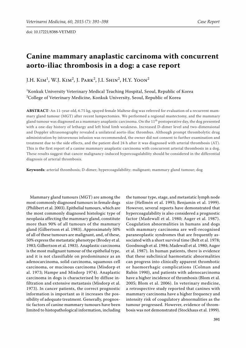
An 11-year-old spayed female Maltese dog was referred to the Veterinary Medical Teaching Hospital of Konkuk University (KU-VMTH) for an evaluation of recurrent MGT after three mammary gland lumpectomies in the previous year for several newly formed MGTs that were histopathologically diagnosed as carcinoma. The dog presented two weeks after the last lumpectomy and had a recur-rently formed MGT that was greater than 4 cm in diameter (Figure 1A). Fine needle aspiration of the MGT revealed a few clusters of epithelial cells with anisocytosis, anisokaryosis, and an increased nu-cleus-to-cytoplasm ratio that was consistent with mammary carcinoma (Figure 1B). Evidence of local metastases to regional lymph nodes and epithelial neoplastic cells were demonstrated (Figure 1C). A physical examination detected a soft systolic mur-mur (grade III/VI) that was localised at the left heart apex. Blood pressure was normal (systolic
blood pressure, 126 mmHg). Nothing remarkable was found with electrocardiography. Thoracic ra-diography showed normal cardiac size (vertebral heart score, 9.4). Two-dimensional echocardiog-raphy disclosed that the anterior leaflet of the mi-tral valve was thickened and elongated. On spectral Doppler echocardiography, mitral regurgitant jet velocity (peak velocity, 3.5 m/s) with low density was detected. Abdominal ultrasound revealed no remarkable findings. Blood work including D-dimer (0.2 μg/ml, reference range [RR]: less than 0.5 μg/ml) was unremarkable. Based on the diagnosis of myxomatous mitral valve disease with asympto-matic congestive heart failure ACVIM stage B1, the patient was managed with furosemide (Lasix; Handok Pharmaceuticals Co., Ltd., Seoul, Korea), 1 mg/kg twice daily by mouth (per os), and ramipril (Vasotop; Intervet Korea Co., Ltd, Seoul, Korea), 0.125 mg/kg once daily per os. In order to assess distance metastasis, thoracic computed tomogra-phy was performed, and several contrast-enhancing nodules within the lung lobes confirmed metas-tasis. The clinical stage based upon the modified World Health Organization classification system was stage V (T2N2M1). The owner agreed to sur-gical excision of the mammary tumour. Before the surgery, a complete blood count and routine hae-mostatic profile were determined in order to detect haemostatic abnormalities, and all of the values were within the normal reference ranges : platelet count, 497 × 109 cells/l (RR: 200–500 × 109 cells/l); prothrombin time, 11.6 s (RR: 11–17 s); activated partial thromboplastic time, 9.6 s (RR, 7.2–10.2 s).
Figure 1. (A) Gross findings during the physical examination. The patient exhibited a recurrently formed mammary gland tumour (4.6 cm × 2.3 cm in size). (B) Fine needle aspiration (FNA) of the mammary gland tumour revealed mammary carcinoma with an increased nucleus:cytoplasm ratio, anisocytosis, and anisokaryosis (Diff-Quik, × 1000, scale bar = 14 µm). (C) FNA of a prescapular lymph node revealed neoplastic epithelial cells (arrows) within the lymph node, indicating local metastasis to the regional lymph node (Diff-Quik, × 1000, scale bar = 14 µm)
393
Veterinarni Medicina, 60, 2015 (7): 391–398 Case Report
doi: 10.17221/8388-VETMED
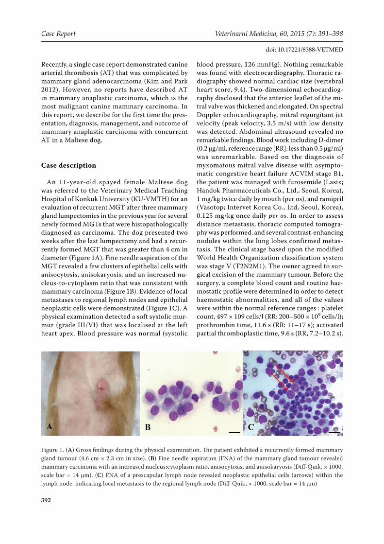
A skin incision was made with a minimum of a 2-cm lateral margin from the MGT. The incision contin-ued through subcutaneous tissue to the fascia of the external abdominal wall, and the tumour appeared to be attached to the fascia (Figure 2A). Tumour removal was performed with a deep margin of one fascial plane, including the external abdominal wall (Figure 2B). The defect was closed with 3-0 polygly-colic acid (Dexon II®; Covidien Animal Health and Dental Division, Mansfield, MA, USA) in a simple continuous pattern. The subcutaneous tissues and skin were closed routinely. Macroscopically, the tumour appeared to have large amounts of colla-gen fibres with necrotic tissues in the central re-gion (Figure 2C). The excised tumour was fixed by immersion in neutral buffered 10% formalin for microscopic evaluation. The histopathological examination revealed that the tumour tissue was comprised of exceedingly invasive and scattered
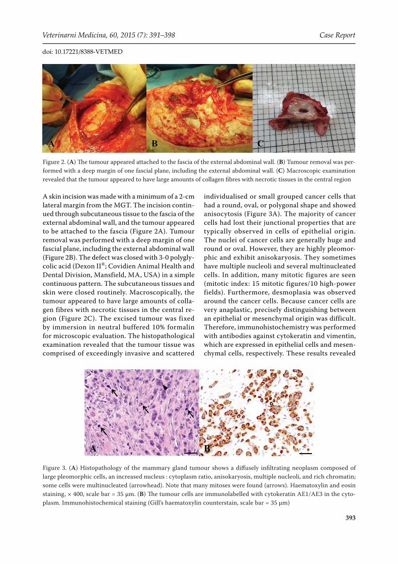
individualised or small grouped cancer cells that had a round, oval, or polygonal shape and showed anisocytosis (Figure 3A). The majority of cancer cells had lost their junctional properties that are typically observed in cells of epithelial origin. The nuclei of cancer cells are generally huge and round or oval. However, they are highly pleomor-phic and exhibit anisokaryosis. They sometimes have multiple nucleoli and several multinucleated cells. In addition, many mitotic figures are seen (mitotic index: 15 mitotic figures/10 high-power fields). Furthermore, desmoplasia was observed around the cancer cells. Because cancer cells are very anaplastic, precisely distinguishing between an epithelial or mesenchymal origin was difficult. Therefore, immunohistochemistry was performed with antibodies against cytokeratin and vimentin, which are expressed in epithelial cells and mesen-chymal cells, respectively. These results revealed
Figure 3. (A) Histopathology of the mammary gland tumour shows a diffusely infiltrating neoplasm composed of large pleomorphic cells, an increased nucleus : cytoplasm ratio, anisokaryosis, multiple nucleoli, and rich chromatin; some cells were multinucleated (arrowhead). Note that many mitoses were found (arrows). Haematoxylin and eosin staining, × 400, scale bar = 35 µm. (B) The tumour cells are immunolabelled with cytokeratin AE1/AE3 in the cyto-plasm. Immunohistochemical staining (Gill’s haematoxylin counterstain, scale bar = 35 µm)
Figure 2. (A) The tumour appeared attached to the fascia of the external abdominal wall. (B) Tumour removal was per-formed with a deep margin of one fascial plane, including the external abdominal wall. (C) Macroscopic examination revealed that the tumour appeared to have large amounts of collagen fibres with necrotic tissues in the central region
394
Case Report Veterinarni Medicina, 60, 2015 (7): 391–398
doi: 10.17221/8388-VETMED
the cytoplasmic staining of cytokeratin in cancer cells (Figure 3B). Positivity of vimentin staining was detected in the surrounding mesenchymal cells but not in the cancer cells. These results indicated that the tumour had an epithelial origin.
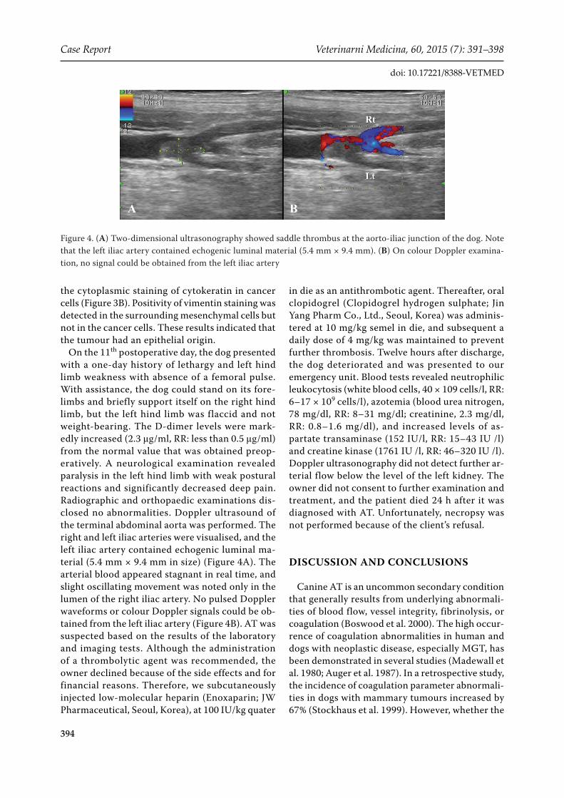
On the 11th postoperative day, the dog presented with a one-day history of lethargy and left hind limb weakness with absence of a femoral pulse. With assistance, the dog could stand on its fore-limbs and briefly support itself on the right hind limb, but the left hind limb was flaccid and not weight-bearing. The D-dimer levels were mark-edly increased (2.3 μg/ml, RR: less than 0.5 μg/ml) from the normal value that was obtained preop-eratively. A neurological examination revealed paralysis in the left hind limb with weak postural reactions and significantly decreased deep pain. Radiographic and orthopaedic examinations dis-closed no abnormalities. Doppler ultrasound of the terminal abdominal aorta was performed. The right and left iliac arteries were visualised, and the left iliac artery contained echogenic luminal ma-terial (5.4 mm × 9.4 mm in size) (Figure 4A). The arterial blood appeared stagnant in real time, and slight oscillating movement was noted only in the lumen of the right iliac artery. No pulsed Doppler waveforms or colour Doppler signals could be ob-tained from the left iliac artery (Figure 4B). AT was suspected based on the results of the laboratory and imaging tests. Although the administration of a thrombolytic agent was recommended, the owner declined because of the side effects and for financial reasons. Therefore, we subcutaneously injected low-molecular heparin (Enoxaparin; JW Pharmaceutical, Seoul, Korea), at 100 IU/kg quater
in die as an antithrombotic agent. Thereafter, oral clopidogrel (Clopidogrel hydrogen sulphate; Jin Yang Pharm Co., Ltd., Seoul, Korea) was adminis-tered at 10 mg/kg semel in die, and subsequent a daily dose of 4 mg/kg was maintained to prevent further thrombosis. Twelve hours after discharge, the dog deteriorated and was presented to our emergency unit. Blood tests revealed neutrophilic leukocytosis (white blood cells, 40 × 109 cells/l, RR: 6–17 × 109 cells/l), azotemia (blood urea nitrogen, 78 mg/dl, RR: 8–31 mg/dl; creatinine, 2.3 mg/dl, RR: 0.8–1.6 mg/dl), and increased levels of as-partate transaminase (152 IU/l, RR: 15–43 IU /l) and creatine kinase (1761 IU /l, RR: 46–320 IU /l). Doppler ultrasonography did not detect further ar-terial flow below the level of the left kidney. The owner did not consent to further examination and treatment, and the patient died 24 h after it was diagnosed with AT. Unfortunately, necropsy was not performed because of the client’s refusal.
DISCUSSION AND CONCLUSIONS
Canine AT is an uncommon secondary condition that generally results from underlying abnormali-ties of blood flow, vessel integrity, fibrinolysis, or coagulation (Boswood et al. 2000). The high occur-rence of coagulation abnormalities in human and dogs with neoplastic disease, especially MGT, has been demonstrated in several studies (Madewall et al. 1980; Auger et al. 1987). In a retrospective study, the incidence of coagulation parameter abnormali-ties in dogs with mammary tumours increased by 67% (Stockhaus et al. 1999). However, whether the
Figure 4. (A) Two-dimensional ultrasonography showed saddle thrombus at the aorto-iliac junction of the dog. Note that the left iliac artery contained echogenic luminal material (5.4 mm × 9.4 mm). (B) On colour Doppler examina-tion, no signal could be obtained from the left iliac artery
395
Veterinarni Medicina, 60, 2015 (7): 391–398 Case Report
doi: 10.17221/8388-VETMED
subclinical haemostatic abnormalities in dogs with tumours could progress into clinically apparent thrombotic or haemorrhagic complications was not demonstrated.
Mammary anaplastic carcinoma is the most ma-lignant canine mammary carcinoma. It is generally firm and ill-defined, and it invades the skin and underlying tissues, which are often oedematous. In many dogs, several adjacent glands of one or both mammary chains are affected (Misdorp et al. 1973). The diagnostic microscopic features of malignant anaplastic carcinoma consist of a diffusely infiltrat-ing neoplasm that is composed of large pleomorphic cells, which often have bizarre nuclei that are rich in chromatin; some cells are multinucleated and exhibit an abundant fibrous reaction around the neoplastic cells (Hampe and Misdorp 1974). Many mitoses are found, which was also evident in this case. The cytoplasm is eosinophilic, and vacuoles are not uncommon. In contrast to most other types of canine mammary carcinomas, the anaplastic type is highly invasive, and it exhibits extensive amounts of collagen fibres (scirrhous carcinoma), which is not typical of human anaplastic carcinomas (Misdorp et al. 1973). Some extreme anaplastic carcinomas that are composed of large pleomorphic cells that are embedded in fibrous stroma are similar to ana-plastic sarcomas and are very hard to recognise as epithelial tumours, as was shown in this case. In the present case, we also performed immunohisto-chemistry with vimentin and cytokeratin in order to confirm the origin. We confirmed that the origin was epithelial based on the immunohistochemical positivity for cytokeratin and negativity for vimen-tin. Carcinomas of this type are difficult to treat with surgery alone because of their early and extensive infiltration into the surrounding tissues and lym-phatics. The regional lymph nodes and lungs are the organs that are most frequently affected by me-tastases (Misdorp et al. 1973). The lungs, when af-fected, are usually extensively involved and appear firm and compact with multiple small nodules, as shown in this case. Although canine mammary ana-plastic carcinomas establish their malignant nature by metastasising, their relationship with hyperco-agulability is unknown (Misdorp et al. 1973). In one report of 64 metastasised canine malignant mam-mary tumours, the anaplastic carcinomas were very malignant and caused death in a short time. The postsurgical survival times in 31 dogs with anaplas-tic carcinoma ranged from one week to 18 months
(average, seven months); in that study of 31 dogs, four had dyspnoea, and four had walking difficul-ties (total, 8; 26%). However, the relationship with coagulability was not defined. Therefore, this case report provides the first evidence that supports a relationship between hypercoagulability and ana-plastic adenocarcinoma in dogs.
In this case, the patient exhibited acute paralytic, pulseless, and cold extremities, which were consist-ent with AT (Schoeman 1999). Ultrasonography is a simple and non-invasive method that can be used to diagnose a thromboembolism by detect-ing areas of decreased blood flow. In the present dog, aorto-iliac thrombosis was identified using ultrasonography. Other potential diagnostic tests for aortic thrombosis include selective angiography (Carter and Van Heerden 1994) and radionuclide angiography (Ramsey et al. 1996), and thermog-raphy (Kim and Park 2012) has also been advo-cated when thromboembolic disease is suspected. However, these tests are very invasive or expensive.
In human and veterinary medicine, D-dimers are laboratory markers that are used clinically to detect early thromboembolism (TE) (Nelson and Andreasen 2003; Owaidah et al. 2014). Other labo-ratory markers of coagulation activation, such as fibrinopeptides A and B, fibrinogen degradation products, prothrombin fragments 1 and 2, and thrombin-antithrombin complexes, have been pro-posed and used for TE diagnosis, however, only the D-dimer assay has been shown to have clinical util-ity in people (Bounameaux 1996). In a recent pro-spective study of dogs with thromboembolic disease, no difference was found between the frequencies of abnormalities of prothrombin time or activated par-tial thromboplastic time in the TE group versus the control group, and fibrinogen degradation products were not abnormal in any TE patient (Nelson and Andreasen 2003). The human literature focuses on imaging modalities and D-dimers as a laboratory marker. In this case, Doppler ultrasonography re-vealed no blood flow below the suggested AT site, and the D-dimer levels were markedly increased af-ter the hind limb paralysis. Additionally, the blood abnormalities in this case, including azotemia, leukocytosis, elevated aspartate transaminase and creatine kinase, could be considered a result of lo-cal ischaemia and skeletal muscle inflammation from the AT because the results of the preopera-tive routine blood tests were within normal limits and no remarkable abnormalities were detected in
396
Case Report Veterinarni Medicina, 60, 2015 (7): 391–398
doi: 10.17221/8388-VETMED
the kidneys with the urinalysis and ultrasonographic assessments. After the hind limb paralysis, arterial flow was not detected, especially below the level of the left kidney on Doppler ultrasonography, and this suggested renal ischaemic damage.
In this case, occlusion of the distal aorta and left iliac artery was found, which suggested complex aetiology. Neoplasia can predispose to thrombosis in numerous ways, including increased coagulation due to platelet activation, thromboplastin release from tumour cells, and the production of factor X activator, as well as reduced clotting factor clear-ance, reduced clotting factor neutralisation, and decreased fibrinolysis, as previously described (O’Keefe and Couto 1988). In addition, in this case, the metastatic neoplastic cells might have eroded into or arisen from the blood vessels, which would directly result in a disruption of the endothelial in-tegrity and disturbed flow. Second, cardiac disease can predispose to thrombosis through the stasis of blood in congested veins and atria and interfere with normal endothelial integrity (Boswood et al. 2000). The present dog had mitral insufficiency, but there was no evidence of left ventricular or atrial dilatation. Mitral insufficiency is a common incidental finding in aging dogs, and it therefore might not have been related to the development of the mural thrombus. In human patients, severe mitral insufficiency is associated with a moderately high incidence of thrombosis (Fuster and Verstraete 1997). The incidence is lower with mild insufficien-cy and when atrial fibrillation is not present (Fuster and Verstraete 1997). The present case presented with both a neoplasia and cardiac disease, and, thus, it is uncertain which disease was the principal cause of the thrombosis. Each condition may have predisposed to thrombosis, and the combination of the factors might then have been sufficient for this to occur. However, this patient did not show cardiomegaly, hypertension, or non-tumour related abnormalities, and the D-dimer levels were normal before the acute signs of AT. Therefore, the malig-nant mammary tumour was the most likely cause of the hypercoagulability and AT in the present case.
Although aorto-iliac thromboses in dogs com-monly cause marked morbidity and mortality, several studies have reported that systemic throm-bolytic therapy can manage this syndrome in hu-mans and other veterinary species (Clare and Kraje 1998; Kim and Park 2012). Early recognition of this clinical condition and the administration of throm-
bolytic therapy may be beneficial. Successful throm-bolytic therapy with streptokinase (Ramsey et al. 1996) and recombinant tissue plasminogen activator (Clare and Kraje 1998) has been reported in cases of canine aortic thromboembolism. Nonetheless, se-vere hyperkalaemia can develop as a reperfusion in-jury after thrombolytic therapy in dogs and cats with AT (Schoeman 1999; Lunsford and Mackin 2007). In the present case, initial thrombotic management was recommended, but the owner declined because of side effects and for financial reasons. Therefore, in order to minimise the risk of any potential fu-ture thrombosis, we administered clopidogrel in combination with low-molecular weight heparin as antithrombotic agents. Recent studies in dogs and cats have reported that clopidogrel therapy has antiplatelet effects and no adverse effects in these species that are unable to tolerate aspirin therapy (Hogan 2004; Mellett et al. 2011). Patients suffering from AT often recover with supportive therapy, but severely affected patients may experience ischaemic acidosis and muscle inflammation, which can be life threatening. In this case, the patient deteriorated and died 24 h after the diagnosis of AT. This is the first known description of a fatal AT that was in-duced by a mammary anaplastic carcinoma in a dog.
In conclusion, canine patients with malignancy could have a higher risk of developing postopera-tive thromboembolic complications, as is seen in human medicine (Blom et al. 2005; Prandoni et al. 2005). We suggest that the prophylactic manage-ment of thromboembolism in canine patients with malignant mammary gland tumours is necessary, and cancer malignancy-induced hypercoagulability should be considered in the differential diagnosis of AT. Additionally, in veterinary medicine, when a malignant mammary tumour is suspected, it is im-portant that veterinarians assess the serial D-dimer levels and perform abdominal ultrasonography in order to improve the therapeutic prognosis.
REFERENCES
Auger MJ, Galloway MJ, Leinster SJ, McVerry BA, Mackie MJ (1987): Elevated fibrinopeptide A levels in patients with clinically localised breast carcinoma. Haemostasis 17, 336–339.
Belt RJ, Leite C, Haas CD, Stephens RL (1978): Incidence of hemorrhagic complications in patients with cancer. JAMA 239, 2571–2574.
397
Veterinarni Medicina, 60, 2015 (7): 391–398 Case Report
doi: 10.17221/8388-VETMED
Benjamin SA, Lee AC, Saunders WJ (1999): Classification and behavior of canine mammary epithelial neoplasms based on life-span observations in beagles. Veterinary Pathology 36, 423–436.
Blom JW, Doggen CJ, Osanto S, Rosendaal FR (2005): Ma-lignancies, prothrombotic mutations, and the risk of ve-nous thrombosis. JAMA 293, 715–722.
Blom JW, Vanderschoot JP, Oostindiër MJ, Osanto S, van der Meer FJ, Rosendaal FR (2006): Incidence of venous thrombosis in a large cohort of 66,329 cancer patients: results of a record linkage study. Journal of Thrombsis and Haemostasis 4, 529–535.
Boswood A, Lamb CR, White RN (2000): Aortic and iliac thrombosis in six dogs. The Journal of Small Animal Prac-tice 41, 109–114.
Bounameaux H (1996): Biological markers of acute venous thrombosis and pulmonary embolism. In: Seghtchian MJ, Samana MN, Hecker SP (eds.): Hypercoagulable States: Fundamental Aspects, Acquired Disorders, and Con-genital Thrombophilia. 1st ed. CRC Press, Boca Raton. 129–137.
Brodey RS, Goldschmidt MA, Roszel JR (1983): Canine mammary gland neoplasms. Journal of the American Animal Hospital Association 19, 61–90.
Carter AJ, Van Heerden J (1994): Aortic thrombosis in a dog with glomerulonephritis. Journal of the South African Veterinary Association 65, 189–192.
Clare AC, Kraje BJ (1998): Use of recombinant tissue-plas-minogen activator for aortic thrombolysis in a hypopro-teinemic dog. Journal of the American Veterinary Medical Association 212, 539–543.
Colman RW, Rubin RN (1990): Disseminated intravascular coagulation due to malignancy. Seminars in Oncology 17, 172–186.
Fuster V, Verstraete M (1997): Hemostasis, thrombosis, fibrinolysis and cardiovascular disease. In: Braunwald E (ed.): Heart Disease. 5th ed. WB Saunders, Philadelphia. 1809–1842.
Gilbertson SR, Kurzman ID, Zachrau RE, Hurvitz AI, Black MM (1983): Canine mammary epithelial neo-plasms: Biologic implications of morphologic charac-teristics assessed in 232 dogs. Veterinary Pathology 20, 127–142.
Goodnough LT, Saito H, Manni A, Jones PK, Pearson OH (1984): Increased incidence of thromboembolism in stage IV breast cancer patients treated with a five-drug chem-otherapy regimen. A study of 159 patients. Cancer 54, 1264–1268.
Hampe JF, Misdorp W. Tumours and dysplasias of the mam-mary gland (1974): Bulletin of the World Health Organ-ization 50, 111–133.
Hellmen E, Bergström R, Holmberg L, Spangberg IB, Hans-son K, Lindgren A (1993): Prognostic factors in canine mammary tumors: a multivariate study of 202 consecutive cases. Veterinary Pathology 30, 20–27.
Hogan DF, Andrews DA, Green HW, Talbott KK, Ward MP, Calloway BM (2004): Antiplatelet effects and pharmaco-dynamics of clopidogrel in cats. Journal of the American Veterinary Medical Association 225, 1406–1411.
Kim JH, Park HM (2012). Unilateral femoral arterial throm-bosis in a dog with malignant mammary gland tumor: clinical and thermographic findings, and successful treat-ment with local intra-arterial administration of streptoki-nase. Journal of Veterinary Medical Science 74, 657–661.
Lunsford KV, Mackin AJ (2007): Thromboembolic therapies in dogs and cats: an evidence-based approach. Veterinary Clinics of North America. Small Animal Practice 37, 579–609.
Madewall BR, Feldman BF, O’Neill S (1980): Coagulation abnormalities in dogs with neoplastic disease. Thrombo-sis and Haemostasis 44, 35–38.
Mellett AM, Nakamura RK, Bianco D (2011): A prospective study of clopidogrel therapy in dogs with primary im-mune-mediated hemolytic anemia. Journal of Veterinary Internal Medicine 25, 71–75.
Misdorp W, Cotchin E, Hampe JF, Jabara AG, Von Sander-sleben J (1973): Canine malignant mammary tumors. 3. Special types of carcinomas, malignant mixed tumors. Veterinary Pathology 10, 241–256.
Nelson OL, Andreasen C (2003): The utility of plasma D-dimer to identify thromboembolic disease in dogs. Jour-nal of Veterinary Internal Medicine 17, 830–834.
O’Keefe DA, Couto CG (1988): Coagulation abnormalities associated with neoplasia. Veterinary Clinics of North America. Small Animal Practice 18, 157–168.
Owaidah T, AlGhasham N, AlGhamdi S, AlKhafaji D, ALAmro B, Zeitouni M, Skaff F, Alzahrani H, AlSayed A, ElKum N, Moawad M, Nasmi A, Hawari M, Maghrabi K (2014): Evaluation of the usefulness of a D dimer test in combination with clinical pretest probability score in the prediction and exclusion of Venous Thromboembolism by medical residents. Thrombosis Journal 12, 28.
Philibert JC, Snyder PW, Glickman N, Glickman LT, Knapp DW, Waters DJ (2003): Influence of host factors on sur-vival in dogs with malignant mammary gland tumors. Journal of Veterinary Internal Medicine 17, 102–106.
Prandoni P, Falanga A, Piccioli A (2005): Cancer and venous thromboembolism. Lancet Oncology 6, 401–410.
Ramsey CC, Burney DP, Macintire DK, Finn-Bodner S (1996): Use of streptokinase in four dogs with thrombo-sis. Journal of the American Veterinary Medical Asso-ciation 209, 780–785.
398
Case Report Veterinarni Medicina, 60, 2015 (7): 391–398
doi: 10.17221/8388-VETMED
Schoeman JP (1999): Feline distal aortic thromboembolism: a review of 44 cases (1990–1998). Journal of Feline Med-icine and Surgery 1, 221–231.
Stockhaus C, Kohn B, Rudolph R, Brunnberg L, Giger U (1999): Correlation of haemostatic abnormalities with
tumour stage and characteristics in dogs with mammary carcinoma. Journal of Small Animal Practice 40, 326–331.
Received: 2015–01–15Accepted after corrections: 2015–07–02
Corresponding Author:
Hun-Young Yoon, Konkuk University, College of Veterinary Medicine, Department of Veterinary Surgery, 120 Neungdong-ro, Gwangjin-gu, Seoul, 143-701, Republic of KoreaE-mail: [email protected]