Embed Size (px)
Citation preview
March 2015 Veterinary Team Brief 27
Most juvenile orthopedic disease is developmental in nature, and early
treatment is key. In addition to a thorough patient history, the orthopedic
examination is a critical first step. Once the abnormal joint(s) is identified,
radiographs should be obtained.
STEP 1:Comprehensive Overview
Canine Juvenile Orthopedic Disease Jonathan Miller, DVM, MS, DACVS Oradell Animal Hospital Paramus, New Jersey
ment is key to reduce the typical life-long progressive arthritis associated with this disease.2
Hip dysplasiaHip dysplasia can be diagnosed as early as 4 months of age3; ventrodor-sal and distracted hip radiographs are useful for diagnosis. Early diagnosis allows early medical treatment and, in some cases, certain therapeutic surgi-cal procedures. A bunny-hopping gait when running, lower energy level, and difficulty climbing stairs or jumping are often seen in hip dysplasia patients.
OTHER DISEASESLess common diseases include con-genital joint luxations, panosteitis, hypertrophic osteodystrophy, cranio-mandibular osteopathy, and angu-lar limb deformity. Dogs of any age may present with a fracture that may manifest as severe pain with non-weight-bearing lameness.
PROGNOSIS & TREATMENTPrognosis depends on disease type and severity, likelihood of arthritis development, and patient response to treatment. Some form of treatment is available for most cases.
CLINICAL SUITE | JUVENILE ORTHOPEDIC DISEASE
STEP 2 Treatment Plan h
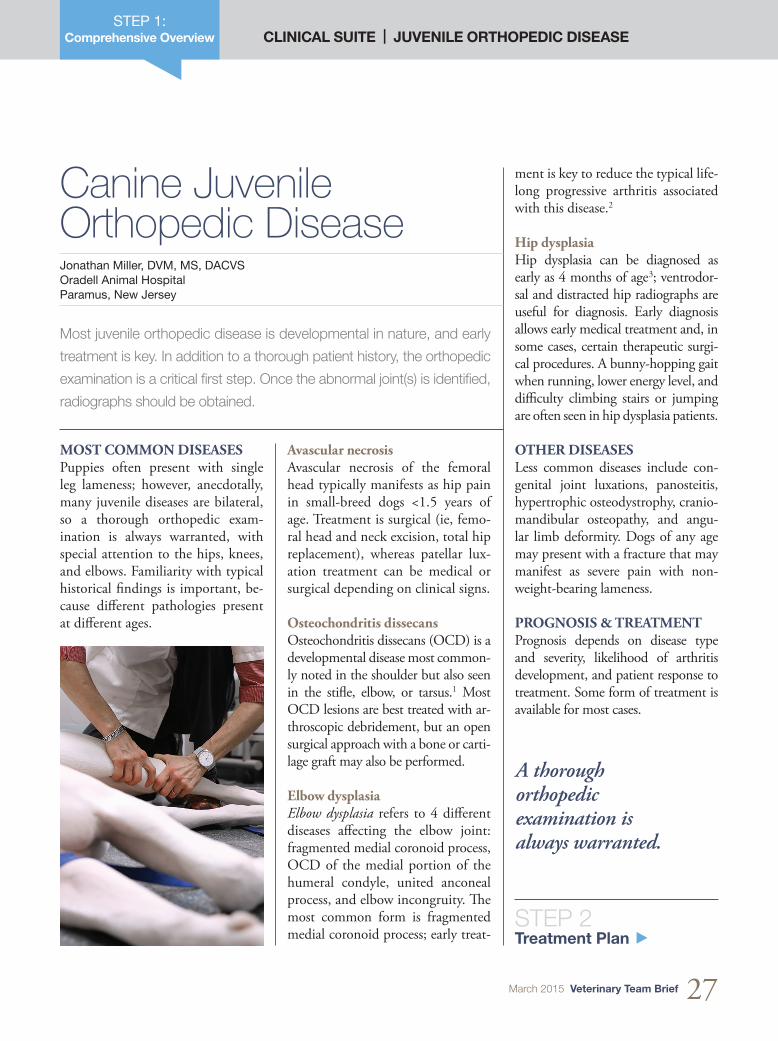
MOST COMMON DISEASESPuppies often present with single leg lameness; however, anecdotally, many juvenile diseases are bilateral, so a thorough orthopedic exam-ination is always warranted, with special attention to the hips, knees, and elbows. Familiarity with typical historical findings is important, be-cause different pathologies present at different ages.
Avascular necrosisAvascular necrosis of the femoral head typically manifests as hip pain in small-breed dogs <1.5 years of age. Treatment is surgical (ie, femo-ral head and neck excision, total hip replacement), whereas patellar lux-ation treatment can be medical or surgical depending on clinical signs.
Osteochondritis dissecansOsteochondritis dissecans (OCD) is a developmental disease most common-ly noted in the shoulder but also seen in the stifle, elbow, or tarsus.1 Most OCD lesions are best treated with ar-throscopic debridement, but an open surgical approach with a bone or carti-lage graft may also be performed.
Elbow dysplasiaElbow dysplasia refers to 4 different diseases affecting the elbow joint: fragmented medial coronoid process, OCD of the medial portion of the humeral condyle, united anconeal process, and elbow incongruity. The most common form is fragmented medial coronoid process; early treat-
A thorough orthopedic examination is always warranted.
CLINICAL SUITE | JUVENILE ORTHOPEDIC DISEASESTEP 2:
Treatment Plan
Examination, Radiographs, & Treatment OptionsJonathan Miller, DVM, MS, DACVS Oradell Animal Hospital Paramus, New Jersey
Early diagnosis and treatment before osteoarthritis formation are often
key to successful management. Congenital problems frequently man-
ifest before 1 year of age, so a limp lasting longer than a day should
be examined by a veterinarian. Thorough orthopedic examination and
radiographs are the foundation of diagnosis and allow formulation of
an appropriate treatment plan.
Physical examination findings help determine if limping (ie, lameness) will resolve independently. Pain med-ications often help, but a definitive diagnosis and radiographs are pru-dent to avoid long-term problems; however, although radiographs show bone and joint spaces clearly, ten-dons, ligaments, and cartilage are not represented.
THE ROLE OF ANESTHETICSThe difference in radiographs of a se-dated dog compared with those of an alert dog is astounding, because small changes in rotation or limb extension can be crucial to identify the problem. For example, femur rotation can hide subtle hip dysplasia, or an OCD lesion could be missed with only a rotated or cranial-caudal view of a shoulder. Se-dation also allows for Ortolani testing to indicate excessive hip laxity second-ary to hip dysplasia; with the dog in lateral recumbency, the pelvic limb is pushed dorsal as the limb is abducted. A distinct clunk can be palpated when
STEP 3 Team Roles h
the femoral head pops back into the acetabulum following subluxation.
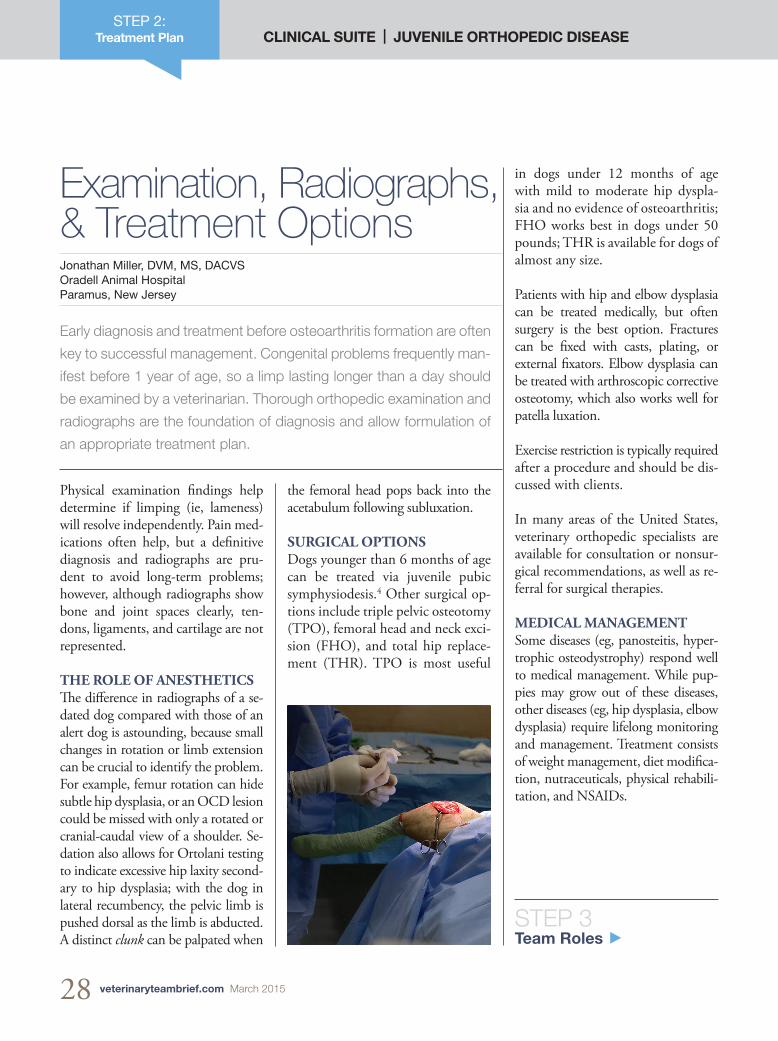
SURGICAL OPTIONSDogs younger than 6 months of age can be treated via juvenile pubic symphysiodesis.4 Other surgical op-tions include triple pelvic osteotomy (TPO), femoral head and neck exci-sion (FHO), and total hip replace-ment (THR). TPO is most useful
in dogs under 12 months of age with mild to moderate hip dyspla-sia and no evidence of osteoarthritis; FHO works best in dogs under 50 pounds; THR is available for dogs of almost any size.
Patients with hip and elbow dysplasia can be treated medically, but often surgery is the best option. Fractures can be fixed with casts, plating, or external fixators. Elbow dysplasia can be treated with arthroscopic corrective osteotomy, which also works well for patella luxation.
Exercise restriction is typically required after a procedure and should be dis-cussed with clients.
In many areas of the United States, veterinary orthopedic specialists are available for consultation or nonsur-gical recommendations, as well as re-ferral for surgical therapies.
MEDICAL MANAGEMENTSome diseases (eg, panosteitis, hyper-trophic osteodystrophy) respond well to medical management. While pup-pies may grow out of these diseases, other diseases (eg, hip dysplasia, elbow dysplasia) require lifelong monitoring and management. Treatment consists of weight management, diet modifica-tion, nutraceuticals, physical rehabili-tation, and NSAIDs.
28 veterinaryteambrief.com March 2015
March 2015 Veterinary Team Brief 29
STEP 4 Team Training Plan h
STEP 3:Team Roles CLINICAL SUITE | JUVENILE ORTHOPEDIC DISEASE
Patient and client bonding expert, client educator
• Encourage clients calling about a limping puppy to come to the practice for an orthopedic examination
• Mention that pain control is very important
• Offer a gurney if multiple limbs are affected
• Reinforce information about adverse effects of medication
• Be familiar with common surgical treatments
Patient caregiver, client educator
• Triage the patient
• Take a thorough history, including when the lameness started, its progression, and any medications
• Evaluate for pain
• Walk the patient down a hallway during the examination
• Know how to obtain high-quality radiographs
• Be familiar with physical therapy modalities
• Educate clients about commonly used medications
Medical expert, client and team educator
• Conduct an orthopedic examination
• Recommend a radiographic examination with the patient sedated or anesthetized
• Confirm a diagnosis
• Devise a medical plan consisting of pain medication, diet, weight management or loss, and nutraceuticals
• Consider referral to an orthopedic specialist
Workflow facilitator, team and education coordinator
• Develop a brochure of common juvenile orthopedic diseases
• Encourage the team to support early aggressive treatment
• Encourage the veterinarian to provide an overview presentation
RE
CE
PT
ION
IST
TE
CH
NIC
IAN
PR
AC
TIC
E M
AN
AG
ER
VE
TE
RIN
AR
IAN
Team Roles & ResponsibilitiesJonathan Miller, DVM, MS, DACVS, Oradell Animal Hospital, Paramus, New Jersey
30 veterinaryteambrief.com March 2015
CLINICAL SUITE | JUVENILE ORTHOPEDIC DISEASESTEP 4:
Team Training Plan
Training a Knowledgeable TeamJennifer Potts, RVT Chimney Hills Animal Hospital Tulsa, Oklahoma
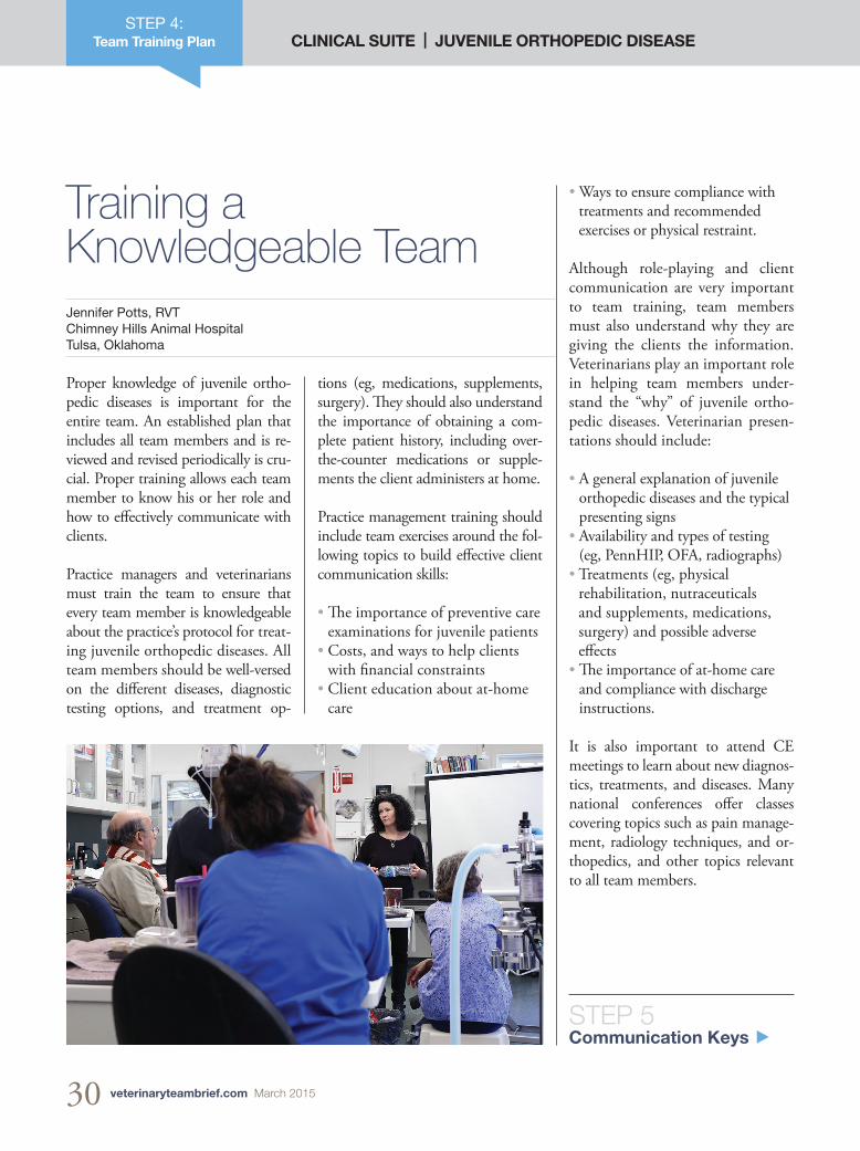
Proper knowledge of juvenile ortho-pedic diseases is important for the entire team. An established plan that includes all team members and is re-viewed and revised periodically is cru-cial. Proper training allows each team member to know his or her role and how to effectively communicate with clients.
Practice managers and veterinarians must train the team to ensure that every team member is knowledgeable about the practice’s protocol for treat-ing juvenile orthopedic diseases. All team members should be well-versed on the different diseases, diagnostic testing options, and treatment op-
STEP 5 Communication Keys h
tions (eg, medications, supplements, surgery). They should also understand the importance of obtaining a com-plete patient history, including over-the-counter medications or supple-ments the client administers at home.
Practice management training should include team exercises around the fol-lowing topics to build effective client communication skills:
• The importance of preventive care examinations for juvenile patients
• Costs, and ways to help clients with financial constraints
• Client education about at-home care
• Ways to ensure compliance with treatments and recommended exercises or physical restraint.
Although role-playing and client communication are very important to team training, team members must also understand why they are giving the clients the information. Veterinarians play an important role in helping team members under-stand the “why” of juvenile ortho-pedic diseases. Veterinarian presen-tations should include:
• A general explanation of juvenile orthopedic diseases and the typical presenting signs
• Availability and types of testing (eg, PennHIP, OFA, radiographs)
• Treatments (eg, physical rehabilitation, nutraceuticals and supplements, medications, surgery) and possible adverse effects
• The importance of at-home care and compliance with discharge instructions.
It is also important to attend CE meetings to learn about new diagnos-tics, treatments, and diseases. Many national conferences offer classes covering topics such as pain manage-ment, radiology techniques, and or-thopedics, and other topics relevant to all team members.
March 2015 Veterinary Team Brief 31
Effective communication skills are important for helping clients under-
stand juvenile orthopedic disease and what to expect after a pet’s
diagnosis. Every team member plays an important role in communi-
cating with clients to help them better understand the condition.
STEP 5:Communication Keys
Client Communication Strategy Jennifer Potts, RVT Chimney Hills Animal Hospital Tulsa, Oklahoma
As with any disease process, finan-cial constraints can affect treatment decision-making. All team members should be able to effectively com-municate every available option.
The most important part of client communication is offering support; all team members should be avail-able to answer questions and listen to client concerns.
References1. Brinker, Piermattei, and Flo’s Handbook of
Small Animal Orthopedics and Fracture Re-pair, 4th ed. Piermattei DL, Flo GL, DeCamp CE–St. Louis: Saunders Elsevier, 2006, pp 208-213.
2. Surgical Treatment of Fragmented Coronoid Process. Krotscheck U. In Bojrab MJ, Waldron DR, Toombs JP (eds): Current Techniques in Small 1. Animal Surger, 5th ed—Jackson, WY: Teton NewMedia, 2014, pp 917-924.
3. Brinker, Piermattei, and Flo’s Handbook of Small Animal Orthopedics and Fracture Re-pair, 4th ed. Piermattei DL, Flo GL, DeCamp CE–St. Louis: Saunders Elsevier, 2006, pp 475-507.
4. Effects of pubic symphysiodesis in dysplas-tic puppies. Dueland RT, Adams WM, Fialkowski JP, et al. Vet Surg 201-217, 2001.
CLINICAL SUITE | JUVENILE ORTHOPEDIC DISEASE
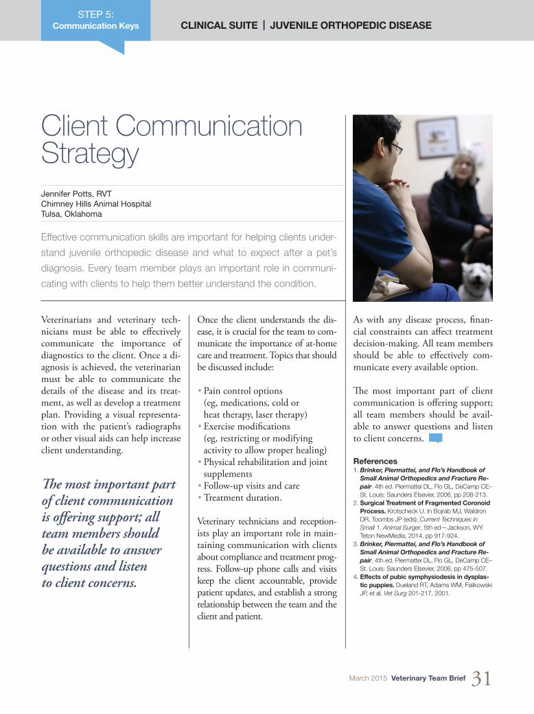
Veterinarians and veterinary tech-nicians must be able to effectively communicate the importance of diagnostics to the client. Once a di-agnosis is achieved, the veterinarian must be able to communicate the details of the disease and its treat-ment, as well as develop a treatment plan. Providing a visual representa-tion with the patient’s radiographs or other visual aids can help increase client understanding.
Once the client understands the dis-ease, it is crucial for the team to com-municate the importance of at-home care and treatment. Topics that should be discussed include:
• Pain control options (eg, medications, cold or heat therapy, laser therapy)
• Exercise modifications (eg, restricting or modifying activity to allow proper healing)
• Physical rehabilitation and joint supplements
• Follow-up visits and care• Treatment duration.
Veterinary technicians and reception-ists play an important role in main-taining communication with clients about compliance and treatment prog-ress. Follow-up phone calls and visits keep the client accountable, provide patient updates, and establish a strong relationship between the team and the client and patient.
The most important part of client communication is offering support; all team members should be available to answer questions and listen to client concerns.