Embed Size (px)
Citation preview

RESEARCH ARTICLE
Candidate epitopes for measurement of hCG and relatedmolecules: the second ISOBM TD-7 workshop
P. Berger & E. Paus & P. M. Hemken & C. Sturgeon & W. W. Stewart &J. P. Skinner & L. C. Harwick & S. C. Saldana & C. S. Ramsay &
K. R. Rupprecht & K. H. Olsen & J.-M. Bidart & U.-H. Stenman &
on behalf of the members of the ISOBM TD-7 Workshop on hCG and Related Molecules
Received: 17 May 2013 /Accepted: 1 July 2013 /Published online: 26 September 2013# The Author(s) 2013. This article is published with open access at Springerlink.com
Abstract Participants of the Second International Workshop(WS) on human chorionic gonadotropin (hCG) of the Inter-national Society of Oncology and Biomarkers Tissue Differ-entiation 7 (ISOBMTD-7) have characterized in detail a panelof 69 antibodies (Abs) directed against hCG and hCG-relatedvariants that were submitted by eight companies and researchgroups. Specificities of the Abs were determined using theFirst WHO International Reference Reagents for six hCGvariants, i.e., hCG, hCGn, hCGβ, hCGβn, hCGβcf, andhCGα, which are calibrated in SI units, and hLH. Molecularepitope localizations were assigned to the ISOBM-mAbs bycomparing ISOBM-Ab specificity, sandwich compatibility,and mutual inhibition profiles, to those of 17 reference mono-clonal (m)Abs of known molecular epitope specificities. It
appeared that 48 Abs recognized hCGβ-, 8 hCGα-, and 13αβ-heterodimer-specific epitopes. Twenty-seven mAbs wereof pan hCG specificity, two thereof with no (<0.1 %; epitopeβ1), 12 with low (<1.0 %; epitopes β2/4), and 13 with high(>>1 %; epitopes β3/5) hLH cross-reactivity. The majority ofhCGβ epitopes recognized were located in two major anti-genic domains, one on the peptide chain of the tips of β-sheetloops 1 and 3 (epitopesβ2–6; 27mAbs) and the second aroundthe cystine knot (e.g., epitopesβ1,β7, andβ10; 9 mAbs). FourmAbs recognized epitopes on hCGβcf-only (e.g., epitopesβ11 and β13) and six mAbs epitopes on the remote hCGβ-carboxyl-terminal peptide (epitopes β8 and β9 correspondingto amino acids 135–144 and 111–116, respectively). For rou-tine diagnostic measurements, methods are used that either
The International Society of Oncology and Biomarkers TissueDifferentiation (TD)-7 Workshop on hCG and Related Molecules.
Electronic supplementary material The online version of this article(doi:10.1007/s13277-013-0994-6) contains supplementary material,which is available to authorized users.
P. Berger (*)Institute for Biomedical Aging Research, University of Innsbruck,Rennweg 10, A6020 Innsbruck, Austriae-mail: [email protected]
E. Paus :K. H. OlsenDepartment of Medical Biochemistry at Radiumhospitalet, OsloUniversity Hospital, Oslo, Norway
P. M. Hemken : J. P. Skinner : L. C. Harwick : S. C. Saldana :C. S. Ramsay :K. R. RupprechtDiagnostic Research and Development, Abbott Diagnostics,Abbott Park, IL, USA
C. SturgeonDepartment of Clinical Biochemistry, Royal Infirmary, UKNEQAS for Peptide Hormones, Edinburgh, UK
W. W. StewartDepartment of Immunology, Ninewells Hospital and MedicalSchool, Dundee, UK
J.<M. BidartDepartment of Clinical Biology, Institut Gustave-Roussy,Villejuif, France
U.<H. StenmanDepartment of Clinical Chemistry, Helsinki University CentralHospital, Helsinki, Finland
Tumor Biol. (2013) 34:4033–4057DOI 10.1007/s13277-013-0994-6

detect hCG-only, hCGβ-only, or hCG together with hCGβ orhCG together with hCGβ and hCGβcf. Sandwich assays thatmeasure hCG plus hCGβ and eventually hCGβcf shouldrecognize the protein backbone of the analytes preferably onan equimolar basis, should not cross-react with hLH and notbe susceptible to blunting of signal by nonmeasured variantslike hCGβcf. Such assays can be constructed using pairs ofmAbs directed against the cystine knot-associated epitope β1
(Asp10, Asp60, and Gln89) in combination with epitopes β2
or β4 located at the top of β-sheet loops 1+3 of hCGβinvolving aa hCGβ20-25+68-77. In summary, the results ofthe First and Second ISOBM TD-7 WSs on hCG provide thebasis for harmonization of specificities and epitopes of mAbsto be used in multifunctional and selective diagnostic hCGmethods for different clinical purposes.
Keywords hCG variants measurement . Antibodystandardization . Epitope standardization . Internationalstandards for hCG . hCG IRR
Objective
Improving between method comparability for measurementof the heterogeneous glycoprotein hCG requires harmoniza-tion of epitopes of the Abs used and broad consensus aboutassay specificity. To address this, the Second InternationalSociety of Oncology and Biomarkers Tissue Differentiation7 (ISOBM TD-7) Workshop (WS) by a three-step algorithmcharacterized and epitope typed 69 Abs directed against hCGand variants submitted by diagnostic companies and researchgroups. The results of this WS in combination with those ofthe First WS enable recommendations to be made regardingepitope combinations to be used for the design of immuno-assays for hCG and its variants [1].
Introduction
Physiology, protein structure, and posttranslational proteinbackbone variants of hCG
The glycoprotein hormone hCG is essential for maintainingpregnancy. Physiologically, it is produced and secreted bythe placental trophoblast and pathophysiologically by tro-phoblastic cancers and by germ cell tumors of the testis andovary [2].
hCG is a protein heterodimer consisting of hCGαnoncovalently linked to the hCGβ subunit. As all glycopro-tein hormone (GPH) subunits, hCGα and hCGβ share struc-tural homology with members of the cystine knot growthfactor superfamily that includes nerve growth factor,platelet-derived growth factor and transforming growth factor
beta [3]. The common structural cystine knot motif consists oftwo disulfide bridges that link adjacent antiparallel strands ofthe single peptide chain to form a ring that is axially permeatedby a third disulfide bond. This central cystine knot determinesthe three-dimensional structure of hCGα and hCGβ. On oneside of the knot, there are two neighboring hairpin-like peptideloops 1 and 3, which, in hCGβ, are stabilized by a disulfidebond between Cys 23 and Cys72. The single larger loop 2 islocated on the opposite side of the knot [3].
The subunits are noncovalently linked in antiparallel, i.e.,a head-to-toe fashion, such that loops 1+3 of one subunit areadjacent to loop 2 of the other subunit [3]. Loops 1 and 3 ofeither subunit and the hCGβ cystine knot, respectively arethe most important antigenic regions [1].
The hCGβ genes have developed from an ancestral LHβgene by gene duplications and mutations [4]. The hCGβprotein is 145 amino acids (aa) in length and encoded by 4genes and 2 alleles (CGβ6/7, CGβ3/9, CGβ5, and CGβ8),while hLHβ is encoded by a single gene, CGβ4, on chromo-some 19q13.3. Thus, hCGβ and hLHβ are highly similar inprotein sequence (>85 %) and are immunologically closelyrelated. Furthermore, LH and hCG activate the same receptor.The major structural difference between hCGβ and hLHβ is acarboxyl-terminal peptide extension of hCGβ (hCGβCTP)encompassing aa 113–145. hCGβCTP evolved through aread-through event due to a mutational loss of the stop codonat the genomic level and the incorporation of a hithertountranslated gene sequence into the coding region [5]. Anti-bodies recognizing epitopes on hCGβCTP are used in a num-ber of highly specific hCG assays [6]. A single gene on humanchromosome 12q21.1-23 encodes the α-subunit, which is92 aa in length and common to all four human GPHs [7].
hCG is heterogeneous with respect to protein backbonestructure and carbohydrate content and is best considered asa complex family of hCG variants occurring in body fluidsand tissues. The unambiguous nomenclature for the mostimportant hCG forms of the protein backbone developedby the International Federation of Clinical Chemistry(IFCC) Working Group for Standardization of hCG Deter-minations is used here (Table 1 and Fig. 1) [1, 8, 9].
Glycosylation isoforms
hCG subunit folding, assembly, intracellular trafficking, se-cretion, receptor activation, and half-life in serum is dependenton glycosylation [10]. Both hCG subunits are glycosylated:hCGα contains twoN-glycosylation sites at Asn52 and Asn78that are either mono-, bi-, or triantennary or are sometimesmissing. Most N-linked carbohydrate antennae at Asn13 andAsn30 of hCGβ are of the bi-antennary type, but malignancy-associated hCG increasingly carries triantennary carbohy-drates at Asn30 and fucosylation at Asn13 (Fig. 2). Fourputative O-glycosylation sites are located at Ser121 (core-2),
4034 Tumor Biol. (2013) 34:4033–4057

Ser127 (core-1), Ser132 (core-1), and Ser138 (core-1) on thehCGβCTP. The pregnancy associated core-1 glycans onSer127 and Ser132 are frequently replaced by core-2 glycansin hCG synthesized in early pregnancy and by tumors [11].
Due to variability in branching of carbohydrate antennaeand terminal sialylation (8–15 sialic acids), numerousisoforms exist [11, 12]. The relative proportions of moreextensively glycosylated and terminally sialylated glycosyla-tion variants change with advancing pregnancy, tumor pro-gression, and between different tumors [13–15]. Consequent-ly, acidic variants (av) of hCG (avhCG) produced by testicularcancer [16], by other tumors, and in early pregnancy [13] have
very low isoelectric points (pIs) and higher MWs [15, 17, 18].The expression avhCG, describing hCG with complex exten-sively terminally sialylated carbohydrate antennae, was laterreplaced by the term “hyperglycosylated” hCG (hCG-h) [19].Presently, hCG-h is defined as hCG isoforms carrying abiantennary core-2 O-glycan on Ser132 that is detected byimmunoassays using a mAb-designated B152 (Fig. 2) [20].
hCG epitopes
Elucidation of the three-dimensional structure of hCG [3]provided the basis for assignment of immunologically and
Table 1 Nomenclature of hCGand hCG-related variants (modi-fied according to [1] withpermission)
Abbreviations and definitions forhCG and hCG-derived mole-cules as established by the IFCCWorking Group for Standardiza-tion of hCG [1, 2].
aa amino acids
Symbol Molecular definition
hCG Intact αβ heterodimer, bioactive
hCGn Nicked αβ heterodimer, nicks in the region of aa hCGβ44-48
hCGβ Intact noncombined free hCGβ-subunit, aa hCGβ1-145
hCGβn Nicked hCGβ, nicks in the region of aa hCGβ44-48
hCGβcf Core fragment of hCGβ; aa hCGβ6-40 linked to hCGβ55-92
hCGα Noncombined free α-subunit of hCG; aa hCGα1-92
Less well-defined hCG variants
hCGβCTP Carboxylterminal extension of hCGβ, aa hCGβ109/114-145
-CTPhCG hCGβ truncated core hCG, missing most of the hCGβCTP (aa hCGβ121-145)
-CTPhCGβ hCGβ truncated core hCGβ (aa hCGβ1-120), missing most of the hCGβCTP
1451
6 40 55 92
127
132
138
13 30
hCG109
CTP
S - S
121
137-144113-11620-25
6
10
frag ment
68-7760/61
61
<<
5
2 4
33
c o r e44-48
nicks
117/120truncations
1
10 11 12 13
98,3
133-139
7
6
89
1
7
8,1
8,2
β
β
β β β β β
β β
β β β β
β β β β
β β β β
β
Fig. 1 Schematic representation of human chorionic gonadotropin β(hCGβ) protein backbone variants and molecular epitope localizationson assembled and free hCGβ (amino acids, aa hCGβ1-145), hCGβcore fragment (hCGβcf, aa hCGβ6-40+β55-92), and the carboxyl-terminal peptide (hCGβCTP, aa hCGβ109/113-145). Modified accordingto [1] with permission (INN). Antigenic determinants are diagrammaticallyrepresented on the linear aa sequence. Non-assembled hCGβ carries nineepitopes (β1–β9), seven are present also on the hCGαβ-heterodimer(β1–β5, β8, β9), and all, except those on the hCGβCTP (β8, β9), arelocated within the amino acid sequences (aa) of hCGβcf. Four additionalspecific epitopes are present on hCGβcf only (β10–β13) but not on intacthCGβ and hCG. All epitopes that are located on core hCGβ (aa 1–112) areconformationally dependent and determined by the tertiary protein struc-ture. Important residues contributing to these epitopes at the primarysequence level were identified by selective mutational analyses: Pro24,
Val25, Arg68, Gly71, and Gly75 contribute to epitope β3, aa Lys20,Glu21, Gln22, Gly75, and Asn77 to free subunit epitope β6 and Arg68 tostructurally overlapping epitopes β2, β3, β4, and β5 [22, 42]. hCG-specificepitope β1 is built up by cystine-knot associated Arg10 and Arg60 and to aminor extent Gln89 as it does in epitope β7. Asp61 plays a role in freesubunit epitopes β6 and β7 [43]. Major antigenic regions of hCGβCTP arerather linear in nature and determined by the primary structure: aahCGβ133-144 comprising epitope β8, that is substructured into β8,1 toβ8,3 and aa hCGβ113-116 corresponding to epitopeβ9 [21, 24, 29, 50, 71].Numbers represent positions of amino acid residues in the peptide chain.The metabolic product hCGβcf consists of two peptide fragments that arelinked via five disulfide bonds (depicted by S–S), and its N-linked carbo-hydrate antennae are truncated.Open circlesN-linked glycans, filled circlesO-linked glycans
Tumor Biol. (2013) 34:4033–4057 4035

biologically important domains to the molecular surface ofhCG and hCG-related molecules. Several strategies werepursued to resolve epitope distribution and arrangement aswell as identification of immunodominant regions. Epitopelocalization and sharing of epitopes among hCG, hCG-variants, subunits, and related hormones like LH and sub-units were determined using molecular chimeras, hCG me-tabolites, homologous and heterologous glycoprotein hor-mones and subunits, chemically modified hormones, proteo-lytic hormone fragments and synthetic peptides, includingpeptide scanning, and most importantly by site-specific mu-tagenesis of hCGβ. It is important to mention that in three
independent laboratories with different sets of mAbs andanalytical techniques similar epitopes and antigenic domainswere defined (for reviews, see [1, 21].
In previous studies, 26 epitopes on hCG and hCG-relatedmolecules were defined (for reviews, see [1, 20, 22]). Sixteenepitopes are located on the intact holo hormone hCG (epi-topes β1–β5, β8, and α9; α1–α5; and c1–c4). Seven of theseare present on both free and assembled hCGβ (β1–β5, β8,and α9; Fig. 1 and Table 2).
Antibodies against epitopes β1–β5 recognize a widerange of hCG and hCGβ variants (pan hCG-mAbs), includinghCG+hCGn+hCGβ+hCGβn+hCGβcf. In a first step, epi-topes recognized by these mAbs can be discerned by theircross-reactivity with hLH or hLHβ: Abs directed against (1)epitopeβ1 have no (<0.1 %), (2) epitopesβ2 and β4 <1%, and(3) epitopes β3 and β5 >>1 % hLH cross-reactivity. Epitopesβ1–β5 are distributed among two antigenic domains: (1) thecystine knot (epitopeβ1) and (2) hCGβ loops 1+3 comprisingthe neighbouring epitopes β2–β6. Epitopes β8 andβ9 arelocated on the hCGβCTP and by definition are specific forhCG and hCGβ [1, 23, 24].
A number of epitopes are of restricted variant specificity.Abs against such epitopes are useful for variant-selectiveimmunoassays designed to measure hCG, hCG+hCGn,hCGβ, hCGβ+hCGβcf, hCGβcf, or hCGα, respectively,in the presence of excess of other hCG protein backbonevariants and GPHs.
Epitopes β6 (hCGβ loops 1+3 related) and β7 (cystineknot related) are shared by free hCGβ and hCGβcf but notby holo-hCG. Epitopeβ14, which is related to the core regionof hCGβ (amino acids 1–112) and maybe cystine knot-related, was defined in the First ISOBM WS. It is specificfor free hCGβ and not shared by hCGβcf [1]. Four hCGβcf-specific epitopes β10–β13 (of which β10 and β12 probablyare hCGβ cystine knot-related, PB unpublished data)are not present on hCGβ, hCGβn, hCG, hCGn, or onhLH/hLHβ/hLHβcf. Two epitopes, α6 (hCGα loop 2 related)and α7 (hCGα carboxyl-terminal related), are specific fornonassembled hCGα.
Some additional Abs recognize epitopes defined onlybroadly at the molecular level, e.g., additional c- or β-mAbs. Within antigenic domains there seem to be epitopesthat remain to be defined more precisely [1].
Algorithm to define hCG epitopes of ISOBM mAbs (INN)
For the Second ISOBM TD-7 WS on hCG, a previouslyreported epitope mapping algorithm [22], whichwas also usedin similar form in the First ISOBM TD-7 hCG WS [1] hasbeen further refined to reliably characterize the specificitiesand epitopes of the 69 ISOBM-Abs. This three-step algorithminvolves:
1451
127 132
113
12113
30
CTP138
hCG pregnancy
1
121
CTP138
hCG
β
βmalignancy
13
30
127 132
145113
Fig. 2 Glycosylation variants of hCGβ (according to [11]). In preg-nancy-derived hCGβ, the N-linked carbohydrates are of thebiantennary type. O-Glycosylation of hCGβ at Ser 121 always containsa biantennary core-2 and at Ser 138 a core-1 structure with one or twosialic acids. Malignancy-derived hCG and very early pregnancy hCG ascompared to middle-to-late pregnancy hCGβ is characterized by in-creased content of triantennary complex-type N-linked carbohydratesattached to hCGβ Asn 30 and fucosylated carbohydrates attached toAsn 13. “Hyperglycosylated” hCGβ contains an increased proportionof triantennary N-linked carbohydrates (Asn 30); core-2 type O-glycansat Ser 127, Ser 132, and Ser 138; and fucosylated Asn 13-linked glycan.Some glycosylation sites were not glycosylated in some variants (Ser138, Ser 121, and Asn 13). Immunoassays for hCG-h based on mAbB152 recognize the encircled glycan at Ser 132 and surrounding peptidestructure. The major differences in carbohydrate antennae compositionbetween early and mid-to-late pregnancy- and malignancy-derived hCGare depicted in red. Filled square GlcNAc, filled diamond Fuc, emptysquare GalNAc, empty circle Man, filled circle Gal, empty diamondNeuAc
4036 Tumor Biol. (2013) 34:4033–4057

1. Determination of intraspecies Ab specificities with hCG,hCG-related variants [six First International Reference Re-agents (IRR) preparations], hLH and synthetic hCGβCTPpeptides to enable grouping of the mAbs according totheir main specificities (α-, β-, and c-mAbs), andtentative assignment of epitopes by comparing specificityprofiles to those of reference mAbs with known epitoperecognition
2. Confirmation of epitope recognition and spatial arrange-ment of epitopes using sandwich assays with mutualantigen recognition or inhibition by pairs ofmAbs to provideinformation about epitope disparity or identity/vicinity
3. Cross-referencing of the ISOBM-Abs’ reaction profilesin specificity and sandwich assays to those of referencemAbs recognizing previously defined epitopes.
This approach frequently enabled definitive assignmentof epitopes at the molecular level. In rare cases where noexact molecular localization could be determined due to thelack of appropriate reference mAbs, additional circumstan-tial evidence, e.g., mutual steric inhibition in simultaneousantigen recognition with mAbs of known epitope
localization or recognition of breakdown products likehCGβcf or inter-species cross-reactivity, was used to eluci-date the epitope’s antigenic domain [1].
Materials and methods
ISOBM-Abs: codes and descriptions
Sixty-nine ISOBM-TD-7 Abs were submitted by eight partic-ipants to Dr. Kjell Nustad at the Central Laboratory, Norwe-gian Radium Hospital (NRH), Oslo, Norway (see Table 7 inAppendix). The Abs were assigned code numbers ISOBM-382 to 450. The panel contained (1) 42 Abs to be tested formolecular epitope recognition, (2) 10 mAbs that were previ-ously specificity- and epitope-typed in the First ISOBM TD-7WS on hCG as blinded internal controls: ISOBM-403 is iden-tical to reference mAb -435 and corresponds to ISOBM-265and ISOBM-274 in the First WS, ISOBM-411 is identical to -275 (First WS), ISOBM-415 to -281; ISOBM-416 to -273;ISOBM-417 to -276; ISOBM-418 to -280; ISOBM-419 to -271; ISOBM-420 to -264 and -277; ISOBM-422 to -272, and
Table 2 hCG reference-mAbs: molecular localization of epitopes and specificity patterns (modified according to [1], with permission)
Epitopes mAb Specificities Reference mAbs1)
Code Molecular Localization
hCG hCG hCG cf hCGn hCG n -CTP hCG
-CTP hCG
hLH hLH hFSH hTSH
GPH mAb-Code Characteristics -mAbs
1Cystine knot
hCG 10+60+89 ISOBM-435/ INN-hCG-2
highly specific
2hCG loops 1+3
hCG 20-25 +
68-77
<1% <1% 449/INN-hCG-22
pan hCG3441/INN-bLH-1
4<1% <1% 445/INN-hCG-24
5442/INN-hCG-58
6? ? 437/INN-hCG-64
free hCG
7Cystine knot hCG 61+89
439/INN-hCG-68
8hCG 135-145 450/h54
hCG CTP
9hCG 111-116 --/FB-12
10
hCG cf
448/INN-hCG-103
hCG cf highly specific
11--/INN-hCG-104 444/INN-hCG-106
12--/INN-hCG-105
13448/INN-hCG-112
-mAbs
1 hCG loop 1 hCG 13-22
INN-hFSH-73
all human GPH and GPH
2INN-hFSH-98 INN-hFSH-100
4INN-hFSH-132
3 hCG loop 3? INN-hFSH-179
5INN-hFSH-158
6hCG loop 2 hCG 33-42
INN-hCG-72, INN-hCG--80
free GPHsbt. interaction
7hCG 87-92 ? ? ? FA36 free GPH
c-mAbs
c1hCG loop 2 hCG loop 1 hCG cystine
knot
? 447/INN-hCG-10 hCG no XR hCGn
minor XR hLHc2436/INN-hCG-40, 438/INN-hCG -53
c3n.t. 446/INN-hCG-45 hCG + hCGn
no XR hLH
c4 hCG ? ? 440/INN-hCG-26 hCG + hCGn + hLH
GPHα glycoprotein hormone alpha subunit, n.t. not testeda “INN-” reference mAbs can be obtained from the author (P.B.); filled squares, strong reactivity; open squares, no reactivity; gray squares, minorreactivity
Tumor Biol. (2013) 34:4033–4057 4037

ISOBM-424 to -279 [1] (Appendix); and (3) 17 referencemAbs (ISOBM-434–450) of known specificity and epitoperecognition provided by Dr. Peter Berger from the Institutefor Biomedical Aging Research, Innsbruck (INN), Austria.The hybridomas producing mouse reference mAbs (INN-mAbs) against hCG, hCGβ, hCGβcf, and hCGβCTP wereestablished as previously described [23, 25–31] and specificity,affinity, and epitope analyses by a panel of immunochemicaltechniques (for reviews, see [1, 22]).
The 17 reference mAbs were directed against 15 epitopeson hCG and hCG-related molecules (Table 2) (for reviews,see [1, 22]). Ten epitopes were located on intact hCG (epi-topes β1–β5 and β8; c1–c4), and six of these shared by hCGβ(β1–β5, and β8). Two Abs recognized epitopes on hCGβplus hCGβn plus hCGβcf (β6 and β7). Three referencemAbs against epitopes β10, β11, and β13 recognized exclu-sively hCGβcf. MAb FB12 recognizing hCGβCTP epitopeβ9 [32], ISOBM-278 (epitope (β8 type 1, β8,1) ISOBM-277(epitope β8 type 2, β8,2) and ISOBM-267 (epitope β14) [1]were additionally used as control reagents. No reference orcontrol mAbs for epitopes α1–α7, β12, and β8/3) were ap-plied in the specificity and epitope typing experiments.
The Abs were checked for purity by sodium dodecylsulfate polyacrylamide gel electrophoresis (SDS-PAGE),the protein content determined by measuring the absorbanceat 280 nm (1 mg/mL=1.43), aliquoted and 1 mg of each sentto the laboratories of the workshop participants performingthe experimental work: Dr. Phil Hemken, Diagnostic Re-search and Development, Abbott Diagnostics (ABB); Dr.Elisabeth Paus, Radiumhospitalet, Oslo University Hospital,(NRH); Dr. Ulf-Håkan Stenman (UHS), Helsinki UniversityCentral Hospital; and Dr. Wilson Stewart, Ninewells Hospi-tal and Medical School, Dundee (NHD).
First international reference reagents for hCG and hCGvariants
The new international standards for hCG, nicked hCG (hCGn),hCGα, hCGβ, hCGβn, and hCGβcf were purified and char-acterized by the IFCC Working Group for Standardization ofhCG Determinations [9] and adopted by the WHO as the FirstIRR for hCG and related variants [33]. The material is intendedfor use in the calibration of immunoassays in substance con-centrations, i.e., moles per liter [6]. One milligram each of thesix First IRRs for hCG and related molecules were kindlysupplied by the NIBSC (Dr. Catharine Sturgeon, CS) to eachof the participants and used to characterize the 69 ISOBM-Abs(Table 3).
For iodination, FRET and BIAcore® specificity and af-finity determinations the carrier-free frozen concentrates(FC) of the six First IRRs were used: hCG (FC 99/688),hCGn (FC 99/642), hCGβ (FC 99/650), hCGβn (FC 99/692), hCGβcf (FC 99/708), and hCGα (FC 99/720).
Other hormones and peptides
Human LH (hLH-I-1) AFP4345B for iodination was obtainedfrom National Hormone & Peptide Program, USA. The pep-tide hCGβCTP135-145, PGPSDTPILPQ, was ordered fromAltaBioscience, UK. The peptide hCGβCTP109-145, TCDDPRFQDSSSSKAPPPSLPSPSRLPGPSDTPILPQ, was pro-vided by Dr. Jean-Michel Bidart.
Biochemical characterization of the mAbs (ABB, NRH)
The mAbs were biochemically characterized by gel perme-ation chromatography–high performance chromatography(GPC-HPLC; ABB; Online Resource 1), SDS-PAGE underreducing (ABB; Online Resource 2) and non-reducing condi-tions (NRH; Online Resource 3), Ab isotyping (ABB, NRH;Online Resource 4), isoelectric focusing (IEF; ABB; OnlineResource 5), and finally mass spectrometry (MS; ABB; On-line Resource 6), which was utilized for further characteriza-tion of Ab samples where double heavy or double light chainbands were observed using SDS-PAGE testing.
Determination of Ab specificity, affinity, and epitopelocalization (ABB, NRH)
The main specificity profiles of mAbs were determined (1)by direct binding RIA (DB-RIA) with 125I-labeled hormonesand hormone fragments with excess Ab (Online Resources 7,8) and (2) with competitive ligand analysis (CLA), a RIAformat, wherein the binding between 125I-hCG and serialdiluted Abs is competed with fixed concentrations of thesix First IRRs of hCG and hCG-related molecules and hLH(75/552), respectively (Online Resource 9). Cross-reactivityof the ISOBM-Abs with hLH was determined by titrationRIA (NRH) by comparing titers of 125I-labeled hCG versus125I-labeled LH (Online Resource 10). Epitope recognitionon the hCGβCTP by ISOBM-Abs was evaluated by com-petitive RIAwith synthetic peptides (NRH; Online Resource
Table 3 The WHO 1st IRRs for hCG and related variants and 5th IS forhCG
Symbol WHO code Content/ampoule
hCG 5th IS 07/364a 0.39 nmol or 179 IU
hCG 1st IRR 99/688 1.88 nmol
hCGn 1st IRR 99/642 0.78 nmol
hCGβ 1st IRR 99/650 0.84 nmol
hCGβn 1st IRR 99/692 0.88 nmol
hCGβcf 1st IRR 99/708 0.33 nmol
hCGα 1st IRR 99/720 1.02 nmol
a The 1st IRR 99/688 for hCG has been adopted as the new 5th IS 07/364 for hCG
4038 Tumor Biol. (2013) 34:4033–4057
11). Ab affinities were determined by Forster ResonanceEnergy Transfer (FRET) (ABB; Online Resource 12) andby BIAcore® (NHD; Online Resource 13). For elucidationof the spatial arrangement of epitopes, Ab compatibility inantigen recognition was evaluated by sandwich RIA (NRH;Online Resource 14).
Results
Biochemical characterization of the mAbs (ABB; NRH)
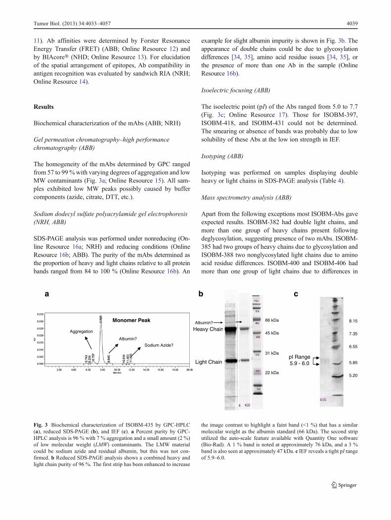
Gel permeation chromatography–high performancechromatography (ABB)
The homogeneity of the mAbs determined by GPC rangedfrom 57 to 99%with varying degrees of aggregation and lowMW contaminants (Fig. 3a; Online Resource 15). All sam-ples exhibited low MW peaks possibly caused by buffercomponents (azide, citrate, DTT, etc.).
Sodium dodecyl sulfate polyacrylamide gel electrophoresis(NRH, ABB)
SDS-PAGE analysis was performed under nonreducing (On-line Resource 16a; NRH) and reducing conditions (OnlineResource 16b; ABB). The purity of the mAbs determined asthe proportion of heavy and light chains relative to all proteinbands ranged from 84 to 100 % (Online Resource 16b). An
example for slight albumin impurity is shown in Fig. 3b. Theappearance of double chains could be due to glycosylationdifferences [34, 35], amino acid residue issues [34, 35], orthe presence of more than one Ab in the sample (OnlineResource 16b).
Isoelectric focusing (ABB)
The isoelectric point (pI) of the Abs ranged from 5.0 to 7.7(Fig. 3c; Online Resource 17). Those for ISOBM-397,ISOBM-418, and ISOBM-431 could not be determined.The smearing or absence of bands was probably due to lowsolubility of these Abs at the low ion strength in IEF.
Isotyping (ABB)
Isotyping was performed on samples displaying doubleheavy or light chains in SDS-PAGE analysis (Table 4).
Mass spectrometry analysis (ABB)
Apart from the following exceptions most ISOBM-Abs gaveexpected results. ISOBM-382 had double light chains, andmore than one group of heavy chains present followingdeglycosylation, suggesting presence of two mAbs. ISOBM-385 had two groups of heavy chains due to glycosylation andISOBM-388 two nonglycosylated light chains due to aminoacid residue differences. ISOBM-400 and ISOBM-406 hadmore than one group of light chains due to differences in
6.55
7.35
8.15
5.85
5.20
a
pI Range5.9 - 6.0
b c
Heavy Chain
Light Chain
Monomer Peak
Aggregation
Albumin?
Sodium Azide?
66 kDa
45 kDa
31 kDa
22 kDa
Albumin?
Fig. 3 Biochemical characterization of ISOBM-435 by GPC-HPLC(a), reduced SDS-PAGE (b), and IEF (c). a Percent purity by GPC-HPLC analysis is 96 % with 7 % aggregation and a small amount (2 %)of low molecular weight (LMW) contaminants. The LMW materialcould be sodium azide and residual albumin, but this was not con-firmed. b Reduced SDS-PAGE analysis shows a combined heavy andlight chain purity of 96 %. The first strip has been enhanced to increase
the image contrast to highlight a faint band (<1 %) that has a similarmolecular weight as the albumin standard (66 kDa). The second striputilized the auto-scale feature available with Quantity One software(Bio-Rad). A 1 % band is noted at approximately 76 kDa, and a 3 %band is also seen at approximately 47 kDa. c IEF reveals a tight pI rangeof 5.9–6.0.
Tumor Biol. (2013) 34:4033–4057 4039
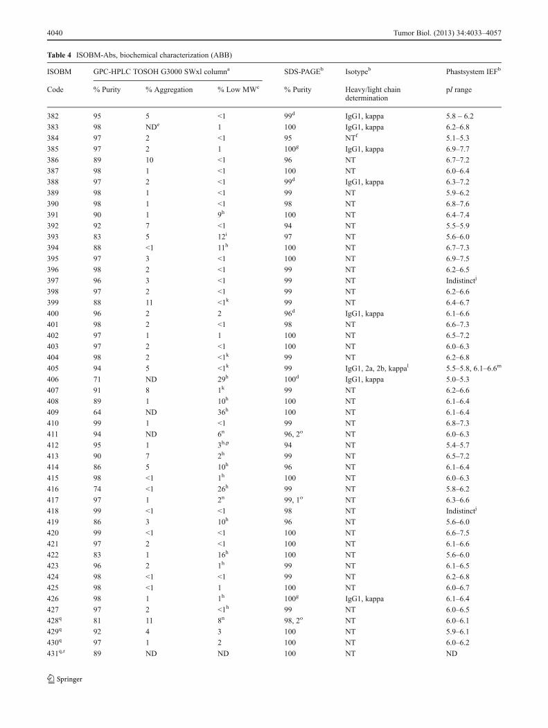
Table 4 ISOBM-Abs, biochemical characterization (ABB)
ISOBM GPC-HPLC TOSOH G3000 SWxl columna SDS-PAGEb Isotypeb Phastsystem IEFb
Code % Purity % Aggregation % Low MWc % Purity Heavy/light chaindetermination
pI range
382 95 5 <1 99d IgG1, kappa 5.8 – 6.2
383 98 NDe 1 100 IgG1, kappa 6.2–6.8
384 97 2 <1 95 NTf 5.1–5.3
385 97 2 1 100g IgG1, kappa 6.9–7.7
386 89 10 <1 96 NT 6.7–7.2
387 98 1 <1 100 NT 6.0–6.4
388 97 2 <1 99d IgG1, kappa 6.3–7.2
389 98 1 <1 99 NT 5.9–6.2
390 98 1 <1 98 NT 6.8–7.6
391 90 1 9h 100 NT 6.4–7.4
392 92 7 <1 94 NT 5.5–5.9
393 83 5 12i 97 NT 5.6–6.0
394 88 <1 11h 100 NT 6.7–7.3
395 97 3 <1 100 NT 6.9–7.5
396 98 2 <1 99 NT 6.2–6.5
397 96 3 <1 99 NT Indistinctj
398 97 2 <1 99 NT 6.2–6.6
399 88 11 <1k 99 NT 6.4–6.7
400 96 2 2 96d IgG1, kappa 6.1–6.6
401 98 2 <1 98 NT 6.6–7.3
402 97 1 1 100 NT 6.5–7.2
403 97 2 <1 100 NT 6.0–6.3
404 98 2 <1k 99 NT 6.2–6.8
405 94 5 <1k 99 IgG1, 2a, 2b, kappal 5.5–5.8, 6.1–6.6m
406 71 ND 29h 100d IgG1, kappa 5.0–5.3
407 91 8 1k 99 NT 6.2–6.6
408 89 1 10h 100 NT 6.1–6.4
409 64 ND 36h 100 NT 6.1–6.4
410 99 1 <1 99 NT 6.8–7.3
411 94 ND 6n 96, 2o NT 6.0–6.3
412 95 1 3h,p 94 NT 5.4–5.7
413 90 7 2h 99 NT 6.5–7.2
414 86 5 10h 96 NT 6.1–6.4
415 98 <1 1h 100 NT 6.0–6.3
416 74 <1 26h 99 NT 5.8–6.2
417 97 1 2n 99, 1o NT 6.3–6.6
418 99 <1 <1 98 NT Indistinctj
419 86 3 10h 96 NT 5.6–6.0
420 99 <1 <1 100 NT 6.6–7.5
421 97 2 <1 100 NT 6.1–6.6
422 83 1 16h 100 NT 5.6–6.0
423 96 2 1h 99 NT 6.1–6.5
424 98 <1 <1 99 NT 6.2–6.8
425 98 <1 1 100 NT 6.0–6.7
426 98 1 1h 100g IgG1, kappa 6.1–6.4
427 97 2 <1h 99 NT 6.0–6.5
428q 81 11 8n 98, 2o NT 6.0–6.1
429q 92 4 3 100 NT 5.9–6.1
430q 97 1 2 100 NT 6.0–6.2
431q,r 89 ND ND 100 NT ND
4040 Tumor Biol. (2013) 34:4033–4057

Table 4 (continued)
ISOBM GPC-HPLC TOSOH G3000 SWxl columna SDS-PAGEb Isotypeb Phastsystem IEFb
Code % Purity % Aggregation % Low MWc % Purity Heavy/light chaindetermination
pI range
432 95 4 1 99 NT 6.5–7.0
433 97 2 <1 99 NT 5.8–6.2
434 88 4 7n 91, 2o NT 6.1–6.3
435 91 7 2n 96, 1o NT 5.9–6.0
436 86 13 1n 86d,g, 1o IgG1, lambda and kappas 5.7–6.1
437 88 10 <1 96 NT 6.0–6.3
438 87 11 1n 96, 1o NT 5.5–5.7
439 93 5 1n 95, 1o NT 6.3–6.9
440 93 2 5n 91d, 2o IgG1, kappa 5.9–6.0
441 83 12 5n 94, 2o NT 6.2–6.5
442q 57 35 8i,n 85d, 1o IgG1, kappa 5.4–5.7
443 90 9 1 99 NT 6.3–6.6
444 93 4 3n 95, 1o NT 6.0–6.3
445 72 25 3n 84, 3o NT 5.3–5.5
446 95 3 2n 97 NT 6.1–6.4
447 97 2 <1 98 NT 6.3–6.6
448 92 3 5n 87g, 3o IgG2a, kappa 5.8–6.9
449 87 12 <1 95 NT 6.1–6.5
450 89 3 7i,n 90d IgG1, kappa 6.0–6.7
a GPC-HPLC samples were run in triplicate. Mean values are ±1.9 %, which is established from the largest standard deviation (SD) observed.Triplicate injections of sample 442 had the largest SD of 1.91.b Single lanes or strips were run for these tests, to preserve sample for additional testing.c All the samples have peaks with the same retention time as sodium azide. The presence of sodium azide could not be confirmed due to lack ofsample volume to perform additional testing.d Double light chains. Possible causes for double chains include but are not limited to; glycosylation differences, amino acid residue differences ormore than one antibody present in the samplee Not Detectedf Not Tested, only samples exhibiting double heavy or light chains by SDS-PAGE or multiple clusters of bands by IEF underwent isotype analysis.g Double heavy chains.h These samples have peaks that could represent high levels of residual citrate in these samples. Residual testing would need to be performed toconfirm this. Testing was not performed due to lack of sample volume. High levels of citrate can react with iron that may be present in HPLCequipment forming iron-citrate complexes. This phenomenon has been observed in other samples containing high citrate levels at retention times ofapproximately 11.3 minutes using a G3000SWxl Column and our Waters HPLC system.i A tailing shoulder is present behind the main antibody peak.j The sample only produced a smear, possibly due to a high salt concentration.k Retention times of 9.2 to 9.6 minutes correspond to a molecular weight of 20–30 kDa and could represent free light chain material. The molecularweight determination was obtained by plotting the logarithm of the GFS molecular weights versus their retention times.l Two types of heavy chain isotypes indicate this sample is probably not derived from a single clone.m Two pI ranges indicate this sample may not be derived from a single clone.n These samples have peaks that have a similar retention time as albumin. The presence of albumin could not be confirmed due to lack of samplevolume to perform additional testing.o A band was observed near the albumin standard.p These samples have peaks that have a similar retention time as DTT. The presence of DTT could not be confirmed due to lack of sample volume toperform additional testing.q Sheep Antibodyr The label concentration may be incorrect. The observed signals were not consistent with the label concentration.s Two types of light chain isotypes indicate this sample is probably not derived from a single clone.
GFS Standards: thyroglobulin MW 670,000, RT 6.0–6.1; gamma-globulin MW 158,000, RT 7.9–8.0; ovalbumin MW 44,000, RT 9.2–9.3,myoglobin MW 17,000, RT 10.3–10.4, vitamin B12 MW 1,350, RT 11.9–12.0
Tumor Biol. (2013) 34:4033–4057 4041

glycosylation and ISOBM-426 complex heavy chains andISOBM-450 complex light chains (Online Resource 18).
Ab affinities, specificities, and epitope localizations
Ab specificities (NRH) Based on the results of DB-RIAs with125I-labeled hCG, hCG-variants, and hLH tracers, the 69ISOBM-Abs were categorized according to their main speci-ficities (α-, β-, and c-mAbs) (Fig. 4): Antibodies either rec-ognized (a) assembled and/or free hCGα (hCGα-mAbs, n=8;α epitopes were not determined) or (b) assembled and/or freehCGβ or hCGβ metabolites such as hCGβcf (hCGβ-mAbs,n=48; epitopes β1–β13), or (c) exclusively the intact±nickedhCGαβ heterodimer, but not the free subunits or metabolicvariants thereof (c-mAbs, n=13; epitopes c1–c4).
Comparing specificity profiles of ISOBM-Abs to those ofreference mAbs permitted preliminary epitope assignment.To discern mAbs against epitopes β1–β5, hLH cross-reactivity was determined by titration RIAs with 125I-hLH(Fig. 4). Recognition of hCGβCTP was investigated bycompetitive RIA using synthetic peptides derived fromhCGβCTP (Fig. 4a; Online Resource 19). No mAbs againstepitope β14 (hCGβ specific) were identified in this ISOBMpanel (Fig. 4a).
Ab affinities and specificities as determined by FRET (ABB) TheISOBM-Abs were grouped according to their specificity pro-files based on affinity for hCG, hCGβ, hCGβcf, and hLH(Fig. 5) determined by FRET. Affinities for the major hCGvariants and hLH, reported as dissociation constants (Kd),ranged from subpicomolar values (0.3 pmol/L for hCGβ ofISOBM-429) to ≥50 nmol/L. The latter value indicated thatbinding was very weak or not detectable.
Only nine Abs (ISOBM-387, ISOBM-399, ISOBM-401,ISOBM-414, ISOBM-416, ISOBM-427, ISOBM-428, ISOBM-429, and ISOBM-444) expressed high affinities (Kd,≤50 pmol/L) for any of the four antigens tested (hCG, hCGβ,hCGβcf, and hLH). It is striking that six of these mAbsrecognize the major antigenic domain on the tips of hCGβloops 1+3. An exception was the hCGβcf-specific mAb ISOBM-444 (reference mAb INN-hCG-106), the epitope ofwhich (β11) does not overlap with epitopes β2–β5 on hCGβloops 1+3 nor with the cystine knot-associated epitopes β1
and β7. Thus, this epitope is remote from either cluster. Thisis an interesting mAb for highly sensitive and specific mea-surement of hCGβcf in particular in combination with β2-mAbs [9, 36].
Another interesting observation is that all three sheepmAbs, ISOBM-428, ISOBM-429 and ISOBM-430, were inthe high affinity group. ISOBM-430 was not tested by theFRET technology but by titration RIA. All four sheep Abs(three mAbs and polyclonal ISOBM-431) were directedagainst hCGβ loops 1+3 epitope β5 that is shared by hLH
and, therefore, in principle, do not seem suitable for hCGmeasurement. Nevertheless, ISOBM-429 seems to have tol-erably low cross-reactivity with hLH (Figs. 4a and 5 andOnline Resource 20), but its suitability for use in hCG+hCGβvariant measurement might still be hampered by preferentialrecognition of hCGβ.
Fig. 4 Specificity profiles of the ISOMB-Abs of the Second TD-7 WSrecognizing hCG and hCGβ variants (a), hCG-only and hCGα, respec-tively (b) were determined by binding of iodinated tracers to excess ofAb (DB-RIA) (NRH). ISOBM-mAbs were classified according to theirmain specificities and their epitopes recognized on the basis of cross-reactivity patterns with hCG, hCG-variants, and hLH: (1) β-mAbscorresponding to epitopes β1–β13, (2) c-mAbs recognizing epitopesc1–c4 on holo-hCG only, and (3) α-mAbs. a MAbs directed againstepitopes β1–β5 are pan-hCG reagents recognizing hCG and hCGβvariantsbut differ in their cross-reactivity with hLH:β1 mAbs are highly specific forhCG and show no hLH cross-reactivity (<0.1%), β2 andβ4 show very lowhLH reactivity (<1 %), whereas β3 and β5 strongly cross-react (>>1 %).Epitopes β6 and β7 are specific for uncombined hCGβ, hCGβn, andhCGβcf. MAbs against epitope β8 at the very carboxyl-terminal end ofhCGβCTP do not cross-react with hCGβcf and hLH but recognize all otherhCG variants except for those lacking the CTP. These mAbs constantlyshow a low bindable fraction of the tracers as only approximately 50 % ofthe tracers can be bound specifically. This is in contrast to theβ1–β5 mAbs.ISOBM-418 seems to be directed against epitope β9 as already typedpreviously in the First WS (ISOBM-280, [1]. Epitopesβ10–β13 are specificfor hCGβcf as no other hCG variants or hLH are recognized by therespective mAbs. b c-mAbs directed against epitopes determined by thequaternary structure of hCG either do not (c1 and c2) or do recognize hCGn(c3 and c4) [56]. The apparent hCGn cross-reactivity of c1 and c2 mAbs isdue to a cross-contamination of this preparation with non-nicked hCG(approximately 20 %) [1]. The presence of non-nicked hCG and recogni-tion by the ISOBM-mAbs of the two-nicked forms in hCGn were investi-gated in detail by LC-MS/MS (see accompanying publication by H. Lund).Epitope c3 (ISOBM-446=INN-hCG-45, reference mAb) is highly specificfor hCG+hCGn. ISOBM-mAb 433 that has the same specificity patternmight be directed against a fifth sterically independent c-epitope as shownby sandwich assay. The exact molecular localization of epitope c4 on hCGis not known, but it is remote from the other c-epitopes. In the First ISOBMTD-7 WS, ISOBM-424 has been characterized (ISOBM-279) and classi-fied as c4 specific [1]. Theα-mAbs have not been investigated in detail as totheir epitope recognition. As they readily recognized iodinated tracers (incontrast to α3- and α5-mAbs), they should be directed against the epitopecluster α1/α2/α4 with the exception of ISOBM-404 that is free hCGα-specific and therefore presumably recognizing the subunit assembly regionof hCGα (aa hCGα33-42). Minor apparent cross-reactivity with hCGn isowed to a cross-contamination of hCGα in that preparation. hLH cross-reactivity of ISOBM-404 might be due to dissociation of highly purifiedhLH that is observed during testing (PB, personal observation). DB-RIAwith 125I-tracers: results are expressed as maximum specific binding inpercent of the “bindable fraction” of added tracer (NRH) [26]. Italics RIAtitration experiments (NRH): Results are expressed as percent hLH cross-reactivities compared to hCG; asterisk 125I-tracers; gray background sig-nificant cross-reactions. Superscripted a Apparent cross-reactivities withhCGn of ISOBM-447–438 are caused by an approximate 20 % cross-contamination of intact hCG (see accompanying publication by H. Lund)and superscripted b of ISOBM-383–404 due to a suchlike with hCGαthat is contained in hCGβn; superscripted c ISOBM-404: apparent cross-reactivity is probably caused by slight dissociation of α-subunit in hLH.Section symbolCompetitive RIAwith hCGβ109-145 vs. hCGβ*, percentcross-reactivity.Double section symbolCompetitive RIAwith hCGβ135-145 vs. hCGβ*, percent cross-reactivity
�
4042 Tumor Biol. (2013) 34:4033–4057

a
Tumor Biol. (2013) 34:4033–4057 4043

Most Abs, including all directed against the cystine knot-associated epitopes β1 and β7, the hCGβCTP epitope β8 types1 and 2, showed moderate affinities (50 pmol/L–5 nmol/L)against their primary target hCG variant. Low affinities couldbe observed for three reasons: (1) the primary antigenic targethCG variant of the mAb in question was not among theantigens tested, as is the case of mAbs against uncombinedhCGα (ISOBM-404), or (2) genuine low affinity to the primarytarget antigens, e.g., ISOBM-418/280 (hCGβCTP epitope β9,aa hCGβ113-116) and ISOBM-443 and 448 against hCGβcf,and (3) FRET labeling affected binding of Abs (ISOBM-445 tohCGβcf; ISOBM-399, ISOBM-436, ISOBM-412, and ISOBM-421 to hLH) (Fig. 5). This is also the case with 125I-labeling of epitopes α3 and α5 [37].
Ab affinities and specificities as determined by BIAcore®(NHD) The specificity patterns of the ISOBM-Abs weredetermined based on their affinity for hCG, hCGβ, andhCGβcf in BIAcore®. The affinities (dissociation constants;Kd) ranged from picomolar values (<10 pM for hCG ofISOBM-399) to >10 nM. An affinity of <100 nM wasobserved for 43 of the Abs for either a single or a combina-tion of the three antigens tested (hCG, hCGβ, and hCGβcf).Five of these Abs, ISOBM-427 (epitope β2), ISOBM-399,ISOBM-423, ISOBM-441 (all three epitope β3), and
ISOBM-428 (epitope β5) had affinities of <10pM for theantigen. It is striking that all of these recognize the tops ofhCGβ loops 1+3; thus, all epitopes were located within thesame antigenic domain. Assignment of epitopes to theISOBM-Abs was achieved by comparing their specificityprofiles to those of reference mAbs (Online Resource 20).
The affinity of many of the ISOBM-Abs appeared to behigher than determined by FRET analysis. This could per-haps be a consequence of having the antigen in a bound formon the BIAcore® chip rather than in a fluid state. The affinityfor this immobilized form of antigen may result in anoverestimate of affinity.
Ab specificities as determined by CLA (NHD) In the CLAapproach, ISOBM-Abs were titrated against 125I-hCG and inparallel competed with a fixed amount (0.5 pmol/mL) ofhCG, hCG-variants, and hLH, respectively. A shift of theAb dilution curves to a lower titre indicated cross-reactivityof this competitor with the Ab. Based on the CLA results, 34ISOBM-Abs, either recognized (a) assembled and/or free
b
Fig. 4 (continued)
Fig. 5 Affinity and specificity of the ISOBM-Abs as determined byFRET (ABB). Preliminary assignment of epitopes was done by com-paring the specificity profiles of the ISOBM-Abs to those of referencemAbs. Specificities based on affinity of mAbs against hCGα could notbe determined with hCG, hCGβ, and hCGβcf
�
4044 Tumor Biol. (2013) 34:4033–4057

ISOBMii Ab Codes
hCG affinity
[nM]
hCGβ affinity
[nM]
hCGβcf affinity
[nM]LH [nM]
Specificity Based on Affinity: hCG, β, βcf
Specificity Based on
Affinity: hLHEpitope
403 1.17 0.65 0.29 >50435* 1.15 0.52 0.32 >50382 0.66 0.51 0.16 3.90388 0.14 0.11 0.11 24.00390 0.14 0.10 0.07 >50402 0.08 0.06 0.18 16.90408 0.10 0.48 0.40 >50416 ? 0.04 0.02 >50417 0.37 0.30 0.10 >50426 0.13 0.17 0.25 >50427 0.08 0.04 0.03 >50449* 0.34 0.15 0.15 12.30396 0.20 0.94 0.90 0.17399 0.13 0.03 0.02 ?400 1.60 0.35 0.23 0.33401 0.86 0.08 0.01 0.30405 0.17 0.13 0.19 0.50423 0.06 0.06 0.10 0.10434* 11.60 16.50 6.10 3.60441* 5.40 0.75 1.20 0.70419 0.34 0.35 0.23 >50445* 0.78 0.76 ? >50
428 sheep 0.01 0.003 0.01 0.10429 sheep 0.004 0.0003 0.03 0.40430 sheep n.d. n.d. n.d. n.d.431 sheep 0.35 0.14 0.20 0.60
442* 0.36 0.25 0.07 0.10410 49.00 0.08 0.07 >50437* >50 2.75 3.75 >50386 6.32 0.23 0.50 >50397 >50 4.99 8.60 >50407 >50 0.70 >50 >50409 >50 1.32 1.55 >50415 40.40 1.27 4.83 >50439* >50 0.54 1.92 >50450* 0.23 2.88 >50 >50395 0.25 1.93 >50 >50413 0.54 1.74 >50 >50420 0.32 1.89 >50 >50394 0.09 0.67 >50 >50418 >50 >50 >50 >50 ? _392 >50 12.40 >50 >50 _406 0.16 0.23 >50 >50 _384 >50 >50 4.10 >50393 >50 >50 4.00 >50443* >50 >50 >50 >50444* >50 >50 0.05 >50448* >50 >50 >50 >50414 0.01 >50 >50 0.04447* 0.44 >50 >50 3.00387 0.05 >50 >50 1.20 hLH411 2.45 >50 >50 >50422 0.06 >50 >50 21.80425 0.12 >50 >50 2.10436* 0.35 >50 >50 ?438* 0.06 >50 >50 0.50
446* 1.07 >50 >50 >50 _ c3
424 0.29 >50 >50 >50 _440* 3.55 >50 >50 1.00 hLH389 0.19 35.70 >50 >50433 0.35 >50 >50 >50383 0.30 >50 >50 0.24385 0.07 >50 >50 0.12391 0.05 >50 >50 0.09398 15.00 29.00 >50 3.80412 0.29 >50 >50 ?421 0.20 >50 >50 ?432 0.34 >50 >50 0.60
404 >50 >50 >50 28.00 _ α6
*reference antibodies Key (kD)< 0.05 nM
0.05 - 0.5 nM0.51 - 5 nM5.1 - 50 nM
>50 nM
β5
hCGβ + hCGβcfβ7
β8hCG + hCGβ
β9
β1
hCG + hCGβ + hCGβcf
β2
β3
β4
hCGβcf only
_
_
β
β6
c1
hCG only
c2
c4
c
_
hLH
_
hLH
βcf
_
hLH α?
I.C. … internal control
_
_
hLH
_
hLH
Tumor Biol. (2013) 34:4033–4057 4045
hCGβ metabolites such as hCGβcf (hCGβ-mAbs, n=24;epitopes β1–β9) or (b) exclusively hCG±hCGβ, but not thefree subunits (c-mAbs, n=10; epitopes c1–c4). Epitope as-signment was achieved by comparing profiles of the refer-ence mAbs with ISOBM-Abs (Online Resource 20).
No CLA analysis was possible for 35 ISOBM-Abs, whichhad very low or no signal, which suggested that the Ab didnot recognize 125I-hCG or could not be competed with theamounts utilized.
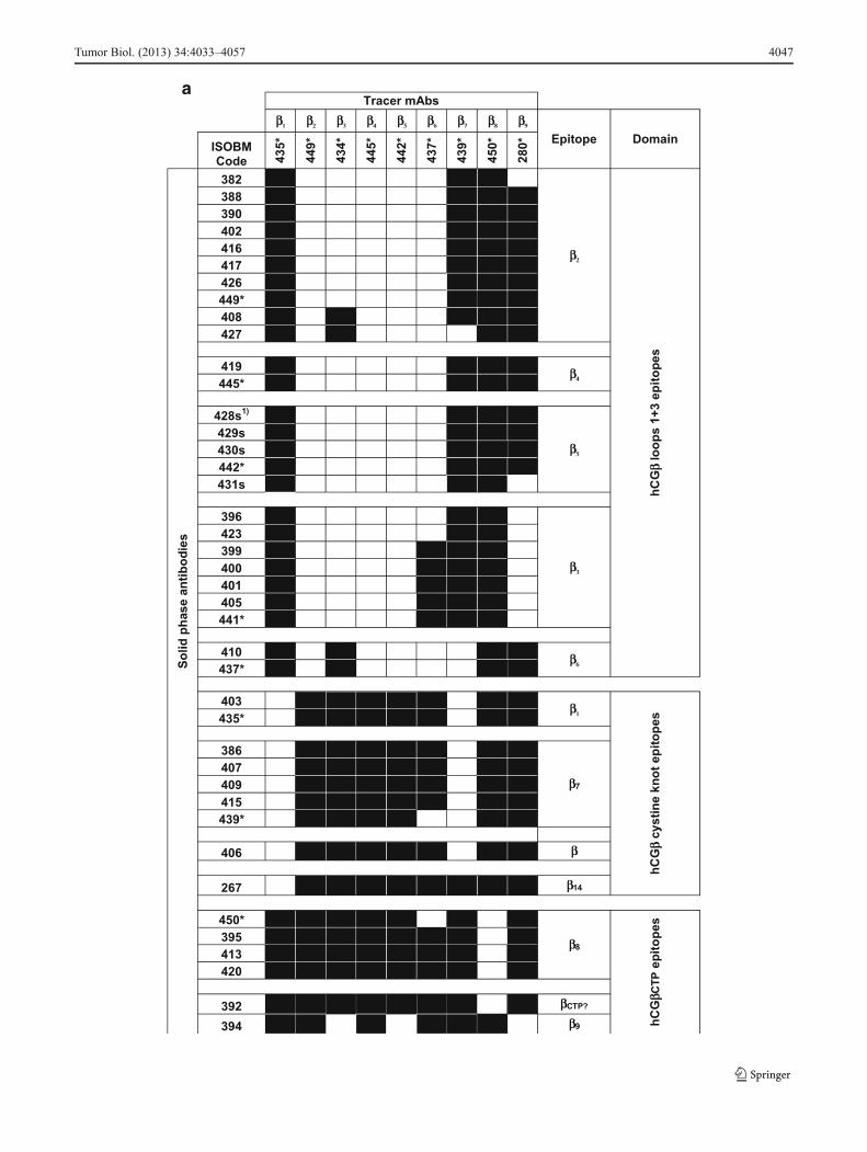
Epitope classification by sandwich assays (NRH) IRMA-like sandwich assays were performed to confirm preliminaryAb epitope classifications by specificity assays and todetermine epitope localization by comparison with ref-erence mAbs. Characteristic reaction patterns were ob-served when solid-phase bound ISOBM-mAbs weretested for their ability to sandwich hCG or hCGβ withthe panel of reference mAbs directed against epitopesβ1–β9 (Fig. 6a) and c1–c4 (Fig. 6b). Patterns observedagreed with previously determined epitope locations forthe reference mAbs [28, 38].
Compatibility of Ab pairs in sandwich assays indicatedthat their epitopes were spatially distinct, e.g., epitopes β1
versus β2–β6 and vice versa (Fig. 6a). Identical or highlysimilar compatibility patterns of Abs to that of reference orother mAbs indicated recognition of identical or of neigh-boring epitopes within the same antigenic domain: e.g., thecystine knot-related epitopes β1 and β7, or epitopes β2, β4,and β5 on hCGβ loops 1+3. Epitopes within a particularantigenic domain can be easily discerned by cross-reactivitypatterns with hCG variants and LH from various species [1].Thus, although β1- and β7-mAbs show identical compatibil-ity patterns in sandwich assays and are not compatible witheach other (Fig. 6a), they recognize different but spatiallyadjacent cystine knot-related epitopes reflected by differingvariant recognition patterns: β1-mAbs recognize a broadspectrum of hCG-variants whereas β7-mAbs are highly se-lective for hCGβ, hCGβn±hCGβcf and do not recognizehCG (Fig. 4).
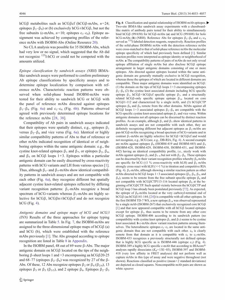
Antigenic domains and epitope maps of hCG and hCGβ(INN) Results of the three approaches for epitope typingare summarized in Table 5. In Fig. 7, the ISOBM-mAbs areassigned to the three-dimensional epitope maps of hCGβ (a)and hCG (b), which were established with the referencemAbs previously [1]. The Abs grouped according to epitoperecognition are listed in Table 8 in Appendix.
In the ISOBM panel, 48 out of 69 were β-Abs. The majorantigenic domain on hCGβ located on the tips of the neigh-boring β-sheet loops 1 and +3 encompassing aa hCGβ20-25and 68–77 (epitopes β2–β6) was recognized by 27 of the β-Abs. Of these, 12 Abs recognize epitopes β2 or β4 (β2/4), 13epitopes β3 or β5 (β3/5), and 2 epitope β6. Epitopes β2–β5
Fig. 6 Classification and spatial relationship of ISOBM-mAb epitopes.Two-site IRMA-like sandwich assay experiments with a chessboard-like matrix of antibody pairs tested for their ability to simultaneouslybind hCGβ (99/650) for hCGβ-mAbs (a) and hCG (99/688) for holohCG-mAbs (b) (NRH). Reference Abs for epitopes β1–β9 and c1–c4served as 125I-labeled detection reagents, respectively. Reaction profilesof the solid-phase ISOBMii mAbs with the detection reference mAbswere cross-matched to that of solid-phase reference mAbs the molecularepitope specificity of which had previously been defined [1]. Similarreaction profiles were interpreted as epitope identity or neighborhood ofmAbs. a The compatibility patterns of pairs of mAbs do not only revealepitope affiliation of single mAbs but also disclose hCGβ epitopearrangement in larger antigenic domains consisting of one or moreepitopes. Abs directed against epitopes located within the same anti-genic domain are generally mutually exclusive in hCGβ recognition,whereas those the epitopes of which are located in different domains arecompatible. Three major antigenic domains were identified on hCGβ:(1) the domain on the tips of hCGβ loops 1+3 encompassing epitopesβ2–β6 (2) the cystine knot associated domain including hCG specificepitope β1, hCGβ+hCGβcf specific epitope β7, and a structurallyrelated hCGβ-only specific epitope epitope β14 located on corehCGβ1-112 and characterized by a single mAb, and (3) hCGβCTPepitopes β8 and β9 remote from the other domains. MAbs against allhCGβ loops 1+3 associated epitopes β2–β6 are compatible with thehCG-specific cystine knot-associated epitope β1 and vice versa. Withinantigenic domains not all epitopes can be discerned by distinct reactionprofiles. As an example, although β1 and β7 show identical patterns insandwich assays and are not compatible with each other, they aredefinitely recognizing different but adjacent epitopes as β1-mAbs arepan-hCGβ-mAbs recognizing a broad spectrum of hCG-variants and incontrast β7-mAbs are highly selective for hCGβ+hCGβcf and wouldnot recognize, e.g., hCG (see, e.g., DB-RIA, Fig. 4). A second exampleare mAbs against epitopes β4 (ISOBM-419 and ISOBM-445) and β5
(ISOBM-428, ISOBM-429, ISOBM-430, ISOBM-431, and ISOBM-442) having an identical compatibility profile, i.e., nicely work withmAbs against epitopes β1 and β7–9 but not with β2–β6. These epitopescan be discerned by their variant recognition profiles whereby β4 mAbsare specific for hCG (≤1 % cross-reactivity with hLH) and β5 mAbsstrongly cross-react with hLH (>>1 %) in titration and competitive RIA(Fig. 4). β3-mAbs, although showing a similar reaction pattern as othermAbs directed to hCGβ loops 1+3 associated epitopes (β2, β4, β5, andβ6), seems to be remote from the free subunit specific epitope β6 andnot compatible with hCGβCTP113-116 located epitope β9 at the be-ginning of hCGβCTP. Such spatial vicinity between the hCGβCTP andhCGβ loop 3 has already been postulated previously [72]. As expected,the epitope of β8-mAbs located at the very carboxyl-terminal end ofhCGβ (aa hCGβ141-144; [24]) is compatible with all other epitopes. Inthe first ISOBM TD-7WS, a new epitope β14 was observed representedby a single mAb (ISOBM-267) that exclusively recognized core hCGβ[1] and that now appeared compatible with all hCGβ located epitopesexcept for epitope β1, thus seems to be remote from any other corehCGβ epitope. ISOBM-406 according to its sandwich pattern (nocompatibility with cystine knot epitopes β1 and β7) seems to be cystineknot associated. b c-mAbs show variant reaction patterns among them-selves. The heterodimeric epitopes c1–c3 are located in the same anti-genic domain thus are not compatible with each other. c4 is clearlyremote from that domain as it is compatible with c1 to c3-mAbs.ISOBM-433 recognizes a previously structurally not defined epitopethat is highly hCG specific as is ISOBM-446 (epitope c3) (Fig. 4).ISOBM-389 a highly hCG specific c-mAb that according to BIAcore®analyses rapidly dissociates (Kd=13E−03), ISOBM-397 and ISOBM-418 (very low affinity in FRET analyses) did not perform well ascapture mAbs in this type of assay and were negative throughout (notshown). Reactions classified as positive (mean+2 standard deviations)are depicted as closed squares. Noncompatible mAb pairs are shown aswhite squares
�
4046 Tumor Biol. (2013) 34:4033–4057
a
Tumor Biol. (2013) 34:4033–4057 4047

are pan hCG specific, i.e., present on hCG, hCGn, hCGβhCGβn, and hCGβcf (Fig. 4a), whereas β6 is present onlyon hCGβ, hCGβn, and hCGβcf.
The cystine knot-associated antigenic domain comprises anumber of epitopes that are recognized by 10 of the 48 β-mAbs (including ISOBM-397 results of which are ambigu-ous). ISOBM-267 that is hCGβ specific and its epitopecystine knot-related (epitope β14) was used as a controlmAb: The pan-hCG epitope, β1 (hCGβ Arg10, Arg60, andGln89), which is not shared by hLH, was recognized by twoISOBM-mAbs (ISOBM-403 and reference mAb ISOBM-435). It is spatially close to epitope β7 (hCGβ Asp61 andGln89) against which six mAbs (including ISOBM-397) weredirected. MAbs classified as β7 recognize either hCGβ+-hCGβn+hCGβcf or mainly hCGβ+hCGβn (ISOBM-407).The cystine knot-related epitope β10 recognized by referencemAb ISOBM-448 is hCGβcf specific. One mAb (ISOBM-406) reacted with a not specified cystine knot epitope(Fig. 6a). This mAb is of restricted pan-hCG specificity anddoes not recognize hCGβcf (Fig. 4a).
In addition to the above-mentioned mAb ISOBM-448 (cys-tine knot related epitope β10), 4 of the 48 β-Abs recognize
epitopes located on hCGβcf only (epitope β11, ISOBM-384and ISOBM-444; epitope β13, ISOBM-443; and one non-coded hCGβcf epitope, ISOBM-393).
Six of the 48 β-Abs are directed against the hCGβCTP.The linear antigenic region (aa hCGβ137–144; epitope β8)at the very end of the hCGβCTP is recognized by fourmAbs; type 2). Three of these (ISOBM-395, ISOBM-413,and ISOBM-420) are mAbs against epitope β8,2 recognizingglycosylated hCGβ much better than the nonglycosylatedsynthetic peptide. Thus, epitope β8,2 might be influenced byglycans on Ser132 and/or Ser138 [1, 39]. One mAb(ISOBM-450; epitope β8,1) recognizes both antigens to thesame extent. Two mAbs, 394 and 418, may be directedagainst epitope β9. One β-mAb (ISOBM-392) could not beclassified but it does not seem to be located on hCGβCTP(Figs. 4a and 7a).
Thirteen of the 69 mAbs reacted with c-epitopes: c1(n=2), c2 (n=6), c3 (n=1), c4 (n=2), c (n=1; ISOBM-433;new noncoded c-epitope). One c-mAb could not be classified(ISOBM-389).
Eight out of 69 mAbs are directed against hCGα. Sixrecognize assembled and one, ISOBM-404, which has beenprepared by immunization with hCGα (Stenman et al.,unpublished data), recognizes only free hCGα. The exactmolecular localization of the hCGα mAbs was not elucidat-ed (Figs. 4b and 7b).
Discussion
Topography of hCG epitopes
Epitopes and antibodies
By definition, epitopes are molecular structures dependenton the existence of complementary Abs. Not the entiresurface of a glycoprotein like hCG is antigenic. Againstcertain molecular areas no Abs exist as they are immunolog-ically inert, e.g., due to insufficient T cell help, or stericallynot accessible due to protein folding or shielding by glycans.In contrast, other areas representing structurally inherentepitopes, which are characterized by high solvent accessibil-ity and high protrusion indices, are often sites of Ab recog-nition [40]. hCGβ cystine knot-associated residues Arg10and Gln89 (epitopes β1 and β7), hCGα loop 1 residuesPro16, Phe17, and Phe 18 (epitopes α1, α2,and α4) and theantigenic domain on hCGβloops 1+3 comprising aa 20–25+68–75 (epitopes β2–β6) all bulge away from the mole-cule forming prominent surfaces that are the major antigenicdomains of hCG [3, 22, 37, 41–43]. There is a good chancethat irrespective of the immunized species these molecularstructures will be recognized as epitopes [38]. For example,
Tracer mAbs
c1 c2 c3 c4
EpitopeISOBM No 44
7*
438*
446*
440*
So
lid p
has
e an
tib
od
ies
414c1
447*
387
c2
411
422
425
436*
438*
446* c3
424a)
c4440*
433 c
*…reference mAbs1) s: sheep
b
Fig. 6 (continued)
4048 Tumor Biol. (2013) 34:4033–4057

the immunodominant antigenic domain on top of hCGβbeta-sheet loops 1 and 3 is recognized by Abs derived frommice and sheep as shown in the present study and interest-ingly by Abs from humans and rabbits (PB, unpublishedobservations). Moreover, hLH cross-reactive mAb B206directed against an epitope within this cluster, presumablyepitope β3/5, inhibited 40–90 % of the binding of humanantisera to hCG [44].
The definition of epitopes by Abs and recognition of themultitudes of possible amino acid combinations within aninherently antigenic structure/domain is dependent on andrestricted by the combinatorial repertoire of the VDJ and VJimmunoglobulin heavy and light chains gene segments, re-spectively, and the cellular capacity to mature the paratope ofa given Ab to optimally fit the antigenic surface. This reper-toire of Ab specificity varies with individual immune re-sponses, haplotypes, and species. Not every amino acidcombination within an antigenic domain will therefore berecognized by Abs of any individual or species. Thus, therepertoire of Ab specificities and corresponding epitopeswithin an antigenic domain is very large but still somewhat
restricted as shown by the present and previous studies. Forexample, the antigenic domain on hCGβ loops 1+3 is rec-ognized by large panels of Abs that differ slightly in hCGvariant recognition, hLH cross-reactivity, affinity, etc. Thishas been shown to be due to variability in amino acidrecognition within the antigenic domain [1].
It is striking that this antigenic region, aa hCGβ20–25+68–75 on the tips of loops 1+3, comprises 16 aminoacids, a number that reasonably well corresponds to thesurface covered by a single complementary paratope of anAb whereby two to three amino acids that vary from Ab toAb provide most of the binding energy and fine specificity[45]. Consequently, dozens of ISOBM-mAbs and Abs ofother panels directed against hCGβ loops 1+3 epitopesβ2–β5 do not behave uniformly in their recognition of theapproximately 15 potential contact amino acids composingdiscontinuous epitopes, even though they cover more or lessthe same surface with their paratope [43]. Thus, all differ-ences in affinity, specificity, and hLH cross-reactivity ofnumerous antibodies directed against this major antigenicregion seem to have their basis in variability of preferential
Table 5 Epitope assignment of the ISOBM-Abs using three approaches
Epitopes n Comments Classification of ISOBM-mAbs by three approachesβ-mAbsa 48 Antigen binding (NRH) Affinity (FRET) (ABB) Sandwich assay (NRH)β1 2 Specific for hCG and hCG derivatives 403, 435* 403, 435* 403, 435*β2,β4 12 <1% cross-reactivity with hLH/hLHβ
Epitopes located on top of loops 1+3β2: 382, 388, 390, 402, 408, 416, 417, 426, 427, 449*β4: 419, 445 *
β2: 382, 388, 390, 402, 408, 416, 417, 426, 427, 449*β4: 419, 445*
β2: 382, 388, 390, 402, 408, 416, 417, 426, 427, 449*β4: 419, 445*
β3,β5 13 High cross-reactivity with hLH/hLHβEpitopes located on top of loops 1+3
β3: 396, 399, 400, 401, 405, 424, 434, 441*β5: 428s to 431s, 442*
β3: 396, 399, 400, 401, 405, 424, 434, 441 β5: 428s to 431s, 442*
β3: 396, 399, 400, 401, 405, 424, 434, 441*β5: 428s to 431s, 442*
β6 2 No recognition of the αβ heterodimer 410, 437 410, 437 410, 437β7 6 No recognition of the αβ heterodimer 386, 397, 407, 409, 415, 439 386, 397, 407, 409, 415, 439 386, 407, 409, 415, 439β8 4 Epitope on hCGβCTP Type 1: 450*;
Type 2: 395, 413, 420450*, 395, 413, 420 450, 395, 413, 420
β9 2 Epitope on hCGβCTP 394, 418 394β10-β13 5 Specific for hCGβcf 384, 393, 443*, 444*, 448* 384, 444* n.d.β 2 Unspecified location 392, 406 392, 406 (cystine knot?)
-mAbsb 13c1 2 In a cluster with c2, sensitive to nicks 414, 447* 414, 447* 414,447*c2 6 In a cluster with c1, sensitive to nicks 387, 411, 422, 425, 436*, 438* 387, 411, 422, 425, 436*, 438* 387, 411, 422, 425, 436*, 438*c3 1 Specific for hCG + hCGn 446* 446* 446*c4 2 Spatially remote from other epitopes 424, 440* 424, 440* 424, 440*c 2 Unspecified location 389, 433 389 433
α-mAbsc 8 Not epitope typed in detail 383, 385, 391, 398, 404, 412, 421, 432
α3 1 Epitope sensitive to iodination 432?α6 1 Specific for non-combined hCGα 404?
Excluded Abs None 383d, 385d, 391d, 398d, 404d, 412d, 418d, 421d, 432d, 443*d,448*d
383, 384, 385, 389e, 391, 393, 397, 398, 404, 412, 418e, 421, 432e, 434e, 443*, 444*, 448*
a Fourteen epitopes, 13 of which recognized by ISOBM-mAbsb At least four epitopes; four plus one recognized by ISOBM-mAbsc Seven epitopes; these ISOBM-mAbs were not characterized in detaild Specificity based on affinity for hCG, hCGβ and hCGβcf could not be determinede These four ISOBM-mAbs did not perform well as capture reagents
*Reference mAbs
Tumor Biol. (2013) 34:4033–4057 4049
recognition of a few amino acids, providing binding energywithin very similar or even identical sets of amino acidscovered by the Abs’ paratopes.
The surface area of an epitope that is covered by acylinder-like antigen binding site of an Ab is approximately700 Å2 in size [38, 46], whereby the radius of the antibody
CTP115
I
NY
T
N
C
N
R
23
Q
VA
T
A
L
C
T
C
R
6
FT 15
RL
P
P
A
A
A
V 80
E
T
I
V
V
G
Y
V
S
Y
E
I
S
D
P
V
D105
10
P
C
L
H
L
T
L
P
G
Q
L
S
T
CC
9
9390
57
R
T
S
G
R
P
G
D
M
P
L
Q
V
A
T
V
R
K
E
110
100
95
50
45
40
5
13
3
55
65
75
85
26
38
34 88
1
25
60
V
30
C
LS
A 61
89
V
CP
1403,435
INN-hCG-2
2/4382,388,390,402,408,416
417,419,426,427?445,449
INN-hCG-22,-24
3/5396,399,400,401,405
423,428,429s,430s,431s434,441,442
INN-hCG-58,-111,-b1
6410,437
INN-hCG-64
9394,418
8 type 1450,h-54
type 2395,413,420
7386,397?,407,409,415,439
INN-hCG-68
14267
Loop 1 Loop 3
Loop 244-48nicks
S
SS
S
P
P
G
P
P
P
S
A
P
K
L
L
T
P
S
L D
P
S
A
S
Q 145
D
140
135
130
1
125
120
truncations
121
127
132
138
Q
D
10β
β
β
6β
1β7β
2β
5β
4β
3β
9β
8β
β
β
β
Γ
β
β
β
β β β
- 13384,393
443,444,448
392406
2
5
1
4
9
c2 c1
c3
2
41
3
5
3
8
hCG CTP
c1389?,414,447INN-hCG-10
c2387,411,422,425,436,438
INN-hCG-40,-53
C3446
INN-hCG-45
c4424?,440
INN-hCG-26
1/ 2/ 4383,385,391398,412,421
432
c433
α α α
αα
αα
α
β
β
β
ββ
β ββ
b
a
4050 Tumor Biol. (2013) 34:4033–4057
binding domain is 8–10 Å and the radius of the epitopecovering area is 15 Å irrespective of Ab specificity [45]. X-ray crystallography studies revealed that core hCG, i.e., hCGwithout hCGβCTP, has a length of 75 Å and a width of 30–35 Å [3, 47] corresponding to a surface area of approximately8,200 Å2. As some regions on assembled hCGβ, such as thestems of β-sheet loops 1+3, are not recognized by any anti-hCG-mAbs [1, 18, 48], the total epitope-covered area on corehCG could be in the range of 5,000 Å2 theoretically accommo-dating simultaneous binding of up to seven Abs to spatiallyindependent epitopes. The minimal spatial requirement forsterical compatibility of two mAbs is that the respective epi-topes are approximately 20–30 Å apart. In fact preliminaryexperiments showed that at least five radiolabeled mAbsagainst epitopes β1+β3+α2+α3+c4 were able to bind to corehCG simultaneously [38].
Glycosylation and epitopes
With two exceptions, glycosylation has little effect on hCG’simmunological make-up, although the glycans, which are
hydrophilic in nature and thus surface exposed, representapproximately 30–35 % of its total molecular mass. Theexceptions are glycans at the very end of hCGβCTP and inthe stem region of hCGβ loop 1. The 14 epitopes on corehCG, which is lacking hCGβCTP, are dependent on theprotein backbone. Neither desialylation, deglycosylation[48], partial natural deglycosylation as in the case of themetabolic product hCGβcf [49], nor intense glycosylationas shown with highly acidic pI variants of pregnancy- andtumor-derived hCG have essential effects on Ab recognitionby the reference mAbs [17, 18]. In addition, the number andthe relative spatial location of epitopes do not differ betweenthe isoforms [1, 18, 48].
The peptidic stem region of assembled hCGβ loop 1,which accommodates the two large N-linked glycans athCGβAsn13 and Asn30 that are spatially near the hCGαglycan at Asn52 [3], is not recognized by any mAb in thepanels of anti-hCG-mAbs of the previous and the presentstudy. Thus, the immune response seems to be attenuatedby the N-linked glycans in this region of hCGβ loop 1[1, 18, 48].
A mAb (B152) that was not included in this studyrecognizes hCG with a core-2 O-glycan at Ser 132 andsurrounding peptide structures [50, 51]. Its epitope,which we termed β8,3, is spatially related to epitope β8,2 thatalso seems to be influenced by the glycans on Ser 132 and/orSer 138 [1, 29].
Some hCG assays have been claimed to underestimatehCG-h [52]. However, these results have been obtained withan hCG-h preparation that also was completely nicked (C5)[39]. Thus, the results most probably reflected failure torecognize hCGn rather than hyperglycosylated hCG.
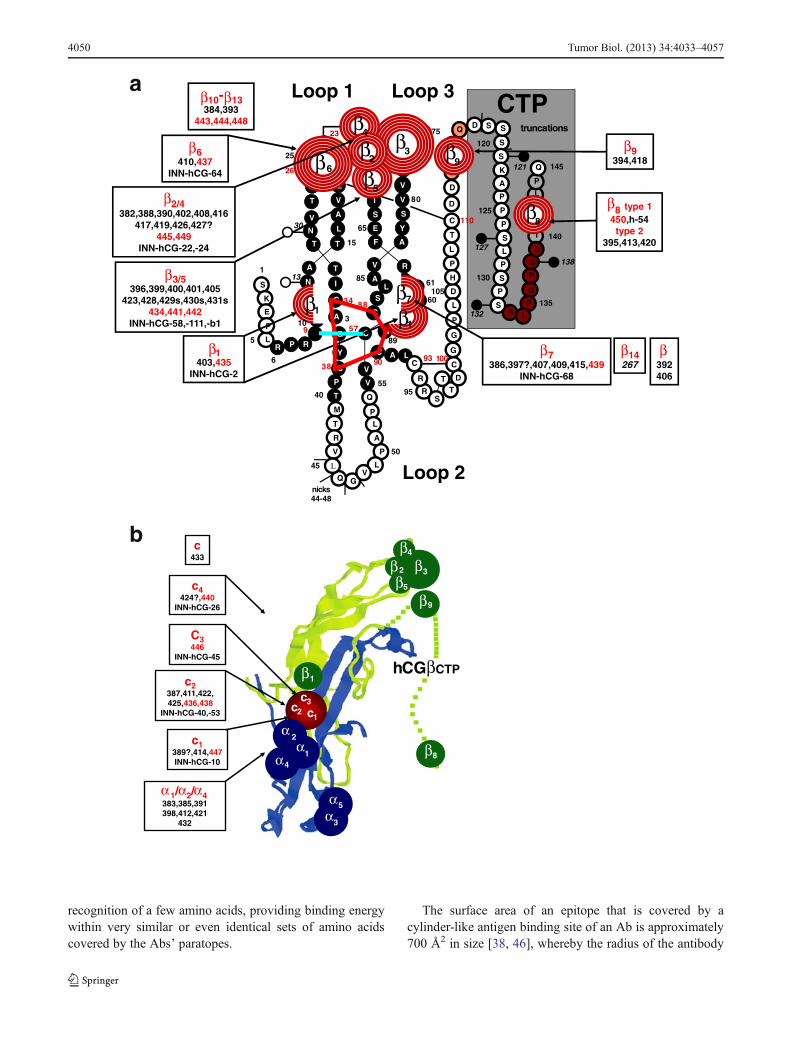
Epitopes on assembled and/or free hCGβ (β1–β9, β14)and hCGβcf only (β10–β13)
The immunodominant structure of hCG and hCGβ-relatedmolecules is the molecular region corresponding to hCGβcf,which has lost its N-terminus, the long loop 2, most of its N-linked carbohydrate antennae, and the hCGβCTP with all O-linked glycans but has retained its protein backbone config-uration [53]. Thus, numerous mAbs against epitopes β1–β7
recognize hCGβ, hCGβn, and hCGβcf. However, one mAb(ISOBM-407) did not react with hCGβcf.
The epitopes on assembled and/or free hCGβ (β1–β9,β14) are located in three molecular regions: (1) hCGβ cystineknot, (2) tips of hCGβ loops 1+3, and (3) hCGβCTP.
The cystine knot-associated antigenic domain includesepitope β1 involving aa hCGβArg10+Arg60 and possiblyGln89 that sterically are in close proximity to each other [42,43]. hCGβArg10 and Gln89 are unique to hCG and notshared by hLH. This presumably explains why epitope β1
is highly specific for hCG and its variants and therefore is not
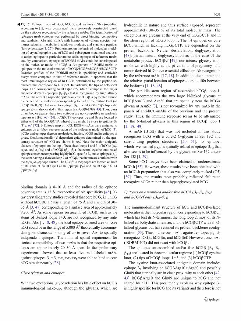
Fig. 7 Epitope maps of hCG, hCGβ, and variants (INN) (modifiedaccording to [1], with permission) were previously constructed basedon the epitopes recognized by the reference mAbs. The identification ofreference mAb epitopes was performed by direct binding, competitiveand sandwich RIA and ELISA with hormones of various species, hor-mones subunits, metabolic breakdown products, and synthetic peptides(for reviews, see [1, 22]). Furthermore, on the basis of molecular model-ing of crystallographic data of hCG and subsequent mutational analysesto assign epitopes to particular amino acids, epitopes of reference mAbsand, by comparison, epitopes of ISOBM-mAbs could be superimposedon the molecular model of hCGβ. a Assignment of ISOBM-mAbs toepitopes on the molecular model of hCGβ/hCGβn/hCGβcf/hCGβCTP.Reaction profiles of the ISOBMii mAbs in specificity and sandwichassays were compared to that of reference mAbs. It appeared that themost immunogenic region of hCGβ is determined by the peptide se-quences that correspond to hCGβcf. In particular, the tips of beta-sheetloops 1+3 corresponding to hCGβ20-25+68–77 comprise the majorantigenic domain (epitopes β2–β6) that is recognized by high affinitymAbs. The only hCG-specific epitope on core hCGβ isβ1 located aroundthe center of the molecule corresponding to part of the cystine knot (aahCGβ10,60,89). Adjacent to epitope β1, the hCGβ/hCGβcf-specificepitope β7 is also located in this region (aa hCGβ61,89) [43]. Thus, pairsof antibodies against these two epitopes are not compatible in sandwichtype assays (Fig. 6a) [24]. hCGβCTP epitopes β9 and β8 are located ateither end of the hCGβCTP, whereby β9 might be close to epitope β3
(Fig. 6a) [72]. b Epitope map of hCG. ISOBM-mAbs were assigned toepitopes on a ribbon representation of the molecular model of hCG [3].hCGα and epitopes thereon are depicted in blue, hCGβ and its epitopes ingreen. Conformationally (c) dependent epitopes determined by the qua-ternary structure of hCG are shown in red. Note the major antigenicclusters of epitopes on the top of beta sheet loops 1 and 3 of hCGα (α1/α2/α4 and α3/α5) and of hCGβ (β2– β5), the central cystine knot-basedepitope cluster encompassing highly hCG-specificβ1 and c-epitopes (c3),the latter having a share on loop 2 of hCGβ, that in turn are confluent withthe α1/α2/α4 epitope cluster. The hCGβCTP epitopes are located on bothof its ends at aa hCGβ113-116 (epitope β9) and aa hCGβ133-144(epitope β8)
�
Tumor Biol. (2013) 34:4033–4057 4051

present on hLH or hLHβ [26]. Due to its superior specificity,it is highly valuable for hCG/hCGβ-variant measurement byimmunoassay with no interference by hLH or hLHβ [1].
The assumed location of epitope β7 on hCGβ, hCGβn,and hCGβcf is based both on mutational analyses andvicinity analysis by sandwich assays: It is associated withthe cystine knot, present on hCGβcf, and Asp61 andGln89 have a role in this epitope. Thus, in sandwich typeassays, β7-mAbs are not compatible with β1-mAbs(Fig. 6a) [1, 22, 24].
MAbs against the cystine knot epitope β7 recognizehCGβcf in addition to hCGβ. ISOBM-407 is an exceptionto this, although other parameters match with epitope β7, itshows an exceptionally low cross-reactivity with hCGβcf(Fig. 4) and thus seems to be suitable for measurement ofhCGβ in urine in the presence of high levels of hCGβcf.The assignment of hCGβ specific epitope β14 to the cystineknot antigenic domain is based on circumstantial evidenceas mAb ISOBM-267 defined in the First ISOBM TD-7 WSto recognize epitope β14 is not compatible withhCGβcystine knot-related epitope β1 but with all otherhCGβ-related epitopes (Fig. 6a). Two hCGβcf epitopesβ10 and β12 are also cystine knot-associated (PB,unpublished data). An additional cystine knot-related epitopeis represented by mAb ISOBM-406.
Antibodies directed against the major hCGβ antigenicdomain on loops 1 and 3 are of significantly higher affinity
compared to those against other antigenic regions ofhCGβ[1, 21, 54]. MAbs against epitopes β2–β5 recognizea wide spectrum of hCG and hCGβ-related variants (hCG,hCGn, hCGβ, hCGβn, and hCGβcf) [1, 17, 18]. MAbsagainst epitopes β3 and β5 additionally react well withhLH and hLHβ, whereas epitopes β2 and β4 are specificfor hCG and hCGβ variants (<1 % hLH and hLHβ cross-reactivity) and thus highly suitable for specific measurementof hCG and hCGβ variants (Fig. 4) [1, 26].
In summary, β-epitopes located on the protein corehCGβ1-112 are discontinuous in nature, determined by thetertiary protein structure, present on hCGβcf, and arrangedin antigenic domains associated with the cystine knotand on the tips of loops 1+3. MAbs directed against theseepitopes are of adequate affinity and suitable for immunoassayapplications.
hCGβ-related epitopes not determined by hCGβcf or corehCGβ1–112 are located in two major regions on the hCGβCTP(aa hCGβ113–145). The immunodominant linear antigenicregion at the very end of the hCGβCTP consists of aahCGβ133–144 and encompasses epitope β8 that is composedof epitope variants β8,1, β8,2, and β8,3) [29, 55]. It partiallyseems to be influenced by glycans on Ser132 and/or Ser138(epitopes β8,2 and β8,3) [29] [1, 50]. One mAb in this WS(epitope β8,1; ISOBM-450) and four mAbs in the First WSrecognized nonglycosylated synthetic peptides andglycosylated hCGβ equally [1]. Epitope β9 at aa hCGβ113–
Table 6 hCG and/or hCG-variants measurements: candidate epitopes for sandwich methods (modified according to [1])
Primary TargetEpitope
Localization 1st mAba
EpitopeLocalization
2nd mAbaAppropriateClinical use
hCG + hCGWide spectrum of hCGβvariants
β1 cystine knothCGβ10+60+89
β2
hCGβ loops 1+3aa 20-25 + 68-77
OncologyEarly pregnancy
Prenatal screening
hCGc2 or c3
hCGβ loop 2,cystine knot hCGβ,
hCGα loop1
OncologyEarly pregnancy
Prenatal screeninghCGβ det. necessary
hCGβ7 cystine knothCGβ61+89
OncologyPrenatal screening
hCG det. mandatory
hCG cfβ11
hCGβcfClinical utility to be
establishedIn urine only
hCGα6
hCGα 33-42α5
Loop 3 (Tyr 65)Oncology (pit./testis)Clinical utility to be
established
a Candidate mAbs for the respective epitopes are listed in Figs. 4, 6, 7, and Appendix 2
4052 Tumor Biol. (2013) 34:4033–4057

116 [21, 24] was recognized by two mAbs (Fig. 7a) wherebyISOBM-394 was of high and ISOBM-418 of very low affinity(Fig. 5).
When immunizing with the glycoprotein hCG, the vastmajority of antibodies will be generated against compositeepitopes on hCGα or the core region of hCGβ (aa 1–112) butonly rarely against linear peptide sequences of low structuralorder like the hCGβCTP. MAbs against hCGβCTP aregenerally of fairly low affinity. Nevertheless, they are usedin diagnostic sandwich-type immunoassays as they do notcross-react with hLH (Fig. 4).
hCGα epitopes (α1–α7)
In the panel of ISOBM-mAbs, 8 of 69 recognize hCGαepitopes. One of these mAbs, ISOBM-404, seems to bespecific for free hCGα, and it is speculated that it mightrecognize the sequence hCGα33-42 on the single loop 2. Asno reference hCGα-mAbs (Table 2) were included, a de-tailed assignment of epitopes was not possible.
Epitopes on the hCG αβ-heterodimer (c1–c4)
At least four epitopes (c1–c4) are present only on hCG±-hCGn but not on either free subunit or hCGβcf [21, 26, 28].Detailed analysis of hCG and hCGn recognition by theISOBM-mAbs was performed by liquid chromatographymass spectrometry (LC-MS/MS) (see accompanying publi-cation by H. Lund). Epitopes c1 (reference mAb INN-hCG-10) and c2 (reference mAbs INN-hCG-40 and INN-hCG-53)are (1) dependent on intact hCG and thus sensitive to nickingof assembled hCGβ loop 2, (2) not compatible in sandwich-type assays with the cystine knot-related hCGβ epitope β1
(aa hCGβArg10+Arg60 and possibly Gln89) [38], and (3)incompatible with mAbs recognizing epitope cluster α1, α2,and α4 [38] on loop 1 in the region of aa hCGα13-22. Aminoacids hCGβ44-48 in loop 2 and hCGα loop 1 have beenshown by X-ray crystallography to be in close proximity asthe subunits are assembled in a head-to-toe fashion [3]. It isstriking that in sandwich assays c1-mAbs show identicalreactivity patterns as α1- and α2-mAbs reflecting stericalepitope relatedness [28, 38].
MAbs against epitope c3 are sterically related to epitopec2, highly specific versus hLH as well as non-combinedintact and modified subunits (<1 % cross-reactivity), notinfluenced by nicking of assembled hCGβ loop 2, and thusrecognize hCGn and hCG equally [1, 56](Fig. 4). They aretherefore highly suitable for simultaneous measurement hCGand hCGn (Table 6).
The exact molecular localization of epitope c4 hasnot been resolved yet. It is present on hCGn andhLH, remote from and thus sterically compatible withall other c-epitopes and to a minor extent determined by
hCGβ as shown by low cross-reactivity [1, 26, 38]. Avariant of the c4-epitope represented by ISOBM-424 (=ISOBM-279, First ISOBM TD-7 WS) that is not sharedwith hLH (cross-reactivity <0.1 %) seems to exist. Apresumably fifth highly specific c-epitope has been ob-served in sandwich assays wherein mAb ISOBM-433 iscompatible with mAbs to c1–c4 (Fig. 6b). Its molecularlocalization is unknown. ISOBM-389 is a c-mAb thatcould not be epitope typed but, according to its speci-ficity profile analyzed by LC-MS/MS, might be a c2mAb (see accompanying publication by H. Lund).
Method-specific recognition of hCG and hCG variants
Sandwich-type assays measuring hCG alone or in com-bination with free hCGβ and metabolites are used fordetection of pregnancy, pregnancy-related disorders, tro-phoblastic disease, and various other female and maletumors [2]. Detailed knowledge of the epitopes recog-nized by the Abs used facilitates development of assaysproviding better comparability of the results betweenmethods. It has been suggested that assays that are mul-tifunctional with respect to clinical use should (1) recog-nize in an equimolar fashion hCG and hCGβ proteinbackbone and glycosylation variants, (2) not cross-reactwith hLH or derivatives, and (3) not be prone to signalblunting by non-measured variants, e.g., caused by ex-cess hCGβcf leading to false low results [36]. This is aproblem when hCG in urine is measured with sandwichassays utilizing a mAb against core hCGβ1-112 in com-bination with an anti-hCGβCTP mAb [57].
While assays measuring hCG and all hCGβ-related vari-ants are useful as first line methods, for diagnosis of pregnan-cy and cancer, it is often advantageous to specifically measureonly selected variants [2]. Thus, specific hCGβ assays areused for first trimester Down’s syndrome screening and alsofor diagnosis of testicular [58, 59] and nontrophoblastic can-cers, 20–50 % of which produce only hCGβ but not hCG[60–62]. However, the concentrations are mostly low, and theassays used need to be highly sensitive. Assays for hCGβ thatare intended for first trimester screening of Down’s syndromeneed to be insensitive to interferences by an approximately100-fold excess of hCG and tuned to measure fairly highconcentrations. They are therefore of limited utility for thediagnosis of nontrophoblastic cancers.
Elevated plasma concentrations of hCGβ are reflected byhigh levels of hCGβcf in urine [63], and specific assay ofthis form has been used for diagnosis of nontrophoblasticcancer [62, 64, 65] and for the characterization of the FirstIRR for hCGβcf [9]. However, commercial assays are notavailable presently.
Tumor Biol. (2013) 34:4033–4057 4053

Candidate epitopes for measurement of hCG and hCGβ
Assays specifically recognizing hCG, hCGβ, and relatedvariants can be constructed using a combination of two panhCGβmAbs with identical specificity profiles [66], i.e., withone partner directed against epitopesβ2 orβ4 (the hCGβloops1 and 3 domain) combined with a mAb-recognizing epitopeβ1 (the hCGβ cystine knot domain; Table 6).
In the two ISOBM TD-7 WSs, 50 of 96 Abs were shown torecognize hCG+hCGβ and 23 of these did not recognize hLH.Theoretically, any of the five mAbs directed against epitope β1
around the cystine knot could be combined with any of the 18mAbs against epitopes β2 or β4 on loops 1+3 for constructionofmultifunctional assays. Epitopesβ1 andβ2/4 are shared by allimportant hCG and hCGβ protein backbone variants and gly-cosylation isoforms including hCG-h and hCGβ-h [17, 18].MAbs against these two discrete epitopes are highly specific forhCG with <0.1 and <1 % cross-reaction for hLH for epitopesβ1 and β2/4, respectively. No other epitope combination pro-vided assays with equally wide and identical recognition ofhCG and hCGβ variants and high specificity versus hLH.
While Abs recognizing these epitopes provide desirablespecificity, variable affinity for hCG variants (Fig. 5) may causenonequimolar recognition of hCG and hCG variants in differentmethods [6]. Although assay specificity can be predicted on thebasis of mAb specificity profiles and epitope recognition [66],ultimate performance can only be evaluatedwith the final assay.An additional source of method variability in hCG measure-ment that cannot be fully predicted is that of Ab synergy, whichmay vary between different Ab pairs [67].
Alternative epitopes for measurement of hCG and/or hCGβand variants
Few manufacturers provide information about the epitopespecificities of Abs used in their assays, but due to variablerecognition of the First IRR preparations for hCG and vari-ants, it is obvious that different epitope combinations are usedin the major commercial assays [6]. In addition to the epitopecombination β1–β2/4, other combinations are possible for theconstruction of assays for hCG and variants, e.g., epitopesβ8,1
and β2, β1–α5, α4–β2, etc., but none of them will fulfill allthree above-mentioned criteria. However, the frequently usedβ8 and β2 combination does not pose problems as long asserum specimen are measured that do not contain hCGβcf,truncated hCG or truncated hCGβ, or clipped hCGβCTP.
For selective measurement of hCG or free hCGβ orhCGβcf certain epitope combinations can be suggested: forhCG (no recognition of hLH or noncombined subunits), a
mAb against epitope c2 or c3 can be combined with oneagainst β2/4 (Table 6). Alternatively, β1–α3 combinations[66] orβ2/4 combined with a tracer mAb against anα epitopeare possible [68]. These designs eliminate cross-reactionswith free subunits but are sensitive to interferences by freesubunits and hCGβcf. For measurement of free hCGβ, amAb to epitope β7 or β14, and for hCGβcf, a mAb to epitopeβ11 can be combined with one to epitope β2/4. For hCGα,combinations of mAbs against epitopes α6 and α5 are rec-ommended [69] (Table 6).
A unique mAb coded B152 is used for the measurementof hCG-h that carries a core-2 glycan on Ser132 located onhCGβCTP [20]. However, the clinical utility of assays usingthis mAb remains to be established [70].
Future perspectives: harmonization of hCG and/or hCGβand variant measurement
Considerable reduction in between-method and between-laboratory variability in results can be achieved by a numberof measures: (1) the establishment and usage of a clear nomen-clature of hCG and its variants [1, 8]; (2) endorsement of thatnomenclature to define what hCG-assay measure [1, 8, 60]; (3)characterization of diagnostic assays with the new six FirstIRRs calibrated in SI units that were adopted by WHO forimmunoassay standardization [6]; (4) standardization ofmethods with the highly pure new WHO Fifth IS for hCGencoded 07/36,4 which is identical to the First IRR for hCG99/688; (5) harmonization of mAb epitopes used in diagnosticmethods for hCG, hCGβ, and their variants; and (6) the estab-lishment of referencemethods for the various forms of hCG [8],which will be supported by the detailed knowledge on Abepitope recognition reported in the present study.
Acknowledgments We would like to thank the International Society ofOncology and BioMarkers for organizing the Tissue Differentiation (TD)-7Workshop on hCG and Related Molecules, the International Federation ofClinical Chemistry (IFCC)Working Group for Standardization of hCG, theNational Institute for Biological Standards and Control (NIBSC) (SouthMimms, UK), and the National Hormone & Peptide Program (NHPP),USA, for the supply of hormones and hormone variants. We would like tothank the following at Abbott Laboratories for their valued contributions:B.L. Dowell, S.Y. Tetin, and J.R. Fishpaugh. The NHS National ServicesScotland, National Services Division, supported the work of Wilson Stew-art. TheAustrian Science Fund (National ResearchNetwork, S9307B05) inpart supported the work of Peter Berger.
Open Access This article is distributed under the terms of the CreativeCommons Attribution License which permits any use, distribution, andreproduction in any medium, provided the original author(s) and thesource are credited.
4054 Tumor Biol. (2013) 34:4033–4057
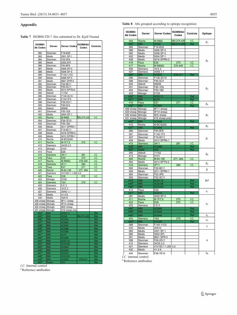
Appendix
Table 7 ISOBM-TD-7 Abs submitted to Dr. Kjell Nustad
ISOBMii Ab Codes
Owner Owner CodesISOBMi&ii
CodesControls
382 Stenman F16-6G5383 Medix 5501 SP-1384 Stenman F52-3F8385 Medix 5503 SPI386 Stenman F94-8F8387 Medix 5009 SP-5388 Medix 5006 SP-5389 Stenman F140-11C5390 Medix 5008 SP-5391 Medix 6601 SPR-5392 Stenman F20-6E11393 Stenman F52-3C11394 Medix 5014 SPTN-5395 Abbott 71752396 Stenman F132-3C10397 Stenman F142-7F3398 Stenman F26-2G11399 Stenman F95-5C4400 Abbott 95658401 Stenman F95-1E8402 Medix 5004 SP-1403 Roche M-INN2 265,274,435 I.C.404 Stenman F26-7E10405 Stenman F95-1B2406 Medix 5011 SPRN-1407 Stenman F19-9C11408 Medix 5016 SPRN-5409 Medix 5012 SPRN-1410 Roche M-BCG005411 Roche M-1F7.9 275 I.C.412 Siemens 34/25.2.2413 Mologic D101414 Paus E26415 Siemens 3A11 281 I.C.416 Paus E30 273 I.C.417 Roche M-INN22 276.449 I.C.418 Siemens 2F11 280 I.C.419 Paus E27 271 I.C.420 Roche M-94.139 277, 264 I.C.421 Siemens 41I/100.1.1.200.4.2422 Paus E28 272 I.C.423 Mologic D102424 Siemens 1G4 279 I.C.425 Siemens 5 E 5426 Siemens 16 E 2427 Siemens 34A8.1.1432 Medix 41-3-9433 Medix 45A10
428 sheep Mologic 8F11 sheep429 sheep Mologic 9F10 sheep430 sheep Mologic 8G5 sheep431 sheep Mologic 618 sheep poly
434a INN hCG111 Ref435a INN hCG2 265,274,403 Ref436a INN hCG40 Ref437a INN hCG64 Ref438a INN hCG53 Ref439a INN hCG68 Ref440a INN hCG26 Ref441a INN bLH1 Ref442a INN hCG58 Ref443a INN hCG112 Ref444a INN hCG106 Ref445a INN hCG24 Ref446a INN hCG45 Ref447a INN hCG10 Ref448a INN hCG103 Ref449a INN hCG22 276.417 Ref450a Stahli h54 Ref
I.C. Internal controla Reference antibodies
Table 8 Abs grouped according to epitope recognition
ISOBMii Ab Codes
Owner Owner CodesISOBMi&ii
CodesControls Epitope
403 Roche M-INN2 265,274,435 I.C.435a INN hCG2 265,274,403 Ref382 Stenman F16-6G5388 Medix 5006 SP-5390 Medix 5008 SP-5402 Medix 5004 SP-1408 Medix 5016 SPRN-5416 Paus E30 273 I.C.417 Roche M-INN22 276.449 I.C.426 Siemens 16 E 2427 Siemens 34A8.1.1449a INN hCG22 276.417 Ref396 Stenman F132-3C10399 Stenman F95-5C4400 Abbott 95658401 Stenman F95-1E8405 Stenman F95-1B2423 Mologic D102434a INN hCG111 Ref441a INN bLH1 Ref419 Paus E27 271 I.C.445a INN hCG24 Ref
428 sheep Mologic 8F11 sheep429 sheep Mologic 9F10 sheep430 sheep Mologic 8G5 sheep431 sheep Mologic 618 sheep poly
442a INN hCG58 Ref410 Roche M-BCG005437a INN hCG64 Ref386 Stenman F94-8F8397 Stenman F142-7F3407 Stenman F19-9C11409 Medix 5012 SPRN-1415 Siemens 3A11 281 I.C.439a INN hCG68 Ref450a Stahli h54 Ref395 Abbott 71752413 Mologic D101420 Roche M-94.139 277, 264 I.C.394 Medix 5014 SPTN-5418 Siemens 2F11 280 I.C.392 Stenman F20-6E11406 Medix 5011 SPRN-1384 Stenman F52-3F8393 Stenman F52-3C11443a INN hCG112 Ref444a INN hCG106 Ref448a INN hCG103 Ref414 Paus E26447a INN hCG10 Ref387 Medix 5009 SP-5411 Roche M-1F7.9 275 I.C.422 Paus E28 272 I.C.425 Siemens 5 E 5436a INN hCG40 Ref438a INN hCG53 Ref
446a INN hCG45 Ref c3
424 Siemens 1G4 279 I.C.440a INN hCG26 Ref389 Stenman F140-11C5433 Medix 45A10383 Medix 5501 SP-1385 Medix 5503 SPI391 Medix 6601 SPR-5398 Stenman F26-2G11412 Siemens 34/25.2.2421 Siemens 41I/100.1.1.200.4.2432 Medix 41-3-9
404 Stenman F26-7E10 6
c2
c4
c
7
8
9
cf
c1
1
2
3
4
5
6
I.C. internal controla Reference antibodies
Tumor Biol. (2013) 34:4033–4057 4055

References
1. Berger P, Sturgeon C, Bidart JM, Paus E, Gerth R, Niang M, et al.The isobm td-7 workshop on hCG and related molecules. Towardsuser-oriented standardization of pregnancy and tumordiagnosis:assignment of epitopes to the three-dimensional structureof diagnostically and commercially relevant monoclonal antibodiesdirected against human chorionic gonadotropin and derivatives.Tumor Biol. 2002;23:1–38.
2. Stenman UH, Alfthan H, Hotakainen K. Human chorionic gonad-otropin in cancer. Clin Biochem. 2004;37:549–61.
3. Lapthorn AJ, Harris DC, Littlejohn A, Lustbader JW, Canfield RE,Machin KJ, et al. Crystal structure of human chorionic gonadotro-pin. Nature. 1994;369:455–61.
4. Talmadge K, Boorstein WR, Fiddes JC. The human genome containsseven genes for the beta-subunit of chorionic gonadotropin but only onegene for the beta-subunit of luteinizing hormone. DNA. 1983;2:281–9.
5. Fiddes JC, Goodman HM. The cdna for the beta-subunit of humanchorionic gonadotropin suggests evolution of a gene by readthroughinto the 3′-untranslated region. Nature. 1980;286:684–7.
6. Sturgeon CM, Berger P, Bidart JM, Birken S, Burns C, Norman RJ,et al. Differences in recognition of the 1st who international referencereagents for hCG-related isoforms by diagnostic immunoassays forhuman chorionic gonadotropin. Clin Chem. 2009;55:1484–91.
7. Fiddes JC, Goodman HM. Isolation, cloning and sequence analysisof the cDNA for the alpha-subunit of human chorionic gonadotro-pin. Nature. 1979;281:351–6.
8. Stenman UH, Bidart JM, Birken S, Mann K, Nisula B, O’Connor J.Standardization of protein immunoprocedures. Choriogonadotropin(cg). Scand J Clin Lab Invest Suppl. 1993;216:42–78.
9. Birken S, Berger P, Bidart JM, Weber M, Bristow A, Norman R,et al. Preparation and characterization of new who referencereagents for human chorionic gonadotropin and metabolites. ClinChem. 2003;49:144–54.
10. Merz WE, Krause JM, Roig J, Singh V, Berger P. Nonassembled humanchorionic gonadotropin subunits and {alpha}{alpha}-homodimers usefast-track processing in the secretory pathway in contrast to{alpha}{beta}-heterodimers. Endocrinology. 2007;148:5831–41.
11. Valmu L, Alfthan H, Hotakainen K, Birken S, Stenman UH. Site-specific glycan analysis of human chorionic gonadotropin beta-subunit from malignancies and pregnancy by liquid chromatogra-phy–electrospray mass spectrometry. Glycobiology. 2006;16:1207–18.
12. Toll H, Berger P, Hofmann A, Hildebrandt A, Oberacher H, LenhofHP, et al. Glycosylation patterns of human chorionic gonadotropinrevealed by liquid chromatography-mass spectrometry and bioin-formatics. Electrophoresis. 2006;27:2734–46.
13. Wide L, Lee JY, Rasmussen C. A change in the isoforms of humanchorionic gonadotropin occurs around the 13th week of gestation. JClin Endocrinol Metab. 1994;78:1419–23.
14. Lopata A, Oliva K, Stanton PG, Robertson DM. Analysis of cho-rionic gonadotrophin secreted by cultured human blastocysts. MolHum Reprod. 1997;3:517–21.
15. Kovalevskaya G, Birken S, Kakuma T, Ozaki N, Sauer M, LindheimS, et al. Differential expression of human chorionic gonadotropin(hCG) glycosylation isoforms in failing and continuing pregnancies:preliminary characterization of the hyperglycosylated hCG epitope. JEndocrinol. 2002;172:497–506.
16. Mann K, Schneider N, Hoermann R. Thyrotropic activity of acidicisoelectric variants of human chorionic gonadotropin from tropho-blastic tumors. Endocrinology. 1986;118:1558–66.
17. Berger P, Schwarz S, Spottl G, Wick G, Mann K. Variants of humanchorionic gonadotropin from pregnant women and tumor patientsrecognized by monoclonal antibodies. J Clin Endocrinol Metab.1993;77:347–51.
18. Lottersberger C, Hoermann R, Mann K, Schwarz S, Berger P.Tumor- and pregnancy-derived isoforms of human chorionic go-nadotropin: biological and diagnostic relevance. Horm Res.2003;59:125–34.
19. Elliott MM, Kardana A, Lustbader JW, Cole LA. Carbohydrate andpeptide structure of the alpha- and beta-subunits of human chorionicgonadotropin from normal and aberrant pregnancy and choriocarci-noma. Endocrine. 1997;7:15–32.
20. Birken S. Specific measurement of o-linked core 2 sugar-containing isoforms of hyperglycosylated human chorionic gonad-otropin by antibody b152. Tumour Biol. 2005;26:131–41.
21. Bidart JM, Birken S, Berger P, Krichevsky A. Immunochemicalmapping of hCG and hCG-related molecules. Scand J Clin LabInvest Suppl. 1993;216:118–36.
22. Berger P, Bidart JM, Delves PS, Dirnhofer S, Hoermann R, IsaacsN, et al. Immunochemical mapping of gonadotropins. Mol CellEndocrinol. 1996;125:33–43.
23. Bidart JM, Troalen F, Bohuon CJ, Hennen G, Bellet DH. Immuno-chemical mapping of a specific domain on human choriogonadotropinusing anti-protein and anti-peptidemonoclonal antibodies. J Biol Chem.1987;262:15483–9.
24. Dirnhofer S, Madersbacher S, Bidart JM, Ten Kortenaar PB, SpottlG, Mann K, et al. The molecular basis for epitopes on the free beta-subunit of human chorionic gonadotrophin (hCG), its carboxyl-terminal peptide and the hCG beta-core fragment. J Endocrinol.1994;141:153–62.
25. Kofler R, Kalchschmid E, Berger P, Wick G. Production and char-acterization of monoclonal antibodies against bovine luteinizinghormone. Immunobiology. 1981;160:196–207.
26. Kofler R, Berger P, Wick G. Monoclonal antibodies against humanchorionic gonadotropin (hCG): I. Production, specificity, and intra-molecular binding sites. Am J Reprod Immunol. 1982;2:212–6.
27. Stahli C, Miggiano V, Stocker J, Staehelin T, Haring P, Takacs B.Distinction of epitopes by monoclonal antibodies. MethodsEnzymol. 1983;92:242–53.
28. Berger P. molecular morphology of placental and pituitary hor-mones: epitope mapping with monoclonal antibodies. Wien KlinWochenschr. 1985;97:573–81.
29. Bidart JM, Bellet DH, Alberici GF, Van Besien F, Bohuon C. Theimmune response to a synthetic peptide analogous to the 109–145beta hCG carboxyl-terminus is directed against two major and twominor regions. Mol Immunol. 1987;24:339–45.
30. Berger P, Panmoung W, Khaschabi D, Mayregger B, Wick G. Anti-genic features of human follicle stimulating hormone delineated bymonoclonal antibodies and construction of an immunoradiomometricassay. Endocrinology. 1988;123:2351–9.
31. Berger P, Klieber R, Panmoung W, Madersbacher S, Wolf H, WickG. Monoclonal antibodies against the free subunits of human cho-rionic gonadotrophin. J Endocrinol. 1990;125:301–9.
32. Bidart JM, Ozturk M, Bellet DH, Jolivet M, Gras-Masse H,Troalen F, et al. Identification of epitopes associated with hCGand the beta hCG carboxyl terminus by monoclonal antibodiesproduced against a synthetic peptide. J Immunol. 1985;134:457–64.
33. Bristow A, Berger P, Bidart JM, Birken S, Norman R, Stenman UH,et al. Establishment, value assignment, and characterization of newwho reference reagents for six molecular forms of human chorionicgonadotropin. Clin Chem. 2005;51:177–82.
34. Tsai PK, Bruner MW, Irwin JI, Ip CC, Oliver CN, Nelson RW, et al.Origin of the isoelectric heterogeneity of monoclonal immunoglob-ulin h1b4. Pharm Res. 1993;10:1580–6.
35. Perkins M, Theiler R, Lunte S, Jeschke M. Determination of theorigin of charge heterogeneity in a murine monoclonal antibody.Pharm Res. 2000;17:1110–7.
36. Madersbacher S, Berger P. Antibodies and immunoassays. Methods.2000;21:41–50.
4056 Tumor Biol. (2013) 34:4033–4057

37. Dirnhofer S, Lechner O, Madersbacher S, Klieber R, de Leeuw R,Wick G, et al. Free alpha subunit of human chorionic gonadotro-phin: Molecular basis of immunologically and biologically activedomains. J Endocrinol. 1994;140:145–54.
38. Schwarz S, Berger P, Wick G. The antigenic surface of humanchorionic gonadotropin as mapped by murine monoclonal antibod-ies. Endocrinology. 1986;118:189–97.
39. Birken S, Krichevsky A, O’Connor J, Schlatterer J, Cole L, KardanaA, et al. Development and characterization of antibodies to a nickedand hyperglycosylated form of hCG from a choriocarcinoma patient:Generation of antibodies that differentiate between pregnancy hCGand choriocarcinoma hCG. Endocrine. 1999;10:137–44.
40. Novotny J, Handschumacher M, Haber E, Bruccoleri RE, CarlsonWB, Fanning DW, et al. Antigenic determinants in proteins coin-cide with surface regions accessible to large probes (antibodydomains). Proc Natl Acad Sci USA. 1986;83:226–30.
41. Charlesworth MC, Bergert ER, Morris JC, McCormick DJ, RyanRJ. The antigenic structure of the human glycoprotein hormonealpha-subunit: Iii Solution- and solid-phase mapping using synthet-ic peptides. Endocrinology. 1991;128:2907–15.
42. Jackson AM, Klonisch T, Lapthorn AJ, Berger P, Isaacs NW,Delves PJ, et al. Identification and selective destruction of sharedepitopes in human chorionic gonadotropin beta subunit. J ReprodImmunol. 1996;31:21–36.
43. Charrel-Dennis M, Jackson AM, Lund T, Lapthorn AJ, Berger P, RoittIM, et al. The major hormone-specific discontinuous epitopes on hu-man chorionic gonadotrophin. J Mol Endocrinol. 2004;32:571–81.
44. Deshmukh US, Pal R, Talwar GP, Gupta SK. Antibody responseagainst epitopes on hCG mapped by monoclonal antibodies inwomen immunized with an anti-hCG vaccine and its implicationsfor bioneutralization. J Reprod Immunol. 1993;25:103–17.
45. Novotny J, Bruccoleri R, Newell J, Murphy D, Haber E, KarplusM. Molecular anatomy of the antibody binding site. J Biol Chem.1983;258:14433–7.
46. Atassi MZ, Smith JA. A proposal for the nomenclature of antigenicsites in peptides and proteins. Immunochemistry. 1978;15:609–10.
47. Wu H, Lustbader JW, Liu Y, Canfield RE, HendricksonWA. Structureof human chorionic gonadotropin at 2.6 a resolution frommad analysisof the selenomethionyl protein. Structure. 1994;2:545–58.
48. Schwarz S, Krude H, Klieber R, Dirnhofer S, Lottersberger C, MerzWE, et al. Number and topography of epitopes of human chorionicgonadotropin (hCG) are shared by desialylated and deglycosylatedhCG. Mol Cell Endocrinol. 1991;80:33–40.
49. Birken S, Kovalevskaya G, O’Connor J. Metabolism of hCG and hLHto multiple urinary forms. Mol Cell Endocrinol. 1996;125:121–31.
50. Kovalevskaya G, Birken S, Kakuma T, O’Connor JF. Early pregnan-cy human chorionic gonadotropin (hCG) isoforms measured by animmunometric assay for choriocarcinoma-like hCG. J Endocrinol.1999;161:99–106.
51. Birken S, Yershova O, Myers RV, Bernard MP, Moyle W. Analysisof human choriogonadotropin core 2 o-glycan isoforms. Mol CellEndocrinol. 2003;204:21–30.
52. Cole LA, Sutton JM, Higgins TN, Cembrowski GS. Between-method variation in human chorionic gonadotropin test results. ClinChem. 2004;50:874–82.
53. Birken S, Armstrong EG, Kolks MA, Cole LA, Agosto GM,Krichevsky A, et al. Structure of the human chorionic gonadotropinbeta-subunit fragment from pregnancy urine. Endocrinology.1988;123:572–83.
54. Berger P, Sturgeon C. Human chorionic gonadotropin isoforms andtheir epitopes: diagnostic utility in pregnancy and cancer. ExpertOpinMedDiagn. 2008;2:1347–64.
55. Dirnhofer S, Klieber R, De Leeuw R, Bidart JM, Merz WE, Wick G,et al. Functional and immunological relevance of the COOH-terminalextension of human chorionic gonadotropin beta: implications for thewho birth control vaccine. FASEB J. 1993;7:1381–5.
56. Hoermann R, Berger P, Spoettl G, Gillesberger F, Kardana A, ColeLA, et al. Immunological recognition and clinical significance ofnicked human chorionic gonadotropin in testicular cancer. ClinChem. 1994;40:2306–12.
57. Grenache DG, Greene DN, Dighe AS, Fantz CR, Hoefner D,McCudden C, et al. Falsely decreased human chorionic gonadotro-pin (hCG) results due to increased concentrations of the free betasubunit and the beta core fragment in quantitative hCG assays. ClinChem. 2010;56:1839–44.
58. Madersbacher S, Berger P, Mann K, Kuzmists R, Wick G. Diag-nostic value of free subunits of serum chorionic gonadotropin intesticular cancer. Lancet. 1990;336:630–1.
59. Lempiainen A, Stenman UH, Blomqvist C, Hotakainen K. Freebeta-subunit of human chorionic gonadotropin in serum is a diag-nostically sensitive marker of seminomatous testicular cancer. ClinChem. 2008;54:1840–3.
60. Stenman UH, Tiitinen A, Alfthan H, Valmu L. The classification,functions and clinical use of different isoforms of hCG. HumReprod Update. 2006;12:769–84.
61. Marcillac I, Troalen F, Bidart JM, Ghillani P, Ribrag V, Escudier B,et al. Free human chorionic gonadotropin beta subunit in gonadal andnongonadal neoplasms. Cancer Res. 1992;52:3901–7.
62. Alfthan H, Haglund C, Roberts P, Stenman UH. Elevation of free betasubunit of human choriogonadotropin and core beta fragment of humanchoriogonadotropin in the serum and urine of patients with malignantpancreatic and biliary disease. Cancer Res. 1992;52:4628–33.
63. Alfthan H, Stenman UH. Pregnancy serum contains the beta-corefragment of human choriogonadotropin. J Clin Endocrinol Metab.1990;70:783–7.
64. Norman RJ, Buck RH, Aktar B, Mayet N, Moodley J. Detection of asmall molecular species of human chorionic gonadotropin in the urineof patients with carcinoma of the cervix and cervical intraepithelialneoplasia: comparison with other assays for human chorionic gonad-otropin and its fragments. Gynecol Oncol. 1990;37:254–9.
65. O’Connor JF, Schlatterer JP, Birken S, Krichevsky A, ArmstrongEG, McMahon D, et al. Development of highly sensitive immuno-assays to measure human chorionic gonadotropin, its beta-subunit,and beta core fragment in the urine: application to malignancies.Cancer Res. 1988;48:1361–6.
66. Schwarz S, Berger P, Wick G. Epitope-selective, monoclonal-antibody-based immunoradiometric assays of predictable specificityfor differential measurement of choriogonadotropin and its subunits.Clin Chem. 1985;31:1322–8.
67. Klonisch T, Panayotou G, Edwards P, Jackson AM, Berger P,Delves PJ, et al. Enhancement in antigen binding by a com-bination of synergy and antibody capture. Immunology. 1996;89:165–71.
68. Stenman UH, Alfthan H, Ranta T, Vartiainen E, Jalkanen J, SeppalaM. Serum levels of human chorionic gonadotropin in nonpregnantwomen and men are modulated by gonadotropin-releasing hor-mone and sex steroids. J Clin Endocrinol Metab. 1987;64:730–6.
69. Madersbacher S, Klieber R, Mann K, Marth C, Tabarelli M, WickG, et al. Free alpha-subunit, free beta-subunit of human chorionicgonadotropin (hCG), and intact hCG in sera of healthy individualsand testicular cancer patients. Clin Chem. 1992;38:370–6.
70. Lempiainen A, Hotakainen K, Blomqvist C, Alfthan H, StenmanUH. Hyperglycosylated human chorionic gonadotropin in serum oftesticular cancer patients. Clin Chem. 2012;58:1123–9.
71. Klonisch T, Delves PJ, Berger P, Panayotou G, Lapthorn AJ, IsaacsNW, et al. Relative location of epitopes involved in synergisticantibody binding using human chorionic gonadotropin as a model.Eur J Immunol. 1996;26:1897–905.
72. Charrel-Dennis M, Terrazzini N, McBride JD, Kaye P, MartensenPM, Justesen J, et al. The human chorionic gonadotropin-betaarginine68 to glutamic acid substitution fixes the conformation ofthe c-terminal peptide. Mol Endocrinol. 2005;19:1803–11.
Tumor Biol. (2013) 34:4033–4057 4057