Embed Size (px)
Citation preview

www.animalaid.org.uk
VICTIMS OFCHARITY...
CANCER RESEARCH UK
BRITISH HEART FOUNDATION
PARKINSON’S UK
ALZHEIMER’S SOCIETY
Researched and written byDr Adrian Stallwood & Andre Menache
A REPORT ON THE CRUEL AND
SCIENTIFICALLY INVALID
EXPERIMENTS FUNDED BY MEDICAL
RESEARCH CHARITIES
Charities Report:Layout 1 21/6/11 11:01 Page 1

VICTIMS OFCHARITY...
Animal AidThe Old Chapel, Bradford Street,Tonbridge, Kent, TN9 1AW.Tel: 01732 364546Email: [email protected]
CONTENTSIntroduction ..............................................................................1
Vital StatisticsThe story of four charities in figures....................................4
The Use of Animals in Cancer Research ........6Incidence and mortality ......................................................6
What is cancer? ....................................................................6
History of cancer research ..................................................7
Animal models in cancer research and theirhistorical failure....................................................................7
Animal suffering in cancer research ..................................10
The Use of Animals inHeart Disease Research ............................................12
Incidence and mortality ....................................................12
History of heart disease research ......................................13
Animal models in heart disease research and theirhistorical failure ..................................................................14
Some current trends in heart research ..............................16
The BHF’s ‘Mending Broken Hearts’ Appeal ....................17
Animal suffering in heart disease research ......................19
The Use of Animals in Parkinson’sDisease Research ........................................................22
Incidence and mortality ....................................................22
History of Parkinson’s Disease research ............................22
Animal models in Parkinson’s Disease research and theirhistorical failure..................................................................23
Contemporary research into Parkinson’s Disease ............25
Animal suffering in Parkinson’s Disease research..............26
The Use of Animals inAlzheimer’s Research..................................................28
Incidence and mortality ....................................................28
What is Alzheimer’s Disease? ............................................28
History of Alzheimer’s Disease research............................28
Animal models in Alzheimer’s Disease research andtheir historical failure ........................................................28
Expensive and time-consuming flops ................................31
Contemporary Alzheimer’s research– more of the same............................................................32
Animal suffering in Alzheimer’s Disease research ............34
Non-Animal Medical Research ................................36
Peer Review ..................................................................39
Conclusion ......................................................................40
Medical Charities That Do Not TestOn Animals......................................................................41
References ....................................................................42
Researched and written by Dr Adrian Stallwood and Andre Menache
Dr Adrian Stallwood (A.S)Dr Adrian Stallwood MB BS is a speciality doctor in emergencymedicine in West Wales, and a clinical teacher of medicalundergraduates from Cardiff University. He grew up in Hampshire andgraduated in 1995 from St Bartholomew’s Hospital Medical School,London. He has worked extensively in both hospital and communitysettings around the UK. In addition to clinical work with patients, hecampaigns nationally against animal cruelty. He lives with his wifeand companion animals in Pembrokeshire.
Andre Menache (A.M)Andre Menache holds degrees in Zoology and Veterinary Medicine.He has served as President of the UK organisation Doctors andLawyers for Responsible Medicine and as Scientific Consultant toAnimal Aid. In 1999, he proposed an amendment to the Declarationof Helsinki to encourage the wider use of non-animal methods inmedical research – a proposal that was accepted at The 52ndGeneral Assembly of the World Medical Association. He is currentlyDirector of Antidote Europe, based in France.
A REPORT ON THE CRUEL AND
SCIENTIFICALLY INVALID
EXPERIMENTS FUNDED BY MEDICAL
RESEARCH CHARITIES
Published by Animal Aid June 2011 | ISBN: 978-1-905327-27-0
Charities Report:Layout 1 21/6/11 11:01 Page 2

The medical research charities that are the focusof this report are well-regarded British institutions,charged with seeking remedies for health problemsthat devastate millions of lives every year. As wellas laboratory research, they devote a proportion oftheir income to providing practical support foraffected patients and their families.
1
INTRODUCTION
Animal Aid’s interest in Cancer Research UK, the British
Heart Foundation, Parkinson’s UK and the Alzheimer’s
Society relates to the animal experiments they fund. The
appalling suffering meted out in the course of such
experiments – to mice, monkeys, goats, pigs, dogs and
other animals – is sufficient reason for them to be stopped.
Animals’ brains are deliberately damaged with toxic
chemicals, or their hearts are slowly and systematically
destroyed. Animals are tormented in water mazes, injected
with cancerous tissue and subjected to breeding
programmes that produce weakened, disease-prone,
mentally deranged ‘mutants’. The agonies they endure are
described – in cold, arcane prose – in the published scientific
papers that serve as the raw material for our report.
Necessary evil?Some people argue that, though regrettable, such suffering
is justified because significant health benefits accrue to
people. The core of our report assesses the validity of
that claim. Researched and written by a hospital doctor
and a veterinary surgeon, the authors examine past and
contemporary accounts of experimental procedures by
the researchers themselves; as well as scientific reviews in
leading specialist journals. They conclude that animal-based
research into cancer, dementia, heart disease and
Parkinson’s has been a wasteful and futile quest – one
that has failed to advance the cause of human medicine.
We have identified 66 charities that use public donations
to fund animal research (and nearly 80 that forswear the
use of animals). We focus on Cancer Research UK, the
British Heart Foundation, Parkinson’s UK and the
Alzheimer’s Society because they are bodies of some
standing and authority. Their collective annual income is
currently more than £710m, with Cancer Research UK taking
£515 million of that total. At the other end of the scale
is Parkinson’s UK, which draws £17 million.
Policy of concealmentHow much of their respective research budgets goes into
funding animal experiments? We asked the charities
directly but received, in response, rhetoric rather than
detail. They would not say how many animals – and of
which species – they use. Or how they are used. Through
intense burrowing into specialist scientific libraries we did
eventually find a good deal of information, and this forms
the backbone of our report. But that material was more
difficult to obtain than it should have been. Occasionally,
code numbers and phrases were favoured in place of
straightforward terms such as ‘non-human primate’, or
‘dog’. Deliberate obfuscation? We cannot know, but what
is clear, is that the four charities concerned are loath to
reveal to the general public details of the scale and nature
of the animal research in which they are engaged.
Animal Aid believes that transparency and accountability are
vital. The public gives huge sums of money to these charities.
In return, they should be told what they are paying for. They
should have available to them details of the torments the
animals experience, and also be offered verifiable
information about the alleged fruits of such activities.
The immune-deficient ‘mouse model’As we have seen, the largest of the four charities is Cancer
Research UK (CRUK). It currently spends more than £300
million on research (of all kinds; not just that which uses
animals), even though it is widely recognised that cancer is
largely preventable – lifestyle and environmental factors
being responsible for more than 90 per cent of new cases.
CRUK, however, continues to fund dozens of animal studies,
mostly on mice, at academic and research institutions
throughout the UK and overseas.
Animal researchers have struggled for decades to mimic
human cancer in mice. The ‘triumph’ of all this activity is
strains of mice who have been stripped of their immune
defences and into whom are introduced human cancer cells.
Researchers often do no better than deposit this alien
material (the ‘xenograft’) under the mouse’s skin, thereby
producing a ‘subcutaneous xenograft’. The result is an
unconvincing ‘model’ of the human condition. People with
cancer generally have an active immune system that affects
the way their cancer develops, whereas the mice are
immune-deficient. And the introduced human tumour is
deposited at a site from where, it is reported, it almost
VICTIMS OF CHARITY Introduction
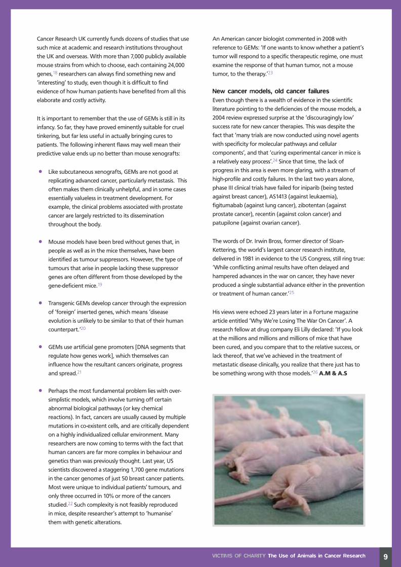
Marmoset monkeys in neurological research
©BUAV
Charities Report:Layout 1 21/6/11 11:01 Page 3

never spreads to other parts of the body – this spreading
(metastasis) being the factor that decreases a patient’s
chances of survival.
A large percentage of the immune-deficient mice die in the
womb or perish soon after birth from conditions that leave
them unable to breathe or feed properly. Those who do
survive face considerable challenges. Some develop
(unplanned for) tumours and degenerative diseases. Others
suffer anxiety – made evident through frenetic plucking of
hair or whiskers from cagemates or from themselves. They
are also susceptible to stress-induced circling, pacing,
jumping or back-flipping.
Destroying the hearts of dogsand pigsFor heart disease research, healthy animals have often
been grievously injured to produce a condition that is
markedly different from those found in human patients.
Dogs have had their hearts systematically destroyed over a
period of months by the injection of polystyrene beads into
their coronary arteries. With pigs, the favoured method is
to place constricting rings around those same arteries.
These narrow gradually over a period of weeks, resulting in
a heart attack. The British Heart Foundation (annual
income £213.7 million; expenditure on research £48 million)
funds highly invasive experiments involving dogs, goats,
pigs and rabbits. More recently, large numbers of fish have
been the victims of their laboratory activities.
Many people will have seen the BHF’s ‘Mending Broken
Hearts‘ advertising campaign, aimed at raising £50 million
for heart failure research. It has featured talking zebrafish
– a luckless minnow whose regenerative powers are
claimed to offer hope for heart disease sufferers. Zebrafish
have already been subjected to years of mutilating
experiments. The BHF plans much more of the same. This
report debunks the ‘science’ behind the BHF hype.
Forced to swim in a water mazeEqually unconvincing are the ‘animal models’ of
Alzheimer’s disease. Neurotoxins have been injected
directly into the brains of rodents and monkeys, while
rabbits have been poisoned with a diet of cholesterol and
copper. The current fad is for genetically manipulated mice,
some of whom are forced to swim around a pool of water
from which they cannot escape or touch the bottom (mice
are scared of being in water). Their task is to find a small
platform on which they can rest. In later tests, the torment
is increased when the platform is submerged.
A recent article in Naturemagazine sums up where such
activities have brought us. ‘…In recent years, and especially
for neurodegenerative disease, mouse model results have
seemed nearly useless.’*
Injecting poison into the brainsof monkeysEven more conspicuously vicious is the history of animal use
for Parkinson’s Disease research. In contrast to the positive
steps achieved as a result of studying human Parkinson’s
sufferers, we show that animal research into PD has failed
to deliver. Researchers, nonetheless, continue to ‘model’
the disease by injecting poison into the brains and
circulation of primates and other animals.
For example, research funded by Parkinson's UK led on
to a 2004 experiment in which 12 monkeys each suffered
18 separate brain injections ‘in the hope of achieving
longer-lasting behavioural deficits’, with needles being
left in their brains for two minutes after instillation of
poison. Recipients of such treatment are likely to be left so
severely disabled that they have to be hand-fed. They will
suffer rigidity, poor coordination and loss of balance.
And highly toxic pesticides have been injected
into the abdomens of mice, in order to kill or severely
incapacitate them.
Valuable workIt is important to make clear that much of the educational
and patient-support work done by the four charities under
review does merit strong public backing. In the case of the
Alzheimer’s Society, more than 70 per cent of its nearly
£60 million budget is devoted to ‘care services’, with ‘just’
£2 million spent on research. Substantially the largest share
of Cancer Research UK’s income, by contrast, goes on
research (at the heart of which is a fixation on the ‘mouse
model’). What all four bodies have in common is a
determination to conceal the nature and extent of the
animal suffering for which they are responsible.
Research relevant to peopleOur objective is to expose what is currently hidden, and
thereby show an unsuspecting public just what their
generosity is paying for. Beyond that, we want to press
the four charities concerned (and others that fund animal
experiments) to reappraise their research agendas. We
wish to see them recognise that their animal research is
as medically unproductive as it is cruel, and that they
should be directing the funds bequeathed to them by
the public into modern, non-animal research methods
(a number of which are outlined in this report) that are
directly relevant to people.
Andrew Tyler, Director Animal Aid
*(Schnabel J (2008). Neuroscience: Standard Model.
Nature. 454:682-685)
2 VICTIMS OF CHARITY Introduction
©ARS
Charities Report:Layout 1 21/6/11 11:02 Page 4

3
©ARS
Charities Report:Layout 1 21/6/11 11:02 Page 5

4
Annual Income: £515 million (2009/10)
Expenditure on Research: £308 million
Expenditure on Information and Advocacy: £14 million
Staff Employed: 3,500
Headquarters:Moving from eight London-based offices to one new site, the
Angel Building in Islington, in autumn 2011
Mission Statement: ‘We are the world’s leading charity dedicated to
beating cancer through research… Our aim is to ensure more people survive
cancer.’ It launched 10 goals in May 2007 to be achieved by 2020, which included
educational goals (for example ‘to make the public aware of the main lifestyle
choices they can make to reduce their risk of getting cancer’).
Original Aims/History: Formed in 2002 as a research initiative, followingthe merger of the Cancer Research Campaign and the Imperial Cancer Research
Fund. Now the biggest single independent funder of cancer research in Europe.
VICTIMS OF CHARITY Vital Statistics: The Story of Four Charities in Figures
CANCER RESEARCH UK
Annual Income: £213.7 million (2009/10)
Expenditure on Research: £48.4 million
Expenditure on Prevention and Care: £37.2 million
Staff employed: 2,000
Headquarters: Head Office in Central London, regionaloffices across the country
Mission Statement: ‘Our mission is to play a leading role in the fight againstdisease of the heart and circulation, so that it is no longer a major cause of
disability and premature death.’ Aims embody both research and education.
Original Aims/History: Founded in 1961 by medical professionalsconcerned about the increasing death rate from cardiovascular disease. Its aim
was to raise money to help fund extra research into causes, diagnosis, treatment
and prevention of heart and circulatory disease. In 1986, it became more
involved in public education. In 1990, it moved into rehabilitation.
VITAL STATISTICS:THE STORY OF FOURCHARITIES IN FIGURES
BRITISH HEART FOUNDATION
Charities Report:Layout 1 21/6/11 11:02 Page 6

5VICTIMS OF CHARITY Vital Statistics: The Story of Four Charities in Figures
Annual Income: £17.1 million (2009)
Expenditure on Research: £4.8 million
Expenditure on Care, Nursing and Service Provision: £10.8 million
Staff Employed: 250
Headquarters: Head Office in Central London, local groups across the country
Mission Statement: ‘Our vision – our ultimate ambition – is to find a cure,and improve life for everyone affected by Parkinson’s.’
Original Aims/History: Founded in 1969 as the Parkinson’s Disease Society,to help patients and their relatives with the problems arising from Parkinson’s, to
collect and disseminate information on Parkinson’s and to encourage and
provide funds for research. Today they focus on research in addition to support,
and want to improve services for people affected by Parkinson’s through
campaigning and education and training for professionals.
Note: Parkinson’s UK has the UK’s largest human brain bank dedicated to the disease.
One of the group’s strategic priorities is to develop new animal models of Parkinson’s
because the current ones ‘don’t recreate the changes that happen in the human brain’.1
Annual Income: £58.7 million (2009/10)
Expenditure on Research: £2 million
Expenditure on Care Services: £42.4 million
Staff Employed: 1,200
Headquarters: Head Office in Central London, services across the country
Mission Statement: ‘We exist to champion the rights of everyone with
dementia and those who care for them.’ One of their goals is to ‘galvanise
investment for research into the causes, prevention, treatment and care of
people with dementia’.
Original Aims/History: Formed in 1979 as the Alzheimer’s Disease Societyby two people who recognised the need to raise awareness of dementia and to
improve the quality of care, support and information for people with dementia
and their carers.
PARKINSON’S UK
ALZHEIMER’S SOCIETY
Charities Report:Layout 1 21/6/11 11:02 Page 7

6
THE USE OFANIMALS IN CANCER RESEARCH
Incidence and mortalityCancer incidence has reached epidemic proportions.
Around 300,000 new cases are diagnosed each year in the
UK, and more than one in three people will develop some
form of the disease during their lifetime. Between 1978
and 2007, incidence rates increased by 25 per cent, with a
14 per cent increase in men and a 32 per cent increase in
women. Cancer is not a single disease. There are more than
200 different types, four of which – breast, lung, large
bowel (colorectal) and prostate – account for more than
half of all new cases. In 2008, there were around 156,000
deaths due to cancer.2
The increase cannot be explained simply in terms of an
ageing population. Not only are rates of juvenile cancer
increasing but, in May 2009, an eight-month old baby boy
became the youngest individual to be diagnosed with
prostate cancer in the UK. In 1960, one hundred children
per million were diagnosed with cancer. By 2005, this figure
had increased to 138 per million.3 Cancer is now the most
common cause of death in children aged 1–14 years.4
It is commonly acknowledged that cancer is largely
preventable. Lifestyle and environmental influences are
responsible for 90-95 per cent of the incidence, while
genetic predisposition accounts for between 5 and 10 per
cent.5 The recognised risk factors include smoking, obesity,
a diet high in saturated animal fats and low in fibre, excess
alcohol consumption, environmental pollution
and over-exposure to sun and radiation.
What is cancer?Cancer is uncontrolled cell growth, beginning at the level of
a single cell. Normal, healthy cells multiply in a controlled
fashion, governed by cellular mechanisms, which are in turn
controlled by proteins, which are encoded by genes. If a
cell becomes stressed or damaged for any reason (e.g.
through exposure to toxic chemicals), it will normally stop
multiplying and try to repair the damage. If the cell is
unable to do so, it will commit suicide (‘apoptosis’) in order
to preserve the integrity of surrounding cells.
Cells have a wide array of mechanisms to protect them
from the effects of stress and DNA damage. But these
can be overcome by, for instance, a highly toxic chemical
VICTIMS OF CHARITY The Use of Animals in Cancer Research
Note on Prevalence and IncidencePrevalence measures how much of a given disease or condition there is in a population at a particular point in time.
Incidence measures the rate of occurrence of new cases of a disease or condition. Simply stated, prevalence is how many
people have the condition at any given time and incidence is new cases in a given time (usually a year) in a given population.
Charities Report:Layout 1 21/6/11 11:02 Page 8
or a cancer-causing virus. Equally, excessive hormonal
stimulation (caused, for instance, by hormone replacement
therapy or hormone-mimicking chemicals such as
pesticides) can lead to uncontrolled cell multiplication –
breast and prostate cancer being the most common
examples of this.
History of cancer researchThe father of 20th-century cancer research is Sir Richard
Doll, whose pioneering work firmly established a link
between smoking and lung cancer, based on
epidemiological (human population) studies. He also did
pioneering work on the relationship between radiation
and leukaemia, as well as that between asbestos and lung
cancer,6 and alcohol and breast cancer.7
Despite his success, attention since the 1990s has turned
increasingly from epidemiology towards the molecular
approach to cancer, using biotechnology. Much of this
research is currently being conducted using animals – most
frequently, mice.
Animal models in cancer researchand their historical failureFrom ‘nude’ to ‘SCID’ to transgenic miceAnimal researchers have struggled for decades to mimic
human cancer in mice. They have failed for a variety of
reasons. Their biggest initial obstacle was that when
human cancer cells were transplanted or injected into
mice they were rejected by the mouse’s immune system.
In an attempt to overcome this problem, an
immunodeficient mouse was developed – known, because
they were without fur – as the nude mouse. Bred
specifically to lack a gene (FOX1) that is critical for the
proper development of the thymus, researchers could
engraft human cancer cells into the nude mouse that
would not be rejected.8 However, because some important
immunity function remained, not all cancers grew well.9
And so a new type of mouse was bred – known as SCID
(severe combined immunodeficiency) – that was, as the
name implies, even more immune-deficient than the nude
variety. The SCID mouse soon became a favourite of
pharmaceutical companies. Cancer researchers could take
an established human cancer cell line and insert it under
the skin of the SCID mouse – producing what is known as a
subcutaneous xenograft – then test the mouse’s response
to an experimental cancer drug.10
Over the years, researchers continued to genetically
manipulate the SCID mouse, knocking out more genes to
further disable its immune defences. But then came an
important realisation: eliminating more and more of the
mouse’s immune system might let an experimenter
introduce foreign cancer tissue and see it ‘successfully’
grow but that is not how cancers work in people. Most
human cancer sufferers have a functioning immune
system, which interacts with the cancer throughout
its development, changing the course and outcome of
the disease.
The multiple failings of the mouse model... by ascientific expertIn fact, there were several ways in which the SCID and
nude mice fell short of providing a solution to the problem
of ‘modelling’ human cancer. An article in a leading
cancer journal summed up the multiple problems: ‘The
subcutaneous xenograft is clearly better than nothing,
but its drawbacks are well known. The mouse has no
functioning immune system; something rarely seen in
human cancer, and the tumor is growing in an artificial site.
Xenograft tumors almost never metastasize [spread to
other parts of the body and thereby decrease the patient’s
chances of survival]... Finally, the tumor does not develop
naturally in the mouse. Instead, it is transplanted from the
7VICTIMS OF CHARITY The Use of Animals in Cancer Research
Stem cell
Charities Report:Layout 1 21/6/11 11:02 Page 9

8
cell line of a fully-grown human tumor – another
divergence from the human situation. When you consider
that drugs behave differently in mice than in humans, it's
not surprising that the subcutaneous xenograft is a poor
predictor of success. In general, it's much easier for a drug
to shrink a tumor in such mice than in humans.’11
There is now acceptance within the cancer research
community that the mouse xenograft model performs very
poorly when it comes to developing useful therapies. The
US National Cancer Institute conducted a retrospective
analysis for 39 drugs in 2000. It compared their
performance in xenograft testing and Phase II human
clinical trials. Only 45 per cent of compounds with anti-
tumour effects in xenografts showed benefit in human
trials. In addition, drugs that worked in a particular fashion
in tumour cells that had been transplanted into mice could
not be relied upon to work in the same way in human
patients with the same type of tumour. 12
A 2003 study compared the value of three cancer models in
predicting drug effects in humans. The models were:
mouse xenografts of human cells, mouse cell tumours
grafted into mice, and human in vitro cell lines. The
researchers concluded (rather timidly) that the human cell
model was ‘of at least equivalent usefulness to mouse
xenografts’.13 In fact, it is clear from the report that it was
more predictive for a greater variety of malignancies.
Further, the study makes clear that the mouse-cell tumour
models were useless.
Two US researchers in 2006 offered further explanations of
why the mouse xenograft model bears such a
‘questionable relation to the naturally occurring human
disease’.14 They pointed out that the living matrix with
which implanted tumours interact is fundamentally
different in mice than in people. There are also ‘intrinsic
differences between mouse and human toxicity features’,
which in practice mean that doses of a candidate drug in
people cannot be increased to levels tolerated in mice.
Despite these fundamental drawbacks, subcutaneous
xenograft studies still provide the most common ‘proof-of-
concept’ data for new cancer therapies submitted to the
Food and Drug Administration, the world’s most important
drug authorisation body. And most drugs released to date
were ‘likely originally tested on SCID mice’.15
CRUK’s use of mouse models – inherentlycontradictoryCancer Research UK is clearly aware of the shortcomings of
mouse xenografts. A promotional poster16 for genetically
modified mice, produced by its Cambridge Research
Institute in 2007, had this to say: ‘Although initially useful,
xenograft models of human cancer do little to replicate the
real disease and are essentially an in vivo Petri dish… It is
therefore not surprising that xenografts have an altered
response to chemotherapeutic drugs. The time for reliance
on such models to determine the response to a new
therapy has passed.’
And yet CRUK researchers at the same institute are
committed to the continued use of xenografts for ‘the
tumours of major interest’.17 Bafflingly, these researchers
are working on experimental therapies that they hope can
move from the laboratory to use in human patients. The
contradiction between word and deed is obvious.
Failings of the genetically engineeredmouse modelsMeanwhile, the long-suffering mouse continues to be
subjected to all manner of genetic experiments aimed at
producing a reliable surrogate for human cancer – a quest
that remains as elusive as ever. Rather than having cancer
cells engrafted, these mice are designed to develop cancers
spontaneously. Genes are deleted (creating ‘knockout’
models) or human genes are added (producing ‘transgenic’
strains). According to official Home Office statistics, more
than one-and-a-half million genetically engineered mice
(GEMs), including those suffering ‘harmful mutations’,
were bred and killed in 2009. The majority of these mice were
used in cancer research, immunology and genetics.
VICTIMS OF CHARITY The Use of Animals in Cancer Research
An illustration of breast cancer
Charities Report:Layout 1 21/6/11 11:02 Page 10
Cancer Research UK currently funds dozens of studies that use
such mice at academic and research institutions throughout
the UK and overseas. With more than 7,000 publicly available
mouse strains fromwhich to choose, each containing 24,000
genes,18 researchers can always find something new and
‘interesting’ to study, even though it is difficult to find
evidence of how human patients have benefited from all this
elaborate and costly activity.
It is important to remember that the use of GEMs is still in its
infancy. So far, they have proved eminently suitable for cruel
tinkering, but far less useful in actually bringing cures to
patients. The following inherent flaws may well mean their
predictive value ends up no better than mouse xenografts:
• Like subcutaneous xenografts, GEMs are not good at
replicating advanced cancer, particularly metastasis. This
often makes them clinically unhelpful, and in some cases
essentially valueless in treatment development. For
example, the clinical problems associated with prostate
cancer are largely restricted to its dissemination
throughout the body.
• Mouse models have been bred without genes that, in
people as well as in the mice themselves, have been
identified as tumour suppressors. However, the type of
tumours that arise in people lacking these suppressor
genes are often different from those developed by the
gene-deficient mice.19
• Transgenic GEMs develop cancer through the expression
of ‘foreign’ inserted genes, which means ‘disease
evolution is unlikely to be similar to that of their human
counterpart.‘20
• GEMs use artificial gene promoters [DNA segments that
regulate how genes work], which themselves can
influence how the resultant cancers originate, progress
and spread.21
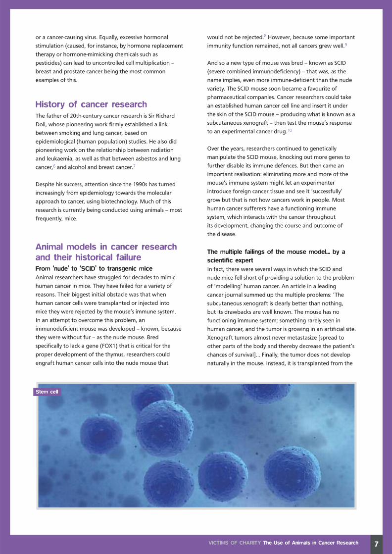
• Perhaps the most fundamental problem lies with over-
simplistic models, which involve turning off certain
abnormal biological pathways (or key chemical
reactions). In fact, cancers are usually caused by multiple
mutations in co-existent cells, and are critically dependent
on a highly individualized cellular environment. Many
researchers are now coming to terms with the fact that
human cancers are far more complex in behaviour and
genetics than was previously thought. Last year, US
scientists discovered a staggering 1,700 gene mutations
in the cancer genomes of just 50 breast cancer patients.
Most were unique to individual patients' tumours, and
only three occurred in 10% or more of the cancers
studied.22 Such complexity is not feasibly reproduced
in mice, despite researcher’s attempt to ‘humanise’
themwith genetic alterations.
An American cancer biologist commented in 2008 with
reference to GEMs: ‘If one wants to knowwhether a patient’s
tumor will respond to a specific therapeutic regime, one must
examine the response of that human tumor, not a mouse
tumor, to the therapy.’23
New cancer models, old cancer failuresEven though there is a wealth of evidence in the scientific
literature pointing to the deficiencies of the mouse models, a
2004 review expressed surprise at the ‘discouragingly low’
success rate for new cancer therapies. This was despite the
fact that ‘many trials are now conducted using novel agents
with specificity for molecular pathways and cellular
components’, and that ‘curing experimental cancer in mice is
a relatively easy process’.24 Since that time, the lack of
progress in this area is even more glaring, with a stream of
high-profile and costly failures. In the last two years alone,
phase III clinical trials have failed for iniparib (being tested
against breast cancer), AS1413 (against leukaemia),
figitumabab (against lung cancer), zibotentan (against
prostate cancer), recentin (against colon cancer) and
patupilone (against ovarian cancer).
The words of Dr. Irwin Bross, former director of Sloan-
Kettering, the world’s largest cancer research institute,
delivered in 1981 in evidence to the US Congress, still ring true:
‘While conflicting animal results have often delayed and
hampered advances in the war on cancer, they have never
produced a single substantial advance either in the prevention
or treatment of human cancer.’25
His views were echoed 23 years later in a Fortune magazine
article entitled ‘WhyWe’re Losing TheWar On Cancer’. A
research fellow at drug company Eli Lilly declared: ‘If you look
at the millions and millions and millions of mice that have
been cured, and you compare that to the relative success, or
lack thereof, that we’ve achieved in the treatment of
metastatic disease clinically, you realize that there just has to
be something wrong with those models.’26 A.M & A.S
9VICTIMS OF CHARITY The Use of Animals in Cancer Research
Charities Report:Layout 1 21/6/11 11:02 Page 11

A daunting list of stresses and hazards
Some techniques for producing genetically alteredmice involve genetic manipulation of DNA, using avirus as a vehicle to insert a gene. With othermethods, programmed stem cells obtained fromembryos or from skin cells are used. Alternativetechniques rely on the effects of toxic chemicalsthat are injected directly into the abdominal cavitiesof young mice.
Such chemicals include N-ethyl-N-nitrosourea (ENU).Where ENU impacts on non-target genes, seriousmalformations can result. They include cleft palate,which can leave the newborn pups in a desperatecondition, unable either to feed or breathe properly.27
But with all the above-described methods of geneticalteration, the chances of achieving the desiredoutcome are in the range of just 1-2 per cent. Thismeans that the vast majority of progeny die either asembryos or shortly after birth. The mice who dosurvive face a daunting variety of stresses andhazards, according to a report published by a keygovernment-appointed laboratory welfareorganisation.28
Immune-deficient mice, such as the SCID and nudestrains, will have a susceptibility to infection. Somedevelop [unplanned for] tumours, degenerativediseases or other dysfunctions. Genetic alterationscan also cause increased anxiety, frustration andheightened aggression. In addition, genetically alteredmice may be prone to frenetically plucking hair orwhiskers from cagemates or from themselves.They are also susceptible to ‘stereotypies’ –stress-induced repetitive movements, such ascircling, pacing, jumping or back-flipping.
VICTIMS OF CHARITY Animal Suffering in Cancer Research10
An example of an animal experimentfunded by Cancer Research UK
As to the experiments themselves, typical is a 2009project funded by Cancer Research UK, in which nudemice were injected with human cancer cells and thenforce-fed, via a tube from the mouth to the stomach,an experimental anti-cancer drug.29 There were dailyforce-feedings over a ten-day period. This was inaddition to painful daily injections, via the tail vein,of a radiotracer chemical to study the cancer’sdevelopment. At the end of the ten-day trial, themice were killed and their organs studied. A.M
ANIMAL SUFFERING IN
CANCER RESEARCH
Charities Report:Layout 1 21/6/11 11:03 Page 12

11
Charities Report:Layout 1 21/6/11 11:03 Page 13

12
Incidence and mortalityCoronary heart disease (CHD), which is characterised by a
narrowing of the coronary arteries due to a build-up of fatty
deposits, is the most common cause of death in the UK,
killing 80,000 people in 2009.30 It accounted for
approximately one in six male deaths and one in eight
female deaths in that year. Other forms of heart disease
affect many thousands, but cause significantly fewer deaths.
Death rates from CHD have been falling in the UK since the
late 1970s. For people under 75, they fell by 75 per cent
between 1985 and 2009. The majority of the fall between
1981 and 2000 has been attributed to reductions in major
risk factors, principally smoking.31
These figures, however, mask a disturbing recent trend.
The fall in death rates has been slowest in younger age
groups (35-44 years), especially among women.32
VICTIMS OF CHARITY The Use of Animals in Heart Disease Research
THE USE OF ANIMALSIN HEART DISEASE RESEARCH
Blood pressure check
Charities Report:Layout 1 21/6/11 11:03 Page 14

The epidemiologists who uncovered this trend concluded in
2009 that it could be ‘the first warning sign of worsening
lifestyle choices and behaviours rather than deterioration
of medical management of coronary heart disease’.
The prevalence of CHD is extremely high. The most recent
data from the British Heart Foundation (BHF) shows that
around 3.4 million adults in the UK report angina and/or a
heart attack.
Heart failure – in essence, the failure of the heart to pump
properly – is now at epidemic proportions in the UK, and
the prognosis remains dismal. Data from the London Heart
Failure Study show that around 40 per cent of people die
within one year of an initial diagnosis of heart failure,
which is worse than expected survival rates for breast,
prostate and bladder cancers.33 Prevalence and incidence
(see page 6) are both increasing, and do so steeply with
age. Around 750,000 people lived with the disease in 2010,
compared with just 100,000 in 1961.34
The commonest cause of heart failure is damage due to
CHD. More patients are surviving the acute phase of a
heart attack, and so the decreased mortality from CHD
parallels the increasing prevalence of heart failure.
History of heart disease researchThe BHF claims that ‘without animal research, many of
today’s life-saving treatments for heart and circulatory
disease could not have been developed’. This categorical
assertion is impossible to prove or to disprove
retrospectively. It is certainly the case that treatments in
use today have employed surgical experimentation or drug
trials on animals. Whether the use of animals was essential,
however, is mere speculation. We cannot know whether
the use of non-animal techniques instead may have
brought benefits of equal or greater medical value. Nor is
it known how many potentially useful treatments have
been lost due to misleading animal data.
Undoubtedly, many surgical techniques developed during
the last century involved animal experimentation during
their development. But it is striking how often the first
human trials led to a dramatic acceleration in progress,
in a way that cannot simply be ascribed to technological
improvements.
The history of heart transplants provides a telling
example.35 Alexis Carrel first experimented with
transplanting dogs’ kidneys into their necks in the 1890s. In
1955, Demikhov transplanted the hearts removed from 22
dogs into the chests of others, and none lived longer than
15 hours. Many experimenters performed dog heart
transplants during this decade, and survival rates were
universally poor. A group of US researchers concluded, by
way of explanation, that ‘there is a specific adverse effect
of severing the heart from the body’.36 In 1964, a team
from Mississippi transplanted a chimpanzee’s heart into a
human recipient – the patient died shortly afterwards, as
the ‘donor’s’ heart was too small.
In 1967, Professor Christiaan Barnard performed the first
human heart transplant, with ten more by Denton Cooley
and associates during the following year. In response,
transplant programmes developed ‘seemingly overnight’.37
By 1974, Shumway’s Stanford team had performed 59
human heart transplants, with a three-year survival rate of
26 per cent. It was careful clinical studies and follow-up of
their patients that was crucial to this progress, something
not possible with short-term animal procedures.
A similar time line can be mapped out for coronary artery
bypass grafting: the first animal procedures took place in
1910, but it needed human success in 1966 before rapid
progress was made.38 And the same pattern can be seen
with regard to interventional cardiology: biventricular
catheterisation of a live horse was first performed in 1711,
but the technique only became successful in humans after
Forssmann guided a catheter into his own right atrium
in 1929.39
There are also instructive historical examples in which
researchers have discovered unacceptable side effects of
new treatments in animal subjects, but forged ahead
regardless, with subsequent human benefit. The first Starr-
Edwards heart valves, when transplanted into dogs, were
plagued by fatal thrombus (blood clot) formation, and the
necessary post-operative anticoagulation caused many
dogs to bleed to death. Modifications to the design
improved canine survival figures – but it was the original,
simpler design that was chosen for placement in people.
The researchers knew that humans were much less likely to
develop thrombi than dogs; one commented: ‘humans will
tolerate this surgery much better than dogs... dogs, for
some reason, don't like to have their blood bubbled
through a pump oxygenator’.40
13VICTIMS OF CHARITY The Use of Animals in Heart Disease Research
Obesity is a risk factor for heart disease
Charities Report:Layout 1 21/6/11 11:03 Page 15

14
Animal models in heart diseaseresearch and their historical failureCrude, cruel and irrelevantThe use of animal ‘models’ – dogs, pigs and rodents are
commonly used – to mimic cardiovascular disease has
always been striking for its crudity and cruelty. For research
into heart attacks and heart failure, healthy animals are
usually grievously injured to produce a disease that is
markedly different from those found in human patients.
Notwithstanding, experimenters have devised many ways
to destroy the circulatory systems of animals in
laboratories:
• Dogs have undergone appalling procedures in the
quest to damage their hearts. They are naturally
resistant to heart attacks, having a rich collateral
coronary circulation, and cannot be induced to
develop heart disease with an artificial fatty diet.
Instead, their hearts are systematically and gradually
destroyed over a period of months by injecting
polystyrene beads into their coronary arteries.41 The
mortality rate after such treatment can approach 30
per cent.42 Tying off the coronary arteries of dogs is
also common, although half of the victims die acutely –
not by design – of malignant ventricular tachycardias.
A leading US veterinarian, Dr. Holly Cheever, observes:
‘The kind of heart disease seen in humans has no
correlation with canine heart problems. Therefore, to
attempt to create artificially human heart disease,
our number one killer, in canines is inappropriate,
ineffective, and diverts funds from the more rational
approach, which is prevention.’43
• As pigs do not possess such an extensive cardiac blood
supply as humans, a favoured method of damaging
their hearts is the placement of constricting rings
(ameroids) around the coronary arteries, which
narrow gradually over a period of weeks resulting in
a heart attack.44
• Millions of rodents have been victims of crude surgical
mutilations to induce heart attacks and resultant heart
failure. ‘Aortic banding’, in which a stricture is placed
around the ascending aorta of weanling rats, is widely
employed. The stricture gradually blocks the blood
flow out of the heart as the rats grow and, by 18
weeks of age, they are breathless and swollen with
fluid collecting in their lungs and abdominal cavity.45
Mice are increasingly used to model heart attacks (MI,
myocardial infarction) by tying off their coronary
arteries, with up to half the subjects dying within
the hour.46
VICTIMS OF CHARITY The Use of Animals in Heart Disease Research
Charities Report:Layout 1 21/6/11 11:03 Page 16

• Other methods of injury include freezing the hearts of
animals with liquid nitrogen, poisoning them with
known cardiotoxins, or electrically forcing their hearts
to beat so fast that they fail.47
The mouse has clearly emerged in the last decade as the
most favoured laboratory species for cardiovascular
experiments. This is largely due to researchers developing
transgenic varieties programmed to be born with or to
develop diseases. These lines include mice liable to die
spontaneously due to rupture of their major vessels, or
who will develop dilated and dysfunctional heart muscle.
The shortcomings of the ‘animal model’ asacknowledged by the research communityWhether surgically or genetically created, the research
community readily admits that these models do not
accurately reproduce human pathology. The animals used
are unlike humans in their basic physiology and anatomy.
Rodents, for example, have a resting heart rate five times
higher than humans, with different electrical impulses
and muscle composition.48
In addition, the damage ‘induced’ in healthy animals is
fundamentally different from the diseases found in
humans. A 2010 review from the National Institute for
Medical Research (NIMR) noted the obvious: ‘[in the
animals] heart failure occurs suddenly post-surgery in the
context of a relatively young heart, whereas in humans, the
onset may be insidious over several years in the context of
comorbidities and age-related changes… The major disease
burden of heart failure in the future is expected to come
from patients with the complex phenotype cluster of
hypertension/hyperlipidaemia/obesity/diabetes… it is not
obvious how closely [it] resembles the current animal
models’.49
Researchers have, however, always justified their use of
animals by claiming that it has led to novel observations
that can then be explored in humans. In many instances,
this claim is spurious as data from the animal experiments
only confirm what is already known to occur in patients.
One paper from 2009 credits a rat myocardial infarction
model with ‘ground-breaking’ significance in the use of
ACE inhibitors.50 A quote from the original paper reveals
otherwise: ‘In the present study, the chronic administration
of captopril [an ACE inhibitor drug] to rats with
myocardial infarction and failure yielded hemodynamic
results similar to those noted above in patients with
congestive heart failure.’51
Neither can it be said that the models are reliably predictive
of human outcomes. The same rat model suggested that
endothelin receptor antagonists would give similar positive
results to captopril but, in fact, patients with heart failure
got worse.52 Mice engineered to overproduce a chemical
suspected to worsen heart failure (TNF-alpha)
unsurprisingly improved when the receptors to this
chemical were blocked. However, a human drug trial using
the same substance failed, leading researchers to caution
that ‘positive results in preclinical rodent studies do not
necessarily translate to clinical benefits when applied to
non-uniform heart failure populations’.53 Such examples of
non-correlation are the rule rather than the exception.
During the last 30 years, hundreds more heart failure drugs
have been developed using animal models, with very few
making it to clinical trials on patients. A particularly
wasteful 20-year obsession has been the search for
antioxidants that could slow cardiovascular damage by
neutralising free radicals, presumed to be toxic. Despite
many studies (often involving rabbits being poisoned with
cholesterol), which showed ‘proof of principle of the
efficacy of antioxidants in animal models of atherogenesis,
atherosclerosis regression, and reperfusion injury’,
randomised trials in humans have been ‘disappointing’.54
Shockingly, BHF researchers have now announced that a
new transgene mouse model shows why this is the case –
free radicals can be cardioprotective.55 This casts enormous
doubt on the validity of the previous models, or suggests
that the animals were manipulated to produce desired but
erroneous conclusions. It is likely that a slew of animal
experiments will now take place to ‘validate’ the new
preferred hypothesis.
Curiosity-driven experimentsExperimental cardiothoracic surgery on animals continues
today, and a good deal of it is funded by the BHF. Even a
cursory probe into the scientific literature reveals that the
charity has funded thousands of terminal experiments in
the name of ‘basic’ research. This Home Office category
refers to speculative, ‘blue skies’ procedures that may or
may not lead to medical advances in the future. One of the
most damning, though by no means isolated, examples of
BHF-supported research is the long-running series of dog
15VICTIMS OF CHARITY The Use of Animals in Heart Disease Research
Charities Report:Layout 1 21/6/11 11:04 Page 17

16
experiments carried out at Leeds Medical School. The
repetitive and self-justifying programme has been roundly
condemned by cardiologist and former dog researcher
John Pippin.56
Some current trends in heartresearchRegenerative medicine – the new Holy Grail ofcardiologyWith the epidemic of heart failure showing no signs of
abating, the last decade has seen an explosion of interest
in ‘regenerative’ cardiac strategies – in essence, helping the
heart to repair itself with functional tissue rather than
scarring. Despite experimenters’ best efforts, ‘significant
cardiac regeneration of any form has not been reported in
mammals after multiple modes of injury, including
ischaemic infarction, burning, freezing, mechanical injury,
chemical injury, etc’.57 This regenerative ability – if it ever
did exist – has disappeared over millions of years,
suggesting that its loss conferred a survival advantage.
Nonetheless, researchers have been trying to challenge
evolution with stem cells and genetic manipulation, so far
with little success.
a) Stem cellsThese are immature cells with the ability to
evolve into various kinds of specialised
tissues. Pluripotent stem cells, which have
the potential to develop into many cell
types, can be found in human embryos or
can be induced in the laboratory, starting,
for instance, with skin cells. Adult tissues
such as bone marrow harbour a lesser
number of differentiated lines, whilst a still
more limited variety (known as progenitor
cells) is found in highly specialised organs
such as the heart. Stem cell research and
trials have involved transplanting cells into
animal or human recipients. There is also
ongoing work exploring gene-based
strategies, with the aim of inducing
indigenous tissues to re-acquire a degree of
‘stemness’.
Stem cell therapies for heart disease are
highly controversial, with numerous unique
methodological and clinical problems.
However, it comes as no surprise to discover
that stem cell trials on animals generated a
mass of ‘positive’ data that was not
replicated in human trials. Heart-damaged
rabbits, for example, showed improvements
when injected with bone marrow or muscle
stem cells. Human trials with these cells have
been universally disappointing. Irrelevant
experiments have also been conducted on
rabbits using fibroblasts.58 These are not even stem cells and
are not able to change into specialised cardiac tissue. Mainly
for this reason, this is not an approach that has been
pursued in humans.
Skeletal myoblasts (SMs) are muscle-derived stem cells. Trials
directly injecting SMs into patients’ hearts during bypass
surgery were curtailed when subjects experienced life-
threatening arrhythmias (irregular heart rhythms), although
‘previous extensive animal experiments provided no hint of
an arrhythmogenic risk’.59 A later trial in 2007, in which all
patients had defibrillators implanted along with the SMs,
was also a failure. The lead researcher has commented:
‘Once again in medicine, clinical outcomes have not
matched the hopes raised by the animal data’60 and he
called the animal models ‘suboptimal’.61 Since then, there
have been numerous further trial failures of stem cells, using
both SMs and bone marrow cells to treat heart attacks,
heart failure and chronic angina.62
Guidelines issued in 2008 for stem cell trials sensibly
recommend that ‘participants should appreciate that
researchers may not know whether or not the stem cell
treatment will be beneficial, that animal studies might not
VICTIMS OF CHARITY The Use of Animals in Heart Disease Research
Heart attack
Charities Report:Layout 1 21/6/11 11:04 Page 18

predict effects of the cells in humans, and that unexpected
adverse events may occur’.63
Leading researchers in this field are now stressing that
human trials, not further animal experiments, will be key
to progress. A European Society of Cardiology task force
made the following recommendation in 2005: ‘No matter
what animal experiments are undertaken, the mechanisms
that may be deduced from them may not be the actual
mechanism pertaining to benefit in the human clinical
situation… We believe that sufficient animal experiments
have been performed in this area to allow clinical studies
to continue’.64
b) Gene therapiesThese therapies usually employ viruses to deliver DNA that
codes for desired proteins into target cells. Administration
of these vectors in heart research is highly invasive,
commonly via the coronary arteries.
Fibroblast growth factor – FGF – helps blood vessels
to develop in humans. When FGF gene viruses
were injected into the hearts of ameroid-
constricted pigs, they showed evidence of
improved cardiac blood flow. However, a
Phase 3 clinical trial in humans with angina
was forced to stop recruiting in 2004 due to
lack of efficacy.65
Dr Paul Williams, a BHF Clinical Researcher,
commented in 2010: ‘… despite a huge amount
of basic science research, promising animal
studies, and numerous clinical trials, to date no
gene therapy has demonstrated unequivocal
benefit in the clinical setting… is all the hype
and research expenditure unwarranted?’66
Part of the reason for this dismal performance
is the use of laboratory-based physiological
outcomes that are derived from animal trials.
This data can be carefully selected to
demonstrate benefits that do not translate
to real-world patient improvements – all too
frequently revealed by a Phase 3 clinical trial.
The NIMR review referred to above states:
‘Many of the clinical end points that are
important to doctors, patients and health
care systems alike, such as quality of life,
exercise tolerance and hospital admission,
are unlikely to be modelled adequately in
any animal system.’67
Obesity researchDespite widespread knowledge and acceptance
of the causes of human obesity, the BHF is
currently funding highly questionable research
that deliberately makes animals ill via dietary modification. In
a repellent 2008 series of experiments, researchers fed female
rats an ‘obesogenic’ unnatural diet, and allowed them to
mate and give birth. After the females had weaned their
pups, they were fasted overnight and ‘sacrificed’, and their
offspring’s eating habits studied. Litters of other obese
females were decapitated at varying intervals after birth so
that their bodies could be dissected. There is no mention of
medical relevance at any point in the experimental write-up.68
The BHF’s ‘Mending Broken Hearts’appealThe therapies described above are key planks of the BHF’s
current programme for heart failure, upon which the
charity ‘needs to spend’ £50 million. To illustrate the
underpinning science, the BHF describes four research
examples – all involve stem cells, and two involve their
testing on animal models. For example, Professor Andy
Baker will use his grant to discover ‘how much the stem
cell treatment can improve the heart’s ability to pump in
17VICTIMS OF CHARITY The Use of Animals in Heart Disease Research
The BHF.s ‘Mending Broken Hearts’ campaign
Charities Report:Layout 1 21/6/11 11:04 Page 19

18
mice’.69 Another project will ‘use the cells to promote the
growth of new heart cells and blood vessels in mice’. Given
the above, it is surely legitimate to question the relevance
of these experiments to medical progress.
Mutilating zebrafishThe advertising motif of the campaign is a talking
zebrafish, said to represent hope for heart failure
sufferers. These small minnows have a remarkable
regenerative ability – researchers have amputated many
different parts of their bodies, and they are able to grow
them back. This ability is not newly discovered, and
zebrafish have been studied for many years. They are
increasingly popular as ‘models’ because they are cheaper
than mammals, reproduce quickly in large numbers, are
transparent when young, and have had their genome
sequenced.
In a series of mutilating experiments over the last decade,
anaesthetised zebrafish have had their scales pulled off with
forceps and portions of their heart chopped out with
scissors. The fish were returned to water after the
procedure. Unsurprisingly, they ‘appeared less active and
less co-ordinated while swimming’ before recovering over a
few days.70 They were later killed and their hearts removed
for study. Fish are capable of feeling pain and possess, in
addition to a central nervous system, pain receptors all over
their bodies – it is hardly surprising that they did not look
well after such brutal surgery.
Last year, experimenters performed similar partial heart
amputations on one-day-old mice and removed their hearts
three weeks later. They found that the organs had
regenerated without scarring. The surgery was also
performed on seven-day-old mice but their hearts did not
heal, suggesting that the regenerative ability was lost by
this age.71
The BHF claims that these experiments could help develop
treatments for human heart failure – an assertion that
merits a rigorous and sceptical examination:
• There are a great many fundamental bio-evolutionary
differences between zebrafish and humans.
Importantly, the former have two-chambered hearts
(compared with the four-chambered human organ),
with different cardiac muscle, and can grow
throughout most of their adult lives. Kenneth Poss, a
leading zebrafish researcher, observes: ‘It would appear
that the teleost heart is better designed for growth and
regeneration, while the mammalian heart is better
designed for sheer contractile force’.72
• Cardiac progenitor cells are present in mammalian
hearts, and it was thought until recently that zebrafish
used these stem cells to regenerate cardiac tissue.
However, zebrafish repair their hearts via a different
mechanism (dedifferentiation),73 which has no
functional analogue in human hearts. After this
discovery, researchers stated lamely: ‘If we could mimic
in mammalian cells what happens in zebrafish, perhaps
we could be in a position to understand why
regeneration does not occur in humans.’ This hardly
suggests curative potential.
• It is clear that the American researchers do not know
how neonatal mice regenerate their hearts, or how this
could lead to human heart failure treatments: ‘…we
can begin to search for drugs or genes or other things
that might reawaken this potential in the adult heart
of mice and eventually of humans.’74
• The BHF’s campaign explicitly references stem cell
treatments as candidates for early clinical trials within
five years. The implication is that these treatments are
novel. As described above, stem cell treatments for
heart failure have already failed in clinical trials
following success in animal models.
• In humans, coronary artery disease is the most
common cause of heart failure. It damages heart
muscle both acutely and chronically via a lack of oxygen
and nutrients. Heart attacks lead to large fibrous scars
in an already diseased organ. Heart failure is associated
with a complex series of long-term physiological
derangements. All these elements are absent in animal
models in which the hearts of mice and fish are
surgically damaged.
In conclusion, there is no evidence whatsoever that these
heart amputation studies will ever translate into clinical
benefit for humans. Last year, a group of leading
cardiologists using stem cells advised that ‘additional
safeguards are warranted because of the innovative nature
of these treatments, differences between animal and
human physiology, limited experience with these cells in
humans, and the high hopes of desperate patients for
whom no alternative effective treatment currently exists’.75
Unfortunately, no such caution is detectable in the BHF’s
current fundraising drive. A.S
VICTIMS OF CHARITY The Use of Animals in Heart Disease Research
©PET
A
Charities Report:Layout 1 21/6/11 11:04 Page 20

19
ANIMAL SUFFERING IN HEART
DISEASE RESEARCH
‘The experiments involved opening the chests of anaesthetised dogs, cutting their spinal cords,draining and re-circulating their blood and cutting nerves to the brain, gut and diaphragm...’
The British Heart Foundation funds invasive animal research, involving dogs, pigs, rabbits, goats, rats and mice. Someresearchers prefer to use dogs and pigs rather than rats because their heart size is comparable to that of humans.However, heart size is not necessarily a useful parameter when trying to compare human and animal heart function.Other factors play a major role, such as differences in blood clotting mechanisms, and the fact that, in quadrupeds, 70per cent of the blood volume is at, or above, the level of the heart, whereas in humans 70 per cent is below the levelof the heart. In an experiment using seven healthy dogs, the animals underwent two procedures.76 In the first,all of the dogs were anaesthetised and their chests surgically opened. The heart sac (pericardium) was cut open toallow the researchers to inject a damaging nerve toxin (phenol) into one of the main blood vessels supplying the heart,which also damaged its associated nerve supply. The heart sac and chest were then closed with sutures andthe dogs allowed to recover. No mention is made by the researchers of the clinical condition of the dogs during thisrecovery period. Three to four weeks later, the dogs were once again anaesthetised, the heart exposed and afluorescent dye injected into it to record blood flow, while various measuring instruments were applied to the heart.Once the experiment was completed, researchers killed the anaesthetised dogs by cutting out their beating hearts.
27 experiments involving 100 dogs
In 2005, the BHF ran into serious controversy over aseries of 27 experiments that it had funded usingmore than 100 dogs. The experiments involved openingthe chests of anaesthetised dogs, cutting their spinalcords, draining and re-circulating their blood andcutting nerves to the brain, gut and diaphragm.77
Former Harvard Medical School faculty member andheart specialist John Pippin MD FACC, who examinedthe research team’s published papers, was scathing.He wrote: ‘Very evident in this collection of papers isthe characteristic use of one study to justify the next.In many cases, unanswered (usually unforeseen)questions arising from one study produced therationale for a later study. In several instances, theteam invokes conflicting or erroneous results fromprevious studies (sometimes their own) to justifyanother study.’78 He continued: ‘This work provides anexceptional example of a common practice: themanipulation of animal models for convenience andusefulness, regardless of the effects upon the validityof results obtained. This is not uncommon amongthose researchers who propose and perform studiesto satisfy their scientific curiosity and sustain theircareers, without sufficient regard for potentialapplications to humans.’79
Given that the BHF continues to fund similar dogexperiments, it would seem that it has ignored DrPippin’s findings. Whilst the dog is a well establishedmodel in heart research, those involved are becomingincreasingly uncomfortable as public awareness oftheir activities grows. This could help to explain whythe following study was conducted on goats, ratherthan dogs – even though goats, of course, have thesame capacity to suffer pain and stress.
©PET
A
Charities Report:Layout 1 21/6/11 11:05 Page 21

Ten healthy adult female goats were anaesthetised andtheir blood pressure was measured by an instrumentinserted into an artery in one of their limbs for theduration of the procedure.80 From the following day,the goats received a heart drug by mouth at differentdoses for seven days. They were also brieflyanaesthetised every day in order for blood pressuremeasurements to be made while they received aninjection of a naturally-occurring chemical that affectsblood pressure. Repeated anaesthesia is a stressfulprocedure for any animal, especially during the recoveryphase, in addition to being physiologically demandingon the liver, which is responsible for metabolisingthe anaesthetic.
A follow-up experiment involved 28 adult goats.81 Allwere anaesthetised, while a pacemaker device wasimplanted via the external jugular vein. The goats wereallowed to recover before undergoing the next stage ofthe experiment, in which the pacemaker was switchedon for three continuous 28-day periods, separated by24-hour rest periods, with the aim of upsetting thenatural electrical activity of the heart. Blood sampleswere taken every few hours for the first week, thenonce a week until the end of each 28-day period –a series of interventions that would have caused theanimals significant pain and distress. At the end ofthe third 28-day period, several of the goats wereanaesthetised for a last time, their chests openedand their hearts examined before being killed. It isunclear from the article what the fate was of theremaining goats.
The researchers concluded that important heart eventsseen in human patients were absent or difficult todetect in their goat experiments. In particular, leftventricular dysfunction and atrial fibrosis – two keystructural heart changes seen in humans – are notreplicated in goats. These major caveats are surelysufficient justification for invalidating the goat model.
Researchers at the University of Bristol were funded bythe British Heart Foundation to study a new techniquefor vein grafts in 56 Large White-Landrace cross pigs.82
The pigs were given a general anaesthetic, while aportion of a leg vein was cut out and inserted into themain artery on one side of their necks, in much thesame way as a coronary bypass is performed. Theveins were then coated with either a low or high doseof an immunosuppressant drug to improve the chancesof a successful outcome of the graft. The pigs wereallowed to recover from the procedureand kept alive for a few more weeks.
The pigs were subsequently re-anaesthetised eitherone, four or 12 weeks later (although not mentioned inthe study, it would appear that the pigs would havebeen killed at this stage of the experiment) in order tocut out the graft for microscopic study and evaluation.The researchers noted that the graft appeared healthyat one week but not at four weeks. Increasing thedose of immunosuppressant in an attempt to prolongits effect led to serious problems. For example, therewere eight deaths from graft rupture in the pigs whohad received the high doses of immunosuppressantdrug. These animals would have experienced atraumatic and painful death.
Researchers at the British Heart Foundation GlasgowCardiovascular Research Centre conducted a studyusing 27 healthy New Zealand White Rabbits.83 Thestated objective was to study the electrical activity ofthe left ventricle following heart attack. The rabbitswere divided into three groups. The first 11 rabbits wereanaesthetised and had a major artery supplying theirhearts tied off with surgical suture, to mimic a seriousheart attack. The second group of four rabbits wasalso anaesthetised and had their chests opened, buttheir hearts were not damaged. Finally, the third groupof 11 rabbits was not anaesthetised or operated on butkept as control animals for later comparison with the‘heart attack’ rabbits.
Eight weeks after surgery, the rabbits’ hearts wereexamined with an ultrasound device prior to them allbeing killed and their hearts studied in the laboratory.It should be noted that there are some significantdifferences between rabbit and human hearts. Theformer is obviously much smaller and also beats muchfaster (180 to 250 beats per minute, compared with72 bpm for a human heart). In the rabbit, the rightatrioventricular valve of the heart has only two valveleaflets (cusps) rather than three in humans.Conclusions drawn from the rabbit heart cannot beapplied predictively to people. A.M
20 VICTIMS OF CHARITY Animal Suffering in Heart Disease Research
Eight pigs suffer graft rupture
‘Heart attack’ induced in rabbits
Protracted and ultimately lethalgoat experiments
Charities Report:Layout 1 21/6/11 11:05 Page 22

21
Charities Report:Layout 1 21/6/11 11:06 Page 23

22
Incidence and mortalityParkinson’s Disease (PD) is a common condition, with both
incidence and prevalence being strongly age-related. It
currently affects 1 in 500 people in the UK, representing
120,000 people or about 1-2 per cent of the population
above the age of 65.84 Prevalence increases sharply to 3.7
per cent in those over 75 years, and 5 per cent in those
aged 80 years and over. The InfoPark international research
project estimates that by 2050, 3-4 million Europeans will
have PD as the population ages.85
The annual incidence is age-dependent, from 17·4 per
100,000 between 50 and 59 years of age, to 93·1 between
70 and 79 years.86
Results of long-term UK studies over the last 40 years
suggest a stable prevalence of PD. There is no good
evidence that PD sufferers die earlier. The UK rate for
mentions of PD on death certificates declined by 22 per
cent for males and 32 per cent for females between 1985
and 2004.87
History of Parkinson’s DiseaseresearchDiscoveries about PD during the last half of the 20th
century proceeded only very slowly. And whilst the
underlying disease process is now clearer, its causes remain
uncertain. There is still no cure, only treatments which can
alleviate symptoms, and whose efficacy tends to decline
over time.
An examination of the major treatment breakthroughs
cited by Parkinson’s UK shows clearly that they have been
due to human studies:
• The drug levodopa remains the single most potent and
useful PD drug. The pioneering discoveries in this area
were made by Oleh Hornykiewicz in the late 1950s.
The breakthrough came when ‘rather than trying to
use animal models of the disease, like many others did,
I felt that the best way to test my idea was to go
directly to the human brain and see whether in PD
there was a dopamine deficiency or not’.88 Autopsy
samples proved Hornykiewicz correct, and his work led
immediately to the first successful trials of dopamine
replacement – in human PD sufferers.
• Selegiline is a monoamine oxidase inhibitor, and
potentiates the effects of levodopa. It was studied first
in humans – 47 parkinsonian subjects – by Birkmayer
and Riederer in 1975,89 and is still in widespread use
today.
• Apomorphine is the strongest of the dopamine
agonists used to treat PD. Again, its use in this context
was pioneered in human drug trials, first by Schwab in
the 1950s and later by Cotzias.90
• Contrary to a campaign of public misinformation by
proponents of animal experiments, deep brain
stimulation treatment for PD was discovered in a
human patient. The technique was used to successfully
treat a series of patients beforemonkey research
took place.91
VICTIMS OF CHARITY The Use of Animals in Parkinson’s Disease Research
THE USE OF ANIMALS INPARKINSON’S DISEASE RESEARCH
Charities Report:Layout 1 21/6/11 11:06 Page 24
Animal models in Parkinson’sDisease research and theirhistorical failureAn immensely expensive blind alleyIn contrast to the positive steps made when studying
human Parkinson’s sufferers, animal research into PD has
proved, especially recently, to be an immensely expensive
blind alley. A recurrent theme has been researchers’
obsession with creating ‘animal models’ that purportedly
mimic the human disease. PD animal researchers openly
acknowledge the numerous shortcomings of these
surrogates, but continue to push them as being essential to
progress – until the next model comes along.
A variety of methods have been used to artificially
generate ‘parkinsonism’ in animals in laboratories.
Researchers continue to inject poison into the brains and
circulation of primates, producing a ‘toxic’ model that is
fundamentally different from human PD. Most notably, the
brain-damaged primates, unlike people, gradually recover.
Paralysis, ulcers, unable to feed or walk...the suffering of monkeys in PD researchMarmosets are the victims of choice, and have to endure
numerous intracerebral injections in order to keep them
sufficiently diseased. Research funded by grants from
Parkinson’s UK led on to a series of appalling experiments
on these animals. In 2004, 31 monkeys were used to
investigate a therapy that had already failed in human
clinical trials.92 The unluckiest dozen suffered 18 separate
brain injections ‘in the hope of achieving longer-lasting
behavioural deficits’, with needles being left in their brains
for two minutes after instillation of poison. The
experimenters concluded that their techniques ‘may cause
a concern for the safety’ of patients – a concern that had
already been clearly established in human trials.
Mice are also routinely poisoned with brain-destroying
chemicals, or genetically modified to develop certain
aspects of neurological disease, none of which have proved
scientifically satisfactory. Highly toxic pesticides have been
injected into the abdomens of mice, specifically in order to
kill or severely incapacitate them.93 One of these was
paraquat, a herbicide so hazardous that it causes
irreversible organ damage and failure when ingested.
The most widely used brain poison is MPTP, which was
discovered when people were accidentally exposed to it.
The substance is usually injected into monkeys and
rodents, under the skin or directly into blood vessels, for
which major surgery can be required. A particularly
saddening and callous review by an American experimenter
describes how she makes monkeys in her care ‘extremely
sick’.94 As well as parkinsonian symptoms – slowness,
lack of movement, stooped posture and trouble walking
– the animals can become paralysed, develop ulcers and
hypothermia and experience severe weakness. Some
are too feeble to eat and require feeding via tubes into
their stomachs.
23VICTIMS OF CHARITY The Use of Animals in Parkinson’s Disease Research
Marmoset monkeys used in Parkinson’s Disease research
©BUAV
Charities Report:Layout 1 21/6/11 11:06 Page 25
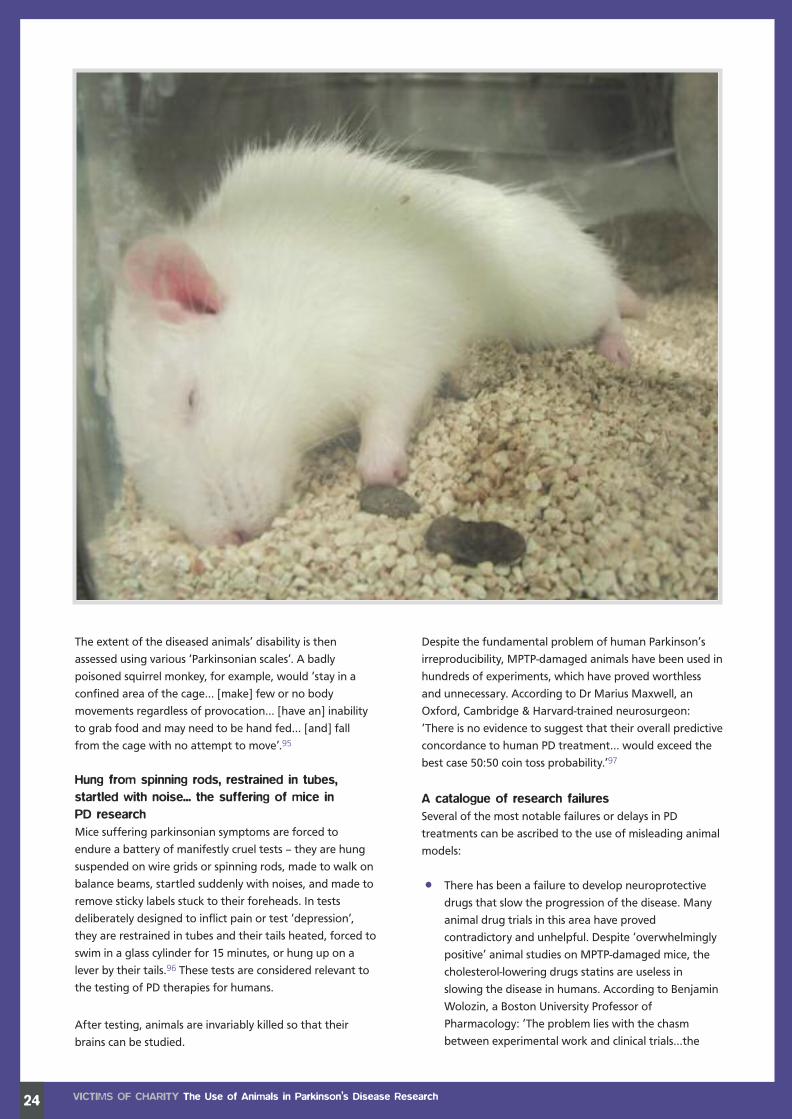
24
The extent of the diseased animals’ disability is then
assessed using various ‘Parkinsonian scales’. A badly
poisoned squirrel monkey, for example, would ‘stay in a
confined area of the cage… [make] few or no body
movements regardless of provocation… [have an] inability
to grab food and may need to be hand fed… [and] fall
from the cage with no attempt to move’.95
Hung from spinning rods, restrained in tubes,startled with noise... the suffering of mice inPD researchMice suffering parkinsonian symptoms are forced to
endure a battery of manifestly cruel tests – they are hung
suspended on wire grids or spinning rods, made to walk on
balance beams, startled suddenly with noises, and made to
remove sticky labels stuck to their foreheads. In tests
deliberately designed to inflict pain or test ‘depression’,
they are restrained in tubes and their tails heated, forced to
swim in a glass cylinder for 15 minutes, or hung up on a
lever by their tails.96 These tests are considered relevant to
the testing of PD therapies for humans.
After testing, animals are invariably killed so that their
brains can be studied.
Despite the fundamental problem of human Parkinson’s
irreproducibility, MPTP-damaged animals have been used in
hundreds of experiments, which have proved worthless
and unnecessary. According to Dr Marius Maxwell, an
Oxford, Cambridge & Harvard-trained neurosurgeon:
‘There is no evidence to suggest that their overall predictive
concordance to human PD treatment… would exceed the
best case 50:50 coin toss probability.’97
A catalogue of research failuresSeveral of the most notable failures or delays in PD
treatments can be ascribed to the use of misleading animal
models:
• There has been a failure to develop neuroprotective
drugs that slow the progression of the disease. Many
animal drug trials in this area have proved
contradictory and unhelpful. Despite ‘overwhelmingly
positive’ animal studies on MPTP-damaged mice, the
cholesterol-lowering drugs statins are useless in
slowing the disease in humans. According to Benjamin
Wolozin, a Boston University Professor of
Pharmacology: ‘The problem lies with the chasm
between experimental work and clinical trials...the
VICTIMS OF CHARITY The Use of Animals in Parkinson’s Disease Research
Charities Report:Layout 1 21/6/11 11:06 Page 26

most reasonable interpretation is to accept that the
disease is more complicated than reflected in the
animal or epidemiological models.’98
• A review in 2003 found that animal neurotoxin models
of PD demonstrated a neuroprotective effect from
iron chelators, radical scavenger antioxidants, MAO-B
inhibitors, glutamate antagonists, nitric oxide synthase
inhibitors, calcium channel antagonists and trophic
factors – none of which are useful in humans. The
review authors, all prominent pharmacologists and PD
researchers, concluded: ‘The animal models of
Parkinson's disease may not be totally reflective of the
disease and, therefore, the chemical pathologies
established in the animal models may not cause, or
contribute to, the progression of the disease
clinically.’99
• One of the latest drugs to be touted as a
neuroprotector is exendin-4. However, the most recent
results of trials in rats do not even match those of
earlier rodent research.100
• Evidence suggests that animal research confused the
issue of cell transplant surgery for PD. In the 1990s,
transplants of foetal nerve cells into the brains of PD
patients were halted after disabling side-effects
relating to the donor tissue became apparent. Animal
trials had not indicated the risk, which researchers
have suggested was due to ‘differences between the
non-human primate and human putamen [a structure
located at the base of the forebrain]’.101 A revisiting of
this research in 2010 was mainly stimulated by brain
imaging of two PD patients who had received
transplants. This study demonstrated an excess of
serotonin-producing cells in the grafted area,102 which
could be damped down by drug therapy.
• Gene therapy for PD (see below) is a relatively recent
development, but, true to history, animal models are
once again proving useless. In 2006, a gene encoding
the growth factor neurturin was introduced into the
brains of MPTP-poisoned rhesus monkeys, using a viral
vector. They demonstrated a dramatic improvement in
their parkinsonian symptoms. In human clinical trials,
however, neurturin gene treatment was no better
than sham surgery103 (whereby the same procedure
was performed but without introducing the ‘active
ingredient’ – i.e. the sham group act as controls).
Contemporary research intoParkinson’s DiseaseParkinson’s UK has identified four priorities for their latest
research programme. Alongside finding biomarkers to help
diagnose PD earlier, and a greater understanding of nerve
cell death, developing new animal models for both
research and drug testing are also key objectives.
In support of these, the organisation quotes Professor J
Timothy Greenamyre, a US-based PD researcher: ‘Current
animal models mimic some of the symptoms of Parkinson’s
but they don’t recreate the changes that happen in the
human brain. To do this, we need better models, where the
nerve cells die slowly and develop characteristic features of
Parkinson’s, such as Lewy bodies. We need to be able to
look at the gradual development of the condition in
animals as they age.’104
It is hard to see why any emphasis should be placed on
developing ‘ageing’ animal models when human studies
and modern technologies offer a wealth of appropriate
and ethical research options. The crudity of such ‘new’
animal models (and their striking similarities to the failed
old ones) is revealed by the latest Parkinson’s UK-funded
experiments. For example, in 2009, scientists poisoned rats
with the pesticide rotenone, already implicated in causing
PD in humans.105 The chemical had been used years earlier
to induce parkinsonism in rats and primates. Thus, in a
baffling reversal of good science, researchers are using
pre-existing knowledge about a likely cause of human PD
to create disease parodies in animals – over and over again.
Genetic research and treatments now form a major part of
PD activity. Large-scale human population studies are
revealing a complex interaction of genetic susceptibility
and environmental factors in PD causation. However,
despite the failure of transgenic rodent models to deliver
medical progress,106 Parkinson’s UK intends to use these
new genetic discoveries to develop more of the same.
In a parallel development, Japanese scientists are
developing genetically altered marmosets who can pass
their altered genome to their offspring.107 They intend to
create colonies of animals born with a PD-type disease for
research purposes. However, marmosets are genetically
more distant from humans than macaques, the non-human
primate of choice until recent years. Marmosets fail many
cognitive ability tests that are used to assess treatments for
neurodegenerative disorders, and their brains are too small
to study with positron emission tomography scans, which
are an important element in human trials.
With regard to contemporary PD research, there are
striking historical echoes. Even though promising avenues
of human-based research are being pursued, there remains
a strong reliance on animal models, a reliance that could
delay – and possibly derail – progress being made by the
human-centred investigations. A.S
25VICTIMS OF CHARITY The Use of Animals in Parkinson’s Disease Research
Charities Report:Layout 1 21/6/11 11:06 Page 27

Parkinson’s Disease is not known to occur naturallyin any species other than humans, which is whyresearchers resort to deliberate brain-damaginginterventions in order to produce Parkinson-likesymptoms in animals. While there may be similaritiesbetween the brains of human and non-humanprimates, monkeys’ brains are not scaled-downversions of the human organ. Their brains are theresult of unique evolutionary biology, moulded overmillions of years in response to environmental, socialand genetic influences. While macaque and marmosetmonkeys are often used in Parkinson’s Diseaseresearch, it is rats who are used more than anyother species.
In two experiments funded by Parkinson’s UK(formerly Parkinson’s Disease Society), 26 and eightmarmoset monkeys were used, respectively.108, 109
They received daily injections of the chemical MPTPfor five consecutive days, which rendered them soseverely disabled that they were unable even to feedthemselves and had to be hand-fed. Over thefollowing eight weeks, the monkeys exhibited rigidity,
poor coordination, loss of balance and an inability tovocalise. Only at the end of the eight-week periodwere some of the monkeys given treatment toalleviate their symptoms. These ‘lucky’ animalsreceived different chemical cocktails by gavage(force feeding), while others were left untreated toendure the full effects of MPTP poisoning. Thebehaviour of the treated and untreated monkeyswas then compared and rated.
In another study, 25 female macaque monkeys wereused.110 Five were killed at the beginning of thestudy so that their brains could be kept forcomparative purposes with those of the monkeysundergoing the actual experiment. The remaining 20monkeys received daily injections of the sameseverely disabling chemical, MPTP, until they toowere killed. Their behaviour and ability to move weremonitored by videotape recordings. Fruit ‘rewards’were used to manipulate the monkeys into doingthe researchers’ bidding. This may sound benign butsuch ‘rewards’ are often provided against abackground of food and water deprivation. Five ofthe monkeys were killed after six days, five at day12 and five at day 15. The monkeys in the day 15group exhibited early symptoms of brain damage(loss of coordination) due to MPTP toxicity. Allanimals were killed with an overdose injection ofanaesthetic. Their brains were removed after deathfor laboratory study.
VICTIMS OF CHARITY Animal Suffering in Parkinson’s Disease Research26
ANIMAL SUFFERING IN PARKINSON’S
DISEASE RESEARCH
Examples of animal experimentsfunded by Parkinson’s UK
Monkeys severely disabled
Charities Report:Layout 1 21/6/11 11:06 Page 28

27
Rats brain-damaged
Inflammation in the brain is thought to play a significant role in PD. In this experiment, researchers injected twodestructive chemicals directly into the brains of rats. One chemical triggered PD-like symptoms, while the othercaused inflammation in their brains.111 In people, inflammation of the brain can cause symptoms ranging fromheadaches and visual disturbances to convulsions and coma.
A week later, the rats were injected with an experimental drug directly into their abdomen. Although the drugappeared to protect the brain against inflammation, the authors could only hypothesise as to whether the effectwould also occur in PD patients. The injections, which were given twice a day for seven days, would have beenparticularly painful, since they penetrate muscle as well as skin. These were followed by behavioural tests in whichthe rats were placed in a circular test arena for up to 60 minutes and observed to see whether they had sufficientcoordination to perform tight turn-arounds when changing direction. For animals who may be suffering pain anddisorientation, this is likely to have been an ordeal. Later the same day, the rats were anaesthetised while samplesof brain chemicals were measured. They were then killed and their brains studied. Although the authors do notmention how the rats were killed, there are several common methods in use today: anaesthetic overdose byinjection; carbon dioxide inhalation; cervical dislocation (breaking the neck without anaesthesia); or decapitation(using a guillotine, with or without prior anaesthetic).
A total of 44 male rats were used in another study.112 The animals underwent major head surgery to implant smalltubes directly into their brains, secured in place with an apparatus fixed to the skull (dental cement cap) and metalscrews. After a 10-day ‘recovery period’, during which the rats received no painkillers, brain damaging chemicalswere administered to the fully conscious rats through the tubes implanted in their brains, in order to replicate PDsymptoms. No mention is made by the authors of how the animals reacted. One hour after the procedure, the ratswere killed by decapitation and their brains examined. A.M
Charities Report:Layout 1 21/6/11 11:07 Page 29

28
Incidence and mortalityDementia – a progressive loss of cognitive
function and memory – is caused by various
diseases and conditions. In 2007, dementia
affected a little more than one per cent of the
UK population, and was predicted to rise by 154
per cent in the next 45 years.113 Dementia is
strongly age-related, with one in six people over
80 having a form of it. Alzheimer's Disease, a
physical brain disorder, is the most common
cause of dementia, affecting around 465,000
people in 2010.114
For people over 65, 15 per cent of deaths in
women and ten per cent of deaths in men are
attributable to dementia. Delaying the onset
of dementia by five years would halve the
number of UK deaths due to dementia to
30,000 a year.115
What is Alzheimer’sDisease?Alzheimer’s Disease changes the chemistry and
structure of the brain, causing brain cells to die.
The other hallmarks of the condition are the
development of plaques and tangles in the
brain. The plaques are composed largely of a protein called
amyloid-beta, which is cut from a naturally occuring
precursor known as APP. Tangles are made up of a protein
known as tau, which is produced in an abnormal form.
The causes of Alzheimer's Disease are believed to be
multiple, with age, genetic inheritance, environmental
factors, diet and health all implicated.
History of Alzheimer’s DiseaseresearchThe Alzheimer's Society, in a sweeping statement that
credits animal research for almost all medical therapies ever
discovered, claims that it is essential for ‘understanding the
biology’ of dementia and for testing new drugs and
treatments. In fact, the biological discoveries are usually
made in humans, with subsequent attempts made to mimic
them in animals, the data obtained from which are
inadequate and misleading. If animal models had produced
a plethora of effective therapies, the position of the charity
would be more defensible. In reality, the failure to translate
results from animal tests into clinical progress is perhaps
most dramatic in the case of Alzheimer's Disease.
Animal models in Alzheimer’sDisease research and theirhistorical failureChosen for convenience and economics ratherthan for their predictive valueAccording to a 2008 article in Nature, the research
community is now worried that ‘it may be unrealistic to
VICTIMS OF CHARITY The Use of Animals in Alzheimer’s Disease Research
THE USE OF ANIMALS INALZHEIMER’S DISEASE RESEARCH
Charities Report:Layout 1 21/6/11 11:07 Page 30

think of modelling the full complexity of ageing-related
human brain disorders in mice whose disease course is
usually accelerated by a single crude genetic
modification’.116 This worry is entirely legitimate.
The ‘experimental Alzheimer's Disease’ that researchers
produce in animals is emphatically not the same as the
human variety. Although some aged primates and dogs
develop a disease with similarities, no animal species suffers
from the same condition. Thus, researchers have had to
produce laboratory facsimiles, with what their creators
admit have been ‘partial and unpredictable’ results.117
Their methods include injection of neurotoxins directly into
the brains of rodents and primates, and poisoning rabbits
with a diet of cholesterol and copper. However, by far the
most popular ‘models’ of recent years have been transgenic
mice, whose genetic make-up has been altered. These
transgenic models have been instrumental in developing a
whole range of ineffective drugs for Alzheimer's.
Alzheimer's in humans leads to complex, progressive
structural brain changes and cognitive losses. Despite years
of tampering with rodents’ genetics, the models produced
have not accurately reproduced these features:
• Some mice with mutant tau genes failed to show any
sign of altered neurological function. Others formed
more Alzheimer's-like tangles but developed lesions in
the spinal cord and brainstem that left them
effectively disabled. They were therefore unable to
properly carry out tests of cognition and memory that
required them, for instance, to run about in a maze.118
More recently still, mice have been created with
tangles in the ‘right’ areas – but they do not form
amyloid plaques.
• Many transgenic animals accumulating amyloid
plaques display only subtle effects, and do not
develop tangles or suffer from significant
neurodegeneration.119
• Many APP over-expressing mice ‘do not develop
pathology at all, probably due to insufficient APP/
amyloid-beta expression‘. Models that did show either
plaques or cognitive impairment did not demonstrate
any cell death.120
Only very recently have mice been created with amyloid-
beta and tangles together. There is no reason to suspect
that they will be any more predictive of human success
than their forebears. The researchers are not blind to this
problem: ‘The relevance of… an artificial model to an aged,
non-mutant human brain is potentially problematic.
Furthermore, there are additional factors that play a role in
triggering a disease and its progression, such as diet and
29VICTIMS OF CHARITY The Use of Animals in Alzheimer’s Disease Research
Charities Report:Layout 1 21/6/11 11:08 Page 31

30
the environment… the impact of these factors have (sic)
not been addressed.’121
In any case, these animals are chosen for convenience and
economics rather than for their predictive value – mice ‘are
relatively cheap to maintain, fecund and have a short life
span; they are easy to manipulate genetically and they
respond reasonably well in cognitive tests.’122
The irrelevance of animal memory testsThe aforementioned tests, supposedly relevant to
Alzheimer's in humans, are extremely crude and
reductionist. Some of the most facile – not to mention
cruel – include:
• The active avoidance task. This is described as ‘a fear-
motivated… test based on electric current as a source
of punishment’.123 In other words, mice are scared
into remembering when and where they will receive
an electric shock.
• Y-mazes and T-mazes. Rodents are placed into these
very simple structures, often after having been starved
or deprived of water, and are forced to make choices
between routes. Experimenters used a T-maze to
discover that ‘for a thirsty rat the rewarding effects
of drinking water are very much greater than those
of airlicking.’124
• Isolation-induced aggression testing.Mice are forced
to attack each other, after being held in solitary
confinement for prolonged periods.
• Morris water maze. This widely used test forces
rodents to swim around a pool of water in order to
find an escape route. The procedure is highly
operator-dependent with myriad variables;
standardisation is difficult.
• Step-down avoidance. Rodents are dropped onto
uncomfortable vibrating platforms, which they can
switch off by stepping down onto a grid with a
built-in sensor.
A wryly humorous internet column by a US-based
pharmacologist, in which ‘Alzheimer’s rodents’ are
nominated as the ‘worst animal model’, effectively sums
up the state of the ‘art’: ‘…the disease is affecting higher
brain functions that are very poorly modelled in any of the
small animals… When I used to work in the field, I would
occasionally wonder about the relevance of watching a
rat run into one half of his cage or another to a person
forgetting an important appointment… the infamous
Morris Swim Maze… needs its own special room, full of
special equipment, and a full-time person trained in its
complications to generate the data that you still don’t
quite trust.’125
VICTIMS OF CHARITY The Use of Animals in Alzheimer’s Disease Research
Neurons and neural system
Charities Report:Layout 1 21/6/11 11:08 Page 32

31Victims of charity The Use of Animals in Alzheimer’s Disease Research
Expensive and time-consuming flopsThe failure of Alzheimer’s animal models indrug development:Time and time again, drugs that seemed powerfully
effective in animal Alzheimer's models have failed in clinical
trials. Experts have suggested many reasons for this
mismatch – the almost ubiquitous poor design of animal
trials, the obvious differences between animal models and
human pathology, and a dramatic proven publication bias
(see page 39) in favour of ‘positive’ results from animal
experiments.126
There is a large and growing list of highly expensive drug
failures for Alzheimer’s. The following are all recent and
represent only the tip of the iceberg:
• Dimebon was found to be useful in avoidance
experiments in brain-poisoned rats,127 but useless in
humans. (Phase 3 clinical trial failed 2010.) The
pharmacology of the drug was always unclear, and it
actually increased levels of amyloid-beta in mouse
brains. The Alzheimer’s Society called the research
‘head scratching’, but still saw fit to claim Dimebon
could be available as a treatment within three to
five years.128
• Tarenflurbil was shown to improve memory and
behavioural performance in transgenic mice, but was
totally ineffective in patients with mild Alzheimer’s.
(Phase 3 trial failed 2008.)129
• Tramiprosate significantly reduced amyloid-beta in the
brains of transgenic mice, but was ineffective in mild
to moderate Alzheimer's sufferers. (Phase 3 trial failed
2007.)130
• Semagacestat reduced levels of amyloid-beta in
plasma, cerebrospinal fluid and the brain in a dose-
dependent manner in animals. However, it worsened
cognition and the ability to perform activities of daily
living in mild to moderate Alzheimer’s patients. (Phase
3 trial failed 2010.)131
Charities Report:Layout 1 21/6/11 11:08 Page 33

32
• Bapineuzumab is a monoclonal antibody to amyloid-
beta. Such drugs given to transgenic mice cleared
some of their brain deposits with ‘cognitive benefit’.132
The drug failed to improve cognitive function in a
Phase 2 trial of 234 Alzheimer's patients in 2008.133
• AN-1792 was a vaccine to amyloid-beta, designed to
stimulate patients’ own immune systems to destroy
the protein. In mouse trials, this immunotherapy was
successful at clearing amyloid-beta with no obvious
side-effects. It was also found to be ‘safe’ in monkeys,
rabbits and guinea pigs. Yet Phase 2 clinical trials were
terminated in 2002 when patients developed serious
brain inflammation, and there were no significant
cognitive benefits.134 Clinical trials of a supposedly
safer ‘second-generation’ vaccine were suspended in
2008, again due to unforeseen side-effects. The AN-
1792 researchers later reported postmortem data on
some of their patients, all of whom had died of severe
dementia. Some had no amyloid in their brains, casting
doubt on whether its removal from animal models was
of any relevance.135
• Nerve Growth Factor (NGF) was shown to safely
prevent the death of nerve cells in various strains of
rats, and in ageing rhesus monkeys. In the earliest
human trial, NGF was infused into the brain ventricular
system of three patients, but it caused severe side-
effects. Following further research, Phase 2 trials are
ongoing, led by the same company that used similar
failed techniques for Parkinson’s disease.136
• Finally, some recent animal experiments proved
unhelpful in illuminating epidemiological data. A link
between Alzheimer’s and type 2 diabetes, for
example, has been known for some time. Metformin, a
commonly prescribed anti-diabetic drug, has been
shown to increase the formation of amyloid-beta yet
decrease the formation of tau in rodents.137 The doses
used for recent animal studies were much higher than
those used in human diabetes, which casts doubt on
whether this drug could ever be safely employed in
people due to side-effects.
The 2008 Nature article points out what is now obvious –
‘in recent years, and especially for neurodegenerative
diseases, mouse model results have seemed nearly
useless’.138
Contemporary Alzheimer’s research– more of the sameMany questions relating to Alzheimer’s remain
unanswered, with crucial links in the chain of causation
still missing. Heated debates are ongoing – in particular,
there is the vexed question of whether amyloid-beta is a
cause or a consequence of the disease. It is clear, however,
that many ‘amyloid-busting’ drugs developed and tested
using simplistic animal models have been a failure. The
obsession with mimicking a disease in animals, without
first understanding it in humans, has cost Alzheimer’s
patients dearly.
However, the Alzheimer’s Society remains committed to
animal-based research. Many of its current projects are still
ploughing the same infertile furrows, with mouse models
of Alzheimer’s being used to test stem cells, amyloid-
reducing antibodies and new drugs. Patently pointless
experiments, such as demonstrating that rat nerve cells do
not function well if deprived of oxygen, are also funded.
Shockingly, the society is funding several studies on animals
that investigate therapies that have already failed in
humans. For example, it has granted more than £200,000
for a researcher to investigate the mechanism of Dimebon
in mouse models. Another is injuring mice to study the
effects of the anti-inflammatory drug Ibuprofen on their
memory, despite numerous clinical trials showing these
drugs do not help in Alzheimer’s and have too many
dangerous side-effects.139
Equally wasteful and pointless is a series of animal brain
damage experiments. It is known from human studies
that severe head injury is a risk factor for Alzheimer’s,
and postmortems in these patients have revealed some
of the disease’s structural changes. Certain chemicals
associated with trauma were also first identified in the
brains of Alzheimer’s patients. Despite continued human
studies in this area, one researcher is destroying the brains
of mice, either by tying off their cerebral arteries or by
direct trauma to their heads to see if the same findings
occur.140A.S
VICTIMS OF CHARITY The Use of Animals in Alzheimer’s Disease Research
Charities Report:Layout 1 21/6/11 11:08 Page 34

33
Charities Report:Layout 1 21/6/11 11:08 Page 35

The search for an animal model for Alzheimer’sDisease began in earnest in 1980.141 Some of theearliest attempts involved injecting aluminium chloride– a chemical so corrosive that people dealing with itshould wear safety glasses and gloves142 – directlyinto the brains of developing rabbits.143 Today, mice,rats, marmoset monkeys and mouse lemurs are theanimals of choice.
Animal researchers have not only developed varioustechniques to cause brain damage, they have alsodevised extremely cruel methods to assess brainfunction afterwards. Transgenic mice are now usedto mimic the excess protein deposition in the brain.These mice are bred with a defective human geneassociated with AD. However, they often do notdevelop the desired pathological damage or elsedisplay it in the ‘wrong’ areas.144
In an experiment funded jointly by the Alzheimer’sSociety, the Alzheimer’s Association, the Alzheimer’sResearch Trust and the Medical Research Council,transgenic mice containing the defective human genewere crossed with another strain of transgenic micecontaining a protective gene. The aim of the researchwas to see whether the protective gene could blockor neutralise the effects of the defective gene, and
so provide leads to the development of therapeuticdrugs for AD.145
The crossbred mice were subjected to variousmemory and behavioural tests as a means of gaugingthe overall effectiveness of the protective gene. Aswould be expected when two different strains ofmice are crossbred, some littermates were born withthe protective gene, while others were not. Thephysical abilities of all the mice were assessed usingthe Morris water maze, a technique developed 30years ago. Although mice are scared of being inwater, they are put into a small round tank fromwhich they cannot escape or touch the bottom. Thewater in the tank is deliberately cold so as toprovoke frantic swimming, which can lead toexhaustion and to some animals drowning when notkept under careful observation.
In this test, mice were forced to swim until theylocated a small platform on the surface of the wateron which they could rest. Once they had been trainedto do this, the platform was submerged so that itwas no longer visible on the water’s surface. Thedeclared object was to test the spatial memory ofthe animals. Mice with the protective gene performedslightly better than those without and could locatethe hidden platform sooner. At the end of the tests,all the mice were killed and their brains examined.
A similar experiment was funded by the Alzheimer’sSociety, this time aimed at determining the effectthat stress might have on the production of ‘bad’protein in the brain.146 Twenty adult male rats weredivided into four groups of five animals: a 20 daysstress group, a 10 days stress group, an acute (1 day)stress group and a control group.
Stress exposure was achieved by placing the rats onan open elevated platform for 60 minutes. While free-roaming rats may choose to climb to an elevatedlevel, locating them off the ground in an exposedposition in a laboratory environment is highlystressful, as indicated by the immediate rise in levelsof stress hormones in their blood. This responsedecreases significantly after 10 to 20 days once theanimals become accustomed to the platform, butthey were nonetheless subjected to fear and stressfor a long period. All rats were killed 24 hours aftertheir last stress exposure to measure the impact ontheir brains of what they had endured. Theresearchers concluded that stress may have aneffect on ‘bad’ protein production in the brain butthat further studies would be required to determinethe exact mechanisms. A.M
VICTIMS OF CHARITY Animal Suffering in Alzheimer’s Disease Research34
ANIMAL SUFFERING IN ALZHEIMER’S
DISEASE RESEARCH
Examples of animal experimentsfunded by the Alzheimer’s Society
Charities Report:Layout 1 21/6/11 11:09 Page 36

35
Charities Report:Layout 1 21/6/11 11:10 Page 37

NON-ANIMALMEDICAL RESEARCH
Non-animal medicalresearch: humane, effectiveand commercially-sound
The evidence presented inthis report demonstrates thatproponents of animal research arebringing false hope to millions ofpatients affected by the medicalconditions examined. They have alsodiverted funds, donated by thepublic in good faith, away frommethods of non-animaltesting that are relevantto the species inquestion – humans.
36
Charities Report:Layout 1 21/6/11 11:10 Page 38

There are many well-established non-animal research
techniques that already play an important role in
developing therapeutic interventions.
In fact, the medical charities studied in this report all,
to a greater or lesser extent, employ such methods.
However, they still insist that this work is complementary
to animal research, and that some animal suffering will
always be necessary to find cures for human disease.
The results-based analysis we present demonstrates that
animal research does not complement good science
– it confounds it.
The options for studying human disease in humans are
growing all the time, and are supported by solid science
and, increasingly, commercial and government funding.
Next to these effective and efficient technologies, animal
tests look cruder than ever.
Scanning technologiesThere is a wide range of scanning technologies that can
reveal processes in living humans. The images produced are
now truly remarkable and are especially useful in
neurodegenerative conditions like Parkinson’s and
Alzheimer's.
Human-derived raw materialsHuman-derived raw materials can be obtained and used in
a range of ways. From donated human cadavers down to
human DNA, all levels of tissue sample can be gainfully
employed. Intact slices of human tissue, ethically obtained
from patients who undergo operations or biopsies, can be
maintained in the laboratory so that they retain their
function. Tumour biopsies, for example, can be used to see
whether a drug has bound to its intended molecular
target. Comparing healthy and diseased donated organs
can provide important information on disease processes.
Stem cells of human origin also have enormous utility.
Human tissues or organ systemsHuman tissues or organ systems can also be recreated in
laboratories. A Cardiff University team led by cell biologist
Dr Kelly Bérubé has grown human lung cells in the
laboratory to form three-dimensional tissue-like structures.
These can be used to test substances for the potential to
cause damage if inhaled.147 Human lymph nodes have
been created in the laboratory, and can be employed to
test vaccines and biologically-based drugs, like the TGN1412
monoclonal antibody, which – having been passed as safe
on the basis of tests on monkeys – went on to cause
catastrophic injuries to human trial subjects.148
Computer programsHuman systems, from individual organs to the whole body,
can be simulated using highly sophisticated computer
programs. These are created using data obtained from
people. Computer simulations have been developed, for
example, to predict the behaviour of a drug in the digestive
system. These simulations are likely to predict such effects
in humans more accurately than animal models, and in a
much more efficient way.
MicrodosingMicrodosing involves giving a tiny amount of a substance –
less than one hundredth of the quantity expected to have a
noticeable effect – to a volunteer or patient. This dose is
sometimes labelled with a safe amount of a radioactive
chemical. Body fluids are then analysed to see how the
body has responded, or PET imaging is employed to
ascertain how the substance behaves in specific organs.
This technique has already been used successfully to test
drugs for cardiovascular disease, pain, Alzheimer’s disease
and gastrointestinal disorders.
MicroarrayCell components, including DNA, RNA and protein
molecules, can be arranged in a microarray, which is,
typically, a tiny chip or slide made from silicon or glass, or it
can be a membrane. The signals produced by microarrays
are read by scanners and the data generated are analysed
by computer. The technology can be used for drug
development, both to identify potential drug targets and
to test for efficacy and toxicity. Thousands of genes can be
monitored simultaneously.
37VICTIMS OF CHARITY Non-Animal Medical Research
Cell under microscope
Charities Report:Layout 1 21/6/11 11:10 Page 39

Microfluidic devicesMicrofluidic devices contain human tissue samples in tiny
chambers linked by microchannels. Fluids and chemicals
flow in a natural way between different compartments,
simulating conditions in the human body. As with
microarrays, microfluidics can produce large amounts of
information very quickly. The technology can help scientists
to understand how cancers spread, for example.
Microfluidics can investigate human tissues and organ
systems, with the creation of ‘bioreactors’ designed to
supply nutrients and remove waste products.149 One team
of researchers has developed a system in which human
liver, brain cortex and bone marrow are interconnected
through a circulatory system mimicking blood flow. These
models can be used to predict the effects of substances as
they move between these organs.
EpidemiologyEpidemiology involves the study of significant numbers of
people over a period of years, comparing their lifestyles,
genes, medical interventions, environments, social status,
etc. It remains a powerful tool with huge potential, and
has already produced enormously valuable findings,
including the link between smoking and lung cancer.
Clinical dataClinical data and observation are greatly under-used,
including the information gathered from minimally or
non-invasive procedures (such as blood or urine sampling).
Data from this type of benign intervention, undertaken
in consenting patients already undergoing procedures,
could be collated much more efficiently than is currently
the case. A.S
38 VICTIMS OF CHARITY Non-Animal Medical Research
A wholesome diet is a major contributor to good health
Woman receiving CAT scan
Charities Report:Layout 1 21/6/11 11:11 Page 40

39VICTIMS OF CHARITY Peer Review
The peer reviewprocess of approvingresearch is biased,unjust, unaccountable...’
PEER REVIEW
Research for this report has involved analysing and
interpreting animal experiments published in leading
scientific, peer-reviewed journals. Publication in such august
periodicals is often cited as a proxy marker of scientific
validity – supposedly, shoddy or poor research does not
pass peer review scrutiny and is rejected.
When it comes to animal research, peer review has
additional stages. Local ethical review committees have to
approve the project and decide whether the experimenters
should move to the next stage and submit their proposal
to the Home Office. Assuming a licence is granted, a
retrospective review is subsequently undertaken by another
in-house committee, to scrutinise and check the validity of
the work before submission for publication.
Unfortunately, there is evidence that this entire peer-review
process is flawed, secretive and biased – at all stages:
• Ethical committees are formed by the same institutions
in which the researchers operate. All too often, they
simply rubber-stamp the experimenters’ proposals
after little or no modification. Cardiff University, for
example, received 29 applications for project licences
between 2006 and 2009. Just one failed to gain
approval. The 28 that were approved encompassed
nearly 200,000 individual experiments.151
• Despite the ease with which approval is gained, the
quality of the experiments is often demonstrably poor.
In 2009, the National Centre for the Replacement,
Refinement and Reduction of Animals in Research
(NC3Rs) commissioned a comprehensive review of
research on rats, mice and primates. It found that
the studies ‘contained a catalogue of basic and
fundamental errors that you would not expect in
any properly constructed paper from a practising
scientist’.152 Another recent review also found that
‘publication bias’* was prevalent in the field of animal
experiments, to a degree that would not be possible
with human clinical trials.153
• The NC3Rs review additionally found that only 59 per
cent of the studies examined stated the hypothesis or
objective of the study, and the number and
characteristics of the animals used. It must be
emphasised that all these experiments had gone
through a multi-stage peer review process, and had
not been found wanting.
• Institutions often claim that animal research is justified
because it has been funded by external grants, such as
those provided by the Medical Research Council.
However, these bodies are strongly supportive of
animal experiments, and often fail to apply the
requisite level of scepticism in response to applicants.
The MRC, for example, is wholly in favour of ‘basic’ (or
speculative) animal research because ‘we don't know
where new advances for acquisition of new treatments
are going to come into play’.154 The lack of rigour was
highlighted by the Home Office (the government
department that grants animal research licences) when
it declared that support for animal research by a major
funding body ‘cannot be taken to guarantee the
relevance, importance or scientific validity of any
individual experiment’.155
• Neither the peer review process that takes place within
the research institution, nor the Home Office ‘cost-
benefit analysis’ aimed at quantifying animal suffering,
is open to public scrutiny. A.S
* Publication bias is a tendency on average to produce
results that appear significant, because negative or
near-neutral results are infrequently published
‘We portray peer review to the public as a quasi-sacred
process that helps to make science our most objective
truth teller. But we know that the system of peer
review is biased, unjust, unaccountable, incomplete,
easily fixed, often insulting, usually ignorant,
occasionally foolish, and frequently wrong.’150
Dr Richard Horton, Editor, The Lancet
Charities Report:Layout 1 21/6/11 11:11 Page 41

CONCLUSION
If our objection to the animal research funded by thefour charities rested exclusively on the brutal treatmentinflicted on the animal ‘models’, we would struggle towin over the public. We do object powerfully, on moralgrounds, to what the animals are put through. But wealso strongly refute the scientifically flawed propositionthat information of direct relevance to human beingscan be procured by surgically mutilating and/orgenetically manipulating mice, dogs, monkeys, goats,rabbits and other animals. Such research is not onlyunproductive and therefore wasteful of preciousresources offered in good faith by the public, it alsouselessly employs scientific minds that might otherwisebe directed at producing something of human benefit.
It must be clear from the above that, in rejecting the‘animal model’ of human disease, we do not rejectthe quest, by means of science, for remedies andpalliatives. In fact, our report includes a section listingthe impressive and growing range of non-animalresearch methodologies.
Furthermore, we are fully aware that the four charitieson which we focus engage in valuable patient supportwork. Our critique is not intended to damage that work,and need not do so. It is open to anyone who wasthinking of donating to one of the four charities butwho has decided against doing so after considering our
arguments, to back those charities’ work with patients.Parkinson’s UK and the Alzheimer’s Society, inparticular, offer a range of volunteering opportunities.They include help at social events and therapysessions, as well as one-to-one befriending andsupport. The British Heart Foundation has nearly 300affiliated Heart Support Groups that are open toanyone with any kind of heart condition, as well as totheir partners and families. Cancer Research UK ismuch more thoroughly concerned than the other threeorganisations with research, and therefore directsprospective volunteers to bodies such as Macmillannurses (Macmillan Cancer Support).
Then there are the scores of medical research charities– no doubt each of them hungry for funds – that covera vast range of human ailments, and which eschewanimal research. Nearly 80 of them are listed overleaf.We would argue that these are the organisations thatmerit the public’s financial support rather than thosethat use their funds to pointlessly wound, torment andkill large numbers of vulnerable animals.
Such ‘research’ is a double betrayal – of the animals,and of the human patients in whose name they aremade to suffer.Andrew Tyler
40
Cancer, heart disease, Parkinson’s and Alzheimer’s are conditions that blight thelives of huge numbers of people – both the sufferers and those close to them. Werecognise this reality at Animal Aid as much as anyone. And, of course, we are notimmune to these diseases and the physical suffering and mental anguish they cause.
Charities Report:Layout 1 21/6/11 11:12 Page 42

41VICTIMS OF CHARITY Medical Charities That Do Not Test On Animals
Medical Charities thatdo not test on animalsThis list is correct at the time of printing. For updates, visit www.animalaid.org.uk
• Action Against Allergy
• Action for Blind People
• Against Breast Cancer (also known
as Action Against Breast Cancer)
• Age Care (formerly Royal Surgical
Aid Society)
• Allergy UK (formerly British
Allergy Foundation)
• Arterial Health Foundation
• The Arthritic Association
• Arthritis Care
• AVERT
• Back-Up Trust
• Bath Cancer Research
• The Big C
• Birmingham Children’s
Hospital Charity
• Breast Cancer Care
• Breast Cancer Survival Trust
• Breast Friends
• British Deaf Association
• British Dyslexia Association
• British Institute for Brain Injured
Children (BIBIC)
• British Kidney Patient Association
• British Organ Donor Society
• British Polio Fellowship
• British Red Cross
• Cancer Active (formerly Research
Into Ovarian Cancer)
• Cancer Kin Centre
• Cancer & Leukaemia in Childhood
(CLIC Sargent)
• Cardiomyopathy Association
• Caring Cancer Trust
• The Children's Cancer and
Leukaemia Group (formerly United
Kingdom Children’s Cancer Study
Group)
• Christian Lewis Children’s Cancer
Care
• Cleft Lip & Palate Association
• Coeliac UK (formerly Coeliac
Society)
• Colostomy Association (formerly
British Colostomy Association)
• Down’s Syndrome Association
• Dr Hadwen Trust for Humane
Research
• Dyslexia Action (formerly The
Dyslexia Institute)
• East Anglia’s Children’s Hospices
• Eating Disorders Foundation
• Elton John AIDS Foundation
• ENABLE
• Epilepsy Action Scotland
• Epilepsy Society (formerly National
Society for Epilepsy
• FORCE Cancer Charity
• Greater London Fund for the Blind
• Headway – The Brain Injury
Association
• Heartbeat
• The Humane Research Trust
• International Glaucoma Association
• John Charnley Trust
• Laura Crane Trust
• Lord Dowding Fund
• Lynn’s Bowel Cancer Campaign
• Macmillan Cancer Support
• Michael Palin Centre for
Stammering Children
• Mid-Kent Breast Cancer
Research Appeal
• Migraine Action (formerly Migraine
Action Association)
• Mind, The Mental Health Charity
• Myasthenia Gravis Association
• National Deaf Children’s Society
• National Kidney Federation
• National Society for Research
into Allergy
• New Approaches to Cancer
• ORBIS UK
• The Pain Relief Foundation
• Penny Brohn Cancer Care
(formerly Bristol Cancer Help
Centre)
• Quest Cancer Research
• Raynaud’s & Scleroderma
Association
• Royal College of Psychiatrists
• Royal National Institute of
Blind People
• SCOPE
• Shaw Trust
• Spinal Injuries Association
• Susan Channon Breast Cancer Trust
• Teenage Cancer Trust
• Terrence Higgins Trust (now
incorporating CRUSAID)
• Values Into Action
• York Against Cancer
Charities Report:Layout 1 21/6/11 11:12 Page 43

42 VICTIMS OF CHARITY References
REFERENCES
1 Parkinson’s UK (2010). The Parkinson’s UK research strategy 2010-2014 [online]. Available from http://www.parkinsons.org.uk/PDF/ResearchStrategy 2010_ 2014.pdf [AccessedMarch 2011]
2 Cancer Research UK (2010). Latest UK Cancer Incidence and Mortality Summary – numbers [online]. Available from http://info.cancerresearchuk.org/prod_ consump/groups/cr_common/@nre/@sta/documents/generalcontent/crukmig_1000ast-2735.pdf [Accessed March 2011]
3 Cancer Research UK (2010). Overall childhood cancer incidence [online]. Available from http://info.cancerresearchuk.org/cancerstats/childhoodcancer/incidence/#Overall[Accessed March 2011]
4 Cancer Research UK (2010). Childhood cancer statistics – mortality [online]. Available from http://info.cancerresearchuk.org/cancerstats/childhoodcancer/mortality/#Main[Accessed March 2011]
5 Preetha A, Ajaikumar B, Kunnumakara CS et al (2008). Cancer is a Preventable Disease that Requires Major Lifestyle Changes. Pharm Res. 25(9):2097–2116
6 Samet JM, Speizer FE (2006). Sir Richard Doll, 1912-2005. Am. J. Epidemiol. 164(1):95-100
7 British Journal of Cancer (2002). Alcohol, Smoking and Breast Cancer: the Definitive Answer. Press Release. 12 Nov
8 Zielinska E (2010). Building a Better Mouse. The Scientist. 24(17):34
9 Ibid
10 Garber K (2006). Realistic rodents? Debate grows over new mouse models of cancer. Journal of the National Cancer Institute. 98(17): 1176-1178
11 Ibid
12 Kelland L R (2004). Of mice and men. European Journal of Cancer. 40 (6): 827-836
13 Voskoglou-Nomikos T, Pater J L, Seymour L (2003). Clinical Predictive Value of the in Vitro Cell Line, Human Xenograft, and Mouse Allograft Preclinical Cancer Models. Clin CancerRes.9:4227-423
14 Sausville E A, Burger A M (2006). Contributions of Human Tumor Xenografts to Anticancer Drug Development. Cancer Res. 66:3351-3354
15 Zielinska E (2010). Building a Better Mouse. The Scientist. 24(17):34
16 Frese K, Tuveson D A (2007). The evolution of genetically engineered mouse models of cancer [poster]. Nature, September 2007.
17 Cancer Research UK (2011). John Griffiths: Cancer Research UK [online]. Available from http://science.cancerresearchuk.org/research/who-and-what-we-fund/browse-by-location/cambridge/cambridge-research-institute/john-griffiths-12 [Accessed May 2011]
18 Bhogal N, Combes R (2006). The Relevance of Genetically Altered Mouse Models of Human Disease. ATLA. 34:429-454
19 Rangarajan A, Weinberg R A (2003). Comparative biology of mouse versus human cells: modeling human cancer in mice. Nature Reviews Cancer. 3: 952-959
20 Abate-Shen C, Brown P H, Nancy H. Colburn N H et al (2008). The Untapped Potential of Genetically Engineered Mouse Models in Chemoprevention Research: Opportunities andChallenges. Cancer Prev Res.1:161-166.
21 Talmadge J E, Singh R K, Fidler I J et al (2007). Murine Models to Evaluate Novel and Conventional Therapeutic Strategies for Cancer. The American Journal of Pathology. 170(3).
22 Wadman M (2011). Fifty genome sequences reveal breast cancer's complexity, Naturenews [online], 2 April. Available fromhttp://www.nature.com/news/2011/110402/full/news.2011.203.html [Accessed May 2011]
23 Richmond A, Su Y (2008). Mouse xenograft models vs GEM models for human cancer therapeutics. Disease Models & Mechanisms. 1: 78-82
24 Schuh J C L (2004). Trials, Tribulations, and Trends in Tumor Modeling in Mice. Toxicol Pathol 32:53
25 Irwin Bross, as quoted in Sharpe R (1988). The Cruel Deception: the use of animals in medical research. Wellingborough, UK: Thorsons
26 Leaf C, Burke D (2004). Why We’re Losing The War On Cancer [And How To Win It] [Avastin, Erbitux, Gleevec... The new wonder drugs might make you think we’re finallybeating this dreaded scourge. We’re not. Here's how to turn the fight around.] [online] Available from http://money.cnn.com/magazines/fortune/fortune_archive/2004/03/22/365076/index.htm [Accessed March 2011]
27 The Jackson Laboratory (2011). Jax Mice Database [online]. Available from http://jaxmice.jax.org/strain/013100.html [Accessed March 2011]
28 GA mouse welfare assessment working group (2006). Assessing the welfare of genetically altered mice [online]. Available from http://www.nc3rs.org.uk/downloaddoc.asp?id=356&page=231&skin=0 [Accessed March 2011]
29 Leyton J, Smith G, Zhao Y, et al (2009). [18F] Fluoromethyl-[1,2-H4]-choline : a novel radiotracer for imaging choline metabolism in tumors by positron emission tomography.Cancer Research. 69(19):7721-8
30 British Heart Foundation (2011). Trends in Cardiovascular Disease Mortality, 1961 to 2011 [online]. Available from http://www.ws3.heartstats.web.baigent.net/uploads/documents% 5C2011trend_mortality_text.pdf [Accessed March 2011]
31 British Heart Foundation (2010). Coronary Heart Disease Statistics 2010 [online]. Available from http://www.ws3.heartstats.web.baigent.net/uploads/documents%5 CChapter_1_2010.pdf [Accessed March 2011]
32 O’Flaherty M, Bishop J, Redpath A et al (2009). Coronary heart disease mortality among young adults in Scotland in relation to social inequalities: time trend study. BMJ. 339:b2613.
33 British Heart Foundation (2006).Mortality from Heart Failure [online]. Available from http://www.heartstats.org.uk/datapage.asp?id=752www.heartstats.org [Accessed March 2011]
34 British Heart Foundation (2006). British Heart Foundation – The Science [online]. Available from http://www.bhf.org.uk/research/mending-broken-hearts/the-science.aspx?sc_id=FP-00300&utm_source=MN-MBH&utm_medium=MN-MBH&utm_campaign=MN-MBH-Science%20280111[Accessed March 2011]
35 DiBardino DJ (1999). The History and Development of Cardiac Transplantation. Tex Heart Inst J. 26:198-205
36 Willman VL, Cooper T, Cian LG et al (1962). Autotransplantation of the canine heart. Surg Gynecol Obstet. 115:299-302
37 DiBardino DJ (1999). The History and Development of Cardiac Transplantation. Tex Heart Inst J. 26:198-205
38 Levinson MM (2011). Coronary Artery Bypass Surgery. The Heart Surgery Forum [online] Available from http://www.hsforum.com/stories/storyReader$1482 [Accessed March 2011]
39 Truss MC, Stief CG, Jonas U (1999). Werner Forssmann: surgeon, urologist, and Nobel Prize Winner.World Journal of Urology. 17(3):184-186
40 Thompson T (1971). Hearts; of surgeons and transplants, miracles and disasters along the cardiac frontier. New York: The McCall Publishing Company:148
41 Klocke R, Tian W, Kuhlmann MT et al (2007). Surgical animal models of heart failure related to coronary heart disease. Cardiovascular Research. 74:29–38
42 Monnet E, Chachques JC (2005). Animal Models of Heart Failure: What Is New? Ann Thorac Surg. 79:1445-1453
43 Physicians Committee for Responsible Medicine (2011). Questionable Research [online]. Available from http://www.pcrm.org/resch/charities/questionable.html [AccessedMarch 2011]
44 Klocke R, Tian W, Kuhlmann MT et al (2007). Surgical animal models of heart failure related to coronary heart disease. Cardiovascular Research. 74: 29–38
45 Patten RD, Hall-Porter MR (2009). Small Animal Models of Heart Failure: Development of Novel Therapies, Past and Present. Circ Heart Fail. 2:138-144
46 Klocke R, Tian W, Kuhlmann MT et al (2007). Surgical animal models of heart failure related to coronary heart disease. Cardiovascular Research. 74:29–38
47 Dixon JA, Spinale FG (2009). Large Animal Models of Heart Failure: A Critical Link in the Translation of Basic Science to Clinical Practice. Circ Heart Fail. 2:262-271
48 Klocke R, Tian W, Kuhlmann MT et al (2007). Surgical animal models of heart failure related to coronary heart disease. Cardiovascular Research. 74:29–38
Charities Report:Layout 1 21/6/11 11:12 Page 44

49 Breckenridge R (2010). Heart Failure and mouse models. Disease Models and Mechanism. 3:138-143
50 Patten RD, Hall-Porter MR (2009). Small Animal Models of Heart Failure: Development of Novel Therapies, Past and Present. Circ Heart Fail. 2:138-144
51 Pfeffer JM, Pfeffer MA, Braunwald E (1985). Influence of chronic captopril therapy on the infarcted left ventricle of the rat. Circ. Res. 57;84-95
52 Patten RD, Hall-Porter MR (2009). Small Animal Models of Heart Failure: Development of Novel Therapies, Past and Present. Circ Heart Fail. 2:138-144
53 Ibid
54 Griendling KK, FitzGerald GA (2003). Oxidative Stress and Cardiovascular Injury, Circulation. 108:2034-2040
55 British Heart Foundation (2010). Are free radicals poachers turned gamekeepers of the heart? [online] Available from http://www.bhf.org.uk/default.aspx?page=12405[Accessed March 2011]
56 Pippin J, Menache A (2005). Curiosity Killed the Dog, Animal Aid
57 Poss KD (2007). Getting to the heart of regeneration in zebrafish. Semin Cell Dev Biol. 18(1):36-45
58 Dimmeler S, Zeiher AM, Schneider MD (2005). Unchain my heart: the scientific foundations of cardiac repair. J. Clin. Invest. 115:572-583
59 Ibid
60 Menasché P (2008). Skeletal Myoblasts for Cardiac Repair: Act II?, J. Am. Coll. Cardiol. 52:1881-1883
61 Menasché P (2009). Cell-based therapy for Heart Disease: A Clinically Oriented Perspective,Molecular Therapy. 17(5):758-766
62 Sanz-Ruiz R, Gutierrez Ibanes E, Villa Arranz A et al (2010). Phases I–III Clinical Trials Using Adult Stem Cells, Stem Cells International [online]. Available fromhttp://www.ncbi.nlm.nih.gov/pmc/articles/PMC2975079/ [Accessed March 2011]
63 Ibid
64 Bartunek J, Dimmeler S, Drexler H et al (2006). The consensus of the task force of the European Society of Cardiology concerning the clinical investigation of the use ofautologous adult stem cells for repair of the heart. European Heart Journal. 27:1338-1340
65 Grines CL (2004). The AGENT clinical trials programme, Eur Heart J Suppl. 6 (supp E): E18-E23
66 Williams P (2010). Gene therapy for cardiovascular disease – white knight or white elephant? [online]. Available from http://www.bcs.com/pages/news_full.asp?NewsID=19599295 [Accessed March 2011]
67 Breckenridge R (2010). Heart Failure and mouse models. Disease Models and Mechanism. 3:138-143
68 Kirk SL, Samuelsson A, Argenton M et al (2009). Maternal obesity induced by diet in rats permanently influences central processes regulating food intake in offspring. PLoS ONE[online]. Available from http://www.plosone.org/article/info%3Adoi%2F10.1371%2Fjournal.pone.0005870 4(6): e5870 [Accessed March 2011]
69 British Heart Foundation (2011). Studying heart repair [online]. Available from http://www.bhf.org.uk/research/mending-broken-hearts/the-science/studying-heart-repair.aspx,[Accessed March 2011]
70 Poss KD, Wilson LG, Keating MT (2002). Heart Regeneration in Zebrafish. Science. 298 (5601);298:2188-2190
71 Porrello ER, Mahmoud AI, Simpson E et al (2011). Transient regenerative potential of the neonatal mouse heart. Science. 331(6020):1078-1080
72 Poss KD (2007). Getting to the heart of regeneration in zebrafish. Semin Cell Dev Biol. 18(1):36-45
73 Jopling C, Sleep E, Raya M et al (2010). Zebrafish heart regeneration occurs by cardiomyocyte dedifferentiation and proliferation [letter]. Nature. 464:606
74 Sample I (2011). Damaged hearts could one day repair themselves, mouse study suggests, The Guardian [online], 24 Feb. Available from http://www.guardian.co.uk/science/2011/feb/24/damaged-hearts-repair-themselves-mouse?INTCMP=SRCH [Accessed March 2011]
75 Sanz-Ruiz R, Gutierrez Ibanes E, Villa Arranz A et al (2010). Phases I–III Clinical Trials Using Adult Stem Cells, Stem Cells International [online]. Available fromhttp://www.ncbi.nlm.nih.gov/pmc/articles/PMC2975079/ [Accessed March 2011]
76 Alders DJC, Cornelussen RN, Prinzen FW et al (2007). Regional sympathetic denervation affects the relation between local canine myocardial blood flow and oxygenconsumption, Exp Physiol. 92:541-548
77 Pippin J, Menache A (2005). Curiosity Killed the Dog, Animal Aid
78 Ibid
79 Ibid
80 Hall MCS, Kirubakaran S, Choudhury R et al (2010). Effects of angiotensin receptor blockade on atrial electrical remodelling and the ‘second factor’ in a goat burst-paced modelof atrial fibrillation. Journal of Renin-Angiotensin-Aldosterone System. 11(4):222-233
81 Ibid
82 Rajathurai T, Rizvi SI, Lin H, et al (2010). Periadventitial Rapamycin-Eluting Microbeads Promote Vein Graft Disease in Long-Term Pig Vein-Into-Artery Interposition Grafts. CircCardiovasc Interv. 3:157-165
83 Myles RC, Burton FL, Cobbe SM et al (2011). Alternans of action potential duration and amplitude in rabbits with left ventricular dysfunction following myocardial infarction.J Mol Cell Cardiol. 50(3):510-21
84 de Rijk MC, Launer LJ, Berger K et al (2000). Prevalence of Parkinson's disease in Europe: A collaborative study of population-based cohorts. Neurology. 54(11)(Supp 5):S21-3
85 Cardiff University School of Medicine (2011). InfoPark [online]. Available from http://medic.cardiff.ac.uk/archive_subsites/_/_/medic/subsites/infopark/index.html, [AccessedMarch 2010]
86 Lees AJ, Hardy J, Revesz T (2009). Parkinson’s Disease. Lancet. 373(9680):2055-66.
87 Griffiths C, Rooney C (2006). Trends in mortality from Alzheimer’s disease, Parkinson’s disease and dementia, England and Wales, 1979-2004. Health Statistics Quarterly, 30:13
88 Hornykiewicz O (2007). Dopamine, Levodopa and Parkinson’s disease. Advances in Clinical Neuroscience and Rehabilitation. 6(6):14
89 Parvez H, Riederer P, eds (2006). Oxidative stress and neuroprotection. New York: Springer WienNewYork
90 Le Witt PA (2004). Subcutaneously administered apomorphine. Neurology. 62(Suppl 4): S8-11
91 Maxwell M (2006). Oxford vivisectionists are swimming against the tide [online]. Available from http://www.vero.org.uk/press7.asp [Accessed March 2011]
92 Eslamboli A, Georgievska B, Ridley RM et al (2005). Continuous Low-Level Glial Cell Line-Derived Neurotrophic Factor Delivery Using Recombinant Adeno-Associated ViralVectors Provides Neuroprotection and Induces Behavioral Recovery in a Primate Model of Parkinson's Disease. The Journal of Neuroscience. 25(4):769-777
93 ElHak SG, Ghanem AA, Abdelghaffar H et al (2010). The role of pramipexole in a severe Parkinson’s disease model in mice. Therapeutic Advances in Neurological Disorders. 3:333-337
94 Emborg M (2007). Nonhuman Primate Models of Parkinson’s Disease. ILAR Journal. 48 (4):339-355
95 Wolfe-Coote S (2005). The Laboratory Primate. London: Elsevier Academic Press:475
96 Perez FA, Palmiter RD (2005). Parkin-deficient mice are not a robust model of parkinsonism, PNAS. 102(6);2174–2179
97 Maxwell M (2006). Oxford vivisectionists are swimming against the tide [online]. Available from http://www.vero.org.uk/press7.asp [Accessed March 2011]
98 Wolozin B (2009). Statins and Parkinson’s Disease: Commentary on Ghosh et al [online] Available from http://www.pdonlineresearch.org/responses/23051/4598/statins-and-parkinson-s-disease-commentary-ghosh-et-al-2009 [Accessed March 2011]
99 Mandel S, Grunblatt E, Riederer P et al (2003). Neuroprotective strategies in Parkinson's disease: an update on progress. CNS Drug. 17(10):729-62
100 Tomaselli K (2011). Pre-clinical Development of a Parkinson's Disease Therapy Using a Glucagon-Like Peptide-1 (GLP-1) Receptor Agonist [online]. Available fromhttp://www.michaeljfox.org/research_MJFFfundingPortfolio_searchableAwardedGrants_3.cfm?ID=549&#Update_1 [Accessed March 2011]
101 Kaplitt MG (2010). Parkinson Disease: Another player in gene therapy for Parkinson’s Disease. Nature Reviews Neurology. 6:7-8
102 Politis M (2010). Dyskinesias after neural transplantation in Parkinson’s disease. BMC Medicine. 8:80
43VICTIMS OF CHARITY References
Charities Report:Layout 1 21/6/11 11:12 Page 45

44 VICTIMS OF CHARITY References
103 Marks WJ, Bartus RT, Siffert J et al (2010). Gene delivery of AAV2-neurturin for Parkinson’s disease. The Lancet Neurology. 9 (12):1164-1172
104 Parkinson’s UK (2010). The Parkinson’s UK research strategy 2010-2014 [online]. Available from http://www.parkinsons.org.uk/PDF/ResearchStrategy2010_2014.pdf [AccessedMarch 2011]
105 Pan-Montojo F, Anichtchik O, Dening Y et al (2010). Progression of Parkinson’s Disease Pathology Is Reproduced by Intragastric Administration of Rotenone in Mice. PLoS ONE[online]. 5(1):e8762. Available from http://www.plosone.org/article/info%3Adoi%2F10.1371%2Fjournal.pone.0008762[Accessed March 2011]
106 Bailey J (2005).Man or Mouse – Genetically Modified Animals in Medical Research, Animal Aid
107 Cyranoski D (2009). Marmoset model takes centre stage. Nature [online]. 459, 492 Available from http://www.nature.com/news/2009/090527/full/459492a.html [AccessedMarch 2011]
108 Iravani MM, Jackson MJ, Kuoppamaki M, et al (2003). 3,4 Methylenedioxymethamphetamine (Ecstasy) inhibits dyskinesia expression and normalizes motor activity in 1-methyl-4-phenyl-1,2,3,6-tetrahydropyridine-treated primates. Journal of Neuroscience. 23(27):9107-9115
109 Hansard MJ, Smith LA, Jackson MJ, et al (2002). Dopamine, but not norepinephrine or serotonin, reuptake inhibition reverses motor deficits in 1-methyl-4-phenyl-1,2,3,6-tetrahydropyridine-treated primates. Journal of Pharmacology and Experimental Therapeutics. 303(3):952-958
110 Bezard E, Dovero S, Prunier C et al (2001). Relationship between the appearance of symptoms and the level of nigrostriatal degeneration in a progressive 1-methyl-4-phenyl-1,2,3,6-tetrahydropyridine-lesioned macaque model of Parkinson’s disease. Journal of Neuroscience. 21(17):6853-6861
111 Harkavyi A, Abuirmeileh A, Lever R, et al (2008). Glucagon-like peptide I receptor stimulation reverses key deficits in distinct rodent models of Parkinson’s disease. Journal ofNeuroinflammation 2008:5(19)
112 Vernon AC, Zbarsky V, Datla KP, et al (2006). Selective activation of group III metabotropic glutamate receptors by L-(+)-2-amino-4-phosphonobutyric acid protects thenigrostriatal system against 6-hydroxydopamine toxicity in vivo. Journal of Pharmacology and Experimental Therapeutics. 320(1):397-409
113 The Alzheimer’s Society (2007). Dementia UK: The Full Report [online]. Available from http://alzheimers.org.uk/site/scripts/download_info.php?fileID=2 [Accessed March 2011]
114 The Alzheimer’s Society,What is Alzheimer’s disease? [online]. Available from http://www.alzheimers.org.uk/site/scripts/documents_info.php?documentID=100, [AccessedMarch 2011]
115 The Alzheimer’s Society (2007). Dementia UK: The Full Report [online]. Available from http://alzheimers.org.uk/site/scripts/download_info.php?fileID=2 [Accessed March 2011]
116 Schnabel J (2008). Neuroscience: Standard Model. Nature.454:682-685
117 Duff K, Suleman F (2004). Transgenic mouse models of Alzheimer’s disease: How useful have they been for therapeutic development? Brief Funct Genomic Proteomic. 3(1):47-59
118 Ibid
119 St George-Hyslop PH, Westaway DA (1999). Alzheimer's disease: Antibody clears senile plaques. Nature. 400:116-117
120 Duff K, Suleman F (2004). Transgenic mouse models of Alzheimer’s disease: How useful have they been for therapeutic development? Brief Funct Genomic Proteomic. 3(1):47-59
121 Ibid
122 Ibid
123 John Hopkins University Dept of Psychological and Brain Sciences (2007).Active Avoidance [online]. Available from http://nbc.jhu.edu/beh/activeavoidance.aspx, [AccessedMarch 2011]
124 Ostroot D, Mendelson J (1974). Airlicking vs. water drinking in a T-maze: Thirsty rats prefer water. Physiology and Behaviour. 13(2):195-199
125 Lowe D (2007).Worst Animal Model: Nominations are Open [online]. Available from http://pipeline.corante.com/archives/2007/04/20/worst_animal_model_ nominations_are_open.php [Accessed March 2011]
126 Schnabel J (2008). Neuroscience: Standard Model. Nature. 454:682-685
127 Lermontova NN, Lukoyanov NV, Serkova T P et al (2000). Dimebon improves learning in animals with experimental Alzheimer's disease. Bull Exp Biol Med. 129(6):544-6
128 Ballard C (2009). Review of the Year. Alzheimer's Society Annual round- up 2008-2009 [online]. Available from http://alzheimers.org.uk/site/scripts/download_info.php?fileID=813 [Accessed March 2011]
129 Neugroschl J, Sano M (2010). Current Treatment and Recent Clinical Research in Alzheimer’s Disease.Mount Sinai Journal Of Medicine. 77:3-16
130 Ibid
131 Eli Lilley (2010). A Study of Semagacestat for Alzheimer's Patients (Identity XT) [online] Available from http://clinicaltrials.gov/ct2/show/NCT01035138. [Accessed March 2011]
132 Morgan D, Diamond DM, Gottschall PE et al (2000). A beta peptide vaccination prevents memory loss in an animal model of Alzheimer's disease. Nature. 408(6815):982-5
133 Neugroschl J, Sano M (2010). Current Treatment and Recent Clinical Research in Alzheimer’s Disease.Mount Sinai Journal Of Medicine. 77:3-16
134 Ibid
135 Ibid
136 Ibid
137 National Health Service (2010). Diabetes drug tested in Alzheimer’s study [online]. Available from http://www.nhs.uk/news/2010/11November/Pages/diabetes-drug-tested-in-dementia-study.aspx [Accessed March 2010]
138 Schnabel J (2008). Neuroscience: Standard Model. Nature. 454:682-685
139 Neugroschl J, Sano M (2010). Current Treatment and Recent Clinical Research in Alzheimer’s Disease.Mount Sinai Journal Of Medicine. 77:3-16
140 Fowler JH, Tang H-Y (2006). P2-012: Humanin is upregulated following in vivo brain injury. Alzheimer's and dementia. 2(3)(supp):S235
141 Vogel FS (1980). Needs for animal models of human diseases of the nervous system. Am J Pathol. 101(Supp 3):S201–S211
142 The Physical and Theoretical Chemistry Laboratory, Oxford University (2008). Safety data for aluminium chloride, anhydrous [online]. Available from http://msds.chem.ox.ac.uk/AL/aluminium_chloride_anhydrous.html [Accessed March 2011]
143 Wisniewski HM, Sturman JA, Shek JW (1980). Aluminum chloride induced neurofibrillary changes in the developing rabbit a chronic animal model. Ann Neurol. 8(5):479-90
144 Kokjohn TA, Roher AE (2009). Amyloid precursor protein transgenic mouse models and Alzheimer’s disease: Understanding the paradigms, limitations and contributions.Alzheimers Dement. 5(4):340–347
145 Mitchell JC, Ariff BB, Yates DM, et al (2009). X11B rescues memory and long-term potentiation deficits in Alzheimer’s disease APPswe Tg2576 mice. Human Molecular Genetics.18(23):4492-4500
146 Sayer R, Robertson D, Balfour DJK, et al (2008). The effect of stress on the expression of the amyloid precursor protein in rat brain. Neuroscience Letters. 431(3):197-200
147 Hughes T, Prytherch Z, Balharry D et al (2007).Why Use a Rat Lung? [online]. Available from http://www.nc3rs.org.uk/bloodsamplingmicrosite/downloaddoc.asp?id=522[Accessed March 2011]
148 Giese C, Lubitz A, Demmler CD et al (2010). Immunological substance testing on human lymphatic micro-organoids in vitro, Journal of Biotechnology 1;148(1):38-45
149 Sonntag F, Schilling N, Mader K et al (2010). Design and prototyping of a chip-based multi-micro-organoid culture system for substance testing, predictive to human (substance)exposure, Journal of Biotechnology 1;148(1):70-5
150 Horton, R (2000). Genetically modified food: consternation, confusion, and crack-up.MJA; 172 (4): 148–9
151 Personal communication from Cardiff University Information Governance Team, 11th October 2010
152 MacCallum CJ (2010). Reporting Animal Studies: Good Science and a Duty of Care. PLoS Biol. 8(6): e1000413
153 van der Worp HB, Howells DW, Sena ES et al (2010). Can Animal Models of Disease Reliably Inform Human Studies? PLoS Med. 7(3): e1000245.
154 Corbyn, Z (2008). Don’t monkey with research, says UK. Times Higher Education [online], 13th November. Available from http://www.timeshighereducation.co.uk/story.asp?storycode=404304 [Accessed March 2011]
155 Home Office letter to Animal Aid, dated 30th August 2005
©PET
A
Charities Report:Layout 1 21/6/11 11:12 Page 46
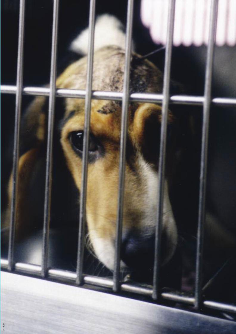
©PET
A
Charities Report:Layout 1 21/6/11 11:12 Page 47

Animal Aid exposes and campaignspeacefully against all animal abuse,and promotes a cruelty-free lifestyle
Animal AidThe Old Chapel, Bradford Street, Tonbridge, Kent, TN9 1AWTel: 01732 364546 | Email: [email protected]
www.animalaid.org.ukPublished by Animal Aid June 2011 | ISBN: 978-1-905327-27-0
©PET
A
Incorporated under the name Animal Abuse Injustice and Defence Society Limited, a company limited by guarantee.Registered in the UK no. 1787309.
Charities Report:Layout 1 21/6/11 11:12 Page 48