Embed Size (px)
Citation preview
Cancer Associated Thrombosis
Vicky Tagalakis MD MSc
Associate Professor McGill University
Division of Internal Medicine
Center for Clinical Epidemiology
Jewish General Hospital
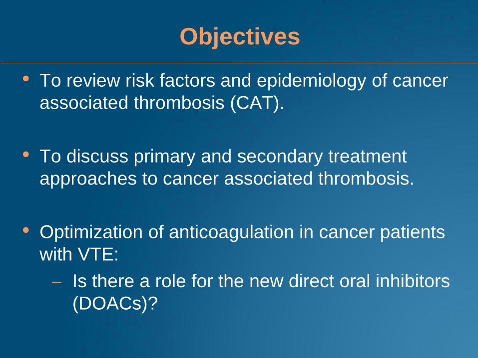
Objectives
• To review risk factors and epidemiology of cancer
associated thrombosis (CAT).
• To discuss primary and secondary treatment
approaches to cancer associated thrombosis.
• Optimization of anticoagulation in cancer patients
with VTE:
– Is there a role for the new direct oral inhibitors
(DOACs)?
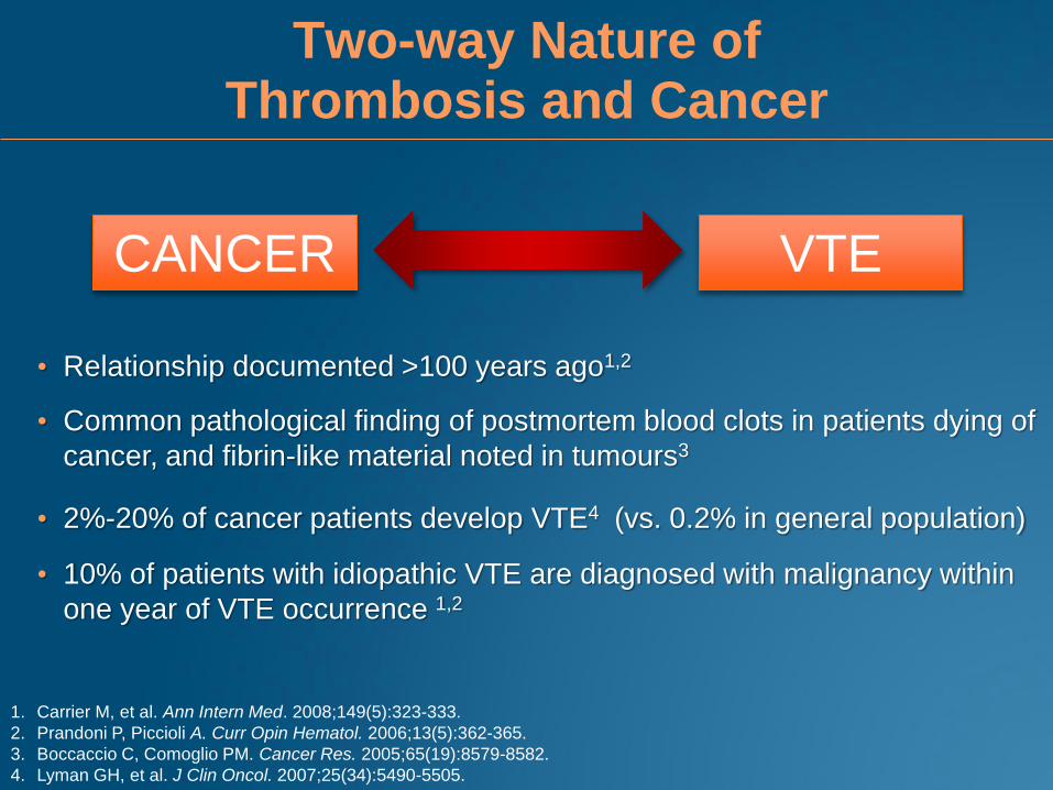
Two-way Nature of Thrombosis and Cancer
CANCER VTE
1. Carrier M, et al. Ann Intern Med. 2008;149(5):323-333.
2. Prandoni P, Piccioli A. Curr Opin Hematol. 2006;13(5):362-365.
3. Boccaccio C, Comoglio PM. Cancer Res. 2005;65(19):8579-8582.
4. Lyman GH, et al. J Clin Oncol. 2007;25(34):5490-5505.
• Relationship documented >100 years ago1,2
• Common pathological finding of postmortem blood clots in patients dying of
cancer, and fibrin-like material noted in tumours3
• 2%-20% of cancer patients develop VTE4 (vs. 0.2% in general population)
• 10% of patients with idiopathic VTE are diagnosed with malignancy within
one year of VTE occurrence 1,2
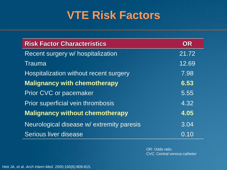
VTE Risk Factors
Risk Factor Characteristics OR
Recent surgery w/ hospitalization 21.72
Trauma 12.69
Hospitalization without recent surgery 7.98
Malignancy with chemotherapy 6.53
Prior CVC or pacemaker 5.55
Prior superficial vein thrombosis 4.32
Malignancy without chemotherapy 4.05
Neurological disease w/ extremity paresis 3.04
Serious liver disease 0.10
OR: Odds ratio
CVC: Central venous catheter
Heit JA, et al. Arch Intern Med. 2000;160(6):809-815.
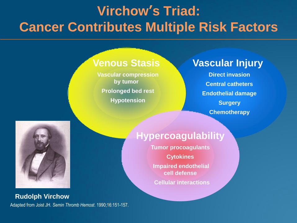
Virchow’s Triad:
Cancer Contributes Multiple Risk Factors
Venous Stasis Vascular compression
by tumor
Prolonged bed rest
Hypotension
Vascular Injury Direct invasion
Central catheters
Endothelial damage
Surgery
Chemotherapy
Hypercoagulability Tumor procoagulants
Cytokines
Impaired endothelial
cell defense
Cellular interactions
Rudolph Virchow
Adapted from Joist JH. Semin Thromb Hemost. 1990;16:151-157.

Factors That May Affect Risk for
Cancer-Associated VTE
Patient-related factors1,2
• Older age
• Female sex
• Black ethnicity
• Comorbid conditions
• Prior history of VTE
Treatment-related factors1,2
• Recent major surgery
• Hospitalization
• Chemotherapy
• Hormonal therapy
• Anti-angiogenic agents
• Erythropoiesis-stimulating agents
Cancer-related factors1,2
• Primary site of cancer and
histology
• Advanced stage
• Initial period after diagnosis
1. Khorana AA, et al. Thromb Res. 2007;120(Suppl 2):S41-50.
2. Lyman GH, et al. J Clin Oncol. 2007;25(34):5490-5505.
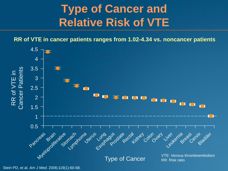
Type of Cancer and
Relative Risk of VTE
Stein PD, et al. Am J Med. 2006;119(1):60-68.
RR of VTE in cancer patients ranges from 1.02-4.34 vs. noncancer patients
Pancr
eas
Brain
Mye
lopr
olife
rativ
e
Stom
ach
Lym
phom
a
Ute
rus
Lung
Esoph
agus
Prost
ate
Rec
tal
Kidne
y
Col
on
Ova
ryLi
ver
Leuk
emia
Breas
t
Cer
vix
Bladd
er
RR
of
VT
E in
Ca
nce
r P
atien
ts
Type of Cancer
4.5
4
3.5
3
2.5
2
1.5
1
0.5
VTE: Venous thromboembolism
RR: Risk ratio
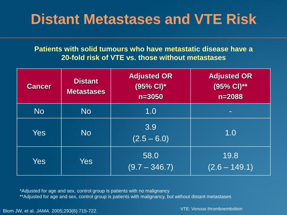
Patients with solid tumours who have metastatic disease have a
20-fold risk of VTE vs. those without metastases
Cancer Distant
Metastases
Adjusted OR
(95% CI)*
n=3050
Adjusted OR
(95% CI)**
n=2088
No No 1.0 -
Yes No 3.9
(2.5 – 6.0) 1.0
Yes Yes 58.0
(9.7 – 346.7)
19.8
(2.6 – 149.1)
Distant Metastases and VTE Risk
Blom JW, et al. JAMA. 2005;293(6):715-722.
*Adjusted for age and sex, control group is patients with no malignancy
**Adjusted for age and sex, control group is patients with malignancy, but without distant metastases
VTE: Venous thromboembolism

Cancer Treatment-Related
Risk Factors
Cancer Treatment VTE Incidence (%)
Chemotherapy1 9.6
Angiogenesis Inhibitorsa
Thalidomide-containing regimens2 3-22
Thalidomide alone2 3.4
Thalidomide + dexamethasone2 14.8
Thalidomide + chemotherapy2 22.0
Lenalidomide + high-dose dexamethasone3 26
Lenalidomide + low-dose dexamethasone3
Bevacizumab4b
12
11.9
Hormone Therapy5
Aromatase inhibitors 1.3-1.6
Tamoxifen 2.4-3.5
Erythropoietin6 7.5
a newly diagnosed patients only b meta-analysis VTE: Venous thromboembolism
1. Sousou T, Khorana A. J Clin Oncol. 2007 (ASCO meeting);25(18S):90-92. 2. Wu CM, et al. J Clin Oncol. 2007 (ASCO meeting);25(18S):9056.
3. Rajkmur SV, et al. Lancet Oncology. 2010;11(1):29-37. 4. Nalluri SR, et al. JAMA. 2008;300(19):2277-2285.
5. Lycette JL, et al. Breast Cancer Res Treat. 2006;99(3):249-255. 6. Bennett CL, et al. JAMA. 2008;299(8):914-924.
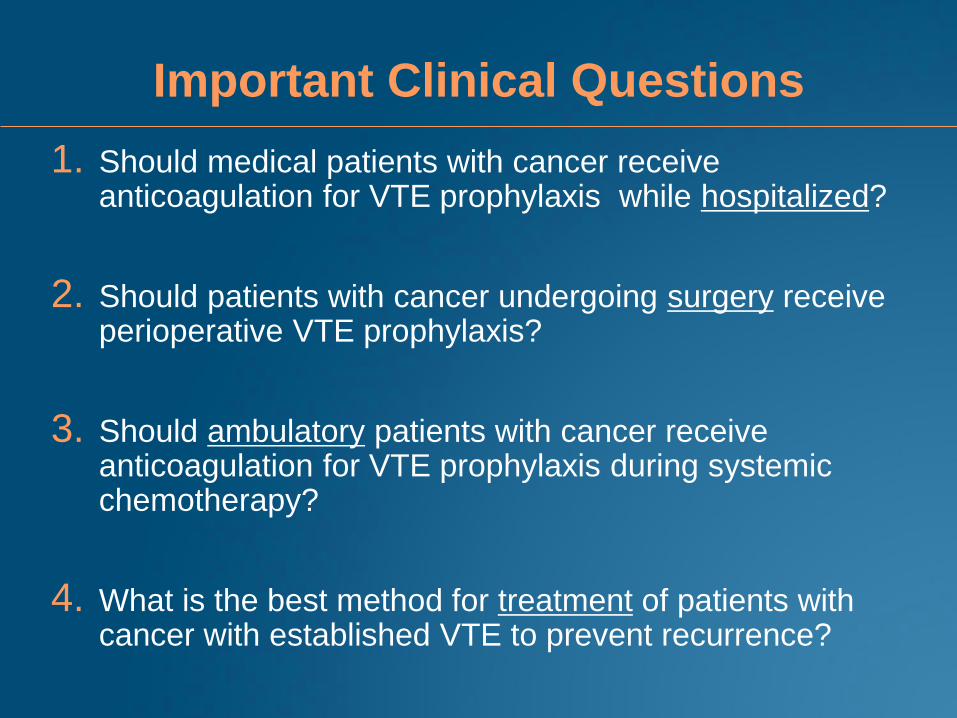
Important Clinical Questions
1. Should medical patients with cancer receive anticoagulation for VTE prophylaxis while hospitalized?
2. Should patients with cancer undergoing surgery receive perioperative VTE prophylaxis?
3. Should ambulatory patients with cancer receive anticoagulation for VTE prophylaxis during systemic chemotherapy?
4. What is the best method for treatment of patients with cancer with established VTE to prevent recurrence?
Prevention of VTE during medical hospitalization
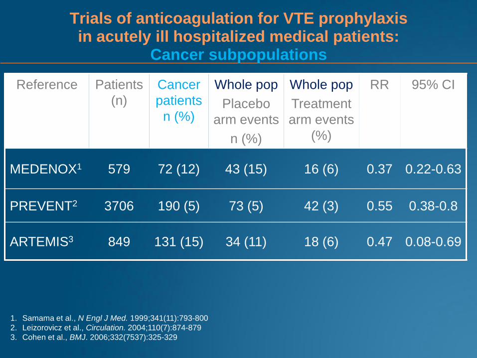
Trials of anticoagulation for VTE prophylaxis in acutely ill hospitalized medical patients:
Cancer subpopulations
Reference Patients
(n)
Cancer
patients
n (%)
Whole pop
Placebo
arm events
n (%)
Whole pop
Treatment
arm events
(%)
RR 95% CI
MEDENOX1 579 72 (12) 43 (15) 16 (6) 0.37 0.22-0.63
PREVENT2 3706 190 (5) 73 (5) 42 (3) 0.55 0.38-0.8
ARTEMIS3 849 131 (15) 34 (11) 18 (6) 0.47 0.08-0.69
1. Samama et al., N Engl J Med. 1999;341(11):793-800
2. Leizorovicz et al., Circulation. 2004;110(7):874-879
3. Cohen et al., BMJ. 2006;332(7537):325-329
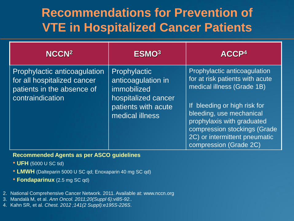
Recommendations for Prevention of
VTE in Hospitalized Cancer Patients
NCCN2 ESMO3 ACCP4
Prophylactic anticoagulation
for all hospitalized cancer
patients in the absence of
contraindication
Prophylactic
anticoagulation in
immobilized
hospitalized cancer
patients with acute
medical illness
Prophylactic anticoagulation
for at risk patients with acute
medical illness (Grade 1B)
If bleeding or high risk for
bleeding, use mechanical
prophylaxis with graduated
compression stockings (Grade
2C) or intermittent pneumatic
compression (Grade 2C)
Recommended Agents as per ASCO guidelines
• UFH (5000 U SC tid)
• LMWH (Dalteparin 5000 U SC qd; Enoxaparin 40 mg SC qd)
• Fondaparinux (2.5 mg SC qd)
2. National Comprehensive Cancer Network. 2011. Available at: www.nccn.org
3. Mandalà M, et al. Ann Oncol. 2011;20(Suppl 6):vi85-92..
4. Kahn SR, et al. Chest. 2012 ;141(2 Suppl):e195S-226S.
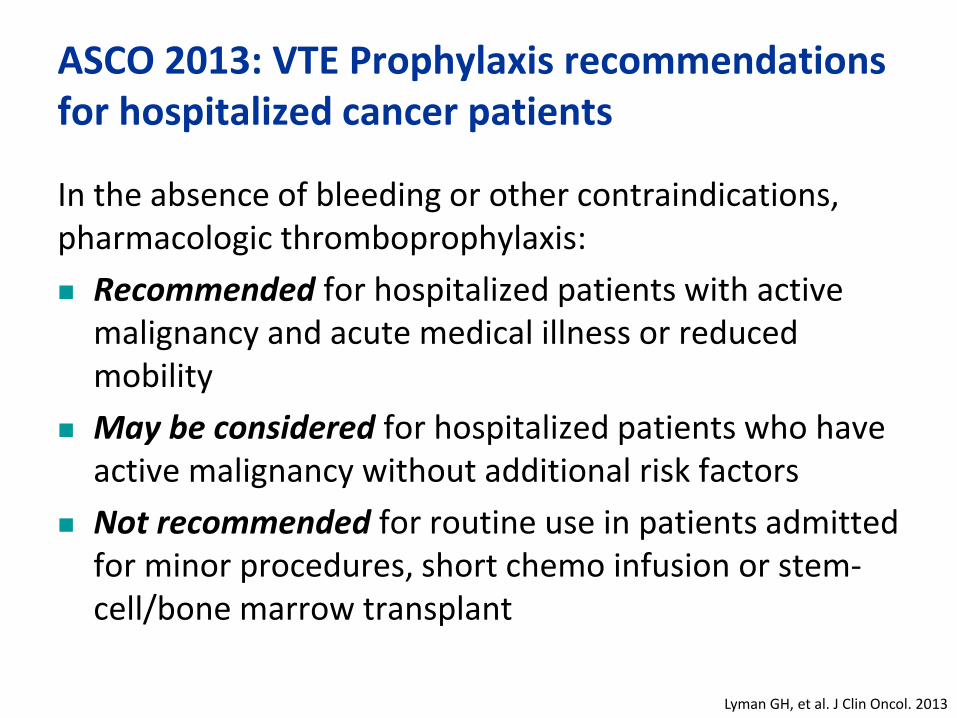
ASCO 2013: VTE Prophylaxis recommendations for hospitalized cancer patients
In the absence of bleeding or other contraindications, pharmacologic thromboprophylaxis:
Recommended for hospitalized patients with active malignancy and acute medical illness or reduced mobility
May be considered for hospitalized patients who have active malignancy without additional risk factors
Not recommended for routine use in patients admitted for minor procedures, short chemo infusion or stem-cell/bone marrow transplant
Lyman GH, et al. J Clin Oncol. 2013

For how long to prophylax
While in hospital?
While immobilized?
While acutely ill?

Prevention of VTE in the surgical setting
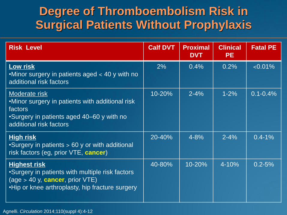
Degree of Thromboembolism Risk in Surgical Patients Without Prophylaxis
Risk Level Calf DVT Proximal
DVT
Clinical
PE
Fatal PE
Low risk
•Minor surgery in patients aged ˂ 40 y with no
additional risk factors
2% 0.4% 0.2% ˂0.01%
Moderate risk
•Minor surgery in patients with additional risk
factors
•Surgery in patients aged 40–60 y with no
additional risk factors
10-20%
2-4%
1-2%
0.1-0.4%
High risk
•Surgery in patients ˃ 60 y or with additional
risk factors (eg, prior VTE, cancer)
20-40%
4-8%
2-4%
0.4-1%
Highest risk
•Surgery in patients with multiple risk factors
(age ˃ 40 y, cancer, prior VTE)
•Hip or knee arthroplasty, hip fracture surgery
40-80% 10-20% 4-10% 0.2-5%
Agnelli. Circulation 2014;110(suppl 4):4-12
Surgery in cancer patients
• Cancer patients have higher rates of postoperative VTE,
PE, fatal PE, and death compared to non-cancer
patients despite thromboprophylaxis
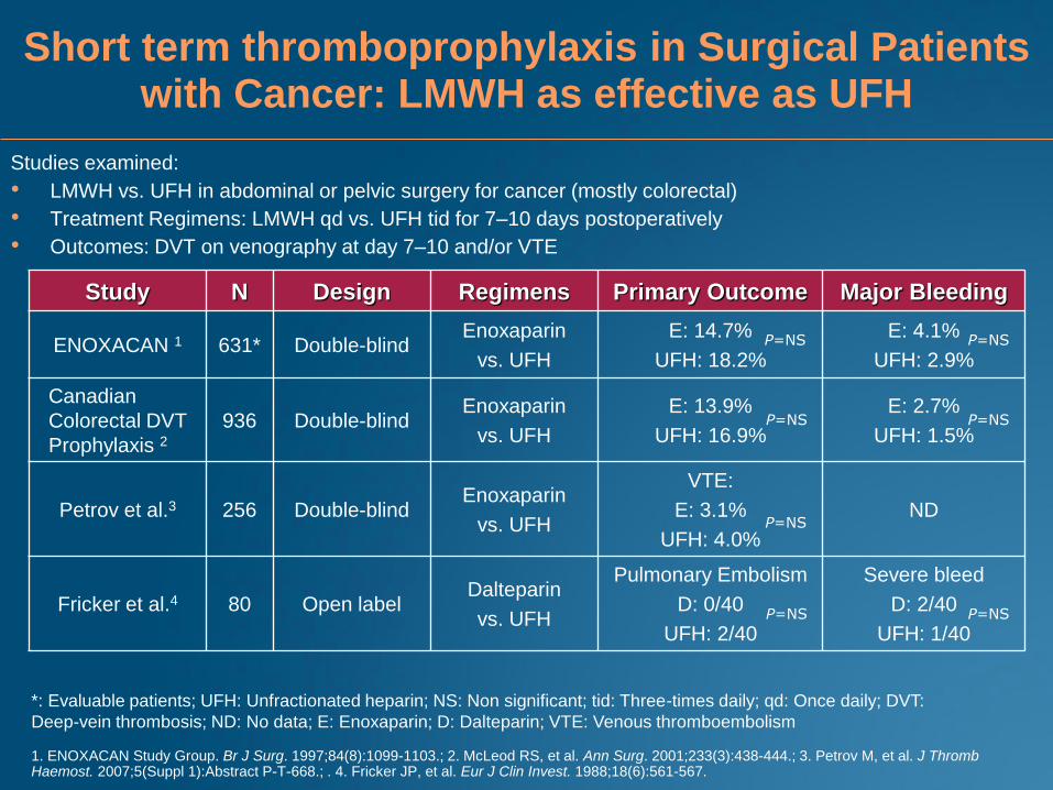
Short term thromboprophylaxis in Surgical Patients with Cancer: LMWH as effective as UFH
Studies examined:
• LMWH vs. UFH in abdominal or pelvic surgery for cancer (mostly colorectal)
• Treatment Regimens: LMWH qd vs. UFH tid for 7–10 days postoperatively
• Outcomes: DVT on venography at day 7–10 and/or VTE
1. ENOXACAN Study Group. Br J Surg. 1997;84(8):1099-1103.; 2. McLeod RS, et al. Ann Surg. 2001;233(3):438-444.; 3. Petrov M, et al. J Thromb Haemost. 2007;5(Suppl 1):Abstract P-T-668.; . 4. Fricker JP, et al. Eur J Clin Invest. 1988;18(6):561-567.
Study N Design Regimens Primary Outcome Major Bleeding
ENOXACAN 1 631* Double-blind Enoxaparin
vs. UFH
E: 14.7%
UFH: 18.2%
E: 4.1%
UFH: 2.9%
Canadian
Colorectal DVT
Prophylaxis 2
936 Double-blind Enoxaparin
vs. UFH
E: 13.9%
UFH: 16.9%
E: 2.7%
UFH: 1.5%
Petrov et al.3 256 Double-blind Enoxaparin
vs. UFH
VTE:
E: 3.1%
UFH: 4.0%
ND
Fricker et al.4 80 Open label Dalteparin
vs. UFH
Pulmonary Embolism
D: 0/40
UFH: 2/40
Severe bleed
D: 2/40
UFH: 1/40
*: Evaluable patients; UFH: Unfractionated heparin; NS: Non significant; tid: Three-times daily; qd: Once daily; DVT:
Deep-vein thrombosis; ND: No data; E: Enoxaparin; D: Dalteparin; VTE: Venous thromboembolism
P=NS P=NS
P=NS P=NS
P=NS
P=NS P=NS
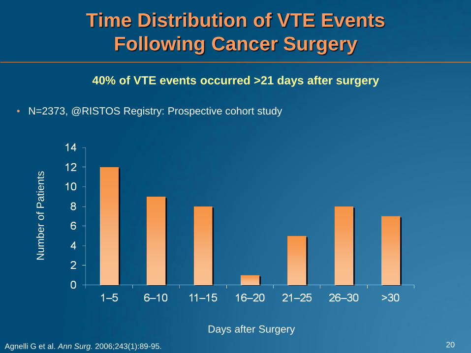
Time Distribution of VTE Events
Following Cancer Surgery
Agnelli G et al. Ann Surg. 2006;243(1):89-95.
• N=2373, @RISTOS Registry: Prospective cohort study
40% of VTE events occurred >21 days after surgery
Num
ber
of
Patients
Days after Surgery
20

Extended prophylaxis in cancer patients undergoing surgery
• Abdominal or pelvic surgery for cancer
• LMWH for 7 days vs. ~28 days
• Endpoints: routine bilateral venography at ~28 days and
symptomatic VTE
N Design Regimens VTE P-value
ENOXACAN II1 332 Double
blind
Enoxaparin
vs. placebo
4.8% (E) vs.
12% (P)
0.02
FAME2
(subgroup)
198 Open label Dalteparin
vs. no
prophylaxis
8.8% (D)
vs. 19.6%
(P)
0.03
1. Bergqvist et al. N Engl J Med. 2002;346(13);975-980
2. Rasmussen et al. J Thromb Haemostat. 2006;4(11):2384-90
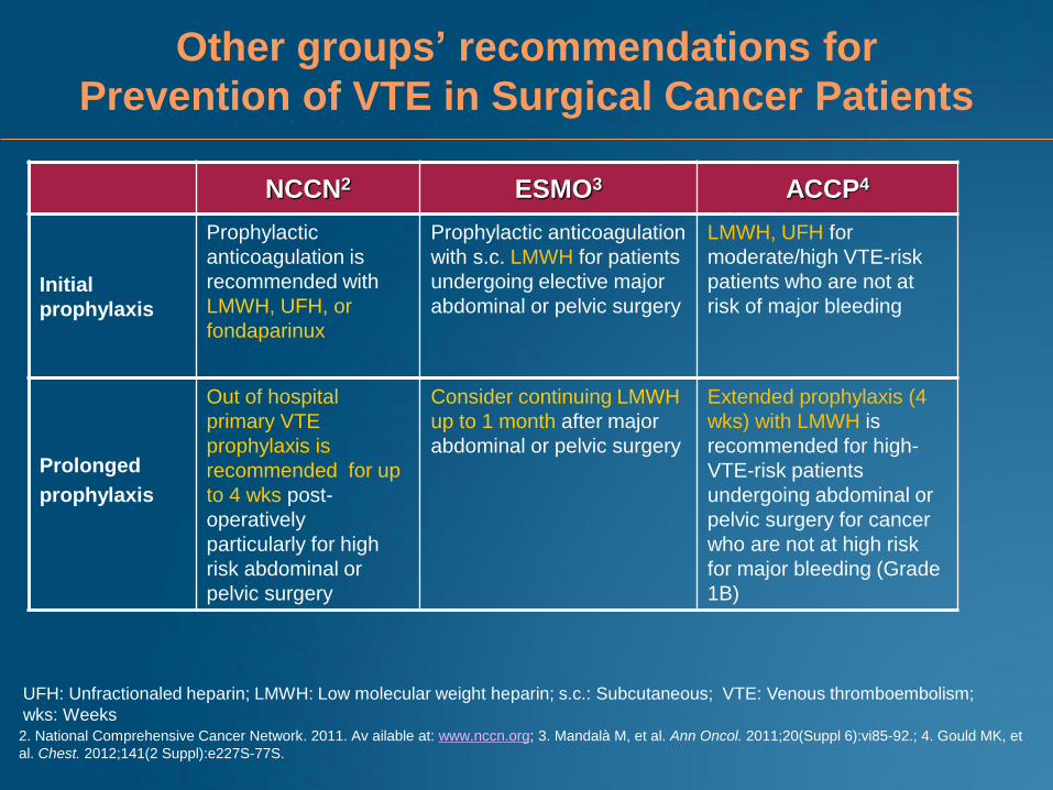
Other groups’ recommendations for
Prevention of VTE in Surgical Cancer Patients
NCCN2 ESMO3 ACCP4
Initial
prophylaxis
Prophylactic
anticoagulation is
recommended with
LMWH, UFH, or
fondaparinux
Prophylactic anticoagulation
with s.c. LMWH for patients
undergoing elective major
abdominal or pelvic surgery
LMWH, UFH for
moderate/high VTE-risk
patients who are not at
risk of major bleeding
Prolonged
prophylaxis
Out of hospital
primary VTE
prophylaxis is
recommended for up
to 4 wks post-
operatively
particularly for high
risk abdominal or
pelvic surgery
Consider continuing LMWH
up to 1 month after major
abdominal or pelvic surgery
Extended prophylaxis (4
wks) with LMWH is
recommended for high-
VTE-risk patients
undergoing abdominal or
pelvic surgery for cancer
who are not at high risk
for major bleeding (Grade
1B)
2. National Comprehensive Cancer Network. 2011. Av ailable at: www.nccn.org; 3. Mandalà M, et al. Ann Oncol. 2011;20(Suppl 6):vi85-92.; 4. Gould MK, et
al. Chest. 2012;141(2 Suppl):e227S-77S.
UFH: Unfractionaled heparin; LMWH: Low molecular weight heparin; s.c.: Subcutaneous; VTE: Venous thromboembolism;
wks: Weeks
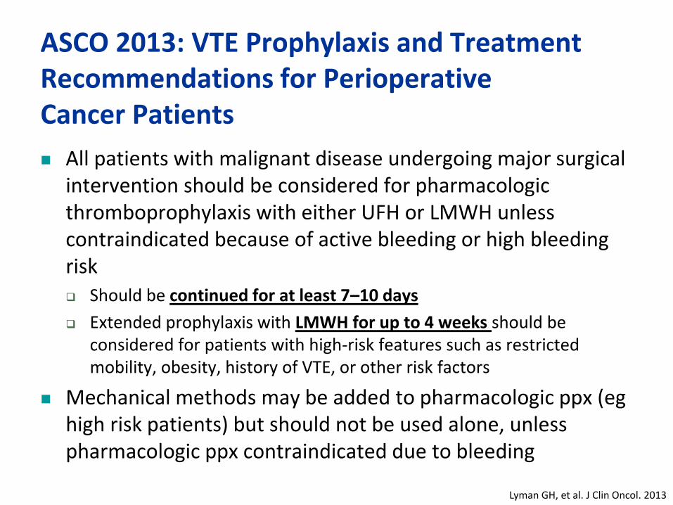
ASCO 2013: VTE Prophylaxis and Treatment Recommendations for Perioperative Cancer Patients
All patients with malignant disease undergoing major surgical intervention should be considered for pharmacologic thromboprophylaxis with either UFH or LMWH unless contraindicated because of active bleeding or high bleeding risk Should be continued for at least 7–10 days
Extended prophylaxis with LMWH for up to 4 weeks should be considered for patients with high-risk features such as restricted mobility, obesity, history of VTE, or other risk factors
Mechanical methods may be added to pharmacologic ppx (eg high risk patients) but should not be used alone, unless pharmacologic ppx contraindicated due to bleeding
Lyman GH, et al. J Clin Oncol. 2013

Summary: Recommendations for VTE prophylaxis in hospitalized medical and surgical cancer patients Most hospitalized medical and surgical cancer patients
should receive thromboprophylaxis
Prophylaxis for patients undergoing major cancer surgery should continue for min. 7–10 days
Consider extending postoperative prophylaxis up to 4 weeks in high-risk patients
Patients should be educated about the signs and symptoms of VTE
Low threshold to investigate for DVT and/or PE

Prophylaxis in ambulatory cancer patients undergoing chemotherapy
Prevention of VTE in patients treated
with thalidomide-based regimens
Cumulative risk of recurrent VTE in patients who
received MPT, MPT and Enoxaparin or MPL and ASA
Palumbo A, et al. J Thromb Haemost. 2006;4(8):1842-1845.
0 5 10 15 20 25
20%
10%
0%
Cum
ula
tive P
erc
enta
ge (
%)
Months
MPT (n=65)
MPT and Enoxaparin (n=78)
VTE: Venous thromboembolism
ASA: Acetylsalicylic acid
MPT: Melphalan and prednisone plus thalidomide
MPL: Melphalan and prednisone plus lenalidomide
• Retrospective analysis
MPL and ASA (n=50)
• Dose: Enoxaparin 40 mg qd or ASA 100 mg qd
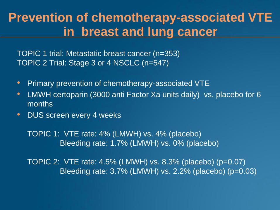
Prevention of chemotherapy-associated VTE in breast and lung cancer
TOPIC 1 trial: Metastatic breast cancer (n=353)
TOPIC 2 Trial: Stage 3 or 4 NSCLC (n=547)
• Primary prevention of chemotherapy-associated VTE
• LMWH certoparin (3000 anti Factor Xa units daily) vs. placebo for 6
months
• DUS screen every 4 weeks
TOPIC 1: VTE rate: 4% (LMWH) vs. 4% (placebo)
Bleeding rate: 1.7% (LMWH) vs. 0% (placebo)
TOPIC 2: VTE rate: 4.5% (LMWH) vs. 8.3% (placebo) (p=0.07)
Bleeding rate: 3.7% (LMWH) vs. 2.2% (placebo) (p=0.03)
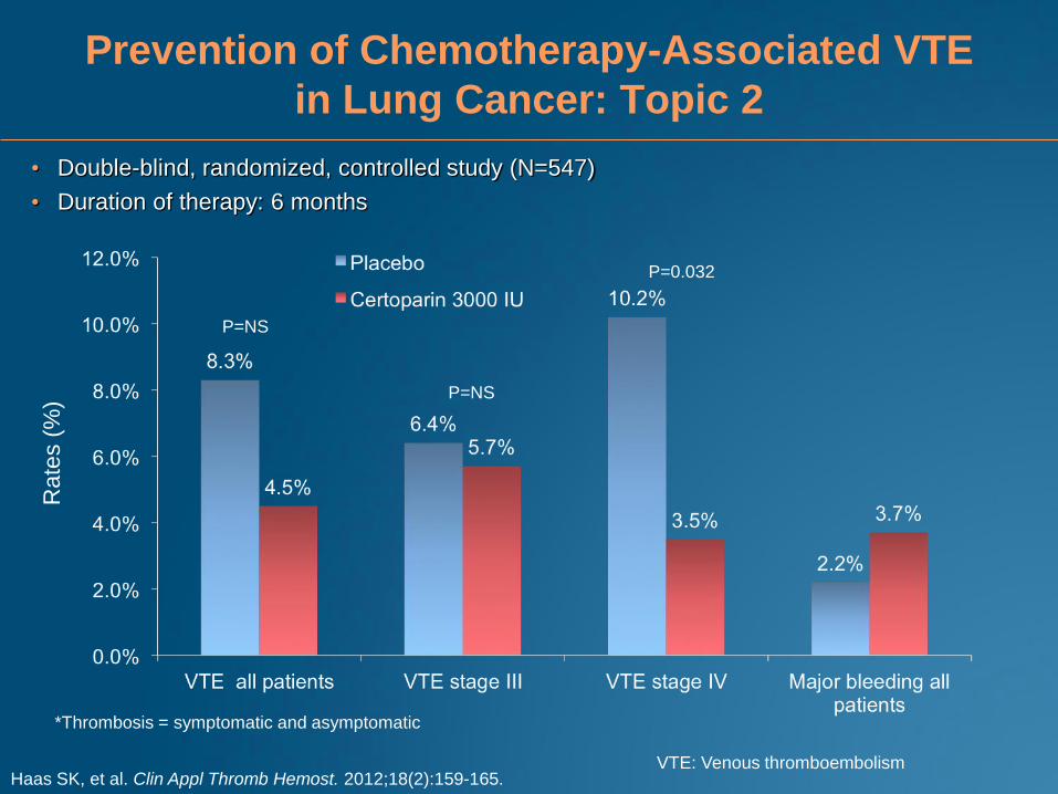
Prevention of Chemotherapy-Associated VTE
in Lung Cancer: Topic 2
Haas SK, et al. Clin Appl Thromb Hemost. 2012;18(2):159-165.
*Thrombosis = symptomatic and asymptomatic
• Double-blind, randomized, controlled study (N=547)
• Duration of therapy: 6 months
Rate
s (
%)
P=NS
P=NS
P=0.032
VTE: Venous thromboembolism

Primary Thromboprophylaxis in Patients
Receiving Chemotherapy: PROTECHT Trial
P=0.02
Cumulative Hazard of Thromboembolic Events by Treatment
• 5/769 (0.7%) patients in the nadroparin group and 0/381 patients in the placebo group had a major bleeding
event (P=0.18)
• The incidence of minor bleeding was 7.4% (57/769) with Nadroparin and 7.9% (30/381) with placebo
Agnelli G, et al. Lancet Oncol. 2009;10(10):943-949.
• Double-blind, randomized study; metastatic or locally advanced lung, GI, pancreatic,
breast, ovarian, or heard and neck
• Duration of therapy: Length of chemotherapy (maximum 4 months)
• Primary outcome: Composite of symptomatic venous and arterial thrombotic events
Rate
s (
%)
Rate of Thromboembolic Events
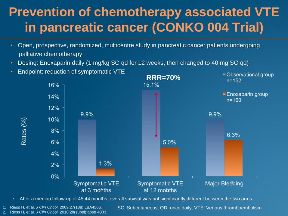
Prevention of chemotherapy associated VTE
in pancreatic cancer (CONKO 004 Trial)
1. Riess H, et al. J Clin Oncol. 2009;27(188):LBA4506.
2. Riess H, et al. J Clin Oncol. 2010:28(suppl):abstr 4033.
RRR=70%
Rate
s (
%)
• Open, prospective, randomized, multicentre study in pancreatic cancer patients undergoing
palliative chemotherapy
• Dosing: Enoxaparin daily (1 mg/kg SC qd for 12 weeks, then changed to 40 mg SC qd)
• Endpoint: reduction of symptomatic VTE
• After a median follow-up of 45.44 months, overall survival was not significantly different between the two arms
1
2 2
SC: Subcutaneous; QD: once daily; VTE: Venous thromboembolism
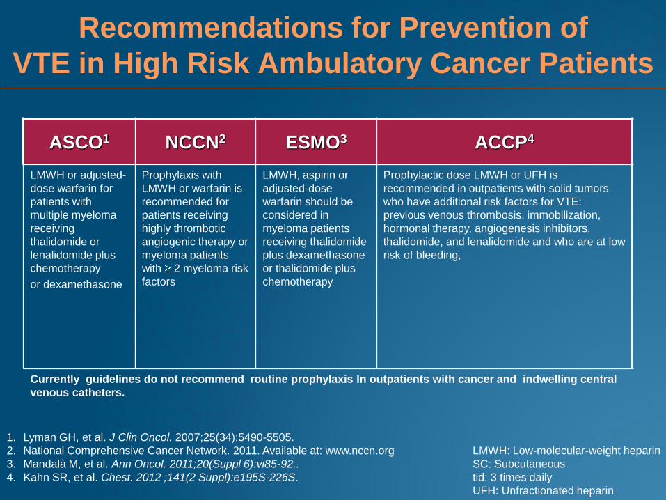
Recommendations for Prevention of
VTE in High Risk Ambulatory Cancer Patients
ASCO1 NCCN2 ESMO3 ACCP4
LMWH or adjusted-
dose warfarin for
patients with
multiple myeloma
receiving
thalidomide or
lenalidomide plus
chemotherapy
or dexamethasone
Prophylaxis with
LMWH or warfarin is
recommended for
patients receiving
highly thrombotic
angiogenic therapy or
myeloma patients
with 2 myeloma risk
factors
LMWH, aspirin or
adjusted-dose
warfarin should be
considered in
myeloma patients
receiving thalidomide
plus dexamethasone
or thalidomide plus
chemotherapy
Prophylactic dose LMWH or UFH is
recommended in outpatients with solid tumors
who have additional risk factors for VTE:
previous venous thrombosis, immobilization,
hormonal therapy, angiogenesis inhibitors,
thalidomide, and lenalidomide and who are at low
risk of bleeding,
Currently guidelines do not recommend routine prophylaxis In outpatients with cancer and indwelling central
venous catheters.
LMWH: Low-molecular-weight heparin
SC: Subcutaneous
tid: 3 times daily
UFH: Unfractionated heparin
1. Lyman GH, et al. J Clin Oncol. 2007;25(34):5490-5505.
2. National Comprehensive Cancer Network. 2011. Available at: www.nccn.org
3. Mandalà M, et al. Ann Oncol. 2011;20(Suppl 6):vi85-92..
4. Kahn SR, et al. Chest. 2012 ;141(2 Suppl):e195S-226S.
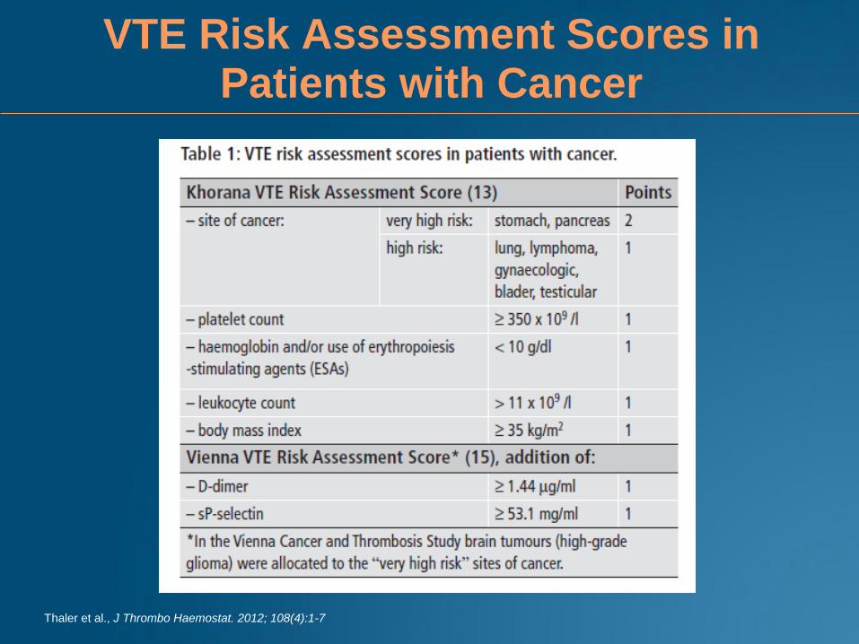
VTE Risk Assessment Scores in Patients with Cancer
Thaler et al., J Thrombo Haemostat. 2012; 108(4):1-7
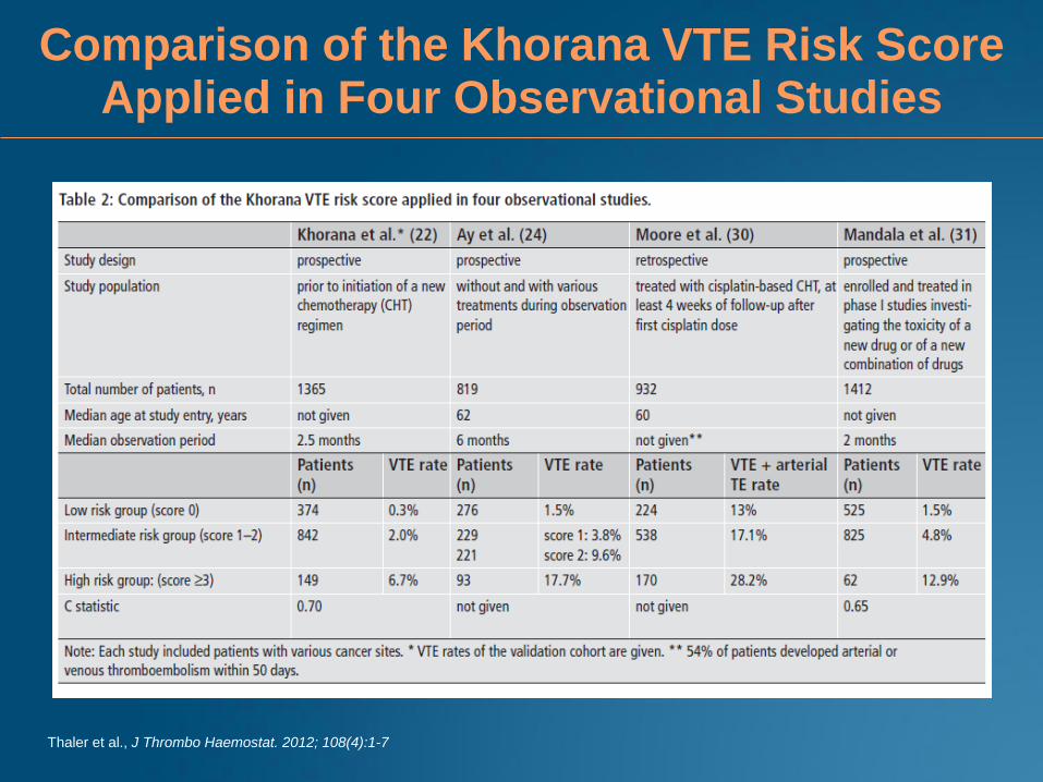
Comparison of the Khorana VTE Risk Score Applied in Four Observational Studies
Thaler et al., J Thrombo Haemostat. 2012; 108(4):1-7
Treatment of
Cancer-Associated Thrombosis
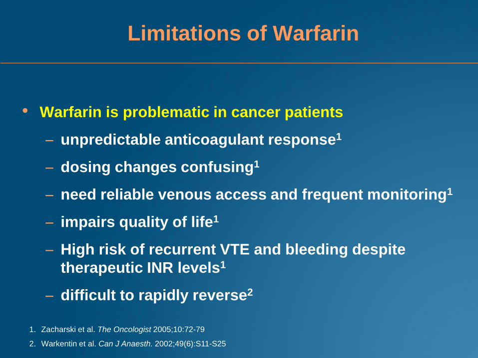
Limitations of Warfarin
• Warfarin is problematic in cancer patients
– unpredictable anticoagulant response1
– dosing changes confusing1
– need reliable venous access and frequent monitoring1
– impairs quality of life1
– High risk of recurrent VTE and bleeding despite
therapeutic INR levels1
– difficult to rapidly reverse2
1. Zacharski et al. The Oncologist 2005;10:72-79
2. Warkentin et al. Can J Anaesth. 2002;49(6):S11-S25
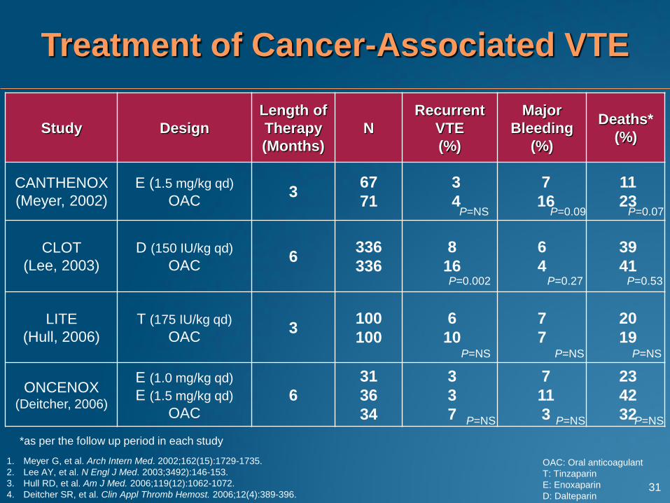
Study Design
Length of
Therapy
(Months)
N
Recurrent
VTE
(%)
Major
Bleeding
(%)
Deaths*
(%)
CANTHENOX
(Meyer, 2002)
E (1.5 mg/kg qd)
OAC 3
67
71
3
4
7
16
11
23
CLOT
(Lee, 2003)
D (150 IU/kg qd)
OAC 6
336
336
8
16
6
4
39
41
LITE
(Hull, 2006)
T (175 IU/kg qd)
OAC 3
100
100
6
10
7
7
20
19
ONCENOX (Deitcher, 2006)
E (1.0 mg/kg qd)
E (1.5 mg/kg qd)
OAC
6
31
36
34
3
3
7
7
11
3
23
42
32
P=0.07
P=NS P=NS
P=NS
P=0.09 P=NS
P=NS
P=NS
P=NS
P=0.002 P=0.53 P=0.27
1. Meyer G, et al. Arch Intern Med. 2002;162(15):1729-1735.
2. Lee AY, et al. N Engl J Med. 2003;3492):146-153.
3. Hull RD, et al. Am J Med. 2006;119(12):1062-1072.
4. Deitcher SR, et al. Clin Appl Thromb Hemost. 2006;12(4):389-396.
OAC: Oral anticoagulant
T: Tinzaparin
E: Enoxaparin
D: Dalteparin
Treatment of Cancer-Associated VTE
31
*as per the follow up period in each study
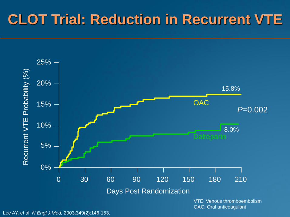
CLOT Trial: Reduction in Recurrent VTE
Lee AY, et al. N Engl J Med, 2003;349(2):146-153.
P=0.002
0%
5%
10%
15%
20%
25%
Days Post Randomization
0 30 60 90 120 150 180 210
Recurr
ent
VT
E P
robabili
ty (
%)
Dalteparin
OAC
15.8%
8.0%
VTE: Venous thromboembolism
OAC: Oral anticoagulant
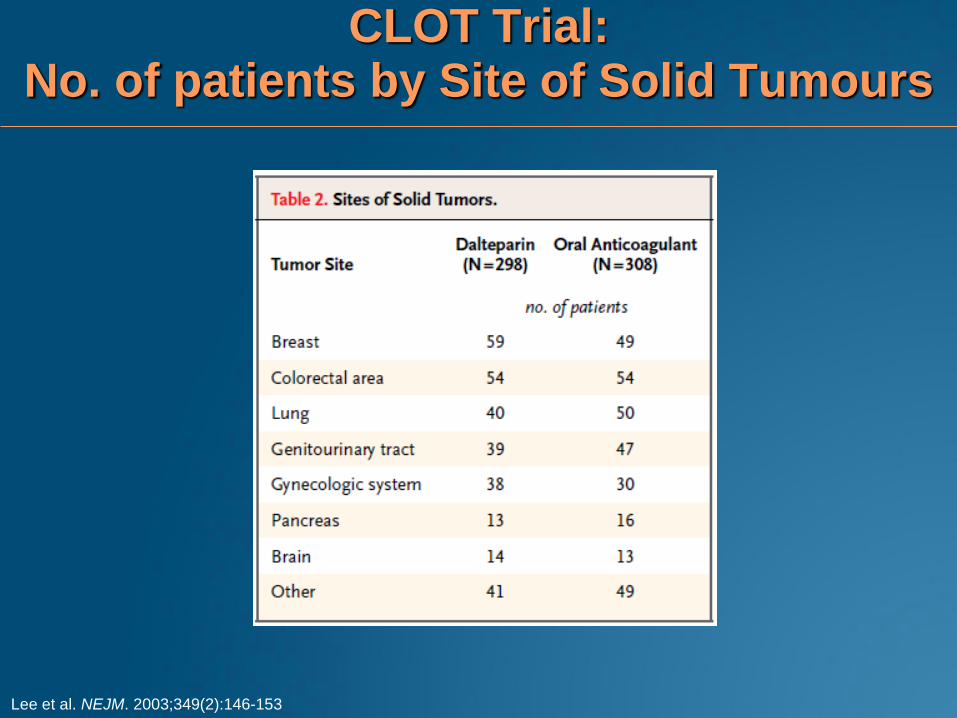
CLOT Trial: No. of patients by Site of Solid Tumours
Lee et al. NEJM. 2003;349(2):146-153
CLOT Trial:
Bleeding Events and Survival* Data
Bleeding1 Dalteparin
(N=338)
OAC
(N=335) P**
Major bleed 6% 4% 0.27
Any bleed 14% 19% 0.09
1. Lee AY, et al. N Engl J Med. 2003;349(2):146-153.
2. Lee AY, et al. J Clin Oncol. 2005; 23(10):2123-2129.
OAC: Oral anticoagulant
Probability of
Death*2 Dalteparin OAC P
Patients with
Metastases 72% 69% 0.46
Patients without
Metastases 20% 36% 0.03
*Posthoc analysis
** Fisher’s exact test
34
0.50 (0.35 – 0.72) 0.80 (0.61 – 1.05)
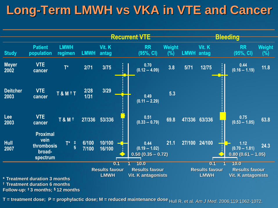
Recurrent VTE Bleeding
Study Patient
population LMWH
regimen LMWH Vit. K antag
RR (95%, CI)
Weight (%) LMWH
Vit. K antag
RR (95%, CI)
Weight (%)
Meyer 2002
VTE cancer
T* 2/71 3/75 0.70
(0.12 – 4.09) 3.8 5/71 12/75
0.44 (0.16 – 1.19)
11.8
Deitcher 2003
VTE cancer
T & M † T 2/28 1/31
3/29
0.49 (0.11 – 2.29)
5.3
Lee 2003
VTE cancer
T & M † 27/336 53/336 0.51
(0.33 – 0.79) 69.8 47/336 63/336
0.75 (0.53 – 1.05)
63.8
Hull 2007
Proximal vein
thrombosis broad-
spectrum
T* ‡
§ 6/100 7/100
10/100 16/100
0.44 (0.19 – 1.02)
21.1
27/100
24/100
1.12 (0.70 – 1.81)
24.3
Long-Term LMWH vs VKA in VTE and Cancer
Hull R, et al. Am J Med. 2006;119:1062-1072.
* Treatment duration 3 months † Treatment duration 6 months
Follow-up: ‡ 3 months; § 12 months
T = treatment dose; P = prophylactic dose; M = reduced maintenance dose
Results favour Results favour Results favour Results favour
LMWH Vit. K antagonists LMWH Vit. K antagonists
0.1 1 10.0 0.1 1 10.0
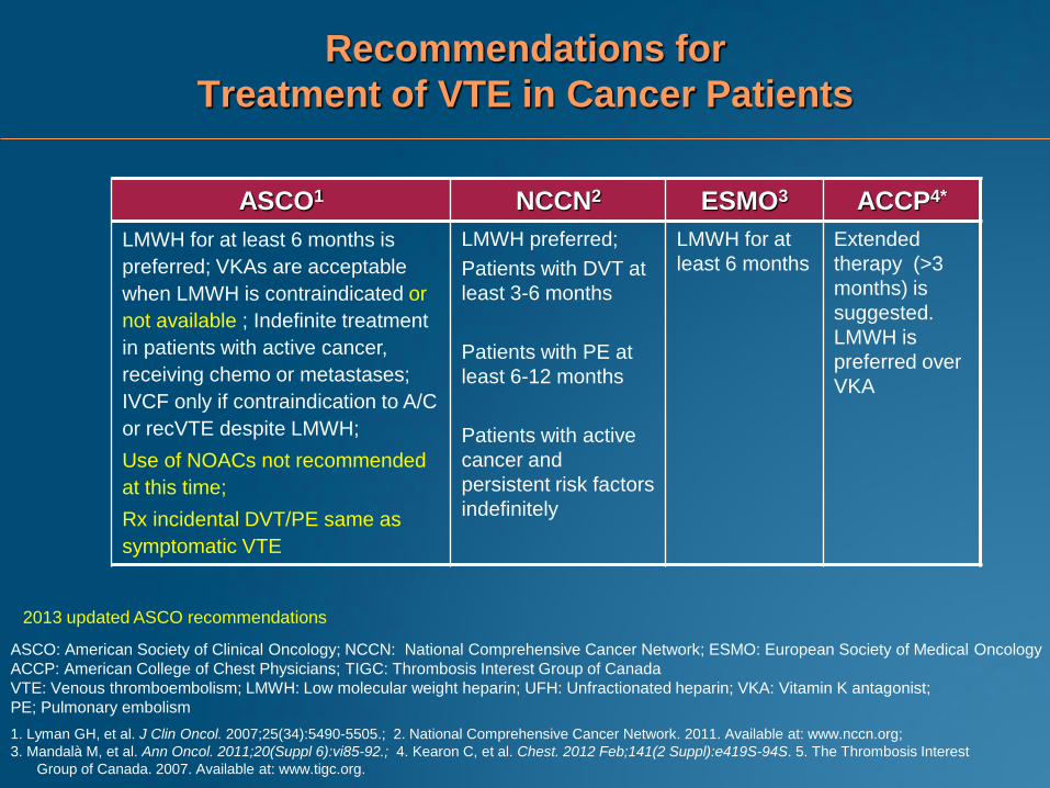
Recommendations for
Treatment of VTE in Cancer Patients
ASCO1 NCCN2 ESMO3 ACCP4*
LMWH for at least 6 months is
preferred; VKAs are acceptable
when LMWH is contraindicated or
not available ; Indefinite treatment
in patients with active cancer,
receiving chemo or metastases;
IVCF only if contraindication to A/C
or recVTE despite LMWH;
Use of NOACs not recommended
at this time;
Rx incidental DVT/PE same as
symptomatic VTE
LMWH preferred;
Patients with DVT at
least 3-6 months
Patients with PE at
least 6-12 months
Patients with active
cancer and
persistent risk factors
indefinitely
LMWH for at
least 6 months
Extended
therapy (>3
months) is
suggested.
LMWH is
preferred over
VKA
ASCO: American Society of Clinical Oncology; NCCN: National Comprehensive Cancer Network; ESMO: European Society of Medical Oncology
ACCP: American College of Chest Physicians; TIGC: Thrombosis Interest Group of Canada
VTE: Venous thromboembolism; LMWH: Low molecular weight heparin; UFH: Unfractionated heparin; VKA: Vitamin K antagonist;
PE; Pulmonary embolism
1. Lyman GH, et al. J Clin Oncol. 2007;25(34):5490-5505.; 2. National Comprehensive Cancer Network. 2011. Available at: www.nccn.org;
3. Mandalà M, et al. Ann Oncol. 2011;20(Suppl 6):vi85-92.; 4. Kearon C, et al. Chest. 2012 Feb;141(2 Suppl):e419S-94S. 5. The Thrombosis Interest
Group of Canada. 2007. Available at: www.tigc.org.
2013 updated ASCO recommendations
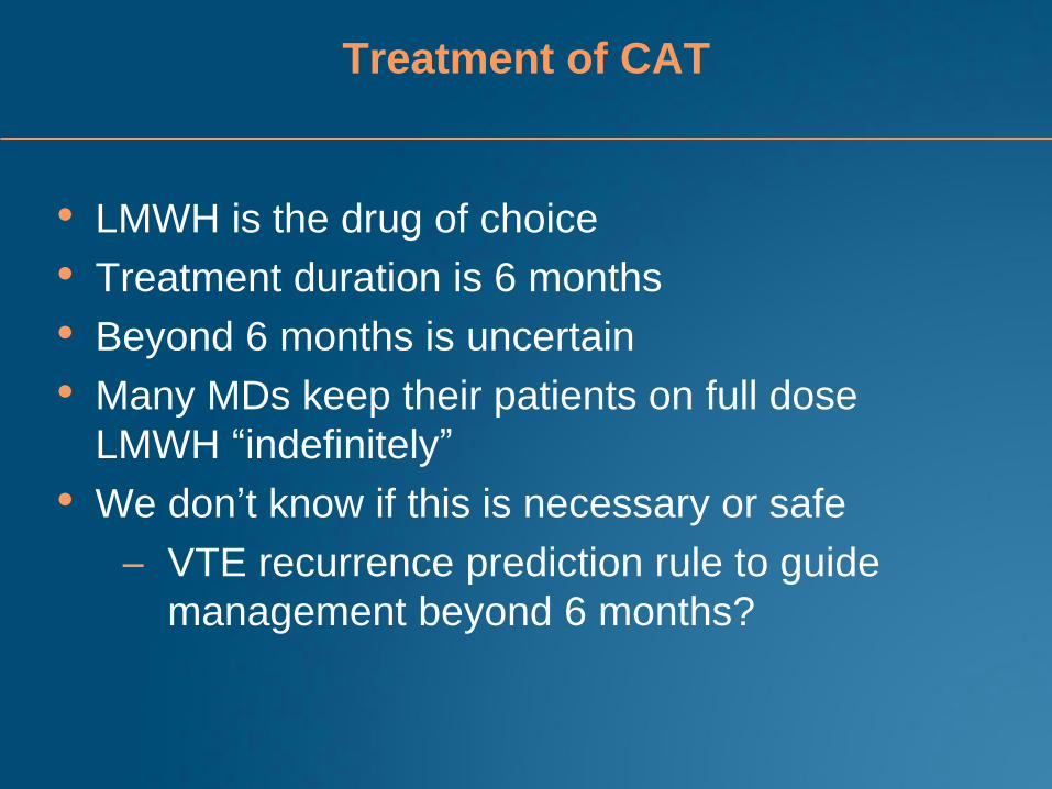
Treatment of CAT
• LMWH is the drug of choice
• Treatment duration is 6 months
• Beyond 6 months is uncertain
• Many MDs keep their patients on full dose
LMWH “indefinitely”
• We don’t know if this is necessary or safe
– VTE recurrence prediction rule to guide
management beyond 6 months?
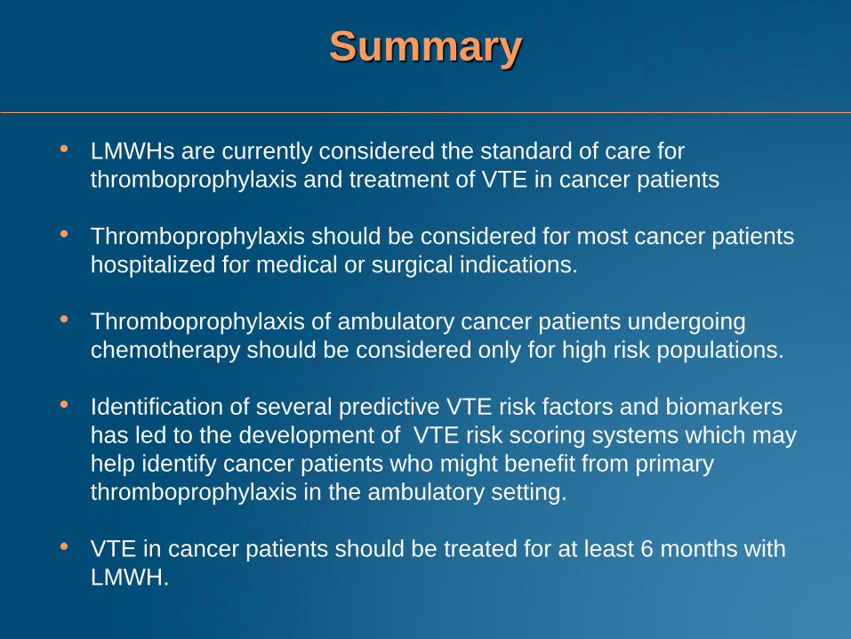
Summary
• LMWHs are currently considered the standard of care for
thromboprophylaxis and treatment of VTE in cancer patients
• Thromboprophylaxis should be considered for most cancer patients
hospitalized for medical or surgical indications.
• Thromboprophylaxis of ambulatory cancer patients undergoing
chemotherapy should be considered only for high risk populations.
• Identification of several predictive VTE risk factors and biomarkers
has led to the development of VTE risk scoring systems which may
help identify cancer patients who might benefit from primary
thromboprophylaxis in the ambulatory setting.
• VTE in cancer patients should be treated for at least 6 months with
LMWH.

Case scenarios Optimizing management of CAT
• Can I use a DOAC to treat CAT?
• What if my patient has a recurrent thrombosis on
LMWH?
• My patient has a catheter-related DVT? How do
I treat? Do I remove the catheter?
44
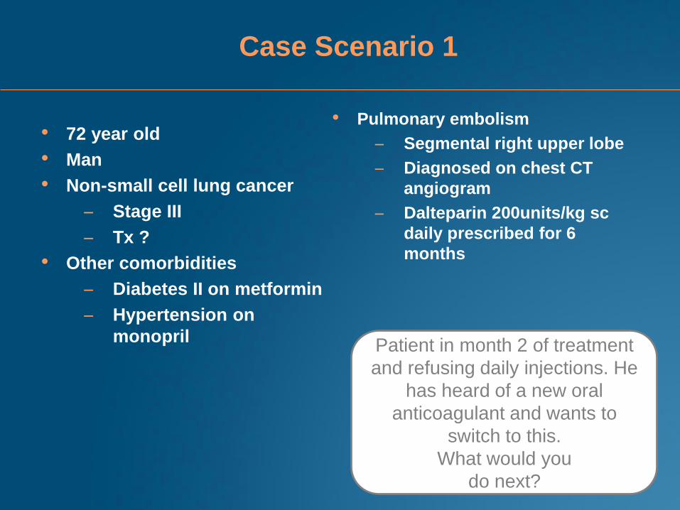
Case Scenario 1
• 72 year old
• Man
• Non-small cell lung cancer
– Stage III
– Tx ?
• Other comorbidities
– Diabetes II on metformin
– Hypertension on
monopril
• Pulmonary embolism
– Segmental right upper lobe
– Diagnosed on chest CT
angiogram
– Dalteparin 200units/kg sc
daily prescribed for 6
months
Patient in month 2 of treatment
and refusing daily injections. He
has heard of a new oral
anticoagulant and wants to
switch to this.
What would you
do next?
Case Scenario 1
• You ask why he is refusing the injections.
• He explains that he is fed up with the injections; they are
too painful
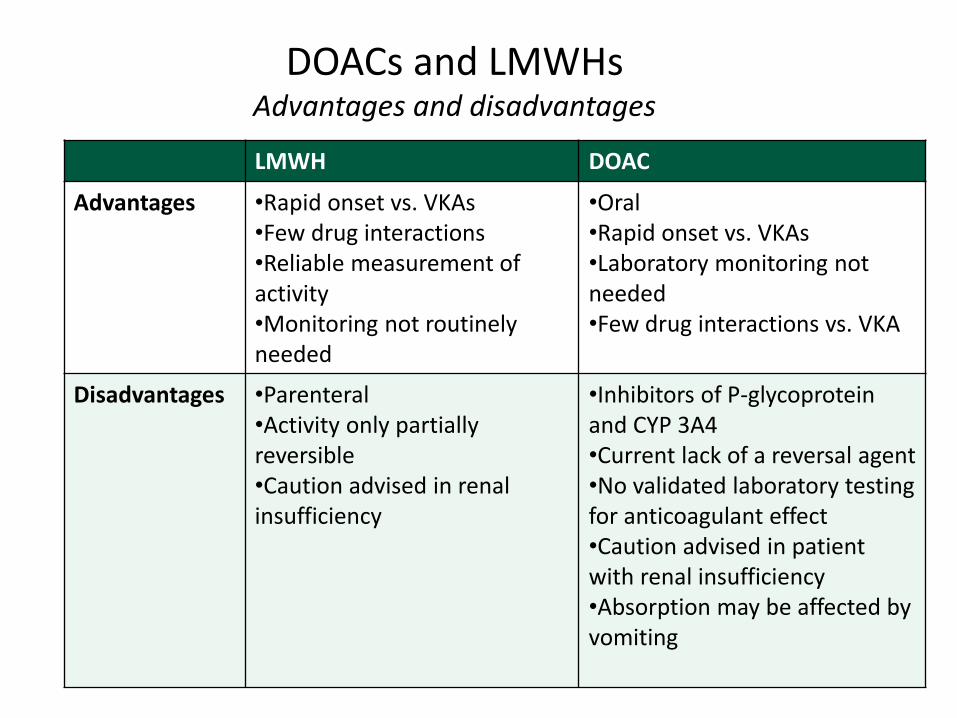
DOACs and LMWHs Advantages and disadvantages
LMWH DOAC
Advantages •Rapid onset vs. VKAs •Few drug interactions •Reliable measurement of activity •Monitoring not routinely needed
•Oral •Rapid onset vs. VKAs •Laboratory monitoring not needed •Few drug interactions vs. VKA
Disadvantages •Parenteral •Activity only partially reversible •Caution advised in renal insufficiency
•Inhibitors of P-glycoprotein and CYP 3A4 •Current lack of a reversal agent •No validated laboratory testing for anticoagulant effect •Caution advised in patient with renal insufficiency •Absorption may be affected by vomiting
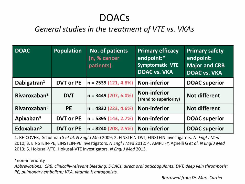
DOACs General studies in the treatment of VTE vs. VKAs
DOAC Population No. of patients (n, % cancer patients)
Primary efficacy endpoint:* Symptomatic VTE
DOAC vs. VKA
Primary safety endpoint: Major and CRB DOAC vs. VKA
Dabigatran1 DVT or PE n = 2539 (121, 4.8%) Non-inferior DOAC superior
Rivaroxaban2 DVT n = 3449 (207, 6.0%) Non-inferior (Trend to superiority)
Not different
Rivaroxaban3 PE n = 4832 (223, 4.6%) Non-inferior Not different
Apixaban4 DVT or PE n = 5395 (143, 2.7%) Non-inferior DOAC superior
Edoxaban5 DVT or PE n = 8240 (208, 2.5%) Non-inferior DOAC superior
1. RE-COVER, Schulman S et al. N Engl J Med 2009; 2. EINSTEIN-DVT, EINSTEIN Investigators. N Engl J Med 2010; 3. EINSTEIN-PE, EINSTEIN-PE Investigators. N Engl J Med 2012; 4. AMPLIFY, Agnelli G et al. N Engl J Med 2013; 5. Hokusai-VTE, Hokusai-VTE Investigators. N Engl J Med 2013. *non-inferiority Abbreviations: CRB, clinically-relevant bleeding; DOACs, direct oral anticoagulants; DVT, deep vein thrombosis; PE, pulmonary embolism; VKA, vitamin K antagonists. Borrowed from Dr. Marc Carrier
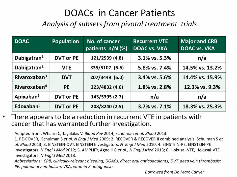
DOACs in Cancer Patients Analysis of subsets from pivotal treatment trials
• There appears to be a reduction in recurrent VTE in patients with cancer that has warranted further investigation.
DOAC Population No. of cancer patients n/N (%)
Recurrent VTE DOAC vs. VKA
Major and CRB DOAC vs. VKA
Dabigatran1 DVT or PE 121/2539 (4.8) 3.1% vs. 5.3% n/a
Dabigatran2 VTE 335/5107 (6.6) 5.8% vs. 7.4% 14.5% vs. 13.2%
Rivaroxaban3 DVT 207/3449 (6.0) 3.4% vs. 5.6% 14.4% vs. 15.9%
Rivaroxaban4 PE 223/4832 (4.6) 1.8% vs. 2.8% 12.3% vs. 9.3%
Apixaban5 DVT or PE 143/5395 (2.7) n/a n/a
Edoxaban6 DVT or PE 208/8240 (2.5) 3.7% vs. 7.1% 18.3% vs. 25.3%
Adapted from: Wharin C, Tagalakis V. Blood Rev 2014; Schulman et al. Blood 2013. 1. RE-COVER, Schulman S et al. N Engl J Med 2009; 2. RECOVER & RECOVER II combined analysis. Schulman S et al. Blood 2013; 3. EINSTEIN-DVT, EINSTEIN Investigators. N Engl J Med 2010; 4. EINSTEIN-PE, EINSTEIN-PE Investigators. N Engl J Med 2012; 5. AMPLIFY, Agnelli G et al., N Engl J Med 2013; 6. Hokusai-VTE, Hokusai-VTE Investigators. N Engl J Med 2013. Abbreviations: CRB, clinically-relevant bleeding; DOACs, direct oral anticoagulants; DVT, deep vein thrombosis; PE, pulmonary embolism; VKA, vitamin K antagonists Borrowed from Dr. Marc Carrier
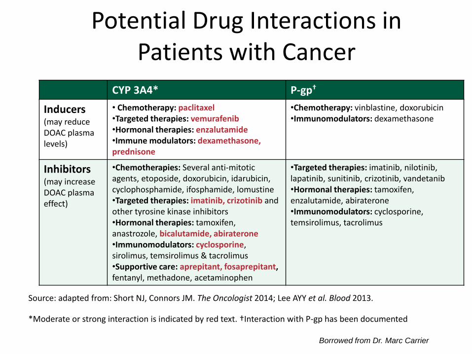
CYP 3A4* P-gp†
Inducers (may reduce DOAC plasma levels)
• Chemotherapy: paclitaxel •Targeted therapies: vemurafenib •Hormonal therapies: enzalutamide •Immune modulators: dexamethasone, prednisone
•Chemotherapy: vinblastine, doxorubicin •Immunomodulators: dexamethasone
Inhibitors (may increase DOAC plasma effect)
•Chemotherapies: Several anti-mitotic agents, etoposide, doxorubicin, idarubicin, cyclophosphamide, ifosphamide, lomustine •Targeted therapies: imatinib, crizotinib and other tyrosine kinase inhibitors •Hormonal therapies: tamoxifen, anastrozole, bicalutamide, abiraterone •Immunomodulators: cyclosporine, sirolimus, temsirolimus & tacrolimus •Supportive care: aprepitant, fosaprepitant, fentanyl, methadone, acetaminophen
•Targeted therapies: imatinib, nilotinib, lapatinib, sunitinib, crizotinib, vandetanib •Hormonal therapies: tamoxifen, enzalutamide, abiraterone •Immunomodulators: cyclosporine, temsirolimus, tacrolimus
Potential Drug Interactions in Patients with Cancer
*Moderate or strong interaction is indicated by red text. †Interaction with P-gp has been documented
Source: adapted from: Short NJ, Connors JM. The Oncologist 2014; Lee AYY et al. Blood 2013.
Borrowed from Dr. Marc Carrier
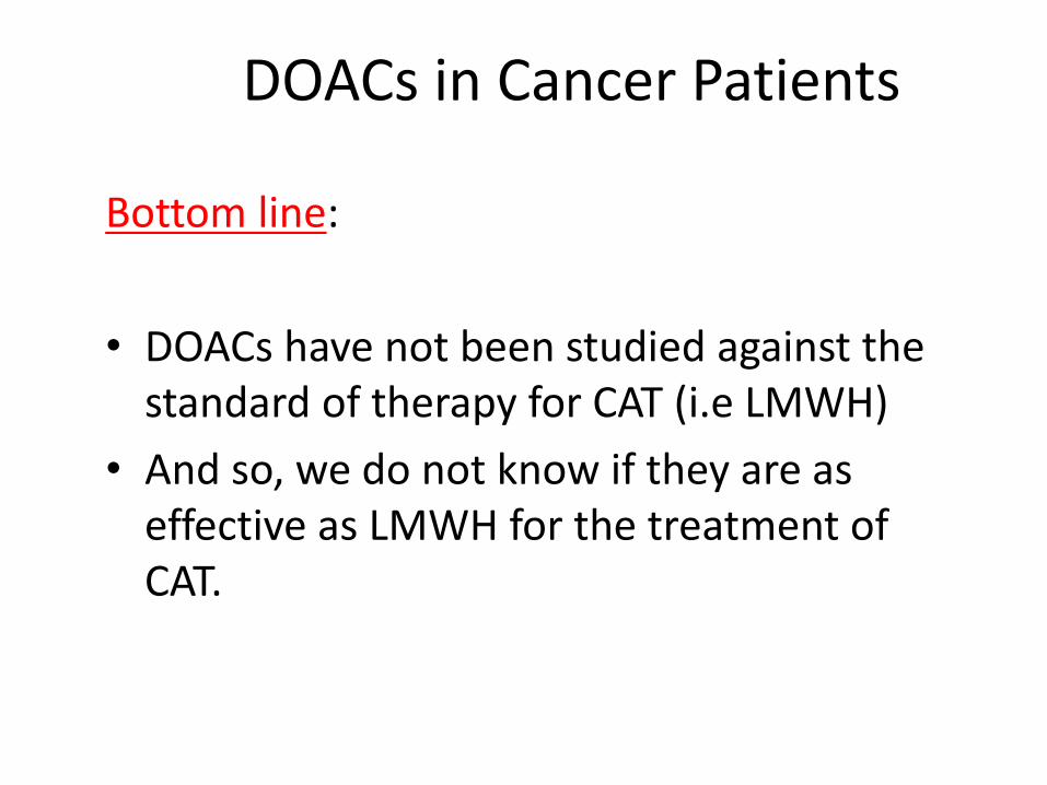
DOACs in Cancer Patients
Bottom line:
• DOACs have not been studied against the standard of therapy for CAT (i.e LMWH)
• And so, we do not know if they are as effective as LMWH for the treatment of CAT.
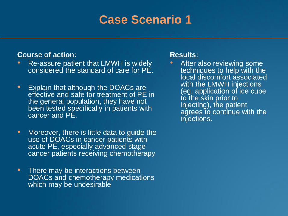
Case Scenario 1
Course of action:
• Re-assure patient that LMWH is widely considered the standard of care for PE.
• Explain that although the DOACs are effective and safe for treatment of PE in the general population, they have not been tested specifically in patients with cancer and PE.
• Moreover, there is little data to guide the use of DOACs in cancer patients with acute PE, especially advanced stage cancer patients receiving chemotherapy
• There may be interactions between DOACs and chemotherapy medications which may be undesirable
Results:
• After also reviewing some techniques to help with the local discomfort associated with the LMWH injections (eg. application of ice cube to the skin prior to injecting), the patient agrees to continue with the injections.

Clinical Scenario 2
• 42 year old man 90kg
• Non small cell lung
cancer IIIb
– cisplantin and
radiotherapy
• Diagnosed with acute
segmental PE
• Started on dalteparin 200
IU/kg (18,000 IU SC) for
3 weeks then 150 IU/kg
(15,000 IU sc)
• Two weeks after reducing
the dalteparin dose, he
presents with a massively
swollen and painful right leg
• Adequate limb perfusion
• Doppler ultrasound confirms
extensive DVT of right
popliteal and femoral veins
• Hg 112; plts 160 (baseline);
coags normal
53
Clinical Scenario 2
• Review weight and
medication compliance
• Patient’s dose was
increased to 18,000 IU sc
daily (wt=90kg)
• Leg symptoms improved
• What if recurrence
occurred on full
therapeutic dose LMWH
(i.e. dalteparin 200
IU/kg)?
54
Clinical Scenario 2
• Recommend increasing the dose by 20-25% for
at least 4 weeks and likely indefinite
• Consider twice daily dosing and not capping
dose when using prefilled syringes
• Do not switch to warfarin, fondaparinux, or
DOACs
•
55

Clinical Scenario 3
• 70 year old woman with DLBCL stage 3b with no prior
comorbidities
• Undergoes 2 cycles of R-CHOP. No complications
• Left arm PICC line inserted due to difficult venous
access
• After 4th cycle of R-CHOP, left arm swollen and painful
• Hemodynamically stable
• PICC line site OK
• Sent for doppler ultrasound which confirms left
subclavian DVT
56
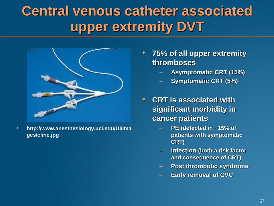
Central venous catheter associated upper extremity DVT
• http://www.anesthesiology.uci.edu/UI/ima
ges/cline.jpg
• 75% of all upper extremity
thromboses
– Asymptomatic CRT (15%)
– Symptomatic CRT (5%)
• CRT is associated with
significant morbidity in
cancer patients
– PE (detected in ~15% of
patients with symptomatic
CRT)
– Infection (both a risk factor
and consequence of CRT)
– Post thrombotic syndrome
– Early removal of CVC
57
Clinical Scenario 3
• Do you treat?
• If yes, which anticoagulant regimen?
• Do you remove the catheter?
58
Clinical Scenario 3
• Treat with LMWH alone
– No direct evidence to support LMWH alone
– Indirect evidence from the CLOT trial
• If the PICC line is functioning, is needed, and symptoms
improving, then no need to remove the catheter.
• ACCP Guideline (Chest 2012)
• Do not recommend removal of an indwelling catheter if the
device is functioning and there is an ongoing need for the
catheter (Grade 2C)
59
Clinical Scenario 3
• Duration of treatment?
– At least 3 months, even if catheter removed before
the completion of therapy (ACCP Grade 2C)
– Many MDs will treat beyond 3 months if catheter
remains in place.
60


![[Cancer-associated cachexia] clean for authors](https://img.pdfslide.us/doc/110x75/61d1ee79118df22edc52f710/cancer-associated-cachexia-clean-for-authors.jpg)