Embed Size (px)
Citation preview
Bennett et al. Pediatric Rheumatology (2019) 17:74 https://doi.org/10.1186/s12969-019-0377-7
RESEARCH ARTICLE Open Access
Can quantitative MRI be used in the clinical
setting to quantify the impact of intra-articular glucocorticoid injection onsynovial disease activity in juvenileidiopathic arthritis? Joshua L. Bennett1, Amanda Wood1, Nicola Smith2, Ravi Mistry3, Karen Allen1, Sharmila Jandial1, John D. Tuckett4,S. Claire Gowdy5, Helen E. Foster1,2, Flora McErlane1,2† and Kieren G. Hollingsworth6*†Abstract
Background: Juvenile idiopathic arthritis (JIA), the most common chronic rheumatic disease of childhood, ischaracterised by synovitis. Clinical assessments of synovitis are imperfect, relying on composite and indirectmeasures of disease activity including clinician-reported measures, patient-reported measures and blood markers.Contrast-enhanced MRI is a more sensitive synovitis assessment technique but clinical utility is currently limited byavailability and inter-observer variation. Improved quantitative MRI techniques may enable future development ofmore stringent MRI-defined remission criteria.The objective of this study was to determine the utility and feasibility of quantitative MRI measurement of synovialvolume and vascularity in JIA before and twelve weeks after intra-articular glucocorticoid injection (IAGI) of the kneeand to assess the acceptability of MRI to participating families.
Methods: Children and young people with JIA and a new episode of knee synovitis requiring IAGI were recruitedfrom the Great North Children’s Hospital in Newcastle upon Tyne. Quantitative contrast-enhanced MRI wasperformed prior to and twelve weeks after IAGI, in addition to standard clinical assessment tools, including thethree-variable clinical juvenile arthritis disease activity score (cJADAS) and active joint count.
Results: Eleven young people (5 male, median age 13 years, range 7–16) with JIA knee flare were recruited and 10completed follow-up assessment. Following IAGI, the median (interquartile range) cJADAS improved from 8.5 (2.7)to 1.6 (3.9), whilst the median synovial volume improved from 38.5cm3 (82.1cm3) to 0.0cm3 (0.2cm3). Six patientspresented with frank synovitis outside normal limits on routine MRI reporting. A further three had baseline MRIreports within normal limits but the quantitative measurements identified measurable synovial uptake. Post-IAGIquantitative measurements highlighted significant improvements in 9 patients.
(Continued on next page)
© The Author(s). 2019 Open Access This article is distributed under the terms of the Creative Commons Attribution 4.0International License (http://creativecommons.org/licenses/by/4.0/), which permits unrestricted use, distribution, andreproduction in any medium, provided you give appropriate credit to the original author(s) and the source, provide a link tothe Creative Commons license, and indicate if changes were made. The Creative Commons Public Domain Dedication waiver(http://creativecommons.org/publicdomain/zero/1.0/) applies to the data made available in this article, unless otherwise stated.
* Correspondence: [email protected]†Flora McErlane and Kieren G. Hollingsworth contributed equally to thiswork.6Newcastle Magnetic Resonance Centre, Institute of Cellular Medicine,Newcastle University, Newcastle upon Tyne, UKFull list of author information is available at the end of the article
Bennett et al. Pediatric Rheumatology (2019) 17:74 Page 2 of 12
(Continued from previous page)
Conclusions: IAGI led to a marked reduction in synovial volume, with quantitative MRI identifying more patientswith an improved synovial volume than routine qualitative clinical reporting. Improvements in cJADAS scores weremore variable with the patient/parent global assessment component contributing most to the scores. Further workis indicated, exploring the utility of quantitative MRI in the assessment of less accessible joints and comparing theimpact of different treatment modalities.
Keywords: Juvenile idiopathic arthritis, Disease activity assessment, Quantitative MRI, Synovitis, Synovial volume,Intra-articular glucocorticoid injection, Remission
BackgroundJuvenile idiopathic arthritis (JIA) is an umbrella term,summarising the International League of Associations forRheumatology (ILAR) classification system for the mark-edly heterogeneous chronic idiopathic paediatric arthriti-des [1, 2]. JIA is the most common chronic rheumaticdisease in children and young people with a UK preva-lence of 1:1000 [3]. Affected joints are characterised bysynovial proliferation and inflammatory cell infiltration,resulting in synovial effusion and hypertrophy.JIA is a relapsing and remitting condition and the majority
of young people will have more than one inflammatory flareduring the two years following initial diagnosis. At least one-third of young people continue to have episodes of activeinflammation during their adult years [4]. Long-standingpoorly controlled synovitis is associated with a high fre-quency of joint damage and joint replacement surgery inadults with JIA [5]. Early aggressive therapy, includingintraarticular glucocorticoid injection (IAGI) and promptintroduction of systemic immunosuppression, is thought toimprove remission rates, prevent joint damage and improvefunctional outcomes in JIA [6]. IAGI induces rapid suppres-sion of inflammation in targeted joints through a complexcombination of effects including reduced leucocyte cellinfiltration, altered leucocyte activity, reduced synovial perfu-sion and reduced vascular permeability [7, 8].The heterogeneous nature of JIA and normal develop-
mental changes of childhood ensure that no singleclinical assessment can reliably capture overall diseaseactivity in all young people with JIA [9]. At the presenttime, clinicians rely on composite measures of diseaseactivity, comprising multiple indirect clinical and bloodmarkers, including the active and limited joint counts(AJC / LJC), disability score (CHAQ) [10], ESR/CRP andglobal assessments by clinicians and parents. None ofthese accurately reflects the synovial state, and many aresubjective in nature with limited repeatability [11]. JIA-specific composite tools have been used to derivemultiple definitions of remission and it is not yet clearwhich, if any, constitutes the optimal definition [12].The lack of a single ‘gold standard’ treatment target isone of the central barriers to implementing moderntreat-to-target regimes in clinical practice [13].
Newer imaging modalities, such as musculoskeletalultrasound (MSUS) and magnetic resonance imaging(MRI), have the potential to further inform our under-standing of remission in JIA. Comparison of clinical andimaging-based definitions of remission is considered ahigh priority for future research [14].Ultrasound (US) and colour Doppler techniques can
detect subclinical synovitis in young people with JIA,and there have been extensive efforts in recent years todefine the normal US appearance of paediatric joints[15], to create definitions for the sonographic features ofsynovitis in young people [16], and to standardise acqui-sition protocols and scoring systems [17]. Although thiswork is important, paediatric MSUS requires specialistskills and training and may not be available at all cen-tres. Furthermore, the non-uniform accessibility of manyjoint spaces limits MSUS scoring to broad grades [18].Contrast-enhanced MRI is able to visualise the syno-
vitis that drives the degradative disease process, even atlow levels, and is recognised to be the most sensitivetechnique for the assessment of synovitis [19–21]. Post-contrast T1-weighted images clearly differentiate theextent of synovial hypertrophy vs. free fluid in the joint.In fact, MRI can identify synovial changes indicative ofactive disease in young people with JIA who fulfil clinicalcriteria for inactive disease [22]. Unenhanced MRI canuniquely detect bone oedema in addition to bone ero-sions. However, the clinical utility of MRI has beenmodest due to availability and the acceptability of MRIscanning in paediatric populations. While there has beenresearch activity demonstrating the potential of MRI inthe assessment of intra-articular disease activity, thereare limited normative paediatric data, recruited cohortshave had mixed presentations and there is a lack of JIA-specific clinical assessment tools to validate the findings.Consequently, there is wide inter-observer variation inthe interpretation and reporting of MRI scans.In clinical practice, synovial enhancement is reported
as either being within or exceeding normal limits, basedon an expected normal appearance. In research, thequalitative JAMRIS score (juvenile arthritis MRI scoringsystem) has been proposed as a classification of synovialhypertrophy on a 3-point scale (< 2 mm, 2-4 mm or > 4
Bennett et al. Pediatric Rheumatology (2019) 17:74 Page 3 of 12
mm thickness) in six anatomical regions but may beinsensitive to longitudinal change [23, 24]. By contrast,we can seek to quantify properties of disease activity oncontinuous scales by image processing, rather thanqualitative description or grading. The potential advan-tage of such an approach is the ability to discriminatechange more finely than qualitative scales, and to meas-ure disease activity that may fall below present qualita-tive reporting standards [25–27]. The cost of suchapproaches is the analysis effort required.In summary, quantitative MRI may have considerable
utility in both clinical research and the clinical care ofyoung people with JIA, since it can provide sensitive,continuous measurement of synovial volume and thevascularity of the synovium through dynamic measure-ment of the uptake of gadolinium contrast agent. Thisstudy investigates whether quantitative MRI can providea sensitive measurement of the effects of IAGI in youngpeople with JIA, and compares synovial volume andcontrast uptake measurements. The study also assessesthe feasibility and acceptability of the imaging for chil-dren and parents through feedback by questionnaire andtelephone interviews.
MethodsSubjectsYoung people were recruited from a tertiary paediatricrheumatology service in the North of England (TheGreat North Children’s Hospital at Newcastle uponTyne Hospitals NHS Foundation Trust). Patients andtheir families attending clinic were invited to considerparticipating in the study if all inclusion criteria werepresent (new or known diagnosis of JIA, new presenta-tion of knee synovitis requiring intra-articular gluco-corticoid injection (IAGI) and age 4–16 years). Exclusioncriteria were steroid injection of involved knee in thepast six months, concurrent use of oral or IV steroids,contraindications to contrast agent or non-sedated MRI,non-English speaking families, or recent trauma to theknee. No changes were made to concomitant immuno-suppressive treatments during the study period. Familiesof young people who were eligible and interested at theirclinic visit were followed up by telephone and invited totake part in the study.Additional file 1: Fig. S1 shows the recruitment flow
chart for the study. The subjects had two MRI andclinical assessments, the first prior to the intra-articularinjection and the second at least twelve weeks after theinjection. Affected knees were injected with 1 mg/kg tri-amcinolone hexacetonide to a maximum of 40 or 60 mgdepending on weight: image guidance was not used, asper local protocol. Where both knees were affected, bothwere injected, and the knee judged most affected at clin-ical examination was followed by MRI. The study,
performed between September 2017 and August 2018,complied with the Declaration of Helsinki and obtaineda favourable opinion from the Newcastle and NorthTyneside 1 Research Ethics Committee and the HealthResearch Authority, with the parents/carers of the sub-jects giving written informed consent, and written assentfrom the young people. Routine clinical care was not al-tered by the addition of the MRI scanning. KGH hadcontrol of the study data and had responsibility for over-seeing data acquisition and processing.
Clinical assessmentsClinical assessment was performed at baseline andfollow-up. This included measurement of the core out-come variables: binary 74-joint active and limited jointcounts, physician global score of disease activity (10 cmvisual analogue scale (VAS)), patient/parent global scoreof disease activity (10 cm VAS), CHAQ and ESR/CRP(where available) [28], a global pain score (10 cm VAS)and the 3 and 4 variable juvenile arthritis disease activityscores (cJADAS and JADAS) [29, 30]. Clinical remissionwas defined according to the JADAS and cJADAS cut-offs for inactive disease (</= 1) [31, 32].
MRI protocolA peripheral venous cannula was inserted in the upperlimb. Subjects were made comfortable in a supine feet-firstorientation on a Siemens 1.5 T Espree, using an extremitybirdcage coil. The MRI sequences (including T2-, T1- andproton density weighted scans, Additional file 1: Table S1)were prescribed so that the bottom slice passed throughthe proximal aspect of the superior tibiofibular joint. Theleg position and anatomical location were carefullymatched at the post-treatment visit. The sequences in-cluded pre- and post-contrast T1-weighted (T1w) imagingto distinguish synovial enhancement and a multiple dy-namic T1w sequence was used to collect dynamic gadolin-ium uptake data before, during and after the injection ofcontrast agent. After two dynamics (26 s), the contrastagent was administered by hand as a standard single doseof gadoterate meglumine (Dotarem, 0.2 ml/kg body weight,Guerbet), followed by a 3ml flush of 0.9% sodium chloride.The uptake of contrast was imaged for five minutes post-injection. The post-contrast T1w fat suppressed turbo spinecho imaging was collected immediately after the dynamicseries ended such that post-contrast data were collected 5min post injection as recommended [33].
Image processingThe MRI images were reported by a consultant radiologist(JDT) and were also quantitatively processed using customsoftware in MATLAB 2017a (Mathworks, UK) and ImageJv1.43u (NIH, USA). The images of the dynamic gradient
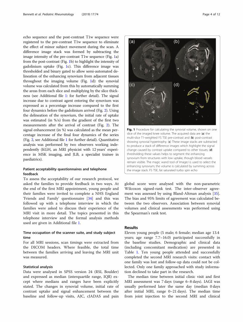
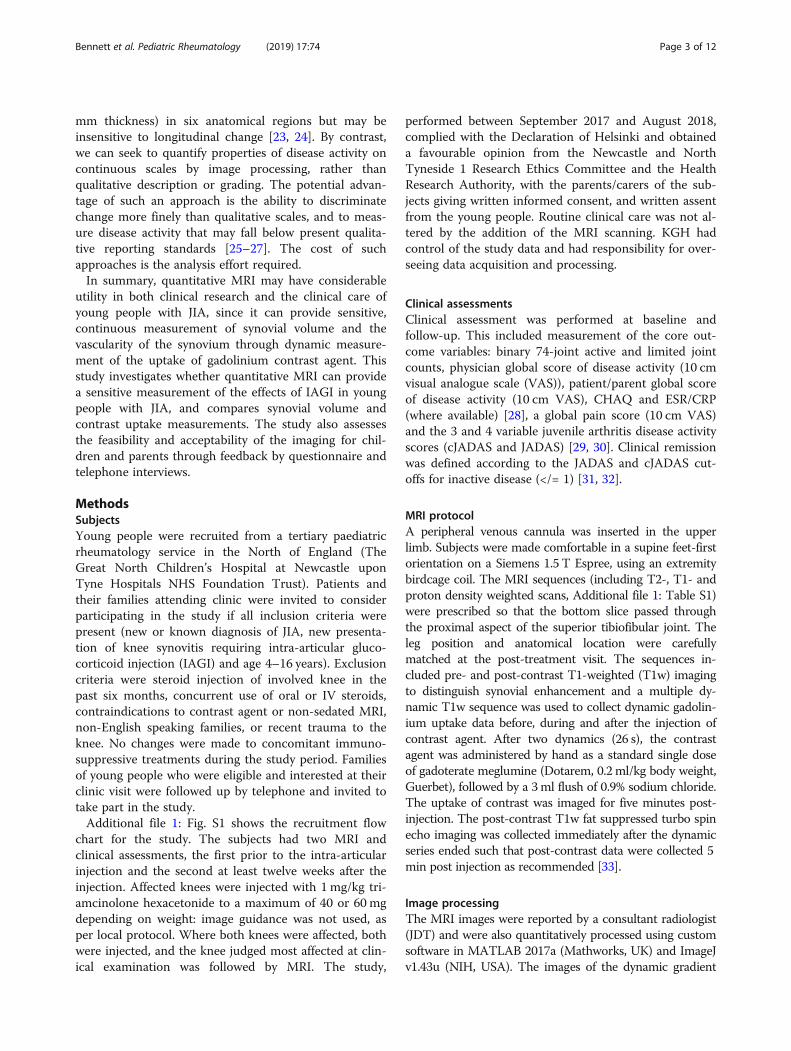
Fig. 1 Procedure for calculating the synovial volume, shown on oneslice of the imaged knee volume. The acquired data are (a) themulti-slice T1-weighted FS TSE pre-contrast and (b) post-contrastshowing synovial hypertrophy. (c) These image stacks are subtractedto produce a stack of difference images which highlight the signalchange caused by contrast uptake compared to other tissues; (d)thresholding these values helps to segment the enhancingsynovium from structures with low uptake, though blood vesselsremain visible. The magic wand tool of ImageJ is used to select theenhancing synovium; the volume is calculated by summing acrossthe image stack. FS TSE, fat saturated turbo spin echo
Bennett et al. Pediatric Rheumatology (2019) 17:74 Page 4 of 12
echo sequence and the post-contrast T1w sequence wereregistered to the pre-contrast T1w sequence to eliminatethe effect of minor subject movement during the scan. Adifference image stack was formed by subtracting theimage intensity of the pre-contrast T1w sequence (Fig. 1a)from the post-contrast (Fig. 1b) to highlight the intensity ofgadolinium uptake (Fig. 1c). This difference image wasthresholded and binary gated to allow semi-automated de-lineation of the enhancing synovium from adjacent tissuesthroughout the imaging volume (Fig. 1d): the synovialvolume was calculated from this by automatically summingthe areas from each slice and multiplying by the slice thick-ness (see Additional file 1: for further detail). The signalincrease due to contrast agent entering the synovium wasexpressed as a percentage increase compared to the firstfour dynamics before the gadolinium arrived (Fig. 2). Usingthe delineation of the synovium, the initial rate of uptakewas estimated (in %/s) from the gradient of the first twomeasurements after the arrival of contrast (Fig. 2). Thesignal enhancement (in %) was calculated as the mean per-centage increase of the final four dynamics of the series(Fig. 2, see Additional file 1 for further detail). Quantitativeanalysis was performed by two observers working inde-pendently (KGH, an MRI physicist with 12 years’ experi-ence in MSK imaging, and JLB, a specialist trainee inpaediatrics).
Patient acceptability questionnaires and telephonefeedbackTo assess the acceptability of our research protocol, weasked the families to provide feedback in two ways. Atthe end of the first MRI appointment, young people andtheir families were invited to complete a NHS England‘Friends and Family’ questionnaire [34] and this wasfollowed up with a telephone interview in which thefamilies were asked to discuss their experience of theMRI visit in more detail. The topics presented in thistelephone interview and the formal analysis methodsused are given in Additional file 1.
Time occupation of the scanner suite, and study subjecttimeFor all MRI sessions, scan timings were extracted fromthe DICOM headers. Where feasible, the total timebetween the families arriving and leaving the MRI unitwas measured.
Statistical analysisData were analysed in SPSS version 24 (RSI, Boulder)and expressed as median (interquartile range, IQR) ex-cept where medians and ranges have been explicitlystated. The changes in synovial volume, initial rate ofcontrast uptake and signal enhancement between thebaseline and follow-up visits, AJC, cJADAS and pain
global score were analysed with the non-parametricWilcoxon signed-rank test. The inter-observer agree-ment was assessed by using Bland-Altman analysis [35].The bias and 95% limits of agreement was calculated be-tween the two observers. Association between synovialvolumes and clinical assessments was performed usingthe Spearman’s rank test.
ResultsEleven young people (5 male; 6 female; median age 13.4years; age range 7.7–16.0) participated successfully inthe baseline studies. Demographic and clinical data(including concomitant medication) are presented inTable 1. Ten young people attended and successfullycompleted the second MRI research visits: contact withone family was lost and follow-up data could not be col-lected. Only one family approached with study informa-tion declined to take part in the research.The median time between initial clinic visit and first
MRI assessment was 7 days (range 6–8 days). IAGI wasusually performed later the same day (median 0 daysafter initial MRI, range 0–21 days). The median timefrom joint injection to the second MRI and clinical
Fig. 2 Percentage gadolinium signal increase across the segmented synovial volume for subject 5. The contrast is injected at the time shown inthe arrow. The initial uptake rate (%/s) is calculated from the gradient of the line of the first two positive signal increases and the signalenhancement (%) is the mean of the last four points. This subject had a change in synovial volume from 72.2cm3 pre-treatment to 0.7cm3 post-treatment. The initial uptake shown reduces from 2.83%/s to 0.51%/s post-treatment and signal enhancement reduces from 190 to 90%
Bennett et al. Pediatric Rheumatology (2019) 17:74 Page 5 of 12
research assessment was 14 weeks (range 12.6–19weeks). The time required for subjects’ MRI appoint-ments is presented in the Additional file 1.The median time (IQR) for contrast to reach the syno-
vium after the beginning of the scan was 63 (14) secondsand contrast never reached synovium before the fifthimage (range fifth-seventh image). Therefore, the uptakeof gadolinium was followed for a median time of 315(14) seconds after injection.
Clinical assessmentsBaseline and follow-up clinical assessments are pre-sented in Table 1 and Fig. 3. Clinician-assessed diseaseactivity in the injected knee changed from present to ab-sent in 7/10 participants post-IAGI. Two of the threeparticipants with persisting clinician-assessed disease ac-tivity had a measurable synovial volume on follow-upMRI; one did not. All three participants with persistingclinically-assessed disease activity had repeat IAGI. Thissubsequently led to clinical improvement in the two witha measurable synovial volume and no improvement inthe third young person. Although the AJC decreased sig-nificantly overall, it did not change in 4/10 patients. Thephysician global score reduced after joint injection in allcases, as did the parent global score for 9/10 cases.Where the ESR was > 5mm at baseline, it always de-creased. 6/10 patients had an AJC of 0 following joint in-jection and 4 of these 6 patients had follow-up JADASand cJADAS scores of less than or equal to 1 (consistentwith inactive disease or remission): no other patients
had JADAS/cJADAS scores indicating remission. Figure 3illustrates that the parent global scores generally contrib-uted more to the overall JADAS score than the AJC orphysician global score with no suggestion of a relation-ship to ILAR subtype or disease duration.
Qualitative MRI reportsFor the first MRI assessment, synovial thickening outsidenormal limits was reported for six of the subjects atbaseline and reported within normal limits for five sub-jects (Tables 1 and 2). Minimal synovial enhancement inkeeping within normal limits was noted in the report ontwo of the latter subjects, and no enhancement noted onthe other three subjects. All reports for the second MRIassessment found no synovial thickening beyond normallimits. Chondral surfaces all appeared to be well main-tained and there was no evidence of bone oedema.
Quantitative MRI measurementsThe synovial volumes measured at baseline and atfollow-up are given in Table 2. Across the 10 subjectswho attended follow-up appointments the median(IQR) synovial volume was reduced from 38.5 (82.1)cm3 to 0.0 (0.2) cm3 (p = 0.005). There were threesignatures of synovial volume response. The firstgroup (n = 6) had frank synovitis, called qualitativelyby the radiologist as outside normal limits and with amedian synovial volume of 79.6cm3 (34.5cm3, Fig. 4).On post-treatment follow-up, these volumes reducedto a median of 0.0cm3 (0.6cm3). The second group
Table
1Patient
characteristicsandsummaryof
clinicalassessmentsatbaseline(B)and
follow-up(F)
Subject
Age
Gen
der
Disease
duratio
n(m
onths)
ILARSubtype&System
icMed
ication
Activejoint
coun
tClinicianassessed
disease
activity
ininjected
knee
(Y/N)
JADAS10
cJADAS10
ESR
CHAQ
Pain
glob
alscore(cm)
BF
BF
BF
BF
BF
BF
BF
112.3
F0
psoriatic
m1_lk
1lk
YY
n/a
5.5
9.3
5.5
n/a
101.4
n/a
6.1
4.0
27.7
F27
peroligo
m2_rk,ra
–Y
–7.0
–7.0
–20
–0.3
–3.2
–
316.0
F8
peroligo
z2lk,_rk
0Y
N8.4
0.0
7.8
0.0
2616
0.5
03.3
0.0
411.6
M140
peroligo
1_rk
1rk
YY
4.2
2.0
4.2
2.0
52
1.0
03.6
0.0
513.4
M53
extoligo
1_lk
1rk
YN
7.1
n/a
7.1
3.0
2n/a
0.0
03.0
1.5
616.2
F6
RF+po
lym,d
2lk,_rk
0Y
N7.2
0.0
7.2
0.0
55
1.0
0.3
2.9
0.9
713.6
F54
peroligo
d2_lk,rk
0Y
N12.4
0.0
11.0
0.0
3415
0.5
06.3
0.0
88.1
M0
peroligo
2lk,_rk
2rk,ra
YY
18.8
8.9
15.6
8.9
529
1.9
1.9
10.0
6.8
913.4
M74
extoligo
d2_lk,rk
0Y
N10.8
1.2
10.0
1.2
2815
1.3
n/a
6.5
6.1
1015.3
F92
RF-po
ly1_rk
0Y
N9.2
n/a
9.2
4.2
16n/a
1.3
n/a
7.1
3.4
1114.3
M14
peroligo
1_rk
0Y
N2.0
0.0
2.0
0.0
25
0.0
n/a
0.0
0.0
Med
ian
13.4
271.5
0.0†
––
8.5
1.6*
––
––
4.9
1.2$
IQR
2.8
571.0
1.0
––
2.7
3.9
––
––
3.4
3.9
Med
ians
andinterqua
rtile
rang
es(IQ
R)compa
ringba
selin
ean
dfollow-upexclud
esubject2who
was
lost
tofollow-up.
Whe
remeasuremen
tsareno
tavailableforothe
rindividu
als,med
ians
have
notbe
enprov
ided
.†p
<0.03
,*p=0.00
5,$p=0.00
8at
follow-upcompa
redto
baselin
e.Ke
y:pe
r/ext.oligo=pe
rsistent/exten
dedoligoa
rticular,R
F+/RF-
poly=rheu
matoidfactor
positiv
e/ne
gativ
epo
lyarticular,JADAS10=juvenile
arthritis
diseaseactiv
ityscore(10joints),cJADAS10=clinical
juvenile
arthritisdiseaseactiv
ityscore(10joints),ESR=erythrocytesedimen
tatio
nrate,C
HAQ=child
health
assessmen
tqu
estio
nnaire,n
/a=no
tavailable.
Foractiv
ejointcoun
ts,sup
erscrip
tsspecify
joints
(lk=leftkn
ee,rk=rig
htkn
ee,ra=rig
htan
kle).The
imag
edkn
eeisun
derline
d.System
icmed
ication:
m=metho
trexate,
z=azathiop
rine,
d=ad
alim
umab
Bennett et al. Pediatric Rheumatology (2019) 17:74 Page 6 of 12
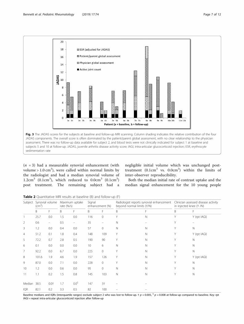
Fig. 3 The JADAS scores for the subjects at baseline and follow-up MRI scanning. Column shading indicates the relative contribution of the fourJADAS components. The overall score is often dominated by the patient/parent global assessment, with no clear relationship to the physicianassessment. There was no follow-up data available for subject 2, and blood tests were not clinically indicated for subject 1 at baseline andsubjects 5 and 10 at follow-up. JADAS, juvenile arthritis disease activity score; IAGI, intra-articular glucocorticoid injection; ESR, erythrocytesedimentation rate
Bennett et al. Pediatric Rheumatology (2019) 17:74 Page 7 of 12
(n = 3) had a measurable synovial enhancement (withvolume > 1.0 cm3), were called within normal limits bythe radiologist and had a median synovial volume of1.2cm3 (0.1cm3), which reduced to 0.0cm3 (0.1cm3)post treatment. The remaining subject had a
Table 2 Quantitative MRI results at baseline (B) and follow-up (F)
Subject Synovial volume(cm3)
Maximum uptakerate (%/s)
Signalenhancement (%)
Radiolbeyon
B F B F B F B
1 25.7 0.0 1.5 0.0 116 0 Y
2 0.6 – 0.5 – 35 – N
3 1.2 0.0 0.4 0.0 57 0 N
4 51.2 0.1 1.8 0.4 148 109 Y
5 72.2 0.7 2.8 0.5 190 90 Y
6 0.1 0.0 0.0 0.0 10 6 N
7 92.2 0.0 6.7 0.0 225 0 Y
8 101.6 1.9 4.6 1.9 157 126 Y
9 87.0 0.0 7.1 0.0 228 0 Y
10 1.2 0.0 0.6 0.0 95 0 N
11 1.1 0.2 1.5 0.8 145 103 N
Median 38.5 0.0† 1.7 0.0$ 147 3† –
IQR 82.1 0.2 3.3 0.5 82 100 –
Baseline medians and IQRs (interquartile ranges) exclude subject 2 who was lost toIAGI = repeat intra-articular glucocorticoid injection after follow-up
negligible initial volume which was unchanged post-treatment (0.1cm3 vs. 0.0cm3) within the limits ofinter-observer reproducibility.Both the median initial rate of contrast uptake and the
median signal enhancement for the 10 young people
ogist reports synovial enhancementd normal limits (Y/N)
Clinician assessed disease activityin injected knee (Y /N)
F B F
N Y Y (rpt IAGI)
– Y –
N Y N
N Y Y (rpt IAGI)
N Y N
N Y N
N Y N
N Y Y (rpt IAGI)
N Y N
N Y N
N Y N
–
–
follow-up. † p = 0.005, $ p = 0.008 at follow-up compared to baseline. Key: rpt
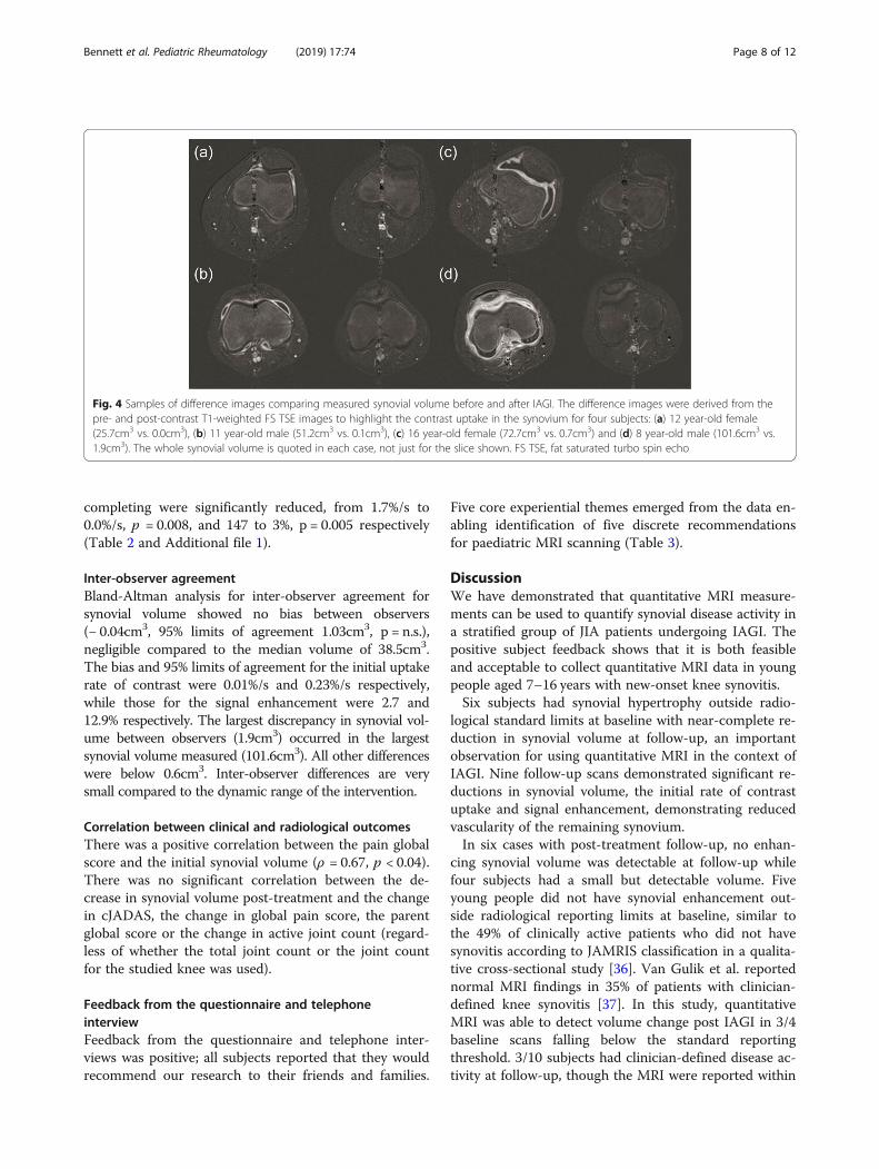
Fig. 4 Samples of difference images comparing measured synovial volume before and after IAGI. The difference images were derived from thepre- and post-contrast T1-weighted FS TSE images to highlight the contrast uptake in the synovium for four subjects: (a) 12 year-old female(25.7cm3 vs. 0.0cm3), (b) 11 year-old male (51.2cm3 vs. 0.1cm3), (c) 16 year-old female (72.7cm3 vs. 0.7cm3) and (d) 8 year-old male (101.6cm3 vs.1.9cm3). The whole synovial volume is quoted in each case, not just for the slice shown. FS TSE, fat saturated turbo spin echo
Bennett et al. Pediatric Rheumatology (2019) 17:74 Page 8 of 12
completing were significantly reduced, from 1.7%/s to0.0%/s, p = 0.008, and 147 to 3%, p = 0.005 respectively(Table 2 and Additional file 1).
Inter-observer agreementBland-Altman analysis for inter-observer agreement forsynovial volume showed no bias between observers(− 0.04cm3, 95% limits of agreement 1.03cm3, p = n.s.),negligible compared to the median volume of 38.5cm3.The bias and 95% limits of agreement for the initial uptakerate of contrast were 0.01%/s and 0.23%/s respectively,while those for the signal enhancement were 2.7 and12.9% respectively. The largest discrepancy in synovial vol-ume between observers (1.9cm3) occurred in the largestsynovial volume measured (101.6cm3). All other differenceswere below 0.6cm3. Inter-observer differences are verysmall compared to the dynamic range of the intervention.
Correlation between clinical and radiological outcomesThere was a positive correlation between the pain globalscore and the initial synovial volume (ρ = 0.67, p < 0.04).There was no significant correlation between the de-crease in synovial volume post-treatment and the changein cJADAS, the change in global pain score, the parentglobal score or the change in active joint count (regard-less of whether the total joint count or the joint countfor the studied knee was used).
Feedback from the questionnaire and telephoneinterviewFeedback from the questionnaire and telephone inter-views was positive; all subjects reported that they wouldrecommend our research to their friends and families.
Five core experiential themes emerged from the data en-abling identification of five discrete recommendationsfor paediatric MRI scanning (Table 3).
DiscussionWe have demonstrated that quantitative MRI measure-ments can be used to quantify synovial disease activity ina stratified group of JIA patients undergoing IAGI. Thepositive subject feedback shows that it is both feasibleand acceptable to collect quantitative MRI data in youngpeople aged 7–16 years with new-onset knee synovitis.Six subjects had synovial hypertrophy outside radio-
logical standard limits at baseline with near-complete re-duction in synovial volume at follow-up, an importantobservation for using quantitative MRI in the context ofIAGI. Nine follow-up scans demonstrated significant re-ductions in synovial volume, the initial rate of contrastuptake and signal enhancement, demonstrating reducedvascularity of the remaining synovium.In six cases with post-treatment follow-up, no enhan-
cing synovial volume was detectable at follow-up whilefour subjects had a small but detectable volume. Fiveyoung people did not have synovial enhancement out-side radiological reporting limits at baseline, similar tothe 49% of clinically active patients who did not havesynovitis according to JAMRIS classification in a qualita-tive cross-sectional study [36]. Van Gulik et al. reportednormal MRI findings in 35% of patients with clinician-defined knee synovitis [37]. In this study, quantitativeMRI was able to detect volume change post IAGI in 3/4baseline scans falling below the standard reportingthreshold. 3/10 subjects had clinician-defined disease ac-tivity at follow-up, though the MRI were reported within
Table 3 Themes and recommendations from the patient’s perspective. Five core themes relating to the patient experienceemerged from the data and from this we identified the following recommendations for paediatric MRI
Theme 1: Clear expectationsKnowing fully what to expect relaxes both the child and their family and helps alleviate any concerns or uncertainties.
Recommendations:• Provide detailed information to alleviate any concerns or uncertainties both prior to the scan and on the day itself. This should include detaileddirections and guidance on what will happen on the day with enough information so the parent/carer can answer any questions the child mayhave.
• Offer the opportunity to ask questions beforehand (e.g. telephone call from clinical team prior to appointment).• Provide the opportunity to view scanner beforehand.
Theme 2: Creation of relaxing environmentExtra touches that make families feel at home help create a relaxing environment and create a more positive experience in which to have the scan.
Recommendations:• Use trained paediatric staff to put both child and parent/carer at ease. Communication to be on a first name basis and whilst familiarity can addan extra layer of relaxation, it is also acceptable to be simply introduced on the day.
• Use staff experienced in inserting cannulas in children to avoid extra stress that difficulties with their insertion can cause.• Give the opportunity for child to listen to music of their choice during the scan.• Give the option for parent/carer or member of staff to accompany child into scanner.• Ensure pace of appointment is not rushed and is led by the child and parent/ carer’s needs.• Care to be taken regarding scan setting wherever possible to reduce unnecessary concerns (e.g. having scan alongside cancer unit viewed as notideal).
Theme 3: Child centred approachScan experience can further be improved by adopting a child centred approach in which the child is seen as key and in control of the situation.
Recommendations:• Direct discussion at child.• Provide child friendly information leaflets as well as parent versions.• Give the option for toys to distract child if required by child and parent/carer.• Provide ‘completion certificate’ at the end and postcard in the post.
Theme 4: Increased understanding of the conditionAn extra layer of positivity can be added to the experience by using the scan as a way to educate the families further about the child’s condition andincrease their understanding of what the scan is able to show.
Recommendations:• Allow the opportunity to view images after the scan alongside detailed guidance on what the images show and where possible enable patientsto take a copy (e.g. photo) they can show other family members, friends or teachers at school.
Theme 5: Linking in to current treatment plan(s)Effective management enabling linking into current treatment plans viewed positively by families.
Recommendations:• Link in to current treatment plan wherever possible. For example providing the opportunity to take bloods at the same time as giving contrastseen as useful especially since many children do not like needles.
Bennett et al. Pediatric Rheumatology (2019) 17:74 Page 9 of 12
normal limits: there was a measurable synovial volumein two of these subjects. In retrospect, the negligible syn-ovial volume for subject 6 suggests a non-synovial aeti-ology to their symptoms, despite the clinical response toIAGI.Although the impact of IAGI on synovial volume and
vascularity was frequently dramatic, the quantitativeMRI changes were not always reflected in the compositeclinical assessments. Of the six children with no detect-able synovial volume post IAGI (0.0cm3), only threewould be regarded as in remission according to cJADAS(</=1), whilst one child with a post-treatment cJADASscore of 0 had a measurable synovial volume. Our studyadds further weight to a growing concern that thepresent definitions of remission may require reconsider-ation in light of future imaging data [14].The post-joint injection clinical assessments illustrate
that, in our cohort with low joint counts, the patient/parent global scores contribute more to the JADAS
score than the AJC/physician global score. This discrep-ancy between family / physician perception of impact ofdisease has been demonstrated previously [38] and ourstudy further highlights the risk of underestimating theimpact of oligoarthritis on everyday life.There have been previous attempts to quantify syn-
ovial volume changes with treatment over similar time-scales using quantitative MRI in JIA. Workie et al.studied the 3-month response to treatment, though onlyfour subjects received IAGI at that time point [27], withmodest synovial volume reduction of 26% and 29% inthe initial uptake rate: the children recruited had a me-dian of eight active joints at baseline.A one-year follow-up of active wrist involvement in 36
children demonstrated a 50% reduction in synovial vol-ume, though treatment types and durations were notspecified [39]. In other arthritides, such as rheumatoidarthritis, more modest changes are reported with quanti-tative methods [25, 39, 40].
Bennett et al. Pediatric Rheumatology (2019) 17:74 Page 10 of 12
Our study suggests that quantitative MRI scanningadds important information both to clinical assessmentsand to current qualitative MRI reporting. We chose toassess the impact of IAGI in new onset knee synovitis,as knee synovitis is common. However, we anticipatethat quantitative MRI reporting may have more potentialin the assessment of disease activity in less accessiblejoints and in the presence of long-standing disease, jointdamage or co-existing pain syndromes.The limitations of our study include the small sample
size studied. Although the study was open to sequential eli-gible families with young people aged 4–16 years, only onechild younger than 7 years was approached. The family de-clined, citing concerns that their child may not lie still.Therefore, we cannot draw conclusions about the accept-ability of MRI in the youngest children. Contact with onesubject was lost before follow-up scanning, though theirresponses to the questionnaire and telephone interview fol-lowing the first MRI visit were positive. The post-IAGIsynovial volumes calculated in this study suggest that thesynovium of healthy knee joints has a negligible volume onquantitative contrast-enhanced imaging. There are noquantitative contrast-enhanced data on young people withhealthy knees: the closest study to date examined youngpeople with knee pain and non-inflammatory diagnosessuch as hypermobility and functional disorders [42].The use of thresholding at defined signal enhancement
levels reduced subjective choice between the analysts,with the decision dominating the analysis time (typically20–30min per volume) being that of separating thesynovium from adjacent small blood vessels. Thesevessels appeared to have a consistent small volume onthe pre- and post- IAGI imaging, so it may be possibleto evaluate the volume much faster, perhaps three mi-nutes, with further development of the analysis softwareand if the inclusion of minor vessels were to be toler-ated, aiding clinical adoption. Translation to clinicalpractice requires careful selection of the managementdecisions in the clinical care pathway for which quantita-tive MRI would be used, since MRI availability is limited.These aspects are the subject of ongoing work.This study focuses on the impact of IAGI in new-onset
knee synovitis, measured by quantitative MRI. Whilst thenear-complete resolution of knee synovitis post IAGI is aninteresting observation, the true value of quantitative MRItechniques may be realised in future studies exploring theimpact of current treatment regimes on less accessiblejoints such as the hip or temporomandibular joints and /or comparing the relative efficacies of different therapeuticregimes. Quantitative MRI has particular potential inyoung people with oligoarticular disease in whom clinicaldisease activity measures may not be sensitive or specificenough to reliably identify changes in the degree oflocalised synovial disease.
ConclusionsThis study has demonstrated that quantitative MRI canmeasure the effect of IAGI on synovial volume and isaccessible and feasible for children aged between 7 and16 years, with positive feedback from families. Synovialvolume was reduced markedly by IAGI, either to zero orclose to zero. Quantitative MRI provided different infor-mation to traditional clinical disease activity measuresand was able to identify more patients with an improvedsynovial volume than routine qualitative reporting. Insummary, quantitative MRI has the potential to add im-portant information to our understanding of disease out-comes in JIA.
Supplementary informationSupplementary information accompanies this paper at https://doi.org/10.1186/s12969-019-0377-7.
Additional file 1. Supplementary Methods, Supplementary Results,Supplementary Fig. S1, Supplementary Fig. S2, Supplementary Table S1,Supplementary References.
AbbreviationscJADAS: three variable clinical juvenile arthritis disease activity score; CRP: C-reactive protein; ESR: Erythrocyte sedimentation rate; IAGI: Intra-articularglucocorticoid injection; ILAR: International League of Associations forRheumatology; IQR: Interquartile range; JADAS: four variable juvenile arthritisdisease activity score; JAMRIS: juvenile arthritis MRI scoring system;JIA: Juvenile idiopathic arthritis; MRI: Magnetic resonance imaging;VAS: Visual analogue scale
AcknowledgementsThe authors would like to thank Louise Fox from the Newcastle upon TyneHospitals NHS Foundation Trust for assistance in setting up our project andof the research radiographers Louise Jordan and Leoana McNamara from theFreeman Hospital, who helped to acquire the MRI data.
Authors’ contributionsAll named authors meet the International Committee of Medical JournalEditors (ICMJE) criteria for authorship for this manuscript, acceptresponsibility for the integrity of the work as a whole, and have providedfinal approval of the manuscript to be published.
FundingThe research costs and the support of JLB’s research time was supported bythe Medical Research Council, UK, by a Confidence in Concept award (MC/PC/15030), with support from the NIHR Clinical Research Network North Eastand North Cumbria.
Availability of data and materialsThe datasets used and/or analysed during the current study are availablefrom the corresponding author on reasonable request, subject to therequirements of the study ethical approval and Caldicott approval.
Ethics approval and consent to participateThe study, performed between September 2017 and August 2018, compliedwith the Declaration of Helsinki and obtained a favourable opinion from theNewcastle and North Tyneside 1 Research Ethics Committee (206363, 16/NE/0299) and the Health Research Authority, with the parents/carers of thesubjects giving written informed consent, and written assent from the youngpeople.
Bennett et al. Pediatric Rheumatology (2019) 17:74 Page 11 of 12
Consent for publicationConsent for the publication of anonymised participant study results andimages was obtained from all participants as part of the study consentprocess above.
Competing interestsThe authors declare that they have no competing interests.
Author details1Paediatric Rheumatology, Great North Children’s Hospital, Newcastle uponTyne Hospitals NHS Foundation Trust, Newcastle upon Tyne, UK.2Musculoskeletal Research Group, Institute of Cellular Medicine, NewcastleUniversity, Newcastle upon Tyne, UK. 3Paediatrics, Newcastle upon TyneHospitals NHS Foundation Trust, Newcastle upon Tyne, UK. 4Radiology,Freeman Hospital, Newcastle upon Tyne Hospitals NHS Foundation Trust,Newcastle upon Tyne, UK. 5Paediatric Radiology, British Columbia Children’sHospital, Vancouver, Canada. 6Newcastle Magnetic Resonance Centre,Institute of Cellular Medicine, Newcastle University, Newcastle upon Tyne, UK.
Received: 7 October 2019 Accepted: 4 November 2019
References1. Petty RE, Southwood TR, Baum J, Bhettay E, Glass DN, Manners P,
Maldonado-Cocco J, Suarez-Almazor M, Orozco-Alcala J, Prieur AM. Revisionof the proposed classification criteria for juvenile idiopathic arthritis: Durban,1997. J Rheumatol. 1998;25(10):1991–4.
2. Petty RE, Southwood TR, Manners P, Baum J, Glass DN, Goldenberg J, He X,Maldonado-Cocco J, Orozco-Alcala J, Prieur AM, Suarez-Almazor ME, Woo P.International league of associations for rheumatology classification ofjuvenile idiopathic arthritis: second revision, Edmonton, 2001. J Rheumatol.2004;31(2):390–2.
3. Symmons DP, Jones M, Osborne J, Sills J, Southwood TR, Woo P. Pediatricrheumatology in the United Kingdom: data from the British pediatricrheumatology group National Diagnostic Register. J Rheumatol. 1996;23(11):1975–80.
4. Foster HE, Marshall N, Myers A, Dunkley P, Griffiths ID. Outcome in adultswith juvenile idiopathic arthritis: a quality of life study. Arthritis Rheum.2003;48(3):767–75.
5. Malviya A, Johnson-Lynn S, Avery P, Deehan D, Foster H. Juvenile idiopathicarthritis in adulthood and orthopaedic intervention. Clin Rheumatol. 2009;28(12):1411–7.
6. Beresford MW, Baildam EM. New advances in the management of juvenileidiopathic arthritis--2: the era of biologicals. Arch Dis Child Educ Pract Ed.2009;94(5):151–6.
7. Coutinho AE, Chapman KE. The anti-inflammatory and immunosuppressiveeffects of glucocorticoids, recent developments and mechanistic insights.Mol Cell Endocrinol. 2011;335(1):2–13.
8. Ostergaard M, Halberg P. Intra-articular corticosteroids in arthritic disease: aguide to treatment. BioDrugs. 1998;9(2):95–103.
9. McErlane F, Beresford MW, Baildam EM, Thomson W, Hyrich KL. Recentdevelopments in disease activity indices and outcome measures for juvenileidiopathic arthritis. Rheumatology (Oxford). 2013;52(11):1941–51.
10. Ruperto N, Ravelli A, Pistorio A, Malattia C, Cavuto S, Gado-West L, TortorelliA, Landgraf JM, Singh G, Martini A. Cross-cultural adaptation andpsychometric evaluation of the childhood health assessment questionnaire(CHAQ) and the child health questionnaire (CHQ) in 32 countries. Review ofthe general methodology. Clin Exp Rheumatol. 2001;19(4 Suppl 23):S1–9.
11. Guzman J, Burgos-Vargas R, Duarte-Salazar C, Gomez-Mora P. Reliability ofthe articular examination in children with juvenile rheumatoid arthritis:interobserver agreement and sources of disagreement. J Rheumatol. 1995;22(12):2331–6.
12. Shoop-Worrall SJW, Kearsley-Fleet L, Thomson W, Verstappen SMM, HyrichKL. How common is remission in juvenile idiopathic arthritis: a systematicreview. Semin Arthritis Rheum. 2017;47(3):331–7.
13. Shoop-Worrall SJW, Verstappen SMM, McDonagh JE, Baildam E, Chieng A,Davidson J, Foster H, Ioannou Y, McErlane F, Wedderburn LR, Thomson W,Hyrich KL. Long-term outcomes following achievement of clinically inactivedisease in juvenile idiopathic arthritis: the importance of definition. ArthritisRheumatol. 2018;70(9):1519–29.
14. Ravelli A, Consolaro A, Horneff G, Laxer RM, Lovell DJ, Wulffraat NM, AkikusaJD, Al-Mayouf SM, Anton J, Avcin T, Berard RA, Beresford MW, Burgos-VargasR, Cimaz R, De Benedetti F, Demirkaya E, Foell D, Itoh Y, Lahdenne P,Morgan EM, Quartier P, Ruperto N, Russo R, Saad-Magalhaes C, Sawhney S,Scott C, Shenoi S, Swart JF, Uziel Y, Vastert SJ, Smolen JS. Treating juvenileidiopathic arthritis to target: recommendations of an international taskforce. Ann Rheum Dis. 2018;77(6):819–28.
15. Collado P, Vojinovic J, Nieto JC, Windschall D, Magni-Manzoni S, Bruyn GA,Iagnocco A, D'Agostino MA, Naredo E. Toward standardized musculoskeletalultrasound in pediatric rheumatology: Normal age-related ultrasoundfindings. Arthritis Care Res (Hoboken). 2016;68(3):348–56.
16. Roth J, Ravagnani V, Backhaus M, Balint P, Bruns A, Bruyn GA, Collado P, De laCruz L, Guillaume-Czitrom S, Herlin T, Hernandez C, Iagnocco A, Jousse-JoulinS, Lanni S, Lilleby V, Malattia C, Magni-Manzoni S, Modesto C, Rodriguez A,Nieto JC, Ohrndorf S, Rossi-Semerano L, Selvaag AM, Swen N, Ting TV,Tzaribachev N, Vega-Fernandez P, Vojinovic J, Windschall D, D’Agostino MA,Naredo E. Preliminary Definitions for the Sonographic Features of Synovitis inChildren. Arthritis Care Res (Hoboken). 2017;69(8):1217–23.
17. Ting TV, Vega-Fernandez P, Oberle EJ, De Ranieri D, Bukulmez H, Lin C,Moser D, Barrowman NJ, Zhao Y, Benham HM, Tasan L, Thatayatikom A,Roth J. Novel ultrasound image acquisition protocol and scoring system forthe pediatric knee. Arthritis Care Res (Hoboken). 2019;71(7):977–85.
18. Damasio MB, Malattia C, Martini A, Toma P. Synovial and inflammatorydiseases in childhood: role of new imaging modalities in the assessment ofpatients with juvenile idiopathic arthritis. Pediatr Radiol. 2010;40(6):985–98.
19. Doria AS, Babyn PS, Feldman B. A critical appraisal of radiographic scoringsystems for assessment of juvenile idiopathic arthritis. Pediatr Radiol. 2006;36(8):759–72.
20. Malattia C, Consolaro A, Pederzoli S, Madeo A, Pistorio A, Mazzoni M,Mattiuz C, Magnano GM, Viola S, Buoncompagni A, Palmisani E, Hasija R,Ruperto N, Ravelli A, Martini A. MRI versus conventional measures of diseaseactivity and structural damage in evaluating treatment efficacy in juvenileidiopathic arthritis. Ann Rheum Dis. 2013;72(3):363–8.
21. Miller E, Uleryk E, Doria AS. Evidence-based outcomes of studies addressingdiagnostic accuracy of MRI of juvenile idiopathic arthritis. AJR Am JRoentgenol. 2009;192(5):1209–18.
22. Magni-Manzoni S, Malattia C, Lanni S, Ravelli A. Advances and challenges inimaging in juvenile idiopathic arthritis. Nat Rev Rheumatol. 2012;8(6):329–36.
23. Hemke R, van Rossum MA, van Veenendaal M, Terra MP, Deurloo EE, deJonge MC, van den Berg JM, Dolman KM, Kuijpers TW, Maas M. Reliabilityand responsiveness of the juvenile arthritis MRI scoring (JAMRIS) system forthe knee. Eur Radiol. 2013;23(4):1075–83.
24. Li M, Sauer A, Holl-Wieden A, Pabst T, Neubauer H. Diagnostic value ofdiffusion-weighted MRI for imaging synovitis in pediatric patients withinflammatory conditions of the knee joint. World J Pediatr. 2019. https://doi.org/10.1007/s12519-019-00232-8.
25. Fritz J, Galeczko EK, Schwenzer N, Fenchel M, Claussen CD, Carrino JA,Horger MS. Longitudinal changes in rheumatoid arthritis after rituximabadministration assessed by quantitative and dynamic contrast-enhanced 3-TMR imaging: preliminary findings. Eur Radiol. 2009;19(9):2217–24.
26. Hodgson RJ, Barnes T, Connolly S, Eyes B, Campbell RS, Moots R. Changesunderlying the dynamic contrast-enhanced MRI response to treatment inrheumatoid arthritis. Skelet Radiol. 2008;37(3):201–7.
27. Workie DW, Graham TB, Laor T, Rajagopal A, O'Brien KJ, Bommer WA,Racadio JM, Shire NJ, Dardzinski BJ. Quantitative MR characterization ofdisease activity in the knee in children with juvenile idiopathic arthritis: alongitudinal pilot study. Pediatr Radiol. 2007;37(6):535–43.
28. Giannini EH, Ruperto N, Ravelli A, Lovell DJ, Felson DT, Martini A.Preliminary definition of improvement in juvenile arthritis. ArthritisRheum. 1997;40(7):1202–9.
29. Consolaro A, Ruperto N, Bazso A, Pistorio A, Magni-Manzoni S, Filocamo G,Malattia C, Viola S, Martini A, Ravelli A. Development and validation of acomposite disease activity score for juvenile idiopathic arthritis. ArthritisRheum. 2009;61(5):658–66.
30. McErlane F, Beresford MW, Baildam EM, Chieng SE, Davidson JE, FosterHE, Gardner-Medwin J, Lunt M, Wedderburn LR, Thomson W, Hyrich KL.Validity of a three-variable juvenile arthritis disease activity score inchildren with new-onset juvenile idiopathic arthritis. Ann Rheum Dis.2013;72(12):1983–8.
31. Consolaro A, Bracciolini G, Ruperto N, Pistorio A, Magni-Manzoni S, MalattiaC, Pederzoli S, Davi S, Martini A, Ravelli A. Remission, minimal disease
Bennett et al. Pediatric Rheumatology (2019) 17:74 Page 12 of 12
activity, and acceptable symptom state in juvenile idiopathic arthritis:defining criteria based on the juvenile arthritis disease activity score.Arthritis Rheum. 2012;64(7):2366–74.
32. Consolaro A, Negro G, Chiara Gallo M, Bracciolini G, Ferrari C, SchiappapietraB, Pistorio A, Bovis F, Ruperto N, Martini A, Ravelli A. Defining criteria fordisease activity states in nonsystemic juvenile idiopathic arthritis based on athree-variable juvenile arthritis disease activity score. Arthritis Care Res(Hoboken). 2014;66(11):1703–9.
33. Ostergaard M, Klarlund M. Importance of timing of post-contrast MRI inrheumatoid arthritis: what happens during the first 60 minutes after IVgadolinium-DTPA? Ann Rheum Dis. 2001;60(11):1050–4.
34. Family and Friends Test (FFT). https://www.nhs.uk/using-the-nhs/about-the-nhs/friends-and-family-test-fft/. Accessed 14th June 2019.
35. Bland JM, Altman DG. Statistical methods for assessing agreement betweentwo methods of clinical measurement. Lancet. 1986;1(8476):307–10.
36. Hemke R, Maas M, van Veenendaal M, Dolman KM, van Rossum MA, vanden Berg JM, Kuijpers TW. Contrast-enhanced MRI compared with thephysical examination in the evaluation of disease activity in juvenileidiopathic arthritis. Eur Radiol. 2014;24(2):327–34.
37. van Gulik EC, Hemke R, Welsink-Karssies MM, Schonenberg-Meinema D,Dolman KM, Barendregt AM, Nusman CM, Maas M, Kuijpers TW, van denBerg JM. Normal MRI findings of the knee in patients with clinically activejuvenile idiopathic arthritis. Eur J Radiol. 2018;102:36–40.
38. Sztajnbok F, Coronel-Martinez DL, Diaz-Maldonado A, Novarini C, Pistorio A,Viola S, Ruperto N, Buoncompagni A, Martini A, Ravelli A. Discordancebetween physician's and parent's global assessments in juvenile idiopathicarthritis. Rheumatology (Oxford). 2007;46(1):141–5.
39. Malattia C, Damasio MB, Basso C, Santoro M, Verri A, Pederzoli S, Mattiuz C,Viola S, Buoncompagni A, Madeo A, Mazzoni M, Rosendahl K, Lambot-JuhanK, Tanturri de Horatio L, Magnano GM, Ravelli A, Martini A. Novel automatedsystem for magnetic resonance imaging quantification of the inflamedsynovial membrane volume in patients with juvenile idiopathic arthritis.Arthritis Care Res (Hoboken). 2012;64(11):1657–64.
40. Hodgson RJ, O'Connor P, Moots R. MRI of rheumatoid arthritis imagequantitation for the assessment of disease activity, progression andresponse to therapy. Rheumatology (Oxford). 2008;47(1):13–21.
41. Tam LS, Griffith JF, Yu AB, Li TK, Li EK. Rapid improvement inrheumatoid arthritis patients on combination of methotrexate andinfliximab: clinical and magnetic resonance imaging evaluation. ClinRheumatol. 2007;26(6):941–6.
42. Hemke R, van den Berg JM, Nusman CM, van Gulik EC, Barendregt AM,Schonenberg-Meinema D, Dolman KM, Kuijpers TW, Maas M. Contrast-enhanced MRI findings of the knee in healthy children; establishing normalvalues. Eur Radiol. 2018;28(3):1167–74.
Publisher’s NoteSpringer Nature remains neutral with regard to jurisdictional claims inpublished maps and institutional affiliations.


![Quantitative and clinical impact of MRI-based attenuation ...ORIGINAL RESEARCH Open Access Quantitative and clinical impact of MRI-based attenuation correction methods in [18F]FDG](https://img.pdfslide.us/doc/110x75/61051ff9fdfc6b2f1701c1a7/quantitative-and-clinical-impact-of-mri-based-attenuation-original-research.jpg)