Embed Size (px)
Citation preview

© 2017 The Linnean Society of London, Biological Journal of the Linnean Society, 2017, 122, 883–896 883
Biological Journal of the Linnean Society, 2017, 122, 883–896. With 4 figures.
Can functional traits help explain the coexistence of two species of Apodemus?
ELIZABETH KERR1,2, RAPHÄEL CORNETTE3, HELDER GOMES RODRIGUES1,4, SABRINA RENAUD5, PASCALE CHEVRET5, ANNE TRESSET2 and ANTHONY HERREL1*
1UMR 7179 CNRS/MNHN, Département d’Ecologie et de Gestion de la Biodiversité, 57 rue Cuvier, Case postale 55, 75231, Paris Cedex 5, France2UMR CNRS/MNHN 7209, ‘Archeozoologie, Archeobotanique: Societes, Pratiques et Environnements’, Museum National d’Histoire Naturelle, 55 Rue Buffon, Case Postale 56, 75005 Paris, France3UMR 7205 CNRS/MNHN/UPMC/EPHE, ‘Institut de Systématique, Evolution, Biodiversité’ (ISYEB), 45 rue Buffon, 75005 Paris, France4UMR CNRS 7207, Centre de Recherche sur la Paléobiodiversité et les Paléoenvironnements (CR2P), CP38, Muséum national d’Histoire naturelle, Univ Paris 6, 8 rue Buffon, 75005 Paris, France5UMR CNRS 5558 – LBBE, ‘Biométrie et Biologie Évolutive’, UCB Lyon 1 – Bât. Grégor Mendel, 43 bd du 11 novembre 1918, 69622 Villeurbanne cedex, France
Received 23 October 2017; revised 26 July 2017; accepted for publication 26 July 2017
Sustainable coexistence of similar, related species is generally expected to be achieved through character displace-ment, resulting in niche partitioning. The species Apodemus sylvaticus and Apodemus flavicollis are very similar both morphologically and ecologically, and have a large geographical overlap. Whether functional or biomechanical differences between these two species contribute to their coexistence remains unknown. A biomechanical model was created based on muscle data derived from dissections to estimate the maximum bite force. In addition, the dental microwear was analysed to test for evidence of a divergence in diet. Finally, geometric morphometric approaches were used to compare mandibular shapes. The results indicate that A. flavicollis, the slightly larger species, is opti-mized for biting at a larger gape angle. Apodemus sylvaticus appears to be slightly more specialized for grinding and biting at a narrower gape angle. However, the majority of shape variation in the mandible across both species follows the same pattern. No significant differences in microwear were observed between species, and thus, they appear to consume similar food types. These results suggest that character divergence resulting in niche partitioning has not occurred, possibly due to low resource competition. Alternatively, resource partitioning may occur through behav-ioural differences or differences in activity patterns.
ADDITIONAL KEYWORDS: biomechanics – ecology – mandible – morphology – rodent – sympatry.
INTRODUCTION
The coexistence of similar, related species is expected to drive character displacement, and niche partition-ing (Brown & Wilson, 1956; Schoener, 1989; Dayan & Simberloff, 1998). Some criteria have been sug-gested as necessary to allow sustainable sympatry of similar, related species such as the Hutchinson ratio of a linear size difference of at least 1.3 for mammals
and birds (Hutchinson, 1959). Related species may additionally require character divergence to sustain-ably coexist (Brown & Wilson, 1956, Okuzaki, Takami & Sota, 2010). Species often diverge along character axes that are directly related to the resources that are being partitioned (Adams & Rohlf, 2000; Grant & Grant, 2006). An example is the middle ground finch, Geospiza fortis, which diverged in beak size after the arrival of a competitor species (Geospiza magni-rostris) on the same island (Grant & Grant, 2006). Competition for dietary resources is therefore an im-portant selective pressure that may drive adaptive *Corresponding author. E-mail: [email protected]
Downloaded from https://academic.oup.com/biolinnean/article-abstract/122/4/883/4259735by Museum National d'Histoire Naturelle useron 29 November 2017

884 E. KERR ET AL.
© 2017 The Linnean Society of London, Biological Journal of the Linnean Society, 2017, 122, 883–896
differences in feeding structures, resulting in differ-ences in function that allow the exploitation of dif-ferent resources (Adams & Rohlf, 2000; Lalis, Evin & Denys, 2009) unless resources are very abundant (Bauduin et al., 2013).
The two species of wood mice considered in the present study, Apodemus sylvaticus (Linnaeus, 1758)and Apodemus flavicollis (Melchior, 1834), have overlap-ping ranges and are commonly found in sympatry, including syntopy, across Western Europe (Michaux et al., 2001; Bugarski-Stanojević et al., 2008). These species are morphologically very similar, such that molecular tests are required to con-firm species identity (Michaux et al., 2001). Although A. sylvaticus and A. flavicollis are closely related (Bugarski-Stanojević et al., 2008), they do not appear to be sister species (Makova, Nekrutenko & Baker, 2000; Michaux et al., 2002; Janzekovic & Krystufek, 2004). In addition, there is no evidence from either morphometric, molecular or experimental data that these species hybridize (Engländer & Amtmann, 1963; Jewell & Fullagar, 1965; Niethammer, 1969; Serizawa, Suzuki & Tsuchiya, 2000; Michaux et al., 2001; Filippucci, Macholán & Michaux, 2002). In tem-perate Europe, the genus Apodemus is the dominant rodent present in most woodland and shrub habi-tats, even near their distribution limits (Kryštufek & Vohralík, 2007; Loman, 2008). These species pref-erentially feed on a variety of nuts and seeds, but will also eat fruit, insects and plant matter (Smal & Fairley, 1980; Rogers & Gorman, 1995; Loman, 2008). In Europe, A. flavicollis is a woodland species, inhabiting mature forests (Marsh & Harris, 2000), with some studies showing higher abundance in an-cient woodland (Marsh, Poulton & Harris, 2001). This species is almost always found in sympatry with at least one other Apodemus species, the sympat- ric specie(s) varying throughout its range (Filippucci et al., 2002; Kryštufek & Vohralík, 2007). Apodemus sylvaticus inhabits a wider range of habitats rang-ing from open grasslands and arable fields to early growth forests, anthropogenic forests and mature woodland (Amori & Contoli, 1994; Filippucci et al., 2002; Barčiová & Macholán, 2006). It is more abun-dant in smaller forest islands as it prefers wood-land edges and arable matrix close to woodland (García et al., 1998). In woodlands, the height and cover of herbaceous plants affect its abundance (Montgomery, 1980; Marsh & Harris, 2000) and it is generally thought to prefer seeds from deciduous trees (Jensen, 1985).
A previous study on A. flavicollis and A. sylvaticus in Prague did not reveal character displacement for pop-ulations in sympatry. There were, however, differences between central urban and peripheral populations of A. sylvaticus (Mikulová & Frynta, 2001), probably due
to the isolation and extreme conditions the central populations experience due to urbanism. A study on the broad latitudinal differences in A. sylvaticus more-over suggested trends in mandible shape and size, with a reduced coronoid and angular processes in more northern populations (Renaud & Michaux, 2003). As the coronoid and the angular processes are important insertion sites for the jaw adductors, this suggests that these species may vary in the utilization of the avail-able resources across their range based on differences in jaw function. Adaptive mandible shape change induced by diet as a selection pressure is also known to occur in other rodent species (Renaud, Chevret & Michaux, 2007).
Here, we investigate whether A. flavicollis and A. syl-vaticus show differences in masticatory biomechanics that might facilitate their coexistence by allowing the use of different resources. We compared the mandible shape, the masticatory muscle morphology and dental microwear and used a biomechanical model to calcu-late functional traits that may help explain how these two species are able to coexist.
MATERIAL AND METHODS
SpecimenS
Three data sets are used in the present study: a larger sample to analyse mandible morphology, a subset of these to analyse dental microwear and an even smaller subset for which dissections were performed. All of the specimens in this study were adults and were genet- ically identified to species level. In total, 119 specimens were used for the morphometric analysis, 54 speci-mens were used for microwear analysis and 15 speci-mens were used for dissection and bite force modelling (Table 1).
Specimens that were used for dissection were trapped in the wild at two sites in France. The indi-viduals from a site close to Orléans were genetic-ally identified as A. flavicollis and were trapped over a period of 3 days in June 2008. The site is within a forest of tall, broad-leafed trees, mostly oak and beech. The specimens genetically identi-fied as A. sylvaticus were trapped at the Parc de la Faisanderie within the forest of Sénart (3000 ha), about 26 km south-east of central Paris, over a 3-day period in May 2008. The park is a lawn area of about 1.5 acres with a few bushes and surrounded on all sides by forest. There are recreational fields about 0.3 km away through the forest from the park, with suburban area surrounding all sides of the forest. The wider sample of genotyped indi-viduals used for morphometrics and microwear analyses included specimens from four sites across Belgium, France and Spain. The sites were Dalhem in
Downloaded from https://academic.oup.com/biolinnean/article-abstract/122/4/883/4259735by Museum National d'Histoire Naturelle useron 29 November 2017

CAN FUNCTIONAL TRAITS HELP EXPLAIN THE COEXISTENCE OF APODEMUS? 885
© 2017 The Linnean Society of London, Biological Journal of the Linnean Society, 2017, 122, 883–896
Belgium, Montpellier in the south of France, and Murcia and Montseny in Spain (Table 1). Although at many sites the genotyped specimens showed the presence of only a single species, this does not
exclude the presence of the other species in low density. Extensive trapping campaigns and genotyp-ing would be required to test this. Preliminary tests showed no effect of geography on mandible shape in our sample despite the fact that previous studies on A. sylvaticus did show such patterns (Renaud & Michaux, 2003).
GenotypinG
DNA was extracted from ethanol-preserved samples using the ‘DNeasy Blood and Tissue’ kit (Qiagen, France) following the manufacturer’s instructions. We used a PCR approach that is based on two sets of spe-cific primers as described previously (Michaux et al., 2001) to determine if our Apodemus specimens belong to the species sylvaticus or flavicollis.
DiSSection
All muscles linking the mandible with the cranium were dissected, and muscle bundles were removed individually and preserved in 70% ethanol (Herrel et al., 2008; see Fig. 1). Twelve muscle bundles were dissected: the digastricus, the superior masseter, the anterior deep masseter, the posterior deep masseter, the anterior zygomaticomandibularis, the posterior zygomaticomandibularis, the infra-orbital zygoma-ticomandibularis, the lateral temporalis, the medial temporalis, the temporalis pars suprazygomatica, the external pterygoid and the internal pterygoid (Cox & Jeffery, 2011; Baverstock, Jeffery & Cobb, 2013). Note that for some of the analyses muscles bundles were grouped into larger functional groups (masster, zygo-maticomandibularis, temporalis and pterygoid). After dissection, the hemi-mandibles were separated and cleaned for photography.
Table 1. Three data sets of genotyped specimens were used in this study
Site Species Microwear Coronoid Non-coronoid
Belgium Apodemus sylvaticus 12 14 naMontpellier Apodemus sylvaticus 10 19 naMontseny Apodemus flavicollis 7 22 na
Apodemus sylvaticus 8 38 naMurcia Apodemus sylvaticus 10 14 naOrleans Apodemus flavicollis 3 4 5Senart Apodemus sylvaticus 4 8 10Total 54 119 15
Specimens with undamaged mandibles were chosen to compare morphology (N = 119) in a data set, which included a landmark on the tip of the coronoid. A subset of these (N = 54) was used for a microwear analysis. A smaller set of specimens (N = 15) from two sites from Northern France (Senart and Orleans) was used to obtain the muscle data needed for the biomechanical model. An additional morphological analysis was performed for these two sites, including some specimens with the coronoid tip missing (N = 15). Microwear = number of specimens used for microwear analysis, coronoid = number of specimens included in the morphological analysis including landmark 29; non-coronoid = number of specimens included in the morphological analysis excluding landmark 29. na, not applicable.
Figure 1. Lateral view (top) and dorsal view (bottom) photograph of the skull and jaw muscles of Apodemus syl-vaticus. Indicated are the X-, Y- and Z-axes, the jaw joint and a bite point on the incisor (Bp1). Also indicated are the origin and insertion of the anterior (O2, I2) and posterior (O1, I1) parts of the superficial masseter. The coordinates determined on dorsal and lateral view images were then used as input for a biomechanical model to estimate bite force.
Downloaded from https://academic.oup.com/biolinnean/article-abstract/122/4/883/4259735by Museum National d'Histoire Naturelle useron 29 November 2017
886 E. KERR ET AL.
© 2017 The Linnean Society of London, Biological Journal of the Linnean Society, 2017, 122, 883–896
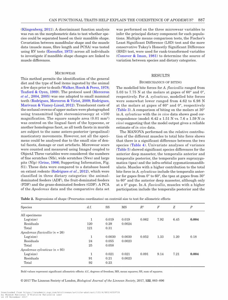
Muscles were blotted dry to remove excess alcohol and weighed using a Mettler AE100 electronic bal-ance (± 0.0001 g). The muscles were then placed in a 30% nitric acid solution (Loeb & Gans, 1986) until the connective tissue was sufficiently dissolved that the fibres would separate easily when gently prodded with a pair of narrow tweezers. At this point, the ni-tric acid was removed and replaced by a 50% aqueous glycerol solution, and the fibres of each muscle bundle were photographed using a Leica macroscope Z6 (×0.5 objective, ×2 zoom) coupled to a Leica digital camera (6 mega pixels). The lengths of ten to 15 fibres were measured using Image J. The volume of each muscle bundle was then calculated by dividing muscle mass by muscle density (1.06 g/cm3; Mendez & Keys, 1960), and the physiological cross-sectional area (PCSA) was calculated by dividing the volume by fibre length.
Biomechanical moDel
The model used is identical to that previously described (Cleuren, Aerts & De Vree, 1995; Herrel, Aerts & De Vree, 1998a, b; Herrel et al., 2008) and relies on the computation of the static force equilibrium. The input for the model consists of the three-dimensional coor-dinates of origin and insertion, the PCSA of the jaw muscles and the three-dimensional coordinates of the point of application of the bite force and the centre of rotation (Fig. 1). The centroid area of insertion was used for muscle bundles with relatively broad areas of origin and insertion. The coordinates were determined from lateral, dorsal and ventral drawings made during the dissection using a stereomicroscope with camera lucida, and from photographs of skulls and mandibles taken using a Leica macroscope Z6 (×0.5 objective, zoom ×2) attached to a Leica digital camera. Separate muscle and bite-point coordinates were taken for one specimen of A. flavicollis and A. sylvaticus each. Complex pennate muscles were separated into their component parts; therefore, no correction for pennation was included. Cross-sectional areas were scaled using a conservative muscle stress estimate of 30 N/cm (Herzog, 1994).
The simulations were run at gape angles of 0°, 30° and 60° with all jaw closer muscles set as maximally active. We ran the model for each individual of each species using the measured variation in muscle PCSA. Bite forces were calculated at a range of orientations for the food reaction forces, and at two different bite points: the incisor and the first molar. Model output consists of the magnitude of the bite forces and joint forces and the orientation of the joint forces at any given orientation of the food reaction forces. The log10-transformed bite and joint forces and the joint force orientations were then used as input for multivariate analyses of variance (MANOVA) with species as fixed
effect to test for differences in the functional output of the jaw system between species.
To validate the output of our model, we measured bite force from 18 individuals of A. sylvaticus caught in the field in Southern France using a piezoelectric force transducer (Kistler, type 9203, range 500 N; Kistler, Winterthur, Switzerland) attached to a handheld charge amplifier (Kistler, type 5995). The transducer was mounted between two bite plates as described by Herrel et al. (1999). The tips of both upper and lower bite plates were covered with a layer of cloth medical tape to provide a non-skid surface and to protect the teeth of the animals. The distance between the bite plates was adjusted to assure a gape of about 20°. Bite force was measured during molar biting. Five trials were conducted for each animal.
morphometricS
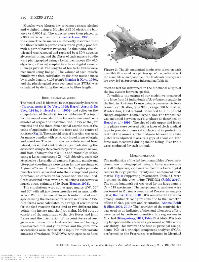
The medial side of the left hemi-mandibles of each spe-cimen was photographed using a Leica macroscope Z6 (×0.5 objective, ×2 zoom) coupled to a Leica digital camera (6 mega pixels). Twenty-nine anatomical land-marks (Fig. 2; Supporting Information, Table S1) were digitized in this view using TPSDIG2 (Rohlf, 2010). The entire landmark set was used for the large sample (N = 119 specimens). The morphometric analyses were performed in R using a generalized Procrustes analysis (GPA; Rohlf & Slice, 1990). GPA removes the variation among landmark configurations due to the isometric effects of size, position and orientation (Adams, Rohlf & Slice, 2004, 2013). The logarithm of the centroid size was used as an indicator of size, and allometric effects were tested by performing multivariate regressions in MorphoJ (Klingenberg, 2011; Table 2). A MANOVA test-ing for species differences was performed on 90% of the variability. This involved the first 22 principal compo-nents (PCs) of a principal component analyses (PCAs) performed on the Procrustes coordinates in MorphoJ
Figure 2. The 29 anatomical landmarks taken on each mandible illustrated on a photograph of the medial side of the mandible of an Apodemus. The landmark descriptions are provided in Supporting Information, Table S1.
Downloaded from https://academic.oup.com/biolinnean/article-abstract/122/4/883/4259735by Museum National d'Histoire Naturelle useron 29 November 2017
CAN FUNCTIONAL TRAITS HELP EXPLAIN THE COEXISTENCE OF APODEMUS? 887
© 2017 The Linnean Society of London, Biological Journal of the Linnean Society, 2017, 122, 883–896
(Klingenberg, 2011). A discriminant function analysis was run on the morphometric data to test whether spe-cies could be separated based on their mandible shape. Covariation between mandibular shape and the muscle data (muscle mass, fibre length and PCSA) was tested using RV tests (Escoufier, 1973) across all individuals to investigate if mandible shape changes are linked to muscle differences.
microwear
This method permits the identification of the general diet and the type of food items ingested by the animal a few days prior to death (Walker, Hoeck & Perez, 1978; Teaford & Oyen, 1989). The protocol used (Merceron et al., 2004, 2005) was one adapted to small mammal teeth (Rodrigues, Merceron & Viriot, 2009; Rodrigues, Marivaux & Vianey-Liaud, 2012). Translucent casts of the occlusal crowns of upper molars were photographed using transmitted light stereomicroscopy at ×100 magnification. The square sample area (0.01 mm2) was centred on the lingual facet of the hypocone, or another homologous facet, as all tooth facets in murids are subject to the same antero-posterior (propalinal) masticatory movements. However, not all the speci-mens could be analysed due to the small size of den-tal facets, damage or cast artefacts. Microwear scars were counted and measured using ImageJ coupled to ObjectJ. Three variables were considered: the numbers of fine scratches (Nfs), wide scratches (Nws) and large pits (Nlp) (Grine, 1986; Supporting Information, Fig. S1). These data were compared to a database based on extant rodents (Rodrigues et al., 2012), which were classified in three dietary categories: the animal-dominated feeders (ADF), the fruit-dominated feeders (FDF) and the grass-dominated feeders (GDF). A PCA of the Apodemus data and the comparative data set
was performed on the three microwear variables to infer the principal dietary component for each popula-tions. Multiple means comparison tests, the Fischer’s Least Significant Difference (LSD) test and the more conservative Tukey’s Honestly Significant Difference (HSD) test, were used for rank-transformed variables (Conover & Iman, 1981) to determine the source of variation between species and dietary categories.
RESULTS
BiomechanicS of BitinG
The modelled bite forces for A. flavicollis ranged from 5.03 to 7.75 N at the molars at gapes of 60° and 0°, respectively. For A. sylvaticus, modelled bite forces were somewhat lower ranged from 4.62 to 6.98 N at the molars at gapes of 60° and 0°, respectively (Table 3). A comparison of biting on the molars at 30° in A. sylvaticus with the in vivo data shows good cor-respondence (model: 6.42 ± 1.51 N vs. 7.6 ± 1.59 N in vivo) suggesting that the model output gives a reliable estimate of in vivo data.
The MANOVA performed on the relative contribu-tion of the different muscles to total bite force shows that there is a significant difference between the two species (Table 4). Univariate analyses of variance (Table 5) showed significant species differences for the anterior deep masseter, the temporalis anterior and temporalis posterior, the temporalis pars suprazygo-matica (tpsz) and the infra-orbital zygomaticomandib-ularis. Muscles with a higher contribution to the total bite force in A. sylvaticus include the temporalis anter-ior for gapes from 0° to 60°, the tpsz at gapes from 30° to 60° and the anterior deep masseter, although only at a 0° gape. In A. flavicollis, muscles with a higher participation include the temporalis posterior and the
Table 2. Regressions of shape (Procrustes coordinates) on centroid size to test for allometric effects
Species d.f. SS MS R2 F Z P
All specimens Log(size) 1 0.019 0.019 0.062 7.92 6.45 0.004 Residuals 120 0.29 0.0024 Total 121 0.31Apodemus flavicollis (n = 26) Log(size) 1 0.0030 0.0030 0.052 1.33 1.20 0.18 Residuals 24 0.055 0.0023 Total 25 0.058Apodemus sylvaticus (n = 93) Log(size) 1 0.021 0.021 0.091 9.14 7.21 0.004 Residuals 91 0.21 0.0023 Total 92 0.23
Bold values represent significant allometric effects. d.f., degrees of freedom; MS, mean squares; SS, sum of squares.
Downloaded from https://academic.oup.com/biolinnean/article-abstract/122/4/883/4259735by Museum National d'Histoire Naturelle useron 29 November 2017

888 E. KERR ET AL.
© 2017 The Linnean Society of London, Biological Journal of the Linnean Society, 2017, 122, 883–896
infra-orbital part of the zygomaticomandibularis. The two temporalis (anterior and posterior) muscles show the greatest difference between species.
A MANOVA on the output of the biomechanical model (Table 4) calculated for a gape angle of 0° showed a sig-nificant species effect (Pillai trace = 0.76; F3,17 = 17.78; P < 0.001). However, subsequent univariate ANOVAs showed no significant difference in bite force (P = 0.57) and joint force (P = 0.91) between the species (Table 5). The angle of the joint force was, however, different be-tween species (P < 0.001) and was significantly greater in A. sylvaticus. These results were consistent at all gape angles tested (0°–60°; see Table 5) and at both bite points. This indicates that the angle of joint force is orientated more posteriad in A. sylvaticus compared to A. flavicollis. A regression of joint force on bite force at
0° of gape and for bite point was significant (R2 = 0.96; P < 0.001; results for other gape angles and bite points are indicated in Supporting Information, Table S2). An analysis of the residual data showed that the joint force is significantly higher in A. sylvaticus for a given bite force (F1,19 = 22.78; P < 0.001), and further analysis shows this is true at all gape angles and bite points (Supporting Information, Table S2).
morphometricS
An ANOVA on morphometric data indicated significant differences between species in centroid size (F1,117 = 4.09; P = 0.046). However, the size ratio difference between species is less than 1.3 (Hutchinson ratio) for both a linear measurement (body length ratio = 1.04) and a
Table 4. Results of MAN(C)OVAs performed on biomechanical and morphometric data testing for differences between species
Pillai trace F d.f.1 d.f.2 P
Biomechanical data by species Biomechanical output 0.76 17.78 3 17 < 0.001 Fractional participation (all) 0.60 15.08 4 40 < 0.0001 Fractional participation (gape × species) 1.31 3.79 20 156 < 0.05 Fractional participation (gape 0) 0.66 4.91 4 10 0.019 Fractional participation (gape 30) 0.73 6.68 4 10 < 0.01 Fractional participation (gape 60) 0.70 5.76 4 10 0.011Morphometric data Shape by species (MANCOVA) 0.74 12.00 1 94 < 0.0001 Shape by size (MANCOVA) 0.58 5.91 1 94 < 0.0001 Shape by species × size (MANCOVA) 0.27 1.59 1 94 0.064 Shape (PC) by sex 0.99 1.93 43 1 0.52 Shape (Procrustes) by sex 0.97 1.67 42 2 0.45
Bold values indicate significant differences. As a covariate, the centroid size was used. MANCOVA, multivariate analysis of covariance; PC, principal component.
Table 3. Summary of the calculated bite force for both species and at different gape angles
Species Bite point Gape Bite force Joint force Angle joint force In vivo bite force
Apodemus flavicollis Molar 60 5.03 ± 1.44 3.12 ± 0.83 136.20 ± 4.4230 7.02 ± 1.93 2.58 ± 0.57 137.12 ± 5.440 7.75 ± 2.16 2.33 ± 0.47 135.40 ± 3.77
Incisor 60 2.96 ± 0.85 4.13 ± 1.09 139.61 ± 3.3830 4.13 ± 1.14 3.98 ± 0.96 131.06 ± 3.590 4.56 ± 1.27 3.64 ± 0.86 117.35 ± 2.89
Apodemus sylvaticus Molar 60 4.62 ± 1.12 3.27 ± 0.72 141.96 ± 2.2230 6.42 ± 1.51 2.83 ± 0.62 142.96 ± 2.48 7.6 ± 1.590 6.98 ± 1.61 2.65 ± 0.58 143.18 ± 4.06
Incisor 60 2.72 ± 0.66 4.21 ± 0.94 143.56 ± 1.7330 3.78 ± 0.89 4.08 ± 0.91 135.71 ± 1.730 4.11 ± 0.95 3.69 ± 0.82 125.03 ± 2.58
Table entries are means ± SD. In vivo bite force in Apodemus sylvaticus was measured at a gape angle of 20°.
Downloaded from https://academic.oup.com/biolinnean/article-abstract/122/4/883/4259735by Museum National d'Histoire Naturelle useron 29 November 2017

CAN FUNCTIONAL TRAITS HELP EXPLAIN THE COEXISTENCE OF APODEMUS? 889
© 2017 The Linnean Society of London, Biological Journal of the Linnean Society, 2017, 122, 883–896
mandible centroid size (1.03). A MANCOVA (Table 4) indicated differences between species in shape (Pillai = 0.74; F1,94 = 11.99; P < 0.001). Although sig-nificant allometry was detected across all specimens (Pillai = 0.58; F1,94 = 5.91; P < 0.01; see also Table 2), no interaction between size and species was observed (Pillai = 0.27; F1,94 = 1.59; P = 0.064). Interestingly, whereas allometric effects were significant for A. syl-vaticus, this was not the case for A. flavicollis when analysing species separately (Table 2). A discriminant function analysis was able to discriminate the two species based on the first 22 PC axes (Fig. 3). Twenty-three out of 26 A. flavicollis were correctly classified; for A. sylvaticus, all specimens were correctly classified when using a leave-one-out cross-validation. The man-dible of A. flavicollis has a more vertical coronoid, and the coronoid process is slanted slightly more anteriad; there is a slightly higher articular, a slightly longer and
narrower angular and a more narrow molar region. Apodemus sylvaticus has a slightly flatter coronoid and articular (= condylar process), a slightly shorter and wider angular and a more robust molar region espe-cially around the m2 and m3 (Fig. 3).
While species differences do exist, they do not account for the majority of variance in the data set. The PCAs show that the mandibles of the two species overlap in shape space (Supporting Information, Fig. S2). The first PC accounts for 16.18% of the variance for the entire sample. The second PC accounts for 9.09% (all sites with coronoid). On the positive side of the first axis, specimens have a lower more concave molar region, coupled with a shorter (but not narrower) angular and a slightly longer articular. On the negative side of PC1 mandibles with a slightly flatter molar alveolar region, a less vertical anterior edge of the coronoid, a shorter articular and a longer angular are found.
Table 5. Results of ANOVAs testing for differences between species in the fractional participation (log10) of the individual muscles, fractional participation of the muscle bundles and of the biomechanical output
Gape 0 Gape 30 Gape 60
P F d.f.1 d.f.2 P F d.f.1 d.f.2 P F d.f.1 d.f.2
Fractional participation Sm 0.43 0.67 1 19 0.49 0.50 1 19 0.30 1.15 1 19 adm 0.011 7.84 1 19 0.052 4.31 1 19 0.30 1.16 1 19 pdm 0.67 0.19 1 19 0.71 0.14 1 19 0.57 0.34 1 19 azm 0.14 2.42 1 19 0.052 4.31 1 19 0.28 1.23 1 19 pzm 0.25 1.40 1 19 0.20 1.80 1 19 0.20 1.78 1 19 iozm 0.041 4.79 1 19 0.045 4.60 1 19 0.045 4.61 1 19 tp < 0.001 87.59 1 19 < 0.001 132.2 1 19 < 0.001 287.6 1 19 ta < 0.001 73.54 1 19 < 0.001 112.2 1 19 < 0.001 344.1 1 19 tpsz 0.073 3.61 1 19 ep 0.72 0.13 1 19 0.54 0.40 1 19 0.25 1.41 1 19 ip 0.19 1.81 1 19 0.98 0.001 1 19 0.46 0.57 1 19 Mass 0.0056 9.75 1 19 0.015 7.24 1 19 0.088 3.24 1 19 Zmand 0.045 4.60 1 19 0.0018 13.11 1 19 0.085 3.31 1 19 Temp 0.72 0.14 1 19 0.56 0.36 1 19 0.65 0.21 1 19 Pter 0.11 2.76 1 19 0.92 0.009 1 19 0.21 1.69 1 19Biomechanical outputBite point 1 prey rf 0.57 0.012 1 20 0.63 0.24 1 20 0.65 0.21 1 20 b-b jf 0.91 0.012 1 20 0.87 0.028 1 20 0.88 0.022 1 20 a.joint f < 0.001 37.03 1 20 0.001 14.55 1 20 0.003 11.21 1 20Bite point 2 prey rf 0.57 0.34 1 20 0.63 0.24 1 20 0.65 0.21 1 20 b-b jf 0.414 0.70 1 20 0.57 0.34 1 20 0.76 0.095 1 20 a.joint f < 0.001 20.23 1 20 0.004 10.73 1 20 0.002 12.45 1 20
Bold values represent significant differences between species. sm, superior masseter; adm, anterior deep masseter; pdm, posterior deep masseter; azm, anterior zygomaticomandibularis; pzm, posterior zygomaticomandibularis; iozm, infra-orbital zygomaticomandibularis; tp, temporalis posterior; ta, temporalis anterior; tpsz, temporalis pars suprazygomatica; ep, external pterygoid; ip, internal pterygoid; Mass, masseter bundle; Zmand, zygomaticomandibularis bundle; Temp, temporalis bundle; Pter, pterygoid bundle; prey rf, prey reaction force (bite force); b-b jf, bone-on-bone joint force; a.joint f, angle joint force. ANOVA, analysis of variance; d.f., degrees of freedom.
Downloaded from https://academic.oup.com/biolinnean/article-abstract/122/4/883/4259735by Museum National d'Histoire Naturelle useron 29 November 2017

890 E. KERR ET AL.
© 2017 The Linnean Society of London, Biological Journal of the Linnean Society, 2017, 122, 883–896
RV tests were performed to investigate relationships between shape and biomechanical properties. There was no significant relationship between shape (PC scores describing 90% of the overall variability) and the output of the biomechanical model or between shape and the PCSA of the muscles (P = 0.07). However, there was a significant relationship (P = 0.03) between the mass of the muscle bundles and mandible shape (both for PC axes and Procrustes coordinates; Supporting Information, Table S3). As separate muscle bundles, only the temporalis muscle bundle mass was signifi-cantly associated with mandible shape (P = 0.01; PC coordinates).
microwear
A PCA performed on the microwear data extracted two axes together explaining 84% of the variance in the data set. On the plot of axes one and two, populations
of both species were distributed in a cloud between two of the standards, grass feeding (GDF) and animal feed-ing (ADF), with no apparent clustering or grouping (Fig. 4). There were significant differences (P < 0.05) be-tween each of the populations and the dietary category standards except for the A. sylvaticus from Montpellier against GDF (Supporting Information, Table S4). However, none of the populations had an exclusive feeding type. The A. sylvaticus from Montpellier appear to be somewhat more prone to grass feeding, while the A. sylvaticus from Belgium were more prone to insect feeding. In addition, no significant differences in the microwear were observed between species or between animals from different sites (Supporting Information, Table S4) with the exception of the Belgian popula-tion that differed from two of the Southerly popula-tions. However, this difference was only observed when using the LSD test for fine scratches and large pits. This indicates that the species and populations are not
Figure 3. Wireframes describing average shapes of each species (A) and the results of a discriminant function analysis (B) showing that species can be discriminated based on mandible shape.
Downloaded from https://academic.oup.com/biolinnean/article-abstract/122/4/883/4259735by Museum National d'Histoire Naturelle useron 29 November 2017

CAN FUNCTIONAL TRAITS HELP EXPLAIN THE COEXISTENCE OF APODEMUS? 891
© 2017 The Linnean Society of London, Biological Journal of the Linnean Society, 2017, 122, 883–896
discriminated by microwear, except for a slight differ-ence between the Belgian population vs. Murcia and Montpellier. However, the populations of the two spe-cies from Montseny cluster close together in the micro-wear space suggesting that they have similar diets in syntopy. Finally, no association between microwear and the mandible shape was observed (P = 0.26; RV coefficient: 0.15).
DISCUSSION
There are significant differences between the two spe-cies in muscle cross-sectional areas, the fractional par-ticipation of the muscles and mandible shape (Tables 3 and 4). Biting appears more optimized in A. flavicollis, with a lower joint force for a given bite force and a smaller joint force angle than in A. sylvaticus. Although there are no differences in the maximal bite force cal-culated, the differences in mandible shape between the species suggest some differences in function that could be related to diet. The more robust molar region in A. sylvaticus may indicate greater amount of grind-ing, as does the reduced angular process and the pos-teriorly shifted coronoid and the higher contribution of the anterior temporalis (Cox & Jeffery, 2011). Similar
differences are also observed in the species A. specio-sus in comparison to Apodemus argenteus (Renaud & Millien, 2001). The lower articular could also reflect a more habitually closed jaw posture while biting or chewing. The more vertical coronoid in A. flavicollis may improve the moment arm of the temporalis, and the longer and narrower angular will affect the areas of insertion of the masseter and the pterygoid mus-cles. These differences are subtle, however, and prob-ably reflect ecological tendencies rather than exclusive feeding patterns. Moreover, local adaptations at the population level may play an important role (Renaud & Michaux, 2003).
In addition to the differences in mandible shape, biomechanical properties other than maximal bite force may indicate species variation in diet. Apodemus flavicollis appears to be more optimized in terms of the efficacy of the production of bite force. The larger angle of joint force in A. sylvaticus may indicate that it tends to masticate at narrow gape, therefore exerting less destabilizing forces on the joint. This is moreover corroborated by the higher relative joint force for a given bite force in A. sylvaticus. Correspondingly, it may be possible that A. flavicollis habitually uses larger bite forces and requires a better optimization of its bite force to reduce the corresponding joint forces.
Figure 4. Plot of the first two principal components of a principal component analysis performed on the tooth wear data. Data for different populations are plotted together with feeding standards (animal, fruit and grass feeders; diamonds). Note how the populations of both species fall between grass- and animal-dominant feeders suggesting an omnivorous diet. The two species from the same locality (Montseny) cluster closely together. Open circles: Apodemus sylvaticus; closed square: Apodemus flavicollis. B, Belgium; MTP, Montpelier; MU, Murcia; MY, Montseny.
Downloaded from https://academic.oup.com/biolinnean/article-abstract/122/4/883/4259735by Museum National d'Histoire Naturelle useron 29 November 2017

892 E. KERR ET AL.
© 2017 The Linnean Society of London, Biological Journal of the Linnean Society, 2017, 122, 883–896
The greater participation of different muscle groups could also reflect feeding tendencies. There is a greater contribution of the masseter in A. sylvaticus, the bun-dle that controls biting and jaw movement at a narrow gape. There is also a greater participation of the zygo-maticomandibularis bundle in A. flavicollis. This bun-dle lies underneath the masseter bundle and passes through the infra-orbital fossa to attach anteriorly on the rostrum. This muscle has been shown to contribute to the stabilization of the mandible (Cox, Kirkham & Herrel, 2013) and may account for the more optimized joint force and joint force angle in A. flavicollis.
Allometric variation explains some of the vari-ation in shape across both species and in A. sylvaticus (Table 2). However, this is not the case for A. flavicol-lis where a regression of shape on centroid size was not significant (Table 2). Despite these differences in allometry between species, both overlap nearly com-pletely on the first two axes of a PCA. In coexisting Japanese Apodemus species, the overall shape vari-ation across the two species is also the same, despite having a slightly higher size ratio difference than the current species, around the Hutchinson value of 1.3 (Renaud & Millien, 2001). This suggests that morphol- ogical variation within Apodemus is often conserved in closely related species, even if they are more dis-parate in size than A. sylvaticus and A. flavicollis, and suggests an important role for allometry in structuring intrapopulation variation (Renaud & Auffray, 2013).
The overall mandible shape across both species is also linked to the mass of the temporalis bundle (Supporting Information, Table 3). It is likely that the temporalis is linked to the entire mandible shape and not only the region of its insertion, as modules do not alter in form independently, as has been observed in other rodents (Zelditch, Wood & Swiderski, 2009). The mass of this muscle bundle appears to be the only mus-cle property linked to mandible shape in these species, in contrast to what has been observed in other taxa (Fabre et al., 2014; Cornette, Tresset & Herrel, 2015a). This is possibly due to the fact that differences in mus-cle cross-sectional area do not directly translate into biomechanical differences, as different combinations of individual muscles or muscle bundles can result in the same or similar biomechanical performance. This many-to-one mapping of morphology on function has been observed in the jaw apparatus of many taxa and is especially common in fish that have extremely com-plex jaw systems with many elements (Alfaro, Bolnick & Wainwright, 2005).
The microwear signature is indistinguishable be-tween the two species and is different from the standards of grass-dominated and animal-dominated feeding, as could be expected for omnivorous species.
Notably, the two species that occur in syntopy at Montseny show very similar microwear signatures suggesting that differences in diet do not contribute to niche differentiation. Overall, our results suggest that the omnivory in these Apodemus species tends towards the plant-eating type. The only source of geographic variation in microwear was observed for the Belgian population (A. sylvaticus), which was significantly different from the populations of the same species in Montpellier and Murcia. The Montpellier population is also the only population with no significant differ-ence from the grass-feeding standard. This suggests that the Belgian population may tend more towards animal (probably insect) and fruit feeding compared to two other populations. Finally, we observed no cor-relation between the microwear profile and mandible shape, suggesting that at least the short-term feeding patterns do not impact overall mandible shape as has been suggested previously (Renaud & Michaux, 2003). Feeding differences that may potentially exist between the species are thus not identifiable through dental microwear, which appears limited to detect subtle variations.
It is often considered that character displacement and niche segregation is required for related, simi-lar species to coexist. However, the two Apodemus species considered in the present study have a very similar size and show no direct evidence for differ-ences in diet. It is likely that different taxa interact differently in terms of how and what kind of selec-tion pressures are mutually imposed due to sympatry. However, character displacement has been observed in taxa as different as birds, salamanders, lizards and shrews (Adams & Rohlf, 2000; Grant & Grant, 2006; Stuart et al., 2014; Cornette et al., 2015b). However, ecologically similar, related species may readily and sustainably coexist in the same habitat where there is no limitation in the vital shared resources (Den Boer, 1979; Okuzaki et al., 2010). The two species densities will then probably fluctuate due to environ- mental fluctuations that alternatively favour the slight differences between the species. These scenarios have been demonstrated to be viable based on sim-ple models (Yo Mo, Xu & Urabe, 1996). An example of this is the successful sympatry of two pairs of bird species within the genus Calidris permitted by differ-ential resource exploitation through fluctuations in yearly population size, differences in distribution and migratory timing (Holmes & Pitelka, 1968). In ana-logy, perhaps the differences seen in the Apodemus species studied here may be due to the allopatric selection pressures driving phenotypic differences that remained upon secondary contact. The selec-tion pressures that are exerted by the coexistence of
Downloaded from https://academic.oup.com/biolinnean/article-abstract/122/4/883/4259735by Museum National d'Histoire Naturelle useron 29 November 2017

CAN FUNCTIONAL TRAITS HELP EXPLAIN THE COEXISTENCE OF APODEMUS? 893
© 2017 The Linnean Society of London, Biological Journal of the Linnean Society, 2017, 122, 883–896
these species do not appear to drive a notable add-itional divergence in size or morphology (Dayan & Simberloff, 2005). Niche segregation may therefore be more subtle, involving behavioural differences as much if not more than biomechanical or mor-phological changes (Simberloff & Boecklen, 1981). Differences in spatial use, staggering of breeding pat-terns and differences in distribution and dispersion also exist between these two Apodemus species, and these may be sufficient to allow differential resource exploitation (Montgomery, 1980, 1981, 1989; Marsh et al., 2001; Mitter, Sumasgutner & Gamauf, 2015).
CONCLUSION
Our data on mandible shape and the biomechanics of biting suggest that A. sylvaticus may have a ten-dency to eat smaller food items that require greater amounts of grinding (e.g. plant leaves, stems and roots). This would account for the more robust molar region, associated changes in shape of the mandibu-lar ramus, the greater development of the masseter bundle and the less optimal joint force to bite force ratio. The anatomy and function of A. flavicollis, on the other hand, suggests a greater tendency to eat larger food items that require the application of greater bite forces (nuts, hard-shelled fruits). This would concur with the more optimized bite force and the vertical shape of the coronoid associated with the attachment of the temporalis muscle. The morphological and biomechanical differences that do exist nonetheless do not seem sufficient to create a consistent dietary niche separation. This is suggested by the microwear data, which show no discernable difference between the species, especially for syntopic populations. Further studies with larger sample sizes and focusing in syntopic populations of the two species in addition to an examination other niche dimensions are required in order to better understand the coexistence of these two species.
ACKNOWLEDGEMENTS
We thank Phil Cox, Sam Ginot and an anonymous reviewer for helpful and constructive comments on the manuscript. This study was funded by a Labex BCDiv (Laboratoires d’Excellence, Diversités Biologiques et Culturelles; http://labex-bcdiv.mnhn.fr/). We thank Johan R. Michaux for providing specimens from Belgium, Montpellier and Murcia; Frank Sauvage for the specimens from Sénart and Orleans; and Alexis Ribas Salvador for the specimens from Montseny.
REFERENCES
Adams DC, Rohlf FJ. 2000. Ecological character displace-ment in Plethodon: biomechanical differences found from a geometric morphometric study. Proceedings of the National Academy of Sciences of the United States of America 97: 4106–4111.
Adams DC, Rohlf FJ, Slice DE. 2004. Geometric morpho-metrics: ten years of progress following the ‘revolution’. Italian Journal of Zoology 71: 5–16.
Adams DC, Rohlf FJ, Slice DE. 2013. A field comes of age: geometric morphometrics in the 21st century. Hystrix 24: 7–14.
Alfaro ME, Bolnick DI, Wainwright PC. 2005. Evolutionary consequences of many-to-one mapping of jaw morphology to mechanics in labrid fishes. The American Naturalist 165: E140–E154.
Amori G, Contoli L. 1994. Morphotypic, craniometric and genotypic diversification in Apodemus flavicollis and Apodemus sylvaticus. Italian Journal of Zoology 61: 353–357.
Barčiová L, Macholán M. 2006. Morphometric study of two species of wood mice Apodemus sylvaticus and A. flavicollis (Rodentia: Muridae): traditional and geometric morphometric approach. Acta Theriologica 51: 15–27.
Bauduin S, Cassaing J, Issam M, Martin C. 2013. Interactions between the short-tailed mouse (Mus spretus) and the wood mouse (Apodemus sylvaticus): diet overlap revealed by stable isotopes. Canadian Journal of Zoology 91: 102–109.
Baverstock H, Jeffery NS, Cobb SN. 2013. The morphology of the mouse masticatory musculature. Journal of Anatomy 223: 46–60.
Brown WL Jr, Wilson EO. 1956. Character displacement. Systematic Zoology 5: 49–64.
Bugarski-Stanojević V, Blagojević J, Adnaðevic ́ T, Jojić V, Vujošević M. 2008. Molecular phylogeny and distri-bution of three Apodemus species (Muridae, Rodentia) in Serbia. Journal of Zoological Systematics and Evolutionary Research 46: 278–286.
Cleuren J, Aerts P, De Vree F. 1995. Bite and joint force ana-lysis in Caiman crocodilius. Belgian Journal of Zoology 125: 79–94.
Conover WJ, Iman RL. 1981. Rank transformations as a bridge between parametric and nonparametric statistics. The American Statistician 35: 124–129.
Cornette R, Tresset A, Herrel A. 2015a. The shrew tamed by Wolff ’s law: do functional constraints shape the skull through muscle and bone covariation? Journal of Morphology 276: 301–309.
Cornette R, Tresset A, Houssin C, Pascal M, Herrel A. 2015b. Does bite force provide a competitive advantage in shrews? The case of the greater white-toothed shrew. Biological Journal of the Linnean Society 114: 795–807.
Cox PG, Jeffery N. 2011. Reviewing the morphology of the jaw-closing musculature in squirrels, rats, and guinea pigs with contrast-enhanced microCt. The Anatomical Record 294: 915–928.
Downloaded from https://academic.oup.com/biolinnean/article-abstract/122/4/883/4259735by Museum National d'Histoire Naturelle useron 29 November 2017

894 E. KERR ET AL.
© 2017 The Linnean Society of London, Biological Journal of the Linnean Society, 2017, 122, 883–896
Cox PG, Kirkham J, Herrel A. 2013. Masticatory biomech-anics of the Laotian rock rat, Laonastes aenigmamus, and the function of the zygomaticomandibularis muscle. PeerJ 1: e160.
Dayan T, Simberloff D. 1998. Size patterns among competi-tors: ecological character displacement and character release in mammals, with special reference to island populations. Mammal Review 28: 99–124.
Dayan T, Simberloff D. 2005. Ecological and community-wide character displacement: the next generation. Ecology Letters 8: 875–894.
Den Boer PJ. 1979. Exclusion or coexistence and the taxonomic or ecological relationship between species. Netherlands Journal of Zoology 30: 278–306.
Engländer H, Amtmann E. 1963. Introgressive hybridisa-tion von Apodemus sylvaticus und A. tauricus in Westeuropa. Naturwissenschaften 50: 312–313.
Escoufier Y. 1973. Le traitement des variables vectorielles. Biometrics 29: 751–760.
Fabre A-C, Andrade DV, Huyghe K, Cornette R, Herrel A. 2014. Interrelationships between bones, muscles, and performance: biting in the lizard Tupinambis merianae. Evolutionary Biology 41: 518–527.
Filippucci MG, Macholán M, Michaux JR. 2002. Genetic variation and evolution in the genus Apodemus (Muridae: Rodentia). Biological Journal of the Linnean Society 75: 395–419.
García FJ, Díaz M, de Alba JM, Alonso CL, Carbonell R, De Carrion ML, Monedero C, Santos T. 1998. Edge effects and patterns of winter abundance of wood mice Apodemus sylvaticus in Spanish fragmented forests. Acta Theriologica 43: 255–262.
Grant PR, Grant BR. 2006. Evolution of character displace-ment in Darwin’s finches. Science 313: 224–226.
Grine FE. 1986. Dental evidence for dietary differences in Australopithecus and Paranthropus: a quantitative analysis of permanent molar microwear. Journal of Human Evolution 15: 783–822.
Herrel A, Aerts P, De Vree F. 1998a. Ecomorphology of the lizard feeding apparatus: a modelling approach. Netherlands Journal of Zoology 48: 1–25.
Herrel A, Aerts P, De Vree F. 1998b. Static biting in lizards: functional morphology of the temporal ligaments. Journal of Zoology 244: 135–143.
Herrel A, De Smet A, Aguirre LF, Aerts P. 2008. Morphological and mechanical determinants of bite force in bats: do muscles matter? The Journal of Experimental Biology 211: 86–91.
Herrel A, Spithoven L, Van Damme R, De Vree F. 1999. Sexual dimorphism of head size in Gallotia galloti; testing the niche divergence hypothesis by functional analyses. Functional Ecology 13: 289–297.
Herzog W. 1994. Muscle. In: Herzog W, Nigg BM, eds. Biomechanics of the musculoskeletal system. Chichester: John Wiley & Sons, 154–187.
Holmes RT, Pitelka FA. 1968. Food overlap among coexisting sandpipers on northern Alaskan Tundra. Systematic Zoology 17: 305–318.
Hutchinson GE. 1959. Homage to Santa Rosalia or why are there so many kinds of animals? The American Naturalist 93: 145–159.
Janzekovic F, Krystufek B. 2004. Geometric morphometry of the upper molars in European wood mice Apodemus. Folia Zoologica 53: 47–55.
Jensen TS. 1985. Seed-seed predator interactions of European beech, Fagus silvatica and forest rodents, Clethrionomys glareolus and Apodemus flavicollis. Oikos 44: 149–156.
Jewell PA, Fullagar PJ. 1965. Fertility among races of the field mouse (Apodemus sylvaticus) and their failure to form hybrids with the yellow-necked mouse (Apodemus flavicol-lis). Evolution 19: 175–181.
Klingenberg CP. 2011. MorphoJ: an integrated software package for geometric morphometrics. Molecular Ecology Resources 11: 353–357.
Kryštufek B, Vohralík V. 2007. Distribution of field mice (Apodemus) (Mammalia: Rodentia) in Anatolia. Zoology in the Middle East 42: 25–36.
Lalis A, Evin A, Denys C. 2009. Morphological identifica-tion of sibling species: the case of West African Mastomys (Rodentia: Muridae) in sympatry. Comptes Rendus Biologies 332: 480–488.
Loeb GE, Gans C. 1986. Electromyography for experimental-ists. Chicago: University of Chicago Press.
Loman J. 2008. Small rodent population synchrony in western Sweden. Effects of landscape structure. Web Ecology 8: 14–21.
Makova KD, Nekrutenko A, Baker RJ. 2000. Evolution of microsatellite alleles in four species of mice (genus Apodemus). Journal of Molecular Evolution 51: 166–172.
Marsh ACW, Harris S. 2000. Partitioning of woodland habitat resources by two sympatric species of Apodemus: lessons for the conservation of the yellow-necked mouse (A. flavicollis) in Britain. Biological Conservation 92: 275–283.
Marsh ACW, Poulton S, Harris S. 2001. The yellow-necked mouse Apodemus flavicollis in Britain: status and ana-lysis of factors affecting distribution. Mammal Review 31: 203–227.
Mendez J, Keys A. 1960. Density and composition of mam-malian muscle. Metabolism-Clinical and Experimental 9: 184–188.
Merceron G, Blondel C, Brunet M, Sen S, Solounias N, Viriot L, Heintz E. 2004. The Late Miocene paleoenvironment of Afghanistan as inferred from dental microwear in artiodac-tyls. Palaeogeography, Palaeoclimatology, Palaeoecology 207: 143–163.
Merceron G, Blondel C, De Bonis L, Koufos GD, Viriot L. 2005. A new method of dental microwear analysis: applica-tion to extant primates and Ouranopithecus macedoniensis (Late Miocene of Greece). Palaios 20: 551–561.
Michaux JR, Chevret P, Filippucci MG, Macholan M. 2002. Phylogeny of the genus Apodemus with a special em-phasis on the subgenus Sylvaemus using the nuclear IRBP gene and two mitochondrial markers: cytochrome b and 12S rRNA. Molecular Phylogenetics and Evolution 23: 123–136.
Michaux JR, Kinet S, Filippucci MG, Libois R, Besnard A, Catzeflis F. 2001. Molecular identification of three sympatric species of wood mice (Apodemus sylvaticus,
Downloaded from https://academic.oup.com/biolinnean/article-abstract/122/4/883/4259735by Museum National d'Histoire Naturelle useron 29 November 2017

CAN FUNCTIONAL TRAITS HELP EXPLAIN THE COEXISTENCE OF APODEMUS? 895
© 2017 The Linnean Society of London, Biological Journal of the Linnean Society, 2017, 122, 883–896
A. flavicollis, A. alpicola) in western Europe (Muridae: Rodentia). Molecular Ecology Notes 1: 260–263.
Mikulová P, Frynta D. 2001. Test of character displacement in urban populations of Apodemus sylvaticus. Canadian Journal of Zoology 79: 794–801.
Mitter G, Sumasgutner P, Gamauf A. 2015. Niche-partitioning of three Apodemus species (Mammalia: Murinae) in an urban environment. Annalen der Naturhistorisches Museum Wien 117: 37–46.
Montgomery WI. 1980. Spatial organization in sympatric pop-ulations of Apodemus sylvaticus and A. flavicollis (Rodentia: Muridae). Journal of Zoology 192: 379–401.
Montgomery WI. 1981. A removal experiment with sympatric populations of Apodemus sylvaticus (L.) and A. flavicollis (Melchior) (Rodentia: Muridae). Oecologia 51: 123–132.
Montgomery WI. 1989. Population regulation in the wood mouse, Apodemus sylvaticus. II. Density dependence in spa-tial distribution and reproduction. The Journal of Animal Ecology 58: 477–494.
Niethammer J. 1969. Zur Frage der Introgression bei den Waldmäusen Apodemus sylvaticus und A. flavicollis (Mammalia, Rodentia). Journal of Zoological Systematics and Evolutionary Research 7: 77–127.
Okuzaki Y, Takami Y, Sota T. 2010. Resource partitioning or reproductive isolation: the ecological role of body size differ-ences among closely related species in sympatry. The Journal of Animal Ecology 79: 383–392.
Renaud S, Auffray J-C. 2013. The direction of main pheno-typic variance as a channel to morphological evolution: case studies in murine rodents. Hystrix 24: 85–93.
Renaud S, Michaux JR. 2003. Adaptive latitudinal trends in the mandible shape of Apodemus wood mice. Journal of Biogeography 30: 1617–1628.
Renaud S, Millien V. 2001. Intra and interspecific morphological variation in the field mouse species Apodemus argenteus and A. spe-ciosus in the Japanese archipelago: the role of insular isolation and biogeographic gradients. Biological Journal of the Linnean Society 74: 557–569.
Renaud S, Chevret P, Michaux J. 2007. Morphological vs. molecular evolution: ecology and phylogeny both shape the mandible of rodents. Zoologica Scripta 36: 525–535.
Rodrigues HG, Marivaux L, Vianey-Liaud M. 2012. Expansion of open landscapes in Northern China dur-ing the Oligocene induced by dramatic climate changes:
paleoecological evidence. Palaeogeography, Palaeoclimatology, Palaeoecology 358: 62–71.
Rodrigues HG, Merceron G, Viriot L. 2009. Dental microwear patterns of extant and extinct Muridae (Rodentia, Mammalia): ecological implications. Die Naturwissenschaften 96: 537–542.
Rogers LM, Gorman ML. 1995. The diet of the wood mouse Apodemus sylvaticus on set aside land. Journal of Zoology 235: 77–83.
Rohlf FJ. 2010. TpsDig v2. 16. Stony Brook: Department of Ecology and Evolution, State Univ. New York.
Rohlf FJ, Slice D. 1990. Extensions of the Procrustes method for the optimal superimposition of landmarks. Systematic Biology 39: 40–59.
Schoener TW. 1989. The ecological niche. In: Cherrett JM, Bradshaw AD, Goldsmith FB, Grubb PJ, Krebs JR, eds. Ecological concepts: the contribution of ecology to an under-standing of the natural world. Oxford: Blackwell Scientific Publications, 79–114.
Serizawa K, Suzuki H, Tsuchiya K. 2000. A phylogen-etic view on species radiation in Apodemus inferred from variation of nuclear and mitochondrial genes. Biochemical Genetics 38: 27–40.
Simberloff D, Boecklen W. 1981. Santa Rosalia reconsid-ered: size ratios and competition. Evolution 35: 1206–1228.
Smal CM, Fairley JS. 1980. Food of wood mice (Apodemus sylvaticus) and bank voles (Clethrionomys glareolus) in oak and yew woods at Killarney, Ireland. Journal of Zoology 191: 413–418.
Stuart YE, Campbell TS, Hohenlohe PA, Reynolds RG, Revell LJ, Losos JB. 2014. Rapid evolution of a native species following invasion by a congener. Science 346: 463–466.
Teaford MF, Oyen OJ. 1989. In vivo and in vitro turnover in dental microwear. American Journal of Physical Anthropology 80: 447–460.
Walker A, Hoeck H, Perez L. 1978. Microwear of mamma-lian teeth as an indicator of diet. Science 201: 908–910.
Yo Mo T, Xu W-Z, Urabe I. 1996. Mathematical model allowing the coexistence of closely related competitors at the initial stage of evolution. Researches on Population Ecology 38: 239–247.
Zelditch ML, Wood AR, Swiderski DL. 2009. Building de-velopmental integration into functional systems: function-induced integration of mandibular shape. Evolutionary Biology 36: 71–87.
SUPPORTING INFORMATION
Additional Supporting Information may be found in the online version of this article at the publisher’s web-site:
Table S1. Definition of the landmarks used for the geometric morphometric analysis.Table S2. Regression of the Log10-transformed bite force vs. the Log10-transformed joint force and the results of an ANOVA testing for differences between species performed on the residual joint force.Table S3. Analyses of covariance between muscle properties and morphology (Principal Components and Procrustes coordinates) of both species using an RV-test. Muscle mass is by muscle bundle.
Downloaded from https://academic.oup.com/biolinnean/article-abstract/122/4/883/4259735by Museum National d'Histoire Naturelle useron 29 November 2017

896 E. KERR ET AL.
© 2017 The Linnean Society of London, Biological Journal of the Linnean Society, 2017, 122, 883–896
Table S4. ANOVA post-hoc tests on the microwear data, using Fisher’s least significance (L) and Tukey’s honestly significant test (H). Grey squares indicate a significant difference at α = 0.05, for number of wide scratches (Nws), number of fine scratches (Nfs) and number of large pits (Nlp). Diet standards are animal dominant feeding (ADF), grass dominant feeding (GDF) and fruit dominant feeding (FDF).Figure S1. Picture taken of a tooth cast illustrating the large pits (Lp), the fine scratches (Fs) and the wide scratches (Ws) that were quantified.Figure S2. Principal components analysis performed on the mandibular shape data. Different symbols repre-sent different populations (circle = Montseny; diamond = Montpellier; hexagon = Murcia; triangle down = Senart; triangle up = Orleans; square = Belgium) and different colours represent different species (black = A. flavicollis; white = A. sylvaticus). Note how the variability of A. sylvaticus largely encompasses the variability of A. flavicollis for the syntopic site of Montseny as illustrated by the minimum convex polygons. Across all sites the variability of A. flavicollis is nearly completely contained within that of A. sylvaticus (dashed lines).
Downloaded from https://academic.oup.com/biolinnean/article-abstract/122/4/883/4259735by Museum National d'Histoire Naturelle useron 29 November 2017