Embed Size (px)
Citation preview
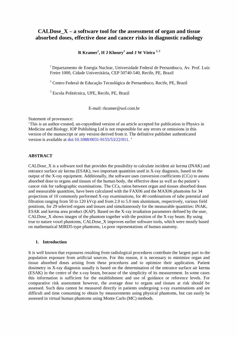
CALDose_X – a software tool for the assessment of organ and tissue
absorbed doses, effective dose and cancer risks in diagnostic radiology
R Kramer1, H J Khoury
1 and J W Vieira
2, 3
1 Departamento de Energia Nuclear, Universidade Federal de Pernambuco, Av. Prof. Luiz
Freire 1000, Cidade Universitária, CEP 50740-540, Recife, PE, Brazil
2 Centro Federal de Educação Tecnológica de Pernambuco, Recife, PE, Brazil
3
Escola Politécnica, UPE, Recife, PE, Brazil
E-mail: [email protected]
Statement of provenance:
„This is an author-created, un-copyedited version of an article accepted for publication in Physics in
Medicine and Biology. IOP Publishing Ltd is not responsible for any errors or omissions in this version of the manuscript or any version derived from it. The definitive publisher authenticated
version is available at doi:10.1088/0031-9155/53/22/011. „
ABSTRACT
CALDose_X is a software tool that provides the possibility to calculate incident air kerma (INAK) and
entrance surface air kerma (ESAK), two important quantities used in X-ray diagnosis, based on the output of the X-ray equipment. Additionally, the software uses conversion coefficients (CCs) to assess
absorbed dose to organs and tissues of the human body, the effective dose as well as the patient‟s
cancer risk for radiographic examinations. The CCs, ratios between organ and tissues absorbed doses and measurable quantities, have been calculated with the FAX06 and the MAX06 phantoms for 34
projections of 10 commonly performed X-ray examinations, for 40 combinations of tube potential and
filtration ranging from 50 to 120 kVcp and from 2.0 to 5.0 mm aluminium, respectively, various field positions, for 29 selected organs and tissues and simultaneously for the measurable quantities: INAK,
ESAK and kerma area product (KAP). Based on the X-ray irradiation parameters defined by the user,
CALDose_X shows images of the phantom together with the position of the X-ray beam. By using
true to nature voxel phantoms, CALDose_X improves earlier software tools, which were mostly based on mathematical MIRD5-type phantoms, i.e.poor representations of human anatomy.
1. Introduction
It is well known that exposures resulting from radiological procedures contribute the largest part to the population exposure from artificial sources. For this reason, it is necessary to minimize organ and
tissue absorbed doses arising from these procedures and to optimize their application. Patient
dosimetry in X-ray diagnosis usually is based on the determination of the entrance surface air kerma (ESAK) in the centre of the x-ray beam, because of the simplicity of its measurement. In some cases
this information is sufficient for the establishment and use of guidance or reference levels. For
comparative risk assessment however, the average dose to organs and tissues at risk should be assessed. Such data cannot be measured directly in patients undergoing x-ray examinations and are
difficult and time consuming to obtain by measurements using physical phantoms, but can easily be
assessed in virtual human phantoms using Monte Carlo (MC) methods.
Conversion coefficients (CCs) between absorbed or equivalent dose to organs at risk and measurable
quantities commonly used in X-ray diagnosis have been calculated by MC methods for the last 30
years mostly with mathematical MIRD5-type phantoms. Kramer and Drexler (1976) and Rosenstein (1976) published independently organ doses in the MIRD5 phantom (Snyder et al 1974) for diagnostic
radiology for the first time. Since then, comprehensive compilations of organ absorbed doses for the
most important as well as for special examinations have been published for the ADAM and EVA phantoms (Kramer et al 1982) and other MIRD5-type phantoms, relating the quantities of interest to
measurable quantities, like exposure in free air, entrance exposure on the surface of the patient or
dose-area product (Jones and Wall 1985, Rosenstein 1988, Drexler et al 1990, Le Heron 1992, Hart et
al 1994, Schultz et al 1994, Schultz et al 1995, Petoussi-Henss et al 1995).
Until the middle of the 90ies of the last century, the results of these studies were usually published in
reports containing extensive tables with CCs for the most frequent X-ray examinations. Later, software tools for absorbed dose calculation in X-ray diagnosis began to appear.
One of the first was published by Rannikko et al (1997), who developed a computer program, called WinODS (WinODS 2008), for organ absorbed dose calculations with a size- and sex-adjustable
phantom. This method uses analytical functions, based on MC-calculated depth-dose distributions for
a semi-infinite homogeneous water slab, and applies them to 35 sagittal slices based on a male
ALDERSON-RANDO (Alderson et al 1962) (AR) phantom containing contours of organ cross-sections. Organ absorbed doses are calculated as sum over the absorbed doses to voxels having a size
of 1cm x 1cm x d, where d = 2.5 cm is the slice thickness. A female phantom was derived from the
male phantom and under-weight as well as over-weight male and female versions have also been constructed based on statistics from the Finnish clothing industry. WinODS can easily be adapted to a
variety of exposure conditions as well as patient anatomies and the results appear almost
instantaneously ; however, being based on depth dose distributions for a homogeneous water phantom,
many corrections had to be made for WinODS in order to fit these distributions into the shape and the inhomogeneous structure of the AR phantom. Still, correction factors for shielding by bone were
applied only to the ovaries and the uterus for posterior-anterior and lateral projections and the voxel
size is quite large to properly represent small organs and tissues. WinODS calculates the effective dose based on ICRP60 (ICRP 1991) and runs only on WINDOWS 98. According to the distributor (Wendin
2008) there are no plans to update WinODS with respect to the operational system and/or the revised
concept of effective dose published by the International Commission on Radiological Protection (ICRP) in its new recommendations ICRP103 (ICRP 2007).
Shortly after, Servomaa and Tapiovaara (1998) published the program PCXMC, which is distributed
from a website of the Finnish Radiation and Nuclear Safety Authority (PCXMC 2008). PCXMC is a MC computer code using the whole hermaphrodite MIRD5 phantom family (Cristy 1980) and
applying scaling factors to modify the size of the phantoms. As opposed to WinODS, PCXMC is not
using pre-calculated data but performing a MC calculation for the exposure conditions defined by the user. Therefore for a given computer, organ and tissue absorbed doses are emitted by PCXMC perhaps
only after some minutes, depending on the level of statistical accuracy required by the user. The latest
version 1.5.2 runs on WINDOWS 95/98/NT/2000/XP and calculates the effective dose based on ICRP60. According to one of the authors (Tapiovaara 2008), PCXMC will be updated to calculate the
effective dose based on ICRP103 and the radiation risk.
DoseCal is a software tool published by Kyriou et al (2000), which uses the CCs determined by Jones and Wall (1985) and by Hart et al (1994) for adult and paediatric MIRD5 phantoms, but this program
does not scale a given phantom for weight or size. Using pre-calculated CCs, DoseCal outputs results
also almost instantaneously. DoseCal calculates the effective dose based on ICRP60 and runs only on WINDOWS 98. According to one of the authors (Kyriou 2008) there are no plans to update DoseCal
with respect to the operational system and/or the revised concept of effective dose published by ICRP
in its new recommendations ICRP103 (ICRP 2007).
The development of tomographic or voxel-based phantoms began in the 80ies of the last century
(Williams et al 1986) in order to overcome the inadequate anatomy of the MIRD5-type phantoms, but
relatively few studies with respect to diagnostic radiology have been published with voxel-based
phantoms over the last 22 years (Zankl et al 2000, Alkahane et al 2001, Winslow et al 2005, Petoussi-
Henss et al 2005, Bozkurt and Bor 2007) and then mainly about special radiographic examinations. None of them presented a comprehensive set of voxel-based CCs for the most important X-ray
examinations and no corresponding software tool was developed so far, i.e. that after having been
upgraded, PCXMC would then be the only software tool available for X-ray diagnosis; however, based on hermaphrodite MIRD5 phantoms, which are poor representations of human anatomy and do
not allow for a sex-specific absorbed dose calculation of the effective dose according to ICRP 103.
The purpose of this study is therefore
a) to calculate organ and tissue CCs for the most common examinations in X-ray diagnosis with
the recently developed MAX06 and FAX06 voxel phantoms for various projections and different X-ray spectra,
b) to make these CCs available to the public through a software tool, called CALDose_X
(CALculation of Dose for X-ray diagnosis), to be used in daily routine work by radiological departments of hospitals, health services, regulatory authorities, educational programs, etc. for
the assessment of the incident air kerma (INAK), the ESAK and organ and tissue absorbed
doses for X-ray examinations and exposure conditions defined by the user, by multiplying the
CCs with the value of the corresponding measurable quantity, for determining the effective dose with sex-specific phantoms according to ICRP103 and for estimating the patient‟s cancer
risks.
Earlier publications have reported about the development and the use of the MAX06 and the FAX06
phantoms, the methods of calculation (Kramer et al 2003a, 2004a, b, 2005a, b, 2006a, c, 2007) and
about comparisons with data from other phantoms, also covering the area of X-ray diagnosis (Kramer
et al 2003b, Vieira et al 2003, Lima et al 2003, 2004, Kramer et al 2006b). Consequently this study will refer to the underlying scientific methods only as far as it is necessary for the understanding of the
program CALDose_X. For specific issues, the reader is kindly asked to consult these references.
2. Materials and methods
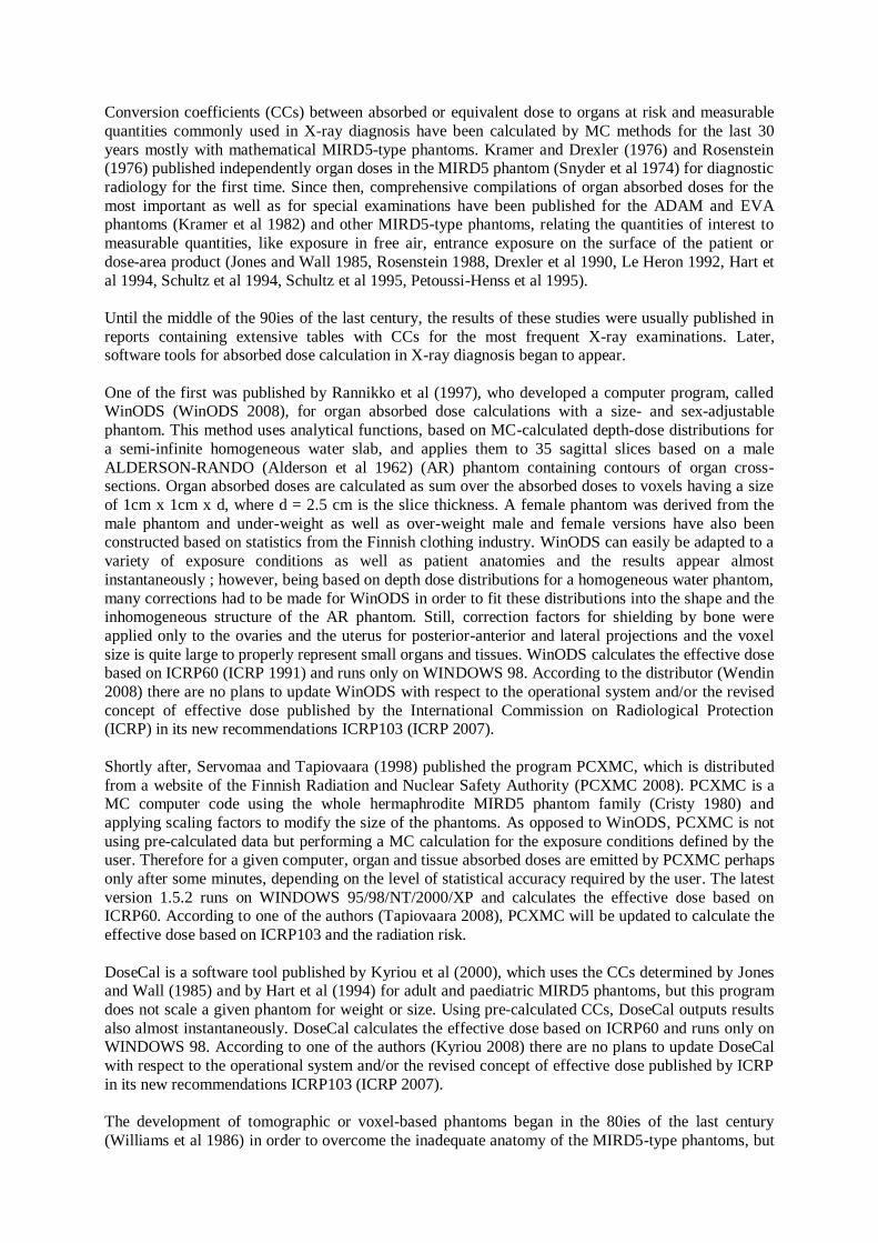
2.1 The MAX06 and the FAX06 phantoms
FAX06 MAX06
Figure 1. The FAX06 and the MAX06 phantoms. (Adipose and muscle tissues removed)
MAX06 and FAX06 (Kramer et al 2006a) are updated versions of the MAX (Kramer et al 2003a) and
of the FAX phantoms (Kramer et al 2004a). The update became necessary because of the inclusion of
the extra-thoracic airways, the oral mucosa, the gall bladder, the heart, the lymphatic nodes, the prostate, the salivary glands and male breasts into the concept of effective dose (ICRP 2007). The
updated phantoms are made of 1.2 mm cubic voxel, and the additional segmentation was based on the
original CT images from which the MAX/FAX phantoms had been developed, on anatomical textbooks, and on skeletal data provided by ICRP70 (ICRP 1995) and ICRP89 (ICRP 2003). Like
MAX and FAX, the MAX06 and the FAX06 phantoms have organ and soft tissue masses in
accordance with the reference data from ICRP89. Soft, muscle, adipose, skin, lung and cartilage tissue
as well as a homogeneous mixture for the skeleton have been used in organs and tissues of the two phantoms. The elemental compositions and densities were taken from or are based on data given in
ICRU44 (ICRU1989), except for the composition of the skeletal mixture which has been taken from
the report of Cristy and Eckerman (1987). The most important soft tissue organs and the skeletons of the two phantoms are shown in figure 1.
2.2 The EGSnrc Monte Carlo code
The EGSnrc MC code (Kawrakow 2000), in the form of version V4-r2-2-3 (Kawrakow 2006), was
used to calculate organ and tissue absorbed doses in the FAX06 and the MAX06 phantoms. The EGSnrc system, which provides a much improved implementation of the condensed history technique
for charged particle transport, and uses single scattering simulation in the vicinity of interfaces
between different materials, is much better suited for the simulation of particle transport in small regions compared to its predecessor EGS4 (Nelson et al 1985) In addition, EGSnrc includes Doppler
broadening and binding corrections in the simulation of Compton scattering, and simulates the full
relaxation cascade of inner shell vacancies thus providing more reliable results at low photon energies.
EGSnrc simulates coupled electron-photon transport through arbitrary media.
2.3 Absorbed dose and normalization quantities
2.3.1 Organ and tissue absorbed dose
For each MC simulation of a diagnostic X-ray examination, average absorbed doses (kerma) have
been calculated in the following 29 organs and tissues of the MAX06 and the FAX06 phantoms:
adrenals, bladder wall, brain, oral mucosa, colon wall, breasts, kidneys, liver, lungs, muscle tissue, oesophagus, ovaries, testes, pancreas, small intestine wall, skin, spleen, stomach wall, salivary glands,
thymus, thyroid, extra-thoracic region, uterus, prostate, heart wall, lymphatic nodes, gall bladder wall,
bone surface cells (BSC) and red bone marrow (RBM). Absorbed dose to the mouth cavity was taken as surrogate absorbed dose to the oral mucosa. The 29 organs and tissues are those specified by the
ICRP for the calculation of the effective dose (ICRP 2007).
Absorbed dose to skeletal soft tissues
Diagnostic X-ray spectra have normally peak potentials below 150 kV, which translates into secondary electron ranges of less than 0.28 mm in soft tissue, 0.16 mm in cortical bone, 1.1 mm in
lung tissue, and 0.26 mm in skin tissue, and since the probability of Bremsstrahlung production is very
low, it is assumed that the energy of the secondary electron is totally absorbed at the interaction site, i.e. that the absorbed dose will be approximated by the kerma. In the energy range below 150 keV,
kerma approximation is therefore sufficiently accurate for the average absorbed dose to the skin, the
lungs and all soft tissue organs, however, for the absorbed dose to the RBM and to the BSC, those soft
tissues which are located in cavities of trabecular bone with diameters in the range between 50 and
2000 m, secondary electrons, especially those released in trabecular bone, cannot be neglected.
Calculations of RBM and BSC absorbed dose for external exposure to photons based on secondary
electron transport in CT images of trabecular bone have been presented earlier (Kramer et al 2006c, 2007). Nevertheless, for the time being the present study continues to use the older 3 correction factor
(3CF) method, which is based on the energy deposited in a homogeneous mixture of marrow and bone in all parts of the skeleton, to which then three correction factors are applied to get an estimate for the
RBM absorbed dose. The average absorbed dose to the skeleton is taken as an estimate for the
absorbed dose to the BSC. It has been shown that skeletal dosimetry with the 3CF method represents a
conservative assessment, i.e. that for external exposure to photons below 150 keV the 3CF RBM and
the 3CF BSC absorbed doses are usually greater than the corresponding data determined with CT
images (Kramer et al 2006c). The CT image-based method is still an ongoing project. Therefore, it
will be added to the X-ray diagnostic MC code once some remaining open questions have been answered. In the meantime, with the 3CF method, RBM and BSC absorbed doses are on the safe side.
Average absorbed dose
Diagnostic X-ray examinations represent quite inhomogeneous exposures to the human body. Depending on its location with respect to the boundaries of the irradiated body volume, a specific
organ or tissue can be exposed by primary radiation completely, partly or not at all. From the
assumption of a linear, non-threshold, dose-response relationship (ICRP 2007) follows the concept of average absorbed dose and this study applies this concept to all of the above mentioned organs and
tissues independent from their location with respect to the beam boundaries, except for the RBM, the
BSC and the skin. Among the tissues which are distributed over the whole body, the RBM, the BSC and the skin have the greatest risk factors. For example, they are listed in the main group of tissues
which are to be taken into account for the determination of the effective dose (ICRP 2007), the RBM
being even among those tissues with the greatest tissue weighting factor.
Bones, and with them the RBM and the BSC, are distributed very heterogeneously throughout the human body. For diagnostic X-ray examinations the average whole body RBM or BSC absorbed doses
are usually much smaller than the absorbed doses to those parts of these skeletal tissues which are
located inside the irradiated volume of the body. Consequently, in this study the absorbed doses to the RBM and the BSC are determined as maximum average absorbed doses selected from the RBM and
BSC absorbed doses to the skull, the mandible, the ribcage (ribs, sternum, clavicles and scapulae), the
spine, the pelvis, upper arm bones and upper leg bones. Depending on the X-ray projection it can still happen that some of these bones or bone groups are located partly inside or outside the beam.
Therefore CALDose_X outputs these RBM and BSC absorbed doses as “mainly in beam volume”,
which makes them more suitable for risk estimates than the RBM and BSC absorbed doses averaged
over the whole body. Similar considerations apply to the skin. For extreme partial body exposures, like in interventional
radiology, even tissue damage may occur while at the same time the average whole body skin
absorbed dose is still relatively small. In this study, a maximum skin entrance absorbed dose is determined, which is the average absorbed dose to a 7.2 cm x 7.2 cm square of skin tissue centered
around the central axis of the beam where it enters the phantom. This quantity is considered to be
more appropriate for risk estimates than the skin absorbed dose averaged over the whole body surface.
2.3.2 Normalization quantities
In order to be helpful for diagnostic radiology, organ and tissue absorbed doses determined for human
phantoms have to be normalized to measurable quantities. Over the years, many normalization
quantities have been used, like exposure at skin entrance, entrance surface dose or kerma and others, sometimes free-in air or sometimes including backscattered radiation. Clarification and guidance for
this issue can be found in report ICRU74 (ICRU 2005) which deals with patient dosimetry in X-ray
diagnosis. According to the concepts outlined in that report, CALDose_X presents organ and tissue
absorbed doses normalized to the following measurable quantities:
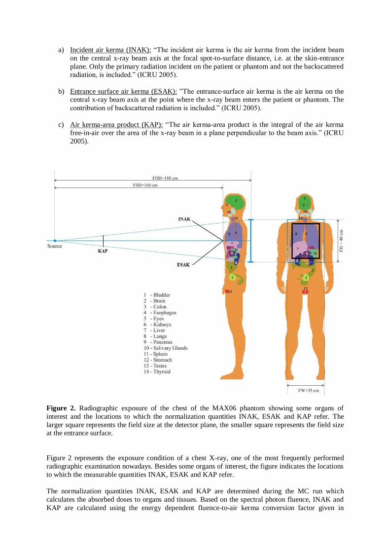
a) Incident air kerma (INAK): “The incident air kerma is the air kerma from the incident beam
on the central x-ray beam axis at the focal spot-to-surface distance, i.e. at the skin-entrance
plane. Only the primary radiation incident on the patient or phantom and not the backscattered radiation, is included.” (ICRU 2005).
b) Entrance surface air kerma (ESAK): ”The entrance-surface air kerma is the air kerma on the central x-ray beam axis at the point where the x-ray beam enters the patient or phantom. The
contribution of backscattered radiation is included.” (ICRU 2005).
c) Air kerma-area product (KAP): “The air kerma-area product is the integral of the air kerma free-in-air over the area of the x-ray beam in a plane perpendicular to the beam axis.” (ICRU
2005).
Figure 2. Radiographic exposure of the chest of the MAX06 phantom showing some organs of
interest and the locations to which the normalization quantities INAK, ESAK and KAP refer. The
larger square represents the field size at the detector plane, the smaller square represents the field size at the entrance surface.
Figure 2 represents the exposure condition of a chest X-ray, one of the most frequently performed
radiographic examination nowadays. Besides some organs of interest, the figure indicates the locations
to which the measurable quantities INAK, ESAK and KAP refer.
The normalization quantities INAK, ESAK and KAP are determined during the MC run which
calculates the absorbed doses to organs and tissues. Based on the spectral photon fluence, INAK and
KAP are calculated using the energy dependent fluence-to-air kerma conversion factor given in
ICRP74 (ICRP 1996), while the ESAK is derived from the above mentioned skin absorbed dose to a
7.2 cm x 7.2 cm square of skin tissue centered around the central x-ray beam axis, by transforming it
into air kerma using the ratio between the mass energy-absorption coefficients of air and skin.
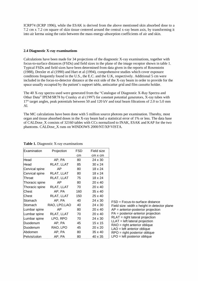
2.4 Diagnostic X-ray examinations
Calculations have been made for 34 projections of the diagnostic X-ray examinations, together with
focus-to-surface distances (FSDs) and field sizes in the plane of the image receptor shown in table 1. Typical FSDs and field sizes have been determined from data given in the reports of Rosenstein
(1988), Drexler et al (1990) and Hart et al (1994), comprehensive studies which cover exposure
conditions frequently found in the U.S., the E.C. and the U.K, respectively. Additional 5 cm were included in the focus-to-detector distance at the exit side of the X-ray beam in order to provide for the
space usually occupied by the patient‟s support table, antiscatter grid and film cassette holder.
The 40 X-ray spectra used were generated from the “Catalogue of Diagnostic X-Ray Spectra and
Other Data” IPEM/SR78 by Cranley et al (1997) for constant potential generators, X-ray tubes with
17° target angles, peak potentials between 50 and 120 kV and total beam filtrations of 2.0 to 5.0 mm
Al.
The MC calculations have been done with 5 million source photons per examination. Thereby, most
organ and tissue absorbed doses in the X-ray beam had a statistical error of 1% or less. The data base of CALDose_X consists of 32160 tables with CCs normalized to INAK, ESAK and KAP for the two
phantoms. CALDose_X runs on WINDOWS 2000/NT/XP/VISTA.
Table 1. Diagnostic X-ray examinations
FSD = Focus-to-surface distance Field size: width x height in detector plane AP = anterior-posterior projection PA = posterior-anterior projection RLAT = right lateral projection LLAT = left lateral projection RAO = right anterior oblique LAO = left anterior oblique RPO = right posterior oblique LPO = left posterior oblique
Examination Projection FSD Field size
cm cm x cm
Head AP, PA 80 24 x 30
Head RLAT, LLAT 85 30 x 24
Cervical spine AP 80 18 x 24
Cervical spine RLAT, LLAT 80 18 x 24
Throat RLAT, LLAT 75 18 x 24
Thoracic spine AP 80 20 x 40
Thoracic spine RLAT, LLAT 70 20 x 40
Chest AP, PA 160 35 x 40
Chest RLAT, LLAT 150 25 x 40
Stomach AP, PA 40 24 x 30
Stomach RAO, LPO,LAO 40 24 x 30
Lumbar spine AP 80 20 x 40
Lumbar spine RLAT, LLAT 70 20 x 40
Lumbar spine LPO, RPO 70 24 x 30
Duodenum AP, PA 45 15 x 15
Duodenum RAO, LPO 45 20 x 20
Abdomen AP, PA 80 35 x 40
Pelvis/colon AP, PA 80 40 x 35
2.5 Sources of uncertainty
2.5.1 The phantoms
Compared to the stylized MIRD5 phantoms, FAX06 and MAX06 are true to nature representations of humans, which make their CCs applicable to individual patients, at least to those with similar
anatomical properties, if one neglects minor differences that still could exist with regard to organs
without fixed positions, like stomach, colon and small intestine. Among the anatomical properties of
adult patients of a given age, the central fat mass (CFM), i.e. the subcutaneous and visceral fat mass of the trunk, can influence organ and tissue absorbed doses significantly, whereas the peripheral fat mass
(PFM), i.e. the subcutaneous fat mass of the extremities, has negligible influence at least for the
examinations mentioned in table 1. Ketel et al (2007) have reported that among the various anthropometric parameters they investigated, the waist circumference showed the best correlation with
the CFM, which had been determined before independently by dual-energy X-ray absorptiometry.
Body mass index and body weight showed also good correlation, but only with the total (CFM + PFM) fat mass. Measuring the circumference of a patient‟s waist can easily be done without impeding the
patient or the medical procedure. The next version of CALDose_X will be updated with CCs for
derived male and female phantoms having different fat distributions. Based on the value for the waist
circumference, CALDose_X will then select a patient-specific set of CCs. In the meantime, CALDose_X is using the FAX06 and the MAX06 phantoms for all adult patients.
2.5.2 The statistical error
Increasing the number of photons can reduce the statistical error of the organ and tissue absorbed dose.
However, there is a limit to the number of particles that can be calculated, set by the speed of the computer. Here, usually five million photons were used per projection of an X-ray examination, which
reduced the statistical error for organs and tissues located inside the beam volume to less than 1% for
INAK- and KAP-CCs, and to less than 2% for ESAK-CCs. For organs and tissues located at the boundaries or outside the beam the statistical error is greater. CALDose_X displays organ and tissue
absorbed doses with statistical errors of up to 10%.
2.5.3 Cross-sections and tissue compositions
Uncertainties of cross-section data have been reported to be approximately +/- 2% (Hubbell 1977), which therefore can be ruled out of being a major source of error.
In the FAX06 and the MAX06 phantoms only one soft tissue composition is used which has been
compiled as the average of many organ-specific compositions. It has been shown that differences between soft tissue compositions can cause differences of the effective dose of up to 10% for external
exposure to photons for energies below 150 keV (Jones 1995, 1997, Lima et al 2004, Kramer et al
2004b). As for the absorbed dose to the RBM and BSC, it has been shown that the data calculated with the
homogeneous skeletal mixture are always greater, for energies below 60 keV up to a factor of 2-3,
than the data determined with CT images of trabecular bone. The reason is that in a real skeleton, especially for photon energies below 150 keV, the cortical bone layer and the trabeculae of the
spongiosa act as a shield for the soft tissues in the marrow cavities, whereas in the homogeneous skeleton the bone volume contributes also to the absorbed dose (Kramer et al 2006c).
2.5.4 Exposure geometry
Focus-to-surface (FSD) or focus-to-detector (FDD) distance
According to Rosenstein (1988), variation of the FDD by about +/-25 cm causes changes of the organ
absorbed CCs of less than +/- 10%. For the FSDs mentioned in table 1 this study found, that variation
of the FSD by about +/-10% causes changes of the CCs of less than +/-10% for organs and tissues inside the beam volume. However, organs located on the boundaries of the beam volume, especially
small ones, may be found inside or outside the beam after a change of the FSD, which in turn may
change the CC by far more than 10%. For the time being, CALDose_X works with one FSD per projection, but the user can apply the inverse square law to the INAK and as a first approximation also
to the ESAK to correct for differences of FSDs.
Field size
Many field sizes shown in table 1 represent commercial X-ray film formats. It is assumed that beam collimation is made properly to comply with these formats, i.e. that variations of the field size have not
been considered here.
Field position
For a given type of examination, projection, X-ray spectrum, FSD and field size, the position of the field on the body is the quantity which has the strongest effect on the absorbed dose of an organ or
tissue. Changing the field position only a few centimetres up or down, to the right or to the left can
decide if an organ lies inside, outside or on the boundaries of the beam volume. For small organs, like the testes, a variation of the field position by only 4 cm in an abdominal examination can cause
differences for the absorbed dose by up to a factor of four (Jones and Wall 1985). The standard field
positions for this study were chosen based on the location of the organ(s) to be examined and on
anatomical landmarks to be visible in the image according to good practice. But one has to be aware about the “inevitable variations that occur in practice from hospital to hospital and patient to patient”
(Jones and Wall 1985). In addition, here the problem is not to calculate CCs for different field
positions, but to know in a real situation with a patient if, for example, the testes or the thyroid were inside the beam or not. Therefore, CALDose_X outputs the results for an X-ray examination for the
standard field position and also for field positions with a 4 cm shift of the field centre in each
direction. Thereby, the user gets an idea how organ and tissue absorbed doses would change as a function of the variation of the field center position by 4 cm up or down, to the right or to the left and
backwards or towards the front.
2.5.5 X-ray spectra
Factors affecting the spectrum shape are
1. tube peak potential
2. tube potential variation during exposure (ripple) 3. X-ray emission angle
4. filtration
5. extra focal radiation
6. target roughness
CALDose_X CCs cover the range of 50 – 120 kVpc for the tube peak potential and of 2.0 – 5.0 mm
Al for the total filtration. A constant potential generator (no ripple) and an X-ray emission angle of 17° have been chosen for the spectra, because they represent the most frequent type of generator and target
angle found presently in X-ray units.
The effect of extra focal radiation on the spectral shape is insignificant (Birch et al 1979). Target
roughness leads to a hardening of the X-ray beam and is significant only for ageing X-ray tubes. These two factors have not been considered here.
2.5.6 Measurement of the normalization quantities
In order to get an organ absorbed dose estimate, one has to multiply the appropriate CC given by CALDose_X with the value of the INAK, the ESAK or the KAP. Petoussi-Henss et al (1995) report
that KAP measurements, “if properly performed, have a 30% uncertainty”. Measurements of INAK or
ESAK with TLD dosimeters, for example, show typically uncertainties of ca.10-20%. X-ray output measurements with ionization chambers are usually accurate within 10 %.
2.6 Effective dose and cancer risk
Effective dose
The concept of effective dose has been introduced by the ICRP for the purpose of radiological
protection of workers and the general public (ICRP 2007). The ICRP considers it useful to apply a
single value of effective dose to both sexes and to all age groups. Consequently, the tissue weighting factors to be applied to the calculation of the effective dose have been determined as sex- and age-
averaged values and therefore the effective dose “cannot be used for the assessment of individual risk”
(ICRP 2007).
Age and gender distributions of patients undergoing radiological examinations can be quite different from the distributions which were used for the derivation of the ICRP tissue weighting factors, and
these distributions may differ from one type of medical procedure to another. Therefore, risk
assessment in diagnostic radiology should better be based on appropriate risk coefficients for the individual tissues at risk, taking into account age and gender distributions of the individuals
undergoing the medical procedure.
As a consequence, the International Commission on Radiation Units and Measurements (ICRU)
rejects the use of the effective dose for medical imaging (ICRU 2005) completely, while the International Commission on Radiological Protection (ICRP) considers at least a restricted use of this
quantity for diagnostic radiology to be possible:
“Effective dose can be of value for comparing the relative doses from different diagnostic procedures and for comparing the use of similar technologies and procedures in different hospitals and countries
as well as the use of different technologies for the same medical examination, provided the reference
patient or patient populations are similar with regard to age and sex.” (ICRP 2007).
For those who want to determine the effective dose for X-ray examinations, CALDose_X is providing
separately calculated weighted MAX06 and FAX06 whole-body absorbed doses, which represent the
sex-specific contributions to the effective dose. According to equation 4.5 in ICRP103 (ICRP 2007), the effective dose is then just the arithmetic mean of the two sex-specific weighted absorbed doses.
Cancer risk
If one is interested in quantifying the cancer risk for an individual patient to be submitted to a radiological procedure, one should find the latest age-, sex- and organ- or tissue-specific risk
coefficients for cancer incidence and cancer mortality and multiply these coefficients with the average
organ or tissue equivalent doses. Following a proposal made by D. Brenner (Brenner and Huda 2008),
the sum over risk-weighted organs and tissues equivalent doses can be considered as “whole-body effective risk”, given by the equation
R = rT HT ,
T
where HT are the average organ and tissue equivalent doses in tissues T and rT are the lifetime
attributable tissue-specific cancer risks per unit organ equivalent dose.
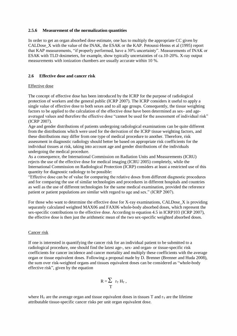
Tables 2 and 3 show adult risk coefficients for cancer incidence and cancer mortality, respectively, as
a function of sex, age and organ taken from the BEIR VII Report (National Research Council 2005).
Females have a significantly higher whole-body effective cancer risk than males mainly because of greater risk coefficients for the sex-specific organs: breast, uterus and ovaries, but also for the lungs.
With aging, risk coefficients decrease for both sexes.
CALDose_X applies the risk coefficients of tables 2 and 3 to calculate sex- and age-specific whole
body risks for cancer incidence and cancer mortality using the equation given above. The organs and
tissues considered are those specified in table 3 of ICRP103 (ICRP 2007) for the calculation of the
effective dose, which have also been mentioned above in section 2.3.1. The risk coefficient for “Other” was equally distributed among organs and tissues, which are not mentioned in tables 2 and 3.
The risk coefficients apply to male and female adults between 20 and 80 years of age, whereas the adult anatomical reference values of ICRP89 (ICRP 2003), used for the development of the MAX06
and the FAX06 phantoms, correspond to adult persons of 35 years. Although not being the main focus
of the data presented in ICRP89, some information about anatomical changes during adulthood can still be found in the report. With respect to organ mass, no significant changes with increasing adult
age have been found for the stomach, the adrenals, the brain and the lungs, while decreases between
15 and 35% have been observed for the spleen, the thymus, the thyroid, the liver, the pancreas and the
kidneys for adults between 30 and 80 years of age for both sexes. Partly, the data are based on Asian populations. Loss of trabecular bone mass can amount to 25-45% of the peak trabecular bone mass,
while body fat increases by ca. 12% during the adult life span between 35 and 70 years for both sexes.
Table 2. Lifetime Attributable Risk of Cancer Incidence given as number of cases per
100,000 persons exposed to a single dose of 0.1 Gy (National Research Council 2005)
Age (y)
Cancer site 20 30 40 50 60 70 80
Males
Stomach 40 28 27 25 20 14 7
Colon 173 125 122 113 94 65 30
Liver 30 22 21 19 14 8 3
Lungs 149 105 104 101 89 65 34
Prostate 48 35 35 33 26 14 5
Bladder 108 79 79 76 66 47 23
Thyroid 21 9 3 1 0.3 0.1 0
Leukaemia 96 84 84 84 82 73 48
Other 312 198 172 140 98 57 23
All cancers 977 686 648 591 489 343 174
Females
Stomach 52 36 35 32 27 19 11
Colon 114 82 79 73 62 45 23
Liver 14 10 10 9 7 5 2
Lungs 346 242 240 230 201 147 77
Breast 429 253 141 70 31 12 4
Bladder 109 79 78 74 64 47 24
Uterus 26 18 16 13 9 5 2
Ovary 50 34 31 25 18 11 5
Thyroid 113 41 14 4 1 0.3 0
Leukaemia 71 63 62 62 57 51 37
Other 323 207 181 148 109 68 30
All cancers 1646 1065 886 740 586 409 214
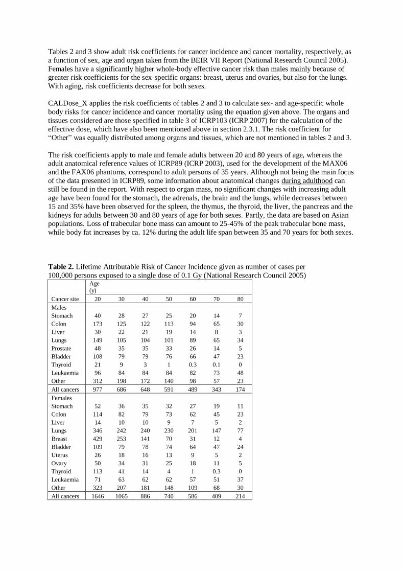
While body height decreases by ca. 2% for both sexes, corresponding data for the total body mass are
not clear. ICRP89 does not recommend reference values for different adult age groups and the above
mentioned data are mostly presented in figures, which make it difficult to use them as information for changes to be made to organ masses. After all, significant changes of organ mass are more critical for
internal dosimetry and less important for external exposures, like X-ray radiographs. Changes of the
bone mass and the trabecular bone structure are important for the absorbed dose to the RBM and the BSC for any type of exposure; however, these can be taken properly into account only after the
implementation of a skeletal dosimetry model based on CT images of spongiosa. Evaluating age-related changes of anatomical parameters during adulthood would be an interesting project for the
future. In the meantime, CALDose_X is using the bodies of the “35-year old” MAX06 and FAX06
phantoms for risk estimates for all age groups.
Table3. Lifetime Attributable Risk of Cancer Mortality given as number of cases per
100,000 persons exposed to a single dose of 0.1 Gy (National Research Council 2005)
Age (y)
Cancer site 20 30 40 50 60 70 80
Males
Stomach 21 16 15 13 11 8 4
Colon 84 61 60 57 49 36 21
Liver 23 16 16 14 12 8 4
Lungs 151 107 107 104 93 71 42
Prostate 9 7 6 7 7 7 5
Bladder 23 17 17 17 17 15 10
Leukaemia 67 64 67 71 73 69 51
Other 134 94 88 77 58 36 17
All cancers 511 381 377 360 319 250 153
Females
Stomach 29 21 20 19 16 13 8
Colon 53 38 37 35 31 25 15
Liver 12 9 8 8 7 5 3
Lungs 305 213 212 204 183 140 81
Breast 101 61 35 19 9 5 2
Bladder 31 23 23 22 22 19 13
Uterus 6 4 4 3 3 2 1
Ovary 28 20 20 18 15 10 5
Leukaemia 51 51 52 54 55 52 38
Other 147 103 97 86 69 47 24
All cancers 762 542 507 469 409 317 190
3. Results
3.1 Comparison with other data
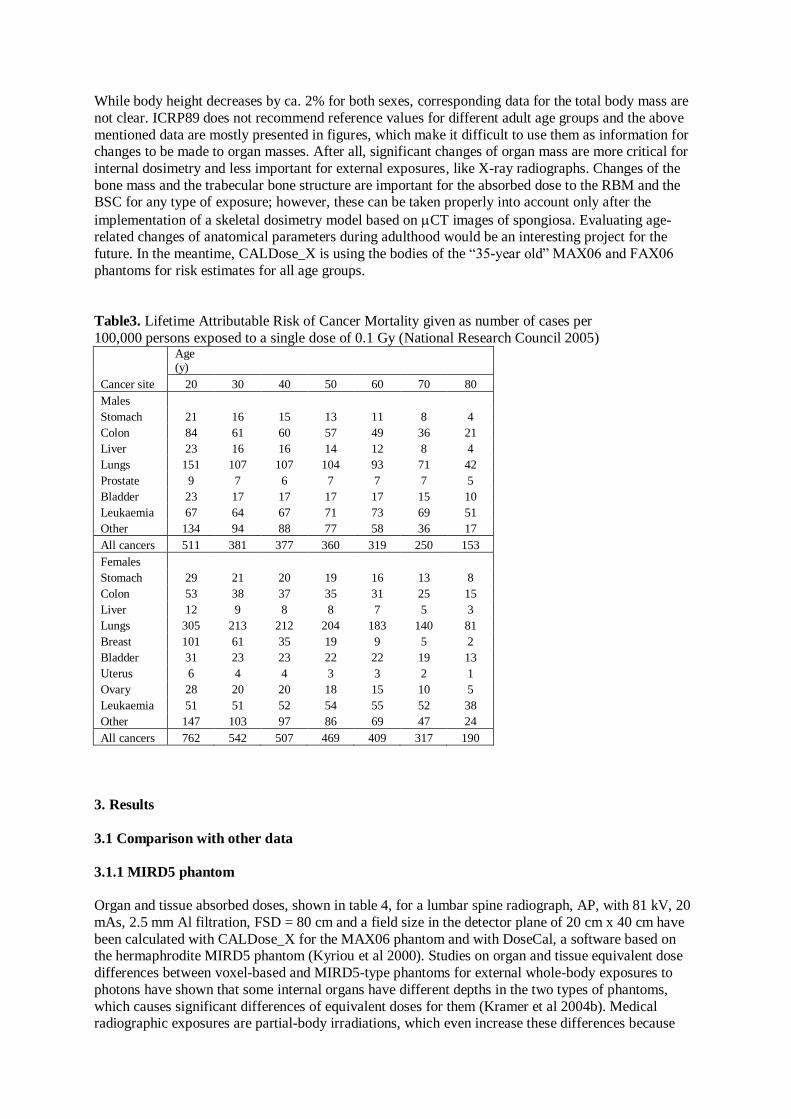
3.1.1 MIRD5 phantom
Organ and tissue absorbed doses, shown in table 4, for a lumbar spine radiograph, AP, with 81 kV, 20
mAs, 2.5 mm Al filtration, FSD = 80 cm and a field size in the detector plane of 20 cm x 40 cm have
been calculated with CALDose_X for the MAX06 phantom and with DoseCal, a software based on the hermaphrodite MIRD5 phantom (Kyriou et al 2000). Studies on organ and tissue equivalent dose
differences between voxel-based and MIRD5-type phantoms for external whole-body exposures to
photons have shown that some internal organs have different depths in the two types of phantoms,
which causes significant differences of equivalent doses for them (Kramer et al 2004b). Medical radiographic exposures are partial-body irradiations, which even increase these differences because
now organs can additionally be located inside or outside the beam volume. Comparing the organ
absorbed doses for the two phantoms shown in table 4, one has to keep the anatomical differences
between them in mind.
Table 4. Absorbed dose to organs in the MAX06 (CALDose_X) and the MIRD5 (DoseCal) phantoms for a lumbar spine radiograph, AP, with 81 kV, 20 mAs, 2.5 mm Al filtration, FSD = 80 cm and a field
size of 20 cm x 40 cm in the detector plane. Mean spectral energy = 42.9 keV. Statistical errors are
given in percent in brackets.
Organ CALDose_X DoseCal
mGy mGy
Adrenals 0.31 (2.1) 0.15 (3.9)
Stomach 1.25 (0.3) 1.04 (1.3)
SMI 1.01 (0.2) 0.70 (1.2)
Colon 0.85 (0.2) 0.61 (1.5)
Kidneys 0.35 (0.4) 0.16 (1.7)
Liver 0.72 (0.1) 0.60 (1.2)
Lungs 0.09 (0.4) 0.05 (1.7)
Pancreas 0.87 (0.4) 0.46 (1.9)
Spleen 0.23 (0.5) 0.22 (2.5)
Bladder 0.34 (1.0) 0.45 (3.2)
RBM 0.06 (0.3) 0.08 (1.1)
SMI = Small intestine, RBM = Red bone marrow
For example, the colon of the MIRD5 phantom is a parallelepiped located at the center of the trunk, whereas in the MAX06 phantom the colon spreads out into the frontal part of the abdomen.
Consequently for a spectrum with a mean energy of 42.9 keV, the MAX06 colon receives a higher
absorbed dose for AP incidence than the MIRD5 colon. Similar considerations apply to the small intestine. Or, while the MIRD5 bladder begins at a depth of 1 cm below the surface of the body, the
same happens for the MAX06 bladder only at a depth of 5 cm. Consequently for AP incidence, the
MAX06 bladder absorbed dose is smaller than the MIRD5 bladder absorbed dose. Analysis of the
locations of other organs in the two phantoms would correspondingly explain the differences or agreements between the absorbed doses to be observed in table 4.
3.1.2 Voxel-based phantom
From the voxel-based studies mentioned above only the paper from Petoussi-Henss et al (2005)
contains tabulated data for the voxel-based phantom GOLEM (Zankl and Wittmann 2001) which can
be compared with CCs calculated in this investigation.
Table 5 shows conversion coefficients between organ and tissue absorbed doses and “entrance dose”
for the MAX06 and the GOLEM phantom for a pelvic examination AP, made with 80 kV tube potential, 2.5mm Al total filtration, FDD=115 cm and a field size of 38cm x 35cm in the plane of the
detector.
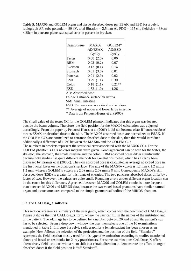
Table 5. MAX06 and GOLEM organ and tissue absorbed doses per ESAK and ESD for a pelvic
radiograph AP, tube potential = 80 kV, total filtration = 2.5 mm Al, FDD = 115 cm, field size = 38cm
x 35cm in detector plane, statistical error in percent in brackets
Organ/tissue MAX06 GOLEM*
AD/ESAK AD/ESD
Gy/Gy Gy/Gy
Testes 0.08 (2.0) 0.06
RBM 0.03 (0.2) 0.07
Skeleton 0.13 (0.1) 0.14
Stomach 0.01 (3.0) 0.01
Pancreas 0.01 (2.9) 0.02
SMI 0.29 (1.1) 0.30
Colon 0.18 (1.1) 0.21**
ESD 1.52 (1.0) 1.26
AD: Absorbed dose
ESAK: Entrance surface air kerma
SMI: Small intestine ESD: Entrance surface skin absorbed dose
** Average of upper and lower large intestine
* Data from Petoussi-Henss et al (2005)
The small value of the testes CC for the GOLEM phantom indicates that this organ was located
outside the beam volume. Therefore, the field position for the MAX06 calculation was adjusted
accordingly. From the paper by Petoussi-Henss et al (2005) it did not become clear if “entrance dose” means ESAK or absorbed dose to the skin. The MAX06 absorbed doses are normalized to ESAK. If
the GOLEM CCs are normalized to entrance absorbed dose to the skin, then this would introduce
additionally a difference of 1.7% between the MAX06 and the GOLEM CCs. The numbers in brackets represent the statistical error associated with the MAX06 CCs. For the
GOLEM phantom‟s CCs no error margins were given. Good agreement can be seen for the testes, the
skeleton, the stomach, the small intestine and the colon. RBM absorbed doses differ significantly because both studies use quite different methods for skeletal dosimetry, which has already been
discussed by Kramer et al (2006c). The skin absorbed dose is calculated as average absorbed dose in
the first voxel layer on the phantom‟s surface. The size of the MAX06 voxels is 1.2 mm x 1.2 mm x
1.2 mm, whereas GOLEM‟s voxels are 2.08 mm x 2.08 mm x 8 mm. Consequently MAX06‟s skin absorbed dose (ESD) is greater for this range of energies. The two pancreas absorbed doses differ by a
factor of two. However, the values are quite small. Rounding errors and/or different organ location can
be the cause for this difference. Agreement between MAX06 and GOLEM results is more frequent than between MAX06 and MIRD5 data, because the two voxel-based phantoms have similar natural
organ and tissue structures compared to the simple geometrical bodies of the MIRD5 phantom.
3.2 The CALDose_X software
This section represents a summary of the user guide, which comes with the download of CALDose_X. Figure 3 shows the first CALDose_X form, where the user can fill in the names of the institution and
of the patient. The adult age has to be defined by a number between 20 and 80 and the patient‟s sex
has to be selected. From a drop down window the user then selects one of the 10 examinations mentioned in table 1. In figure 3 a pelvic radiograph for a female patient has been chosen as an
example. Next follows the selection of the projection and the position of the field. “Standard”
represents the field location mostly used for this type of examination according to studies mentioned
above and based on textbooks for X-ray practitioners. For some examinations CALDose_X offers alternatively field locations with a 4 cm shift in a certain direction to demonstrate the effect on organ
absorbed doses if the field position is “off Standard”.
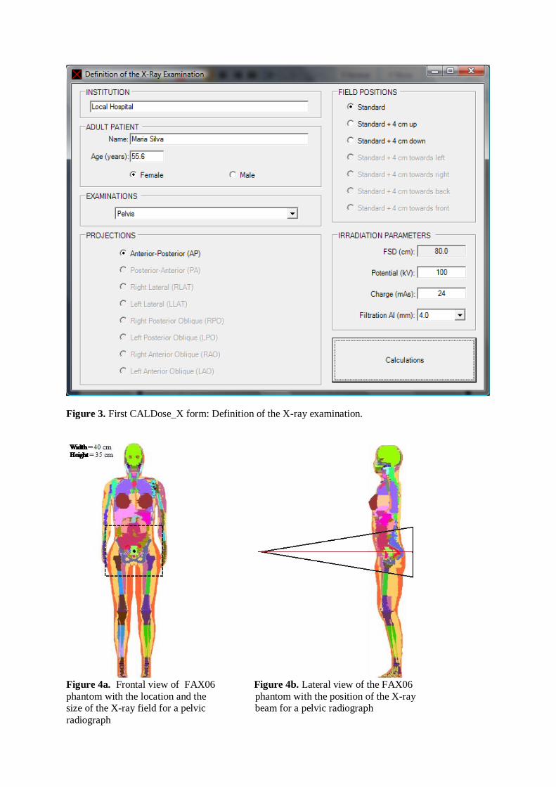
Figure 3. First CALDose_X form: Definition of the X-ray examination.
Figure 4a. Frontal view of FAX06 Figure 4b. Lateral view of the FAX06
phantom with the location and the phantom with the position of the X-ray size of the X-ray field for a pelvic beam for a pelvic radiograph
radiograph
Once the field position has been selected, two images pop up, like the examples shown in figures 4a
and 4b, which show the phantom and the position of the X-ray beam relative to the body. Field height
and field width are given in cm for the plane of the detector (film). These images can be printed and/or saved. For oblique projections, this feature will be added to a later version of CALDose_X. The focus
to skin distance (FSD) for the selected examination appears in the first field in the area called
“irradiation parameters” .
Users who only want to visualize the selected examination could close CALDose_X at this point.
However, if calculations of ESAK or organ and tissue absorbed doses are requested, then the user has
to fill in the tube potential in kV with a number between 50 and 120 and the charge in mAs. Filters between 2 and 5 mm Al can be selected from the drop down window.
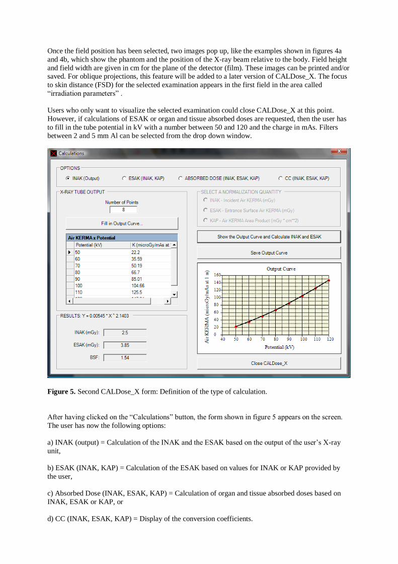
Figure 5. Second CALDose_X form: Definition of the type of calculation.
After having clicked on the “Calculations” button, the form shown in figure 5 appears on the screen.
The user has now the following options:
a) INAK (output) = Calculation of the INAK and the ESAK based on the output of the user‟s X-ray
unit,
b) ESAK (INAK, KAP) = Calculation of the ESAK based on values for INAK or KAP provided by
the user,
c) Absorbed Dose (INAK, ESAK, KAP) = Calculation of organ and tissue absorbed doses based on
INAK, ESAK or KAP, or
d) CC (INAK, ESAK, KAP) = Display of the conversion coefficients.
In the example shown in figure 5 the first option has been chosen, which presumably represents the
most frequent case in radiodiagnosis. The user has to inform the values for the tube potential in kV
and for the output in Gy/mAs at 1m distance from the tube. CALDose_X offers the possibility to save the output curve for future calculations. After having clicked on the button “Show the output curve and calculate INAK and ESAK”, CALDose_X shows a graph of the output curve, calculates the
INAK and the ESAK for the FSD, potential and charge given by the user and shows the results. The
ESAK is the INAK multiplied by the backscatter factor (BSF), which is examination-specific and
provided by the Monte Carlo calculation.
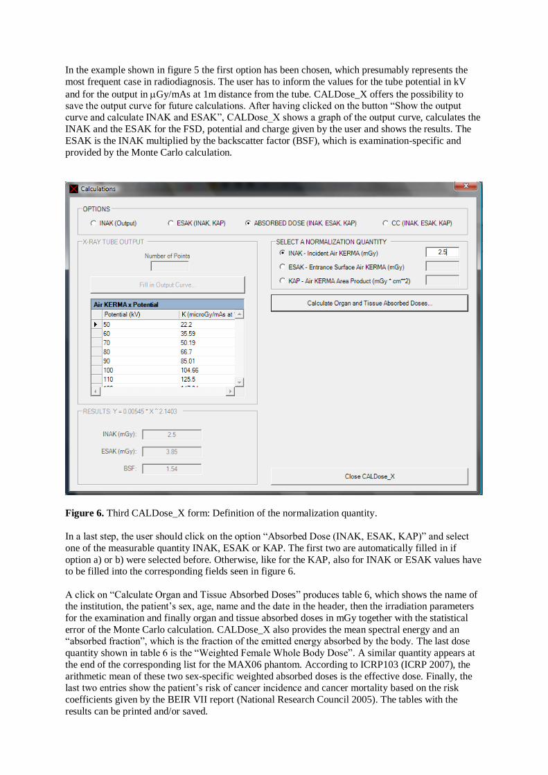
Figure 6. Third CALDose_X form: Definition of the normalization quantity.
In a last step, the user should click on the option “Absorbed Dose (INAK, ESAK, KAP)” and select
one of the measurable quantity INAK, ESAK or KAP. The first two are automatically filled in if
option a) or b) were selected before. Otherwise, like for the KAP, also for INAK or ESAK values have to be filled into the corresponding fields seen in figure 6.
A click on “Calculate Organ and Tissue Absorbed Doses” produces table 6, which shows the name of the institution, the patient‟s sex, age, name and the date in the header, then the irradiation parameters
for the examination and finally organ and tissue absorbed doses in mGy together with the statistical
error of the Monte Carlo calculation. CALDose_X also provides the mean spectral energy and an “absorbed fraction”, which is the fraction of the emitted energy absorbed by the body. The last dose
quantity shown in table 6 is the “Weighted Female Whole Body Dose”. A similar quantity appears at
the end of the corresponding list for the MAX06 phantom. According to ICRP103 (ICRP 2007), the
arithmetic mean of these two sex-specific weighted absorbed doses is the effective dose. Finally, the last two entries show the patient‟s risk of cancer incidence and cancer mortality based on the risk
coefficients given by the BEIR VII report (National Research Council 2005). The tables with the
results can be printed and/or saved.
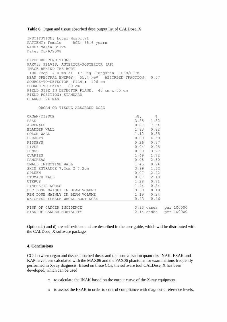
Table 6. Organ and tissue absorbed dose output list of CALDose_X
INSTITUTION: Local Hospital
PATIENT: Female AGE: 55.6 years
NAME: Maria Silva
Date: 26/6/2008
EXPOSURE CONDITIONS
FAX06: PELVIS, ANTERIOR-POSTERIOR (AP)
IMAGE BEHIND THE BODY
100 kVcp 4.0 mm Al 17 Deg Tungsten IPEM/SR78
MEAN SPECTRAL ENERGY: 51.4 keV ABSORBED FRACTION: 0.57
SOURCE-TO-DETECTOR (FILM): 106 cm
SOURCE-TO-SKIN: 80 cm
FIELD SIZE IN DETECTOR PLANE: 40 cm x 35 cm
FIELD POSITION: STANDARD
CHARGE: 24 mAs
ORGAN OR TISSUE ABSORBED DOSE
ORGAN/TISSUE mGy %
ESAK 3.85 1.32
ADRENALS 0.07 7.64
BLADDER WALL 1.83 0.82
COLON WALL 1.12 0.35
BREASTS 0.00 4.69
KIDNEYS 0.26 0.87
LIVER 0.04 0.95
LUNGS 0.00 3.27
OVARIES 1.49 1.72
PANCREAS 0.08 2.30
SMALL INTESTINE WALL 1.45 0.24
SKIN ENTRANCE 7.2cm X 7.2cm 3.99 1.32
SPLEEN 0.07 2.42
STOMACH WALL 0.07 2.18
UTERUS 1.28 0.71
LYMPHATIC NODES 1.44 0.34
BSC DOSE MAINLY IN BEAM VOLUME 3.30 0.19
RBM DOSE MAINLY IN BEAM VOLUME 1.19 0.24
WEIGHTED FEMALE WHOLE BODY DOSE 0.43 0.46
RISK OF CANCER INCIDENCE 3.93 cases per 100000
RISK OF CANCER MORTALITY 2.14 cases per 100000
Options b) and d) are self-evident and are described in the user guide, which will be distributed with
the CALDose_X software package.
4. Conclusions
CCs between organ and tissue absorbed doses and the normalization quantities INAK, ESAK and
KAP have been calculated with the MAX06 and the FAX06 phantoms for examinations frequently
performed in X-ray diagnosis. Based on these CCs, the software tool CALDose_X has been
developed, which can be used
o to calculate the INAK based on the output curve of the X-ray equipment,
o to assess the ESAK in order to control compliance with diagnostic reference levels,
o to calculate organ and tissue absorbed doses for patients with anatomies similar to the
MAX06 and the FAX06 phantoms,
o to assess the effective dose based on ICRP103 and/or the patient‟s cancer risk based
on the BEIR VII report,
o to demonstrate how organ and tissue absorbed doses, i.e. the radiation risk for the
patient, depend on the proper selection of the exposure parameters. This information
can be used in educational programs to train radiologists and technicians to
understand how to perform X-ray examinations with the minimum exposure to the patient,
o to compare organ and tissue absorbed doses, effective doses or radiation risks from
different radiological procedures, or from different X-ray units, or from different hospital, etc., to identify high and low risk examinations, or cases of good and bad
practice and
o to make risk assessments for surveys on radiological exposures, taking into account
risk factors for the age and gender distribution of the patient population under
consideration.
Uncertainties from the statistical error, the cross-sections, the tissue compositions, the focus-to-skin distance and the measurement of the normalization quantities can easily sum up to ca. 65%. The field
position can be very critical to organ and tissue absorbed doses and then there are also properties of
the detector system, which can influence the organ and tissue absorbed doses, which have not been
considered here. Therefore, an overall uncertainty of up to 100% is easily possible, without taking into account patient anatomies, which may deviate from the anatomies of the MAX06 and the FAX06
phantom. M. Stabin has recently found similar estimates for internal dosimetry. He concluded that
“the combined uncertainties in any given radiopharmaceutical dose estimate are typically, at a minimum, a factor of 2 and may be considered greater, in general because of normal human
variability, and particularly in disease states” ( Stabin 2008). According to Stabin, for therapeutic
applications this uncertainty can be reduced to 10-20%. Similar conditions exist for external medical
exposures. In radiation therapy, where all exposure parameter are known and well under control, uncertainties can be made very small, while X-ray diagnosis unfortunately has to live with larger
uncertainties. Still, reduction of uncertainties for CALDose_X is possible, especially by using adult
phantoms with different fat distributions, which is already in preparation for the next update of the software.
Compared to MIRD5-based software tools for diagnostic radiology, CALDose_X presents two improvements: First, using two adult voxel phantoms allows for organ and tissue absorbed dose
calculations based on a true to nature representation of the human anatomy as well as for the correct
sex-specific calculation of the effective dose according to ICRP103 and second, the cancer risk
assessment offers an alternative to the effective dose, which cannot be used for an individual patient.
CALDose_X stands in the tradition of software tools developed earlier. It represents an improvement
compared to them, but on the other hand it is only another milestone towards the aim of making such software tools for diagnostic radiology every time more patient-specific.
Consequently, CALDose_X has to be understood as an open project, which will be updated from time to time with new features, like patient-specific fat distributions, age-related anatomical changes during
adult life span, two updates, which would reduce some of the above mentioned uncertainties, children
phantoms, improved skeletal dosimetry, other types of examinations, etc.
CALDose_X and/or the phantoms FAX06 and MAX06 are available from the following website of the
Federal University of Pernambuco: http://www.grupodoin.com or from the authors.
5. Acknowledgement
The authors would like to thank Dr. John Kyriou for introducing the DoseCal software to our department and also for many interesting discussions on this subject.
The authors would also like to thank the Conselho Nacional de Desenvolvimento Científico e
Tecnológico - CNPq and the Fundação de Amparo à Ciência do Estado de Pernambuco - FACEPE for financial support.
6. References
Akahane K Kai M Kusama T and Saito K 2001 Dose estimation of patients from diagnostic X-ray
based on CT-voxel phantom, Proceedings of the Ninth EGS4 Users‟ Meeting in Japan, KEK Proceedings 2001-22 p.87-91
Alderson S W Lanzl L H Rollins M and Spira J 1962 An instrumented phantom system for
analog computation of treatment plans, Am.J.Roentg.87,185
Bozkurt A and Bor D 2007 Simultaneous determination of equivalent dose to organs and tissues of the
patient and of the physician in interventional radiology using the Monte Carlo method, Phys Med Biol 52 317-330
Brenner D and Huda W 2008 Effective Dose: A Useful Concept in Diagnostic Radiology Rad. Prot.
Dos. Advanced Access published March 28
Cranley K Gilmore B J Fogarty G W A and Desponds L 1997 Catalogue of Diagnostic X-ray Spectra and Other Data the Institute of Physics and Engineering in Medicine (IPEM) Report No. 78
Electronic version prepared by D.Sutton September 1997
Cristy M 1980 Mathematical phantoms representing children of various ages for use in estimates of internal dose NUREG/CR-1159, Oak Ridge National Laboratory TN USA
Cristy M and Eckerman K F 1987 Dose-Response Functions for soft tissues of the skeleton In:
Specific Absorbed Fractions of Energy at Various Ages from Internal Photon Sources. I. Methods ORNL/TM-8381/V1 Oak Ridge National Laboratory TN USA
Drexler G Panzer W Widenmann L Williams G and Zankl M 1990 The Calculation of Dose from
External Photon Exposures Using Reference Human Phantoms and Monte Carlo Methods,
Part III: Organ Doses in X-Ray Diagnosis, Institut für Strahlenschutz, GSF- Gesellschaft für Umwelt und Gesundheit, D-8042 Neuherberg, GSF-Bericht S-11, revised and amended
version of S-1026, 1984
Hart D Jones D G and Wall B F 1994 Estimation of Effective Dose in Diagnostic Radiology from Entrance Surface Dose and Dose-Area Product Measurements, National Radiological
Protection Board, Chilton, Didcot, Oxon OX11 0RQ, UK, NRPB-R262
Hubbell J H 1977 Photon mass attenuation and energy-absorption coefficients for H, C, N, O, Ar, and
seven mixtures from 0.1 keV to 20 MeV Radiat. Res. 70 58
ICRP 1991 1990 Recommendations of the International Commission on Radiological Protection ICRP
Publication 60 (Oxford: Pergamon)
ICRP 1995 Basic Anatomical and Physiological Data for use in Radiological Protection: The Skeleton. ICRP Publication 70 (Oxford: Pergamon)
ICRP 1996 Conversion Coefficients for use in Radiological Protection against External Radiation
ICRP Publication 74.International Commission on Radiological Protection, Pergamon Press, Oxford
ICRP 2003 Basic Anatomical and Physiological Data for Use in Radiological Protection: Reference
Values ICRP Publication 89 Ann. ICRP 32 (3-4) Elsevier Science Ltd., Oxford.

ICRP 2007 Recommendations of the International Commission on Radiological Protection ICRP
Publication 103 Ann. ICRP 37 (2-3) Elsevier Science Ltd., Oxford
ICRU 1989 Tissue Substitutes in Radiation Dosimetry and Measurement ICRU Report No. 44 International Commission On Radiation Units And Measurements, Bethesda, MD, USA
ICRU 2005 Patient dosimetry for X-rays used in medical imaging ICRU Report No. 74 International
Commission On Radiation Units And Measurements, Bethesda, MD, USA
Jones D G and Wall B F 1985 Organ Doses from Medical X-ray Examinations Calculated Using
Monte Carlo Techniques, National Radiological Protection Board, Chilton, Didcot, Oxon
OX11 0RQ, UK, NRPB-R186
Jones D G 1995 Use of voxel phantoms in organ dose calculations In: Proceedings of an International Workshop on Voxel Phantom Development held at the National Radiological Protection
Board, Chilton, UK, 6-7 July
Jones D G 1997 A Realistic Anthropomorphic Phantom For Calculating Organ Doses Arising From External Photon Irradiation Rad Prot Dos Vol.72, No.1, pp. 21-29
Kawrakow I 2000 Accurate condensed history Monte Carlo simulation of electron transport. I.
EGSnrc, the new EGS4 version, Med.Phys. 27, 485-498
Kawrakow I 2006 Version V4-r2-2-3 of the EGSnrc code system
http://www.irs.inms.nrc.ca/EGSnrc/EGSnrc.html
Ketel I J G Volman M N M Seidell J C Stehouwer C D A Twisk J W and Lambalk C B 2007
Superiority of skinfold measurements and waist over waist-to-hip ratio for determination of body fat distribution in a population-based cohort of Caucasian Dutch adults Europ. J.
Endocrinology156 655-661
Kramer R and Drexler G 1976 Zum Verhältnis von Oberflächen- und Körperdosis in der Röntgendiagnostik, 7. Wissenschaftliche Tagung der Deutschen Gesellschaft für Medizinische
Physik e.V. Heidelberg, 5.-7.5.1976, In: Medizinische Physik, Band 2, p. 683-695,
Herausgegeben von W. J. Lorenz, Hüthig Verlag, Heidelberg.
Kramer R Zankl M Williams G and Drexler G 1982 The calculation of dose from external photon exposures using reference human phantoms and Monte Carlo methods. Part I: The male
(ADAM) and female (EVA) adult mathematical phantoms GSF-Report S-885, Institut für
Strahlenschutz, GSF-Forschungszentrum für Umwelt und Gesundheit, Neuherberg-München
Kramer R Vieira J W Khoury H J Lima F R A and Fuelle D 2003a All About MAX: a Male Adult
voXel Phantom for Monte Carlo Calculations in Radiation Protection Dosimetry, Phys. Med.
Biol., 48, No. 10, 1239-1262
Kramer R Vieira J W Lima F R A Khoury J H 2003b Coeficientes de conversão para dose equivalente
em órgãos e dose efetiva normalizadas pelo produto dose-área para radiografias do tórax
usando o fantõma MAX. In: VIII CONGRESSO BRASILEIRO DE FÍSICA MÉDICA Porto
Alegre/Brazil
Kramer R Vieira J W Khoury H J Lima F R A Loureiro E C M Lima V J M and Hoff G 2004a All
about FAX: a Female Adult voXel Phantom for Monte Carlo Calculation in Radiation
Protection Dosimetry, Phys. Med. Biol. 49, 5203-5216
Kramer R Vieira J W Khoury H J and Lima F R A 2004b MAX meets ADAM: a Dosimetric
Comparison Between a Voxel-Based and a Mathematical Model for External Exposures to
Photons, Phys. Med. Biol., 49, 887-910
Kramer R Khoury H J and Vieira J W 2005a Comparison between effective doses for voxel-based
and stylized exposure models from photon and electron irradiation Phys. Med. Biol 50: 5105-
5126
Kramer R Santos A M Brayner C A O Khoury H J Vieira J W and Lima F R A 2005b Application of
the MAX/EGS4 exposure model to the dosimetry of the Yanango radiation accident Phys Med
Biol 50 3681-3695
Kramer R Khoury H J Vieira J W and Lima V J M 2006a MAX06 and FAX06: Udate of two adult
human phantoms for radiation protection dosimetry Phys. Med. Biol.51: 3331-3346
Kramer R Khoury H J Lopes C and Vieira J W 2006b Equivalent dose to organs and tissues in hysterosalpingography calculated with the FAX (Female Adult voXel) phantom, Br J Radiol
79, 893-898
Kramer R Khoury H J Vieira J W and Kawrakow I 2006c Skeletal dosimetry in the MAX06 and the
FAX06 phantoms for external exposure to photons based on vertebral 3D-microCT images Phys.Med.Biol. 51 6265-6289
Kramer R, Khoury H J, Vieira J W and Kawrakow I 2007 Skeletal dosimetry for external exposure to
photons based on CT images of spongiosa from different bone sites Phys Med Biol. 52 6697-6716
Kyriou J C Newey V and Fitzgerald M C 2000 Patient doses in diagnostic radiology at the touch of a button The Radiological Protection Center, St. George`s Hospital, London, United Kingdom
Kyriou J C 2008 Email to Kramer R from June 25, 2008
Le Heron J C 1992 Estimation of effective dose to the patient during medical x-ray examinations from measurements of the dose-area product, Phys Med Biol Vol 37, No 11, 2117-2126
Lima F R A Kramer R Vieira J W Khoury J H 2003 Organ Dose Conversion Coefficients for Lumbar
Spine Examinations calculated for the MAX phantom. In: RADIATION PROTECTION SYMPOSIUM OF THE NORTH WEST EUROPEAN RP SOCIETIES 2003 Utrecht.
Extended Abstracts of the Radiation Protection Symposium of the North West European RP
Societies. UTRECHT/Netherlands
Lima F R A Kramer R Vieira J W Khoury H J Loureiro E C M and Hoff G 2004 Effective dose conversion coefficients with gender-sepcific, adult voxel phantoms for radiographic
examinations common in diagnostic radiology, 11th International Congress of the International
Radiation Protection Association, Madrid, Spain 23.-28.5 2004
National Research Council 2005 Health Risks from Exposure to Low Levels of Ionizing Radiation –
BEIR VII. The National Academies Press, Washington DC
Nelson W R, Hirayama H and Rogers D W O 1985 The EGS4 Code System SLAC-265, Stanford Linear Accelerator Center, Stanford University, Stanford, CA,USA
PCXMC 2008 http://www.stuk.fi/sateilyn_kayttajille/ohjelmat/PCXMC/en_GB/pcxmc/ Accessed
April 28, 2008
Petoussi-Henss N Panzer W Zankl M and Drexler G 1995 Dose-Area Product and Body Doses, Rad Prot Dos 57, Nos 1-4, 363-366
Petoussi-Henss N Zankl M and Panzer W 2005 Estimation of organ doses in radiology using voxel
models describing different patients, Biomedizinische Technik, Vol 50, Supplementary volume 1, Part 1, 664-665
Rannikko S Ermakov I Lampinen J S Toivonen M Karila K T K and Chervjakov A 1997 Computing
patient doses of X-ray examinations using a patient size- and sex- adjustable phantom Br. J.
Radiol. 70 708-718
Rosenstein M 1976 Organ doses in Diagnostic Radiology, US Department of Health, Education and
Welfare, Bureau of Radiological Health, BRH Tech Publ DA 76-8030
Rosenstein M 1988 Handbook of Selected Tissue Doses for Projections Common in Diagnostic Radiology, US Department of Health, Education and Welfare, Bureau of Radiological Health,
BRH Tech Publ DA 89-8031
Schultz F W Geleijns J and Zoetelief J 1994 Calculation of dose conversion factors for posterior-
anterior chest radiography of adults with a relatively high-energy X-ray spectrum, Br J Radiol,
67 775-785
Schultz F W Geleijns J and Zoetelief J 1995 Effetive Doses for different techniques used for PA chest
radiography, Rad Prot Dos 57 Nos 1-4 371-376
Servomaa A and Tapiovaara M 1998 Organ Dose Calculation in Medical X-Ray Examinations by the Program PCXMC, Rad Prot Dos Vol 80, Nos 1-3 pp213-219
Snyder W S Ford M R Warner G G Watson G G 1974 Revision of MIRD Pamphlet No 5 Entitled
“Estimates of absorbed fractions for monoenergetic photon sources uniformly distributed in
vaious organs of a heterogeneous phantom” ORNL-4979 Oak Ridge National Laboratory, Oak Ridge,TennUSA
Stabin M 2008 Uncertainties in Internal Dose Calculations for Radiopharmaceuticals J Nucl Med
49:853-860
Tapiovaara M 2008 Email to Kramer R from June 17, 2008
Vieira J W Lima F R A Kramer R and Khoury J H 2003 Organ and Tissue Equivalent Doses from
Abdominal X-ray Examinations calculated with the MAX/EGS4 exposure model. In: IRPA VI LATIN AMERICAN REGIONAL CONGRESS 2003 Lima/Peru. Anais do VI Congresso
sobre Seguridad Radiologica y Nuclear
Wendin L 2008 Email to Kramer R from June 19, 2008
Williams G Zankl M Abmayr W Veit R and Drexler G 1986 The calculation of dose from external photon exposures using reference and realistic human phantoms and Monte Carlo methods,
Phys Med Biol 31 347-354
WinODS 2008 http://www.rti.se/download_software/index.html Accessed April 28, 2008
Winslow M Huda W Xu X G Chao T C Shi C Y Ogden K M and Scalzetti M, 2004 Use of the VIP-
Man Model to Calculate Energy Imparted and Effective Dose for X-Ray Examinations, Health
Physics 86(2):174-182
Zankl M Panzer W and Herrmann C 2000 Calculation of Patient Doses Using a Human Voxel Phantom of Variable Diameter, Rad Prot Dos Vol 90, Nos 1-2 pp 155-158
Zankl M and Wittmann A 2001 The adult male voxel model GOLEM segmented from whole-body CT
patient data, Radiat Environ Biophys, 40: 153-162


![[x+o] PM-Tool](https://img.pdfslide.us/doc/110x75/56813739550346895d9ec819/xo-pm-tool.jpg)