Embed Size (px)
Citation preview
1/27/2016
1
Calcaneal Fractures: Lateral Extensile Incision
AS Flemister JR, MDUniversity of Rochester
1/27/2016 2
Disclosures
I have no financial disclosures
Mechanism
Axial Loading Fall From Height MVA
BAD SOFT TISSUE INJURY
1/27/2016
2
Mechanism
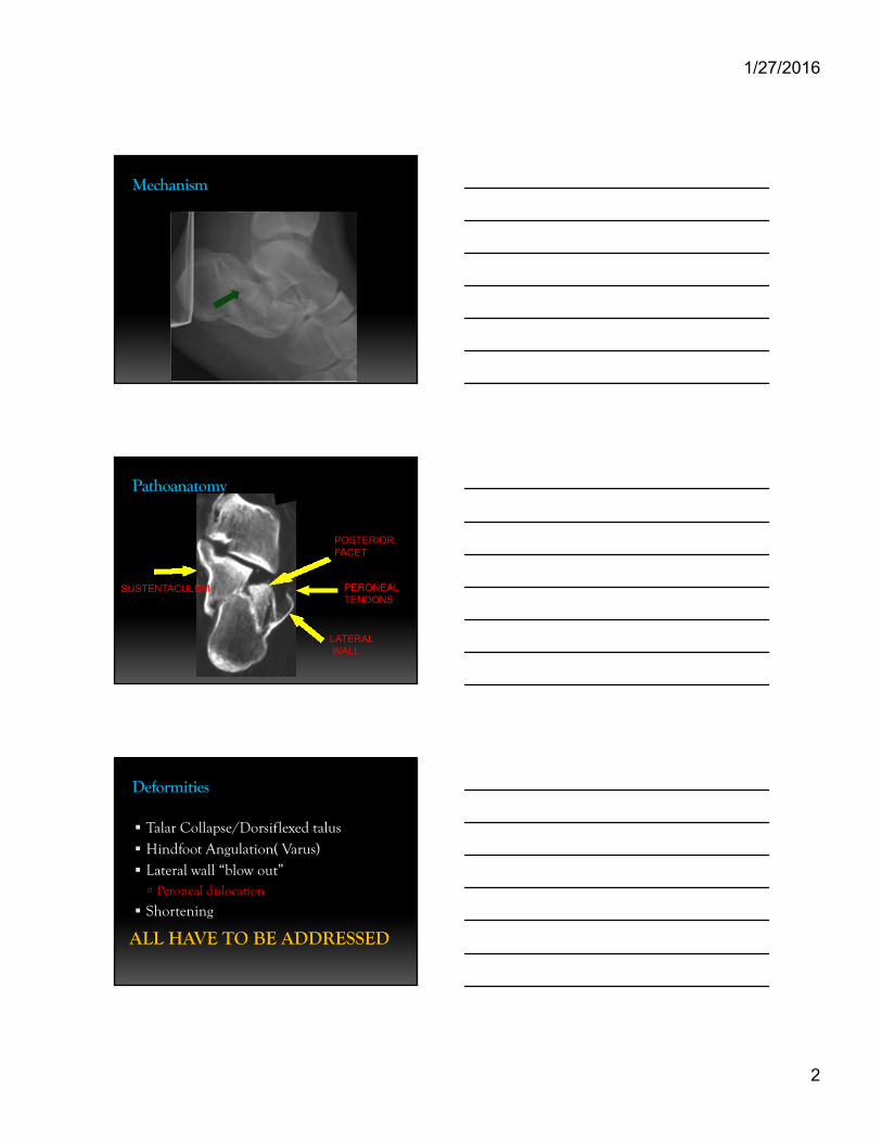
Pathoanatomy
SUSTENTACULUM
POSTERIORFACET
PERONEALTENDONS
LATERALWALL
Deformities
Talar Collapse/Dorsiflexed talus Hindfoot Angulation( Varus) Lateral wall “blow out” Peroneal dislocation
Shortening
ALL HAVE TO BE ADDRESSED
1/27/2016
3
Surgical Treatment
Traditional Extensile Lateral Approach
Sinus Tarsi Approach
Percutaneous Techniques
External Fixation
1/27/2016 8
Question
What method of calcaneal treatment do you most commonly use?
1. ORIF through lateral extensile incision
2. ORIF through sinus tarsi incision3. Percutaneous techniques4. Nonoperative treatment
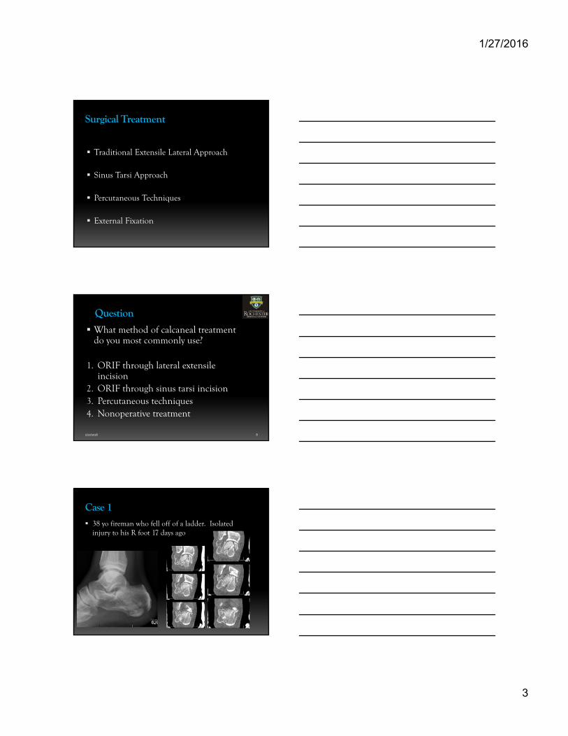
Case 1 38 yo fireman who fell off of a ladder. Isolated
injury to his R foot 17 days ago
1/27/2016
4
Question
What is the ideal surgical approach for this patient?
1. ORIF through lateral extensile incision
2. ORIF through sinus tarsi incision
3. Percutaneous techniques
4. Nonoperative treatment
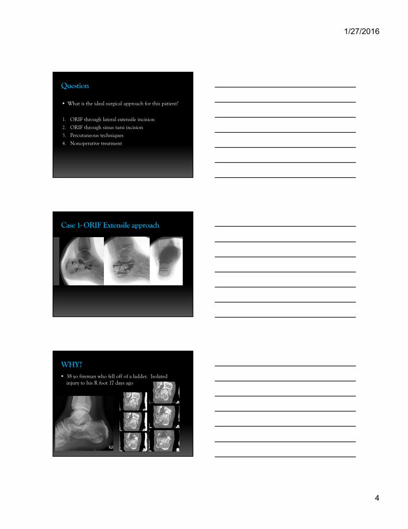
Case 1- ORIF Extensile approach
WHY? 38 yo fireman who fell off of a ladder. Isolated
injury to his R foot 17 days ago
1/27/2016
5
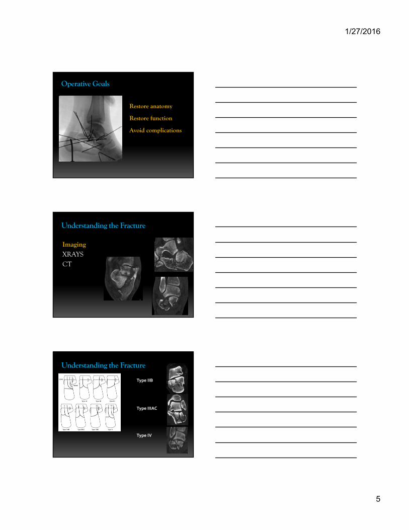
Operative Goals
Restore anatomy
Restore function
Avoid complications
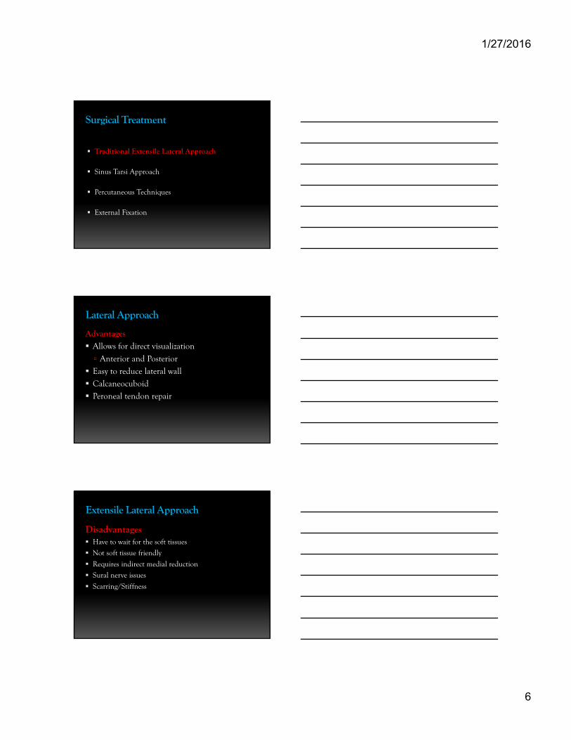
Understanding the Fracture
ImagingXRAYSCT
Understanding the Fracture
15
Type IIB
Type IIIAC
Type IV
1/27/2016
6
Surgical Treatment
Traditional Extensile Lateral Approach
Sinus Tarsi Approach
Percutaneous Techniques
External Fixation
Lateral Approach
Advantages
Allows for direct visualization
Anterior and Posterior
Easy to reduce lateral wall
Calcaneocuboid
Peroneal tendon repair
Extensile Lateral Approach
Disadvantages Have to wait for the soft tissues
Not soft tissue friendly
Requires indirect medial reduction
Sural nerve issues
Scarring/Stiffness
1/27/2016
7
1/27/2016 19
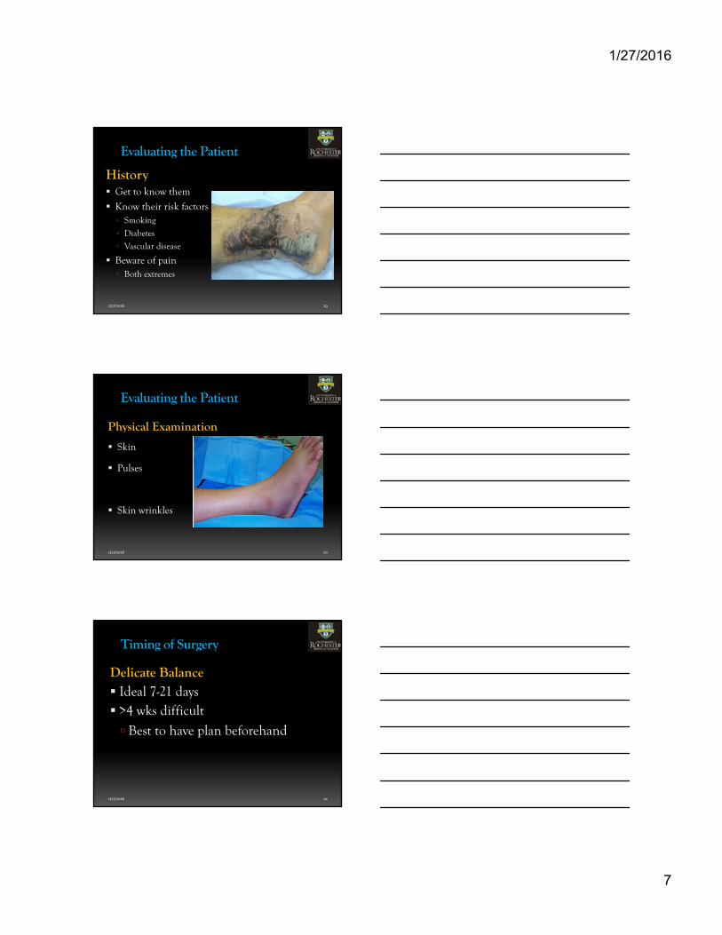
Evaluating the Patient
History Get to know them
Know their risk factors Smoking
Diabetes
Vascular disease
Beware of pain Both extremes
1/27/2016 20
Evaluating the Patient
Physical Examination
Skin
Pulses
Skin wrinkles
1/27/2016 21
Timing of Surgery
Delicate Balance Ideal 7-21 days >4 wks difficult
Best to have plan beforehand
1/27/2016
8
1/27/2016 22
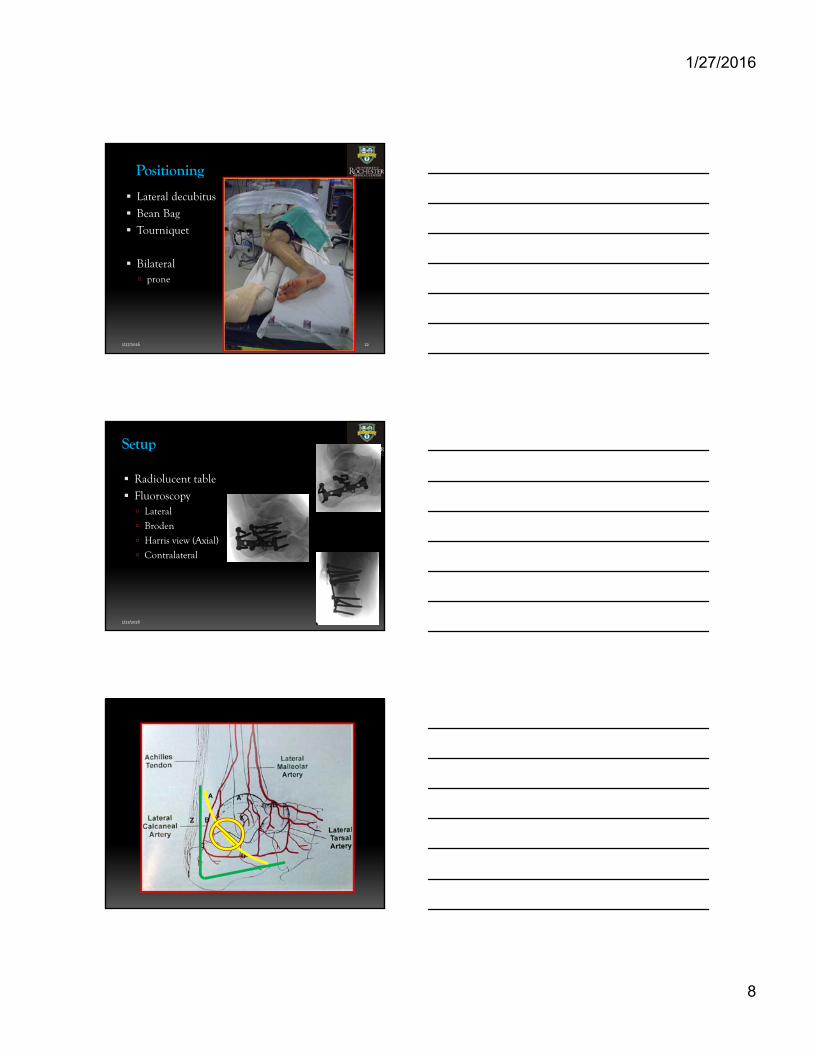
Positioning
Lateral decubitus
Bean Bag
Tourniquet
Bilateral prone
1/27/2016 23
Setup
Radiolucent table
Fluoroscopy Lateral
Broden
Harris view (Axial)
Contralateral
1/27/2016
9
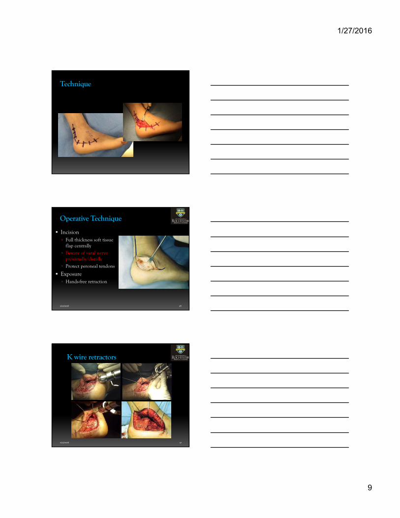
Technique
1/27/2016 26
Operative Technique
Incision Full thickness soft tissue
flap centrally
Beware of sural nerve proximally/distally
Protect peroneal tendons
Exposure Hands-free retraction
1/27/2016 27
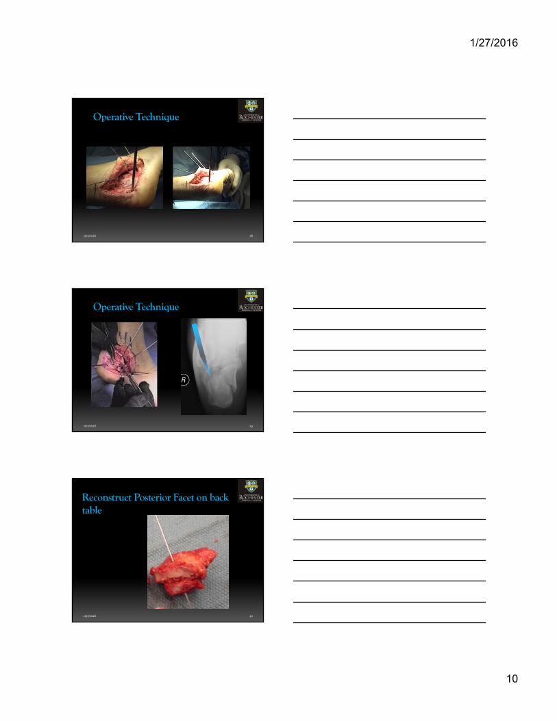
K wire retractors
1/27/2016
10
1/27/2016 28
Operative Technique
1/27/2016 29
Operative Technique
1/27/2016 30
Reconstruct Posterior Facet on back table
1/27/2016
11
1/27/2016 31
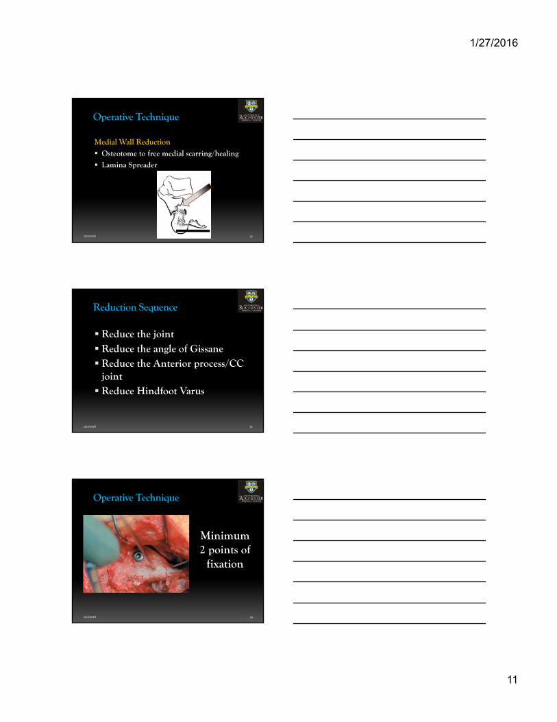
Operative Technique
Medial Wall Reduction
Osteotome to free medial scarring/healing
Lamina Spreader
1/27/2016 32
Reduction Sequence
Reduce the joint Reduce the angle of Gissane Reduce the Anterior process/CC
joint Reduce Hindfoot Varus
1/27/2016 33
Operative Technique
Minimum 2 points of
fixation
1/27/2016
12
1/27/2016 34
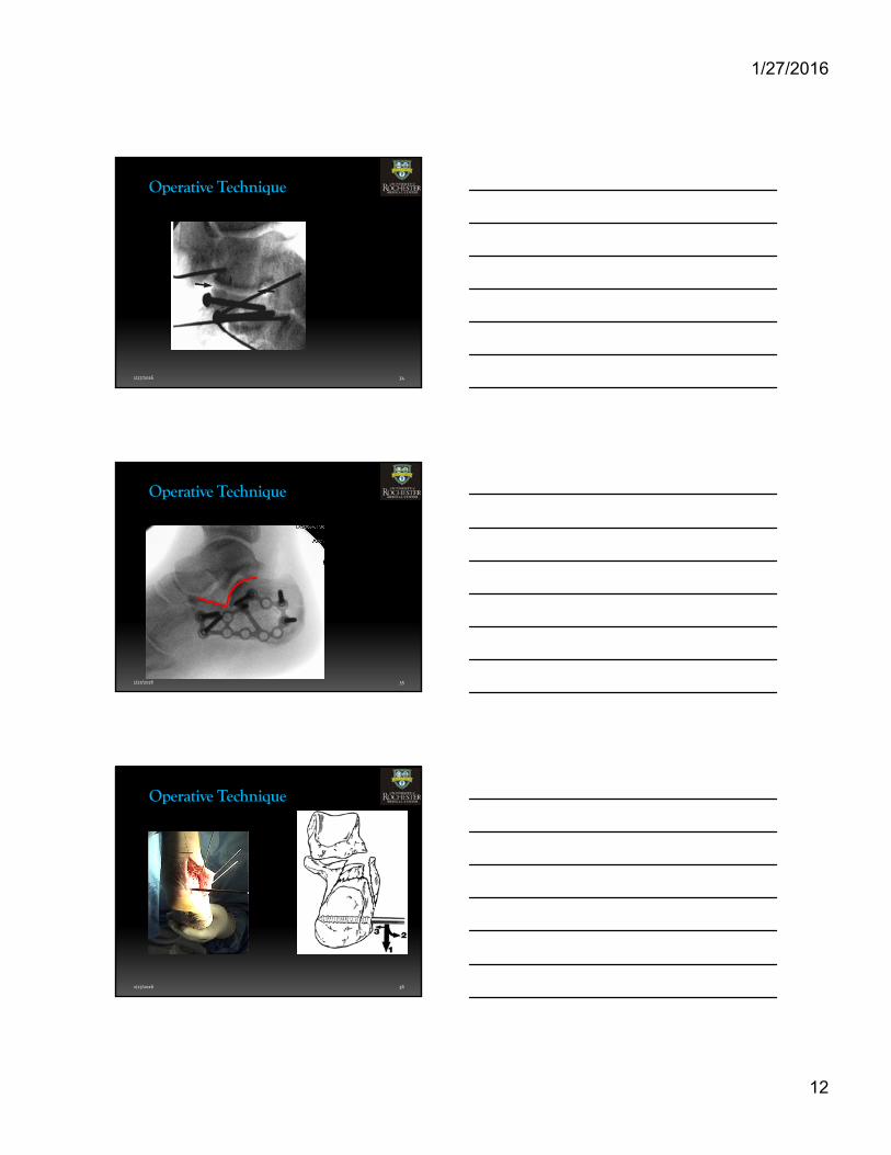
Operative Technique
1/27/2016 35
Operative Technique
1/27/2016 36
Operative Technique
1/27/2016
13
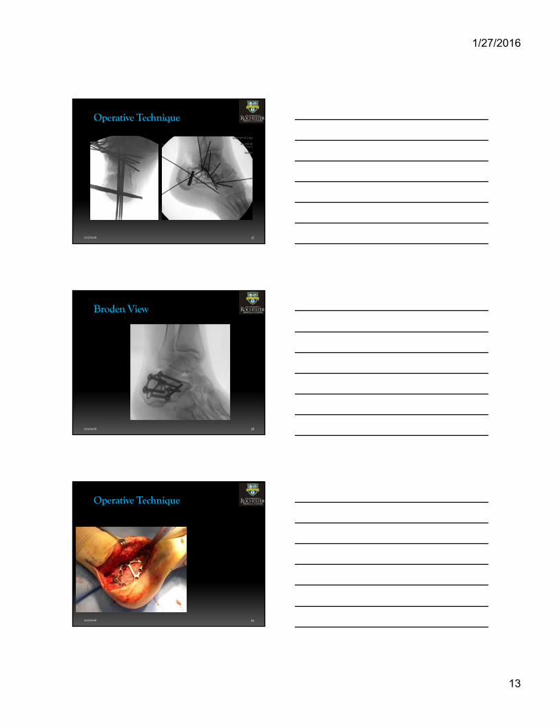
1/27/2016 37
Operative Technique
1/27/2016 38
Broden View
1/27/2016 39
Operative Technique
1/27/2016
14
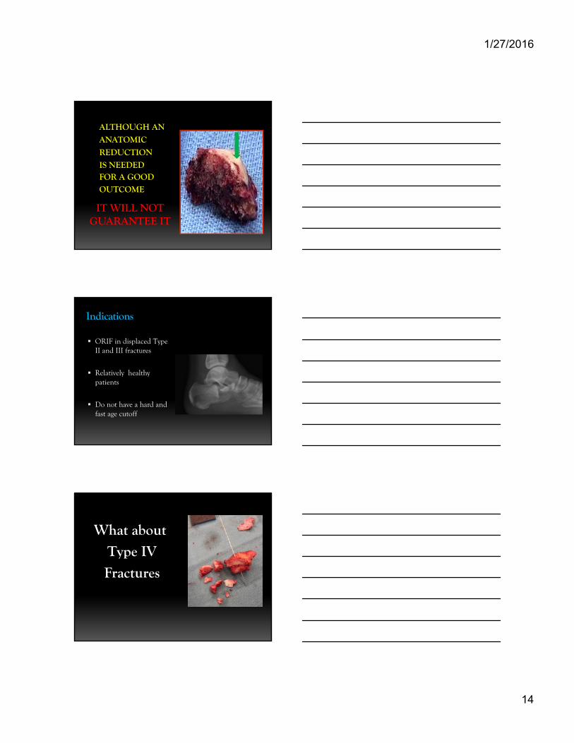
IT WILL NOTGUARANTEE IT
ALTHOUGH AN
ANATOMIC
REDUCTION
IS NEEDED FOR A GOOD
OUTCOME
Indications
ORIF in displaced Type II and III fractures
Relatively healthy patients
Do not have a hard and fast age cutoff
What about
Type IV
Fractures
1/27/2016
15
1/27/2016 43
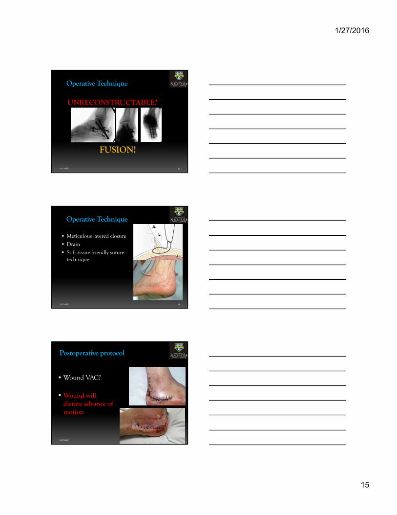
Operative Technique
UNRECONSTRUCTABLE?
FUSION!
1/27/2016 44
Operative Technique
Meticulous layered closure
Drain
Soft tissue friendly suture technique
1/27/2016 45
Postoperative protocol
Wound VAC?
Wound will dictate advance of motion
1/27/2016
16
1/27/2016 46
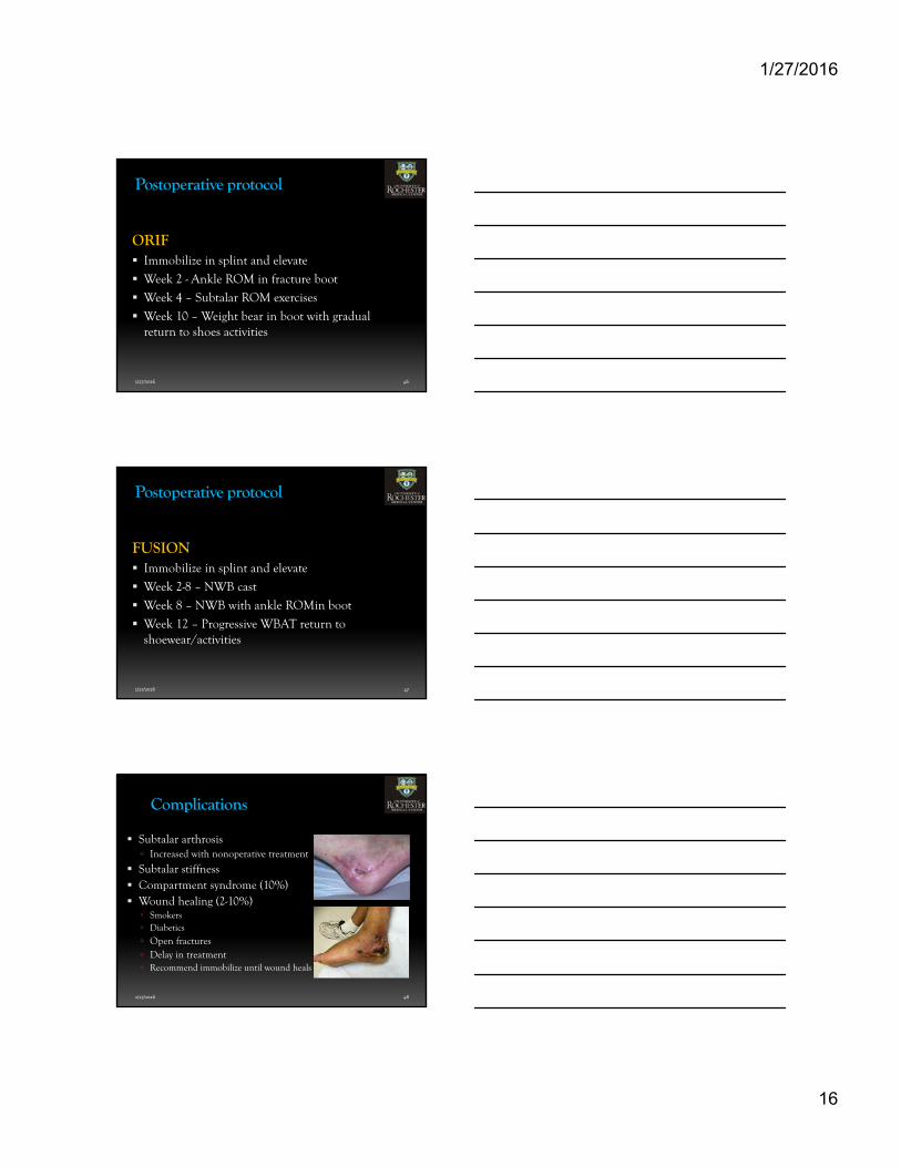
Postoperative protocol
ORIF Immobilize in splint and elevate
Week 2 - Ankle ROM in fracture boot
Week 4 – Subtalar ROM exercises
Week 10 – Weight bear in boot with gradual return to shoes activities
1/27/2016 47
Postoperative protocol
FUSION Immobilize in splint and elevate
Week 2-8 – NWB cast
Week 8 – NWB with ankle ROMin boot
Week 12 – Progressive WBAT return to shoewear/activities
1/27/2016 48
Complications
Subtalar arthrosis Increased with nonoperative treatment
Subtalar stiffness Compartment syndrome (10%) Wound healing (2-10%) Smokers Diabetics
Open fractures Delay in treatment Recommend immobilize until wound heals
1/27/2016
17
1/27/2016 49
Why Extensile Lateral Approach?
Late Presentation
Large Deformity
Posterior articular comminution/stepoff
Primary Fusion
1/27/2016 50
Thank You


![Extensile Exposure (2nd Edition) [Section 03]...EXTENSILE EXPOSURE VISCERAL BRANCHES OF THE INTERNAL ILIAC ARTERY The origins of these branches vary so much that Poirier (Traite d'Anatomie](https://img.pdfslide.us/doc/110x75/5e2e1a225edfe2293b42f1f5/extensile-exposure-2nd-edition-section-03-extensile-exposure-visceral-branches.jpg)