Embed Size (px)
Citation preview

C1-C2 X-Ray assessment of misalignment parameters in patients with Chronic Cerebra-spinal Venous Insufficiency and Multiple Sclerosis versus patients with other pathologies.
Mandolesi S (1), Marceca G (9), d'Alessandro A (4), Ciccone MM (8), Zito A (8), Manconi E (2), Niglio T (3), Ricci D (5), Mandolesi D (6), d’Alessandro A (7), Fedele F (1)
1. Department of Cardio-vascular and Respiratory Sciences – Sapienza University Roma, Italy 2. Department of Cardiovascular and Neurological Sciences, University of Cagliari, Italy 3. Istituto Superiore di Sanità, Roma, Italy 4. Department of Angiology " T. Masselli-Mascia " Hospital - San Severo (FG), Italy 5. Medical Office “Ricci” Bari, Italy 6. Medicina del lavoro Sapienza University Roma, Italy 7. Faculty of Medicine Catholic University “ Our Lady of Good Counsel “ of Tirana, Albanian 8. Cardiovascular Diseases Section, Department of Emergency and Organ Transplantation (DETO),
University of Bari, Italy 9. Unitelma Sapienza Università Roma Address for correspondence: Sandro Mandolesi MD, Via San Martino della Battaglia 25 00185, Rome, Italy. Tel: +39-335-6512303; Fax:+39-06-4873984; e-mail: [email protected]
Abstract
Purpose: The complete compression of the internal jugular veins, in front position, shows a prevalence of 48% and it is equally distributed in the various segments of these veins in patients with Chronic Cerebro-Spinal Venous Insufficiency (CCSVI) and Multiple Sclerosis (MS) . The passage from the supine to the upright position causes an increased incidence of compressions. Such a high incidence of Venous Compression Syndrome of the internal jugular veins prompts us to seek specific postural alterations in these patients. The aim of this search is to identify radiological dislocation of C1-C2 as specific markers in patients with CCSVI and Multiple Sclerosis (MS). Method: We investigated 386 patients suffering from CCSVI and Multiple Sclerosis and a control group of 156 patients without MS. We assessed all these patients by X-rays upright examination and the sample with CCSVI and MS also by cerebral venous ultrasound EchoColorDoppler, EDSS severity scale and clinical types Relapsing-remitting MS –RR, Secondary Progressive MS –SP, Primary progressive MS -PP. We analyzed these conditions of misalignment of C1-C2 by X-ray : left laterality, right laterality, left anterior rotation, right anterior rotation, left post rotation, right post rotation, tilt superior, tilt inferior, anterior intrusion (antero-listesis). Results: The assessment of Anterior Intrusion shows the following average values: in the group with CCSVI and MS: 4.29 ±1.48 mm while in the control group: 3.78 ±1.45 mm (p = 0.0008).The evaluation of the Right Laterality shows the following average values: in group with CCSVI and MS: 2.31±1.41 mm, in control group: 1.97 ±1.28 mm (p = 0.0426). We found also that a longer duration of the disease corresponds to a higher severity of the pathological condition (p <0.0001). Conclusion: Data analysis of C1-C2 X-Ray parameters shows statistical significance of a particular profile of people with CCSVI and MS, severe anterior intrusion and right laterality

misalignment that are two to three times more frequent as compared to controls. These two specific radiological dislocation of the C1-C2 vertebrae allows us to hypothesize, in these subjects, a risk of presence of a CCSVI and of developing a MS. Considering the novelty of this work and the total absence of scientific similar works able to confirm this data, it is necessary to continue these studies in order to improve the clinical management of these patients and to perform therapeutic strategies based on venous decompressive treatments both surgical that manipulatives.
Key Words: Multiple Sclerosis, CCSVI, X-Ray C1-C2, Venous Compressive syndrome
Introduction
Multiple sclerosis (MS) is a chronic, inflammatory and demyelinating disease that affects the central nervous system [1]. Myelin, in fact, is a coating that covers the majority of axons and it allows high speed of nerve impulse transmission. Any pathologic process that damages the myelin, called demyelination, causes an alteration in the transmission of nerve impulses.
The different localization of the demyelination process in MS produces a variety of neurological symptoms that vary from individual to individual. However, in many cases, there is a gradual evolution of the pathology to severe functional disability [2]. The impact of MS is important because it affects mainly young people, aged between 25 and 35 years, preferring female [3].
The pathogenesis of MS is multifactorial and, for this reason, still not fully elucidated [4].
The focus on this pathology has awakened in recent years for a work of Zamboni in 2009. He evaluated the role of cerebrospinal venous drainage in the pathogenesis of MS [5]. In this work, he described a particular impaired cerebrospinal venous flow that involves the internal jugular veins, vertebral, deep cerebral and azygos in patients with MS. From this evidence it was assumed a role of Chronic Cerebra-Spinal Venous Insufficiency (CCSVI) in the development and progression of MS.
Scientific evidences suggest the diagnosis of CCSVI through the Doppler Sonography (DS) identification of at least two of the five hemodynamic criteria proposed by Zamboni [6]. The DS diagnosis of CCSVI is then confirmed by Catheter Venography (CV), which is the gold standard test for assessing cervical venous anatomy. Despite the high quality of the images obtained by CV, its invasiveness, the use of ionizing radiation and the inability to provide information on the changes of the venous flow with the posture of the patient, makes it difficult for clinical application. In addition, this examination requires about 45 minutes [7]. There is still no clear evidence about the gold standard for the evaluation of extracerebral venous anomalies [8].
For this reason, many works suggest the use of a multimodal approach [9]. However, the DS, for the fact of being a non-invasive method, free of ionizing radiation, capable of obtaining high resolution images in real time at a relatively modest cost, it has been proposed as a method of choice in the screening of CCSVI [10]. This method is still the subject of many discussions in the scientific field, especially about the lack of a standardized executive protocol and the huge variability of ultrasound window from individual to individual. The biggest problem is the difficulty of applying this diagnostic method in a routine clinical setting. A study evaluated the inter-agreement in echo-color-Doppler (ECD), in accordance with the Zamboni’s criteria for the diagnosis of cerebrovascular venous alterations, among eight different, experienced sonographers. The authors showed a low agreement among the operators for the diagnosis of cerebrovascular venous alterations. This result makes ECD less accurate and reproducible than other examinations [11].

On the contrary, our working group has demonstrated a high level of inter-observer procedure reproducibility of DS in the evaluation of cerebrospinal venous drainage in patients with MS [12].
Zamboni and his collaborators showed that the diagnosis of CCSVI is highly associated with all forms of MS [5]. Since 2009, several research groups have ventured into the evaluation of the possible involvement of CCSVI in the pathogenesis of MS, but with controversial results. On the one hand, in fact, some studies confirmed the association between CCSVI and MS [12- 13]; on the other hand, other works point out that CCSVI is not specific for MS, but it is also present in healthy subjects or patients with other neurological diseases [14-15]. On this line of research it is present our study. On the basis of an obstacle to the venous outflow in patients with MS, we wanted to investigate the existence of alterations in the cervical spine that may be specific and predictive factors for this neurologic disease.
Methods
We enrolled 386 patients with MS, diagnosed according to the revised McDonald criteria [16], and 156 patients with other not neurological diseases.
We enrolled patients with all three forms of MS [17-18] divided as follows:
• Relapsing-remitting MS -RR- (No. 159 pts): it is characterized by a partial or full recovery (remission) after an acute exacerbation (relapse). In this case, patients can live a relatively quiet life from a relapse and the other;
• Secondary Progressive MS -SP- (n ° 44 pts): is characterized by a persistent and worsening degree of disability, with the presence or absence of exacerbations superimposed.
• Primary progressive MS -PP- (No. 30 pts): disability and symptoms worsen continuously since the onset of the disease, without phases of remission or exacerbation of disability.
All patients were subjected to X-ray examination of the cervical spine in the upright position, in order to assess the possible presence of misalignment between the first two cervical vertebrae, called C1 and C2.
Ultrasound evaluation of cerebrospinal venous flow
Patients with MS (n ° 386) underwent a ultrasound examination of the veins of the neck, in order to assess the presence of alterations in the flow of blood from the head to the neck, according to the Zamboni’s criteria [5] .
The ultrasound parameters evaluated for the diagnosis of CCSVI are the following:
1. Reflux in internal jugular veins (IJVs) and / or vertebral veins (VVs) in the supine and sitting position; reflux disease is defined as a reverse flow that lasts more than 0.88 seconds [19];
2. Reflux in intracranial veins (internal cerebral vein, basal vein of Rosenthal, and great cerebral vein of Galen);
3. Presence of stenosis in the internal jugular vein using B-mode high-resolution echography; in this regard it is possible to identify hemodynamically (intraluminal defects such as fixed valves and / or malformed valves, spider webs, septa) or hemodynamically presence of stenosis internal jugular vein B-mode high-resolution in this regard it is possible to identify stenosis
hemodynamically (intraluminal defects such as fixed valves and/or malformed, spider webs, septa) or not- hemodynamically significant stenosis;
4. No detectable flow using Doppler in the internal jugular veins and/or vertebral veins;
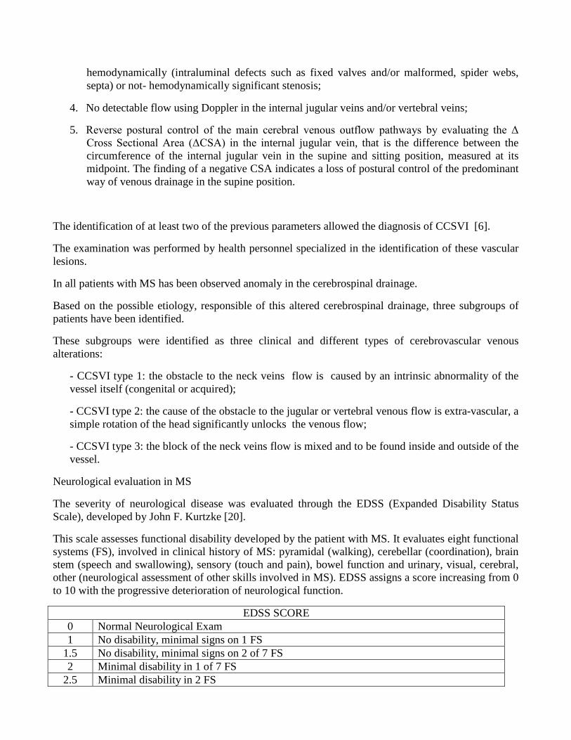
5. Reverse postural control of the main cerebral venous outflow pathways by evaluating the Δ Cross Sectional Area (ΔCSA) in the internal jugular vein, that is the difference between the circumference of the internal jugular vein in the supine and sitting position, measured at its midpoint. The finding of a negative CSA indicates a loss of postural control of the predominant way of venous drainage in the supine position.
The identification of at least two of the previous parameters allowed the diagnosis of CCSVI [6].
The examination was performed by health personnel specialized in the identification of these vascular lesions.
In all patients with MS has been observed anomaly in the cerebrospinal drainage.
Based on the possible etiology, responsible of this altered cerebrospinal drainage, three subgroups of patients have been identified.
These subgroups were identified as three clinical and different types of cerebrovascular venous alterations:
- CCSVI type 1: the obstacle to the neck veins flow is caused by an intrinsic abnormality of the vessel itself (congenital or acquired);
- CCSVI type 2: the cause of the obstacle to the jugular or vertebral venous flow is extra-vascular, a simple rotation of the head significantly unlocks the venous flow;
- CCSVI type 3: the block of the neck veins flow is mixed and to be found inside and outside of the vessel.
Neurological evaluation in MS
The severity of neurological disease was evaluated through the EDSS (Expanded Disability Status Scale), developed by John F. Kurtzke [20].
This scale assesses functional disability developed by the patient with MS. It evaluates eight functional systems (FS), involved in clinical history of MS: pyramidal (walking), cerebellar (coordination), brain stem (speech and swallowing), sensory (touch and pain), bowel function and urinary, visual, cerebral, other (neurological assessment of other skills involved in MS). EDSS assigns a score increasing from 0 to 10 with the progressive deterioration of neurological function.
EDSS SCORE 0 Normal Neurological Exam 1 No disability, minimal signs on 1 FS
1.5 No disability, minimal signs on 2 of 7 FS 2 Minimal disability in 1 of 7 FS
2.5 Minimal disability in 2 FS

3 Moderate disability in 1 FS; or mild disability in 3 - 4 FS, though fully ambulatory 3.5 Fully ambulatory but with moderate disability in 1 FS and mild disability in 1 or 2 FS; or
moderate disability in 2 FS; or mild disability in 5 FS 4 Fully ambulatory without aid, up and about 12hrs a day despite relatively severe
disability. Able to walk without aid 500 meters 4.5 Fully ambulatory without aid, up and about much of day, able to work a full day, may
otherwise have some limitations of full activity or require minimal assistance. Relatively severe disability. Able to walk without aid 300 meters
5 Ambulatory without aid for about 200 meters. Disability impairs full daily activities 5.5 Ambulatory for 100 meters, disability precludes full daily activities 6 Intermittent or unilateral constant assistance (cane, crutch or brace) required to walk 100
meters with or without resting 6.5 Constant bilateral support (cane, crutch or braces) required to walk 20 meters without
resting 7 Unable to walk beyond 5 meters even with aid, essentially restricted to wheelchair,
wheels self, transfers alone; active in wheelchair about 12 hours a day 7.5 Unable to take more than a few steps, restricted to wheelchair, may need aid to transfer;
wheels self, but may require motorized chair for full day's activities 8 Essentially restricted to bed, chair, or wheelchair, but may be out of bed much of day;
retains self care functions, generally effective use of arms 8.5 Essentially restricted to bed much of day, some effective use of arms, retains some self
care functions 9 Helpless bed patient, can communicate and eat
9.5 Unable to communicate effectively or eat/swallow 10 Death due to MS
According to the score achieved, the patients with MS were divided into three groups:
- EDSS between 0 and 3: patients with minimal reduction in functional capacity ”mild” EDSS;
- EDSS between 4 and 6: patients with moderate reduction of functional capacity, with negative impact on the personal autonomy “medium EDSS”
- EDSS more than 7: patients with severe reduction of functional capacity with complete loss of personal autonomy “severe” EDSS.
X-ray Cervical Examination
Each patient received a series of cervical radiographs. The following projections were taken and
measured for each sample group: APOM (anterior posterior open mouth), lateral cervical and base
posterior.

APOM projection
This projection is used to measure left or right laterality of C1. To measure this horizontal line was
drawn through the upper ½ to 1/3 portion of the foramen magnum. The foramen magnum is then
bisected with a vertical line. A line is then drawn from the vertical line to the edge of the lateral mass
on the right and left side. Measure both sides and the longer side is the side of laterality of C1.

Base Posterior projection
This projection is used to measure the rotational component of C1. To measure the anterior or
posterior rotation first a dot must be placed in the center of each transverse process. Then a line is
drawn connecting the two dots. The is called the atlas line. A dot is then placed in the middle of the
nasal septum. Draw a line through the dot in the middle of the nasal septum through the middle of the
basilar process. This line should intersect the atlas line. The angle is measured on the side of laterality
to check for posterior or anterior rotation.

Lateral Cervical projection
This projection is used to measure anterior intrusion of C1. This is measured by drawing a line from
the posterior foramen magnum to the posterior neural canal of axis. This will be labeled as line A. A
line is then drawn from the posterior portion of the neural canal of the posterior arch of atlas to the
posterior of foramen magnum. This line will be labeled line B. A line is then drawn from the posterior
of the neural canal of the posterior arch of the atlas to the posterior neural canal of axis. This line will
be labeled line C. Measure the distance from the point where B and C meet at posterior neural canal at
the posterior arch of atlas to line A at a perpendicular angle.
The normal values for these radiographs are as follows: tilt of 18-22 degrees, no laterality, no rotation,
and not intrusion into the spinal laminar line (neural canal). The moderate categorey values are as
follows: inferior tilt of 16-18 degrees, superior tilt of 22-24 degress, laterality of < 1.5mm, roation of
< 1 dgeree, and anterior intrusion of < 1.8mm. The severe categorey values are as follows: inferior tilt

of <16 degrees, superior tilt of > 24, laterality > 1.5mm, rotation of > 1 degree, and anterior intrusion
of > 1.8 mm.
Statistical Analysis
All data were analyzed by SPSS software to perform a stratified data description for numeric parametric variables. Statistical significance “between” and “within” groups was calculated on continuous variables by the analysis of variance (ANOVA) to test the equality of means. The Chi-square (χ²) Yates corrected test was used for non-continuous variables by Statcalc and Analysis programs from Epi-Info (2008, NIH & CDC Atlanta, USA; Italian version 3.5.1).
A p value less than 0.05 was considered significant, and 95% confidence intervals were also calculated. The collected data of this study were assessed by the Kruskal-Wallis statistical test, that is the non-parametric equivalent test of the analysis of variance in which the data are replaced by their rank; it was used because the studied population did not follow a normal Gaussian distribution. This non-parametric method has the purpose of comparing the equality of medians in the different groups.
Results
The demographic characteristics of the patients are summarized in Table 1.
Table 1. Demographic and clinical characteristics of the patients
542 Patients
(333 F + 209 M)
386 Multiple Sclerosis
(229 F + 157 M)
158 CCSVI-type 1 (101 F + 57 M)
5 CCSVI-type 2 (2 F + 3 M)
124 CCSVI-type 3 (70 F + 54 M)
99 Patients Not Classified Yet
(56 F + 43 M)
156 Without Multiple Sclerosis
(104 F + 52 M)
The female sex is more widely represented in both the population affected by MS and in the control population (229 female vs. 157 male in the group with MS; 104 female vs. 52 male in the control population). This date reflects the actual greater prevalence of this neurological disease in female rather than in male [3]. For this reason, even in the control group we wanted to recruit a greater number of female than male.
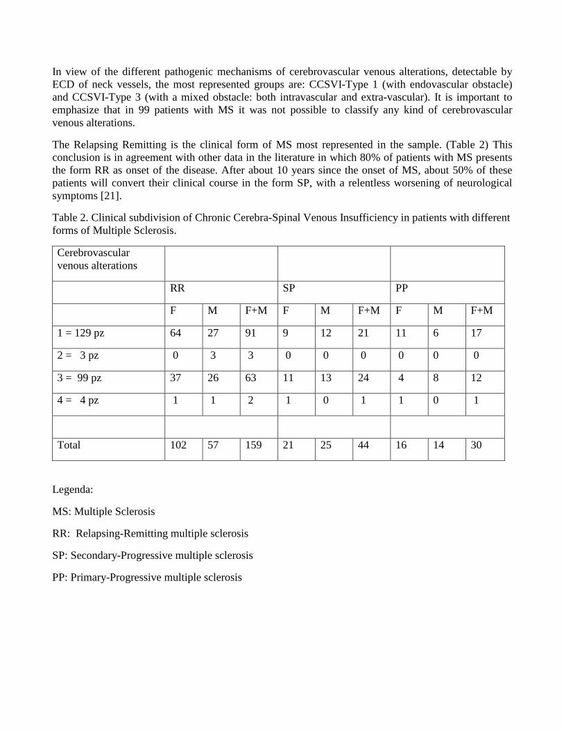
In view of the different pathogenic mechanisms of cerebrovascular venous alterations, detectable by ECD of neck vessels, the most represented groups are: CCSVI-Type 1 (with endovascular obstacle) and CCSVI-Type 3 (with a mixed obstacle: both intravascular and extra-vascular). It is important to emphasize that in 99 patients with MS it was not possible to classify any kind of cerebrovascular venous alterations.
The Relapsing Remitting is the clinical form of MS most represented in the sample. (Table 2) This conclusion is in agreement with other data in the literature in which 80% of patients with MS presents the form RR as onset of the disease. After about 10 years since the onset of MS, about 50% of these patients will convert their clinical course in the form SP, with a relentless worsening of neurological symptoms [21].
Table 2. Clinical subdivision of Chronic Cerebra-Spinal Venous Insufficiency in patients with different forms of Multiple Sclerosis.
Cerebrovascular venous alterations
RR SP PP
F M F+M F M F+M F M F+M
1 = 129 pz 64 27 91 9 12 21 11 6 17
2 = 3 pz 0 3 3 0 0 0 0 0 0
3 = 99 pz 37 26 63 11 13 24 4 8 12
4 = 4 pz 1 1 2 1 0 1 1 0 1
Total 102 57 159 21 25 44 16 14 30
Legenda:
MS: Multiple Sclerosis
RR: Relapsing-Remitting multiple sclerosis
SP: Secondary-Progressive multiple sclerosis
PP: Primary-Progressive multiple sclerosis
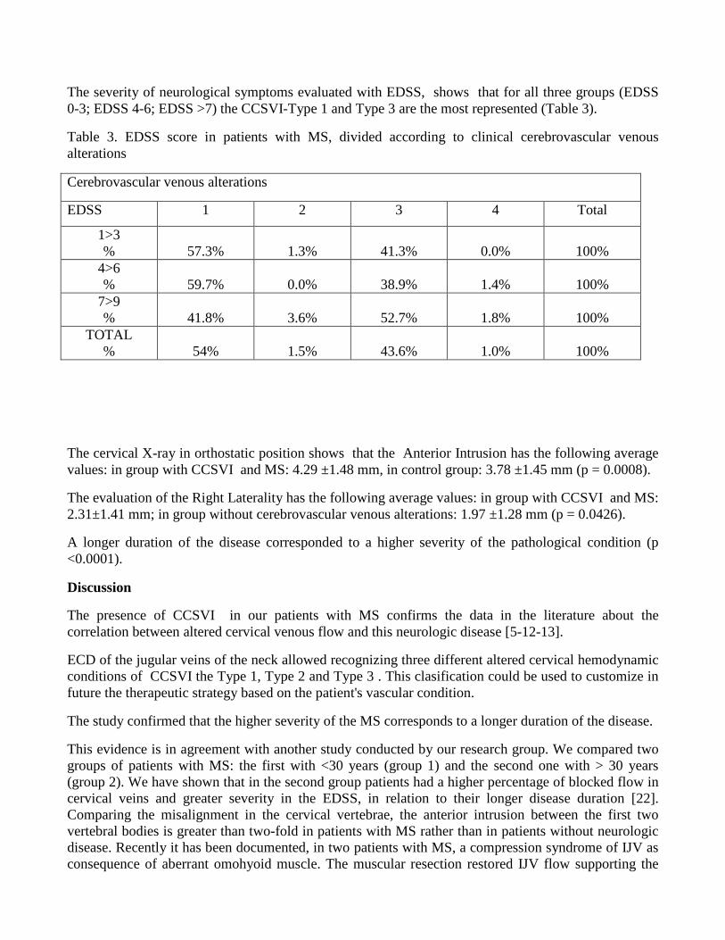
The severity of neurological symptoms evaluated with EDSS, shows that for all three groups (EDSS 0-3; EDSS 4-6; EDSS >7) the CCSVI-Type 1 and Type 3 are the most represented (Table 3).
Table 3. EDSS score in patients with MS, divided according to clinical cerebrovascular venous alterations
Cerebrovascular venous alterations
EDSS 1 2 3 4 Total
1>3 %
57.3%
1.3%
41.3%
0.0%
100%
4>6 %
59.7%
0.0%
38.9%
1.4%
100%
7>9 %
41.8%
3.6%
52.7%
1.8%
100%
TOTAL %
54%
1.5%
43.6%
1.0%
100%
The cervical X-ray in orthostatic position shows that the Anterior Intrusion has the following average values: in group with CCSVI and MS: 4.29 ±1.48 mm, in control group: 3.78 ±1.45 mm (p = 0.0008).
The evaluation of the Right Laterality has the following average values: in group with CCSVI and MS: 2.31±1.41 mm; in group without cerebrovascular venous alterations: 1.97 ±1.28 mm (p = 0.0426).
A longer duration of the disease corresponded to a higher severity of the pathological condition (p <0.0001).
Discussion
The presence of CCSVI in our patients with MS confirms the data in the literature about the correlation between altered cervical venous flow and this neurologic disease [5-12-13].
ECD of the jugular veins of the neck allowed recognizing three different altered cervical hemodynamic conditions of CCSVI the Type 1, Type 2 and Type 3 . This clasification could be used to customize in future the therapeutic strategy based on the patient's vascular condition.
The study confirmed that the higher severity of the MS corresponds to a longer duration of the disease.
This evidence is in agreement with another study conducted by our research group. We compared two groups of patients with MS: the first with <30 years (group 1) and the second one with > 30 years (group 2). We have shown that in the second group patients had a higher percentage of blocked flow in cervical veins and greater severity in the EDSS, in relation to their longer disease duration [22]. Comparing the misalignment in the cervical vertebrae, the anterior intrusion between the first two vertebral bodies is greater than two-fold in patients with MS rather than in patients without neurologic disease. Recently it has been documented, in two patients with MS, a compression syndrome of IJV as consequence of aberrant omohyoid muscle. The muscular resection restored IJV flow supporting the

hypothesis that muscular compression may be responsible of venous angioplasty inefficacy.[23] Other muscles can be involved in such venous compression: for example, the scalene muscle could entrap the J1 terminal segment of the internal jugular vein, while the sterno-mastoid muscle can compress the J3 segment of the internal jugular vein. We suppose that all these compressions may be caused by the misalignment of cervical vertebrae with stretching of the muscles and aponeurosis with effect on neck veins. This intermittent compression block of vertebral and jugular veins could be one of the multi-factorial causes of the worst clinical conditions in MS patients with CCSVI. These patients frequently had head and neck trauma. There is a proposed relationship between MS and upper cervical subluxation. Elster conducted a study on upper cervical subluxation and it’s connection to MS and Parkinson’s disease[24]. These data, absolutely innovative in scientific literature, not only confirmed that CCSVI are related to MS, but also that the structure of cervical spine may altered flow neck of these patients. In addition, the clinical classification of CCSVI about the etiopathogenesis of altered cervical flow can be a real novelty in the therapeutic approach to MS.
Conclusions
The X-Ray results illustrated above show that in patients with MS, compared to control group without neurological disease, there is a statistically significant difference about the effective anterior intrusion of C1 and C2 (p = 0.0008) and a right laterality (p = 0.0426). The presence of an endovascular hemodynamic conditions CCSVI-type 1, intrinsic to the vessel itself, suggests the treatment of these patients with an endovascular approach. This approach instead would be not recommended in patients with extrinsic compression CCSVI-type 2. This second kind of patients may take the specific physiotherapy in order to decompress the neck veins. The treatment of patients with mixed obstruction CCSVI-type 3 may be an indication to both angioplasty treatment than extra-vascular decompression.
The detection of an alteration in the alignment of the first two cervical vertebrae in patients with MS further emphasizes the role of cerebrospinal venous drainage in the pathogenesis and progression of this disease. To find these two specific radiological dislocation of the C1-C2 vertebrae allows us to hypothesize, in these subjects, a risk of presence of a CCSVI and of developing a MS.
Considering the novelty of this work and the total absence of scientific similar works able to confirm this data, it is necessary to continue these studies in order to improve the clinical management of these patients and to perform therapeutic strategies based on venous decompressive treatments both surgical that manipulatives.
References
[1] Compston A, Coles A. Multiple sclerosis. Lancet 2008; 372: 1502-1517.
[2] Ramagopalan SV, Dobson R, Meier UC et al. Multiple sclerosis: risk factors, prodromes, and potential causal pathways. Lancet Neurol 2010; 9: 27-39.
[3] Ascherio A, Munger K. Epidemiology of multiple sclerosis: from risk factors to prevention. Semin Neurol 2008; 28: 17-28.
[4] Pithadia A, Jain S, Navale A. Pathogenesis and treatment of multiple sclerosis (MS). Int J Neurol 2009; 10:1-20.

[5] Zamboni P, Galeotti R, Menegatti E et al. Chronic cerebrospinal venous insufficiency in patients with multiple sclerosis. J Neurol Neurosurg Psychiatry 2009; 80: 392-399.
[6] Menegatti E, Zamboni P. Doppler haemodynamics of cerebral venous return. Curr Neurovasc Res 2008; 5(4): 260-265.
[7] Dolic K, Siddiqui AH, Karmon Y et al. The role of noninvasive and invasive diagnostic imaging techniques for detection of extra-cranial venous system anomalies and developmental variants. BMC Medicine 2013; 11:155.
[8] Zivadinov R, Karmon Y, Dolic K. Multimodal non-invasive and invasive imaging of extracranial venous abnormalities indicative of CCSVI: Results of the PREMiSe pilot study. BMC Neurology 2013; 13:151.
[9] Dolic K, Siddiqui AH, Karmon Y et al. The role of noninvasive and invasive diagnostic imaging techniques for detection of extra-cranial venous system anomalies and developmental variants. BMC Medicine 2013; 11:155.
[10] Zamboni P, Galeotti R. The chronic cerebrospinal venous insufficiency syndrome. Phlebology 2010; 25 (6): 269-279.
[11] Leone MA, Raymkulova O, Lucenti A et al. A reliability study of colour-Doppler sonography for the diagnosis of chronic cerebrospinal venous insufficiency shows low inter-agreement. BMJ Open 2013; 3:e003508.
[12] Ciccone MM, Galeandro AI, Scicchitano P et al. Multigate quality Dopler profiles and morphological/hemodynamic alterations in multiple sclerosis patients. Curr Neurovasc Res 2012; 9: 120-127.
[13] Al-Omari MH, Rousan LA. Internal jugular vein morphology and hemodynamics in patients with multiple sclerosis. Int Angiol 2010; 29: 115-120.
[14] Zivadinov R, Marr K, Cutter G et al. Prevalence, sensitivity, and specificity of chronic cerebrospinal venous insufficiency in MS. Neurol 2011; 77(2): 138-144.
[15] Centonze D, Floris R, Stefanini M et al. Proposed chronic cerebrospinal venous insufficiency criteria do not predict multiple sclerosis risk or severity. Ann Neurol 2011; 70(1): 51-58.
[16] Polman CH, Reingold SC, Banwell B et al. Diagnostic criteria for multiple sclerosis: 2010 revisions to the McDonald criteria. Ann Neurol 2011; 69(2): 292-302.
[17] Lublin FD, Reingold SC. Defining the clinical course of multiple sclerosis: results o fan International survey. National multiple sclerosis society (USA) advisory committee on clinical trials of new agents in multiple sclerosis. Neurology 1996; 46: 907-911.
[18] Lassmann H. Multiple sclerosis pathology: evolution of pathogenetic concepts. Brain Pathol 2005; 15:217-222.
[19] Valdueza JM, Schmierer K, Mehraein S et al. Assessment of normal flow velocity in basal cerebral veins. A transcranial Doppler ultrasound study. Stroke 1996; 27: 1221-1225.

[20] Kurtzke JF. Rating neurological impairment in multiple sclerosis: an expanded disability scale (EDSS). Neurology 1983; 33(11): 1444-1452.
[21] Lublin FD, Reingold SC. Guidelines for clinical trials of new therapeutic agents in multiple sclerosis: relations between study investigators, advisors, and sponsors. National Multiple Sclerosis Society (USA) Advisory Committee on Clinical Trials of New Agents in Multiple Sclerosis. Neurology. 1997; 48:572–574.
[22] Mandolesi S, Ciciarello F, Galeandro AI et al. Age-related differences between patients suffering from multiple sclerosis and Chronic Cerebra-spinal Venous Insufficiency(CCSVI). Curr Neurovasc Res 2014; 11(1): 23-30. 20. Simka M, Majewski E, Fortuna M, Zaniewski M. Internal jugular vein entrapment in a multiple sclerosis patient. Case Rep Surg. 2012;2012:293568.
[23] Gianesini S, Menegatti E, Mascoli F, Salvi F, Bastianello S, Zamboni P. The omohyoid muscle entrapment of the internal jugular vein. A still unclear pathogenetic mechanism. Phlebology. 2013 May 16.
[24] Elster E. , Eighty-one patients with multiple sclerosis and Parkinson disease undergoing Upper Cervical chiropractic care to correct vertebral subluxation: a retrospective analysis. Journal of Vertebral Subluxation Research. August 2, 2004. Pages 1-9.