Embed Size (px)
Citation preview

C
fo
Chromos
or thera
T
some in
peutic d
Ana
Tese de D
nstabilit
drugs to
a Filipa B
Doutoram
ty in Fa
o preve
diseas
Brinco de
mento em
2012
nconi A
ent the p
se
Oliveira
Ciências
Anemia:
progres
Ponte
s Bioméd
: search
ssion of
icas
hing
f the

II
III
Ana Filipa Brinco de Oliveira Ponte
Chromosome instability in Fanconi Anemia: searching for
therapeutic drugs to prevent the progression of the disease
Tese de Candidatura ao grau de Doutor em
Ciências Biomédicas submetida ao Instituto
de Ciências Biomédicas Abel Salazar da
Universidade do Porto.
Orientadora – Professora Doutora Beatriz
Porto
Categoria – Professora Auxiliar
Afiliação – Instituto de Ciências Biomédicas
Abel Salazar da Universidade do Porto
Co-orientador – Professor Doutor Félix
Carvalho
Categoria – Professor Catedrático
Afiliação – Faculdade de Farmácia da
Universidade do Porto

Os
REQ
Facu
Micro
Estes
Tecn
3990
estudos a
QUIMTE/Lab
uldade de F
oscopia, do
s estudos
nologia (FC
03 / 2007).
presentado
boratório d
Farmácia d
o Instituto de
contaram
CT), através
s nesta te
e Toxicolo
do Porto e
e Ciências B
com o ap
s da atribuiç
ese foram
gia do De
no Labora
Biomédicas
poio finance
ção de uma
realizados
epartamento
atório de C
s Abel Salaz
eiro da Fu
a Bolsa de
s no Labo
o de Ciênc
itogenética,
zar.
undação pa
Doutorame
oratório As
cias Biológ
, Departam
ara a Ciên
ento (SFRH
IV
ssociado
gicas da
mento de
ncia e a
H / BD /
V
Author’s declaration
The author states to have afforded a major contribution to the conceptual design,
technical execution of the work, interpretation of the results and manuscript preparation of
the published or under publication articles included in this thesis.
Publications
According to nº 2, alínea a, do artigo 31º do Decreto-Lei nº 230/2009 the following
published or under publication articles were prepared in the scope of this thesis:
Articles in international peer-review journals
Ponte F, Carvalho F and Porto B (2011) Protective effect of acetyl-L-carnitine and a-lipoic
acid against the acute toxicity of diepoxybutane to human lymphocytes. Toxicology, 289
(1): 52-52.
Ponte F, Sousa R, Fernandes AP, Gonçalves C, Barbot J, Carvalho F and Porto B (2012)
Improvement of genetic stability in lymphocytes from Fanconi Anemia patients through the
combined effect of α-lipoic acid and N-acetylcysteine. Orphanet Journal of Rare Diseases
(in press).
Abstracts in international peer-review journals
Ponte F, Sousa R, Carvalho F, Porto B (2011) α-Lipoic acid decreases chromosome instability in lymphocytes from Fanconi Anemia patients. Toxicology Letters, 205 Suppl, S108. Ponte F, Sousa R, Carvalho F, Porto B (2011) The combined used of a-lipoic acid plus N-acetyl-cysteine as an effective cocktail for decreasing chromosome instability in Fanconi Anemia patients. Chromosome Research, 19 (1) S25. Sousa R, Ponte F, Porto B (2011) Accuracy in the cytogenetic diagnosis of Fanconi Anemia and other chromosome instability syndromes. Chromosome Research, 19 (1) S36. Ponte F, Carvalho F, Porto B (2010) Acute toxicity of diepoxybutane to mononuclear leukocytes. Multifactorial mechanisms? Toxicology Letters, 196 Suppl, S86. Ponte F, Carvalho F, Porto B (2009) Acute toxicity of diepoxybutane to human mononuclear lymphocytes. Toxicology Letters 189, Suppl, S121.

VI

VII
Sinto-te como um rio na exata medida que desvendas caminhos misteriosos. Da nascente até ao mar cruzas-te com obstáculos na tentativa vã de pararem a torrente mas teu rio corre inteiro cálido, rebelde, impetuoso sem se perder na paisagem que desperta outras correntes. Quando o objetivo é seguir o caminho é sempre em frente. Sentes a limpidez das águas que correm nas tuas veias e teu caudal majestoso quando em ti confluem rios que com o mesmo fulgor têm pressa na chegada. Ficas contente e sorris mas segues o teu traçado e quando estás já tão perto quase a desaguar olhas para ti surpresa dizes com voz sublime: - afinal sou uma gota majestoso é o mar! Sinto-te navegar...
Teresa Brinco

VIII

IX
Agradecimentos
Agradeço à Rosa tudo o que passámos juntas no nosso pequeno e ao mesmo
tempo, grande Laboratório de Citogenética. Todas as alegrias partilhadas (que
foram muitas) no decorrer destes 4 anos, as tristezas de não conseguirmos o que
queríamos, as brigas e discussões científicas, que por vezes ainda mais confusão
geravam nas nossas cabeças, e de onde mais tarde saía sempre uma luz, a amizade
que criámos e sei que perdurará para além da ciência. As viagens hilariantes mundo
fora, sempre com a anemia de Fanconi ao lado, nunca serão esquecidas. Obrigada
por me fazeres ainda gostar mais de ciência e de anemia de Fanconi. Sei que sem ti
não seria igual. Obrigada por partilhares comigo os teus conhecimentos. Por me
ouvires quando estava desanimada e me sentia incapaz de continuar. As tuas
palavras sempre me ajudaram e passaram além do trabalho. Foi um prazer
encontrar-te e espero que os nossos caminhos se cruzem sempre e o nosso objetivo
final seja cumprido. Tenho a certeza que sem ti nada disto seria possível, pelo
menos desta forma. És uma pessoa incrível.
Agradeço à Sara Teixeira e à Débora Franco, as minhas colegas mais novas do
laboratório de Citogenética, todo o incentivo que me deram e a alegria que
proporcionaram no laboratório, o convívio sempre salutar de onde sempre saía
ciência! Foi bom ajudar e receber o vosso apoio. Um agradecimento muito especial à
nossa “informática” sempre de serviço, a Lara Andrade! A tua ajuda foi preciosa!
Sempre pronta a resolver os problemas do meu PC, que foram muitos, e a ajudar
a utilizar softwares como o EndNote e na formatação da tese! À Maria, apesar do
pouco tempo de convívio, foi bom ter-te aí também e teres-nos adotado como o
teu laboratório! À Joana, apesar de por lá passar pouco tempo, o pouco tempo em

X
que lá esteve mais que uma hora, foi sempre surreal! Ao nosso primeiro membro
masculino, o João, obrigada por conseguires tantos doentes para nós.
Ao Nuno Santos um muito obrigado por toda a ajuda informática prestada! Foste
incansável, amigo, compreensivo e nunca te “revoltaste” pelos imensos pedidos de
ajuda… ultimamente diários, muitos deles de simples resolução, o que demonstra as
minhas imensas capacidades na área da informática! Bem, isto sem ti também tinha
sido bastante mais complicado! E, no fim de tantas horas passadas em frente ao
PC, até ficámos amigos!
Um agradecimento especial à Alice Porto pela dica para a capa da tese! Gostei!
À Ana Paula agradeço a dedicação em cuidar do nosso pequeno recanto, sem ela não
teria sido tão fácil trabalhar.
Obrigada a todos os clínicos que trabalharam comigo nestes anos, dos quais saliento
o Dr. Barbot, a Dra. Ana Paula Fernandes e a Dra. Cristina Gonçalves que tanto
fizeram por conseguir amostras de sangue dos nossos meninos doentes. Obrigada ao
serviço de dadores do HGST e às simpáticas enfermeiras que sempre me ajudaram.
Obrigado aos dadores, por contribuírem para este estudo. Obrigada à Sandra Maia
do Laboratório de Imunologia do ICBAS por nos ajudar nas recolhas de amostras de
sangue.
À Maria João Valente, à Helena Faria e à Diana Dias da Silva agradeço ter-vos
conhecido no meio da confusão do Laboratório de Toxicologia da FFUP! Como foi
possível no meio de tanta gente encontrar-vos como amigas? Obrigada por tudo,
pelas partidas que por vezes me deixaram louca, e pela ajuda e convívio nos nossos
maravilhosos Eurotox, dos quais saliento o de Barcelona e o de Paris!

XI
Agradeço às colegas mais experientes, Vera, Renata e Helena Pontes, a ajuda no
início, quando comecei os trabalhos no Laboratório de Toxicologia. As dicas, a
paciência e os vossos ensinamentos foram preciosos. Ao Ricardo Oliveira um grande
obrigada pela ajuda prestada, ao ensinar-me a fazer a determinação das caspases,
que não foi fácil. Ao Professor Fernando Remião e à Professora Helena Carmo
obrigado por serem sempre prestáveis e me ajudarem, quando nunca foi vossa
obrigação. À Engenheira Elisa obrigada pelos caramelos, que tanto adoçaram os
nossos dias, e obrigada pelos seus elogios sempre muito simpáticos! À doutora
Maria de Lourdes Bastos obrigada por me ter deixado integrar a fantástica equipa
do Laboratório de Toxicologia. À restante equipa do laboratório obrigada pelo bom
ambiente criado e o espirito de interajuda.
À Dona Conceição, à Dona Júlia e à Ana Maria um obrigado por permitirem que
que o laboratório de Toxicologia fosse utilizável! Sem a vossa cooperação seria
impossível fazer ciência!
Agradeço às minhas amigas, Lília Pereira, Sara Assunção, Ana Lúcia Pedrosa, Raquel
ou Tinka, Diana Ledo, Marta Ledo, Filipa Moreira, Filipa Cortes, Isabel Gonçalves,
Adriana Moura, Carla Rocha, Nídia Rocha e amigos, Jorge Martinho, Rui Amorim,
Carlos Martins, André Ferreira, Joel Pereira, Gonçalo Barreto, João Sousa, Thiago
Silva, Diogo Oliveira e Silva, Hugo Oliveira, Pedro Borges e Samuel Reis as palavras
de apoio e carinho que me foram dando nestes anos. Obrigada por compreenderem
que nem sempre foi possível estar perto e por acreditarem em mim e no meu
trabalho. Hoje todos vocês sabem o que é anemia de Fanconi, o que me deixa
extremamente satisfeita. Agradeço, principalmente às meninas, nunca me deixarem
desistir e serem sempre boas ouvintes. Muitos acompanharam-me desde o início e

XII
sabem o quanto passei para estar aqui hoje, obrigada por serem meus amigos de
verdade.
Ao Dr. Miguel Ricou agradeço nunca me ter deixado desistir da vida académica. De
sempre me ter incentivado e mostrado que tinha capacidades para fazer um
doutoramento brilhante. De me ter mostrado que a vida é mais que um trabalho e
que do trabalho teria de tirar o melhor partido, para assim ser mais feliz. Penso
que consegui. Obrigada por toda a ajuda, que sabe que foi essencial para hoje ser
uma pessoa bem sucedida.
Ao meu irmão Ricardo, o Brinco, obrigada por me pores sempre o astral para cima
e me achares a mana genial, mais uma das vedetas da casa! As tuas brincadeiras,
mesmo nos meus momentos de tristeza, sempre me fizeram sorrir. Obrigada por
existires na minha vida. Espero para ti ser um exemplo de luta e persistência,
mostrando-te que com garra vencemos tudo. Para mim já sabes que és o maior
exemplo de felicidade. E isso, mesmo que não tenhas dado conta, foi-me
contagiando. Óbvio que continuo à espera da minha casa feita por ti, meu projecto
de Arquitecto!
Obrigado Pai por, mesmo por vezes ausente fisicamente, estares sempre tão perto.
És o meu guru. Em ti sempre procurei palavras sábias e os caminhos certos a
percorrer. Nunca me deixaste desistir com as tuas palavras, por vezes austeras,
mas sempre racionais. E a ti também te consegui passar o “bichinho” das doenças
raras. Também tu hoje sabes falar de anemia de Fanconi. Os teus sorrisos aquando
dos meus pequenos momentos de vitória encheram-me o coração de uma felicidade
inexplicável. Sem ti, não teria sido igual.

XIII
Mãe, a tua presença constante, foi preponderante nesta minha luta, nem sempre
fácil. As palavras de apoio e coragem foram incessantes. Obrigado por acreditares
que um dia isto seria mesmo verdade. Sempre me mostraste que eu ia conseguir,
mesmo quando eu acreditava que era impossível. Desculpa o mau humor antes dos
congressos, os “não vou conseguir”, seguidos de umas lágrimas e tu sempre lá
estiveste, ao meu lado, com um sorriso de uma mãe que olha com orgulho para a
sua filha.
Sem vocês os três este caminho seria outro, talvez não tão bem sucedido. Obrigada
pelos valores e amor que sempre me transmitiram e ajudarem-me a construir a
pessoa e cientista que hoje sou.
Às avós Maria e Georgete obrigada pela preocupação e por mesmo não sendo fácil
para vocês entenderem aquilo que faço, me darem valor. À avó Georgete agradeço
ainda a velinha que acendia nos meus momentos cruciais, para que tudo me corresse
sempre bem! Ao avô Zé, obrigado por teres gostado tanto de mim e te orgulhares
da neta que fui para ti. Estarás sempre no meu coração. Gostava tanto que aqui
estivesses neste momento tão especial na minha vida… Sinto a tua ausência e vou
sentir falta de ouvir os teus parabéns emocionados… Aos tios e tias obrigada pelos
incentivos e pelos parabéns constantes aquando das minhas pequenas conquistas.
Ao Boris obrigado por seres um ouvinte sempre tão atento e fiel. És o meu leal
companheiro e a ti agradeço toda a atenção e companhia que sempre me fazes, nos
bons e maus momentos. Em ti procurei o amor que só os animais de estimação
sabem oferecer…e o conforto e o calor quando nem as palavras dos humanos me
faziam sentir mais calma.

XIV
Ao Professor Félix agradeço todo o apoio que sempre me deu. Lembro-me dos dias
em que entrava no seu gabinete cabisbaixa e triste, porque os resultados nunca
eram aquilo que eu esperava. As suas palavras de incentivo foram fulcrais para que
nunca desistisse: “Mas estes resultados são fantásticos! Parabéns Filipa!”. Obrigada
por toda a ajuda, compreensão e por todos os ensinamentos que me transmitiu e
ainda me ter incentivado a ser melhor. Se hoje percebo alguma coisa de toxicologia
a si o devo. Se hoje sou persistente aprendi consigo. Se tenho ambição e coragem
também. Obrigada por acreditar que um dia iriamos chegar a um resultado positivo.
À professora Beatriz agradeço tudo. Não há palavras que descrevam a gratidão que
por si tenho. Todas seriam poucas… Mesmo assim arrisco a dizer que se hoje sou
feliz no meu trabalho a si o devo. O amor à ciência também. A dedicação à anemia
de Fanconi. Obrigada por todos os sorrisos, que foram muitos, quando eu estava
desanimada e não acreditava em mim. Por todas as conversas científicas e não só,
que sempre me ajudaram a percorrer este caminho. Se hoje sinto amor à camisola e
desejo continuar esta luta, a si o devo. É o meu exemplo.
Sei que tive sorte em ter o Professor Félix e a Professora Beatriz como
orientadores. Além de me terem transmitido todo o saber, cada um da sua área,
são pessoas excecionais. Sem eles tenho a certeza que os resultados que obtive não
seriam os mesmos. Obrigada

XV
Table of Contents
Preface
1
Abbreviations
5
Resumo
7
Abstract
9
PART I – GENERAL INTRODUCTION 11
Introduction: Fanconi anemia, from the past towards present
13
1. Clinical features of Fanconi anemia 15
1.1. Fanconi anemia phenotype 15
1.1.1. Congenital malformations 15
1.1.2. Hematologic manifestations 17
1.1.3. Impairment in immunological function 18
1.1.4. Cancer susceptibility 21
1.2. Fanconi anemia genotype 23
1.2.1. The FA pathway 25
1.3. Diagnostic tests for Fanconi anemia 27
1.3.1. The chromosomal fragility test 27
1.3.2. Other diagnostic methods 28
1.4. Fanconi anemia treatments
30
2. Diepoxybutane as a golden standard test for Fanconi anemia diagnosis and as
an oxidative stress inducer – Implications on Fanconi anemia research 31
2.1. DEB: why is it a golden standard test for Fanconi anemia diagnosis? 31
2.2. DEB cytotoxicity: biotransformation as a potential cause 32
2.3. DEB cytotoxicity: generation of reactive oxygen species and oxidative stress 34
2.4. DEB cytotoxicity: relation with mitochondrial dysfunction
35
3. Oxidative stress in Fanconi anemia: from cells and molecules towards
prevention against chromosome instability 36
3.1. Pro-oxidant state in Fanconi anemia cells and patients 36

XVI
3.2. Redox related functions of FANC proteins 38
3.2.1. FANCA, FANCC and FANCG: link with xenobiotic processing pathway 38
3.2.2. FANCD2 and FANCJ: multiple identities and redox-related roles 40
3.3. Mitochondrial dysfunction in Fanconi anemia cells 41
3.4. In vitro and in vivo experimental therapies – Antioxidant agents as
protectors of chromosome instability
44
PART II – OBJECTIVES OF THE THESIS
47
PART III – ORIGINAL RESEARCH
51
Chapter I. Protective effect of acetyl-L-carnitine and α-lipoic acid against the acute
toxicity of diepoxybutane to human lymphocytes
53
Chapter II. Improvement of genetic stability in lymphocytes from Fanconi Anemia
patients through the combined effect of α-lipoic acid and N-acetylcysteine
63
PART IV – GLOBAL DISCUSSION
81
PART V – FINAL CONCLUSIONS AND FUTURE PERSPECTIVES
95
PART VI – REFERENCES 99

1
Preface
Four years ago it was straightforward to choose Fanconi anemia (FA) as a model for
studying chromosome instability (CI) disorders and relate them with oxidative stress (OS).
In the Laboratory of Cytogenetics of Institute of Biomedical Sciences (ICBAS) a special
focus has long been given to FA. The blood samples that arrived there were frequently to
confirm the diagnosis of FA. Thus, the interest for this disease slowly “grew” inside me
and the will to learn more about it became natural. The relation with OS came later, after
reading several papers about this disease, in which the main theory to explain the
phenotype and the molecular aspects of the disease was impairment in DNA repair
system. Notwithstanding this postulate, in my perspective, many aspects were not
explained based on it and a conjunction of the two theories would make much more
sense. The works of Pagano and co-workers were very important for establishing my
perspective. So, the idea about antioxidant treatments simply arose. I even remember the
first discussion about this with Professor Beatriz and since the beginning we believed that
we could aim to solve some of the FA problems, finding the right antioxidant! And the work
began! But I didn't know back then how difficult it was to study such a rare disease...
The works in the Laboratory of Toxicology of Faculty of Pharmacy of University of
Porto (FFUP) were the beginning of a long and not always happy journey. It took almost
one year to establish the right techniques and procedures. The next step was to test a
variety of drugs in healthy donors, in the hope of finding a successful one to test in the
blood of FA patients. One after another and the results were always negative…One year
later, after testing 8 or 9 compounds, I got the first positive result and a big smile in my
face! "Maybe I will achieve something", I thought. Working in FFUP, although it was very
difficult, due to the scarcity of space and available material (we were 20 colleagues
working at the same time in a class laboratory!!!), it was funny, even when everything
went wrong and I wanted to do something and I wasn’t able to! Working in FFUP taught
me to deal with stress and to work as a team, a very important lesson to a future scientist.
In the beginning of the third year of my PhD I came back to laboratory of
Cytogenetics. The work with FA patients was finally about to start! First we tested one
compound with good results! All the good results were celebrated with lots of smiles,
because getting blood samples from FA patients was always so difficult… I remember all
the phone calls to all the clinicians of North of Portugal to remember them that we needed
blood samples of all their patients… The work was hard and the phone calls were almost
daily! We had to move clinicians to help us. And the work went on… We waited many
weeks for blood samples, we spent many hours on the microscope and in the end, I was
tired but happy. Then we tried a combination of compounds and the work gained another
shape. The results were simply unbelievable! We wanted to jump but at the same time we

2
were afraid. Was this real?? More blood please! More calls and the confirmation arrived! It
was true, we found something. And the recognition started. First, with the European
Cytogenetic Conference where I was selected to present our work and 3 months later the
unthinkable happened. I sent an abstract for Fanconi Anemia Research Fund Scientific
Symposium, with our results, but to be selected for an oral presentation seemed out of
range…I didn’t know the real importance of our results. And one day, when I opened my
e-mail I saw an answer. I read once, twice, three times until I believe in those words! I had
been selected for an oral communication! My colleague Rosa and I screamed, laughed,
jumped, cried…and we ran off the lab to go tell Professor Beatriz! After this day I started
to believe in me… In October we went to Barcelona for the congress. It was one of the
most satisfying experiences in my life. To speak for 300 FA researchers and to listen
"congratulations!" from everyone got me with a huge smile on my face and made me feel
like I was living a dream. And there, in Barcelona, I saw that my work was valid and more
important, that I wanted to do more to save the life of these children, that born fated to an
early death. The FA children and parents were there, and seeing them was very exciting
and gave us another strength to go on. And of course, getting to know Professor Pagano
was another unthinkable happening! Our guru, the one that always associated FA with
OS, found me to personally say that he shares the same idea about the work I was
developing and that he also has proposed this in his new paper! Could this be real?! And,
with this meeting, arose the collaboration for an European Project…
Working with rare disease is not easy. We are a small country and the scarcity of
patients is a big challenge. It needs dedication, patience and, above all, to know how to
wait! We don´t work with cell lineages, we have to wait for the right patient! We don’t put
solutions in a machine, click the button and make the results show up! We have to pass
many hours and many days counting chromosomes on the microscope, like an ant
collecting its food. We don´t have a true government support. We don’t have projects
accepted for founding, with the justification that it is not a priority to study rare diseases.
Once it was even stated that our work was esoteric! We do almost everything by our own!
And, sometimes, I feel misunderstood. So, this journey was hard, but now I can smile and
think that it is so much better than I thought it would be! Four years ago this was a dream,
now I know it’s true, and this fills my heart. I feel very close, but I know I won't stop until
my work can significantly contribute to save these children.

3
I devote this thesis to all FA patients and parents, especially to
those that continue their hard life battle.
Filipa Ponte

4

5
Abbreviations
1O2 – Singlet oxygen
3-AT – 3-amino-1,2,4-triazole
8-OHdG – 8-hydroxy-2’-deoxyguanosine
ALC – Acetyl-L-carnitine
AML – Acute myeloid leukemia
ANC – Absolute neutrophil count
ATM – Ataxia telangiectasia mutated protein
AIDS – Acquired immune deficiency syndrome
BMF – Bone marrow failure
BMT – Bone marrow transplant
CDU – Cyclohexyl-3-dodecyl urea
CHX – Cycloheximide
CI – Chromosome instability
CYP – Cytochrome
DC – Dendritic cells
DEB – 1,2:3,4-diepoxybutane
DHPLC – Denaturing high performance liquid chromatography
DHLA – Dihydrolipoic acid
EBV – Epstein-Barr virus
EH – Epoxide hydrolase
EPO – Erythropoietin
FA – Fanconi anemia
FARF – Fanconi Anemia Research Fund
FOXO 3a – Forkhead box 3a
G-CSF – Granulocyte colony-stimulating factor
GM-CFS – Granulocyte macrophage colony-stimulating factor
GR – Glutathione reductase
GSH – Reduced glutathione
GPx – Glutathione peroxidase
GST – Glutathione S-transferase
GSTP1 – Glutathione S-transferase P1
GSTT1 – Glutathione S-transferase T1
H2O2 – Hydrogen peroxide
HClO – Hypochlorous acid
Hb – Hemoglobin

6
HbF – Fetal hemoglobin
HD – Healthy donor
HO-1 – Heme oxygenase 1
HSTC – Hematopoietic stem cell transplantation
ICL – Interstrand cross-link
IFAR – International Fanconi Anemia Registry
IFN-γ – Interferon gamma
IL – Interleukin
IR – Ionizing radiation
LPS – Lipopolysaccharides
MDS – Myelodysplasic syndrome
mEH – Microsomal EH
MLPA – Multiplex ligation-dependent probe amplification
MMC – Mitomycin C
mtDNA – Mitochondrial DNA
NAC – N-acetylcysteine
NF-kB – Nuclear factor kappa-light-chain-enhancer of activated B cells
NK – Natural killer
O2•- – Superoxide anion
•HO – Hydroxyl radical
OS – Oxidative stress
PCR – Polymerase chain reaction
PRDX3 – Peroxyredoxin 3
RBC – Red blood cells
ROS – Reactive oxygen species
SCC – Squamous cell carcinoma
sEH – Soluble EH
SH – Sulfhydryl
SOD – Superoxide dismutase
TNF-α – Tumor necrosis factor alfa
TNF-β – Tumor necrosis factor beta
UVA – Ultraviolet A
Zn2+ – Zinc
α-LA – α-Lipoic acid

7
Resumo
A anemia de Fanconi (AF) é uma doença genética rara, caracterizada pela falha
progressiva da medula óssea e elevada predisposição para o cancro, o que implica uma
esperança média de vida reduzida. A instabilidade cromossómica (IC) é um dos principais
defeitos celulares, e a hipersensibilidade das células AF a agentes “crosslinking”, tais
como o diepoxibutano (DEB), constitui um marcador único para o diagnóstico.
Geneticamente a AF é uma doença muito heterogénea, com 15 genes até agora
caracterizados (FANC-A, B, C, D1, D2, E, F, G, I, J, L, M, N, O, P). A função destes
genes está ligada principalmente à reparação de “DNA crosslinks”. O aumento da IC, e
consequente aumento do risco para a malignidade, mostram a importância de
compreender onde e como falham os mecanismos envolvidos na defesa celular. Além do
conhecido envolvimento dos genes AF na reparação do DNA, vários estudos relacionam
o fenótipo AF com desequilíbrio redox, demonstrando que o stress oxidativo (SO) é uma
das principais causas para os defeitos celulares. Estudos recentes demonstram também
defeitos ao nível das mitocondrias das células AF, o que está intimamente relacionado
com o aumento da produção de espécies reativas de oxigénio e pela concomitante
depleção das defesas antioxidantes, das quais a glutationa (GSH) é um conhecido
biomarcador. Além disso, está bem documentada na literatura a relação entre a
toxicidade do DEB e o SO, bem como a relação destes com disfunção mitocondrial. Daí
que a caracterização das defesas antioxidantes contra a toxicidade induzida pelo DEB
possa ser útil para uma melhor compreensão do que está a falhar nas células AF.
Na presente tese o grande objectivo foi conseguir uma abordagem profilática com recurso
a antioxidantes, com o intuito de atrasar a progressão dos sintomas clínicos causados
pela IC em doentes AF. O plano de trabalhos foi dividido em duas partes distintas mas
com objetivos complementares. Na parte I o propósito global foi contribuir para uma
melhor compreensão da toxicidade induzida pelo DEB em linfócitos humanos, através do
estudo da contribuição de alguns parâmetros bioquímicos potencialmente envolvidos na
reatividade deste composto. Este estudo foi realizado em suspensões de linfócitos
humanos isolados de dadores saudáveis que foram expostos ao DEB e a uma variedade
de compostos, como a ciclohexil-3-dodecil ureia (CDU), a elaidamida, o zinco (Zn2+), a N-
acetilcisteína (NAC), a acetil-L-carnitina (ALC), o ácido lipóico (α-LA), o ácido tânico, a 3-
amino-1,2,4-traizole (3-AT) e a cicloheximida (CHX). Na parte II o propósito global foi
selecionar os melhores compostos testados na parte I e que poderiam ser utilizados para
diminuir a IC espontânea e induzida pelo DEB em linfócitos de doentes AF. Dos
compostos inicialmente testados somente a ALC, o NAC, o α-LA e o Zn2+ foram
selecionados para testar, numa primeira fase, em culturas de linfócitos primários de

8
dadores saudáveis e, numa segunda fase, em culturas de linfócitos primários de doentes
AF.
Os ensaios efetuados demonstraram alguns novos e importantes resultados.
Relativamente à toxicidade induzida pelo DEB foi claramente evidenciado, pela primeira
vez, que a exposição aguda de suspensões de linfócitos humanos a este agente resulta
na depleção severa da GSH e na perda de ATP, seguida de morte celular. Além disso, foi
demonstrado que a ALC contribui para um significativo efeito protetor da toxicidade
induzida pelo DEB, efeito esse potenciado pelo α-LA. Coletivamente, estes resultados
contribuíram para aumentar o nosso conhecimento sobre a toxicidade induzida pelo DEB
e que foi bastante útil para aplicar nos estudos subsequentes. Os resultados obtidos
considerando o objetivo final revelaram que um cocktail com α-LA e NAC melhora
drasticamente a estabilidade genética em culturas de linfócitos de doentes AF in vitro,
diminuindo a IC em 60% e 80% em culturas primárias de doentes AF e doentes AF com
mosaicismo e/ou quimerismo, respetivamente. Os resultados finais apresentados nesta
tese sugerem que o cocktail estudado deve ser usado como uma medida profilática para
atrasar a progressão dos sintomas clínicos da doença causados pela IC, e portanto
atrasar a falha progressiva da medula óssea e diminuir a suscetibilidade ao
desenvolvimento de cancro. Desta forma, este estudo revela uma elevada importância
clinica, pelo que é esperado que inspire os clínicos a começar ensaios clínicos do tipo III,
especialmente porque tanto o α-LA como o NAC são compostos pouco dispendiosos e
que apresentam um perfil farmacocinético bom e seguro, tendo sido já aprovados para
uso humano.

9
Abstract
Fanconi anemia (FA) is a rare genetic disorder, characterized by progressive bone
marrow failure and increased predisposition to cancer, which accounts for a reduced life
expectancy. Chromosome instability (CI) is a major cellular defect and the unique
hypersensitivity of FA cells to interstrand crosslinking agents, such as diepoxybutane
(DEB), is an exclusive marker for the diagnosis. Genetically FA is a very heterogeneous
disease, with 15 genes so far characterized (FANC-A, B, C, D1, D2, E, F, G, I, J, L, M, N,
O, P). Functions of these genes are mostly attributed to DNA interstrand crosslinks repair.
The increased CI with consequent increased risk of malignancy highlights the importance
of understanding where and how the mechanisms involved in cellular defense fail. Apart
from the known involvement of FA genes in DNA repair, several studies have related
redox imbalances with FA phenotype, showing that oxidative stress (OS) is a major
feature in FA’s cellular defect. Recent studies point to defective mitochondria in FA cells,
which is closely related with increased production of reactive oxygen species and
concomitant depletion of antioxidant defenses, of which glutathione (GSH) is a well-known
biomarker. Besides, established literature also relates DEB toxicity to OS and
mitochondrial dysfunction, and so characterization of oxidant defenses against this toxicity
can also help to understand what is failing in FA cells.
In the present thesis the big aim was to get an antioxidant prophylactic approach to delay
progressive clinical symptoms in FA patients caused by CI. The work plan was divided in
two distinct parts, with complementary objectives. In part I the global purpose was to
contribute for a better understanding of DEB-induced toxicity to human lymphocytes, by
studying the putative contribution of biochemical pathways postulated to be involved in the
reactivity of this compound. This study was done in freshly isolated human lymphocyte
suspensions from healthy donors (HD) exposed to DEB and to a variety of compounds,
such as cyclohexyl-3-dodecyl urea (CDU), elaidamide, zinc (Zn2+), N-acetylcysteine
(NAC), acetyl-L-carnitine (ALC), α-lipoic acid (α-LA), tannic acid, 3-amino-1,2,4-triazole (3-
AT) and cycloheximide (CHX). In part II the global purpose was to select the best
compounds found in part I that could turn out to be useful to prevent spontaneous and
DEB-induced CI in lymphocytes from FA patients. From the compounds firstly tested only
ALC, NAC, α-LA and Zn2+ were selected to test in primary lymphocyte cultures from HD
and FA patients, to study the spontaneous and DEB-induced CI.
The results showed some novel and important findings. Relatively to DEB induced toxicity
it was clearly evidenced, for the first time, that acute exposure of freshly isolated human
lymphocytes to DEB results in severe GSH depletion and loss of ATP, followed by cell
death. Moreover, it was demonstrated that ALC elicits a significant protective effect on

10
DEB induced toxicity, which was potentiated by α-LA. Collectively, these findings
contribute to increase our knowledge of DEB-induced toxicity and they were very useful to
apply in the subsequent studies. The obtained results considering the ultimate goal
revealed that a cocktail of α-LA and NAC can drastically improve the genetic stability in FA
lymphocytes in vitro, decreasing CI by 60% and 80% in cultures from FA patients and FA
mosaic/chimera patients, respectively. The final results presented in this thesis suggest
that the studied cocktail can be used as a prophylactic approach to delay progressive
clinical symptoms in FA patients caused by CI, which can culminate in the delay of the
progressive bone marrow failure and decrease in the predisposition to cancer
development. Therefore this study is of great clinical importance and it is expected that it
will hopefully inspire clinicians to begin phase III clinical trials, especially because α-LA
and NAC are inexpensive drugs that present a good and safety profile, being already
approved for human use.

PART I – GENERAL INTRODUCTION
11
PA
RT
I –
GE
NE
RA
L IN
TR
OD
UC
TIO
N

PART I – GENERAL INTRODUCTION
12

PART I – GENERAL INTRODUCTION
13
Introduction: Fanconi anemia, from the past towards present
Fanconi anemia (FA) was first described in 1927 by the Swiss pediatrician Guido
Fanconi (Fanconi, 1927), who described a familial form of aplastic anemia in three
brothers between the ages of 5 and 7 with short stature, hypogonadism, skin pigmentation
and pancytopenia. Fanconi’s observations formed the basis for the diagnosis of FA for
many years and included hyperpigmentation, pancytopenia, small stature, skeletal
malformations, urogenital malformations and familial occurrence. In earlier times, FA
children had the inevitable outcome of death, due to progressive aplastic anemia, with no
supportive care available. In the first part of the twentieth century, the advent of modern
blood banking allowed the clinician to stem the immediacy of anemia with transfusions.
Another major problem became infection, even with the development of antibiotics.
Neutropenic infections were generally not well tolerated and typically not cured with
antibiotics alone, and many FA children succumbed to bacterial and fungal infections.
Finally, even when a child could be supported through the huge problem of aplastic
anemia, the looming problem of acute myelogenous leukemia (AML) inevitably and
inexorably presented itself. It was the exceptional rare patients who survived to adulthood
(Green and Kupfer, 2009).
During many years the diagnosis of FA was only based on clinical observed features
and most cases remained unknown. In 1981 the diagnosis gained accuracy through the
work of Auerbach and co-workers (Auerbach et al., 1981). It was well evidenced, for the
first time, that FA lymphocytes were hypersensitive to the clastogenic effect of DNA
crosslinking agents, such as mitomycin C (MMC) and diepoxybutane (DEB). Since that
time, the chromosome fragility test with MMC, and especially with DEB, provides the
unique marker for the diagnosis of FA. According to these findings, this cellular
characteristic can be used to identify pre-anemic cases, as well as patients with aplastic
anemia, who do not have characteristic physical signs. Besides, it is now known that FA
patients may also present a phenotype similar to Seckel syndrome, Nijmegen breakage
syndrome, Dubowitz syndrome, Holt-Oram syndrome, thrombocytopenia absent radius
syndrome, Townes-Brocks syndrome, Saethre-Chotzen syndrome (TWIST1 mutation),
velocardiofacial syndrome, Diamond-Blackfan anemia, and dyskeratosis congenita
(Auerbach, 2009). Therefore, the clinician must recognize the considerable overlap of FA
phenotype with these other syndromes and not be misled by preexisting ‘diagnostic
labels’. The issues of misdiagnosis and therefore mismanagement have decreased with
the applicability of chromosome fragility test, as a gold standard for the diagnosis.
FA is found in all populations and ethnic groups. It has been widely reported to have a
carrier frequency of 1 in 300 in Europe and United States, the incidence being

PART I – GENERAL INTRODUCTION
14
approximately three per million births. The true gene frequency is likely to be considerably
higher than this; a low estimate can result from an incomplete ascertainment of positive
cases before the widespread application of chromosomal breakage tests for FA diagnosis.
In 1982, in order to study a large number of patients with a so rare genetic disorder, and
find the full spectrum of diverse features of the syndrome, including chromosome
instability (CI), the International Fanconi Anemia Registry (IFAR) was established at The
Rockefeller University. The registry serves as a centralized repository for clinical and
genetic information on patients with FA, as well as biological samples from patients.
Patients with one or more clinical features associated with FA are referred to the IFAR by
their physicians. Patients in the IFAR have the diagnosis confirmed by chromosomal
breakage studies, mostly using the DNA crosslinking agent DEB. In agreement with IFAR,
in 1989 was founded the Fanconi Anemia Research Fund (FARF), Inc., to provide support
to FA families and to raise money for scientific research.
With the support of IFAR and FARF and with the progressive advances of technology
and science, particularly in the field of molecular biology, FA is now a well-known genetic
disease. So far 15 genes have been characterized and cloned, which are responsible for
the known FA complementation groups (A, B, C, D1, D2, E, F, G, I, J, L, M, N, O, P) (Kim
et al., 2011). The discovery of such genes gave rise to new knowledge about the
pathogenesis of the disease and also provided new methodologies for the accurate
diagnosis. Approximately 85% of all IFAR patients with known complementation group are
defective in one of the three most common disease-causing genes FANCA, FANCC,
FANCG. The FA proteins have been extensively studied and divided according to their
function. It is consensual that FA genes and proteins function as a FA pathway in the
repair of DNA damage, most probably caused by excess of oxidative damage. Although
this field has undergone a long way within the past decades, there is still much to learn.
Elucidation of the complexities of the FA pathway and its relation with oxidative pathways
will ultimately allow for more individualized and efficacious treatment of FA patients.
Recent years have revolutionized the medical care of FA patients. Blood counts can
be improved with androgens administration and hematopoietic growth factors can be used
in cases of neutropenic infections (Guinan et al., 1994; Rackoff et al., 1996). However,
hematopoietic stem cell transplantation (HSCT) is at present, the only curative therapy for
the hematologic manifestations. Although HSCT is being performed for almost 30 years,
and it is, till now, the truly hope for FA patients, it was only in recent years that such
approach has been done more safely and successfully. Even with greater survival of
children into adulthood as a result of HSCT the potential development of solid tumors,
such as squamous cell carcinomas (SCC) of the head, neck and genitourinary track,
remains a serious problem.

PART I – GENERAL INTRODUCTION
15
We are now in the era of genomics, proteomics, gene therapy, embryonic stem cells
and induced pluripotent cells, and all these new fields are being used for future therapies
and for a better understanding of FA. Despite all improvements in therapy, FA patients
continue, nowadays, to die very early due to progressive bone marrow failure (BMF) and
cancer development. It is time to do something more, to avoid such inexorable fate and
increase the hope. And the way must pass through prevention, although that hasn’t been
properly addressed in FA. Sometimes, it is with simple thoughts, simple methods and
simple ways that science makes great advances, as we hope to contribute with the
present thesis.
1. Clinical features of Fanconi anemia
1.1. Fanconi anemia phenotype
FA is a rare genetic disorder with an estimated incidence of 1:360,000 births, based
on an assumed carrier frequency of 1:300 and an autosomal recessive model (Swift,
1971). In some populations, like Ashkenazi Jewish, Spanish Gypsy and black South
African, the carrier frequency of FA is estimated at around 1:100 (Callén et al., 2005;
Kutler and Auerbach, 2004; Morgan et al., 2005). This disorder is characterized by several
congenital malformations, progressive BMF and higher predisposition to cancer. However,
the clinical phenotype is not always conclusive for the diagnosis, since 25% of FA patients
are phenotypically normal (Shimamura and Alter, 2010). With a so complex clinical
condition the average life expectancy of FA patients is around 20 years, and patients
reaching the age of 50 are extremely rare (Kalb et al., 2006).
1.1.1. Congenital malformations
The likelihood of physical abnormalities is approximately 75%, though generally
these are not a cause of mortality in individuals with FA (Alter and Kupfer, 2002 (update
2011)). The most commonly reported abnormalities and their frequencies are described in
Table 1.

PART I – GENERAL INTRODUCTION
16
Table 1. Congenital malformations in patients with Fanconi anemia (adapted from Shimamura and Alter, 2010).
Low birth weight 5%*
Microsomia Short stature 40%
Skin Generalized hyperpigmentation; café au lait
spots, hypopigmented areas 40%
Skeletal
Upper limb, unilateral
and bilateral 35%
Thumbs
Absent or hypoplast, bifid, duplicated,
rudimentary, attached by thread, triphalangeal,
long, low set
35%
Radii Absent or hypoplastic (only with abnormal
thumbs), absent or weak pulse 7%
Hands Flat thenar eminence, absent first metacarpal,
clinodactyly, polydactyly 5%
Ulnae Dysplastic, short 1%
Lower limbs 5%
Feet Toe syndactyly, abnormal toes, club feet 5%
Legs Congenital hip dislocation 5%
Neck Sprengel deformity, Kippel-Fiel anomaly, short,
low hairline, webbed 1%
Spine Spina bifida, scoliosis, hemivertebrae, abnormal
ribs, coccygeal aplasia 2%
Craniofacial
Head Microcephaly 20%
Face Triangular, birdlike, dysmorphic, micrognathia,
mid-face hypoplasia
Eyes
Small, cataracts, astigmatism; strabismus,
epicanthal folds, hypotelorism, hypertelorism,
ptosis
20%
20%

PART I – GENERAL INTRODUCTION
17
Renal
Kidneys: horseshoe, ectopic or pelvic, abnormal,
hypoplastic or dysplastic, absent; hydronephrosis
or hydroureter
20%
Gonads
Males Hypospadias, micropenis; undescended testes,
absent testes 25%
Females Bicornuate uterus, malposition, small ovaries 2%
Developmental delay Intellectual disability, developmental delay 10%
Ears Hearing loss, abnormal shape 10%
Cardiopulmonary Congenital heart defect 6%
Gastrointestinal
Esophageal, duodenal, jejunal atresia;
imperforate anus; tracheoesophageal fistula;
annular pancreas; malrotation of the gut
5%
Central nervous
system
Small pituitary, pituitary stalk, interruption
syndrome, absent corpus callossum, cerebellar
hypoplasia, hydrocephalus, dilated ventriculus
3%
* Percentages are calculated from 2000 cases reported in the literature from 1927 to 2009. Frequencies are approximate, since many reports did not mention physical description.
The clinical manifestations can be highly variable. Some patients can have many
congenital malformations at the same time, while in others none of such findings are
observed at the time of diagnosis. Furthermore, other non-FA disorders can also have
many of the clinical findings associated with FA. In conclusion, the presence or absence
of physical malformations does not fully establish, neither rule out the diagnosis of FA.
1.1.2. Hematologic manifestations
Hematologic abnormalities in FA patients typically occur within the first decade of
life, at a median age of 7 years, but are highly variable. Thrombocytopenia is often
associated with elevated levels of fetal hemoglobin (HbF) and macrocytosis, and usually
precedes onset of anemia or neutropenia. Pancytopenia generally worsens over time, and
can be present as early as in the newborn period. Sweet syndrome (neutrophilic skin
infiltrations) has been reported in a few individuals with FA and myelodysplasic syndrome
(MDS).
PART I – GENERAL INTRODUCTION
18
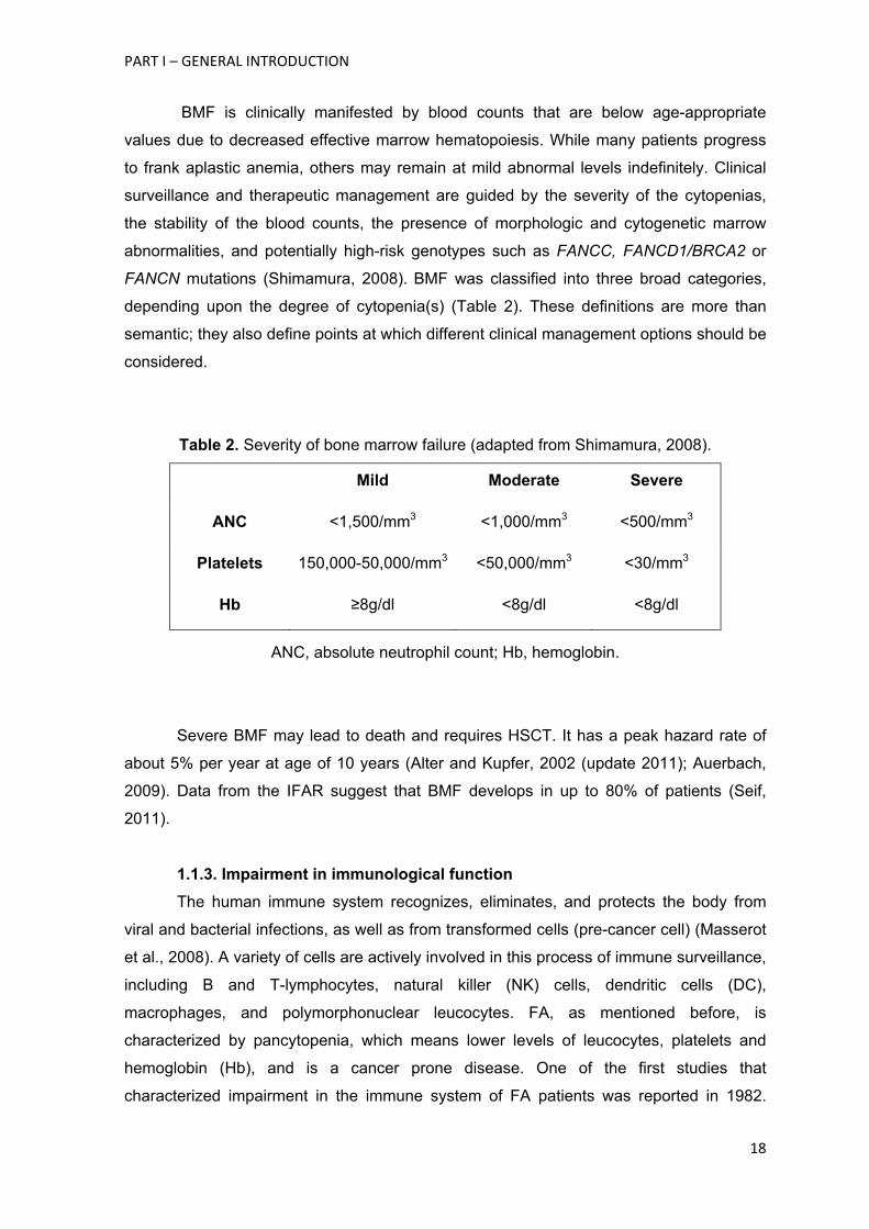
BMF is clinically manifested by blood counts that are below age-appropriate
values due to decreased effective marrow hematopoiesis. While many patients progress
to frank aplastic anemia, others may remain at mild abnormal levels indefinitely. Clinical
surveillance and therapeutic management are guided by the severity of the cytopenias,
the stability of the blood counts, the presence of morphologic and cytogenetic marrow
abnormalities, and potentially high-risk genotypes such as FANCC, FANCD1/BRCA2 or
FANCN mutations (Shimamura, 2008). BMF was classified into three broad categories,
depending upon the degree of cytopenia(s) (Table 2). These definitions are more than
semantic; they also define points at which different clinical management options should be
considered.
Table 2. Severity of bone marrow failure (adapted from Shimamura, 2008).
Mild Moderate Severe
ANC <1,500/mm3 <1,000/mm3 <500/mm3
Platelets 150,000-50,000/mm3 <50,000/mm3 <30/mm3
Hb ≥8g/dl <8g/dl <8g/dl
ANC, absolute neutrophil count; Hb, hemoglobin.
Severe BMF may lead to death and requires HSCT. It has a peak hazard rate of
about 5% per year at age of 10 years (Alter and Kupfer, 2002 (update 2011); Auerbach,
2009). Data from the IFAR suggest that BMF develops in up to 80% of patients (Seif,
2011).
1.1.3. Impairment in immunological function
The human immune system recognizes, eliminates, and protects the body from
viral and bacterial infections, as well as from transformed cells (pre-cancer cell) (Masserot
et al., 2008). A variety of cells are actively involved in this process of immune surveillance,
including B and T-lymphocytes, natural killer (NK) cells, dendritic cells (DC),
macrophages, and polymorphonuclear leucocytes. FA, as mentioned before, is
characterized by pancytopenia, which means lower levels of leucocytes, platelets and
hemoglobin (Hb), and is a cancer prone disease. One of the first studies that
characterized impairment in the immune system of FA patients was reported in 1982.
PART I – GENERAL INTRODUCTION
19
Hersey and co-workers, (Hersey et al., 1982) found that the immune function of one FA
patient revealed selective defects in NK cell activity, which is known to be important in
surveillance against tumors. This case report suggested that the absence of NK activity
was secondary to a defect in interferon release from lymphocytes on exposure to tumor
antigens. They considered that these defects may be an important predisposing factor in
the development of malignancy, not only in this patient but possibly other patients with FA.
Lebbé and co-workers (Lebbé et al., 1993) reported the same results in another patient,
where NK activity was undetectable even with a developing carcinoma.
Another study revealed that both lymphoblasts and fibroblasts from FA patients
demonstrated a reduction in interleukin (IL) 6 production. IL-6 is an interleukin that acts as
both a proinflammatory and anti-inflammatory cytokine. It is secreted by T cells and
macrophages to stimulate immune response. This study showed that this lymphokine is
not induced by tumor necrosis factors α and β (TNF-α and TNF-β) in FA cells, as is the
case in normal cells. It was suggested that the observed deficiency in IL-6 production may
account for one of the major characteristics of FA disease, the defect in differentiation of
the hematopoietic system (Rosselli et al., 1992). The same group also showed that, in
comparison to normal cells, TNF-α is overproduced by FA lymphoblasts from the four
genetic complementation groups A, B, C and D. Indeed, up to an eight-fold increase in
TNF-α is observed in the growth medium of FA cells. Moreover, addition of anti-TNF-α
antibodies partially corrects the FA hypersensitivity to MMC. Treatment of FA cells with IL-
6, which partially restored an almost normal sensitivity to MMC, also reduced the TNF-α
overproduction in FA lymphoblasts (Rosselli et al., 1994). With this study, the authors
concluded that abnormal TNF-α production seems to be associated with the FA genetic
background, which was later confirmed by authors of the same group (Briot et al., 2008).
One important activity of this cytokine is its cytotoxic/cytostatic effect on normal and
cancer cells (Sugarman et al., 1985). Moreover, TNF-α enhances intracellular and
extracellular superoxide anion (O2•-), production, and can induce DNA breakage and cell
death (Rubin et al., 1988; Yamauchi et al., 1989). This protein is of interest in the context
of the FA phenotype, not only for its relation with IL-6 expression, but also for the large
spectrum of its biologic activities (Schindler et al., 1990), which are completely
deregulated in FA cells. Further investigations using FA mouse models revealed that
Fancc(-/-) mice underwent excess inflammatory response, as a result of hematopoietic
suppression, that was corrected by wild-type Fancc gene, suggesting a potential role of
the FANCC protein in innate immunity (Sejas et al., 2007). Fancc(-/-) mice challenged in
vivo with lipopolysaccharides (LPS) at doses that induce septic shock have increased
peripheral blood levels of inflammatory mediators (Sejas et al., 2007), although it remains
unknown what cell type(s) is responsible for this response.

PART I – GENERAL INTRODUCTION
20
As mentioned above, a number of clinical studies indicate that FA patients have
altered levels of circulating cytokines. In addition, it has been suggested that FA patients
may have an increased susceptibility to a variety of pathogens (Fagerlie and Bagby,
2006), although it is unclear whether this observation is a result of a subtle
immunodeficiency or secondary to leukopenia from evolving BMF. A study of Liu and co-
workers, (Liu et al., 2012) provides compelling evidence for a cell-autonomous defect in
Fancc(-/-) macrophages. Specifically, functions requiring dynamic cytoskeletal changes
are impaired, including adhesion, migration, and phagocytosis, as well as in vivo
inflammatory monocyte mobilization and recruitment. Macrophages are a primary line of
defense in the innate immune system (Rees, 2010). The biologic functions of
macrophages are complex, including elimination of pathogens via phagocytosis and
cytokine/chemokine production and repairing damaged tissues during inflammation
(Gordon and Taylor, 2005; Rees, 2010). Most of these functions require macrophages to
migrate to an inflammatory site, and these dysfunctions could explain the cytokine
deregulation and increased susceptibility to pathogens.
Recently it was performed a study that reported a cross-sectional immunological
assessment in 10 children with FA, representing the first attempt at a comprehensive
quantitative and functional evaluation of immune function (Myers et al., 2011). The study
reported a significant, novel, and previously unappreciated abnormality in cytotoxic T cell
function, despite normal quantitative evaluation, and significantly reduced NK cell number
and function in the majority of these children, this last result being confirmed by previous
studies. Additionally, Castello and co-workers (Castello et al., 1998) demonstrated a
decrease in CD4+ T lymphocytes, which was not detected in Myers’ study. Moreover,
Myers’s cohort of patients demonstrated a quantitative abnormality in the B cell
compartment, with significantly lower absolute number of B cells relative to age-based
normal. The phenotype of the remaining B-cells was nearly identical in these patients,
showing reduced percentage of CD5 and CD10 expressing B cells. This particular B-cell
phenotype would be expected in adults, especially at advanced age (Bleesing, 2004). As
B and NK cells mature in the bone marrow, they may probably be most affected as BMF
develops (Myers et al., 2011). Figure 1 resumes the information known till now about
impairment in immunological system in FA patients and possible relation with FA
phenotype.

PART I – GENERAL INTRODUCTION
21
Figure 1. Impairment in immunological function of Fanconi anemia patients. Possible interactions among immunological cells, cytokines and Fanconi anemia phenotype (original figure). Fanconi anemia patients show an impairment in immunological system, characterized by low absolute number of B lymphocytes, abnormal function of T lymphocytes, reduced number of NK cells, cytoskeletal abnormalities in macrophages, low levels of IL-6 and overproduction of TNF-α. In turn, abnormal function of T lymphocytes, reduced number of NK cells and overproduction of TNF-α increase the probability of cancer cells proliferation. The typical deregulation of IL-6 can be aggravated by the abnormal function of T lymphocyte and macrophages, the cells that produce this cytokine. Additionally, overproduction of TNF-α could be regulated by IL-6 activity, but this event apparently doesn’t occur. Therefore, with abnormal macrophages and IL-6 production, FA patients have high susceptibility to pathogens and, thus, to infections. In BMF conditions, B cells and NK cells are more affected, as they mature in bone marrow, so their number and activity keep impaired. BMF, bone marrow failure; IL-6, interleukin 6; NK, natural killer; TNF-α, tumor necrosis factor alpha.
1.1.4. Cancer susceptibility
In addition to the early onset of BMF, FA is a cancer prone disorder, being the one
with higher risk of cancer development among BMF syndromes. Hematologic
malignancies are the first to appear and approximately 10% of patients develop leukemia
(Kutler et al., 2003). Over 90% of these hematologic malignancies and pre-malignancies
are myeloid in origin, representing a 600-fold increase in risk of AML and more than a

PART I – GENERAL INTRODUCTION
22
5,000-fold risk increase for MDS compared to the general population (Shimamura and
Alter, 2010). In a review of the literature reporting clinical cases of patients with FA, 9%
developed AML and 7% developed MDS (Alter et al., 2003). The median age of leukemia
diagnosis in FA patients is between 11 and 14 years, with almost all cases arising before
25 years old (Alter et al., 2003; Rosenberg et al., 2003).
Table 3. Malignancies in FA cases cited in the literature between 1927 and 2001 (adapted from Alter, 2003).
Characteristics All Leukemia MDS* Solid tumors Liver tumors
No. of cases 1301 116 89 68 37
% of total 100 8.9 6.8 5.3 2.8
Male:female 711:578 70:46 46:42 23:45† 22:15
Ratio 1.2 1.5 1.1 0.5† 1.5
FA diagnosis age (yrs)
Mean 8.3 10.1† 11.3† 12.7† 9.2
Median 7 8.6 9.3 9 7
Range 0-48 0.13-28 0.2-43 0-44 3-48
Complications age (yrs)
Mean _ 14.5 15.7 22.6† 15.7
Median _ 14 14 25.5 13
Range _ 0.13-29 1.8-43 0.2-45 6-48
No. of report deceased 488 84 44 41 30
% of report deceased 38 72 51 61 81
Estimate median
survival age (yrs) 20 16 21 31† 16
Age on general
population (yrs) 68 68
_ 47-68 68
* Includes 13 patients who subsequently developed leukemia; † P<0.01 compared with patients without that complication.

PART I – GENERAL INTRODUCTION
23
Hematologic malignancies are not the only cause of early death in patients with
FA. Nonhematologic malignancies are especially striking in these patients. They are
unusually young when they develop cancer and the incidence of the malignancy probably
would be considerably higher if patients had a longer life expectancy. Most of the
nonhematologic tumors in FA patients are solid tumors, especially SCC of the head and
neck and anogenital regions. The risk of head and neck SCC is 1000-fold greater in
patients with FA than that of the general population, and also occurs at an earlier age
(Myers et al., 2011). Epidemiologic analyses strongly suggest that solid tumors will
become the predominant clinical problem of post-transplanted FA patients. HSCT is
becoming available for a growing number of patients, because of an increased pool of
alternative donor options and new transplant protocols. The result is an improved
probability of survival to an age when the incidence of solid tumors begins to increase
(Auerbach, 2009). There is also a probably of development of liver tumors, especially in
patients receiving androgen treatment for BMF. This information is summarized in Table
3.
Interestingly, patients with no congenital anomalies have the highest risk of AML
(cumulative incidence 23.7%) (Rosenberg et al., 2008). Significant dysmorphia is more
closely associated with BMF and with a much lower total cancer cumulative risk (1.4%),
although more seriously affected children may die of BMF before attaining their full cancer
risk (Rosenberg et al., 2008). Importantly, in approximately 25% of patients, FA is
identified only after the leukemia diagnosis as a result of associated complex cancer-
related cytogenetic aberrations or excessive therapy-related toxicity (Alter, 2007; Gyger et
al., 1989).
1.2. Fanconi anemia genotype
Based on somatic cell fusion studies, at least 15 complementation FA groups have
been identified each one with a corresponding gene. Each of these genes, when
biallelically mutated, causes FA. Gene denomination and chromosome location, as well as
their proportion among FA patients are summarized in Figure 2 and Table 4 based on the
work of Alter and Kupfer (Alter and Kupfer, 2002 (update 2011)) and Green and Kupfer
(Green and Kupfer, 2009).

PART
TablGree
Com
T I – GENERA
e 4. The 15en and Kupf
mplementatio
group
FA-A
FA-B
FA-C
FA-D1
FA-D2
FA-E
FA-F
FA-G
L INTRODUC
5 FA genesfer, 2009).
on Respo
ge
FAN
FAN
FAN
BR
FAN
FAN
FAN
XR
CTION
s and their
onsible
ene
C
NCA
NCB
NCC
RCA2
NCD2
NCE
NCF
CC9
location (a
Chromosom
location
16q24.3
Xp22.31
9q22.3
13q12.13
3p25.3
6p21-22
11p15
9q13
dapted from
me
% o
attribu
mutatio
ge
60%
~2
~1
~3
~3
~3
~2
~1
Figure 2. frequencycomplem(genes). Fand FANCthe most cpopulationfrequencyconsiderinethnic gro
m Alter and
of FA
table to
on in this
ene
- 70% 1
2% (M
4%
3%
3%
3%
2%
0%
Relative y of the FA
mentation gFANCA, FACG mutationcommon in n: however,y can be varng the race oup.
d Kupfer, 20
Referen
(Apostolou
996; Lo Te
al., 199
Meetei et a
(Strathdee
1992
(Howlett e
2002
(Timmers
2001
(de Winter
2000
(de Winter
2000
(de Winter
2000
24
A roups
ANCC ns are general the riable and
011 and
nces
u et al.,
n Foe et
96)
l., 2004)
e et al.,
)
et al.,
)
et al.,
)
r et al.,
)
r et al.,
)
r et al.,
)

PART I – GENERAL INTRODUCTION
25
FA-I KIAA1794 15q25-26 ~1%
(Dorsman et al.,
2007; Sims et al.,
2007;
Smogorzewska et
al., 2007)
FA-J BRIP1/BACH1 17q22-24 ~2%
(Levitus et al., 2005;
Levran et al., 2005;
Litman et al., 2005)
FA-L PHF9 2p16.1 ~0.2% (Meetei et al., 2003)
FA-M FANCM 14q21.3 ~0.2% (Meetei et al., 2005)
FA-N PALB2 16p12 ~0.7% (Reid et al., 2007;
Xia et al., 2007)
FA-O RAD51C 17q22 ~0.2% (Vaz et al., 2010)
FA-P SLX4 16p13.3 ~0.2%
(Kim et al., 2011;
Stoepker et al.,
2011)
These genes account for over 95% of all known FA patients. Some patients do not
appear to have mutations in any of these 15 genes, so it can be anticipated that additional
FA genes will be discovered in the future.
1.2.1. The FA pathway
FA genes and proteins function together in a common pathway, involved in DNA
repair and in the maintenance of genomic stability (Bogliolo et al., 2002b). The encoded
proteins for each gene can be subdivided into three groups: (1) eight proteins that make
up the core complex of the upstream FA proteins, FANCA, FANCB, FANCC, FANCE,
FANCF, FANCG, FANCL and FANCM; (2) the FANCD2 and FANCI proteins, which
compose the ID complex; and (3) the downstream effector proteins, FANCD1, FANCJ,
FANCN, and presumably FANCO and FANCP (Kim et al., 2011; Moldovan and D'Andrea,
2009; Vaz et al., 2010).

PART I – GENERAL INTRODUCTION
26
Figure 3. Schematic representation of the FA pathway and associated proteins in response to DNA damage (original figure). A cascade of events starts with DNA damage, being activated the FA core complex, which in turn activates de monoubiquitination of ID complex. These events activate other FA proteins, culminating in the rearrangement of the damage through different repair processes.
In a simple manner and summarizing all this information (Figure 3) the eight
upstream FA proteins together with FAAP100 and FAAP24 form a complex with ubiquitin
ligase activity, termed the FA core complex. This complex is required for
monoubiquitination of FANCD2 and FANCI in response to DNA interstrand crosslink (ICL)
lesions during replication in S phase. FANCD2 and FANCI thereafter localize to DNA
repair foci together with FA effector proteins and other proteins. BRCA1 is required for
FANCD2 foci formation in response to DNA damage. FANCC, FANCE, and FANCG also
form nuclear foci and co-localize with FANCD2. All these factors are required for cellular
resistance to interstrand DNA crosslinking agents, through homologous recombination,
trans-lesion synthesis and some other unknown mechanisms. A few other gene products
were found to be associated with the FA protein complexes and are required for FA
activation. Thus, it is presumed that their inactivation would lead to FA; however, patients

PART I – GENERAL INTRODUCTION
27
with such mutations have not yet been described (Hucl and Gallmeier, 2011; Moldovan
and D'Andrea, 2009).
Interestingly, many of the FA proteins contain no recognizable motifs. Therefore,
discovering their contributions to the FA pathway and the main function of the FA pathway
will be an important challenging in the future (Green and Kupfer, 2009).
1.3. Diagnostic tests for Fanconi anemia
Considering the high genetic FA variability and the vast clinical phenotype, with a
great overlap with other BMF syndromes, and the particularity that 25% of FA patients are
phenotypically normal, a rapid and correct clinical diagnosis is difficult and may be
delayed or even missed. Fortunately more accurate diagnostic methods were developed,
the most important being the chromosomal fragility test.
1.3.1. The chromosomal fragility test
Schroeder and co-workers were the first to report that FA is a CI syndrome
(Schroeder et al., 1964). They first suggested the use of spontaneous chromosomal
breakage as a cellular marker for FA, but subsequent studies of CI in more FA patients
showed these findings to be inconsistent. Years later, Auerbach and co-workers
evidenced that FA cells have an unique hypersensitivity to the clastogenic (chromosome
breaking) effect of cross-linking agents and so this characteristic became a reliable
cellular marker for the diagnosis of FA (Auerbach et al., 1981). DEB and MMC are the
most widely used agents for the diagnosis, but DEB demonstrated to be the one that
proportionated more accuracy.
The chromosomal fragility test with DEB is the cytogenetic method for excellence
of FA diagnosis (Auerbach et al., 1989). Peripheral blood lymphocytes are cultured in the
presence and absence of DEB and a minimum of 50 cells arrested in metaphase are
scored and analyzed for chromosomal breakage (the most used parameter is the number
of breaks per cell) and formation of tri- and tetraradial figures. These figures are the
hallmark of FA diagnosis, since this parameter is the only one that can differentiate FA
from the other CI syndromes. Results are compared with those of normal control cells.
Normal cells are able to correct most of the damage and are not severely affected by
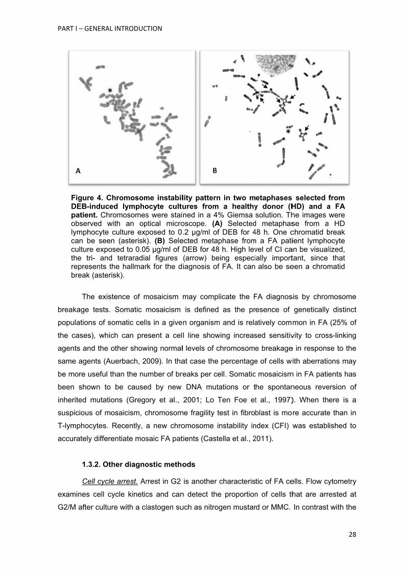
DEB, whereas FA cells show marked chromosome breakage, as can be seen in Figure 4.
This test can also be performed prenatally on cells from chorionic villi or amniotic fluid
(Auerbach et al., 1981).
PART
FDpolycctrb
brea
popu
the c
agen
same
be m
been
inher
susp
T-lym
accu
exam
G2/M
T I – GENERA
Figure 4. CDEB-inducepatient. Chobserved wymphocyte can be seeculture expothe tri- andrepresents tbreak (aster
The exis
kage tests.
ulations of s
cases), whi
nts and the
e agents (A
more useful
n shown to
rited mutati
picious of m
mphocytes.
urately differ
1.3.2. Ot
Cell cycl
mines cell c
M after cultu
L INTRODUC
Chromosomed lymphoromosomes
with an opculture exp
en (asteriskosed to 0.0d tetraradiathe hallmarrisk).
stence of m
. Somatic
somatic cell
ich can pre
other show
Auerbach, 2
than the nu
o be cause
ions (Grego
mosaicism, c
Recently,
rentiate mos
ther diagno
le arrest. Ar
cycle kinetic
ure with a cl
CTION
me instabilocyte cultus were staiptical microposed to 0
k). (B) Sele5 µg/ml of Dal figures rk for the d
mosaicism
mosaicism
ls in a give
esent a cel
wing normal
009). In tha
umber of bre
ed by new
ory et al.,
chromosom
a new chro
saic FA pat
ostic metho
rrest in G2
cs and can
lastogen su
lity patternures from ned in a 4%oscope. (A.2 µg/ml of
ected metapDEB for 48(arrow) beiagnosis of
may compl
is defined
n organism
l line show
l levels of c
at case the
eaks per ce
DNA muta
2001; Lo T
me fragility t
omosome i
tients (Caste
ods
is another
n detect the
uch as nitrog
n in two ma healthy
% Giemsa A) Selectedf DEB for 4phase from h. High lev
eing especif FA. It can
licate the F
as the pr
m and is rela
wing increas
chromosome
percentage
ell. Somatic
ations or t
Ten Foe et
test in fibrob
nstability in
ella et al., 2
characteris
e proportion
gen mustar
etaphasesy donor (Hsolution. Th
d metaphas48 h. One cm a FA pativel of CI caally importalso be se
FA diagnos
esence of
atively com
sed sensitiv
e breakage
e of cells w
c mosaicism
he spontan
t al., 1997)
blast is mo
ndex (CFI) w
2011).
tic of FA ce
n of cells th
rd or MMC.
s selected HD) and ahe images se from achromatid bient lympho
an be visualtant, since een a chrom
sis by chrom
genetically
mon in FA
vity to cros
e in respons
with aberratio
m in FA patie
neous reve
). When th
ore accurate
was establ
ells. Flow cy
hat are arr
In contrast
28
from a FA were HD
break ocyte lized,
that matid
mosome
y distinct
(25% of
ss-linking
se to the
ons may
ents has
ersion of
ere is a
e than in
ished to
ytometry
ested at
with the

PART I – GENERAL INTRODUCTION
29
100 cells examined microscopically for chromosomal aberrations, flow cytometry can have
the advantage of examining thousands of cells and being less labor-intensive and
subjective. However, it requires sophisticated instrumentation. This test is usually done in
a specialized laboratory and is not used as widely as the chromosome breakage assay.
Flow cytometry may give a false negative result in FA patients with MDS or AML (Alter,
2008).
Immunoblot assay of FANCD2 protein monoubiquitination. Following DNA
damage, the complex of upstream FA gene products (A, B, C, E, F, G, L, M) leads to
ubiquitination of the product of FANCD2, forming a longer protein (D2-L), which can be
distinguished from the shorter non-ubiquitinated form (D2-S) on a Western blot with a D2-
specific antibody. This relatively inexpensive assay may be useful for screening patients
for whom FA is in the differential diagnosis, such as those with radial ray anomalies, short
stature, and hypogonadism or café au lait spots or for population-based FA incidence
studies; however, it is usually a limited tool. FA patients whose gene defect is downstream
of FANCD2 (FANCD1, FANCI, FANCJ, FANCN, FANCP and FANCO) will not be detected
with a D2 Western blot, as well as mosaic individuals (Alter, 2008).
Complementation analysis. Patient lymphocytes, EBV (Epstein-Barr virus)-
lymphoblasts or fibroblasts can be cultured with retroviruses that introduce known normal
FANC genes into the patient’s cells, leading to correction of the FA cellular phenotype
(Alter, 2008).
Mutation testing. Determination of the specific mutation in FA genes is complicated
and is done in laboratories with specific expertise. It requires sophisticated methods and
involves DNA amplification, sequencing and detection of large deletions. Many
laboratories rely on knowing the complementation group before sequencing, while in
some contexts targeted sequencing of candidate genes is more appropriate. One center
goes directly to gene sequencing for patients with a positive DEB test: FANCA by
multiplex ligation-dependent probe amplification (MLPA) for large deletions and full
sequencing; FANCB by MLPA and full sequencing, if indicated; FANCC, E, F, G by
denaturing high performance liquid chromatography (DHPLC) and sequencing; FANCD2
by Western blot; FANCD2 sequencing if D2 bands are absent; FANCL and FANCM
sequencing if only D2-S is seen; FANCD1/BRCA2 sequencing, if indicated; FANCJ/BRIP1
and FANCN/PALB2 sequencing; and finally NBS1 and ESCO2 sequencing for Nijmegen
breakage and Roberts syndromes. Mutation testing is used to confirm known cases and
for family studies to determine affected or carrier status. Genetic counseling should be
included in these processes (Alter, 2008).

PART I – GENERAL INTRODUCTION
30
1.4. Fanconi anemia treatments
Hematologic complications represent a major problem to FA patients and are a major
cause of death. Additionally, SCC is another dramatic problem, especially in adult
patients. Some therapies are available, but no one can avoid the drastic problem of CI,
which is a common feature to all FA complications.
Hematopoietic stem cells transplantation (HSCT). Also known as BMT is the only
curative therapy for the hematologic manifestations of FA, including aplastic anemia, MDS
and AML, although it does not cure other non-hematopoietic complications. Donor stem
cells may be obtained from bone marrow, peripheral blood or cord blood. Ideally the
HSTC is performed prior the onset of MDS/AML and before multiple transfusions are
given for hematopoietic support (MacMillan and Wagner, 2010). However, issues
regarding timing of transplant are complicated by the up-front risk of transplant-related
mortality and the unknown long-term side effects of transplant. Since individuals with FA
are extremely sensitive to the toxicity of the usual chemotherapy and radiation regimens
used in preparation for BMT, reduced doses are typically used. Unfortunately, individuals
whose hematologic manifestations have been successfully treated with HSCT appear to
be at an increased risk for solid tumors, particular tongue SCC. In a study of Rosenberg
and co-workers, (Rosenberg et al., 2005) the risk of developed SCC was increased
fourfold and the median age of onset was 16 years younger than in persons with FA who
were not transplanted.
Androgen administration. Androgens have been widely used for the treatment of
cytopenias in FA. The effects of androgens are most pronounced in the red cells and
platelets, but neutrophil counts may also improve (Diamond and Shahidi, 1967). The
major effect of androgen therapy is to increase Hb levels, though it can also improve the
platelet count. Since there is no evidence that androgens can forestall BMF, treatment is
initiated when cytopenias drop to clinically significant levels but before the marrow
becomes completely devoid of hematopoietic stem cells for androgens to stimulate. The
mechanism(s) whereby androgens raise blood counts is currently unclear. The
advantages of androgens include the low risk of therapy-related mortality and the long
history of experience with their use. The major potential side effects associated with
androgen therapy include liver toxicity reflected as elevated liver enzymes, cholestasis,
peliosis hepatis and hepatic tumors (Shimamura and Alter, 2010). The standard
recommended androgen is oxymetholone, although danazol and oxandrolone have also
been used. About half of all treated patients will respond to androgen therapy, and a

PART I – GENERAL INTRODUCTION
31
subset of those who initially respond may become refractory over time. An additional
significant risk is that androgens do not prevent progression to AML that, once developed,
creates a significantly higher transplant risk (Shimamura, 2008).
Cytokines. Studies have demonstrated that granulocyte colony-stimulating factor (G-
CSF) or granulocyte macrophage colony-stimulating factor (GM-CSF) can improve the
neutrophil counts in FA patients (Guinan et al., 1994; Rackoff et al., 1996). Treatment with
G-CSF or GM-CSF should be considered if the neutropenia is associated with recurrent or
serious infections, particularly if the neutrophil counts persistently fall below 500/mm3 or
fail to rise in response to infection. A few patients have also shown improvements in Hb or
platelet counts during G-CSF or GM-CSF therapy. No comparative trials of G-versus GM-
CSF are available in FA patients. Hematopoietic growth factors should be administered
cautiously or not at all in the setting of a clonal cytogenetic bone marrow abnormality
(Alter and Kupfer, 2002 (update 2011); Shimamura, 2008), in order to prevent proliferation
of the abnormal clone that may lead to cancer development.
2. Diepoxybutane as a golden standard test for Fanconi anemia diagnosis
and as an oxidative stress inducer – Implications on Fanconi anemia
research
2.1. DEB: why is it a golden standard test for Fanconi anemia
diagnosis?
DEB (1,2:3,4 diepoxybutane) is a genotoxic metabolite of 1,3-
butadiene produced by further epoxidation of 1,2-epoxybutane
(Vlachodimitropoulos et al., 1997). It is also known to be a
carcinogenic agent (Boogaard and Bond, 1996) and a highly
reactive specie due to its chemical structure (Figure 5).
DEB is a bifunctional alkylating agent that can drastically
damage DNA, specially causing DNA ICL (Moldovan and
D'Andrea, 2009). ICLs are formed when the bifunctional
alkylating molecule reacts with bases on opposing strands of
the DNA double helix forming covalent links (Figure 6).
Figure 5. Chemical structure of DEB, showing the typical epoxide rings.

PART
and r
quan
clinic
Heal
affec
unda
2
C
comp
activ
prod
clarif
prote
capa
for its
by gl
mech
is wi
detox
expre
to DE
perfo
T I – GENERA
These links
recombinat
ntification of
cal diagnos
thy cells a
cted by the
amaged hom
2.2. DEB
Cellular an
pletely unde
vity exhibitin
uce intrastr
fied. Beside
ein sulfhydr
ability to pro
s higher tox
lutathione S
hanism of t
idely ackno
xification o
ession of g
EB toxicity
ormed by B
L INTRODUC
s prevent D
ion (Pang a
f chromoso
sis of the F
re capable
crosslinking
mologous s
cytotoxic
nd molecula
erstood. As
ng, especial
rand DNA c
es, DEB-ind
ryl (SH) g
oduce DNA-
xicity too. D
S-transferas
oxicity (Ahm
owledged th
of DEB an
lutathione S
(Kligerman
Boogaard a
CTION
DNA unwind
and Andrea
mal abnorm
FA, being t
to repair
g moiety re
equence (S
city: biotra
ar mechan
it was men
ly, ICLs, alt
cross-links (
duced cytot
roups (Loe
-protein cro
DEB can for
se (GST) (B
med et al., 2
hat GSH-ba
nd its meta
S-transferas
et al., 1999
nd co-work
ding, thereb
ssen, 2009
malities indu
the most s
ICL, where
enders error
Shen and Li
ansformat
nisms medi
ntioned prev
though it wa
(Millard et a
toxicity has
ecken and
oss-links (M
rm glutathio
Boogaard e
2010; Bogl
ased pathw
abolites. F
se T1 (GST
9; Vlachodi
kers contrib
by blocking
; Shen and
uced by this
striking cell
eas in FA c
r-free repai
, 2010).
ion as a p
ating DEB
viously, DE
as suggeste
al., 2005). T
s been rela
Guengeric
ichaelson-R
one (GSH)
et al., 1996)
iolo et al., 2
ways are inv
Furthermore
TT1) gene h
mitropoulos
buted for th
Figure 6formed cross-linkBifunctionform moaffect a sithese comadducts nucleotidestrand or to generaand interrespectiveAnderson
DNA replic
Li, 2010). C
s chemical h
ular hallma
cells the tw
r unfeasible
otential ca
-induced c
EB has a bif
ed that it ha
This sugges
ated to its a
ch, 2007),
Richie et al.
conjugates
), this being
2002b; Pag
volved in th
, it is als
has been re
s et al., 199
his assumpt
6. DNA aby inte
king nal compouono-adductingle nucleompounds c
affect es in thetwo paired
ate DNA intrstrand croely (from P, 2009).
cation, tran
Cellular tox
have been
ark of the
wo opposin
e in the abs
ause
cytotoxicity
functional a
as also cap
stion remai
ability to re
which exp
, 2010), acc
in reaction
g another im
ano et al.,
he metabol
so recogniz
elated to re
97). Pioneer
tion, demon
32
adducts erstrand agents. nds can ts that otide. Or
can form two
e same strands trastrand osslinks, ang and
scription
xicity and
used for
disease.
g bases
sence of
are not
alkylating
ability to
ns to be
eact with
lains its
counting
ns driven
mportant
2005). It
ism and
zed that
esistance
r studies
nstrating

PART I – GENERAL INTRODUCTION
33
that human, rat and mouse liver cytosolic fractions were able to conjugate [3H]glutathione
with DEB, in a concentration-dependent manner and further confirmed this pathway in rat
and mouse lung cytosolic fractions (Boogaard et al., 1996). Corroborating this
assumption, GSH depletion was later reported to increase DEB-induced cytotoxicity on
mouse germ cells (Spanò et al., 1998).
DEB was also shown to deplete GSH in sea urchin embryos (Korkina et al., 2000).
However, the depletion of GSH after exposure to DEB, in mammalian cells, hadn’t been
demonstrated.
Figure 7. Proposed scheme for the detoxification reaction of DEB (Boogaard and Bond 1996). DEB can be conjugated with glutathione through glutathione S-transferase (GST) or can be subject to hydrolysis via epoxide hydrolase (EH).
Since DEB has a particular structure, 2 epoxide rings, its metabolism may be also
related with epoxide hydrolase (EH), mainly microsomal EH (mEH), which is implicated in
the metabolism of the majority of xenobiotics where epoxidation is involved (Boogaard
and Bond, 1996; Morisseau and Hammock, 2005). Although mEH and soluble EH (sEH)

PART I – GENERAL INTRODUCTION
34
are highly concentrated in the liver, they are also found in nearly all tissues that were
assayed for EH activity (Morisseau and Hammock, 2005), including lymphocytes.
Boogaard and Bond (Boogaard and Bond, 1996) suggested that DEB metabolism and
detoxification in humans are, in its majority, effectuated by EH enzymes in liver and lungs,
mainly by mEH, and that metabolites formed in this process are less toxic than
metabolites formed by DEB conjugation with GSH. The relative contribution of these EH
and the GSH to the toxic mechanism of DEB remains to be clarified (Figure 7).
Knowing that biotransformation can contribute to DEB-induced cytotoxicity a good
approach to better clarify this matter is to study the contribution of several biochemical
pathways for DEB-induced acute toxicity in human lymphocytes, using inhibitors of EHs,
inhibitors of protective enzymes as GST and determine the level of GSH depletion and
compounds that can counteract it, as performed in the present thesis.
2.3. DEB cytotoxicity: generation of reactive oxygen species and
oxidative stress
It has already been postulated that FA cell sensitivity to DEB should also reflect some
deficiency or deficiencies in coping with oxidative stress (OS) (Mukhopadhyay et al.,
2006). In fact, DEB toxicity has been associated with OS. First, the epoxide structure of
DEB implies redox-mediated catalysis in the rearrangement of oxygen bonds (Bartók and
Láng, 1980). Secondly, DEB-induced cytogenetic damage can be partially corrected by
low molecular weight antioxidants in lymphocytes from FA patients and FA heterozygotes,
namely all antioxidants that are known to be donors of SH groups) (Dallapiccola et al.,
1985) and by overexpression of thioredoxin gene (thioredoxin is an important intracellular
antioxidant and an effective regulator of redox sensitive gene expression) in FA fibroblasts
(Ruppitsch et al., 1998). These evidences indirectly show that DEB toxicity is, in some
way, associated with OS and that the GSH system has an important role in this toxicity, as
explained in the previous chapter.
From a more direct approach, the work of Korkina and co-workers (Korkina et al.,
2000) demonstrated that DEB induces high mortality and modulates catalase activity,
affecting hydrogen peroxide (H2O2) balance in sea-urchin embryos. Moreover, they found
a highly significant correlation of DEB-induced toxicity and oxygen levels. In the same
study model, Pagano and co-workers (Pagano et al., 2001) demonstrated that DEB
induces DNA oxidative damage, measured as 8-hydroxy-2’-deoxyguanosine (8-OHdG).
They observed a dose-dependent increase in 8-OHdG levels, which was significantly
correlated with developmental defects in sea urchin embryos. In addition, 8-OHdG is

PART I – GENERAL INTRODUCTION
35
recognized as an outcome of pro-oxidant state. Erexson and Tindall (Erexson and
Tindall, 2000b) in a study using fibroblast of big blue mouse and rat showed that
glutathione peroxidase (GPx) (which primary role is the protection of red blood cells –
RBC - against damage from reactive oxygen species - ROS) and RBC (that are known to
be a major source of GSH) may be important in the detoxification of DEB-induced DNA
damage. This suggests that DEB can generate ROS that, in turn, can either damage DNA
or produce H2O2. Altogether, these results gave further support to the body of evidence
associating DEB toxicity with OS, including DNA oxidative damage.
2.4. DEB cytotoxicity: relation with mitochondrial dysfunction
Gille and Joenje (Gille and Joenje, 1992) have shown that OS affects key
mitochondrial enzymes and lead to a decline in ATP production in mammalian cell
cultures. Carcinogens, and in particular alkylating agents, have long been known to
preferentially target mitochondrial DNA (mtDNA) (Backer and Weinstein, 1980; Backer
and Weinstein, 1982; Clarke et al., 1998; Wunderlich et al., 1972). The mtDNA can bind
20–600 times the amount of alkylating agent compared to genomic DNA. This difference
may in part be due to mtDNA not being protected by histone binding. Thus, it is
reasonable to suggest that DEB also damages mitochondria. A recent study reported that
DEB-induced cell death in human TK6 lymphoblasts exposed to low concentrations of
DEB was due to the occurrence of apoptosis, and not necrosis (Yadavilli and Muganda,
2004). Apoptosis in response to DEB exposure has also been observed in the big blue rat
cultured cells, mouse L929 cultured cells, as well as in human CD34+ bone marrow cells
(Brockmann et al., 2006; Erexson and Tindall, 2000a; Irons et al., 2000). The molecular
mechanisms by which DEB induces apoptosis was resolved by the study of Yadavilli and
co-workers (Yadavilli et al., 2007). This study reported that DEB-induced apoptosis in TK6
lymphoblasts involves activation and up-regulation of pro-apoptotic Bcl-2 proteins (Bax,
Bak), dissipation of mitochondrial membrane potential, depletion of mitochondrial
cytochrome c levels, and activation of initiator caspase 9, accompanied by activation of
executioner caspase 3 by caspase 9. The occurrence of these rate-limiting steps
implicates the activation and involvement of the mitochondrial apoptotic pathway in
mediating DEB-induced apoptosis in human lymphoblasts. These findings also point to
the involvement of an OS-mediated mechanism in the execution of DEB-induced
apoptosis in human lymphoblasts. Since the DEB-dependent generation of ROS is an
early event and since the activation of DEB-induced apoptotic signaling pathways (MAP
kinases and p53) is prevented by the ROS scavenger N-acetylcysteine (NAC), then the

PART I – GENERAL INTRODUCTION
36
DEB-dependent OS is likely to exert its effect on the apoptotic pathway at early times,
upstream of mitochondria (Yadavilli et al., 2007). Through this point of view is also
reasonable to suppose that DEB can induce ATP depletion in human lymphocytes, but
this information is still lacking.
The knowledge of DEB induced cytotoxicity is an important route to understand what
is falling in FA cells, principally related to OS induction. Studies with mitochondrial
protective agents may be very useful to clarify mitochondrial damage as well as studies
with compounds that can inhibit the activity of enzymes related with ROS production and
OS and studies of cell signaling pathways.
3. Oxidative stress in Fanconi anemia: from cells and molecules towards
prevention against chromosome instability
3.1. Pro-oxidant state in Fanconi anemia cells and patients
OS causes severe damage to membranes, proteins, and DNA and is regarded as an
important factor in the development of many chronic and degenerative aging diseases
(Packer, 1995). The role of OS in FA cells damage was described for the first time by
Nordenson (Nordenson, 1977) followed by Joenje and co-workers (Joenje et al., 1981).
Two years later, Joenje and Oostra (Joenje and Oostra, 1983) established a direct
correlation between high tensions of oxygen and increased damage in FA lymphocytes.
After these early discoveries, oxygen levels were found to modulate cell growth and cycle
in FA fibroblasts, pointing to toxicity at high oxygen tension, with accumulation in S and
G2/M phases compared to control cells (Schindler and Hoehn, 1988). Korkina and co-
workers (Korkina et al., 1992) also showed the overproduction of ROS by FA leukocytes,
resulting from activation of NADPH oxidase, decrease in the cellular antioxidant defense
systems, increase in cellular free iron levels and a deficiency in erythrocyte superoxide
dismutase (SOD). Another work suggested that the high susceptibility of FA
lymphoblastoid cells to oxidative DNA damage is possibly due to a decrease in catalase
activity (Takeuchi and Morimoto, 1993). In addition, a study reported by Ruppitsch and co-
workers (Ruppitsch et al., 1997) indicated that cytochrome P-450 enzymes, especially
CYP1A2, play a crucial role in ROS metabolism in FA cells that showed reduced oxygen
consumption and increased activity of the antioxidant phospholipid-hydroperoxide-
glutathione-peroxidase. This enzyme plays a key role in protecting cells from lipid
peroxidation. It is the only enzyme known to metabolize phospholipid hydroperoxides and,

PART
toget
mem
in FA
a slig
activ
I
(Figu
T
even
Figuits Overapopprogetranscellsboneoxygacutelight-Du e
T I – GENERA
ther with α
mbrane. In fa
A cells (Rup
ght inductio
vity is still no
It has been
ure 8) (Du e
The interac
nts as nucle
ure 8. The potential
rproduced ptotic signenitor act
sformation , which lead
e marrow; Bgen speciese myeloid -chain-enha
et al, 2008).
L INTRODUC
α-tocophero
act, malond
ppitsch et a
n of this enz
ot enough to
n reported t
et al., 2008)
ctions betwe
ar factor ka
proinflammrole i
TNF-α planal supprtivity, but of FA he
d to typical BMF, bone s; MDS, myleukemia;
ancer of ac
CTION
ol, is the o
dialdehyde,
l., 1997). E
zyme, like o
o counterac
that plasma
.
een TNF-α
appa-light-ch
matory cytin FA
ays a role ressing F
also promatopoieticphenotype marrow fail
yelodysplasNF-kB, nuctivated B c
nly known
a product
levated leve
other antiox
ct the increa
a from FA p
and its rece
hain-enhan
tokine TNFpathophysnot only
FA hematomoting lc stem/ prof FA patielure; ROS, ic syndromclear factorcells (Adapt
protection
of lipid pero
els of ROS
xidant enzym
ased produc
patients an
h
i
c
p
c
(
2
f
a
f
a
a
c
p
t
S
r
t
e
o
i
m
a
s
s
p
eptors, in p
cer of activ
F-α and siology. in pro-
topoietic eukemic rogenitor nts. BM, reactive
me; AML, r kappa-ted from
against R
oxide degra
in FA fibrob
mes. The re
ction of ROS
d, in a less
heterozygot
increased
clastogenic
peroxidation
cytokines
(Emerit, 20
2011). Exc
found in bo
and periph
from patie
anemia (Sc
1994) and
also found h
cytokine in
patients, ab
than in con
Shahidi, 19
reported th
FA fibrobla
than in nor
et al., 199
opened the
investigatio
major role
another cyt
since it is k
significantly
production
articular, en
ated B cells
ROS within
adation, is e
blasts may
esultant enz
S.
ser extent,
tes
levels
c factors
n produc
such as
007; Petrov
xcess TNF
one marrow
heral blood
ents with
chultz and
the same
higher leve
n plasma
bout 4 time
ntrols (Sch
993). Anoth
hat TNF-α
asts is 8-fold
rmal cells (
94). These
e way of a s
ons ascerta
e for TNF
tokines in F
known that t
y enhance
by phagocy
nhance intr
s (NF-kB) a
37
the cell
elevated
result in
zymatic
from FA
contains
of
(lipid
ts and
TNF-α)
ic et al.,
F-α was
w plasma
plasma
aplastic
Shahidi,
e group
ls of this
of FA
s higher
ultz and
her work
level in
d higher
(Rosselli
reports
series of
aining a
F-α and
FA cells,
they can
e ROS
ytic cells
acellular
ctivation

PART I – GENERAL INTRODUCTION
38
and transcriptional activity, which are enhanced in FA cells compared to normal cells. NF-
kB is a potent indicator for stress factors such as ROS and is capable to activate the
transcription of various genes that are involved in stress defense. Therefore it can be
postulated that a defect in the NF-kB system would thus limit the ability of the cell to
respond to OS (Ruppitsch et al., 1997). Moreover, TNF-α treatment induces FANCA and
FANCG via NF-kB (Macé et al., 2007).
Another marker of pro-oxidant state in FA cells is the excess accumulation 8-OHdG.
This excess was correlated with spontaneous chromosomal breaks (Degan et al., 1995;
Pagano et al., 2004). Another work suggested that FA cells, at least FA complementation
group A cells, have increased susceptibility to oxidative DNA damage possibly due to
decreased catalase activity, which indicates that catalase plays an important role in
protecting DNA from oxidative damage (Takeuchi and Morimoto, 1993). Zunino and co-
workers (Zunino et al., 2001), also showed an increased basal level of 8-OHdG in FA
patients from the complementation group C and E. Besides, the same group also
demonstrated that H2O2-induced 8-OHdG formation is higher in FA than in normal cells.
On the other hand, more recently, Castillo and co-workers (Castillo et al., 2011) concluded
that the FA pathway is redundant for the repair of this oxidative damage. Therefore, the
accumulation of 8-OHdG in FA cells reflects sustained overproduction of ROS rather than
defective processing of oxidized bases.
Beyond the direct information about an in vivo pro-oxidant state, it should be
remembered that the FA phenotype is characterized by some clinical features, or
complications, that find recognized mechanistic explanations on the grounds of redox
imbalances. Together, the available literature provides both direct and indirect evidence
that OS is at least a component of FA cellular and clinical phenotype.
3.2 . Redox-related functions of FANC proteins
3.2.1. FANCA, FANCC and FANCG: links with xenobiotic-processing pathway
The FANCC protein was found to be associated with redox-related activities,
namely interacting with enzymes involved with bioreductive action of xenobiotics. Kruyt
and co-workers (Kruyt et al., 1998) reported that FANCC functions in the cytoplasm, at a
step before DNA repair, and binds to NADPH cytochrome-P450 reductase, a microsomal
membrane protein involved in transferring electrons from NADPH to an isoenzyme of the
cytochrome P450 family, as well as to cytochrome c, in both transfected COS-1 and
normal murine liver cells. Cumming and co-workers (Cumming et al., 2001) also showed
that FANCC interacts with glutathione S-transferase P1-1 (GSTP1) and that

PART I – GENERAL INTRODUCTION
39
overexpression of both proteins in a myeloid progenitor cell line prevents apoptosis
following factor deprivation. GSTP1 is an enzyme that catalyzes the detoxification of
xenobiotics and by-products of OS by conjugation with GSH, and it is frequently
upregulated in neoplastic cells. Studies in animal models also showed a relation between
FANCC and OS. Hadjur and co-workers (Hadjur et al., 2001) found defective
hematopoiesis and hepatic steatosis in double knock-out mice with Fancc(-/-) and SOD1(-
/-). The authors suggested that the altered redox state likely present in Fancc(-/-) and
sod1(-/-) hematopoietic progenitors was responsible for an impairment of cell proliferation
and survival.
FANCG protein is localized both in the nucleus and cytoplasm. After treatment with
clastogenic agents it increases in cells, with predominant expression in the cytoplasm
(Futaki et al., 2002). FANCG was found to interact with cytochrome P450 2E1 (CYP2E1),
which is associated with the production of ROS and the bioactivation of carcinogens.
These results suggest that FANCG has an additional role in protection against oxidative
DNA damage. Additionally, Park and co-workers (Park et al., 2004) showed that FANCA
and FANCG are redox-sensitive proteins that are multimerized and/or form a nuclear
complex in response to OS/damage. Both FANCA and FANCG proteins exist as
monomers under non-oxidizing conditions, whereas they become multimers following
H2O2 treatment. Treatment of cells with oxidizing agent not only triggers the multimeric
complex of FANCA and FANCG in vivo but also induces the interaction between FANCA
and FANCG. Finally, in another study (Noll et al., 2002) mice were genetically modified
with a targeted mutation in FANCA and were crossed with FANCC disrupted animals,
revealing that FANCC and FANCA are part of a multi-protein nuclear FA complex with
identical function in cellular responses to DNA damage and germ cell survival. This group
showed that fibroblast cells and hematopoietic precursors from FANCA/FANCC double-
mutant mice were not more sensitive to MMC than those of both single mutants and that
FANCA/FANCC double mutants had no evidence for an additive phenotype at the cellular
or organism level.
A report by Reuter and co-workers (Reuter et al., 2003) identified 69 proteins
which have not previously been linked to the FA pathway as direct interactors of FANCA,
FANCC, or FANCG. Most of these proteins are associated with four functional classes
including transcription regulation (21 proteins), signaling (13 proteins), oxidative
metabolism (10 proteins), and intracellular transport (11 proteins). The potential
interactors found in this study point towards an additional involvement of the FA pathway
in four other major functional purposes, namely transcription, cell signaling, oxidative
metabolism, and cellular transport.

PART I – GENERAL INTRODUCTION
40
3.2.2. FANCD2 and FANCJ: multiple identities and redox-related roles
The FANCD2 protein features an unique role in the FA pathway, due to its
interactions in the activation of the FA core complex, in cooperation with FANCI and
following monoubiquitination (Smogorzewska et al., 2007). Mammalian forkhead members
of the class O (FOXO) transcription factors, are implicated in the regulation of diverse
physiologic processes, including cell-cycle arrest, apoptosis, DNA repair, stress
resistance, and metabolism (Brunet et al., 2004). Among these FOXO proteins, forkhead
transcription factor forkhead box O 3a (FOXO3a) functions as a major regulator of OS
(Huang and Tindall, 2011). It is known that FANCD2 interacts with the FOXO3a in
response to OS (Li et al., 2010). In a very recent study presented at the American Society
of Hematology Annual Meeting, in December 2011, it was shown that expression of
FOXO3a is associated with HbF levels in hereditary persistence of HbF. Thus, study
FANCD2-FOXO3a pathway in cells from FA patients, which have an increased level of
HbF, may be an important question to address and probably FOXO3a is over expressed
in FA cells. Another recognized interaction of FANCD2 is with ataxia telangiectasia
mutated protein (ATM) (Castillo et al., 2011). ATM has been suggested to function, at
least in part, in the cellular response to oxidative damage (Barzilai et al., 2002). Support
for this hypothesis comes from observations that ATM-deficient cells are unusually
sensitive to the toxic effects of H2O2, nitric oxide, and superoxide treatments as
determined by colony-forming efficiency assays (Barlow et al., 1999).
The FANCJ protein was recognized to coincide with two previously known BRCA1-
interaction entities, the DNA helicase BRIP1 (Levitus et al., 2005; Levran et al., 2005) and
the transcription factor BACH1 (Litman et al., 2005). This protein features a helicase
activity and ATPase activity (Cantor et al., 2004; Cantor et al., 2001). It may be noted that
ATPases are frequently involved in OS related mechanisms (Bigelow and Squier, 2005).
Thus, it might be suggested that the ATPase component of helicase activity plays a direct
role in redox-active functions of these DNA repair proteins. Before the recognition of its
identity with FANCJ, BACH1 was termed as a transcription repressor and found to
regulate redox-related activities, as heme oxygenase-1 (HO-1) while, in turn, increased
levels of HO-1 resulted in BACH1 inactivation (Kitamuro et al., 2003; Okada et al., 2010;
Reichard et al., 2007). Moreover, BACH1 was reported to form a p53-containing complex,
and was regulated by OS and HO-1; thus BACH1 was related to OS as a negative
regulator of p53 (Dohi et al., 2008).

PART I – GENERAL INTRODUCTION
41
3.3. Mitochondrial dysfunction in Fanconi anemia cells
Mitochondria contain about 1% of the cell’s DNA. These organelles account for more
than 90% of cellular oxygen consumption and are prone to DNA oxidative mutation
(Richter et al., 1988). Indeed, mitochondrial dysfunction, as a result of accumulative
oxidative lesions has been correlated with ageing (Shigenaga et al., 1994). It is also
known that mitochondrial dysfunctions ultimately lead to a defect in energy transduction,
with increased formation of ROS, which, by itself, can lead to lower cellular energy
charge, oxidative modification of DNA, protein and lipids. In addition, there is also a
possibility of a positive feedback cycle between ROS formation and mitochondrial
damage, exacerbating the processes of cellular dysfunction (Tarnopolsky, 2008). In a
preliminary study Liebetrau and co-workers (Liebetrau et al., 1997) have examined the
mtDNA profile in tissues of FA patients, using polymerase chain reaction (PCR) primer
pairs to encompass a 5 kb ‘hot spot’ region for deletion mutations seen in ageing and in
some mitochondrial diseases. Two of the seven studied patients showed evidence of the
common 5 kb deletion, another indication that mtDNA is involved in the mechanism of
oxidative sensitivity in FA cells.
More recently, direct evidence for mitochondrial dysfunction in FA cells was provided
by Mukhopadhyay and co-workers (Mukhopadhyay et al., 2006) who reported that
FANCG protein is found in mitochondria and interacts with the mitochondrial peroxidase,
peroxyredoxin 3 (PRDX3). Mitochondrial PRDX3 is an important cellular antioxidant that
regulates physiological levels of H2O2, leading to decreased cell growth while protecting
cells from the apoptosis-inducing effects of high H2O2 levels (Nonn et al., 2003). PRDX3
depletion results in the acceleration of apoptosis, with increased rates of mitochondrial
membrane potential collapse, cytochrome c release, and caspase activation (Chang et al.,
2004). PRDX3 was found to be deregulated in FA-G cells, which displayed distorted
mitochondrial structures, and mitochondrial extracts had a significant decrease in
thioredoxin-dependent peroxidase activity. In addition, overexpression of PRDX3
suppressed the sensitivity of FA-G cells to H2O2, and a decreased PRDX3 expression
increased the sensitivity to alkylating agents (Chang et al., 2004). Moreover, cells from
FA-A and FA-C subtypes also had PRDX3 cleavage and decrease peroxidase activity
(Mukhopadhyay et al., 2006).
Previous reports had provided evidence for mitochondrial dysfunction in FA cells
(Afanas'ev, 2006; Bogliolo et al., 2002a; Rousset et al., 2002). Paulin-Levasseur and co-
workers (Paulin-Levasseur et al., 1998) reported that mitochondria in FA cells have a
filamentous shape and form an interconnected cytoplasmic reticulum running in parallel
with both vimentin filaments and microtubules. This was unlike control (HeLa) cells, where

PART I – GENERAL INTRODUCTION
42
mitochondria appears as individual tubular compartments of variable length and are
closely associated with vimentin filaments. Bogliolo and co-workers (Bogliolo et al.,
2002a), tried to understand the intriguing relationships between apoptotic susceptibility
and alterations in redox state and energy metabolism, studying the response of FA cells to
a number of metabolic inhibitors known to interfere with ATP production and the
consequences of these treatments on proliferation and survival of FANCA and FANCC-
defective lymphoblastoid cells. They reported that FA cells, unlike control cells, were not
affected by treatments with rhodamine-1,2,3 (induces membrane depolarization, affects
the efficiency of electron transport and impairs the respiratory chain and oxidative
phosphorylation) and doxycycline (inhibits synthesis of mitochondrially encoded proteins
by 50% at every cell division), which result in acute ATP depletion and in a significant
enhancement of the fraction of cells undergoing apoptotic death. Additionally, apoptosis is
particularly sensitive to the redox state of the cells and is strictly associated with
alterations in mitochondrial functionality (Kannan and Jain, 2000). On the other hand, FA
cells were very sensitive to 2-deoxy-D-glucose and iodetic acid, two inhibitors of the
glycolytic metabolism. These findings suggested that FA cells are adapted to withstand
mitochondrial stress, while relying on glycolytic metabolism. Rousset and co-workers
(Rousset et al., 2002) reported an oxygen-dependent sensitivity of mitochondria in FA-A
fibroblasts. FA cells, following 8-methoxypsoralen photoreaction or ultraviolet A (UVA)
irradiation, underwent mitochondrial matrix densification, unlike control cells, which was
accompanied by some modifications in transmembrane potential. This effect was oxygen
dependent because it was more enhanced at 20% than at 5% oxygen tension. It is
important to note that UVA exposure leads to the formation of free radicals and that
psolaren acts through photobinding, by means of UVA excitation, to pyrimidine bases of
nucleic acids, to amino acids and proteins and to membrane constituents (Schmitt et al.,
1995). Moreover, psolaren photoreaction and UVA radiation are also responsible for the
induction of lipid peroxidation, which can damage membranes by preventing the
methylation of phospholipids (Caffieri et al., 1996), known to be implicated in the
adjustment of membrane fluidity and in enzyme activities associated with the cell
membranes (Kaneko et al., 1990).
The evidence for mitochondrial dysfunctions in FA-G, FA-A and FA-C cells are well
documented. Thus, it might be questioned if FA-J cells are involved in mitochondrial
dysfunction too. Since FANCJ is related with ATPase activity it is linked by definition to the
availability of ATP that is the major product of mitochondrial function. However, this is a
currently open question that needs to be addressed. Figure 9 summarizes the possible
interactions between FANC proteins, mitochondria and oxygen metabolism.

PART
FiguFANantiotelanFOXNF-kperox
T
can
patho
mitoc
preex
mean
meas
T I – GENERA
ure 9. ROS C proteinsoxidant enngiectasia mXO 3a, forkkB, Nucleaxyredoxin 3
The use of
be a goo
ogenesis c
chondrial cy
xisting clin
ns of thera
sures to avo
L INTRODUC
metaboliss, mitochonzymes (admutated; Cykhead box 3ar factor 3; ROS, rea
“mitochond
od approac
can also be
ytopathies,
ical experie
peutic mito
oid the pro-
CTION
sm in Fancndria, DNA
dapted andyp 2E1, cyt3a; GST, gkappa-light-ctive oxyge
drial nutrient
ch to eval
e a part o
such as ag
ence in ma
chondrial c
-oxidant sta
coni anemiA damage, modified
ytochrome Pglutathione -chain-enha
en species.
ts” as antio
luate mitoc
f a set of
ging (Pallar
anaging pa
cocktails ma
ate and dise
a cells. Po, redox strfrom PagaP450 2E1; S-transferaancer of
xidants to c
chondrial d
diseases
rdó et al., 2
atients with
ay provide u
ease progres
ossible interess signano et al, 2Cyt P450, se; HO-1,hactivated
counteract t
dysfunction
and condit
2010; Tarno
mitochond
useful back
ssion in FA
eractions baling prote2011). ATM
cytochromheme oxyge
B cells;
the ROS pro
in FA ce
tions assoc
opolsky, 200
drial cytopa
kground in
A patients.
43
between ins and
M, ataxia me P450; enase 1; PRDX3,
oduction
ells. FA
ciated to
08). The
thies by
planning

PART I – GENERAL INTRODUCTION
44
3.4. In vitro and in vivo experimental therapies – Antioxidant agents as
protectors of chromosomal instability
Some in vitro studies have been performed testing the use of antioxidants for
decreasing chromosome damage in FA cells. Dallapiccola and co-workers (Dallapiccola et
al., 1985) obtained a partial correction of CI in FA lymphocytes treated with several
antioxidants, namely L-cysteine, 2-mercaptoethanol, α-mercaptopropionylglycine and
GSH, all chemicals which are known to be donors of the SH group. Later, the same group
(Porfirio et al., 1989) extended this study to desferoxamine, an iron chelator, with 50%
reduction of the spontaneous chromosome breakage. Korkina and co-workers (Korkina et
al., 1992) suggested that hydroxyl radical (•OH) scavengers and chelators can be used for
the treatment of FA patients. Rutin, a nontoxic natural bioflavonoid (vitamin P), was found
to be an efficient inhibitor of luminol-dependent chemiluminescence (a technique that
measures the production of oxygen radicals) produced by FA leukocytes. After
administration of rutin to three FA patients they observed that it substantially decreased
oxygen radical production by leukocytes, diminished to some degree the amount of
chromosomal aberrations, and improved hematologic characteristics of patients. Pincheira
and co-workers (Pincheira et al., 2001) demonstrated that Vitamin E (α-tocopherol)
decreases the frequency of chromosomal damage in 50.8% and the duration of G2 in FA
lymphocytes. More recently, it was reported that honey can have a cytoprotective effect
against CI in FA cells, due to the antioxidant capacity of flavonoids and other polyphenols,
which are common constituents of honey (Mogib El-Dahtory and Yahia, 2011). 2 ml of a
solution of 10% of honey can abolish the induced chromosomal aberrations in FA cells.
The effect of these studies once more agrees with the assumption of oxygen sensitivity in
FA cells and the putative beneficial effect of antioxidant treatments.
Some in vivo studies were also performed in animal models. Zhang and co-workers
(Zhang et al., 2008) showed that dietary supplements with the antioxidant tempol, a
nitroxide antioxidant and a SOD mimetic, can delay the age of onset of epithelial tumors in
Fancd2(-/-) knock-out mice model. More recently, this group (Zhang et al., 2010) also
demonstrated that the antioxidant resveratrol maintains Fancd2(-/-) KSL cells in
quiescence, improves the marrow microenvironment, partially corrects the abnormal cell
cycle status, and significantly improves the spleen colony-forming capacity of Fancd2(-/-)
bone marrow cells.
In spite of the promising results from previous studies, no therapy using antioxidants,
megavitamins, or micronutrients has been shown to be effective in treatment of FA using
evidence-based criteria. Thus, further studies with new protective agents are required for

PART I – GENERAL INTRODUCTION
45
the development of drugs with high effectiveness. Ideally, the protective
agents/antioxidants should be already approved for human use or promptly bioavailable
by dietary administration (Pagano et al., In press; Pagano and Korkina, 2000). For a rare
disease like FA, insuperable obstacles to clinical chemoprevention trials may currently be
faced due to the scanty numbers of patients susceptible to be recruited, compromising the
best efforts in drawing adequate conclusions from too small scale clinical studies. As a
possible means for overcoming the restraint imposed by the scarcity of human patients,
and on ethical grounds, studies could be designed by utilizing knock-out mice from
different complementation groups (FANCA, FANCC, FANCD2 and FANCG). However,
animal models not always are a good approach, and in vitro studies with cells from FA
patients could sometimes be a better way. Testing antioxidants to counteract the initial
pro-oxidant state of FA cells or in cells of healthy donors (HD) subjected to the effects of
alkylating agents, should rely on the choice of the best appropriate antioxidants to be used
in animal models or even in clinical trials, with the ultimate goal of mitigating or delaying
FA clinical progression.

PART I – GENERAL INTRODUCTION
46

PART II – OBJECTIVES OF THE THESIS
47
PA
RT
II
- O
BJE
CT
IVE
S O
F T
HE
TH
ES
IS

PART II – OBJECTIVES OF THE THESIS
48

PART II – OBJECTIVES OF THE THESIS
49
Objectives of the Thesis
The present work was divided in two parts, performed in two different laboratories, in
which two complementary objectives were pursued.
The first objective, performed during the first two years, in the Laboratory of
Toxicology of Faculty of Pharmacy of University of Porto (FFUP), was the following:
Characterization of the acute toxicity of DEB in normal lymphocytes, by evaluation
of cell viability as well as GSH and ATP levels in an in vitro cellular suspension model.
Following the results obtained with the acute toxicity study, a mechanistic approach was
further applied by using:
Putative inhibitors of DEB bioactivation: cyclohexyl-3-dodecyl urea (CDU),
elaidamide, Zinc (Zn2+)
Antioxidant agents: NAC
Mitochondrial protective agents: acetyl-L-carnitine (ALC) and α-lipoic acid (α-
LA)
Inhibitors of putative protective enzymes and inhibitors of protein synthesis:
tannic acid, 3-amino-1,2,4-triazole (3-AT) and cycloheximide (CHX).
The global purpose of the first objective was to contribute for a better understanding
of DEB-induced toxicity to human lymphocytes, by studying the putative contribution of
biochemical pathways postulated to be involved in the reactivity of this compound. The
specific goal was to search for protective drugs that could turn out to be useful to prevent
spontaneous and DEB-induced CI in lymphocytes from FA patients.
The second objective, performed during the last two years, in the Laboratory of
Cytogenetics of Institute of Biomedical Sciences Abel Salazar (ICBAS), was the following:
Evaluation of the putative protective effect of previously selected drugs in
spontaneous and DEB-induced CI in lymphocytes cultures from FA patients and control
population.
Optimization of a cocktail with the compounds that demonstrated better results in
the decrease of CI in lymphocyte cultures from FA patients.
Since the available information points to an association of cellular phenotype and
clinical features in the occurrence of both cancer-proneness and OS in FA disorder, the
final goal was to find out an effective antioxidant prophylactic cocktail to be applied, in
vivo, in FA patients, with a consequent block or delay in their characteristic BMF and
predisposition to cancer development.

PART II – OBJECTIVES OF THE THESIS
50

PART III – ORIGINAL RESEARCH
51
PA
RT
III –
OR
IGIN
AL
RE
SE
AR
CH

PART III – ORIGINAL RESEARCH
52

PART III – ORIGINAL RESEARCH
53
CHAPTER I
Protective effect of acetyl-L-carnitine and α-lipoic acid against toxicity of diepoxybutane to human lymphocytes
Reprinted from Toxicology 289 (2011): 52-58
Copyright (2012) with kind permission from Elsevier

PART III – ORIGINAL RESEARCH
54

Toxicology 289 (2011) 52– 58
Contents lists available at ScienceDirect
Toxicology
j ourna l ho me pag e: www.elsev ier .com/ locate / tox ico l
Protective effect of acetyl-l-carnitine and �-lipoic acid against the acute toxicityof diepoxybutane to human lymphocytes
Filipa Pontea,b,∗, Félix Carvalhoa, Beatriz Portob,∗∗
a REQUIMTE, Laboratory of Toxicology, Department of Biological Sciences, Faculty of Pharmacy, University of Porto, Rua Anibal Cunha, 164, 4099-030 Porto, Portugalb Laboratory of Cytogenetic, Department of Microscopy, Institute of Biomedical Sciences Abel Salazar, University of Porto, Largo Professor Abel Salazar, 2, 4099-003 Porto, Portugal
a r t i c l e i n f o
Article history:Received 8 June 2011Received in revised form 15 July 2011Accepted 16 July 2011Available online 22 July 2011
Keywords:Fanconi AnemiaHuman lymphocytes1,2:3,4-DiepoxybutaneAcute toxicityAcetyl-l-carnitine�-Lipoic acid
a b s t r a c t
The biotransformation and oxidative stress may contribute to 1,2:3,4-diepoxybutane (DEB)-induced toxi-city to human lymphocytes of Fanconi Anemia (FA) patients. Thus, the identification of putative inhibitorsof bioactivation, as well as the determination of the protective role of oxidant defenses, on DEB-inducedtoxicity, can help to understand what is failing in FA cells. In the present work we studied the contribu-tion of several biochemical pathways for DEB-induced acute toxicity in human lymphocyte suspensions,by using inhibitors of epoxide hydrolases, inhibitors of protective enzymes as glutathione S-transferaseand catalase, the depletion of glutathione (GSH), and the inhibition of protein synthesis; and a variety ofputative protective compounds, including antioxidants, and mitochondrial protective agents. The presentstudy reports two novel findings: (i) it was clearly evidenced, for the first time, that the acute exposureof freshly isolated human lymphocytes to DEB results in severe GSH depletion and loss of ATP, followedby cell death; (ii) acetyl-l-carnitine elicits a significant protective effect on DEB induced toxicity, whichwas potentiated by �-lipoic acid. Collectively, these findings contribute to increase our knowledge ofDEB-induce toxicity and will be very useful when applied in studies with lymphocytes from FA patients,in order to find out a protective agent against spontaneous and DEB-induced chromosome instability.
© 2011 Elsevier Ireland Ltd. All rights reserved.
1. Introduction
Fanconi Anemia (FA) is a rare recessive disorder, clinically char-acterized by progressive bone marrow failure, diverse congenitalabnormalities and increased predisposition to cancer. Thirteencomplementation groups have been identified and the genes forall of them (FANCA, -B, -C, -D1, -D2, -E, -F, -G, -I, -J, -L, -M and-N) appear to function in the maintenance of genomic stability(Moldovan and D’Andrea, 2009). The hypersensitivity of FA lym-phocytes to the clastogenic (chromosome breaking) effect of DNA
Abbreviations: �-LA, �-lipoic acid; 3-AT, 3-amino-1,2,4-triazole; 8OHdG, 8-hydroxydeoxyguanosine; ALC, acetyl-l-carnitine; BSA, bovine serum albumin; CDU,cyclohexyl-3-dodecyl urea; CHX, cycloheximide; DEB, 1,2:3,4-diepoxybutane; EH,epoxide hydrolase; FA, Fanconi Anemia; FBS, fetal bovine serum; mEH, microsomalEH; MTs, metallothioneins; NAC, N-acetyl-cysteine; PBS, phosphate buffered salinesolution; ROS, reactive oxygen species; RT, room temperature; sEH, soluble EH; SH,sulfhydryl; SOD, superoxide dismutase.
∗ Corresponding author at: Laboratory of Cytogenetic, Department of Microscopy,Institute of Biomedical Sciences Abel Salazar, University of Porto, Largo ProfessorAbel Salazar, 2, 4099-003 Porto, Portugal. Tel.: +351 222062241; fax: +351 222 062232.∗∗ Corresponding author. Tel.: +351 222062241; fax: +351 222 062 232.
E-mail addresses: [email protected] (F. Ponte), [email protected](F. Carvalho), [email protected] (B. Porto).
cross-linking agents, in particular to 1,2:3,4-diepoxybutane (DEB),provides a unique marker for the diagnosis (Auerbach et al., 1981,1989).
It has been postulated that FA cell sensitivity to DEB shouldreflect some deficiency or deficiencies in coping with oxidativestress (Mukhopadhyay et al., 2006). In accordance, a direct asso-ciation of oxidative stress with the primary genetic defect in FAwas suggested through the interaction of the FANCC protein withNADPH cytochrome P450 reductase and glutathione S-transferase(GST), two enzymes involved in the detoxification of reactiveintermediates (Cumming et al., 2001; Kruyt et al., 1998). On theother hand, the cellular and molecular mechanisms mediatingDEB-induced cytotoxicity are not completely understood. Previousstudies have shown that DEB acts as a bifunctional alkylating agentthat exhibits both inter-strand and intra-strand DNA cross-linkingability (Millard et al., 2006). DEB is also redox-active, generatingreactive oxygen species (ROS) that can damage DNA (Erexson andTindall, 2000; Yadavilli et al., 2007).
DEB can form glutathione (GSH) conjugates in reactions drivenby GST (Boogaard et al., 1996), this being an important mech-anism of toxicity (Ahmad et al., 2002; Bogliolo et al., 2002b;Pagano et al., 2005). Since DEB has a particular structure, 2epoxide rings, its metabolism may be also related with epox-ide hydrolase (EH), mainly microsomal EH (mEH), which is
0300-483X/$ – see front matter © 2011 Elsevier Ireland Ltd. All rights reserved.doi:10.1016/j.tox.2011.07.009
ORIGINAL RESEARCH - CHAPTER I
55

F. Ponte et al. / Toxicology 289 (2011) 52– 58 53
implicated in the majority of xenobiotic metabolism where epox-idation is involved (Boogaard and Bond, 1996; Morriseau andHammock, 2005). Although mEH and soluble EH (sEH) are highlyconcentrated in the liver, they are found in nearly all tissuesthat were assayed for EH activity (Morriseau and Hammock,2005), including lymphocytes. The relative contribution of thesetwo enzymes to the toxic mechanism of DEB remains to beclarified.
DEB acts not only at cytoplasmic and nuclear level but alsoinside the mitochondria. This agent induces production of super-oxide anion, causes mitochondrial DNA damage, and interfereswith redox cellular balance and ATP production (Bogliolo et al.,2002a; Clarke et al., 1998). The consequences are reduction incellular growth and cellular viability, as well as increase ofapoptosis (Bogliolo et al., 2002a; Clarke et al., 1998; Yadavilli et al.,2007).
Knowing that biotransformation and oxidative stress con-tribute to DEB-induced toxicity (Dallapicola et al., 1985; Moldovanand D’Andrea, 2009; Porto et al., 2003; Takeuchi and Morimoto,1993), the identification of putative inhibitors of bioactivation,as well as the determination of the protective role of oxidantdefenses, on DEB-induced toxicity, can help to understand whatis failing in FA cells. For that purpose, in the present workwe studied the contribution of several biochemical pathwaysfor DEB-induced acute toxicity in human lymphocyte suspen-sions, by using inhibitors of EHs, inhibitors of protective enzymesas GST and catalase, the depletion of GSH, and the inhibitionof protein synthesis; and a variety of putative protective com-pounds, including antioxidants, and mitochondrial protectiveagents.
2. Materials and methods
2.1. Chemicals
Histopaque 1077, isotonic phosphate buffered saline solution (PBS), fetalcalf serum (FBS), l-glutamine, luciferin, luciferase, reduced glutathione (GSH),glutathione reductase, reduced nicotinamide adeninedinucleotide phosphate(NADPH), bovine serum albumin (BSA), 1,2:3,4-diepoxybutane (DEB), adenosinetriphosphate (ATP), ammonium chloride, cycloheximide (CHX), N-acetyl-cysteine(NAC), trypan blue, Giemsa, acetyl-l-carnitine (ALC), �-lipoic acid (�-LA), tannicacid, elaidamide, 3-amino-1,2,4-triazole (3-AT) and RPMI 1640 were obtained fromSigma (St. Louis, MO). Antibiotics (penicillin and streptomycin) were obtained fromGIBCO. 1-Cyclohexyl-3-dodecyl urea (CDU) was obtained from Calbiochem. Sodiumhydroxide (NaOH), perchloric acid (HClO4), potassium chloride (KCl) and zinc sul-phate (ZnSO4) were obtained from Merck (Darmstadt, Germany). Tris-base wasobtained from Promega Corporation (Madison, U.S.A.).
2.2. Blood samples
Blood samples were collected from normal subjects recruited amonghealthy male and female students and blood donors, aged 20–40 years,with the informed consent obtained from all the participants and accord-ing to the protocol approved by the Ethics Committee of Hospital Geralde Sto António. From each subject, 10 ml of venous blood was col-lected by antecubital venipuncture, into vacuum tubes with lithiumheparin.
2.3. Isolation of lymphocytes
Lymphocytes were isolated from whole blood by a gradient density centrifu-gation method, using Histopaque solution 1077 in polypropylene centrifuge tubes,as previously described (Freitas et al., 2010). Briefly, 3 ml of collected blood wascarefully layered on top of 3 ml of Histopaque 1077 in each 12 ml polypropy-lene tube. The tube was centrifuged at 890 × g for 30 min at room temperature(RT). The lymphocyte layer was carefully removed, using a glass Pasteur pipette,for another tube. The lymphocyte pellet was gently mixed with 10 ml of PBS andthen centrifuged at 840 × g for 10 min at 4 ◦C, to remove platelets. The supernatantwas decanted and 10 ml of a lysis solution (composed of Tris-base and ammo-nium chloride, in a pH of 7.2) was added to lymphocyte pellet, during 10 min, tolyse any remaining red blood cells. This suspension was centrifuged at 840 × g for10 min at 4 ◦C, after which the supernatant was decanted and the lymphocyte pel-let resuspended in 1 ml of RMPI 1640, supplemented with FBS, l-glutamine andantibiotics (penicillin and streptomycin). Isolated lymphocytes were kept on ice
until use, within the following 30 min. Cell viability and cell yield were evalu-ated by the Trypan blue exclusion assay, using a neubauer chamber and an opticmicroscope (40×).
2.4. Experimental protocol for toxicological assays
Lymphocyte suspensions were diluted to a final concentration of1 × 106 cells/ml, adding an adequate volume of supplemented culture medium.Subsequently, cell suspensions were equally distributed by 25 ml flat bottompolyethylene tubes (1 × 10⁶ cells/ml) in a total volume of 2.5 ml/flask and incubatedin a shaking water bath (90 oscillations/min) at 37 ◦C, for toxicological assays.Before any manipulation of lymphocyte suspensions, they were pre-incubated for30 min, to adapt to new conditions.
2.4.1. Acute toxicity of DEBLymphocytes were exposed to DEB and 4 treatments were defined: a control
group and three treatments with different DEB concentrations: 25 �g/ml, 100 �g/mland 400 �g/ml. Lymphocytes were incubated for 5 h and samples were collectedat the beginning of the experiment, t = 0 h, and 2, 3, 4 and 5 h after DEB expo-sure. The evaluated parameters were cell viability as well as glutathione and ATPlevels. From the results obtained in these first experiments, 400 �g/ml DEB wasestablished as the toxic concentration to study putative modulating drugs, sinceat this concentration the all measured toxicological parameters were severelyaffected.
2.4.2. Treatment with putative modulating drugsLymphocytes were exposed to putative modulating drugs, for possible pre-
vention, or aggravation of DEB-induced toxicity. The use of both types ofmodulating drugs could give important clues on the mechanism of DEB-induced acute toxicity. To this end, we used concentrations that provedto be effective in in vitro studies, preferably with lymphocytes. In addi-tion, prospective studies were always performed to find the most efficientconcentrations.
The following putative protective treatments were used (drugs grouped accord-ing to their effects):
- Inhibitors of EHs: CDU (0.01 �M, 0.1 �M and 1 �M), as a sEH inhibitor (Daviset al., 2006), elaidamide (10 �M), as a mEH inhibitor (Morisseau et al., 2001). Zn2+
(10 mM), was also used as an inhibitor of both EHs (Draper and Hammock, 1999),though it is also known that this metal may behave as an antioxidant;
- Antioxidant: NAC (500 �M), as an antioxidant agent (Kligerman and Tennant,2007);
- Mitochondrial protective agents: ALC (5 mM), a potent restorer of mitochondriaenergy production (Boerrigter et al., 1993) and �-LA (100 �M), a restorer of mito-chondrial redox status (Han et al., 1997).
The following treatments were performed for putative aggravation of DEB-induced toxicity:
- Inhibitors of protective enzymes: tannic acid (5 �M) as a GST inhibitor (Wanget al., 2009), and 3-AT (20 mM) as a catalase inhibitor (Takeuchi and Morimoto,1993);
- Inhibitor of protein synthesis: CHX (100 �g/ml) (Oda et al., 2007).
For each drug, 4 treatments were defined: (i) control, (ii) 400 �g/ml DEB, (iii)drug under study, (iv) drug under study plus 400 �g/ml DEB. The control groupswere treated with the same vehicle used to dissolve the studied compounds, whichwas RPMI, except for �-LA that required 1% DMSO in RPMI. The putative modulatingdrugs were added to lymphocytes one hour before DEB exposure. Lymphocytes werethen incubated 5 h and for each treatment, samples were collected at 2, 3, 4 and 5 hafter DEB exposure.
2.5. Analytical techniques
2.5.1. Cell viabilityCell number and viability, as denoted by Trypan blue exclusion, were determined
at all sampling points, on a neubauer chamber under an optic microscope (40×).
2.5.2. Sample processing for ATP and glutathione quantificationAt the established sampling times, cell suspension aliquots were spinned
(12,300 × g, 10 s, 4 ◦C) and the supernatants rejected. The pellet was then treatedwith HClO4 5%. After a brief vortexing, the homogenates were centrifuged(16,000 × g, 3 min, 4 ◦C). Aliquots of the resulting supernatants were immediatelystored at −80 ◦C for the measurement of ATP and GSH, within one week. The pelletwas dissolved in NaOH 0.3 M and stored at −20 ◦C for posterior protein quantifica-tion.
ORIGINAL RESEARCH - CHAPTER I
56

54 F. Ponte et al. / Toxicology 289 (2011) 52– 58
2.5.3. ATP quantificationATP was measured using a bioluminescence assay, based in the fact that
luciferase catalyzes the formation of light from ATP and luciferin (Capela et al.,2007). The emitted light intensity is linearly related to the ATP concentration andwas measured using a 96-well Microplate Luminometer (BioTek Instruments).
2.5.4. Glutathione quantificationGlutathione quantification was performed by the DTNB-glutathione reductase
recycling assay, based on the oxidation of GSH by 5,5′-dithiobis(2-nitrobenzoic acid)(DTNB), as first described by Vandeputte et al. (1994) with some modifications(Carvalho et al., 2004). The rate of TNB formation is monitored at 415 nm and isproportional to the sum of GSH and GSSG present in the sample.
2.5.5. Protein quantificationProtein quantification was performed according to the method of Lowry et al.
(1951) with some modifications (Dinis-Oliveira et al., 2007), using BSA as standard.
2.6. Statistical analysis
Results are expressed as mean ± SEM. The number of experiments for each assayis mentioned in the legend of each figure. Statistical comparison between groups wasestimated using two-way ANOVA, followed the Bonferroni post hoc test with Graph-Pad Prism, version 5.0. P values lower than 0.05 were considered as statisticallysignificant.
3. Results
3.1. Acute toxicity of DEB
Lymphocytes incubated for 5 h with DEB underwent glutathionedepletion (Fig. 1A) and loss of ATP (Fig. 1B), followed by loss of cellviability (Fig. 1C). While glutathione depletion was already maxi-mal at the second hour of incubation (down to about 35% of controllevels), ATP depletion and cell death followed a time and concen-tration dependent pattern. Of note is also the severe depletion ofATP observed at the second hour of incubation (down to about 25%of control levels), and the fact that 400 �g/ml DEB was the onlyconcentration achieving significant loss of viability in the stud-ied human lymphocytes in the first 2 h of incubation. From theseresults, exposure of isolated human lymphocytes at the 400 �g/mlDEB concentration was chosen for the subsequent treatment withputative modulating drugs.
3.2. Epoxide hydrolase inhibition
Inhibition of sEH by CDU and inhibition of mEH by elaidamidehad no effect on DEB (400 �g/ml)-induced toxicity, as measured bycellular levels of glutathione and ATP, as well as by cell viability(data not shown). Interestingly, pretreatment of lymphocytes withZn2+, an inhibitor of both EHs, prevented cell death in the first 3 hof incubation with DEB (Fig. 2), though it did not protect cells fromDEB-induced depletion of glutathione and ATP (data not shown).After 3 h, Zn2+ showed to be toxic by itself and the protective effectwas lost (Fig. 2).
3.3. Antioxidants
The antioxidant NAC protected lymphocytes from acute toxicityof DEB (400 �g/ml), as measured by cellular levels of glutathioneand ATP, as well as by cell viability, though it only reached signifi-cance for glutathione at the third hour of exposure and viability atthe fourth hour of exposure (Fig. 3).
3.4. Mitochondrial protective agents
ALC elicited a partial protective effect on DEB (400 �g/ml)-induced toxicity. Despite no significant differences were found incellular levels of glutathione and ATP in DEB-induced lymphocytesco-exposed simultaneously to ALC (data not shown), ALC reducedcell death at the fourth and fifth hours of exposure to DEB (Fig. 4).
Fig. 1. DEB-induced acute toxicity in human lymphocytes, in vitro. (A) GSH levels,(B) ATP levels and (C) cellular viability in suspensions of human mononuclear lym-phocytes (1 × 10⁶ cells/ml) exposed during 5 h to DEB 25 �g/ml (�), DEB 100 �g/ml(�), DEB 400 �g/ml (�) and the control group (�). Results are expressed as themean ± SEM of seven different experiments. *P < 0.05, **P < 0.01 and ***P < 0.001compared with control.
�-LA alone did not protect lymphocytes from DEB-inducedacute toxicity (data not shown). However, it potentiated ALC pro-tective effect, since protection against DEB-induced cell deathbegan earlier, at the third hour of incubation (Fig. 5).
3.5. Putative aggravation of DEB-induced toxicity
Inhibition of GST by tannic acid, inhibition of catalase by 3-AT and inhibition of protein synthesis by CHX had no effect on
ORIGINAL RESEARCH - CHAPTER I
57

F. Ponte et al. / Toxicology 289 (2011) 52– 58 55
Fig. 2. Effect of Zn2+ on DEB-induced loss of viability in human lymphocytes, in vitro. Cellular viability in suspensions of human mononuclear lymphocytes (1 × 10⁶ cells/ml),exposed to Zn2+ 10 mM, DEB 400 �g/ml, simultaneously to DEB 400 �g/ml and Zn2+ 10 mM, and the control group. Results are expressed as the mean ± SEM of six differentexperiments. +P < 0.05, ++P < 0.01, +++P < 0.001, compared with control. ***P < 0.001 compared with DEB 400 �g/ml.
DEB (400 �g/ml)-induced toxicity, as measured by cellular levelsof glutathione and ATP, as well as by cell viability (data not shown).
4. Discussion
The purpose of the present study was to contribute for a betterunderstanding of DEB-induced toxicity to human lymphocytes, bystudying the putative contribution of biochemical pathways postu-lated to be involved in the reactivity of this compound. Another goalwas to search for protective drugs that may turn out to be usefulto prevent or delay the development of FA. The study reports twonovel findings: (i) it was clearly evidenced, for the first time, thatthe acute exposure of freshly isolated human lymphocytes to DEBresults in severe GSH depletion and loss of ATP, followed by celldeath; (ii) ALC elicits a significant protective effect on DEB inducedtoxicity, which was potentiated by �-LA.
It is widely acknowledged that GSH-based pathways areinvolved in the metabolism and detoxification of DEB and itsmetabolites. For this assumption contributed pioneer studies per-formed by Boogaard et al. (1996), who demonstrated that human,rat and mouse liver cytosolic fractions were able to conjugate10 mM [3H] glutathione with DEB, in a concentration-dependentmanner (DEB concentration ranging from 0.1 to 100 mM), andfurther confirmed this pathway in rat and mouse lung cytosolicfractions. Corroborating this assumption, GSH depletion was laterreported to increase DEB-induced cytotoxicity on mouse germ cells(Spanò et al., 1998). DEB was also shown to deplete GSH in seaurchin embryos (Korkina et al., 2000). However, from the best ofour knowledge, the depletion of GSH after acute exposure to DEB,in mammal cells, has never been demonstrated before. The presentresults, not only demonstrate this effect in the human target cells,the lymphocytes, but also show, for the first time, that followingGSH depletion these cells suffer a severe loss of ATP levels, whichlead to a significant cell death. It is noteworthy that the glutathionesystem has an important role in maintaining the integrity of mito-chondria in these cells, well evidenced by the fact that GSH protectsmtDNA from oxidative damage in human lymphocytes and thatGSH depletion increases their susceptibility to mtDNA depletion(Hollins et al., 2006).
The depletion of glutathione will result in two major blows in thecell defense systems, one of them being the higher susceptibility toelectrophilic agents, namely DEB itself and its metabolites, and theother being the higher susceptibility to prooxidant reactive species(which may also result from DEB reactivity), due to the loss of thisimportant endogenous antioxidant. In fact, DEB-induced cytotoxi-city, despite hitherto not completely understood, has been related
to its ability to react with protein sulfhydryl (SH) groups (Loeckenand Guengerich, 2008), to mediate the formation of DNA–DNA andDNA–protein cross-links (Michaelson-Richie et al., 2010), as well asto its capability to induce the production of ROS and induce oxida-tive stress (Erexson and Tindall, 2000; Yadavilli et al., 2007). Theuse of SH-donor antioxidants like NAC, may help to support thistheory. In fact, NAC is a known precursor for glutathione synthesisthat has also been shown to act by itself on redox balance and to sig-nificantly improve the antioxidant potential by increasing reducedglutathione (Mantovani et al., 2003). In accordance, in the presentstudy we verified that NAC can protect human lymphocytes fromacute toxicity of DEB at all measured parameters, corroboratinga previous study showing that NAC inhibits ROS production andapoptosis induced by DEB in human lymphoblasts (Yadavilli et al.,2007).
From the results here obtained and those obtained by othersusing human TK6 lymphoblast as experimental model (Yadavilliet al., 2007), mitochondria seems to be substantially affected in theevent of exposure to DEB. ALC and �-LA are potent antioxidantswith their action reflected in mitochondrial stability, their benefi-cial effects being well established in several diseases related withoxidative stress, like diabetes mellitus, AIDS and various types ofcancer (Calabrese et al., 2006; Devasagayam et al., 1993; Han et al.,1997; Mantovani et al., 2003). ALC, being a mitochondrial metabo-lite, improves the mitochondrial function and increases generalmetabolic activity (Calabrese et al., 2006). Here we demonstrated,for the first time, that ALC elicits a partial protective effect on DEBinduced toxicity. �-LA, besides its potential to induce a substantialincrease in cellular reduced glutathione, is a scavenger of hydroxylradicals, singlet oxygen and hypochlorous acid (Han et al., 1997)and functions as an essential cofactor in metabolic reactions involv-ing energy utilization in mitochondria (Prahalathan et al., 2006).Our results showed that �-LA did not protect lymphocytes fromDEB-induced acute toxicity. However, the combined use of �-LAand ALC resulted in a potentiation of ALC protective effect, whichagrees with the assumption that �-LA shows beneficial effectsin oxidative stress conditions, own to its synergistic action withother antioxidants (Hagen et al., 2002; Mantovani et al., 2003;Prahalathan et al., 2006).
Boogaard and Bond (1996) suggested that DEB metabolism anddetoxification in humans are in its majority effectuated by EHenzymes in liver and lungs, mainly mEH, and that metabolitesformed in this process are less toxic than metabolites formed byDEB conjugation with GSH. Our results indicate that inhibition ofsEH and mEH, in human lymphocytes, by CDU and elaidamide,respectively, does not affect DEB toxicity. Tentative inhibition of
ORIGINAL RESEARCH - CHAPTER I
58

56 F. Ponte et al. / Toxicology 289 (2011) 52– 58
Fig. 3. Effect of NAC on DEB-induced acute toxicity in human lymphocytes, in vitro. (A) GSH levels, (B) ATP levels and (C) cellular viability in suspensions of human mononuclearlymphocytes (1 × 10⁶ cells/ml) exposed to NAC 500 �M, DEB 400 �g/ml, simultaneously to DEB 400 �g/ml and NAC 500 �M, and the control group. Results are expressed asthe mean ± SEM of six different experiments. *P < 0.05 compared with DEB 400 �g/ml.
both EHs by Zn2+ elicited a protective effect in the first 3 h ofincubation, avoiding cell death. However, Zn2+ does not protectlymphocytes from depletion of glutathione and ATP, which sug-gests that this protective effect may be related with a mechanismother than inhibition of EHs. In fact, it has recently been recog-nized that Zn2+ has a protective role by counteracting oxidativestress (Bray and Bettger, 1990; Prasad, 2008; Prasad et al., 2004;Stehbens, 2003). Zn2+ decreases ROS generation through inhibi-tion of NADPH oxidase, is an excellent scavenger of HO•, and is
a co-factor of cytosolic superoxide dismutase (SOD) (Prasad, 2008;Prasad et al., 2004). Zn2+ may further protect lymphocytes by induc-ing the synthesis of metallothioneins (MTs) (Yamada and Koizumi,1991). Since MTs contain a high amount of cysteine (to the extentof one third of the total amino acids), this effect may contributefor the antioxidant effect of Zn2+. In line with this, Zn2+ was pre-viously shown to protect human peripheral blood lymphocytesfrom Cr(III)(phenanthroline)3-induced oxidative stress and apo-ptosis (Sankaramanivel et al., 2010). Together, these findings can
ORIGINAL RESEARCH - CHAPTER I
59
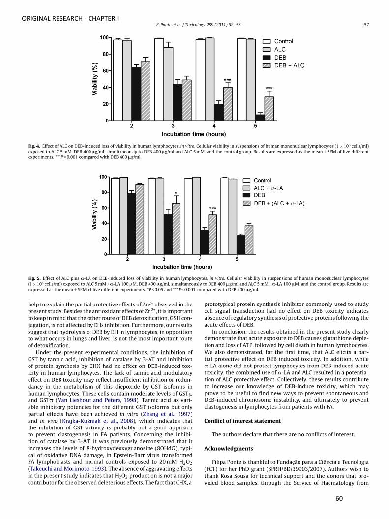
F. Ponte et al. / Toxicology 289 (2011) 52– 58 57
Fig. 4. Effect of ALC on DEB-induced loss of viability in human lymphocytes, in vitro. Cellular viability in suspensions of human mononuclear lymphocytes (1 × 10⁶ cells/ml)exposed to ALC 5 mM, DEB 400 �g/ml, simultaneously to DEB 400 �g/ml and ALC 5 mM, and the control group. Results are expressed as the mean ± SEM of five differentexperiments. ***P < 0.001 compared with DEB 400 �g/ml.
Fig. 5. Effect of ALC plus �-LA on DEB-induced loss of viability in human lymphocytes, in vitro. Cellular viability in suspensions of human mononuclear lymphocytes(1 × 10⁶ cells/ml) exposed to ALC 5 mM + �-LA 100 �M, DEB 400 �g/ml, simultaneously to DEB 400 �g/ml and ALC 5 mM + �-LA 100 �M, and the control group. Results areexpressed as the mean ± SEM of five different experiments. *P < 0.05 and ***P < 0.001 compared with DEB 400 �g/ml.
help to explain the partial protective effects of Zn2+ observed in thepresent study. Besides the antioxidant effects of Zn2+, it is importantto keep in mind that the other route of DEB detoxification, GSH con-jugation, is not affected by EHs inhibition. Furthermore, our resultssuggest that hydrolysis of DEB by EH in lymphocytes, in oppositionto what occurs in lungs and liver, is not the most important routeof detoxification.
Under the present experimental conditions, the inhibition ofGST by tannic acid, inhibition of catalase by 3-AT and inhibitionof protein synthesis by CHX had no effect on DEB-induced tox-icity in human lymphocytes. The lack of tannic acid modulatoryeffect on DEB toxicity may reflect insufficient inhibition or redun-dancy in the metabolism of this diepoxide by GST isoforms inhuman lymphocytes. These cells contain moderate levels of GST�and GST� (Van Lieshout and Peters, 1998). Tannic acid as vari-able inhibitory potencies for the different GST isoforms but onlypartial effects have been achieved in vitro (Zhang et al., 1997)and in vivo (Krajka-Kuzniak et al., 2008), which indicates thatthe inhibition of GST activity is probably not a good approachto prevent clastogenesis in FA patients. Concerning the inhibi-tion of catalase by 3-AT, it was previously demonstrated that itincreases the levels of 8-hydroxydeoxyguanosine (8OHdG), typi-cal of oxidative DNA damage, in Epstein-Barr virus transformedFA lymphoblasts and normal controls exposed to 20 mM H2O2(Takeuchi and Morimoto, 1993). The absence of aggravating effectsin the present study indicates that H2O2 production is not a majorcontributor for the observed deleterious effects. The fact that CHX, a
prototypical protein synthesis inhibitor commonly used to studycell signal transduction had no effect on DEB toxicity indicatesabsence of regulatory synthesis of protective proteins following theacute effects of DEB.
In conclusion, the results obtained in the present study clearlydemonstrate that acute exposure to DEB causes glutathione deple-tion and loss of ATP, followed by cell death in human lymphocytes.We also demonstrated, for the first time, that ALC elicits a par-tial protective effect on DEB induced toxicity. In addition, while�-LA alone did not protect lymphocytes from DEB-induced acutetoxicity, the combined use of �-LA and ALC resulted in a potentia-tion of ALC protective effect. Collectively, these results contributeto increase our knowledge of DEB-induce toxicity, which mayprove to be useful to find new ways to prevent spontaneous andDEB-induced chromosome instability, and ultimately to preventclastogenesis in lymphocytes from patients with FA.
Conflict of interest statement
The authors declare that there are no conflicts of interest.
Acknowledgments
Filipa Ponte is thankful to Fundac ão para a Ciência e Tecnologia(FCT) for her PhD grant (SFRH/BD/39903/2007). Authors wish tothank Rosa Sousa for technical support and the donors that pro-vided blood samples, through the Service of Haematology from
ORIGINAL RESEARCH - CHAPTER I
60

58 F. Ponte et al. / Toxicology 289 (2011) 52– 58
the Hospital Geral de Santo António and Institute of BiomedicalSciences Abel Salazar.
References
Ahmad, S.I., Hanaoka, F., Kirk, S.H., 2002. Molecular biology of Fanconi anaemia – anold problem, a new insight. BioEssays 24, 439–448.
Auerbach, A.D., Adler, B., Chaganti, R.S.K., 1981. Prenatal and postnatal diagnosisand carrier detection of Fanconi anaemia by a cytogenetic method. Pediatrics67, 128–135.
Auerbach, A.D., Rogatko, A., Shroeder-Kurth, T.M., 1989. International Fanconianaemia registry: relation of clinical symptoms to diepoxybutane sensitivity.Blood 73, 391–396.
Boerrigter, M.E.T.I., Franceschi, C., Arrigoni-Martelli, E., Wei, J.Y., Vijg, J., 1993. Theeffect of l-carninite and acetyl l-carnitine on the disappearance of DNA single-strand breaks in human peripheral blood lymphocytes. Carcinogenesis 14 (10),2131–2136.
Bogliolo, M., Borghini, S., Abbondandolo, A., Degan, P., 2002a. Alternative metabolicpathways for energy supply and resistance to apoptosis in Fanconi anaemia.Mutagenesis 17 (1), 25–30.
Bogliolo, M., Cabré, O., Callén, E., Castillo, V., Creus, A., Marcos, R., Surrallés, J., 2002b.The Fanconi anaemia genome stability and tumour suppressor network. Muta-genesis 17 (6), 529–538.
Boogaard, P.J., Bond, J.A., 1996. The role of hydrolysis in the detoxification of 1,2:3,4-diepoxybutane by human, rat and mouse liver and lung in vitro. Toxicol. Appl.Pharmacol. 141, 617–627.
Boogaard, P.J., Sumner, S.C.-J., Bond, J.A., 1996. Glutathione conjugation of 1,2:3,4-diepoxybutane in human liver and rat and mouse liver and lung in vitro. Toxicol.Appl. Pharmacol. 136, 307–316.
Bray, T.M., Bettger, W.J., 1990. The physiological role of zinc as an antioxidant. FreeRadical Biol. Med. 8, 281–291.
Calabrese, V., Stella, A.M.G., Calvani, M., Butterfield, D.A., 2006. Acetylcarnitine andcellular stress response: roles in nutritional redox homeostasis and regulationof longevity genes. J. Nutr. Biochem. 17 (2), 73–88.
Capela, J.P., Macedo, C., Branco, P.S., Ferreira, L.M., Lobo, A.M., Fernandes, E., Remião,F., Bastos, M.L., Dirnagl, U., Meisel, A., Carvalho, F., 2007. Neurotoxicity mecha-nisms of thioether ecstasy metabolites. Neuroscience 146, 1743–1757.
Carvalho, M., Milhazes, N., Remião, F., Borges, F., Fernandes, E., Amado,F., Monks, T.J., Carvalho, F., Bastos, M.L., 2004. Hepatotoxicity of 3,4-methylenedioxyamphetamine and alpha-methyldopamine in isolated rathepatocytes: formation of glutathione conjugates. Arch. Toxicol. 78, 16–24.
Clarke, A.A., Marsh, J.C., Gordon –Smith, E.C., Rutherford, T.R., 1998. Molecular genet-ics and Fanconi anaemia: new insights into old problems. Br. J. Haematol. 103,287–296.
Cumming, R.C., Lightfoot, J., Beard, K., Youssoufian, H., OıBrian, P.J., Buchwald, M.,2001. Fanconi anemia group C protein prevents apoptosis in hematopoietic cellsthrough redox regulation of GSTP1. Nat. Med. 7, 814–820.
Dallapicola, B., Porfirio, B., Mokini, V., Alimena, G., Isacchi, G., Gandini, E., 1985. Effectof oxidants and antioxidants on chromosomal breakage in Fanconi anaemialymphocytes. Hum. Genet. 69, 62–65.
Davis, B.B., Morrisseau, C., Newman, J.W., Pedersen, T.K., Hammock, B.D., Weiss, R.H.,2006. Attenuation of vascular smooth muscle cell proliferation by 1-cyclohexyl-3-dodecyl urea is independent of soluble EH inhibition. J. Pharmacol. Exp. Ther.316 (2), 815–821.
Devasagayam, T.P.A., Subramanian, M., Pradhan, D.S., Sies, H., 1993. Preventionof singlet oxygen-induced DNA damage by lipoate. Chem. Biol. Interact. 86,79–92.
Dinis-Oliveira, R.J., Sousa, C., Remião, F., Duarte, J.A., Navarro, A.S., Bastos, M.L.,Carvalho, F., 2007. Full survival of paraquat-exposed rats after treatment withsodium salicylate. Free Radical Biol. Med. 42, 1017–1028.
Draper, A.J., Hammock, B.D., 1999. Inhibition of soluble and microsomal epoxidehydrolase by zinc and other metals. Toxicol. Sci. 52, 26–32.
Erexson, G.L., Tindall, K.R., 2000. Reduction of diepoxybutane-induced sister chro-matid exchanges by glutathione peroxidase and erythrocytes in transgenic BigBlue mouse and rat fibroblasts. Mutat. Res. 447, 267–274.
Freitas, M., Porto, G., Lima, J.L.T.C., Fernandes, E., 2010. Zinc activation neutrophils’oxidative burst. Biometals 23, 31–41.
Hagen, T.M., Liu, J., Lykkesfeldt, J., Wehr, C.M., Ingersoll, R.T., Vinarsky, V.,Bartholomew, J.C., Ames, B.N., 2002. Feeding acetyl-l-carnitine and lipoic acidto old rats significantly improves metabolic function while decreasing oxidativestress. Proc. Natl. Acad. Sci. U.S.A. 99 (4), 1870–1875.
Han, D., Handelman, G., Marcocci, L., Sen, C.K., Roy, S., Kobuchi, H., Tritschler,H.J., Flohé, L., Packer, L., 1997. Lipoic acid increases de novo synthe-sis of cellular glutathione by improving cystine utilization. BioFactor 6,321–338.
Hollins, D.L., Suliman, H.B., Piantadosi, C.A., Carraway, M.S., 2006. Glutathione reg-ulates susceptibility to oxidant-induced mitochondrial DNA damage in humanlymphocytes. Free Radical Biol. Med. 40 (7), 1220–1226.
Kligerman, A.D., Tennant, A.H., 2007. Insights into the carcinogenic mode of actionof arsenic. Toxicol. Appl. Pharmacol. 222, 281–288.
Korkina, L.G., Deva, I.B., De Biase, A., Iaccarino, M., Oral, R., Warnau, M., Pagano,G., 2000. Redox-dependent toxicity of diepoxybutane and Mitomycin C in seaurchin embryogenesis. Carcinogenesis 21 (2), 213–220.
Krajka-Kuzniak, V., Kaczmarek, J., Baer-Dubowska, W., 2008. Effect of natu-rally occurring phenolic acids on the expression of glutathione S-transferaseisozymes in the rat. Food Chem. Toxicol. 46 (3), 1097–1102.
Kruyt, F.A., Hoshino, T., Liu, J.M., Joseph, P., Jaiswal, A.K., Youssoufian, H., 1998.Abnormal microsomal detoxification implicated in Fanconi anemia group C byinteraction of the FAC protein with NADPH cytochrome P450 reductase. Blood92, 3050–3056.
Loecken, E.M., Guengerich, F.P., 2008. Reactions of glyceraldehyde 3-phosphatedehydrogenase sulfhydryl groups with bis-electrophiles produce DNA-proteincross-links but not mutations. Chem. Res. Toxicol. 21 (2), 453–458.
Lowry, O.H., Rosebrough, N.J., Farr, A.L., Randall, R.J., 1951. Protein measurementwith the folin phenol reagent. J. Biol. Chem. 193 (1), 265–275.
Mantovani, G., Macciò, A., Madeddu, C., Mura, L., Gramignano, G., Lusso, M.R.,Massa, E., Mocci, M., Serpe, R., 2003. Antioxidant agents are effective in induc-ing lymphocytes progression through cell cycle in advanced cancer patients:assessment of the most important laboratory indexes of cachexia and oxidativestress. J. Mol. Med. 81, 664–673.
Michaelson-Richie, E.D., Loeber, R.L., Codreanu, S.G., Ming, X., Liebler, D.C., Campbell,C., Tretyakova, N.Y., 2010. DNA-protein cross-linking by 1,2:3,4-diepoxybutane.J. Proteome Res. 9 (9), 4356–4367.
Millard, J.T., Hanly, T.C., Murphy, K., Tretyakova, N., 2006. The 50-GNC site for DNAinterstrand cross-linking is conserved for diepoxybutane stereoisomers. Chem.Res. Toxicol. 19, 16–19.
Moldovan, G.L., D’Andrea, A.D., 2009. How the Fanconi anemia pathway guards thegenome. Ann. Rev. Genet. 43, 223–249.
Morisseau, C., Newman, J.W., Dowdy, D.L., Goodrow, M.H., Hammock, B.D., 2001.Inhibition of microsomal epoxide hydrolases by ureas, amines and amides.Chem. Res. Toxicol. 14 (4), 409–415.
Morriseau, C., Hammock, B.D., 2005. Epoxide hydrolases: mechanisms, inhibitorsdesigns, and biological roles. Pharmacol. Toxicol. 45, 311–333.
Mukhopadhyay, S.S., Leug, K.S., Hicks, M.J., Hastings, P.J., Youssoufian, H., Plon, S.P.,2006. Defective mitochondrial peroxiredoxin-3 results in sensitivity to oxidativestress in Fanconi anemia. J. Cell Biol. 175, 225–235.
Oda, T., Hayano, T., Miyaso, H., Takahashi, N., Yamashita, T., 2007. Hsp90 regulatesthe Fanconi anemia DNA damage response pathway. Blood 109, 5016–5026.
Pagano, G., Degan, P., d’Ischia, M., Kelly, F.J., Nobili, B., Pallardó, F.V., Youssoufian, H.,Zatterale, A., 2005. Oxidative stress as a multiple effector in Fanconi anaemiaclinical phenotype. Eur. J. Haematol. 75, 93–100.
Porto, B., Chiecchio, L., Gaspar, J., Faber, A., Pinho, L., Rueff, J., Malheiro, I., 2003. Roleof haemoglobin in the protection of cultured lymphocytes against diepoxybu-tane (DEB), assessed by in vitro induced chromosome breakage. Mutat. Res. 536,61–67.
Prahalathan, C., Selvakumar, E., Varalakshmi, P., Kumarasamy, P., Sarevanan, R.,2006. Salubrious effects of lipoic acid against adriamycin-induced clastogenesisand apoptosis in Wistar rat bone marrow cells. Toxicology 222, 225–232.
Prasad, A.S., 2008. Clinical, immunological, anti-inflammatory and antioxidant rolesof zinc. Exp. Gerontol. 43, 370–377.
Prasad, A.S., Bao, B., Beck, F.W.J., Kucuk, O., Sarkar, F.H., 2004. Antioxidant effect ofzinc in humans. Free Radical Biol. Med. 37 (8), 1182–1190.
Sankaramanivel, S., Rajaram, A., Rajaram, R., 2010. Zinc protects human peripheralblood lymphocytes from Cr(III)(phenanthroline)3-induced apoptosis. Toxicol.Appl. Pharmacol. 243 (3), 405–419.
Spanò, M., Cordelli, E., Leter, G., Pacchierotti, F., 1998. Diepoxybutane cytotoxicity onmouse germ cells is enhanced by in vivo glutathione depletion: a flow cytometricapproach. Mutat. Res. 397, 37–43.
Stehbens, W.E., 2003. Oxidative stress, toxic hepatitis, and antioxidants with partic-ular emphasis on zinc. Exp. Mol. Pathol. 75, 265–276.
Takeuchi, T., Morimoto, K., 1993. Increased formation of 8-hydroxydeoxyguanosine,an oxidative DNA damage, in lymphoblast from Fanconi anaemia patients dueto possible catalase deficiency. Carcinogenesis 14 (6), 1115–1120.
Van Lieshout, E.M., Peters, W.H., 1998. Age and gender dependent levels of glu-tathione and glutathione S-transferases in human lymphocytes. Carcinogenesis19 (10), 1873–1875.
Vandeputte, C., Guizon, I., Genestie-Denis, I., Vannier, B., Lorenzon, G., 1994. Amicrotiter plate assay for total glutathione and glutathione disulfide contents incultured/isolated cells: performance study of a new miniaturized protocol. CellBiol. Toxicol. 5–6 (10), 415–421.
Wang, Z., Jin, L., Wegrzyn, G., Wegrzyn, A., 2009. A novel method for screening theglutathione transferase inhibitors. BMC Biochem. 10, 6.
Yadavilli, S., Martinez-Ceballos, E., Snowden-Aikens, J., Hurst, A., Joseph, T., Albrecht,T., Muganda, M.P., 2007. Diepoxybutane activates the mitochondrial apoptoticpathway and mediates apoptosis in human lymphoblasts through oxidativestress. Toxicol. In Vitro 21, 1429–1441.
Yamada, H., Koizumi, S., 1991. Metallothionein induction in human peripheral bloodlymphocytes by heavy metals. Chem. Biol. Interact. 78 (3), 347–354.
Zhang, K., Mack, P., Wong, K.P., 1997. Inhibition of various isoforms of rat liver glu-tathione S-transferases by tannic acid and butein. Biochem. Mol. Biol. Int. 42 (4),701–710.
ORIGINAL RESEARCH - CHAPTER I
61

ORIGINAL RESEARCH – CHAPTER I
62

PART III ‐ ORIGINAL RESEARCH
63
CHAPTER II
Improvement of genetic stability in lymphocytes from Fanconi Anemia patients through the combined effect of α-lipoic acid and N-acetylcysteine
Accepted for publication in Orphanet Journal of Rare Diseases
Copyright (2012) with kind permission from BioMed Central

PART III ‐ ORIGINAL RESEARCH
64

ORIGINAL RESEARCH – CAPTER II
65
Improvement of genetic stability in lymphocytes from Fanconi anemia patients through the combined effect of
α-lipoic acid and N-acetylcysteine
FILIPA PONTE1,2*, ROSA SOUSA
2, ANA PAULA FERNANDES3, CRISTINA GONÇALVES
4, JOSÉ BARBOT5,
FÉLIX CARVALHO1, BEATRIZ PORTO
2*
1 Chemistry and Technology Network (REQUIMTE), Laboratory of Toxicology, Department of Biological Sciences, Faculty of Pharmacy, University of Porto, Porto, Portugal, 2 Laboratory of Cytogenetic, Department of Microscopy,
Institute of Biomedical Sciences Abel Salazar (ICBAS), Porto, Portugal, 3 Hemato-oncologic Unity, Hospital São João (HSJ), Porto, Portugal, 4 Service of Hematology of Hospital Center of Porto (CHP), Porto, Portugal, 5 Unity of Pediatric
Hematology of CHP, Porto, Portugal *Correspondence should be addressed to [email protected] or [email protected]
Abstract
Fanconi Anemia (FA) is a rare genetic disorder, characterized by progressive bone marrow failure and increased predisposition to cancer. Despite being highly heterogeneous, all FA patients are hypersensitive to alkylating agents, in particular to 1,2:3,4-diepoxybutane (DEB), and to oxidative damage. Recent studies point to defective mitochondria in FA cells, which is closely related with increased production of reactive oxygen species (ROS) and concomitant depletion of antioxidant defenses, of which glutathione is a well-known biomarker.
The objective of the present work is to evaluate the putative protective effect of α-lipoic acid (α-LA), a mitochondrial protective agent, and N-acetylcysteine (NAC), a direct antioxidant and a known precursor for glutathione synthesis, in spontaneous and DEB-induced chromosome instability (CI) in lymphocyte cultures from FA patients.
For that purpose, lymphocyte cultures from 15 FA patients and 24 healthy controls were pre-treated with 20 μM α-LA, 500 μM NAC and α-LA plus NAC at the same concentrations, and some of them exposed to DEB (0.05 μg/ml). A hundred metaphases per treatment were scored to estimate the relative frequency of spontaneous and DEB-induced chromosome breakage.
The obtained results reveal that a cocktail of α-LA and NAC can drastically improve the genetic stability in FA lymphocytes in vitro, decreasing CI by 60% and 80% in cultures from FA patients and FA mosaic/chimera patients, respectively. These results suggest that the studied cocktail can be used as a prophylactic approach to delay progressive clinical symptoms in FA patients caused by CI, which can culminate in the delay of the progressive bone marrow failure and early cancer development.
Keywords
Fanconi Anemia, oxidative stress, antioxidants, α-lipoic acid, N-acetylcysteine, chromosome instability, bone marrow failure, cancer susceptibility.

ORIGINAL RESEARCH – CAPTER II
66
Introduction
Fanconi Anemia (FA) is a rare genetic disorder characterized by chromosome instability (CI), several congenital malformations, progressive bone marrow failure, and increased predisposition to cancer, particularly acute myelogenous leukemia [1]. Genetically, FA is a highly heterogeneous disease with 15 genes so far characterized and cloned (FA-A, B, C, D1, D2, E, F, G, I, J, L, M, N, O, P) [2] and that appears to influence genomic stability. The clinical manifestations and the onset of disease are also extremely variable. However, all FA patients have, in common, unique markers that characterize the disease: hypersensitivity to the clastogenic effect of DNA cross-linking agents, in particular to diepoxybutane (DEB), [3] and hypersensitivity to oxidative damage [4]. DEB-induced cytotoxicity, despite hitherto not completely understood, has been related to oxidative damage and to glutathione (GSH) depletion [5-7]. The activation of the mitochondrial apoptotic pathway in the event of DEB-induced oxidative stress (OS) was more recently demonstrated [8]. Importantly, DEB-induced DNA-DNA and DNA-protein cross-links [9] are a characteristic feature of ROS-mediated damage. In accordance to the involvement of mitochondria and OS in the mechanisms of DEB toxicity, we have recently demonstrated that acute exposure to DEB both causes glutathione (GSH) depletion and loss of ATP, followed by cell death in lymphocytes collected from healthy humans [10]. The role of oxygen and oxidative damage in FA cells was described for the first time by Nordenson [11] followed by Joenje et al [12]. Two years later, Joenje and Oostra [13] established a direct correlation between high tensions of oxygen and increased damage in FA lymphocytes. A direct association of OS with the primary genetic defect in FA was suggested through the interaction of the FANCC protein with NADPH cytochrome P450 reductase [14] and glutathione S-transferase [15], two enzymes involved in the detoxification of reactive intermediates. At
least FANCC, FANCG and FANCA have been shown to be associated with redox-related imbalance and to a unique functional and structural sensitivity to OS [16] and, altogether, these proteins are also related with mitochondrial dysfunction [14-18]. Recently, Mukhopadhyay et al [18] reported that FANCG protein physically interacts with the mitochondrial peroxidase peroxiredoxin 3 (PRDX3), FA-G cells display distorted structures, and FA-A, -C and -G have PRDX3 cleavage and decreased peroxidase activity. Mitochondrial dysfunction ultimately leads to oxidative modifications of DNA, proteins and lipids. It is recognized that FA cells show abnormal accumulation of 8-hydroxy-2’-deoxyguanosine (8-OHdG), a sub-product of oxidative DNA damage, and also a mutagenic substance by itself [19]. However, Castillo et al [20] concluded that the FA pathway is redundant for the repair of oxidative DNA damage (7,8-dihydro-8-hydroxyguanine, 8-oxoG). Therefore, the accumulation of 8-oxoG in FA cells reflects sustained ROS overproduction rather than defective processing of oxidized bases. These evidences, altogether, point towards a pro-oxidant state associated with dysfunctional mitochondria, in FA cells (reviewed by Pagano et al) [4]. Some studies have been performed testing the use of antioxidants for decreasing chromosome damage in FA cells. Dallapiccola et al [21] obtained a partial correction of CI in FA lymphocytes treated with the antioxidants sodium ascorbate, L-cysteine, 2-mercaptoethanol, α-mercaptopropionylglycine, and GSH. Later, the same group [22] extended this study to desferoxamine, also with partial correction of CI. Pincheira et al [23] demonstrated that Vitamin E decreases the frequency of chromosomal damage and the duration of G2 in FA lymphocytes. Some in vivo studies were also performed in animal models. Zhang et al [24] showed that dietary supplements with antioxidants can delay the age of onset of epithelial tumors in FA mouse models. More recently, this group [25] also demonstrated that the antioxidant resveratrol maintains Fancd2(−/−) KSL cells in quiescence, improves the marrow

ORIGINAL RESEARCH – CAPTER II
67
microenvironment, partially corrects the abnormal cell cycle status, and significantly improves the spleen colony-forming capacity of Fancd2(−/−) bone marrow cells. In spite of the promising results from previous studies, no therapy using antioxidants, megavitamins, or micronutrients has been shown to be effective in treatment of FA using evidence-based criteria. Thus, further studies with new protective agents are required for the application of drugs with high effectiveness. Among a number of potential candidate molecules with antioxidant properties, some can be identified as “mitochondrial nutrients” and substances that normalize GSH balance. α-Lipoic acid (α-LA) is a natural and endogenous compound that occurs in mitochondria and is an essential cofactor for many mitochondrially localized enzyme complexes. Furthermore, α-LA features a cyclic disulfide moiety which exists in redox equilibrium with its reduced dithiol form, dihydrolipoic acid. Dihydrolipoic acid acts in GSH replenishment and is a potent antioxidant in a redox-cycling environment, by reducing vitamin C, vitamin E and coenzyme Q10 [26,27]. Previous studies also proved that α-LA can be a protective agent against clastogenic [28] and genotoxic agents [29]. N-acetylcysteine (NAC), the acetylated variant of the amino acid L-cysteine, is an excellent source of sulfhydryl groups, and is converted in the body into metabolites capable of stimulating GSH synthesis, promoting detoxification, and acting directly as free radical scavengers [30]. Recently, a study from Yadavilli et al [8] showed that NAC has the ability to inhibit ROS production and apoptosis induced by DEB in human lymphoblasts. Importantly, there are some clinical studies referring the benefit of the combined use of α-LA and NAC by ameliorating the hematological response in advanced cancer patients [31] and in the event of muscle-damaging exercise [32]. In a previous study [10], we showed that α-LA and NAC have a partial protective effect on acute DEB-induced toxicity to normal human lymphocytes, though the effect of these compounds in CI of lymphocytes has never
been tested before. Thus, the aim of the present work was to evaluate the putative protective effect of α-LA and NAC, in spontaneous and DEB induced CI lymphocytes cultures from FA patients.
Methods
Subjects
This study includes 15 FA patients, referred by several Hospitals, and already diagnosed with certainty on the basis of DEB-induced cytogenetic tests, and 24 healthy donors (HD), recruited among healthy male and female donors, aged 20–40 years. The study was approved by the Ethical Committe of Hospital Geral de Santo António, CHP, Porto. All procedures were done with the written informed consent of the participants.
Cells and cell cultures
From each HD participant, 10 ml of venous blood was collected by antecubital venipuncture, into vacuum tubes with lithium heparin. From FA patients, 3 ml to 6 ml of venous blood were collected, depending on the age and healthy conditions of the patients, into vacuum tubes with lithium heparin. Whole blood (0.5 ml) was cultured in RPMI 1640 (Sigma) complete medium supplemented with 15% fetal calf serum (GIBCO), antibiotics (10,000 units/ml of penicillin and 10,000 μg/ml of streptomycin) (GIBCO) and 29 mg/ml of L-glutamine (Sigma). Cultures were stimulated with 5 μg/ml of phytohemagglutinin (GIBCO) and placed in an incubator at 37°C with 5% CO2 atmosphere, for 72 h. Exposure to DEB DEB ((±)-1,2:3,4-diepoxybutane, [298-18-0], D-7019 Lot 34 H3683, Sigma), prepared in RPMI 1640, was added to lymphocyte cultures 24 h after their initiation, thus exposing cells to the chemical for 48 h. In lymphocyte cultures from FA patients, DEB was added at the concentration of 0.05 μg/ml (the same

ORIGINAL RESEARCH – CAPTER II
68
concentration used in the diagnostic test). As the lymphocytes from HD are not so sensitive to DEB at that concentration, and have variable responses, 3 higher concentrations were used to obtain a significant toxic response (see Figure 1 for a description of DEB-induced CI pattern in HD and FA patients). Therefore, a concentration of 0.1 μg/ml was selected in cultures from 5 individuals (HD1-5), a concentration of 0.2 μg/ml was selected in cultures from 11 individuals (HD6-11 and HD14-18) and a
concentration of 0.4 μg/ml was selected in cultures from 8 individuals (HD12-13 and HD19-24). Since DEB is a suspected carcinogen with unknown risk, appropriate precautions were taken. All culture procedures were handled using appropriate gloves and in a vertical laminar flow hood. As DEB is rapidly inactivated by concentrated HCl, all disposable culture bottles and pipettes were rinsed with HCl before being discarded.
Figure 1. CI pattern in metaphases from DEB-induced lymphocyte cultures from a healthy donor and FA patient. Chromosomes were stained in a 4% Giemsa solution. The images were observed with an optical microscope (Olympus CX31) and captured with a digital camera (Nikon Sightds-smc), with the software DP20-5E microscope digital camera. (A) Selected metaphase from a HD lymphocyte culture exposed to 0.2 μg/ml of DEB for 48 h. One chromatid break can be seen (asterisk). (B) Selected metaphase from a FA patient lymphocyte culture exposed to 0.05 μg/ml of DEB for 48 h. High level of chromosome instability can be visualized, especially being important the tri and tetraradial figures (arrow) that are the hallmark for the diagnosis of FA. It can also be seen a dicentric chromosome (head arrow) and 3 chromatid breaks (asterisks).
Antioxidant treatments
We used two antioxidants, α-LA 20 μM [33] (Sigma) and NAC 500 μM [10] (Sigma) at concentrations that proved to be effective in in vitro studies, preferably with lymphocytes. In addition, prospective studies were always performed to find the most efficient concentrations (data not shown). Additionally, α-LA and NAC concentrations used in the present study are within the plasma concentrations found in previous human pharmacokinetic studies using these drugs [34,35]. α-LA was dissolved in 1% dimetilsulfoxide (DMSO) in RPMI 1640 and NAC was dissolved in RPMI 1640. α-LA was stored in the dark at room temperature. NAC was stored at 4°C. The two antioxidants were used from the same batch in all experiments.
Antioxidant treatments in DEB-exposed lymphocyte cultures from healthy donors Two sets of experiments were done. In the first set, lymphocyte cultures were pre-treated with α-LA 20 μM 1.5 h before DEB exposure. In the second set of experiments lymphocyte cultures were pre-treated with NAC 500 μM, 1.5 h before DEB exposure. DMSO toxicity was found to be absent within our experimental conditions. Antioxidant treatments in spontaneous and DEB-exposed lymphocyte cultures from FA patients Three sets of experiments were done. In the first set, lymphocyte cultures were treated with α-LA 20 μM for 48 h. In DEB-induced lymphocyte cultures α-LA was added 1.5 h before DEB exposure. In the second, lymphocyte cultures were treated with NAC 500 μM for 48 h. In DEB-exposed lymphocyte cultures, NAC was added 1.5 h before DEB
ORIGINAL RESEARCH – CAPTER II
69
exposure. In the last set, both antioxidants were added simultaneously to lymphocyte cultures, at the same concentrations, for 48 h. In DEB-exposed lymphocyte cultures antioxidants were added 1.5 h before DEB exposure. DMSO toxicity was found to be absent within our experimental conditions.
Cytogenetic analysis
After 3 days of culture, cells were harvested after 1 h of incubation with colcemid (GIBCO) (4 μg/ml), followed by hypotonic treatment with 75 mM KCl (Sigma) and fixed 3 times in a 1:3 iced solution of acetic acid (Merck): methanol (Merck). The resulting suspensions were dropped onto microscope slides and stained for 5 min in a 4% Giemsa solution (Merck) diluted in phosphate buffer saline solution (Sigma). Analysis was performed on 50–100 metaphases from each experiment, by an independent scorer and in a blinded fashion. Only when the mitotic index (MI) was very low and the number of breaks was high, a minimum of 22 metaphases was counted. Each cell was scored for chromosome number and the number and type of structural abnormalities. Gaps (acromatic areas less than a chromatid in width) were excluded in the calculation of chromosome breakage frequencies, and rearrangements (triradials and quadriradials, dicentrics and ring chromosomes) were scored as two breaks.
Mitotic index
From the same microscope stained slides, used for chromosome breakage analysis, a thousand cells were randomly scored, distinguishing between interphase nuclei and metaphase nuclei. MI was calculated as follows: number of metaphase nuclei /number of total nuclei.
Statistical analysis
Graph results are expressed as mean ± SEM and table results are expressed as mean ± SD. Statistical comparison among groups was estimated using one-way ANOVA, followed
by the Bonferroni post hoc test, and comparison between two groups was estimated using paired t-test, both with GraphPad Prism, version 5.0 software. P values lower than 0.05 were considered as statistically significant.
Results
Effect of α-lipoic acid and N-acetylcysteine on DEB-induced CI in lymphocyte cultures from healthy donors
As shown in Table 1, lymphocyte cultures from HD exposed to DEB and pre-treated with α-LA or NAC showed a reduction in the number of breaks per cell compared to those only exposed to DEB (P>0.05 and P<0.05, respectively). Noteworthy, two distinct groups can be considered concerning the protective effect: a group of responders and another one of non-responders. Among DEB-induced cultured lymphocytes pre-treated with α-LA 54% are responders whereas among those pre-treated with NAC, 82% are responders. However, the response with α-LA treatment is greater, while with NAC the potency is not only lower but also more variable. Globally, the percentage of reduction in lymphocyte cultures pre-treated with α-LA was 25% and with NAC was 20%.
Effect of α-lipoic acid, N-acetylcysteine and α-lipoic acid plus N-acetylcysteine on spontaneous CI in lymphocyte cultures from FA patients As shown in Table 2, lymphocyte cultures from FA patients treated with α-LA, NAC and α-LA plus NAC showed a significant reduction in the number of breaks per cell, compared to cultures without antioxidant treatment (P < 0.01, P < 0.01 and P < 0.01, respectively). Comparing α-LA vs NAC treatments, no difference (P>0.05) was found in the reduction of the number of breaks per cell. However, comparing both isolated α-LA

ORIGINAL RE
and NAC tplus NAC number osignificant respectivelyreduction inα-LA was 40% (P<058% (P<0Concerning
Tab
In control percentage― Unavaianalyzed. P
ESEARCH – C
treatments wtreatment
f breaks (P < 0
y). Globaln lymphocy34% (P <
0.01) and w0.001), as shg the α-LA
ble 1. Numbe
group no antie of reduction ilable data; α-LPaired t-test: C
CAPTER II
with the com, the reduper cell
0.05 and ly, the peyte cultures
< 0.01), witwith α-LA phown in FiguA antioxidan
er of DEB-ind
ioxidant treatmof the numbe
LA indicates αControl group
mbined α-Luction in th
was highP < 0.0
ercentage s treated with NAC wlus NAC wure 2. nt effect tw
duced breaks
ment was doneer of breaks peα-lipoic acid; p vs NAC 500
LA the hly 01, of ith
was was
wo
direlyanvaNobNdeno
s per cell in cu
e. The effect oer cell in α-LANAC indicate
0 μM P<0.05
istinct grouesponders anymphocyte nd in two alues (belo
NAC treatedbserved. Be
NAC and fouecrease in ormal value
ultured lymp
of the antioxidA and NAC trees N-acetylcys
ups were, nd non-respcultures, 8of them CIw 0.1). In d cultures esides, in twur treated wthe number
es was also o
hocytes from
dant treatmenteatments relatsteine; n indic
once moreponders; in 85% were I decreasedNAC and
only respowo cultures with α-LA pr of breaksobserved (T
m healthy don
ts was calculatively to contrcates the numb
70
e, observeda total of 13responders
d to normaα-LA plus
onders weretreated withplus NAC a per cell to
Table 2).
nors.
ted by the rol group. ber of cells
0
d: 3 s, al s e h a o

In redUngrNA
ORIGINAL RE
control grouduction of thenavailable datoup vs NAC AC 500 μM P
ESEARCH – C
Table 2
up no antioxide number of bta. Abbreviati500 μM P <
P<0.05; NAC 5
CAPTER II
2. Number of
dant treatmenbreaks per cellions are expla0.01; Control500 μM vs α-
f breaks per c
nt was done. l in the α-LA,ained in Tabll group vs α-LLA 20 μM + N
cell in culture
The effect of, NAC and α-le 1. Paired t-LA 20 μM + NAC 500 μM
ed lymphocyt
f the antioxid-LA plus NAC-test: Control NAC 500 μM
M P<0.01.
tes from FA p
dants was calC treatments rgroup vs α-L
M P<0.01; α-L
Figure 2. EffLA+NAC oncultured lypatients. Lytreated with 2and both antithe same conpresented as tper cell (percComparison breaks/cell wgroup and e(*** P<0.00between treatmrepresentativeexperiments d
patients.
lculated by threlatively to cLA 20 μM P LA 20 μM vs
ffect of α-LA,n chromosomymphocytes ymphocyte c20 μM α-LA, ioxidants sim
ncentrations. Tthe relative m
cen- tage of coof the relat
was made beeach antioxid01 and **P tments (P>0.0e of the mdepicted from
71
he percentagecontrol group.
< 0.01; Conts α-LA 20 μM
, NAC and αme breaks in
from FAcultures wer500 μM NAC
multaneously aThe results armean of breakontrol values)tive mean otween contro
dant treatmen< 0.01) and
5). Results aremean of 14
m Table 2.
1
of ―
trol M +
α-n A e
C, at e
ks ).
of ol nt d e 4

E
p
P
ORIGINAL RE
Effect of α-lipoic acspontaneouparticular gchimera Consideringpatients refFA10 werea post-tranhigher frequsee is that,breaks per with α-LAtranslates inumber ofP<0.001 (F
ESEARCH – C
-lipoic acid,cid plus us CI in lymgroup of FA
g the cytogferred in Tae classified splant chimuency of no, for these
cell decreA plus Nn an even
f breaks peFigure 3).
Table 3
In control gPaired t-tes
CAPTER II
N-acetylcyN-acetylc
mphocyte cuA patients -
genetic diagable 2, patieas mosaics
mera. Thus,ormal cells. patients, th
eased to noNAC treatm
greater reder cell, of
3. Determinat
group no antist: Control gro
ysteine and cysteine oultures from- mosaics an
gnosis of thents FA9 anand FA11 they haveWhat we c
he number ormal valument, whiduction in th
about 80%
tion of mitotic
oxidant treatmoup vs α-LA 2
α-on
m a nd
the nd as
e a an of
ues ch
the %,
EonlyMsutrshinto0.ofinthin0.
c index in cul
ment was don20 μM + NAC
ffect of α-n cell cymphocytes
MI was meubmitted toreatments. Thow that, ncreased theo 0.18, wh.19, and, mf both antioncrease to 0hat α-LA pncreased the.05 (Figure
ltured lymph
e. AbbreviatioC 500 μM P<0
lipoic acid ycle progfrom FA p
easured in o the threeThe results in averagee MI from 0
hile NAC tost importaoxidants re0.27, which
plus NAC te MI of cult4).
Figure 3 EffLA + NAC ocultured lymosaics/chimcultures wereLA, 500 antioxidants same concenpresented asbreaks per cvalues). Commean of breacontrol groutreatment (**and betweeResults are rof 3 experim2.
hocytes from F
ons are explai0.05
plus N-acgression inpatients
lymphocye types of displayed
e, the α-LA0.17 (in contreatment inantly, the coesulted in ah clearly dtreatment stured lymph
ffect of α-LA, on chromosomymphocytes meras. re treated witμM NAC, simultaneous
ntrations. Thes the relativ
cell (percentagmparison of aks/cell was mup and each** P<0.001 anen treatmentrepresentative
ments depicted
FA patients.
ined in Table
72
cetylcysteinen cultured
yte culturesantioxidanin Table 3
A treatmenntrol groupncreased to
ombined usea significandemonstratessignificantlyhocytes, P <
NAC and α-me breaks in
from FA Lymphocyte
th 20 μM α-and both
s- ly at the e results are ve mean of ge of control
the relative made between h antioxidant nd **P<0.01) ts (P>0.05). of the mean d from Table
1.
2
e d
s nt 3
nt ) o e
nt s y <
f
t

ORIGINAL RESEARCH – CAPTER II
73
Figure 4. Effect of α-LA + NAC on mitotic index of cultured lymphocytes from FA patients. Lymphocyte cultures were treated with 20 μM α-LA plus 500 μM NAC. Mitotic index was evaluated as described in “methods” and the results are presented as absolute frequencies depicted from Table 3 (*P<0.05).
Effect of α-lipoic acid, N-acetylcysteine and α-lipoic acid plus N-acetylcysteine on DEB- induced CI in lymphocyte cultures from FA patients As shown in Table 4, lymphocyte cultures from FA patients pre-treated with α-LA, NAC, and α-LA plus NAC, and after exposure to DEB, showed a significant reduction in the number of breaks per cell compared to the control group (P<0.05, P<0.05 and P<0.05, respectively). Comparing α-LA vs NAC treatments (P > 0.05), α-LA vs α-LA plus NAC treatments (P<0.05) and NAC vs α-LA plus NAC treatments (P>0.05) no differences were found in the reduction in the number of breaks per cell. Globally, the percentage of reduction in lymphocyte cultures pre-treated with α-LA was 28% (P>0.05), with NAC was 54% (P < 0.001) and with α-LA plus NAC was 60% (P<0.001), as shown in Figure 5. Once more, two distinct groups can be considered, a group of responders and another one of non-responders. In a total of 12 α-LA pre-treated lymphocyte cultures, 67% were responders, while in a total of 7 NAC pre-treated lymphocyte cultures, 86% were responders. In α-LA plus NAC pre-treated cultures 100% were responders. In a culture from one patient belonging to this last group, the number of breaks per cell decreased to values considered normal (below 0.1).
Discussion
In the present work we evaluated the protective effect of α-LA and NAC and, more importantly, the protective effect of a cocktail with these two compounds, against spontaneous and DEB-induced CI in lymphocyte cultures from FA patients. The
obtained results provide an important and novel finding: it was clearly evident, for the first time, that the concomitant exposure to α-LA and NAC can drastically improve the genetic stability in lymphocytes from FA patients, in vitro. Various reports have previously shown mitochondrial dysfunctions in FA cells, including distorted mitochondrial structures and peroxyredoxin 3 deregulation in FA-A, FA-C and FA-G cells[18] and mitochondrial matrix densification in FA-A fibroblasts after 8-methoxypsolaren photoreaction or UVA irradiation [17]. It is known that mitochondrial dysfunctions ultimately lead to a defect in energy transduction, with increased formation of ROS, which, by itself, can lead to lower cellular energy charge, oxidative modification of DNA, protein and lipids. In addition, there is also a possibility of a positive feedback cycle between ROS formation and mitochondrial damage, exacerbating the processes of cellular dysfunction (reviewed in Tarnopolsky) [26]. It was already demonstrated that GSH protects mitochondrial DNA from oxidative damage in human lymphocytes and that GSH depletion increases their susceptibility to mitochondrial DNA damage [36]. It is noteworthy that GSH levels are downregulated in FA patients [37]. Therefore, it may be postulated that such imbalance in the redox state of FA cells can be counteracted by antioxidants, in particular mitochondrial nutrients, like α-LA, and other antioxidants that can replace or replenish GSH levels, like NAC, as it was demonstrated in the present study. Hypersensitivity of lymphocytes to the clastogenic effect of DEB is a common biomarker of FA. Previous studies have shown

ORIGINAL RE
T
In control groantioxidants wplus NAC tretest: Control NAC 500 μM
ESEARCH – C
Table 4. Numb
oup no antioxiwas calculatedeatments relatigroup vs α-L
M P<0.05.
Figure 5. induced ctreated witsame concpresented Compari- each antioxrepresentat
CAPTER II
ber of DEB-in
idant treatmend by the perceively to contr
LA 20 μM P<0
Effect of α-Lcultured lymth 20 μM α-L
centrations, 1.5as the relatison of the relxidant treatmetive of the me
nduced break
nt was done anentage of redurol group. ― U0.05; Control
LA, NAC anmphocytes froLA, 500 μM N5 h before expive mean of lative mean oent (*** P<0.
ean of 14 expe
ks per cell in
nd lymphocytuction of the nUnavailable dl group vs NA
nd α-LA+NAom FA patieNAC, and botposure to DEB
breaks per of breaks/cell .001) and betweriments depic
cultured lym
tes were exponumber of bredata. AbbreviaAC 500 μM P
AC on chroments. Lymphoh antioxidantsB 0.05 μg/ml cell (percentwas made between treatmencted from Tab
mphocytes fro
sed to DEB 0aks per cell inations are exp
P<0.05; Contro
osome breakocyte cultures s simul- taneofor 48 h. Theage of contrtween controlnts (P>0.05). le 4.
om FA patien
0.05 μg/ml. Thn the α-LA, Nplained in Tabol group vs α
ks in DEB-s were pre-ously at the e results are rol values). l group and Results are
74
nts.
he effect of thNAC and α-LAble 1. Paired tα-LA 20 μM +
4
e A t-+

ORIGINAL RESEARCH – CAPTER II
75
that DEB acts as a bifunctional alkylating agent, exhibiting both inter-strand and intra-strand DNA cross-linking ability [38] and that is redox-active, generating ROS that can damage DNA [8]. Furthermore, acute exposure of normal human lymphocytes to DEB is associated with GSH and ATP depletion [10], which indicates that DEB can also cause mitochondrial dysfunction. The exacerbation of OS and DNA cross-linking in FA lymphocytes, through DEB-mediated effects, can reproduce damaging conditions present in severe clinical FA situations, which theoretically extends the applicability of the present results from bench to bedside. Under the present experimental conditions, the α-LA effect, measured by the reduction in the number of breaks per cell, was similar (around 30%) in the three studied groups: DEB-
induced lymphocyte cultures from HD and spontaneous and DEB-induced cultures from FA patients (Figure 6A). This indicates that α-LA effect was independent of DEB exposure or health condition of the studied subject. On the other hand, we verified that NAC effect in these same three groups seems to be dependent of the treatment and health condition (Figure 6B), since the reduction in the number of breaks per cell was greater in DEB-induced lymphocytes from FA patients. These results are in agreement with previous studies, showing that NAC inhibits ROS production and apoptosis induced by DEB in human lymphoblasts [8] and that NAC protects human lymphocytes from acute in vitro exposure to DEB [10]. Last but not least, we also verified that the effect of the combined use of α-LA plus NAC is independent of DEB exposure, as shown in
Figure 6. Comparison between the different studied groups according to the antioxidant treatments. Comparative analysis of the effect of α-LA (A), NAC (B) and α-LA plus NAC treatments was performed between lymphocyte cultures from the four studied groups: healthy donors, FA patients (spontaneous breaks), FA patients (DEB-induced breaks) and FA mosaic/chimera patients. All values were depicted from Figures 2, 3 and 5.
Figure 6C, with a 60% of reduction in the number of breaks per cell in both groups of FA patients, and even more effective in cultures from FA patients with higher frequency of normal cells, such as FA mosaics or FA chimera, in which the reduction was of about 80%. Furthermore, the measurement of spontaneous chromosomal breaks was of particular importance, as it rules out the
possibility that the main anti-chromosomal break effect of α-LA and NAC is due to a direct interaction of these agents with DEB. Additionally, MI was measured in lymphocyte cultures from FA patients submitted to all antioxidant treatments. The reason for this assay was to evaluate if the decrease in CI was due to an increase in mitotic proliferation, with a consequent shortening of the cell cycle arrest

ORIGINAL RESEARCH – CAPTER II
76
in G2, as it was demonstrated for α-tocopherol by Pincheira et al [23]. What we observed is that MI increased in all antioxidant treatments, especially with α-LA plus NAC treatment, suggesting a relation between a decrease in CI and a shortening of cell arrest in G2. We cannot exclude, however, that, in lymphocyte cultures from mosaic and chimera patients, the increase in MI can be due to a positive selection of normal cells, with a consequent increase in apoptosis. The results now obtained suggest that α-LA plus NAC cocktail may be useful to keep chromosome stability in FA patients, which can be very important to block or delay the progression of the disease. This cocktail may be even more effective when applied to FA mosaics and FA chimeras, after bone marrow transplant, as we observed in vitro. It must be stressed that bone marrow transplant only resolves bone marrow failure, curing the hematological manifestations. However, CI is not mitigated, and predisposition to cancer, other than leukemia, persists, especially solid tumors [3,39,40]. Thus, controlling CI in FA patients after bone marrow transplant could be very important. Additionally to increasing chromosomal stability, α-LA has also important cellular roles in the regulation of gene transcription, inhibition of the activation of NF-kB (nuclear factor kappa-light-chain-enhancer of activated B cells) and activating protein 1, [41,42] while NAC apparently modulates cytokine concentrations, as interleukin(IL)-1, TNF-α (tumor necrosis factor-alpha) and IFN-γ (interferon-gamma) [30]. These pharmacological effects may further contribute for the therapeutic usefulness of α-LA plus NAC cocktail by avoiding the inflammatory process, mostly frequent in FA children patients [43,44]. Of note, some studies concerning the immunological response of FA patients demonstrated lymphocyte dysfunction and high levels of TNF-α [45]. Low production of IL including IL1, IL2, and IL6, as well as IFN-γ and granulocyte-macrophage colony stimulating factor have also been reported [46].
The present work might open a novel strategy to deal with FA disease. The choice of these antioxidants, apart from their pharmacodynamics, takes into account that both α-LA and NAC have been already approved for safe human use with minimal or no adverse effect, showing an excellent safety profile. NAC has been in clinical use for more than 30 years, in conditions characterized by depletion of GSH or OS, as cancer, AIDS, pulmonary diseases, heart diseases, as well as in acetaminophen overdose [30,47,48]. Thus, in conditions characterized by a chronic excessive OS and in clinical situations where GSH levels are decreased, NAC appears to be a highly effective drug, and may be also recommendable in the prevention/treatment of FA. α-LA has been used, especially in Germany, for over 30 years, in the treatment of diabetic polyneuropathies. Several evidences support a role for α-LA as a mitigator of OS in type 2 diabetes [49], this drug being also used in the treatment of atherosclerosis, cardiovascular diseases, cataract, neurodegenerative diseases, liver diseases and AIDS [50]. Besides the well-known characteristics as antioxidant and as mitochondrial nutrient, α-LA can be administrated orally, since it is easily absorbed in the stomach, it crosses blood–brain barrier, due to its amphipathic properties, and is not toxic at doses used for prophylactic and therapeutic purposes [50]. Furthermore, α-LA and NAC are also inexpensive drugs, which is a great advantage compared with other proposed therapies, namely gene therapy [51], and others that are now available, such as androgen therapy [52], allowing an easy access by all FA patients. Therefore, this cocktail may be immediately tested in phase III clinical trials for treating chromosome instability. Moreover, a FA prevention trial could also be designed to see whether the administration of these drugs can prevent the development of leukemia, in an initial phase, and solid tumors in an advanced form of the disease. The proposed clinical trial, using the combined administration of α-LA and NAC also poses a few challenges. FA is a rare disease, which implies a small population of
ORIGINAL RESEARCH – CAPTER II
77
patients, most of them children or teenagers. This requires a strong adhesion to the new approach by parents and family. In addition, the efficacy may vary among FA patients, as we observed by the presence of responders and non responders. However, this challenge may be easily overcome by studying the response of each individual in an in vitro assay before starting the therapy. A large number of epidemiological studies have shown health improvements in association with the consumption of dietary antioxidants as part of food. Several studies have shown that the provision of individual antioxidants as supplements are either ineffective [53], or can be deleterious to health [54]. Part of the reason that large doses of exogenous single antioxidants can be deleterious is that every antioxidant can also function as a pro-oxidant and antioxidants function optimally when several are present and can function as redox couples [26]. This fact may explain the success of our cocktail, α-LA plus NAC. They act as an effective redox couple, and in two distinct but closely related processes: α-LA acts as a mitochondrial nutrient and NAC as a GSH precursor and direct ROS scavenger.
Conclusions
The present study provides an important and novel finding that may have immediate clinical applicability: it was clearly evident, for the first time, that the concomitant exposure to α-LA and NAC can drastically improve the genetic stability in lymphocytes from FA patients. Since the available information points to an association of cellular phenotype and clinical features with the occurrence of both cancer-proneness and OS in FA disorder, these results suggest that α-LA plus NAC can be an effective antioxidant prophylactic cocktail to be applied, in vivo, in FA patients, with a consequent block or delay in their characteristic bone marrow failure and early cancer development. On the pre-clinical perspective, mechanistic studies are still required to understand why these antioxidants
together are so effective in the reduction of CI in lymphocytes from FA patients, providing, this way, further clues to know what is falling in FA cells.
Abbreviations
8-OHdG, 8-hydroxy-2’-deoxyguanosine; 8-oxoG, 7,8-dihydro-8-hydroxyguanine; AIDS, Acquired immune deficiency syndrome; CI, Chromosome instability; DEB, Diepoxybutane; DMSO, Dimetilsulfoxide; FA, Fanconi anemia; GSH, Reduced glutathione; HD, Healthy donor; IL, Interleukin; INF-γ, Interferon-gamma; MI, Mitotic index; NAC, N-acetylcysteine; NF-kB, Nuclear factor kappa-light-chain-enhancer of activated B cells; OS, Oxidative stress; PRDX3, Peroxiredoxin 3; ROS, Reactive oxygen species; TNF-α, Tumor necrosis factor alpha; α-LA, α-lipoic acid.
Competing interests
The authors declare no competing financial interest.
Acknowledgements
Filipa Ponte is thankful to Fundação para a Ciência e Tecnologia (FCT) for her PhD grant (SFRH / BD / 39903 / 2007). Authors wish to thank to volunteers that provided blood samples, through the Service of Hematology from the Hospital Geral de Santo António and Institute of Biomedical Sciences Abel Salazar, and to FA patients and their families. A special appreciation to all the clinicians around the country that provided the access to blood samples from FA children. Authors also thank to Doctor Graça Porto for the helpful discussion.
Authors’ contributions
FP designed experiments, performed research, analyzed data and drafted the manuscript. RS

ORIGINAL RESEARCH – CAPTER II
78
performed research and analyzed data. APF, CG and JB provided blood samples and clinical data from FA patients, analyzed the experimental data. BP and FC coordinated the study, designed experiments, analyzed data, critically revised the manuscript and gave final approval of the version to be published. All authors read and approved the final manuscript.
References
1. Kutler DI, Singh B, Satagopan J, Batish SD, Berwick M, Giampietro PF, Hanenberg H, Auerbach AD: A 20-year perspective on the International Fanconi Anemia Registry (IFAR). Blood 2003, 101:1249–1256. 2. Kim Y, Lach FP, Desetty R, Hanenberg H, Auerbach AD, Smogorzewska A: Mutations of the SLX4 gene in Fanconi anemia. Nat Genet 2011, 43:142–146. 3. Auerbach AD: Fanconi anemia and its diagnosis. Mutat Res/Fundam Mol Mech Mutagenesis 2009, 668:4–10. 4. Pagano G, Aiello Talamanca A, Castello G, Pallardó FV, Zatterale A, Degan P: Oxidative stress in Fanconi anaemia: from cells and molecules toward prospects in clinical management. Biol Chem In press. 5. Korkina LG, Deeva IB, De Biase A, Iaccarino M, Oral R, Warnau M, Pagano G: Redox-dependent toxicity of diepoxybutane and mitomycin C in sea urchin embryogenesis. Carcinogenesis 2000, 21:213–220. 6. Vlachodimitropoulos D, Norppa H, Autio K, Catalan J, Hirvonen A, Tasa G, Uuskula M, Demopoulos NA, Sorsa M: GSTT1-dependent induction of centromere-negative and -positive micronuclei by 1,2:3,4-diepoxybutane in cultured human lymphocytes. Mutagenesis 1997, 12:397–403. 7. Spano M, Cordelli E, Leter G, Pacchierotti F: Diepoxybutane cytotoxicity on mouse germ cells is enhanced by in vivo glutathione depletion: a flow cytometric approach. Mutat Res-Fund Mol M 1998, 397:37–43. 8. Yadavilli S, Martinez-Ceballos E, Snowden-Aikens J, Hurst A, Joseph T, Albrecht T, Muganda PM: Diepoxybutane activates the mitochondrial apoptotic pathway and mediates apoptosis in human lymphoblasts through oxidative stress. Toxicology in Vitro 2007, 21:1429–1441. 9. Michaelson-Richie ED, Loeber RL, Codreanu SG, Ming X, Liebler DC, Campbell C, Tretyakova NY: DNA − protein cross-linking by 1,2,3,4-diepoxybutane. J Proteome Res 2010, 9:4356–4367. 10. Ponte F, Carvalho F, Porto B: Protective effect of acetyl-l-carnitine and α-lipoic acid against the acute
toxicity of diepoxybutane to human lymphocytes. Toxicology 2011, 289:52–58. 11. Nordenson I: Effect of superoxide dismutase and catalase on spontaneously occurring chromosome breaks in patients with Fanconi's anemia. Hereditas 1977, 86:147–150. 12. Joenje H, Arwert F, Eriksson AW, de Koning H, Oostra AB: Oxygen-dependence of chromosomal aberrations in Fanconi's anaemia. Nature 1981, 290:142–143. 13. Joenje H, Oostra B: Effect of oxygen tension on chromosomal aberrations in Fanconi anaemia. Hum Genet 1983, 65:99–101. 14. Kruyt FAE, Hoshino T, Liu JM, Joseph P, Jaiswal AK, Youssoufian H: Abnormal microsomal detoxification implicated in Fanconi anemia group C by interaction of the FAC protein with NADPH cytochrome P450 reductase. Blood 1998, 92:3050–3056. 15. Cumming RC, Lightfoot J, Beard K, Youssoufian H, O'Brien PJ, Buchwald M: Fanconi anemia group C protein prevents apoptosis in hematopoietic cells through redox regulation of GSTP1. Nat Med 2001, 7:814–820. 16. Futaki M, Igarashi T, Watanabe S, Kajigaya S, Tatsuguchi A, Wang J, Liu JM: The FANCG Fanconi anemia protein interacts with CYP2E1: possible role in protection against oxidative DNA damage. Carcinogenesis 2002, 23:67–72. 17. Rousset S, Nocentini S, Rouillard D, Baroche C, Moustacchi E: Mitochondrial alterations in Fanconi anemia fibroblasts following ultraviolet A or psoralen photoactivation. Photochem Photobiol 2002, 75:159–166. 18. Mukhopadhyay SS, Leung KS, Hicks MJ, Hastings PJ, Youssoufian H, Plon SE: Defective mitochondrial peroxiredoxin-3 results in sensitivity to oxidative stress in Fanconi anemia. J Cell Biol 2006, 175:225–235. 19. Degan P, Bonassi S, Caterina MD, Korkina LG, Pinto L, Scopacasa F, Zatterale A, Calzone R, Pagano G: In vivo accumulation of 8-hydroxy-2'-deoxyguanosine in DNA correlates with release of reactive oxygen species in Fanconi's anaemia families. Carcinogenesis 1995, 16:735–742. 20. Castillo P, Bogliolo M, Surralles J: Coordinated action of the Fanconi anemia and ataxia telangiectasia pathways in response to oxidative damage. DNA Repair 2011, 10:518–525. 21. Dallapiccola B, Porfirio B, Mokini V, Alimena G, Isacchi G, Gandini E: Effect of oxidants and antioxidants on chromosomal breakage in Fanconi anemia lymphocytes. Hum Genet 1985, 69:62–65. 22. Porfirio B, Ambroso G, Giannella G, Isacchi G, Dallapiccola B: Partial correction of chromosome instability in Fanconi anemia by desferrioxamine. Hum Genet 1989, 83:49–51. 23. Pincheira J, Bravo M, Santos MJ, de la Torre C, López-Sáez JF: Fanconi anemia lymphocytes: effect of dl-α-tocopherol (Vitamin E) on chromatid breaks

ORIGINAL RESEARCH – CAPTER II
79
and on G2 repair efficiency. Mutat Res 2001, 461:265–271. 24. Zhang Q-S, Eaton L, Snyder ER, Houghtaling S, Mitchell JB, Finegold M, Van Waes C, Grompe M: Tempol protects against oxidative damage and delays epithelial tumor onset in Fanconi anemia mice. Cancer Res 2008, 68:1601–1608. 25. Zhang Q-S, Marquez-Loza L, Eaton L, Duncan AW, Goldman DC, Anur P, Watanabe-Smith K, Rathbun RK, Fleming WH, Bagby GC, Grompe M: Fancd2−/− mice have hematopoietic defects that can be partially corrected by resveratrol. Blood 2010, 116:5140–5148. 26. Tarnopolsky MA: The mitochondrial cocktail: rationale for combined nutraceutical therapy in mitochondrial cytopathies. Adv Drug Deliv Rev 2008, 60:1561–1567. 27. Pallardó F, Lloret A, Lebel M, d’Ischia M, Cogger V, Le Couteur D, Gadaleta M, Castello G, Pagano G: Mitochondrial dysfunction in some oxidative stress-related genetic diseases: Ataxia-Telangiectasia, Down Syndrome, Fanconi Anaemia and Werner Syndrome. Biogerontology 2010, 11:401–419. 28. Prahalathan C, Selvakumar E, Varalakshmi P, Kumarasamy P, Saravanan R: Salubrious effects of lipoic acid against adriamycin-induced clastogenesis and apoptosis in Wistar rat bone marrow cells. Toxicology 2006, 222:225–232. 29. Dadhania VP, Tripathi DN, Vikram A, Ramarao P, Jena GB: Intervention of α-lipoic acid ameliorates methotrexate-induced oxidative stress and genotoxicity: a study in rat intestine. Chem Biol Interact 2010, 183:85–97. 30. Kelly GS: Clinical applications of N-acetylcysteine. Altern Med Rev 1998, 3:114–127. 31. Mantovani G, Macciò A, Madeddu C, Mura L, Gramignano G, Lusso M, Massa E, Mocci M, Serpe R: Antioxidant agents are effective in inducing lymphocyte progression through cell cycle in advanced cancer patients: assessment of the most important laboratory indexes of cachexia and oxidative stress. J Mol Med 2003, 81:664–673. 32. Zembron-Lacny A, Slowinska-Lisowska M, Szygula Z, Witkowski K, Szyszka K: The comparison of antioxidant and hematological properties of N-acetylcysteine and alpha-lipoic acid in physically active males. Physiol Res 2009, 58:855–861. 33. Han D, Handelman G, Marcocci L, Sen CK, Roy S, Kobuchi H, Tritschler HJ, Flohe L, Packer L: Lipoic acid increases de novo synthesis of cellular glutathione by improving cystine utilization. Biofactors 1997, 6:321–338. 34. Teichert J, Hermann R, Ruus P, Preiss R: Plasma kinetics, metabolism, and urinary excretion of Alpha-Lipoic acid following oral administration in healthy volunteers. J Clin Pharmacol 2003, 43:1257–1267. 35. Brown M, Bjorksten A, Medved I, McKenna M: Pharmacokinetics of intravenous <i>N -
acetylcysteine in men at rest and during exercise. Eur J Clin Pharmacol 2004, 60:717–723. 36. Hollins DL, Suliman HB, Piantadosi CA, Carraway MS: Glutathione regulates susceptibility to oxidant-induced mitochondrial DNA damage in human lymphocytes. Free Radic Biol Med 2006, 40:1220–1226. 37. Pagano G, Degan P, d'Ischia M, Kelly FJ, Pallardó FV, Zatterale A, Anak SS, Akişık EE, Beneduce G, Calzone R, et al: Gender- and age-related distinctions for the in vivo prooxidant state in Fanconi anaemia patients. Carcinogenesis 2004, 25:1899–1909. 38. Millard JT, Hanly TC, Murphy K, Tretyakova N: The 5‘-GNC site for DNA interstrand cross-linking is conserved for Diepoxybutane Stereoisomers. Chem Res Toxicol 2005, 19:16–19. 39. Alter B, Kupfer G: Fanconi Anemia. In GeneReviews. Edited by Pagon RA, Bird TD, Dolan CR, Stephens K. Seattle: University of Washington, Seattle; 2011. 40. Spanier G, Pohl F, Giese T, Meier JK, Koelbl O, Reichert TE: Fatal course of tonsillar squamous cell carcinoma associated with Fanconi anaemia: a mini review. J Craniomaxillofac Surg In press. 41. Shay K, Moreau RF, Smith EJ, Hagen TM: Is α-lipoic acid a scavenger of reactive oxygen species in vivo? Evidence for its initiation of stress signaling pathways that promote endogenous antioxidant capacity. IUBMB Life 2008, 60:362–367. 42. Mizuno M, Packer L: Effects of α-Lipoic acid and Dihydrolipoic acid on expression of proto-oncogene c-fos. Biochem Biophys Res Commun 1994, 200:1136–1142. 43. Myers KC, Bleesing JJ, Davies SM, Zhang X, Martin LJ, Mueller R, Harris RE, Filipovich AH, Kovacic MB, Wells SI, Mehta PA: Impaired immune function in children with Fanconi anaemia. Br J Haematol 2011, 154:234–240. 44. Liu Y, Ballman K, Li D, Khan S, Derr-Yellin E, Shou W, Haneline LS: Impaired function of Fanconi anemia type C-deficient macrophages. J Leukoc Biol In press. 45. Rosselli F, Sanceau J, Gluckman E, Wietzerbin J, Moustacchi E: Abnormal lymphokine production: a novel feature of the genetic disease Fanconi anemia. II. In vitro and in vivo spontaneous overproduction of tumor necrosis factor alpha. Blood 1994, 83:1216–1225. 46. Roxo PJ, Arruda LK, Nagao AT, Carneiro-Sampaio MM, Ferriani VP: Allergic and immunologic parameters in patients with Fanconi's anemia. Int Arch Allergy Immunol 2001, 125:349–355. 47. Atkuri KR, Mantovani JJ, Herzenberg LA, Herzenberg LA: N-Acetylcysteine—a safe antidote for cysteine/glutathione deficiency. Curr Opin Pharmacol 2007, 7:355–359. 48. Millea PJ: N-acetylcysteine: multiple clinical applications. Am Fam Physician 2009, 80:265–269.

ORIGINAL RESEARCH – CAPTER II
80
49. Golbidi S, Laher I: Antioxidant therapy in human endocrine disorders. Med Sci Monit 2010, 16:RA9–RA24. 50. Bilska A, Wlodek L: Lipoic acid - the drug of the future? Pharmacol Rep 2005, 57:570–577. 51. Tolar J, Becker S, Clapp D, Hanenberg H, Heredia C, Kiem H-P, Navarro S, Qasba P, Rio P, Schmidt M, et al: Gene Therapy for Fanconi anemia: one step closer to the clinic. Hum Gene Ther 2012, 23:141-144. 52. Scheckenbach K, Morgan M, Filger-Brillinger J, Sandmann M, Strimling B, Scheurlen W, Schindler D, Göbel U, Hanenberg H: Treatment of the bone marrow failure in Fanconi anemia patients with danazol. Blood Cells Mol Dis 2012, 48:128–131. 53. Jialal I, Devaraj S: Scientific evidence to support a vitamin e and heart disease health claim: research needs. J Nutr 2005, 135:348–353. 54. Lonn E, Bosch J, Yusuf S, Sheridan P, Pogue J, Arnold J, Ross C, Arnold A, Sleight P, Probstfield J, Dagenais G: Effects of long-term vitamin e supplementation on cardiovascular events and cancer: a randomized controlled trial. JAMA 2005, 293:1338–1347.

PART IV ‐ GLOBAL DISCUSSION
81
PA
RT
IV –
GL
OB
AL
DIS
CU
SS
ION

PART IV ‐ GLOBAL DISCUSSION
82
PART IV ‐ GLOBAL DISCUSSION
83
Global Discussion
In order to achieve the ultimate goal of the present thesis, that is, to develop
protective and/or curative therapeutic measures for the characteristic CI in cells from FA
patients, two complementary objectives were considered. In the first objective the global
purpose was to contribute for a better understanding of DEB-induced toxicity to normal
human lymphocytes, by studying the putative contribution of biochemical pathways
postulated to be involved in the reactivity of this compound, and to select the better
compounds to be applied in lymphocyte cultures from FA patients. In the second objective
the global purpose was to find which of the compounds selected in the first objective could
turn out to be useful to prevent spontaneous and DEB-induced CI in lymphocytes from FA
patients.
In order to reach the first objective, cell viability as well as GSH and ATP levels
were evaluated in an in vitro cellular suspension model. The results reported in article I
allowed us to establish that the acute exposure of human lymphocytes, freshly isolated
from human volunteers, to DEB, results in severe GSH depletion and loss of ATP,
followed by cell death. Besides the novelty of these findings, they also provided a better
clarification of DEB’s mechanism of toxicity to lymphocytes. The pro-oxidant nature of
DEB is recognized and characterized in other cell types (Bartók and Láng, 1980; Erexson
and Tindall, 2000b; Korkina et al., 2000; Pagano et al., 2001; Spanò et al., 1998). In
addition, it is also known that DEB may interact with mitochondria (Yadavilli et al., 2007),
which could explain ATP depletion observed in our study. Moreover, it has been referred
that the levels of GSH in FA cells may be lower than in normal cells (Dallapiccola et al.,
1985) and some studies have shown that there are already mitochondrial dysfunctions in
FA cells (Afanas'ev, 2006; Bogliolo et al., 2002a; Mukhopadhyay et al., 2006; Paulin-
Levasseur et al., 1998; Rousset et al., 2002), especially at the membrane level, which
compromises its normal function.
Following the results obtained with the acute toxicity study, a mechanistic
approach was further applied, by studying the putative contribution of biochemical
pathways postulated to be involved in the reactivity of this compound. Furthermore, the
final goal of the first objective was to search for protective drugs that could turn out to be
useful to prevent spontaneous and DEB-induced CI in lymphocytes from FA patients.
Therefore, we chose putative inhibitors of DEB bioactivation, antioxidant agents and
mitochondrial protective agents. Besides, inhibitors of protective enzymes and an inhibitor
of protein synthesis were also tested. The compounds and their principal functions are
summarized in Table 5.
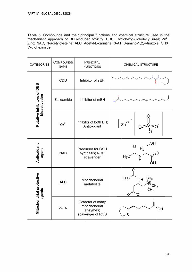
PART
TablmechZinc;Cyclo
CAT
T IV ‐ GLOBAL
e 5. Comphanistic ap; NAC, N-aoheximide.
TEGORIES
Pu
tati
ve in
hib
ito
rs o
f D
EB
b
ioac
tiva
tio
n
An
tio
xid
ant
agen
t M
ito
cho
nd
rial
pro
tect
ive
agen
ts
L DISCUSSIO
pounds andproach of cetylcystein
COMPOUND
NAME
CDU
Elaidamid
Zn2+
NAC
ALC
α-LA
N
d their princDEB-induce
ne; ALC, Ac
DS PFU
Inhib
de Inhib
InhibitoAn
Precusynth
sc
Mitom
Cofacmito
escave
cipal functied toxicity.cetyl-L-carn
RINCIPAL
UNCTIONS
bitor of sEH
bitor of mEH
or of both Entioxidant
rsor for GShesis; ROScavenger
ochondrial metabolite
ctor of manyochondrial nzymes;
enger of RO
ons and chCDU, Cyc
nitine; 3-AT
H
H
EH;
SH S
y
OS
hemical strclohexyl-3-d, 3-amino-1
CHEMICAL
ructure usedodecyl ure1,2,4-triazol
L STRUCTUR
84
d in the ea; Zn2+, le; CHX,
E
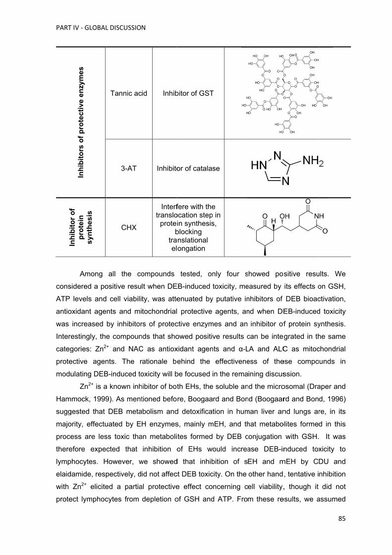
PART
Inh
ibit
or
of
cons
ATP
antio
was
Intere
categ
prote
modu
Ham
sugg
majo
proce
there
lymp
elaid
with
prote
T IV ‐ GLOBAL
Inh
ibit
ors
of
pro
tect
ive
enzy
mes
In
hib
ito
r o
f p
rote
in
syn
thes
is
Among
sidered a po
levels and
oxidant age
increased
estingly, the
gories: Zn2
ective agen
ulating DEB
Zn2+ is a
mmock, 1999
gested that
ority, effectu
ess are les
efore expe
phocytes. H
damide, resp
Zn2+ elicite
ect lymphoc
L DISCUSSIO
Tannic ac
3-AT
CHX
all the co
ositive resu
cell viabili
nts and mi
by inhibitor
e compoun+ and NAC
nts. The r
B-induced to
a known inh
9). As ment
DEB meta
uated by E
ss toxic tha
cted that
However, w
pectively, d
ed a partia
cytes from
N
id Inhib
Inhibito
Interftranslo
proteib
trael
ompounds
lt when DE
ty, was atte
tochondrial
rs of protec
nds that sho
C as antiox
rationale be
oxicity will b
hibitor of bot
tioned befo
abolism and
H enzymes
an metaboli
inhibition o
we showed
id not affec
al protective
depletion o
bitor of GST
or of catalas
fere with theocation stepin synthesisblocking nslational ongation
tested, on
EB-induced
enuated by
protective
ctive enzym
owed positiv
xidant agen
ehind the
be focused
th EHs, the
re, Boogaa
d detoxifica
s, mainly m
tes formed
of EHs w
d that inhib
ct DEB toxic
e effect co
of GSH and
T
se
e p in s,
nly four s
toxicity, me
y putative in
agents, an
mes and an
ve results c
nts and α-L
effectivene
in the rema
e soluble an
rd and Bon
tion in hum
mEH, and th
by DEB c
ould increa
bition of s
city. On the
ncerning c
d ATP. Fro
howed po
easured by
nhibitors of
nd when DE
inhibitor of
can be inte
LA and ALC
ess of the
ining discus
nd the micro
nd (Boogaar
man liver an
hat metabo
conjugation
ase DEB-in
EH and m
other hand
ell viability,
m these re
ositive resu
its effects o
DEB bioac
EB-induced
f protein sy
egrated in th
C as mitoc
ese compo
ssion.
osomal (Dra
rd and Bon
nd lungs ar
olites forme
with GSH.
nduced tox
mEH by C
, tentative i
, though it
esults, we a
85
ults. We
on GSH,
ctivation,
d toxicity
ynthesis.
he same
chondrial
unds in
aper and
d, 1996)
re, in its
d in this
It was
xicity to
DU and
nhibition
did not
assumed

PART IV ‐ GLOBAL DISCUSSION
86
that this protective effect with Zn2+ is related to a mechanism other than inhibition of EHs.
We suggested that the mechanism behind this result could be due to the antioxidant
activity of this metal, already demonstrated by several authors (Bray and Bettger, 1990;
Prasad, 2008; Prasad et al., 2004; Sankaramanivel et al., 2010; Stehbens, 2003; Yamada
and Koizumi, 1991). In fact, all these authors recognized that Zn2+ has a protective role by
counteracting OS by four principal ways: it can decrease ROS generation, it is a
scavenger of •OH, it can induce the synthesis of metallothioneins (that contain a high
amount of cysteine) and finally, it acts as a co-factor of SOD. Additionally, the tentative
inhibition of DEB detoxification by EHs inhibitors also showed that hydrolysis of DEB by
EHs in lymphocytes, in opposition to what occurs in lungs and liver, is not the most
important route of DEB detoxification and that the GSH system can be more relevant than
it was firstly supposed.
The use of SH-donor antioxidants, like NAC, in an attempt to reduce DEB acute
toxicity in human lymphocytes can be justified by two principal ways. First, NAC is a
known precursor for GSH synthesis, increasing its intracellular levels and, therefore,
significantly improving the cellular antioxidant potential of the cells (Atkuri et al., 2007).
Secondly, NAC can act directly as free radical scavenger (Kelly, 1998). GSH depletion by
DEB compromises endogenous antioxidant system of the cells, making lymphocytes more
susceptible to pro-oxidant reactive species as well as to electrophilic agents, like DEB and
its metabolites. In fact, DEB-induced cytotoxicity, despite hitherto not completely
understood, has been related to its ability to react with protein SH groups (Loecken and
Guengerich, 2007), to mediate the formation of DNA-DNA and DNA-protein cross-links
(Michaelson-Richie et al., 2010) as well as to its capability to induce the production of
ROS and induce OS (Erexson and Tindall, 2000a; Yadavilli et al., 2007). In accordance, in
the present study we verified that NAC can protect human lymphocytes from acute toxicity
of DEB at all measured parameters, corroborating a previous study showing that NAC
inhibits ROS production and apoptosis induced by DEB in human lymphoblasts (Yadavilli
et al., 2007). Furthermore, the protective effect of NAC also suggests how relevant is DEB
conjugation with GSH in the process of detoxification of this compound, at least in
lymphocytes.
Mitochondria seems to be substantially affected in the event of exposure to DEB.
This postulate can be assumed from the results obtained with the characterization of the
acute toxicity of DEB in lymphocytes, showing a depletion on ATP levels, and from those
obtained by others, being especially relevant the work of Yadavilli and co-workers
(Yadavilli et al., 2007), that using human TK6 lymphoblast as experimental model,
reported that DEB-induced apoptosis is related with dissipation of mitochondrial
membrane potential and depletion of mitochondrial cytochrome c levels. Considering this

PART IV ‐ GLOBAL DISCUSSION
87
rationale, the use of mitochondrial protective agents to counteract DEB-induced toxicity
seems to be a logic and pertinent way. ALC, an ester of L-carnitine, serves as a source of
acetylcholine and L-glutamate, and also contributes to energy-producing reactions
(Calabrese et al., 2006). In article I we demonstrated, for the first time, that ALC elicits a
partial protective effect on DEB induced toxicity, specifically avoiding cell death, while not
presenting ATP depletion. However, it had been recently reported that ALC may lead to
SOD stabilization (Haorah et al., 2011) and it can significantly prevent the decrease of
SOD activity (Zhang et al., 2012). This means that ALC can also indirectly control ROS
levels and this may reduce mitochondrial injury, although it cannot increase ATP
production. Therefore, these facts can explain why ALC only prevents cell death, similarly
of the effect of NAC in the study of Yadavilli, and once more related with OS. α-LA, the
other mitochondrial protective agent in study, functions as an essential cofactor in
metabolic reactions involving energy utilization in mitochondria, being an essential
cofactor for many mitochondrially localized enzyme complexes (Prahalathan et al., 2006).
Besides, it has the potential to induce a substantial increase in intracellular GSH and is a
scavenger of •OH, singlet oxygen (1O2) and hypochlorous acid (HClO) (Han et al., 1997).
Our results showed that α-LA did not protect lymphocytes from DEB-induced acute
toxicity, in spite of its wide range of action. However, the combined use of α-LA and ALC
resulted in a potentiation of ALC protective effect, which agrees with the assumption that
α-LA shows beneficial effects in OS conditions, own to its synergistic action with other
antioxidants (Hagen et al., 2002; Mantovani et al., 2003; Prahalathan et al., 2006). These
results suggest that α-LA and ALC may eventually increase the total antioxidant status of
the lymphocytes, instead of increasing directly GSH and ATP levels, interacting with some
antioxidant systems, both directly and by regulation of gene transcription: ALC modulates
SOD activity, as previously explained, and α-LA, either in its oxidized or reduced form,
upregulates the transcription of the GSTP gene, (Lii et al., 2010) and also increases the
GPx, glutathione reductase (GR) and SOD activities (Odabasoglu et al., 2011), all
enzymes involved directly or indirectly in defense against OS.
The other three compounds tested were supposed to aggravate the deleterious
effect of DEB-induced toxicity: tannic acid by inhibition of GST, 3-AT by inhibition of
catalase and CHX by inhibition of protein synthesis. In fact, no aggravation was observed
in ATP and GSH levels and cell death. Firstly, these results show that the lack of tannic
acid modulatory effect on DEB toxicity may reflect redundancy on biotransformation of
DEB by GST isoforms in human lymphocytes. Besides, these cells contain moderate
levels of GSTµ and GSTπ (van Lieshout and Peters, 1998) and tannic acid has variable
inhibitory potencies for the different GST isoforms. Secondly, the results show that H2O2
production is not a major contributor for the observed DEB deleterious effects. Finally, cell

PART IV ‐ GLOBAL DISCUSSION
88
signal transduction had no effect on DEB toxicity, indicating absence of regulatory
synthesis of protective proteins following the acute effects of DEB.
Thereafter, the first objective of the present thesis was achieved: to increase the
knowledge of DEB-induced toxicity as a potential way to find out protective drugs that
could turn out to be useful to prevent spontaneous and DEB-induced CI in cultured
lymphocytes from FA patients. The selected drugs were Zn2+, NAC, ALC and α-LA.
In order to reach the second objective the putative protective effect of the
previously selected drugs, against spontaneous and DEB-induced CI in lymphocytes
cultures from FA patients, was evaluated. Considering the previous results we started to
evaluate the effect of ALC and α-LA, alone and in combination, in DEB-induced
lymphocyte cultures from HD. While α-LA (20 µM) was mostly effective in reducing the
DEB-induced CI, the response to ALC (5 mM) was not so linear, and the conjugation of
the two mitochondrial nutrients wasn’t better than α-LA alone. Some studies in vivo and in
vitro demonstrated amelioration in OS and mitochondrial activity after treatment with ALC
and α-LA. An in vitro study from Lal and co-workers (Lal et al., 2008) in primary human
fibroblast demonstrated that a combination of ALC and α-LA is highly effective in reversing
OS arising from iron overload. An in vivo study of Hagen and co-worker (Hagen et al.,
1998) in old rats with the aim to improve mitochondrial-supported bioenergetics decline
and OS increase during aging, demonstrated that feeding old rats with ALC in
combination with α-LA increased metabolism and lowered OS more than either compound
alone. In human lymphocytes exposed to DEB the effect of both compounds didn’t
corroborate these studies, at least they didn’t show a relevant capability to reduce CI.
In the present work it was firstly observed that α-LA alone, at the concentration of
20 µM, showed anti-clastogenic activity in DEB-induced lymphocyte cultures from HD.
These results are in accordance with a study of Prahalathan and co-workers (Prahalathan
et al., 2006) who demonstrated that α-LA can be a protective compound against
adriamycin, a clastogenic agent, in the erythropoietic system of rats. Another study from
Dadhania and co-workers (Dadhania et al., 2010) reported that α-LA pretreatment
ameliorates methotrexate-induced intestinal toxicity in rats. Afterwards the study was
enlarged, in the control population, testing one more concentration of α-LA, 60 µM, to
analyze if its effect was concentration dependent. It was verified that α-LA effect in DEB-
induced lymphocyte cultures was not concentration dependent (data not shown), once the
results obtained for both concentrations were similar. Therefore, the concentration of 20
µM was chosen to study the effect of α-LA in cells from FA patients. The effect in
spontaneous and DEB-induced lymphocyte cultures from FA patients, as stated in article
II, was effective in the reduction of CI in both types of cultures, being mostly pronounced

PART IV ‐ GLOBAL DISCUSSION
89
in spontaneous cultures. However, under the present experimental conditions, the α-LA
effect, measured by the reduction in the number of breaks per cell, was similar (around 30
%) in the three studied groups: DEB-induced lymphocyte cultures from HD and
spontaneous and DEB-induced cultures from FA patients.
α-LA, an endogenous and natural compound, is a mitochondrial nutrient, with a
great range of action. Liu (Liu, 2008) described a mitochondrial nutrient as an agent that
can directly or indirectly protect mitochondria from oxidative damage and improve
mitochondrial function. α-LA and its reduced form, dihydrolipoic acid (DHLA), form a redox
couple, DHLA being the predominant form (Gorąca et al., 2011). Both are small molecules
that contain two oxidized or reduced SH groups. Direct protection includes its capability to
act as a potent antioxidant in redox-cycling environment, reducing vitamin C, vitamin E
and coenzyme Q10 (Tarnopolsky, 2008), as a ROS scavenger (Han et al., 1997), as a
possible chelator of redox active metals (Gorąca et al., 2011) and as an essential cofactor
for many mitochondrial localized enzymes (Prahalathan et al., 2006). Indirect protection
includes regulation of enzymes that act in biotransformation of xenobiotics or have an
antioxidant activity, as the case of GST, GPx, SOD and GR (Lii et al., 2010; Odabasoglu
et al., 2011). Furthermore, the α-LA/DHLA couple has the ability to increase or maintain
GSH levels, by acting as a transcriptional inducer of genes governing GSH synthesis and,
potentially, by increasing substrate availability, mainly cysteine (Han et al., 1997; Shay et
al., 2008). Consequently, α-LA has the potential to maintain the antioxidant status of the
cells, as demonstrated by Zembron-Lacny and co-workers (Zembron-Lacny et al., 2009),
protecting mtDNA from oxidation, since its activity occurs specially in mitochondria. In
turn, it is commonly known that FA cells, as mentioned in the introduction of this thesis,
show overproduction of ROS, resulting in decrease in the cellular antioxidant defense
systems, increase in cellular free iron levels and a deficiency in erythrocyte SOD (Korkina
et al., 1992). Catalase activity is also decreased (Takeuchi and Morimoto, 1993) and an
association of FANCC gene with enzymes involved in the process of biotransformation is
also recognized (Kruyt et al., 1998), being related with the transcription of GSTP1 gene
(Cumming et al., 2001). It is also noteworthy that GSH levels are downregulated in FA
patients (Pagano et al., 2004). Moreover, many reports had provided evidence for
mitochondrial dysfunction in FA cells, either in its metabolism as well as in its structure
(Afanas'ev, 2006; Bogliolo et al., 2002a; Mukhopadhyay et al., 2006; Paulin-Levasseur et
al., 1998; Rousset et al., 2002). Additionally, α-LA/DHLA may reduce the proinflammatory
conditions by its interaction with NF-kB, inhibiting its activity via TNF-α (Shay et al., 2008).
In fact, FA cells show enhanced intracellular NF-kB activation and transcriptional activity
compared to normal cells (Macé et al., 2007), presenting an impairment in immunological
system. NF-kB, besides being a potent indicator of stress factors such as ROS, is also a

PART IV ‐ GLOBAL DISCUSSION
90
transcription factor that induces the expression of many genes involved in inflammation
pathway, endothelial cell migration and apoptosis.
The next step was to evaluate the effect of NAC in DEB-induced lymphocyte
cultures from HD, at the same concentration tested in lymphocyte suspensions. The
results stated in article II showed that NAC has the capability to decrease the CI in HD,
although the effect demonstrated by NAC was not only lower but also more variable
comparing with α-LA. However, concerning the effect of NAC in spontaneous and DEB-
induced lymphocyte cultures from FA patients, the results clearly demonstrated that NAC
has a better capability in decreasing the CI, being the effect most pronounced in DEB-
induced lymphocyte cultures. In opposite to the effect of α-LA, NAC effect seems to be
dependent of the treatment and health condition of the donor, since the reduction in the
number of breaks per cell was greater in DEB-induced lymphocytes from FA patients.
NAC, the acetylated variant of the amino acid L-cysteine, has been shown to
increase GSH levels, the body’s major antioxidant, as mentioned before. Of glutathione’s
three component amino acids (glutamate, glycine, and cysteine), cysteine has the lowest
intracellular concentration. Because de novo synthesis is the primary mechanism by
which GSH is replenished, cysteine availability can limit the rate of GSH synthesis in the
event of OS (Millea, 2009). Moreover, NAC has the ability to reduce extracellular cystine
to cysteine (Kelly, 1998). For this activity, also contributes the capability of NAC to be a
source of SH metabolites. As a source of SH groups, NAC can also enhance GST activity,
promote detoxification, and act directly on ROS. Thus, it is not surprising that NAC acts as
powerful scavenger of HClO and is capable of reducing •OH and H2O2 (Kelly, 1998). Once
more, NAC abilities partially correct FA cells deficiencies, mainly overproduction of ROS,
low levels of GSH and an obviously imbalance in OS defense. Additionally, NAC
apparently modulates proinflammatory cytokine concentrations, as IL-1, TNF-α and IFN-γ
(interferon gamma) (Kelly, 1998), which can be very useful to prevent infections in FA
patients, similarly to the effect of α-LA. A recent report also demonstrated that NAC can
modulate the hematological response in healthy and active males (Zembron-Lacny et al.,
2009), by measuring some hematological parameters, in particular erythropoietin (EPO),
which is a cytokine that controls erythropoiesis. It has been shown that EPO production
and erythroid differentiation are regulated by ROS, especially H2O2, which are involved in
redox-sensitive signaling pathways through downregulation of transcription factors
(Fandrey et al., 1994; Huang et al., 1996; Nagata et al., 2007). This means that ROS
generation can suppress EPO synthesis, whereas antioxidants can stimulate its synthesis.
This particularity can be very useful to regulate the erythropoiesis in FA cells, avoiding the
characteristic BMF.

PART IV ‐ GLOBAL DISCUSSION
91
Finally the last goal was to find out a cocktail with the compounds that
demonstrated better results in the decrease of CI. Therefore, the combined effect of α-LA
and NAC in spontaneous and DEB-induced cultured lymphocytes from FA patients, was
evaluated. The results were surprising, especially in spontaneous breaks, in which the
reduction was about 60% globally, and in patients with mosaicism or chimerism of about
80%, as reported in article II. The measurement of spontaneous chromosomal breaks was
of particular importance, as it goes beyond the possibility of the main anti-chromosomal
break effect of α-LA and NAC being due to a direct interaction of these agents with DEB.
Moreover, the mitotic index increased with all antioxidant treatments, especially with α-LA
plus NAC treatment, suggesting a relation between a decrease in CI and a shortening of
cell arrest in G2. It was also tried to optimize the cocktail adding Zn2+, however CI was not
improved relatively of the effect of α-LA and NAC (data not shown).
In conclusion, the main objective, to find an effective antioxidant prophylactic
cocktail to be applied, in vivo, in FA patients, was achieved. The results obtained suggest
that α-LA plus NAC cocktail may be useful to keep chromosome stability in FA patients,
which can be very important to block or delay the progression of the disease. This cocktail
may be even more effective when applied to FA mosaics and FA chimeras, after BMT, as
observed in vitro. It must be stressed that BMT only resolves BMF, curing the
hematological manifestations. However, CI is not mitigated, and predisposition to cancer,
other than leukemia, persists, especially solid tumors (Alter and Kupfer, 2002 (update
2011); Auerbach, 2009; Spanier et al., In press). Thus, controlling CI in FA patients after
BMT could be very important. Thereby, with the present thesis it is expected to open a
novel strategy to deal with FA disease.
As previously described, α-LA and NAC have excellent properties, and their use
has already shown great advantages. Both have been already approved for safe human
use with minimal or no adverse effect, showing an excellent safety profile, and they are
also inexpensive drugs. Because of that, NAC and α-LA have been in clinical use for more
than 30 years. The use of NAC is recommendable in conditions characterized by
depletion of GSH or OS, as cancer, AIDS, pulmonary diseases, influenza, heart diseases,
as well as in acetaminophen overdose (Atkuri et al., 2007; Kelly, 1998; Millea, 2009),
demonstrating to be a highly effective drug. α-LA has been used in the treatment of
diabetic polyneuropathies. Several evidences support a role for α-LA as a mitigator of OS
in type 2 diabetes (Golbidi and Laher, 2010), this drug being also used in the treatment of
atherosclerosis, cardiovascular diseases, cataract, neurodegenerative diseases, liver
diseases, AIDS and cancer (Bilska and Wlodek, 2005; Gorąca et al., 2011). Besides,

PART IV ‐ GLOBAL DISCUSSION
92
considering its mitochondrial activity, α-LA is used for aging prevention. Mitochondria
provide energy for basic metabolic processes, and their decay with age impairs cellular
metabolism and leads to cellular decline, with an increase production of damaging free
radicals. α‐LA reverses the age-associated decline in mitochondrial enzymes and,
therefore, may lower the increased risk of oxidative damage that occurs during the aging
process (Arivazhagan et al., 2003). Both drugs show excellent pharmacokinetics. NAC is
rapidly absorbed following an oral dose. Although its deacetylation in the intestinal
mucosa and lumen probably limits the absorption of intact molecules of NAC, this
apparent low bioavailability is misleading, since in the degradation process a variety of
physiologically-beneficial, SH-containing metabolites are formed (Kelly, 1998). Humans
can synthesize α-LA de novo from fatty acids and cysteine, but only in very small
amounts. Therefore, α-LA needs to be absorbed from exogenous sources, food or
nutritional complements, being easily absorbed in the stomach and crossing blood-brain
barrier, due to its amphipathic properties, which allows prevention of lipid peroxidation
(Bilska and Wlodek, 2005; Gorąca et al., 2011).
Although the question posed in the present thesis is not “new”, in that a long line of
studies, dating back to Nordenson in 1977, have been trying to correct CI with a variety of
antioxidants (see Introduction), in this work it was presented for the first time a cocktail
focused on the effects of two well-selected antioxidants in compensating CI in primary
lymphocyte cultures from FA patients.
The success of this cocktail is that α-LA and NAC act as an effective redox couple
and in two distinct but closely related processes: α-LA acts principally as a mitochondrial
nutrient and NAC as a pro-glutathione dietary supplement, being both direct ROS
scavengers. It is also very important the fact that both compounds may modulate
proinflammatory cytokine activities and concentrations, which opens a possibility that they
could prevent infections in FA patients, particularly FA children, which are even more
susceptible than normal children. Additionally, NAC also modulates EPO, a cytokine
responsible for hematopoiesis response, which could be of great importance for FA
patients, with an impaired hematopoietic system, that leads to BMF. Therefore it is
suggested that this cocktail can be tested in phase III clinical trials for decreasing CI in FA
patients, a preventive approach that has not been proposed before (Figure 10).

PART
Figu(origshowincreFA charasysteBMFNACitselfglutaα-LA
T IV ‐ GLOBAL
ure 10. Proinal figure)
w increasedeased iron lcells demacteristics rem and hem
F, respectivC can potentf together. Cathione; SOA, α-lipoic ac
L DISCUSSIO
oposed sch. Fanconi a
d levels of lievels, beingonstrate dresult in chmatopoieticely. This ctially prevenCI, chromosD, superoxcid; NAC, N
N
heme for αanemia celipid peroxidg the last c
deregulationromosome
c system, inondition cunt all this FAsome instab
xide dismutaN-acetylcyste
α-LA and ls are in p
dation, demclosely relatn in antiox
instability, ncreasing thulminates inA features.bility; ROS,ase; CAT, ceine; BMF,
NAC actioermanent p
monstrate mted with ovexidant enzfollowed byhe risk of in higher caMoreover, α, reactive ocatalase; Gbone marro
on in Fancpro-oxidant itochondriaerproductionzymes andy impairmennfection an
ancer susceα-LA and Nxygen specST, glutathow failure.
coni Anemstate. The
l dysfunction of ROS. B
d systems.nt in immunnd predispoeptibility. α
NAC can regcies; GSH, hione S-tran
93
mia cells ese cells on, show Besides, These
nological osition to -LA and generate reduced
nsferase;

PART IV ‐ GLOBAL DISCUSSION
94
.

PART V ‐ FINAL CONCLUSIONS AND FUTURE PERSPECTIVES
95
PA
RT
V –
FIN
AL
CO
NC
LU
SIO
NS
AN
D
FU
TU
RE
PE
RS
PE
CT
IVE
S

PART V ‐ FINAL CONCLUSIONS AND FUTURE PERSPECTIVES
96

PART V ‐ FINAL CONCLUSIONS AND FUTURE PERSPECTIVES
97
Final Conclusions and Future Perspectives
The present thesis provides an important and novel finding that may have
immediate clinical applicability: it was clearly evident, for the first time, that the
concomitant exposure to α-LA and NAC can drastically improve the genetic stability in
lymphocytes from FA patients. Since the available information points to an association of
cellular phenotype and clinical features with the occurrence of both cancer-proneness and
OS in FA disorder, the results presented here suggest that α-LA plus NAC can be an
effective antioxidant prophylactic cocktail to be applied, in vivo, in FA patients, with a
consequent block or delay in their characteristic BMF and early cancer development.
Because α-LA and NAC are already approved for human use and they are also
inexpensive drugs, allowing an easy access by all FA patients, the preventive application
of the cocktail proposed in this study can be of a great advantage, before the use of other
proposed therapies, namely gene therapy (Tolar et al., 2012), and others that are now
available, such as androgen therapy (Scheckenbach et al., 2012). Being this study of
great clinical potential, it is expected that it will hopefully inspire clinicians to perform the
required clinical trials. Moreover, with the application of the proposed α-LA and NAC
cocktail, future prospective studies can be designed in order to see if this preventive
approach can decrease the incidence of leukemia, in early stages of the disease, and the
incidence of solid tumors in an advanced stage of the disease.
The proposed clinical trial, using the combined administration of α-LA and NAC,
poses a few challenges. FA is a rare disease, which implies a small population of
patients, most of them children or teenagers. This requires a strong adhesion to the new
approach by parents and families. In addition, the efficacy may vary among FA patients,
as it was observed by the presence of responders and non-responders in the present
studies (see article II). However, this challenge may be easily overcome by studying the
response of each individual in an in vitro assay before starting the therapy.
On the pre-clinical perspective, mechanistic studies are still required to evaluate if
the improvement of CI due to α-LA and NAC is accompanied by an improvement of the
hematopoietic status of the patients. To achieve this goal various parameters should be
measured in primary lymphocytes from FA patients before and after the exposure to α-LA
and NAC cocktail. An initial approach would be the characterization of the general
antioxidant status of those cells, measuring haemoglobin oxidation, RBC protein oxidation
(carbonyl groups) and lipid peroxidation. Characterization of non-enzymatic antioxidant
defence (GST/GSSG ratio and endogenous levels of α-LA) and enzymatic antioxidant

PART V ‐ FINAL CONCLUSIONS AND FUTURE PERSPECTIVES
98
defense (catalase, SOD, GSTP and GSH-Px GR) should also be performed. Another
approach would be the study of cell signal transduction, which can be helpful to see if the
exposure to the α-LA and NAC cocktail can lead to synthesis of regulatory and protective
proteins. In a recent study, persistence of HbF was related with increased expression of
the FOXO3a pathway involved in OS response. Knowing that FA patients are
characterized by increased levels of HbF, the evaluation of FOXO3a pathway expression
can answer the question if it is overexpressed in FA cells and if the cocktail modulates its
regulation.
With the present thesis we expect to open a novel strategy to deal with FA
disease, focused essentially on prophylactic therapeutics. It is our hope to increase the life
expectancy of FA patients and improve their life quality, avoiding the early onset of BMF,
the recurrent infections and the development of early cancer. We tightly think that our aim
can be achieved and our mission fulfilled. With simple thoughts, simple methods and
simple ways, shown with the present thesis, we believe to have contributed to an
important advance in Fanconi anemia research.

PART VI ‐ REFERENCES
99
PA
RT
VI -
RE
FE
RE
NC
ES

PART VI ‐ REFERENCES
100

PART VI ‐ REFERENCES
101
References Afanas'ev, I.B. (2006). Interplay between superoxide and nitric oxide in thalassemia and Fanconi's anemia. Hemoglobin 30, 113‐118. Ahmed, T., Tripathi, A.K., Ahmed, R.S., and Banerjee, B.D. (2010). Assessment of phosphamidon‐induced apoptosis in human peripheral blood mononuclear cells: Protective effects of N‐acetylcysteine and curcumin. Journal of Biochemical and Molecular Toxicology 24, 286‐292. Alter, B., and Kupfer, G. (2002 (update 2011)). Fanconi Anemia. In GeneReviews, Pagon RA, Bird TD, Dolan CR, and Stephens K, eds. (Seattle (WA): University of Washington, Seattle), pp. 1993‐. Alter, B.P. (2007). Diagnosis, Genetics, and Management of Inherited Bone Marrow Failure Syndromes. ASH Education Program Book 2007, 29‐39. Alter, B.P. (2008). Diagnostic evaluation of FA. In Fanconi Anemia Guidelines for the Diagnosis and Management, M. Eiler, D. Frohnmayer , L. Frohnmayer, K. Larsen, and J. Owen, eds. (Oregon: thirth edition, FARF), pp. 33‐48. Alter, B.P., Greene, M.H., Velazquez, I., and Rosenberg, P.S. (2003). Cancer in Fanconi anemia. Blood 101, 2072. Apostolou, S., Whitmore, S.A., Crawford, J., Lennon, G., Sutherland, G.R., Callen, D.F., lanzano, L., Savino, M., D'Apolito, M., Notarangeio, A., et al. (1996). Positional cloning of the Fanconi anaemia group A gene. Nature Genetics 14, 324‐328. Arivazhagan, P., Panneerselvam, S.R., and Panneerselvam, C. (2003). Effect of DL‐alpha‐lipoic acid on the status of lipid peroxidation and lipids in aged rats. Journal of Gerontology 58A, 788‐791. Atkuri, K.R., Mantovani, J.J., Herzenberg, L.A., and Herzenberg, L.A. (2007). N‐Acetylcysteine—a safe antidote for cysteine/glutathione deficiency. Current Opinion in Pharmacology 7, 355‐359. Auerbach, A.D. (2009). Fanconi anemia and its diagnosis. Mutation Research/Fundamental and Molecular Mechanisms of Mutagenesis 668, 4‐10. Auerbach, A.D., Adler, B., and Chaganti, R. (1981). Prenatal and postnatal diagnosis and carrier detection of Fanconi anemia by a cytogenetic method. Pediatrics 67, 128. Auerbach, A.D., Rogatko, A., and Schroeder‐Kurth, T.M. (1989). International Fanconi Anemia Registry: relation of clinical symptoms to diepoxybutane sensitivity. Blood 73, 391‐396. Backer, J., and Weinstein, I. (1980). Mitochondrial DNA is a major cellular target for a dihydrodiol‐epoxide derivative of benzo[a]pyrene. Science 209, 297‐299. Backer, J.M., and Weinstein, I.B. (1982). Interaction of Benzo(a)pyrene and Its Dihydrodiol‐Epoxide Derivative with Nuclear and Mitochondrial DNA in C3H10T½ Cell Cultures. Cancer Research 42, 2764‐2769. Barlow, C., Dennery, P.A., Shigenaga, M.K., Smith, M.A., Morrow, J.D., Roberts, L.J., Wynshaw‐Boris, A., and Levine, R.L. (1999). Loss of the ataxia–telangiectasia gene product causes oxidative damage in target organs. Proceedings of the National Academy of Sciences 96, 9915‐9919. Bartók, M., and Láng, K.L. (1980). Oxiranes. In The Chemistry of Functional Groups, S. Patai, ed. (Chichester, UK: Jonh Wiley & Sons), pp. 609‐673. Barzilai, A., Rotman, G., and Shiloh, Y. (2002). ATM deficiency and oxidative stress: a new dimension of defective response to DNA damage. DNA Repair 1, 3‐25. Bigelow, D.J., and Squier, T.C. (2005). Redox modulation of cellular signaling and metabolism through reversible oxidation of methionine sensors in calcium regulatory proteins. Biochimica et Biophysica Acta (BBA) ‐ Proteins & Proteomics 1703, 121‐134. Bilska, A., and Wlodek, L. (2005). Lipoic acid ‐ the drug of the future? Pharmacological reports : PR 57, 570‐577. Bleesing, J.J.H. (2004). Assays for B Cell and Germinal Center Development. In Current Protocols in Immunology (John Wiley & Sons, Inc.), pp. 7.35.31–37.35.21.
PART VI ‐ REFERENCES
102
Bogliolo, M., Borghini, S., Abbondandolo, A., and Degan, P. (2002a). Alternative metabolic pathways for energy supply and resistance to apoptosis in Fanconi anaemia. Mutagenesis 17, 25‐30. Bogliolo, M., Cabré, O., Callén, E., Castillo, V., Creus, A., Marcos, R., and Surrallés, J. (2002b). The Fanconi anaemia genome stability and tumour suppressor network. Mutagenesis 17, 529‐538. Boogaard, P.J., and Bond, J.A. (1996). The Role of Hydrolysis in the Detoxification of 1,2:3,4‐Diepoxybutane by Human, Rat, and Mouse Liver and Lungin Vitro. Toxicology and Applied Pharmacology 141, 617‐627. Boogaard, P.J., Sumner, S.C.J., and Bond, J.A. (1996). Glutathione Conjugation of 1,2:3,4‐Diepoxybutane in Human Liver and Rat and Mouse Liver and Lungin Vitro. Toxicology and Applied Pharmacology 136, 307‐316. Bray, T.M., and Bettger, W.J. (1990). The physiological role of zinc as an antioxidant. Free Radical Biology and Medicine 8, 281‐291. Briot, D., Macé‐Aimé, G., Subra, F., and Rosselli, F. (2008). Aberrant activation of stress‐response pathways leads to TNF‐α oversecretion in Fanconi anemia. Blood 111, 1913‐1923. Brockmann, W.G., Kostoryz, E.L., and Eick, J.D. (2006). Correlation of apoptotic potential of simple oxiranes with cytotoxicity. Toxicology in Vitro 20, 729‐735. Brunet, A., Sweeney, L.B., Sturgill, J.F., Chua, K.F., Greer, P.L., Lin, Y., Tran, H., Ross, S.E., Mostoslavsky, R., Cohen, H.Y., et al. (2004). Stress‐dependent regulation of FOXO transcription factors by the SIRT1 deacetylase. Science (New York, NY) 303, 2011‐2015. Caffieri, S., Zarebska, Z., and Dall'Acqua, F. (1996). Psoralen photosensitization: damages to nucleic acid and membrane lipid components. Acta Biochim Polonica 43, 241‐246. Calabrese, V., Stella, A.M.G., Calvani, M., and Butterfield, D.A. (2006). Acetylcarnitine and cellular stress response: roles in nutritional redox homeostasis and regulation of longevity genes. The Journal of Nutritional Biochemistry 17, 73‐88. Callén, E., Casado, J.A., Tischkowitz, M.D., Bueren, J.A., Creus, A., Marcos, R., Dasí, A., Estella, J.M., Muñoz, A., Ortega, J.J., et al. (2005). A common founder mutation in FANCA underlies the world's highest prevalence of Fanconi anemia in Gypsy families from Spain. Blood 105, 1946‐1949. Cantor, S., Drapkin, R., Zhang, F., Lin, Y., Han, J., Pamidi, S., and Livingston, D.M. (2004). The BRCA1‐associated protein BACH1 is a DNA helicase targeted by clinically relevant inactivating mutations. Proceedings of the National Academy of Sciences of the United States of America 101, 2357‐2362. Cantor, S.B., Bell, D.W., Ganesan, S., Kass, E.M., Drapkin, R., Grossman, S., Wahrer, D.C.R., Sgroi, D.C., Lane, W.S., Haber, D.A., et al. (2001). BACH1, a Novel Helicase‐like Protein, Interacts Directly with BRCA1 and Contributes to Its DNA Repair Function. Cell 105, 149‐160. Castella, M., Pujol, R., Callén, E., Ramírez, M.J., Casado, J.A., Talavera, M., Ferro, T., Muñoz, A., Sevilla, J., Madero, L., et al. (2011). Chromosome fragility in patients with Fanconi anaemia: diagnostic implications and clinical impact. Journal of Medical Genetics 48, 242‐250. Castello, G., Gallo, C., Napolitano, M., and Ascierto, P.A. (1998). Immunological phenotype analysis of patients with Fanconi's anaemia and their family members. Acta Haematologica 100, 39‐43. Castillo, P., Bogliolo, M., and Surralles, J. (2011). Coordinated action of the Fanconi anemia and ataxia telangiectasia pathways in response to oxidative damage. DNA Repair 10, 518‐525. Chang, T.‐S., Cho, C.‐S., Park, S., Yu, S., Kang, S.W., and Rhee, S.G. (2004). Peroxiredoxin III, a Mitochondrion‐specific Peroxidase, Regulates Apoptotic Signaling by Mitochondria. Journal of Biological Chemistry 279, 41975‐41984. Clarke, A.A., Marsh, J.C., Gordon‐Smith, E.C., and Rutherford, T.R. (1998). Molecular genetics and Fanconia anaemia: new insights into old problems. British Journal of Haematology 103, 287‐296. Cumming, R.C., Lightfoot, J., Beard, K., Youssoufian, H., O'Brien, P.J., and Buchwald, M. (2001). Fanconi anemia group C protein prevents apoptosis in hematopoietic cells through redox regulation of GSTP1. Nature Medicine 7, 814‐820.

PART VI ‐ REFERENCES
103
Dadhania, V.P., Tripathi, D.N., Vikram, A., Ramarao, P., and Jena, G.B. (2010). Intervention of α‐lipoic acid ameliorates methotrexate‐induced oxidative stress and genotoxicity: A study in rat intestine. Chemico‐Biological Interactions 183, 85‐97. Dallapiccola, B., Porfirio, B., Mokini, V., Alimena, G., Isacchi, G., and Gandini, E. (1985). Effect of oxidants and antioxidants on chromosomal breakage in Fanconi anemia lymphocytes. Human Genetics 69, 62‐65. de Winter, J.P., Rooimans, M.A., van der Weel, L., van Berkel, C.G.M., Alon, N., Bosnoyan‐Collins, L., de Groot, J., Zhi, Y., Waisfisz, Q., Pronk, J.C., et al. (2000). The Fanconi anaemia gene FANCF encodes a novel protein with homology to ROM. Nature Genetics 24, 15‐16. Degan, P., Bonassi, S., Caterina, M.D., Korkina, L.G., Pinto, L., Scopacasa, F., Zatterale, A., Calzone, R., and Pagano, G. (1995). In vivo accumulation of 8‐hydroxy‐2'‐deoxyguanosine in DNA correlates with release of reactive oxygen species in Fanconi's anaemia families. Carcinogenesis 16, 735‐742. Diamond, L., and Shahidi, N. (1967). Treatment of aplastic anemia in children. Seminars in Hematology 4, 278‐288. Dohi, Y., Ikura, T., Hoshikawa, Y., Katoh, Y., Ota, K., Nakanome, A., Muto, A., Omura, S., Ohta, T., Ito, A., et al. (2008). Bach1 inhibits oxidative stress‐induced cellular senescence by impeding p53 function on chromatin. Nature Structural & Molecular Biology 15, 1246‐1254. Dorsman, J.C., Levitus, M., Rockx, D., Rooimans, M.A., Oostra, A.B., Haitjema, A., Bakker, S.T., Steltenpool, J., Schuler, D., Mohan, S., et al. (2007). Identification of the Fanconi anemia complementation group I gene, FANCI. Cellular Oncology: The Official Journal of the International Society for Cellular Oncology 29, 211‐218. Draper, A.J., and Hammock, B.D. (1999). Inhibition of soluble and microsomal epoxide hydrolase by zinc and other metals. Toxicological Sciences 52, 26‐32. Du, W., Adam, Z., Rani, R., Zhang, X., and Pang, Q. (2008). Oxidative stress in Fanconi anemia hematopoiesis and disease progression. Antioxidants & Redox Signaling 10, 1909‐1921. Emerit, I. (2007). Clastogenic Factors as Potential Biomarkers of Increased Superoxide Production. Biomarker Insights 2, 429‐438. Erexson, G.L., and Tindall, K.R. (2000a). Micronuclei and gene mutations in transgenic Big Blue® mouse and rat fibroblasts after exposure to the epoxide metabolites of 1,3‐butadiene. Mutation Research/Genetic Toxicology and Environmental Mutagenesis 472, 105‐117. Erexson, G.L., and Tindall, K.R. (2000b). Reduction of diepoxybutane‐induced sister chromatid exchanges by glutathione peroxidase and erythrocytes in transgenic Big Blue® mouse and rat fibroblasts. Mutation Research/Fundamental and Molecular Mechanisms of Mutagenesis 447, 267‐274. Fagerlie, S.R., and Bagby, G.C. (2006). Immune defects in Fanconi anemia. Critical Reviews in Immunology 26, 81‐96. Fanconi, G. (1927). Familiaere infantile perniziosaartige Anaemie (pernizioeses Blutbild und Konstitution). Jahrbuch Kinderheild 117, 257‐280. Fandrey, J., Frede, S., and Jelkmann, W. (1994). Role of hydrogen peroxide in hypoxia‐induced erythropoietin production. Biochemistry Journal 303, 507‐510. Futaki, M., Igarashi, T., Watanabe, S., Kajigaya, S., Tatsuguchi, A., Wang, J., and Liu, J.M. (2002). The FANCG Fanconi anemia protein interacts with CYP2E1: possible role in protection against oxidative DNA damage. Carcinogenesis 23, 67‐72. Gille, J.J., and Joenje, H. (1992). Cell culture models for oxidative stress: superoxide and hydrogen peroxide versus normobaric hyperoxia. Mutatation Research/DNAging 275, 405‐414. Golbidi, S., and Laher, I. (2010). Antioxidant therapy in human endocrine disorders. Medical Science Monitor : International Medical Journal of Experimental and Clinical Research 16, RA9‐24. Gorąca, A., Huk‐Kolega, H., Piechota, A., Kleniewska, P., Ciejka, E., and Skibska, B. (2011). Lipoic acid ‐ biological activity and therapeutic potential. Pharmacological reports : PR 63, 849‐858. Gordon, S., and Taylor, P.R. (2005). Monocyte and macrophage heterogeneity. Nature Reviews Immunology 5, 953‐964.

PART VI ‐ REFERENCES
104
Green, A.M., and Kupfer, G.M. (2009). Fanconi Anemia. Hematology/Oncology Clinics of North America 23, 193‐214. Gregory, J.J., Wagner, J.E., Verlander, P.C., Levran, O., Batish, S.D., Eide, C.R., Steffenhagen, A., Hirsch, B., and Auerbach, A.D. (2001). Somatic mosaicism in Fanconi anemia: Evidence of genotypic reversion in lymphohematopoietic stem cells. Proceedings of the National Academy of Sciences 98, 2532‐2537. Guinan, E.C., Lopez, K.D., Huhn, R.D., Felser, J.M., and Nathan, D.G. (1994). Evaluation of granulocyte‐macrophage colony‐stimulating factor for treatment of pancytopenia in children with Fanconi anemia. The Journal of Pediatrics 124, 144‐150. Gyger, M., Perreault, C., Belanger, R., Bonny, Y., Forest, L., and Lussier, P. (1989). Unsuspected Fanconi's Anemia and Bone Marrow Transplantation in Cases of Acute Myelomonocytic Leukemia. New England Journal of Medicine 321, 120‐121. Hadjur, S., Ung, K., Wadsworth, L., Dimmick, J., Rajcan‐Separovic, E., Scott, R.W., Buchwald, M., and Jirik, F.R. (2001). Defective hematopoiesis and hepatic steatosis in mice with combined deficiencies of the genes encoding Fancc and Cu/Zn superoxide dismutase. Blood 98, 1003‐1011. Hagen, T.M., Ingersoll, R.T., Wehr, C.M., Lykkesfeldt, J., Vinarsky, V., Bartholomew, J.C., Song, M.‐H., and Ames, B.N. (1998). Acetyl‐l‐carnitine fed to old rats partially restores mitochondrial function and ambulatory activity. Proceedings of the National Academy of Sciences 95, 9562‐9566. Hagen, T.M., Liu, J., Lykkesfeldt, J., Wehr, C.M., Ingersoll, R.T., Vinarsky, V., Bartholomew, J.C., and Ames, B.N. (2002). Feeding acetyl‐l‐carnitine and lipoic acid to old rats significantly improves metabolic function while decreasing oxidative stress. Proceedings of the National Academy of Sciences 99, 1870‐1875. Han, D., Handelman, G., Marcocci, L., Sen, C.K., Roy, S., Kobuchi, H., Tritschler, H.J., Flohe, L., and Packer, L. (1997). Lipoic acid increases de novo synthesis of cellular glutathione by improving cystine utilization. Biofactors 6, 321‐338. Haorah, J., Floreani, N.A., Knipe, B., and Persidsky, Y. (2011). Stabilization of superoxide dismutase by acetyl‐l‐carnitine in human brain endothelium during alcohol exposure: Novel protective approach. Free Radical Biology and Medicine 51, 1601‐1609. Hersey, P., Edwards, A., Lewis, R., Kemp, A., and McInnes, J. (1982). Deficient natural killer cell activity in a patient with Fanconi's anaemia and squamous cell carcinoma. Association with defect in interferon release. Clinical and Experimental Immunology 48, 205‐212. Howlett, N.G., Taniguchi, T., Olson, S., Cox, B., Waisfisz, Q., De Die‐Smulders, C., Persky, N., Grompe, M., Joenje, H., Pals, G., et al. (2002). Biallelic inactivation of BRCA2 in Fanconi anemia. Science (New York, NY) 297, 606‐609. Huang, H., and Tindall, D.J. (2011). Regulation of FOXO protein stability via ubiquitination and proteasome degradation. Biochimica et Biophysica Acta (BBA) ‐ Molecular Cell Research 1813, 1961‐1964. Huang, L.E., Arany, Z., Livingston, D.M., and Bunn, H.F. (1996). Activation of Hypoxia‐inducible Transcription Factor Depends Primarily upon Redox‐sensitive Stabilization of Its α Subunit. Journal of Biological Chemistry 271, 32253‐32259. Hucl, T., and Gallmeier, E. (2011). DNA repair: exploiting the Fanconi anemia pathway as a potential therapeutic target. Physiological Research / Academia Scientiarum Bohemoslovaca 60, 453‐465. Irons, R.D., Pyatt, D.W., Stillman, W.S., Som, D.B., Claffey, D.J., and Ruth, J.A. (2000). Comparative toxicity of known and putative metabolites of 1,3‐butadiene in human CD34+ bone marrow cells. Toxicology 150, 99‐106. Joenje, H., Arwert, F., Eriksson, A.W., de Koning, H., and Oostra, A.B. (1981). Oxygen‐dependence of chromosomal aberrations in Fanconi's anaemia. Nature 290, 142‐143. Joenje, H., and Oostra, B. (1983). Effect of oxygen tension on chromosomal aberrations in Fanconi anaemia. Human Genetics 65, 99‐101.

PART VI ‐ REFERENCES
105
Kalb, R., Neveling, K., Nanda, I., Schindler, D., and Hoehn, H. (2006). Fanconi anemia: causes and consequences of genetic instability. Genome dynamics 1, 218‐242. Kaneko, M., Panagia, V., Paolillo, G., Majumder, S., Ou, C., and Dhalla, N.S. (1990). Inhibition of cardiac phosphatidylethanolamine N‐methylation by oxygen free radicals. Biochimica et Biophysica Acta (BBA) ‐ Biomembranes 1021, 33‐38. Kannan, K., and Jain, S.K. (2000). Oxidative stress and apoptosis. Pathophysiology 7, 153‐163. Kelly, G.S. (1998). Clinical applications of N‐acetylcysteine. Alternative Medicine Review : A Journal of Clinical Therapeutic 3, 114‐127. Kim, Y., Lach, F.P., Desetty, R., Hanenberg, H., Auerbach, A.D., and Smogorzewska, A. (2011). Mutations of the SLX4 gene in Fanconi anemia. Nature Genetics 43, 142‐146. Kitamuro, T., Takahashi, K., Ogawa, K., Udono‐Fujimori, R., Takeda, K., Furuyama, K., Nakayama, M., Sun, J., Fujita, H., Hida, W., et al. (2003). Bach1 Functions as a Hypoxia‐inducible Repressor for the Heme Oxygenase‐1 Gene in Human Cells. Journal of Biological Chemistry 278, 9125‐9133. Kligerman, A.D., DeMarini, D.M., Doerr, C.L., Hanley, N.M., Milholland, V.S., and Tennant, A.H. (1999). Comparison of cytogenetic effects of 3,4‐epoxy‐1‐butene and 1,2:3, 4‐diepoxybutane in mouse, rat and human lymphocytes following in vitro G0 exposures. Mutation Research/Genetic Toxicology and Environmental Mutagenesis 439, 13‐23. Korkina, L.G., Deeva, I.B., De Biase, A., Iaccarino, M., Oral, R., Warnau, M., and Pagano, G. (2000). Redox‐dependent toxicity of diepoxybutane and mitomycin C in sea urchin embryogenesis. Carcinogenesis 21, 213‐220. Korkina, L.G., Samochatova, E.V., Maschan, A.A., Suslova, T.B., Cheremisina, Z.P., and Afanas'ev, I.B. (1992). Release of active oxygen radicals by leukocytes of Fanconi anemia patients. Journal of Leukocyte Biology 52, 357‐362. Kruyt, F.A.E., Hoshino, T., Liu, J.M., Joseph, P., Jaiswal, A.K., and Youssoufian, H. (1998). Abnormal Microsomal Detoxification Implicated in Fanconi Anemia Group C by Interaction of the FAC Protein With NADPH Cytochrome P450 Reductase. Blood 92, 3050‐3056. Kutler, D.I., and Auerbach, A.D. (2004). Fanconi anemia in Ashkenazi Jews. Familial Cancer 3, 241‐248. Kutler, D.I., Singh, B., Satagopan, J., Batish, S.D., Berwick, M., Giampietro, P.F., Hanenberg, H., and Auerbach, A.D. (2003). A 20‐year perspective on the International Fanconi Anemia Registry (IFAR). Blood 101, 1249‐1256. Lal, A., Atamna, W., Killilea, D., Suh, J., and Ames, B. (2008). Lipoic acid and acetyl‐carnitine reverse iron‐induced oxidative stress in human fibroblasts. Redox Report 13, 2‐10. Lebbé, C., Pinquier, L., Rybojad, M., Chomienne, C., Ochonisky, S., Miclea, J.M., Gluckman, E., and Morel, P. (1993). Fanconi's anaemia associated with multicentric Bowen's disease and decreased NK cytotoxicity. The British Journal of Dermatology 129, 615‐618. Levitus, M., Waisfisz, Q., Godthelp, B.C., Vries, Y.d., Hussain, S., Wiegant, W.W., Elghalbzouri‐Maghrani, E., Steltenpool, J., Rooimans, M.A., Pals, G., et al. (2005). The DNA helicase BRIP1 is defective in Fanconi anemia complementation group J. Nature Genetics 37, 934‐935. Levran, O., Attwooll, C., Henry, R.T., Milton, K.L., Neveling, K., Rio, P., Batish, S.D., Kalb, R., Velleuer, E., Barral, S., et al. (2005). The BRCA1‐interacting helicase BRIP1 is deficient in Fanconi anemia. Nature Genetics 37, 931‐933. Li, J., Du, W., Maynard, S., Andreassen, P.R., and Pang, Q. (2010). Oxidative stress–specific interaction between FANCD2 and FOXO3a. Blood 115, 1545‐1548. Liebetrau, W., Rünge, T.M., Baumer, A., Henning, C., Gross, O., Schindler, D., Poot, M., and Hoehn, H. (1997). Exploring the role of oxygen in Fanconi's anemia. Recent Results in Cancer Research 143, 353‐367. Lii, C.‐K., Liu, K.‐L., Cheng, Y.‐P., Lin, A.‐H., Chen, H.‐W., and Tsai, C.‐W. (2010). Sulforaphane and α‐Lipoic Acid Upregulate the Expression of the π Class of Glutathione S‐Transferase through c‐Jun and Nrf2 Activation. The Journal of Nutrition 140, 885‐892.
PART VI ‐ REFERENCES
106
Litman, R., Peng, M., Jin, Z., Zhang, F., Zhang, J., Powell, S., Andreassen, P.R., and Cantor, S.B. (2005). BACH1 is critical for homologous recombination and appears to be the Fanconi anemia gene product FANCJ. Cancer Cell 8, 255‐265. Liu, J. (2008). The Effects and Mechanisms of Mitochondrial Nutrient α‐Lipoic Acid on Improving Age‐Associated Mitochondrial and Cognitive Dysfunction: An Overview. Neurochemical Research 33, 194‐203. Liu, Y., Ballman, K., Li, D., Khan, S., Derr‐Yellin, E., Shou, W., and Haneline, L.S. (2012). Impaired function of Fanconi anemia type C‐deficient macrophages. Journal of Leukocyte Biology 91, 333‐340. Lo Ten Foe, J.R., Kwee, M.L., Rooimans, M.A., Oostra, A.B., Veerman, A.J., van Weel, M., Pauli, R.M., Shahidi, N.T., Dokal, I., Roberts, I., et al. (1997). Somatic mosaicism in Fanconi anemia: molecular basis and clinical significance. European Journal Human Genetics 5, 137‐148. Lo Ten Foe, J.R., Rooimans, M.A., Bosnoyan‐Collins, L., Alon, N., Wijker, M., Parker, L., Lightfoot, J., Carreau, M., Callen, D.F., Savoia, A., et al. (1996). Expression cloning of a cDNA for the major Fanconi anaemia gene, FAA. Nature Genetics 14, 320‐323. Loecken, E.M., and Guengerich, F.P. (2007). Reactions of Glyceraldehyde 3‐Phosphate Dehydrogenase Sulfhydryl Groups with Bis‐Electrophiles Produce DNA–Protein Cross‐Links but Not Mutations. Chemical Research in Toxicology 21, 453‐458. Macé, G., Briot, D., Guervilly, J.H., and Rosselli, F. (2007). L'anémie de Fanconi : aspects cellulaires et moléculaires. Pathologie Biologie 55, 19‐28. MacMillan, M.L., and Wagner, J.E. (2010). Haematopoeitic cell transplantation for Fanconi anaemia – when and how? British Journal of Haematology 149, 14‐21. Mantovani, G., Macciò, A., Madeddu, C., Mura, L., Gramignano, G., Lusso, M., Massa, E., Mocci, M., and Serpe, R. (2003). Antioxidant agents are effective in inducing lymphocyte progression through cell cycle in advanced cancer patients: assessment of the most important laboratory indexes of cachexia and oxidative stress. Journal of Molecular Medicine 81, 664‐673. Masserot, C., Peffault de Latour, R., Rocha, V., Leblanc, T., Rigolet, A., Pascal, F., Janin, A., Soulier, J., Gluckman, E., and Socié, G. (2008). Head and neck squamous cell carcinoma in 13 patients with Fanconi anemia after hematopoietic stem cell transplantation. Cancer 113, 3315‐3322. Meetei, A.R., de Winter, J.P., Medhurst, A.L., Wallisch, M., Waisfisz, Q., van de Vrugt, H.J., Oostra, A.B., Yan, Z., Ling, C., Bishop, C.E., et al. (2003). A novel ubiquitin ligase is deficient in Fanconi anemia. Nature Genetics 35, 165‐170. Meetei, A.R., Levitus, M., Xue, Y., Medhurst, A.L., Zwaan, M., Ling, C., Rooimans, M.A., Bier, P., Hoatlin, M., Pals, G., et al. (2004). X‐linked inheritance of Fanconi anemia complementation group B. Nature Genetics 36, 1219‐1224. Meetei, A.R., Medhurst, A.L., Ling, C., Xue, Y., Singh, T.R., Bier, P., Steltenpool, J., Stone, S., Dokal, I., Mathew, C.G., et al. (2005). A human ortholog of archaeal DNA repair protein Hef is defective in Fanconi anemia complementation group M. Nature Genetics 37, 958‐963. Michaelson‐Richie, E.D., Loeber, R.L., Codreanu, S.G., Ming, X., Liebler, D.C., Campbell, C., and Tretyakova, N.Y. (2010). DNA−Protein Cross‐Linking by 1,2,3,4‐Diepoxybutane. Journal of Proteome Research 9, 4356‐4367. Millard, J.T., Hanly, T.C., Murphy, K., and Tretyakova, N. (2005). The 5‘‐GNC Site for DNA Interstrand Cross‐Linking Is Conserved for Diepoxybutane Stereoisomers. Chemical Research in Toxicology 19, 16‐19. Millea, P.J. (2009). N‐acetylcysteine: multiple clinical applications. American Family Physician 80, 265‐269. Mogib El‐Dahtory, F.A., and Yahia, S. (2011). Cytoprotective effect of honey against chromosomal breakage in Fanconi anemia patients in vitro. Indian Journal of Human Genetics 17, 77‐81. Moldovan, G.L., and D'Andrea, A.D. (2009). How the fanconi anemia pathway guards the genome. Annual Review of Genetics 43, 223‐249.

PART VI ‐ REFERENCES
107
Morgan, N.V., Essop, F., Demuth, I., de Ravel, T., Jansen, S., Tischkowitz, M., Lewis, C.M., Wainwright, L., Poole, J., Joenje, H., et al. (2005). A common Fanconi anemia mutation in black populations of sub‐Saharan Africa. Blood 105, 3542‐3544. Morisseau, C., and Hammock, B.D. (2005). Epoxide Hydrolases: Mechanisms, Inhibitor Designs, and Biological Roles. Annual Review of Pharmacology and Toxicology 45, 311‐333. Mukhopadhyay, S.S., Leung, K.S., Hicks, M.J., Hastings, P.J., Youssoufian, H., and Plon, S.E. (2006). Defective mitochondrial peroxiredoxin‐3 results in sensitivity to oxidative stress in Fanconi anemia. Journal of Cell Biology 175, 225‐235. Myers, K.C., Bleesing, J.J., Davies, S.M., Zhang, X., Martin, L.J., Mueller, R., Harris, R.E., Filipovich, A.H., Kovacic, M.B., Wells, S.I., et al. (2011). Impaired immune function in children with Fanconi anaemia. British Journal Haematology 154, 234‐240. Nagata, M., Arimitsu, N., Ito, T., and Sekimizu, K. (2007). Antioxidant N‐acetyl‐l‐cysteine inhibits erythropoietin‐induced differentiation of erythroid progenitors derived from mouse fetal liver. Cell Biology International 31, 252‐256. Noll, M., Battaile, K.P., Bateman, R., Lax, T.P., Rathbun, K., Reifsteck, C., Bagby, G., Finegold, M., Olson, S., and Grompe, M. (2002). Fanconi anemia group A and C double‐mutant mice: Functional evidence for a multi‐protein Fanconi anemia complex. Experimental Hematology 30, 679‐688. Nonn, L., Berggren, M., and Powis, G. (2003). Increased Expression of Mitochondrial Peroxiredoxin‐3 (Thioredoxin Peroxidase‐2) Protects Cancer Cells Against Hypoxia and Drug‐Induced Hydrogen Peroxide‐Dependent Apoptosis11CA52995 and CA772049. Molecular Cancer Research 1, 682‐689. Nordenson, I. (1977). Effect of superoxide dismutase and catalase on spontaneously occurring chromosome breaks in patients with Fanconi's anemia. Hereditas 86, 147‐150. Odabasoglu, F., Halici, Z., Aygun, H., Halici, M., Atalay, F., Cakir, A., Cadirci, E., Bayir, Y., and Suleyman, H. (2011). α‐Lipoic acid has anti‐inflammatory and anti‐oxidative properties: an experimental study in rats with carrageenan‐induced acute and cotton pellet‐induced chronic inflammations. British Journal of Nutrition 105, 31‐43. Okada, S., Muto, A., Ogawa, E., Nakanome, A., Katoh, Y., Ikawa, S., Aiba, S., Igarashi, K., and Okuyama, R. (2010). Bach1‐dependent and ‐independent Regulation of Heme Oxygenase‐1 in Keratinocytes. Journal of Biological Chemistry 285, 23581‐23589. Packer, L. (1995). Antioxidants: molecular interactions between vitamins E and C, biothiols and carotenes. In Signalling mechanisms – from transcription factors to oxidative stress, L. Packer, and K. Wirtz, eds. (Springer, Berlin Heidelberg New York: NATO ASI Series, H 92), pp. 187‐216. Pagano, G., Aiello Talamanca, A., Castello, G., Pallardó, F.V., Zatterale, A., and Degan, P. (In press). Oxidative stress in Fanconi anaemia: from cells and molecules toward prospects in clinical management. Biological Chemistry. Pagano, G., Degan, P., D'Ischia, M., Kelly, F.J., Nobili, B., Pallardó, F.V., Youssoufian, H., and Zatterale, A. (2005). Oxidative stress as a multiple effector in Fanconi anaemia clinical phenotype. European Journal of Haematology 75, 93‐100. Pagano, G., Degan, P., d'Ischia, M., Kelly, F.J., Pallardó, F.V., Zatterale, A., Anak, S.S., Akişık, E.E., Beneduce, G., Calzone, R., et al. (2004). Gender‐ and age‐related distinctions for the in vivo prooxidant state in Fanconi anaemia patients. Carcinogenesis 25, 1899‐1909. Pagano, G., Degan, P., De Biase, A., Iaccarino, M., and Warnau, M. (2001). Diepoxybutane and mitomycin C toxicity is associated with the induction of oxidative DNA damage in sea urchin embryos. Human & Experimental Toxicology 20, 651‐655. Pagano, G., and Korkina, L.G. (2000). Prospects for nutritional interventions in the clinical management of Fanconi anemia. Cancer Causes and Control 11, 881‐889. Pallardó, F., Lloret, A., Lebel, M., d’Ischia, M., Cogger, V., Le Couteur, D., Gadaleta, M., Castello, G., and Pagano, G. (2010). Mitochondrial dysfunction in some oxidative stress‐related genetic diseases: Ataxia‐Telangiectasia, Down Syndrome, Fanconi Anaemia and Werner Syndrome. Biogerontology 11, 401‐419.

PART VI ‐ REFERENCES
108
Pang, Q., and Andreassen, P.R. (2009). Fanconi anemia proteins and endogenous stresses. Mutation Research/Fundamental and Molecular Mechanisms of Mutagenesis 668, 42‐53. Park, S.‐J., Ciccone, S.L.M., Beck, B.D., Hwang, B., Freie, B., Clapp, D.W., and Lee, S.‐H. (2004). Oxidative Stress/Damage Induces Multimerization and Interaction of Fanconi Anemia Proteins. Journal of Biological Chemistry 279, 30053‐30059. Paulin‐Levasseur, M., Chen, G., and Larivieére, C. (1998). The 2G2 Antibody Recognizes an Acidic 110‐kDa Human Mitochondrial Protein. The Histochemical Journal 30, 617‐625. Petrovic, S., Leskovac, A., Kotur‐Stevuljevic, J., Joksic, J., Guc‐Scekic, M., Vujic, D., and Joksic, G. (2011). Gender‐related differences in the oxidant state of cells in Fanconi anemia heterozygotes. Biological Chemistry 392, 625‐632. Pincheira, J., Bravo, M., Santos, M.J., de la Torre, C., and López‐Sáez, J.F. (2001). Fanconi anemia lymphocytes: effect of dl‐α‐tocopherol (Vitamin E) on chromatid breaks and on G2 repair efficiency. Mutation Research/DNA Repair 461, 265‐271. Porfirio, B., Ambroso, G., Giannella, G., Isacchi, G., and Dallapiccola, B. (1989). Partial correction of chromosome instability in Fanconi anemia by desferrioxamine. Human Genetics 83, 49‐51. Prahalathan, C., Selvakumar, E., Varalakshmi, P., Kumarasamy, P., and Saravanan, R. (2006). Salubrious effects of lipoic acid against adriamycin‐induced clastogenesis and apoptosis in Wistar rat bone marrow cells. Toxicology 222, 225‐232. Prasad, A.S. (2008). Clinical, immunological, anti‐inflammatory and antioxidant roles of zinc. Experimental Gerontology 43, 370‐377. Prasad, A.S., Bao, B., Beck, F.W.J., Kucuk, O., and Sarkar, F.H. (2004). Antioxidant effect of zinc in humans. Free Radical Biology and Medicine 37, 1182‐1190. Rackoff, W., Orazi, A., Robinson, C., Cooper, R., Alter, B., Freedman, M., Harris, R., and Williams, D. (1996). Prolonged administration of granulocyte colony‐stimulating factor (filgrastim) to patients with Fanconi anemia: a pilot study. Blood 88, 1588‐1593. Rees, A.J. (2010). Monocyte and Macrophage Biology: An Overview. Seminars in Nephrology 30, 216‐233. Reichard, J.F., Motz, G.T., and Puga, A. (2007). Heme oxygenase‐1 induction by NRF2 requires inactivation of the transcriptional repressor BACH1. Nucleic Acids Research 35, 7074‐7086. Reid, S., Schindler, D., Hanenberg, H., Barker, K., Hanks, S., Kalb, R., Neveling, K., Kelly, P., Seal, S., Freund, M., et al. (2007). Biallelic mutations in PALB2 cause Fanconi anemia subtype FA‐N and predispose to childhood cancer. Nature Genetics 39, 162‐164. Reuter, T.Y., Medhurst, A.L., Waisfisz, Q., Zhi, Y., Herterich, S., Hoehn, H., Gross, H.J., Joenje, H., Hoatlin, M.E., Mathew, C.G., et al. (2003). Yeast two‐hybrid screens imply involvement of fanconi anemia proteins in transcription regulation, cell signaling, oxidative metabolism, and cellular transport. Experimental cell research 289, 211‐221. Richter, C., Park, J.W., and Ames, B.N. (1988). Normal oxidative damage to mitochondrial and nuclear DNA is extensive. Proceedings of the National Academy of Sciences 85, 6465‐6467. Rosenberg, P.S., Alter, B.P., and Ebell, W. (2008). Cancer risks in Fanconi anemia: findings from the German Fanconi Anemia Registry. Haematologica 93, 511‐517. Rosenberg, P.S., Greene, M.H., and Alter, B.P. (2003). Cancer incidence in persons with Fanconi anemia. Blood 101, 822‐826. Rosenberg, P.S., Socié, G., Alter, B.P., and Gluckman, E. (2005). Risk of head and neck squamous cell cancer and death in patients with Fanconi anemia who did and did not receive transplants. Blood 105, 67‐73. Rosselli, F., Sanceau, J., Gluckman, E., Wietzerbin, J., and Moustacchi, E. (1994). Abnormal lymphokine production: a novel feature of the genetic disease Fanconi anemia. II. In vitro and in vivo spontaneous overproduction of tumor necrosis factor alpha. Blood 83, 1216‐1225. Rosselli, F., Sanceau, J., Wietzerbin, J., and Moustacchi, E. (1992). Abnormal lymphokine production: a novel feature of the genetic disease Fanconi anemia. Human Genetics 89, 42‐48.

PART VI ‐ REFERENCES
109
Rousset, S., Nocentini, S., Rouillard, D., Baroche, C., and Moustacchi, E. (2002). Mitochondrial Alterations in Fanconi Anemia Fibroblasts Following Ultraviolet A or Psoralen Photoactivation. Photochemistry and Photobiology 75, 159‐166. Rubin, B.Y., Smith, L.J., Hellermann, G.R., Lunn, R.M., Richardson, N.K., and Anderson, S.L. (1988). Correlation between the Anticellular and DNA Fragmenting Activities of Tumor Necrosis Factor. Cancer Research 48, 6006‐6010. Ruppitsch, W., Meißlitzer, C., Hirsch‐Kauffmann, M., and Schweiger, M. (1998). Overexpression of thioredoxin in Fanconi anemia fibroblasts prevents the cytotoxic and DNA damaging effect of mitomycin C and diepoxybutane. FEBS Letters 422, 99‐102. Ruppitsch, W., Meißlitzer, C., Weirich‐Schwaiger, H., Klocker, H., Scheidereit, C., Schweiger, M., and Hirsch‐Kauffmann, M. (1997). The role of oxygen metabolism for the pathological phenotype of Fanconi anemia. Human Genetics 99, 710‐719. Sankaramanivel, S., Rajaram, A., and Rajaram, R. (2010). Zinc protects human peripheral blood lymphocytes from Cr(III)(phenanthroline)3‐induced apoptosis. Toxicology and Applied Pharmacology 243, 405‐419. Scheckenbach, K., Morgan, M., Filger‐Brillinger, J., Sandmann, M., Strimling, B., Scheurlen, W., Schindler, D., Göbel, U., and Hanenberg, H. (2012). Treatment of the bone marrow failure in Fanconi anemia patients with danazol. Blood Cells, Molecules, and Diseases 48, 128‐131. Schindler, D., and Hoehn, H. (1988). Fanconi anemia mutation causes cellular susceptibility to ambient oxygen. American Journal of Human Genetics 43, 429‐435. Schindler, R., Mancilla, J., Endres, S., Ghorbani, R., Clark, S., and Dinarello, C. (1990). Correlations and interactions in the production of interleukin‐6 (IL‐ 6), IL‐1, and tumor necrosis factor (TNF) in human blood mononuclear cells: IL‐6 suppresses IL‐1 and TNF. Blood 75, 40‐47. Schmitt, I.M., Chimenti, S., and Gasparro, F.P. (1995). Psoralen‐protein photochemistry — a forgotten field. Journal of Photochemistry and Photobiology B: Biology 27, 101‐107. Schroeder, T., Anschultz, F., and Knoff, A. (1964). Spontane chromosomenaberrationen bei familiärer Panmyelopathie. Humangenetik 1, 194‐196. Schultz, J., and Shahidi, N. (1993). Tumor necrosis factor‐alpha overproduction in Fanconi’s anemia. American Journal of Hematology 42, 96‐201. Schultz, J.C., and Shahidi, N.T. (1994). Detection of tumor necrosis factor‐alpha in bone marrow plasma and peripheral blood plasma from patients with aplastic anemia. American Journal Hematology 45, 32‐38. Seif, A.E. (2011). Pediatric leukemia predisposition syndromes: clues to understanding leukemogenesis. Cancer Genetics 204, 227‐244. Sejas, D.P., Rani, R., Qiu, Y., Zhang, X., Fagerlie, S.R., Nakano, H., Williams, D.A., and Pang, Q. (2007). Inflammatory Reactive Oxygen Species‐Mediated Hemopoietic Suppression in Fancc‐Deficient Mice. The Journal of Immunology 178, 5277‐5287. Shay, K., Moreau, R.F., Smith, E.J., and Hagen, T.M. (2008). Is α‐lipoic acid a scavenger of reactive oxygen species in vivo? Evidence for its initiation of stress signaling pathways that promote endogenous antioxidant capacity. IUBMB Life 60, 362‐367. Shen, X., and Li, L. (2010). Mutagenic repair of DNA interstrand crosslinks. Environmental and Molecular Mutagenesis 51, 493‐499. Shigenaga, M.K., Hagen, T.M., and Ames, B.N. (1994). Oxidative damage and mitochondrial decay in aging. Proceedings of the National Academy of Sciences 91, 10771‐10778. Shimamura, A. (2008). Treatment of hematologic abnormalities in Fanconi anemia. In Fanconi Anemia Guidelines for the Diagnosis and Management, M. Eiler, D. Frohnmayer , L. Frohnmayer, K. Larsen, and J. Owen, eds. (Oregon: thirth edition, FARF), pp. 49‐75. Shimamura, A., and Alter, B.P. (2010). Pathophysiology and management of inherited bone marrow failure syndromes. Blood Reviews 24, 101‐122. Sims, A.E., Spiteri, E., Sims, R.J., Arita, A.G., Lach, F.P., Landers, T., Wurm, M., Freund, M., Neveling, K., Hanenberg, H., et al. (2007). FANCI is a second monoubiquitinated member of the Fanconi anemia pathway. Nature Structural & Molecular Biology 14, 564‐567.

PART VI ‐ REFERENCES
110
Smogorzewska, A., Matsuoka, S., Vinciguerra, P., McDonald Iii, E.R., Hurov, K.E., Luo, J., Ballif, B.A., Gygi, S.P., Hofmann, K., D'Andrea, A.D., et al. (2007). Identification of the FANCI Protein, a Monoubiquitinated FANCD2 Paralog Required for DNA Repair. Cell 129, 289‐301. Spanier, G., Pohl, F., Giese, T., Meier, J.K., Koelbl, O., and Reichert, T.E. (In press). Fatal course of tonsillar squamous cell carcinoma associated with Fanconi anaemia: A mini review. Journal of Cranio‐Maxillofacial Surgery. Spanò, M., Cordelli, E., Leter, G., and Pacchierotti, F. (1998). Diepoxybutane cytotoxicity on mouse germ cells is enhanced by in vivo glutathione depletion: a flow cytometric approach. Mutation Research/Fundamental and Molecular Mechanisms of Mutagenesis 397, 37‐43. Stehbens, W.E. (2003). Oxidative stress, toxic hepatitis, and antioxidants with particular emphasis on zinc. Experimental and Molecular Pathology 75, 265‐276. Stoepker, C., Hain, K., Schuster, B., Hilhorst‐Hofstee, Y., Rooimans, M.A., Steltenpool, J., Oostra, A.B., Eirich, K., Korthof, E.T., Nieuwint, A.W.M., et al. (2011). SLX4, a coordinator of structure‐specific endonucleases, is mutated in a new Fanconi anemia subtype. Nature Genetics 43, 138‐141. Strathdee, C.A., Gavish, H., Shannon, W.R., and Buchwald, M. (1992). Cloning of cDNAs for Fanconi's anaemia by functional complementation. Nature 356, 763‐767. Sugarman, B., Aggarwal, B., Hass, P., Figari, I., Palladino, M., and Shepard, H. (1985). Recombinant human tumor necrosis factor‐alpha: effects on proliferation of normal and transformed cells in vitro. Science 230, 943‐945. Swift, M. (1971). Fanconi's anaemia in the genetics of neoplasia. Nature 230, 370‐373. Takeuchi, T., and Morimoto, K. (1993). Increased formation of 8‐hydroxydeoxyguanosine, an oxidative DNA damage, in lymphoblasts from Fanconi's anemia patients due to possible catalase deficiency. Carcinogenesis 14, 1115‐1120. Tarnopolsky, M.A. (2008). The mitochondrial cocktail: rationale for combined nutraceutical therapy in mitochondrial cytopathies. Advanced Drug Delivery Reviews 60, 1561‐1567. Timmers, C., Taniguchi, T., Hejna, J., Reifsteck, C., Lucas, L., Bruun, D., Thayer, M., Cox, B., Olson, S., D'Andrea, A.D., et al. (2001). Positional Cloning of a Novel Fanconi Anemia Gene, FANCD2. Molecular Cell 7, 241‐248. Tolar, J., Becker, S., Clapp, D., Hanenberg, H., Heredia, C., Kiem, H.‐P., Navarro, S., Qasba, P., Rio, P., Schmidt, M., et al. (2012). Gene Therapy for Fanconi Anemia: One Step Closer to the Clinic Human Gene Therapy 23, 141‐144. van Lieshout, E.M., and Peters, W.H. (1998). Age and gender dependent levels of glutathione and glutathione S‐transferases in human lymphocytes. Carcinogenesis 19, 1873‐1875. Vaz, F., Hanenberg, H., Schuster, B., Barker, K., Wiek, C., Erven, V., Neveling, K., Endt, D., Kesterton, I., Autore, F., et al. (2010). Mutation of the RAD51C gene in a Fanconi anemia‐like disorder. Nature Genetics 42, 406‐409. Vlachodimitropoulos, D., Norppa, H., Autio, K., Catalán, J., Hirvonen, A., Tasa, G., Uusküla, M., Demopoulos, N.A., and Sorsa, M. (1997). GSTT1‐dependent induction of centromere‐negative and ‐positive micronuclei by l,2:3,4‐diepoxybutane in cultured human lymphocytes. Mutagenesis 12, 397‐403. Wunderlich, V., Tetzlaff, I., and Graffi, A. (1972). Studies on nitrosodimethylamine: Preferential methylation of mitochondrial DNA in rats and hamsters. Chemico‐Biological Interactions 4, 81‐89. Xia, B., Dorsman, J.C., Ameziane, N., de Vries, Y., Rooimans, M.A., Sheng, Q., Pals, G., Errami, A., Gluckman, E., Llera, J., et al. (2007). Fanconi anemia is associated with a defect in the BRCA2 partner PALB2. Nature Genetics 39, 159‐161. Yadavilli, S., Martinez‐Ceballos, E., Snowden‐Aikens, J., Hurst, A., Joseph, T., Albrecht, T., and Muganda, P.M. (2007). Diepoxybutane activates the mitochondrial apoptotic pathway and mediates apoptosis in human lymphoblasts through oxidative stress. Toxicology in Vitro 21, 1429‐1441. Yadavilli, S., and Muganda, P.M. (2004). Diepoxybutane induces caspase and p53‐mediated apoptosis in human lymphoblasts. Toxicology and Applied Pharmacology 195, 154‐165.

PART VI ‐ REFERENCES
111
Yamada, H., and Koizumi, S. (1991). Metallothionein induction in human peripheral blood lymphocytes by heavy metals. Chemical Biological Interactions 78, 347‐354. Yamauchi, N., Kuriyama, H., Watanabe, N., Neda, H., Maeda, M., and Niitsu, Y. (1989). Intracellular Hydroxyl Radical Production Induced by Recombinant Human Tumor Necrosis Factor and Its Implication in the Killing of Tumor Cells in Vitro. Cancer Research 49, 1671‐1675. Zembron‐Lacny, A., Slowinska‐Lisowska, M., Szygula, Z., Witkowski, K., and Szyszka, K. (2009). The comparison of antioxidant and hematological properties of N‐acetylcysteine and alpha‐lipoic acid in physically active males. Physiological Research 58, 855‐861. Zhang, Q.‐S., Eaton, L., Snyder, E.R., Houghtaling, S., Mitchell, J.B., Finegold, M., Van Waes, C., and Grompe, M. (2008). Tempol Protects against Oxidative Damage and Delays Epithelial Tumor Onset in Fanconi Anemia Mice. Cancer Research 68, 1601‐1608. Zhang, Q.‐S., Marquez‐Loza, L., Eaton, L., Duncan, A.W., Goldman, D.C., Anur, P., Watanabe‐Smith, K., Rathbun, R.K., Fleming, W.H., Bagby, G.C., et al. (2010). Fancd2−/− mice have hematopoie c defects that can be partially corrected by resveratrol. Blood 116, 5140‐5148. Zhang, R., Zhang, H., Zhang, Z., Wang, T., Niu, J., Cui, D., and Xu, S. (2012). Neuroprotective Effects of Pre‐Treament with l‐Carnitine and Acetyl‐l‐Carnitine on Ischemic Injury In Vivo and In Vitro. International Journal of Molecular Sciences 13, 2078‐2090. Zunino, A., Degan, P., Vigo, T., and Abbondandolo, A. (2001). Hydrogen peroxide: effects on DNA, chromosomes, cell cycle and apoptosis induction in Fanconi's anemia cell lines. Mutagenesis 16, 283‐288.

112