Embed Size (px)
Citation preview
Cell, Vol. 54, 423-431, July 29, 1988, Copyright 0 1988 by Cell Press
The Xenopus cdc2 Protein Is a Component of MPF, a Cytoplasmic Regulator of Mitosis
William G. Dunphy; Leonardo Brizuela,t David Beach,7 and John Newport’ l Department of Biology University of California, San Diego La Jolla, California 92093 rCold Spring Harbor Laboratory Cold Spring Harbor, New York 11724
Summary
In Xenopus, a cytoplasmic agent known as MPF in- duces entry into mitosis. In fission yeast, genetic studies have shown that the cdc2 kinase regulates mitotic initlation. The 13 kd product of the sucl gene interacts with the cdc2 kinase in yeast cells. We show that the yeast sucl gene product (~13) is a potent in- hibitor of MPF in cell-free extracts from Xenopus eggs. ~13 appears to exert its antagonistic effect by binding directly to MPF. MPF activity is quantitatively depleted by chromatography on a p13 affinity column. Con- comitantly, the Xenopus counterpart of the yeast cdc2 protein is adsorbed to the column. A 42 kd protein also binds specifically to the ~13 affinity matrix. These findings suggest that the Xenopus cdc2 protein and the 42 kd protein are components of MPF.
Introduction
During the cell cycle, most eukaryotic cells double in mass, replicate their DNA, and then distribute identical copies of their genome to progeny ceils at mitosis. It is widely assumed that an internal biochemical clock en- sures that the diverse events of the cell cycle occur in the proper sequence and at the appropriate time. A critical component of this regulatory clock is maturation, or M phase promoting factor (MPF). As cells enter mitosis, MPF sets in motion the widespread enzymatic and ultrastruc- tural changes that are necessary for cell division. In higher eukaryotes, these mitosis-specific alterations in- clude the disassembly of the nuclear envelope, the pack- aging of the genome into chromosomes, and the con- struction of a cytoskeletal spindle that will faithfully distribute the replicated genomes to each daughter cell. MPF was originally described as a cytoplasmic activity from metaphase-arrested frog eggs that would induce oo- cytes to resume meiosis prematurely, and in the absence of protein synthesis (Masui and Markert, 1971; Smith and Ecker, 1971). Subsequently, it has become clear that MPF plays a general role in mitotic induction in somatic cells ranging from yeast to man (Gerhart et al., 1984; Newport and Kirschner, 1984; Sunkara et al., 1979; Weintraub et al., 1982; Kishimoto et al., 1982; Tachibana et al., 1987).
Over the past several years, it has become possible to reconstitute many of the events of the cell cycle in cell-free extracts, mainly from amphibian eggs. In interphase ex- tracts, nuclear reassembly around chromatin or DNA
(Lohka and Masui, 1983,1984; Forbes et al., 1983; Burke and Gerace, 1988; Newport, 1987; Wilson and Newport, 1988), DNA replication (Blow and Laskey, 1986; Newport, 1987), and nuclear transport (Newmeyer et al., 1986) pro- ceed efficiently. In mitotic extracts, nuclear envelope dis- assembly, chromosome condensation, and spindle for- mation occur faithfully (Lohka and Mailer, 1985; Miake-Lye and Kirschner, 1985; Suprynowicz and Gerace, 1986; Newport and Spann, 1987; Dunphy and Newport, 1988). The conversion from interphase to mitosis follows the ad- dition of MPF-containing fractions to interphase extracts (Miake-Lye and Kirschner, 1985; Lohka and Mailer, 1985; Lohka et al., 1988; Dunphy and Newport, 1988). These cell-free systems have greatly facilitated the study of the dynamics of MPF and its molecular nature. MPF is very unstable, and its purification has proven difficult (Wu and Gerhart, 1980; Gerhart et al., 1985; Nguyen-Gia et al., 1986; Adlakha et al., 1985). However, Lohka et al. (1988) have recently purified MPF extensively, and found that two polypeptides (32 kd and 45 kd) cofractionate with the ac- tivity.
Genetically tractable organisms such as the fungi have provided a different avenue for the study of the mecha- nisms of cell-cycle progression. In Saccharomyces cerevisiae (Hartwell, 1974) Schizosaccharomyces pombe (Nurse et al., 1976), and Aspergillus nidulans (Morris, 1976) a variety of mutations that perturb the regulation of cell division have been isolated. For mitotic control in the fission yeast S. pombe, a network of interacting genes regulates the onset of mitosis. A key component in this network is the cdc2 protein kinase (Nurse and Bisset, 1981; Beach et al., 1982; Hindley and Phear, 1984; Simanis and Nurse, 1986) which is required for both the G2-M and Gl-S transitions of the cell cycle. The homolog of cdc2 in S. cerevisiae (C/X28) is likewise a protein ki- nase (Reed et al., 1985) that is required for “start” (Hart- well, 1974) and possibly mitosis (Piggott et al., 1982). In human cells, the cdc2 homolog (Draetta et al., 1987; Lee and Nurse, 1987) is present in a complex, and this com- plex possesses elevated kinase activity during mitosis (Draetta and Beach, 1988).
A number of genes that interact with cdc2 in the regula- tion of mitosis have been identified. These include cdc25 (Russell and Nurse, 1986; Fantes, 1979), wee1 (Russell and Nurse, 1987) cdcl3 (Booher and Beach, 1987), and sucl (Hayles et al., 1986a). Among these, only the interac- tion between cdc2 and sucl ha8 been elaborated in mo- lecular detail (Brizuela et al., 1987). sucl was isolated as a plasmid-borne, allele-specific suppressor of cdc2 mu- tants (Hayles et al., 1988a). It has been found that the sucl gene encodes a 13 kd protein (~13; Hindley et al., 1987) that exists in a complex with the cdc2 protein (Brizuela et al., 1987). The precise function of sucl has been un- known. Its product is not, for example, a substrate of the cdc2 kinase (Brizuela et al., 1987) but interaction be- tween sucl and cdc2 is essential for the regulation of mi- totic initiation (Hayles et al., 1986b; Hindley et al., 1987).
Cell 424
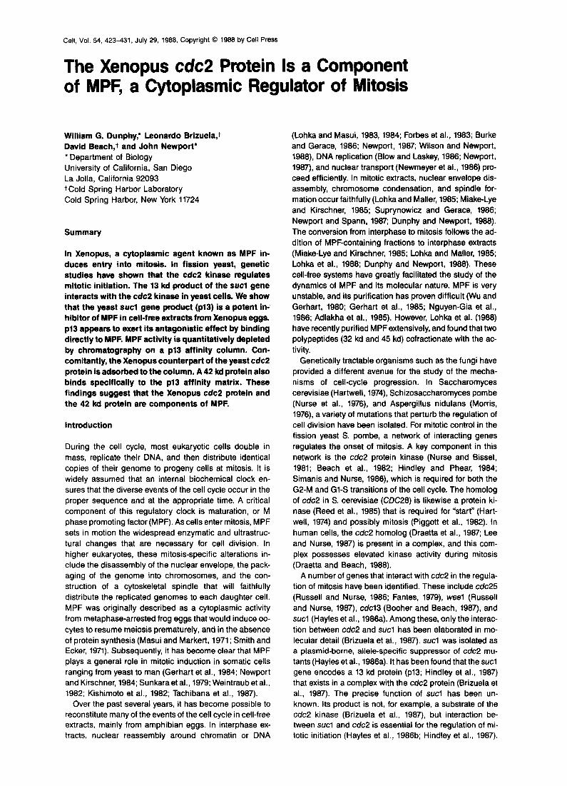
Figure 1. Fission Yeast p13 Antagonizes MPF- induced Nuclear Disassembly in a Cell-Free System Reconstituted nuclei were formed by the addi- tion of sperm chromatin to interphase egg ex- tracts (Lohka and Masui, 1983). Interphase ex- tracts containing the in vitro-assembled nuclei were incubated in the absence (A, B) or pres- ence (C-F) of 2 U of MPF per assay. In E and F, p13 (50 pglml) was included. After a 90 min incubation, nuclear disassembly was ascer- tained by the loss of the nuclear envelope (ar- rowhead). Phase-contrast microscopy (A, C, E). Hoechst staining of nuclear DNA (6, D, F). Bar = 20 urn.
Cell-free assays for mitotic induction have not yet been developed in fission yeast or the other fungal systems. Thus, it has not been possible to approach biochemically the role of the known cdc gene products in mitotic regula- tion. In particular, the relationship of the cdc proteins to MPF has been unclear. We show that it is possible to com- bine the respective technical advantages of Xenopus and yeast for analysis of the cell cycle. We demonstrate that a purified fission yeast cdc gene product can function in vitro to regulate the entry into mitosis in Xenopus egg ex- tracts. Specifically, the product of the sucl gene (~13) blocks the action of MPF by direct physical association. We have been able to take advantage of this interaction to identify and isolate polypeptide components of &lPF. One such component is the frog homolog of cdc2.
Results
Yeast p13 Blocks Mitotic Conversion in Xenopus Egg Extracts Several groups (Lohka and Mailer, 1985; Miake-Lye and Kirschner, 1985; Dunphy and Newport, 1988) have de- scribed cell-free systems from Xenopus eggs that recreate the MPF-dependent entry into mitosis in vitro. In brief, reconstituted nuclei are formed by the addition of sperm chromatin, metaphase chromosomes, or protein-free DNA to interphase cytoplasmic extracts from activated Xenopus eggs (Lohka and Masui, 1983; Blow and Laskey, 1986; Newport, 1987). Upon the addition of crude MPF, the inter- phase cytoplasm is converted to the mitotic state. As a re- sult of this mitotic conversion, the nuclear envelope sur-
Identification of MPF 425
4 6 7 8 9 10 11 12 14 16
--I3 kD
0 4 8 12 16 20
Fraction
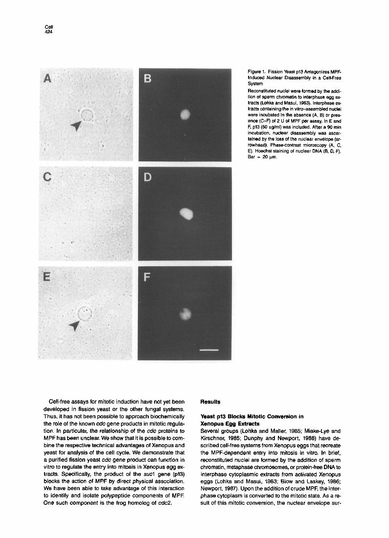
Figure 2. Mitotic Inhibitory Activity Cofractionates with Homogeneous PI3 For FPLC chromatography, p13 (4 mg protein) was applied to a Mono Q column (HR51.5; Pharmacia) equilibrated in 50 mM Tris (pH 6.0). The column was muted with a gradient of O-400 mM NaCl in the same buffer. Fractions (1 ml) were collected and assayed for protein (bottom panel) and for the maximum dilution at which inhibition of MPF- induced nuclear disassembly occurred (middle panel). p13 eluted at 140 mM NaCI. Each fraction was also electrophoresed in a 12.5% poly- acrylamide gel and stained with Coomassie blue (top panel).
rounding the reconstituted nuclei is disassembled. Thus, one can assess conveniently and rapidly the MPF-depen- dent entry into the mitotic state by observing the dissolu- tion of the nuclear envelope (see Figure 1).
To define the molecular components that are responsi- ble for the MPF-induced nuclear disassembly, we have at- tempted to develop a heterologous system by including known yeast cdc proteins in the Xenopus cell-free mitotic conversion assay. We found that one protein-the product of the fission yeast sucl gene (pl3)-inhibits MPF- induced nuclear disassembly. The p13 used in these ex- periments was derived from a strain of Escherichia coli that expresses the protein at high levels (Brizuela et al., 1987). Since p13 remains soluble in the bacteria, the pro- tein can be readily purified. In the presence of ~13, the MPF-induced loss of the nuclear envelope was completely neutralized (Figure 1). Under these MPF assay condi- tions, chromosome condensation occurs poorly (see Dun- phy and Newport, 1988). Under conditions favorable for chromosome formation, p13 also inhibited MPF-induced DNA condensation (not shown).
p13 Concentration, PM
Figure 3. Dose Response of Mitotic Inhibition by p13
MPF-induced nuclear disassembly was quantitated in the presence of 2 U of MPF per assay and the indicated concentrations of ~13. Fifty randomly selected nuclei were examined for each data point.
To confirm that p13 was the active agent responsible for the inhibition, we rechromatographed highly purified p13 on a Mono Q ion-exchange column. The observed inhibi- tory activity co-chromatographed precisely with the p13 protein (Figure 2). No other detectable polypeptides were present in the inhibitory fractions as determined by SDS gel electrophoresis. We found that the block in nuclear disassembly caused by p13 required an intact polypeptide chain. Digestion of p13 with trypsin (5% by weight of ~13) for 15 min at 37% destroyed the ability of p13 to an- tagonize mitotic conversion. On the other hand, p13 is a thermostable protein. Treatment at 100% for 10 min had no effect on the inhibitory activity of ~13.
A variety of observations suggest that the inhibitory ef- fect of p13 is physiologically significant. First, p13 acts at low concentrations. In the standard MPF assay, maximal inhibition occurred at a concentration of 2.5 uM or approxi- mately 30 uglml (Figure 3). This represents approximately 1 part in 1000 of the total protein in the in vitro incubation (~25 mglml). We also found that p13 blocked MPF- induced oocyte maturation (Wu and Gerhart, 1980) when injected into oocytes at comparable concentrations (not shown).
Second, p13 is not a general inhibitor of nuclear events in the egg extract. Even at *30-fold higher concentrations (1 mglml; the highest concentration tested), pi3 had no ef- fect on either the rate or extent of in vitro nuclear assembly around sperm chromatin (not shown). This suggests that p13 does not block MPF-induced mitotic conversion by a nonspecific mechanism such as protein denaturation or ATP depletion, since the very complex events of nuclear assembly proceed normally.
A further demonstration that the effect of p13 is specific is that the inhibition of mitotic conversion is reversible. At an MPF concentration of 2 U per assay, 2.5 uM p13 was sufficient for complete inhibition. However, at 10 U of MPF per assay, 5- to lo-fold higher concentrations of p13 were required for the block in disassembly. This observation en- abled us to perform the following experiment. We in-
1 2 12 3
p34 -
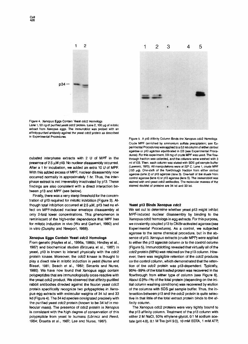
Figure 4. Xenopus Eggs Contain Yeast c&2 Homologs Lane 1, 50 ng of purified yeast c&2 protein. Lane 2, 100 ug of mitotic extract from Xenopus eggs. The immunoblot was probed with an affinity-purified antibody against the yeast cdc2 protein as described in Experimental Procedures.
cubated interphase extracts with 2 U of MPF in the presence of 2.5 uM ~13. No nuclear disassembly occurred. After a 1 hr incubation, we added an extra 10 U of MPF. With this added excess of MPF, nuclear disassembly now occurred normally in approximately 1 hr. Thus, the inter- phase extract is not irreversibly inactivated by ~13. These findings are also consistent with a direct interaction be- tween p13 and MPF (see below).
Finally, there was a very steep threshold for the concen- tration of p13 required for mitotic inhibition (Figure 3). Al- though total inhibition occurred at 2.5 PM, p13 had no ef- fect on MPF-induced nuclear envelope disassembly at only 2-fold lower concentrations. This phenomenon is reminiscent of the high-order dependence that MPF has for mitotic induction in vivo (Wu and Gerhart, 1980) and in vitro (Dunphy and Newport, 1988).
Xenopus Eggs Contain Yeast cdc2 Homologs From genetic (Hayles et al., 1988a, 1986b; Hindley et al., 1967) and biochemical studies (Brizuela et al., 1987) in yeast, p13 is known to interact physically with the c&2 protein kinase. Moreover, the c&2 kinase is thought to play a direct role in mitotic induction in yeast (Nurse and Bisset, 1981; Beach et al., 1982; Simanis and Nurse, 1966). We have now found that Xenopus eggs contain polypeptides that are immunologically cross-reactive with the yeast cdc2 product. We observed that affinity-purified rabbit antibodies directed against the fission yeast c&2 protein specifically recognize two polypeptides in Xeno- pus egg extracts with molecular weights of 34 kd and 33 kd (Figure 4). The 34 kd species comigrated precisely with the purified yeast cdc2 protein (known to be 34 kd in mo- lecular mass). The presence of cdc2 protein in Xenopus is consistent with the high degree of conservation of this polypeptide from yeast to humans (Ldrincz and Reed, 1984; Draetta et al., 1987; Lee and Nurse, 1987).
*
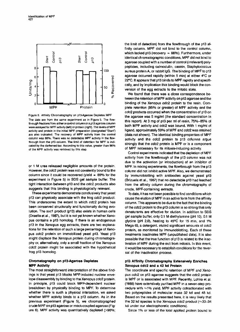
Figure 5. A p13 Affinity Column Binds the Xenopus cdc2 Homologs
Crude MPF (enriched by ammonium sulfate precipitation; see Ex- perimental Procedures) was applied to a 0.3 ml column of either control agarose or p13 agarose equilibrated in EB (see Experimental Proce- dures). For this experiment, 0.9 mg of crude MPF was used. The flow- through fraction was collected, and the columns were washed with 3 ml of EB. Then, each column was eluted with SDS gel sample buffer (Laemmli, 1970). All manipulations were at 220 C. Lane 1, crude MPF (150 ug). One-sixth of the flowthrough fraction from either control agarose (lane 2) or pi3 agarose (lane 3). One-half of the eluate from control agarose (lane 4) or p13 agarose (lane 5). The immunoblot was stained with anti-yeast cdc2 antibodies. The molecular masses of the stained doublet of proteins are 34 kd and 33 kd.
Yeast p13 Binds Xenopus cdc2 We set out to determine whether yeast p13 might inhibit MPF-induced nuclear disassembly by binding to the Xenopus c&2 homologs in egg extracts. For this purpose, we covalently coupled p13 to CNBr-activated agarose (see Experimental Procedures). As a control, we subjected agarose to the same chemical procedure, but in the ab- sence of ~13. Xenopus extracts (crude MPF) were applied to either the p13 agarose column or to the control column (Figure 5). lmmunoblotting revealed that virtually all of the cdc2 protein (98%) was retained on the p13 column. How- ever, there was negligible retention of the cdc2 products on the control column, which demonstrated that the reten- tion of the c&2 protein was pl9dependent Typically, 95%-99% of the total loaded protein was recovered in the flowthrough from either type of column (see Figure 6). About 0.3%-l% of the total protein (depending on the ini- tial column washing conditions) was recovered by elution of the columns with SDS gel sample buffer. Thus, the in- teraction between p13 and the c&2 protein is quite selec- tive in that little of the total extract protein binds to the af- finity column.
The Xenopus cdc2 proteins were very tightly bound to the p13 affinity column. Treatment of the p13 column with either 2 M NaCI, 50% ethylene glycol, 0.1 M sodium ace- tate (pH 4.0), 0.1 M Tris (pH 9.0), 10 mM EDTA, 1 mM ATP,
Identification of MPF 427
m I- MPF Protein
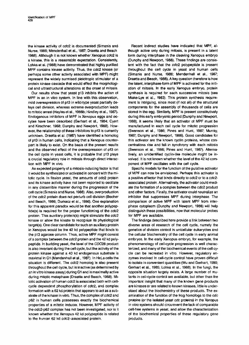
Figure 6. Affinity Chromatography on pl3-Agarose Depletes MPF
The data are from the same experiment as in Figure 5. The flow- through fractions from either a control column or a pi3 agarose column were assayed for MPF activity (left) or protein (right). The levels of MPF activity and protein in the initial MPF preparation (designated “Start”) are also indicated. The recovery of MPF activity from the control column was 66%. There was no detectable MPF activity in the flow- through from the p13 column. The limit of detection for MPF is indi- cated by the darkened bar. According to this value, greater than 96% of the MPF activity was removed by this step.
or 1 M urea released negligible amounts of the protein. However, the c&2 protein was not covalently bound to the column since it could be recovered (yield = 89% for the experiment in Figure 5) in SDS gel sample buffer. The tight interaction between p13 and the cdc2 products also suggests that this binding is physiologically relevant.
These experiments demonstrate conclusively that yeast p13 can physically associate with the frog cdc2 product. This underscores the extent to which cdc2 protein has been conserved structurally and functionally during evo- lution. The sucl product (~13) also exists in human cells (Draetta et al., 1987), but it is not yet known whether Xeno- pus contains a p13 homolog. If there is an endogenous p13 in the Xenopus egg extracts, there are two explana- tions for the retention of such a large percentage of Xeno- pus cdc2 protein on immobilized yeast ~13. Yeast p13 might displace the Xenopus protein during chromatogra- phy or, alternatively, only a small fraction of the Xenopus cdc2 protein might be associated with the hypothetical frog p13 homolog.
Chromatography on pl3-Agarose Depletes MPF Activity The most straightforward interpretation of the above find- ings is that yeast p13 blocks MPF-induced nuclear enve- lope disassembly by binding to the Xenopus cdc2 protein. In principle, p13 could block MPF-dependent nuclear breakdown by physically binding to MPF. To determine whether there is such a physical interaction, we asked whether MPF activity binds to a p13 column. As in the previous experiment (Figure 5), we chromatographed crude MPF on p13 agarose or mock-coupled agarose (Fig- ure 8). MPF activity was quantitatively depleted (>98%;
the limit of detection) from the flowthrough of the p13 af- finity column. MPF did not bind to the control column, which lacked p13 (recovery = 88%). Furthermore, under identical chromatographic conditions, MPF did not bind to agarose coupled with a number of control (irrelevant) poly- peptides, including calmodulin, casein, Staphylococcus aureus protein A, or rabbit IgG. The binding of MPF to p13 agarose occurred rapidly (within 5 min) at either 4OC or 22°C. It appears that pi3 binds to MPF rapidly and specifi- cally, and by implication this binding would block the con- version of the egg extracts to the mitotic state.
We found that there was a close correspondence be- tween the retention of MPF activity on p13 agarose and the binding of the Xenopus cdc2 protein to the resin. Com- plete retention (95% or greater) of MPF activity and the cdc2 products occurred when the concentration of p13 on the agarose was 5 mglml (the standard concentration in this report). At 3 mg of p13 per ml of resin, 750/o-85% of both MPF activity and cdc2 was bound. With 1 mglml of ligand, approximately 50% of MPF and cdc2 was retained (data not shown). The identical binding properties of MPF activity and the cdc2 protein to p13 columns argue strongly that the cdc2 protein is MPF or is a component of MPF necessary for its mitosis-inducing activity.
Control experiments indicated that the depletion of MPF activity from the flowthrough of the p13 column was not due to the activation (or introduction) of an inhibitor of MPF. In mixing experiments, the flowthrough from the p13 column did not inhibit active MPF. Also, we demonstrated by immunoblotting with antibodies against yeast p13 (Brizuela et al., 1987) that no detectable p13 had leached from the affinity column during the chromatography of crude, MPF-containing extracts.
To date, it has not been possible to find conditions which cause the elution of MPF in an active form from the affinity column. This appears to be due to the fact that the binding of the cdc2 protein to the p13 column is so strong that only denaturants are effective for elution. In addition to SDS gel sample buffer, only 0.1 M diethylamine (pH lo), 0.1 M glycine (pH 2.0), heating to 45OC for 15 min, and 1% Mega-10, a detergent, eluted significant amounts of cdc2 protein, as monitored by immunoblotting. Each of these treatments inactivates MPF (unpublished data). It is also possible that the true function of p13 is related to the inac- tivation of MPF during the exit from mitosis. In this event, it would be necessary to establish conditions for the rever- sal of the inactivation process.
~13 Affinity Chromatography Extensively Enriches Xenopus cdc2 and a 42 kd Protein The coordinate and specific retention of MPF and Xeno- pus cdc2 on p13 agarose suggests that the cdc2 protein is MPF or is associated with MPF. Recently, Lohka et al. (1988) have extensively purified MPF in a seven-step pro- cedure with -1% yield. MPF activity cofractionated with two polypeptides of molecular mass 32 kd and 45 kd. Based on the results presented here, it is very likely that the 32 kd species is the Xenopus cdc2 product (~33-34 kd under our electrophoretic conditions).
Since 1% or less of the total applied protein bound to

Cell 420
42 kD
33 kD
Figure 7. The ~13 Affinity Column Enriches Extensively the Xenopus c&2 Protein and a 42 kd Polypeptide The protocol was similar to that described in Figure 5, except that the SDS-containing eluates from either a control column (lane 1) or a p13 agarose column (lane 2) were silver-stained after electrophoresis. Pro- teins (33 kd and 42 kd) that reproducibly bound to ~13 agarose in a spe- cific manner are indicated. Only the 33 kd form of Xenopus cdc2 was evident in this MPF preparation, as noted by immunoblotting. Molecu- lar size standards (not shown) were: phosphorylase b (97 kd); bovine serum albumin (66 kd); ovalbumin (43 kd); carbonic anhydrase (31 kd); soybean trypsin inhibitor (21 kd); and lysozyme (14 kd).
either p13 agarose or control agarose, we investigated whether p13 affinity chromatography might enrich the polypeptide components of MPF extensively. Xenopus ex- tract proteins were applied to a control column and a p13 agarose column, and the columns were washed exten- sively with buffer. The columns were then eluted with SDS gel sample buffer, and the eluted proteins were fraction- ated by gel electrophoresis and silver-stained (Figure 7). We observed a background of proteins that bound nonspecifically to both control and p13 agarose. For the most part, these proteins were abundant silver-staining species in the crude MPF extract. However, we found a protein (33 kd) bound to p13 agarose but not to the control column (Figure 7, lane 2). The silver-stained 33 kd protein corresponded exactly to a band that reacted with anti- cdc2 antibodies. In comparison with the silver-staining in- tensities of known amounts of purified yeast c&2 protein, we estimate that there is -0.4 ug of Xenopus cdc2 protein
per mg of crude MPF (or 0.04% of the total protein). The Xenopus cdc2 protein did not correspond to a major silver- staining species in the extract that we applied to the column.
In addition to the cdc2 protein, we consistently ob- served a 42 kd protein that also was specifically retained on the p13 agarose column (Figure 7). This was the only additional specifically bound protein that we observed in ten similar experiments. The 42 kd protein did not comi- grate with a major polypeptide in the crude MPF extract. This polypeptide might bind directly to p13 or, alternatively, it might exist in a complex with the Xenopus cdc2 protein that is bound to the column. The 42 kd protein did not re- act, even weakly, with anti-cdc2 antibodies. There are precedents in yeast and human cells for proteins that bind to cdc2. In S. cerevisiae, the cdc2 homolog (CDC28) binds to a 40 kd protein (Mendenhall et al., 1967) whereas in human cells (Draetta and Beach, 1988) and S. pombe (Booher and Beach, unpublished data), cdc2 complexes with a 62 kd protein. Recently, Lohka et al. (1988) have ob- served a 45 kd Xenopus polypeptide in highly enriched MPF. Based on the data presented above, it seems quite likely that the 42 kd protein that binds to p13 agarose and the 46 kd protein reported by Lohka et al. (1988) are related.
Discussion
In the experiments reported here, we found that the iso- lated 13 kd product (~13) of the yeast sucl gene blocks the ability of MPF to convert interphase extracts from Xeno- pus eggs to the mitotic state. In yeast it has been estab- lished that p13 binds to the cdc2 kinase (Brizuela et al., 1987). We found that Xenopus contains homologs of the cdc2 protein, and that yeast p13 also binds to these homo- logs. We also showed that the Xenopus cdc2 homologs are efficiently retained on a p13 affinity column. Simul- taneously, MPF is quantitatively retained on the same p13 affinity support. The Xenopus cdc2 homologs (~33-34 kd) and a 42 kd polypeptide are the predominant proteins that bind specifically to p13 agarose. This polypeptide composition is very similar to that reported by Lohka et al. (1968) for MPF that was purified by conventional chro- matographic steps (32 kd and 45 kd). Taken together, these findings suggest that MPF is a mitotic regulator composed of two proteins: the Xenopus cdc2 homolog and a 42 kd protein, In addition, our results suggest that p13 regulates the function of this complex.
A body of circumstantial evidence suggests that protein phosphorylation is an important aspect of mitotic COntrOl.
Many proteins (e.g., the nuclear lamins and histones) are hyperphosphorylated during mitosis (Gerace and Blobel, 1980; Davis et al., 1983; Karsenti et al., 1987; Lohka et al., 1967). Also, many of the fungal cdc gene products are ki- nases or possess sequence motifs typical of kinases (Lorincz and Reed, 1984; Hindley and Phear, 1984; Simanis and Nurse, 1986; Russell and Nurse, 1987; Os- mani et al., 1988). Similarly, indirect evidence has sug gested that MPF might be a phosphoprotein (Wu and Gerhart, 1960; Gerhart et al., 1985). In yeast and humans,
identification of MPF 429
the kinase activity of cdc2 is documented (Simanis and Nurse, 1988; Mendenhall et al., 1987; Draetta and Beach 1988). Although it is not known whether Xenopus cdc2 is a kinase, this is a reasonable expectation. Consistently, Lohka et al. (1988) have demonstrated that highly purified MPF contains kinase activity. Thus, the cdc2 kinase (or perhaps some other activity associated with MPF) might represent the widely surmised pleiotropic stimulator of a protein kinase cascade that would effect the morphologi- cal and ultrastructural alterations at the onset of mitosis.
Our results show that yeast pl3 inhibits the action of MPF in an in vitro system. In line with this observation, mild overexpression of p13 in wild-type yeast partially de- lays cell division, whereas extreme overproduction leads to mitotic arrest (Hayles et al., 1986b; Hindley et al., 1987). Endogenous inhibitors of MPF in Xenopus eggs and oo- cytes have been described (Gerhart et al., 1984; Cyert and Kirschner, 1988; Dunphy and Newport, 1988). How- ever, the relationship of these inhibitors to p13 is currently unknown. Draetta et al. (1987) have identified a homolog of p13 in human cells, indicating that a Xenopus counter- part is likely to exist. On the basis of the present results and the observed effect of the overexpression of p13 on the cell cycle in yeast cells, it is probable that p13 plays a crucial regulatory role in mitosis through direct interac- tion with MPF in vivo.
An expected property of a mitosis-inducing factor is that it would be synthesized or activated in concert with the mi- totic cycle. In fission yeast, the amounts of cdc2 protein and its kinase activity have not been reported to oscillate in any discernible manner during the progression of the cell cycle (Simanis and Nurse, 1986). Also, overproduction of the cdc2 protein does not perturb cell division (Booher and Beach, 1986; Durkacz et al., 1966). One explanation for this apparent paradox would be that another polypep- tide(s) is required for the proper functioning of the cdc2 protein. This auxiliary protein(s) might stimulate the cdc2 kinase or allow the kinase to recognize its physiological target(s). One clear candidate for such an auxiliary protein in Xenopus would be the 42 kd polypeptide that binds to the p13 agarose column. Thus, active MPF might consist of a complex between the cdc2 protein and the 42 kd poly- peptide. In budding yeast, the level of the CDC28 product is also invariant during the cell cycle, but the activity of the protein kinase against a 40 kd endogenous substrate is maximal in Gl (Mendenhall et al., 1987). In HeLa cells the situation is different. The cdc2 homolog is also present throughout the cell cycle, but is inactive (as determined by an in vitro kinase assay) during Gl and is maximally active during mitotic metaphase (Draetta and Beach, 1988). Mi- totic activation of human cdc2 is associated both with cell- cycle dependent phosphorylation of cdc2, and complex formation with a 62 kd protein that appears to act as a sub- strate of the kinase in vitro. Thus, the complex of cdc2 and p62 in human cells possesses exactly the biochemical properties of a mitotic inducer. However, MPF activity of the cdc2-p62 complex has not been investigated, nor is it known whether the Xenopus 42 kd polypeptide is related to the human 62 kd cdc2-associated protein.
Recent indirect studies have indicated that MPF, al- though active only during mitosis, is present in a latent form during interphase in the cleaving Xenopus embryo (Dunphy and Newport, 1986). These findings are consis- tent with the fact that the cdc2 polypeptide is present throughout the cell cycle in yeast and human cells (Simanis and Nurse, 1986; Mendenhall et al., 1967; Draetta and Beach, 1988). A key question therefore is how the latent, interphase-form of MPF is activated for the initi- ation of mitosis. In the early Xenopus embryo, protein synthesis is required for each successive mitosis (see Miake-Lye et al., 1983). This protein synthesis require- ment is intriguing, since most (if not all) of the structural components for the assembly of thousands of cells are stored in the egg. Similarly, MPF is present constitutively during this early embryonic period (Dunphy and Newport, 1988). It seems likely that an activator of MPF must be manufactured in each cell cycle for mitotic progression (Swenson et al., 1986; Pines and Hunt, 1987; Murray, 1987; Dunphy and Newport, 1988). Good candidates for this activator are the known cyclin proteins, whose con- centrations rise and fall in synchrony with each mitosis (Swenson et al., 1966; Pines and Hunt, 1987). Alterna- tively, an unidentified cyclin-like molecule might be in- volved. It is not known whether the level of the 42 kd com- ponent of MPF oscillates with the cell cycle.
Specific models for the function of the putative activator of MPF can now be envisioned. Perhaps this activator is a positive effector that binds directly to cdc2 or to a cdcB associated protein. Alternatively, the activator could medi- ate the formation of a complex between the cdc2 product and other factors. Finally, the activator could neutralize an inhibitor that suppresses MPF during interphase. The comparison of active MPF with latent MPF from inter- phase cytoplasm (Dunphy and Newport, 1988) will help distinguish these possibilities, now that molecular probes for MPF are available.
The findings described here provide a link between two diverse areas of research of the cell cycle, namely the genetics of division control in unicellular eukaryotes and the cellular biochemistry of the cell cycle in early animal embryos. In the early Xenopus embryo, for example, the phenomenology of cell-cycle progression is well charac- terized, and many of the biochemical events of the cell cy- cle can be recreated in vitro. However, regulatory en- zymes involved in cell-cycle control have proven difficult to isolate in convenient quantities (Wu and Gerhart, 1980; Gerhart et al., 1985; Lohka et al., 1988). In the fungi, the opposite situation largely exists. A large number of mu- tants in cell-cycle control are available, but other than the important insight that many of the known gene products are kinases or are related to known kinases, little is under- stood about the biochemistry of these products. The ex- amination of the function of the frog homologs to the cdc proteins (or the isolated yeast cdc proteins) in the Xenopus in vitro systems should circumvent the lack of comparable cell-free systems in yeast, and allow the characterization of the biochemical properties of these regulatory gene products.
Cell 430
In summary, we have shown that the Xenopus cc/c2 pro- tein is a component of MPF. We have established a heter- ologous system in which a purified yeast protein (~13) regulates the function of Xenopus MPF in vitro. Moreover, the polypeptide components of MPF now can be isolated conveniently and in high yield by affinity chromatography on p13 agarose. These findings make it technically feasi- ble to address long-standing questions about the enzy- mology of MPF and the cell-cycle clock to which it be- longs.
Experimental Procedures
Cell-Flee Assay for MPF Cell-free assays for MPF (Lohka and Maker, 1985; Miake-Lye and Kirschner, 1985; Dunphy and Newport, 1988) were performed exactly with the modifications described previously (Dunphy and Newport, 1988). Preparation of mitotic cytoplasm and crude MPF (enriched and concentrated by ammonium sulfate precipitation) from unfertilized Xenopus eggs, based largely on the method of Wu and Gerhart (1980) was conducted exactly as published before (Dunphy and Newport, 1988). The crude MPF extract (6 mg/ml protein) used in this paper was enriched 8-fold (yield = 80%) for mitotic induction relative to unfractio- nated mitotic cytoplasm. MPF-extraction buffer (EB; Wu and Gerhart, 1980) contained 80 mM glycerol phosphate (pH 7.3) 20 mM EGTA, 15 mM MgClz , and 1 mM dithiothreitol. The definition of a unit of MPF activity in the cell-free assay was described in Dunphy and Newport (1988). In brief, U of MPF were defined as the inverse of the maximum dilution at which an MPF-containing extract would induce mitotic con- version. The specific activity of the crude MPF extract used in this pa- per was 8.3 U per mg protein.
Preparation of Flssion Yeast p13 pl3 was purified from an overproducing strain of E. coli by gel filtration on Sepharose CL-66 (Pharmacia) as described previously (Brizuela et al., 1987). For some experiments, ~13 was further purified by chroma- tography on a Mono Q column (see legend to Figure 2 for details) or a Biogel P-100 column (equilibrated with 0.1 M sodium borate; pH 8.0). Prior to addition to the cell-free Xenopus extracts, pl3 was equilibrated in EB by dialysis.
Preparation of p13 Affinity Columns p13 was conjugated to either CNBr-activated Sepharose 48 (Pharma- cia) or Affigel-10 (Bio-Rad) according to the instructions of the manufacturer. Unreacted groups on the gels were quenched with 1 M ethanolamine (pH 8.0). The concentration of coupled ~13 was 5 mg pro- tein per ml of gel unless indicated otherwise.
Electrophoretic Techniques Typically, samples were electrophoresed in 10% polyacrylamide gels (Laemmli, 1970). For visualization of ~13, a 12.5% gel was used. Elec- trophoresed proteins were transferred to lmmobilon membranes (Milli- pore) with the Polyblot apparatus (American Bionetics). Before incuba- tion with antibodies, immunoblots were treated with Tris-buffered saline (10 mM Tris-Cl, 150 mM NaCI; pH 8.0) containing 5% powdered milk and 0.1% Tween-20. After antibody binding, the blots were ex- posed to lz51-protein A (ICN). Affinity-purified antibodies against the fission yeast cdc2 and sucl proteins were described previously (Draetta et al., 1987; Brizuela et al., 1987). The batch of cdc2 antibody used in this paper is the same one used to characterize the human homolog of cdc2 (Draetta et al., 1987). The human cdc2 polypeptide was shown to be related to the fission yeast cdc2 protein by peptide mapping.
Miscellaneous Protein concentration was determined with the Bio-Rad protein assay kit with rabbit immunoglobulin as the standard. Silver staining was per- formed with the kit from Bio-Rad.
Acknowledgments
We thank G. Draetta for a generous supply of anti-cdc2 antibodies. We are also grateful to our colleagues for comments on the manuscript. This research was supported by NIH grants GM33623 and GM34607 to J. N. and D. B., respectively. J. N. is a Searle Scholar. W. G. D. is a Lucille l? Markey Scholar and was supported by funds from the Lucille P Markey Charitable Trust. L. B. received an FMGA Scholarship (Vene- zuela).
The costs of publication of this article were defrayed in part by the payment of page charges. This article must therefore be hereby marked “aoVerrisement” in accordance with 18 U.S.C. Section 1734 solely to indicate this fact.
Received June 1. 1988.
References
Adlakha, R. C., Wright, D. A., Sahasrabuddhe, C. G., Davis, F. M., Prashad, N., Bigo, H., and Rao, P N. (1985). Partial purification and characterization of mitotic factors from HeLa cells. J. Exp. Cell Res. 160, 471-482.
Beach, D. H., Durkacz, B., and Nurse, f? M. (1982). Functionally homol- ogous cell cycle control genes in budding and fission yeast. Nature 300, 706-709.
Blow, J. J., and Laskey, R. A. (1986). Initiation of DNA replication in nuclei and purified DNA by a cell-free extract of Xenopus eggs. Cell 47, 577-587.
Booher, R., and Beach, D. H. (1986). Site-specific mutagenesis of cdc2+, a cell cycle control gene of the fission yeast Schizosac- cheromyces pombe. Mol. Cell Biol. 6, 3523-3530.
Booher, R., and Beach, D. (1987). Interaction between cdcl3+ and cdc2+ in the control of mitosis in fission yeast; dissociation of the Gl and G2 roles of the cd@ protein kinase. EMBO J. 6, 3441-3447.
Brizuela, L., Draetta, G., and Beach, D. (1987). ~13~~ acts in the fis- sion yeast cell division cycle as a component of the ~34~’ protein ki- nase. EMBO J. 6, 3507-3514.
Burke, B., and Gerace, L. (1986). A cell free system to study reassembly of the nuclear envelope at the end of mitosis. Cell 44, 639-652.
Cyert, M. S., and Kirschner, M. W. (1988). Regulation of MPF activity in vitro. Cell 53, 185-195.
Davis, F. M., Tsao, T Y., Fowler, S. K., and Rao, f? N. (1983). Monoclo- nal antibodies to mitotic cells. Proc. Natl. Acad. Sci. USA 80, 2926-2930.
Draetta, G., and Beach, D. (1988). Activation of cdc2 protein kinase during mitosis in human cells: cell-cycle dependent phosphorylation and subunit rearrangement. Cell 54. 17-26.
Draetta, G., Brizuela. L., Potashkin, J., and Beach, D. (1987). Identifica- tion of p34 and ~13, human homologs of the cell cycle regulators of fis- sion yeast encoded by cdc2+ and suci+. Cell 50, 319-325. Dunphy, W. G., and Newport, J. W. (1988). Mitosis-inducing factors are present in a latent form during interphase in the Xenopus embryo. J. Cell Biol., in press.
Durkacz, B., Carr, A., and Nurse, P. (1986). Transcription of the cdc2 cell cycle control gene of the fission yeast Schizosaccharomyces pombe. EMBO J. 5, 369-373. Fantes, F! A. (1979). Epistatic gene interactions in the control of division in fission yeast. Nature 279, 428-430.
Forbes, D. J., Kirschner, M. W., and Newport, J. W. (1983). Spontane- ous formation of nucleus-like structures around bacteriophage DNA microinjected into Xenopus eggs. Cell 34, 13-23.
Gerace, L., and Blobel, G. (1980). The nuclear envelope lamina is reversibly depolymerized during mitosis. Cell 79, 277-287.
Gerhart, J., Wu, M., and Kirschner, M. (1984). Cell cycle dynamics of an M-phase-specific cytoplasmic factor in Xenopus laevis oocytes and eggs. J. Cell Biol. 98, 1247-1255. Gerhart, J., Wu, M., Cyert, M., and Kirschner, M. (1985). M-phase promoting factors from eggs of Xenopus laevis. Cytobios 43, 335-348.
Identification of MPF 431
Hartwell, L. H. (1974). Saccharomyces cerevisiae cell cycle. Bacterial. Rev. 38, 164-198. Hayles, J., Aves, S.. and Nurse, P (1988a). sucl is an essential gene involved in both the cell cycle and growth in fission yeast. EMBO J. 5, 3373-3379. Hayles, J., Beach, D., Durkacz, B., and Nurse, P (1986b). The fission yeast cell cycle control gene cdc2: isolation of a sequence sucl that suppresses c&2 mutant function. Mol. Gen. Genet. 202, 291-293.
Hindley, J., and Phear, G. A. (1984). Sequence of the cell division gene c&2 from Schizosaccharomyces pombe; patterns of splicing and ho- mology to protein kinases. Gene 31, 129-134.
Hindley, J., Phear, G. A., Stein, M., and Beach, D. (1987). sucl+ en- codes a predicted 13-kilodalton protein that is essential for cell viability and directly involved in the division cycle of Schizosaccharomyces pombe. Mol. Cell Biol. 7, 504-511. Karsenti, E.. Bravo, FL, and Kirschner, M. (1987). Phosphorylation changes associated with the early cell cycle in Xenopus eggs. Dev. Biol. 119, 442-453.
Kishimoto, T., Kuriyama, R., Kondo, H., and Kanatani, H. (1982). Gener- ality of the action of various maturation promoting factors. Exp. Cell Res. 137, 121-126.
Laemmli, U. K. (1970). Cleavage of structural proteins during the as- sembly of the head of bacteriophage T4. Nature 277, 680-685.
Lee, M. G., and Nurse, l? (1987). Complementation used to clone a hu- man homologue of the fission yeast cell cycle control gene c&2. Na- ture 327, 31-35.
Lohka, M. J., and Maker, J. L. (1985). Induction of nuclear envelope breakdown, chromosome condensation, and spindle formation in cell- free extracts. J. Cell Biol. 107, 518-523.
Lohka, M. J.. and Masui, Y. (1983). Formation in vitro of sperm pronuclei and mitotic chromosomes induced by amphibian ooplasmic components. Science 220, 7l9-721.
Lohka, M. J. and Masui, Y. (1984). Roles of cytosol and cytoplasmic particles in nuclear envelope assembly and sperm pronuclear forma- tion in cell-free preparations from amphibian eggs. J. Cell Biol. 98, 1222-1230.
Lohka. M. J., Kyes, J. L., and Mailer, J. L. (1987). Metaphase protein phosphorylation in Xenopus laevis. Mol. Cell Biol. 7, 760-768.
Lohka, M. L., Hayes, M. K., and Mailer, J. L. (1988). Purification of maturation-promoting factor, an intracellular regulator of early mitotic events. Proc. Natl. Acad. Sci. USA 85, 3009-3013.
Lorincz, A. T., and Reed, S. I. (1984). Primary structure homology be- tween the product of yeast cell division control gene CDC28 and ver- tebrate oncogenes. Nature 307, 183-185.
Masui, Y., and Markert, C. L. (197l). Cytoplasmic control of nuclear be- havior during meiotic maturation of frog oocytes. J. Exp. 2001. 177, 129-146.
Mendenhall, M. D., Jones, C. A., and Reed, S. I. (1987). Dual regula- tion of the yeast CDC28p40 protein kinase complex: cell cycle, phero- mone, and nutrient limitation effects. Cell 50, 927-935. Miake-Lye, R., and Kirschner, M. W. (1985). Induction of early mitotic events in a cell-free system. Cell 47. 165-175. Miake-Lye, R., Newport, J., and Kirschner, M. (1983). Maturation- promoting factor induces nuclear envelope breakdown in cyctohexi- mide-arrested embryos of Xenopus laevis. J. Cell Biol. 97, 81-91. Morris, N. R. (1976). Mitotic mutants of Aspefgi//us nidulans. Genet. Res. 26, 237-254.
Murray, A. W. (1987). Cyclins in meiosis and mitosis. Nature 326, 542-543.
Newmeyer, D. D., Finlay, D. R., and Forbes, D. J. (1986). In vitro trans- port of a fluorescent nuclear protein and exclusion of non-nuclear pro- teins. J. Cell Biol. 103, 2091-2102.
Newport, J. (1987). Nuclear reconstitution in vitro: stages of assembly around protein-free DNA. Cell 48, 205-217.
Newport, J. W., and Kirschner, M W. (1984). Regulation of the cell cy- de during early Xenopus development. Cell 37. 731-742. Newport, J., and Spann, T. (1987). Disassembly of the nucleus in mi-
totic extracts: membrane vesicularization, lamin disassembly, and chromosome condensation are independent processes. Cell 48, 219-230.
Nguyen-Gia, P., Bomsel, M., Labrousse, J. P, Gallien, C. L., and Wein- traub, H. (1986). Partial purification of the maturation-promoting factor MPF from unfertilized eggs of Xenopus laevis. Eur. J. Biochem. 167, 771-777.
Nurse, P., and Bissett, Y. (1981). Gene required in Gl for commitment to cell cycle and in G2 for control of mitosis in fission yeast. Nature 292, 558-560.
Nurse, P., Thuriaux, f?, and Nasmyth, K. A. (1976). Genetic control of the division cycle of the fission yeast Schizosaccharomes pombe. Mol. Gen. Genet. 146, 167-178.
Osmani, S. A., Pu, R. T., and Morris, N. R. (1988). Mitotic induction and maintenance by overexpression of a GP-specific gene that encodes a potential protein kinase. Cell 53, 237-244.
Piggott, J. A., Rai, R., and Carter, B. L. A. (1982). A bifunctional gene product involved in two phases of the cell cycle. Nature 298, 391-394.
Pines, J., and Hunt, T. (1987). Molecular cloning and characterization of the mRNA for cyclin from sea urchin eggs. EMBO J. 6, 2987-2995.
Reed, S. I., Hadwiger, J. A., and Lorincz, A. T. (1985). Protein kinase activity associated with the product of the yeast cell cycle gene CDC28. Proc. Natl. Acad. Sci. USA 82, 4055-4059.
Russell, f?, and Nurse, P (1986). cdc25+ functions as an inducer in the mitotic control of fission yeast. Cell 45, 145-153.
Russell, P, and Nurse, l? (1987). Negative regulation of mitosis by weel+, a gene encoding a protein kinase homolog. Cell 49, 559-567.
Simanis, V., and Nurse, P (1986). The cell cycle control gene cdc2+ of fission yeast encodes a protein kinase potentially regulated by phos- phorylation. Cell 45, 261-268.
Smith, L. D., and Ecker, R. E. (1971). The interaction of steroids with Rana pipiens oocytes in the induction of maturation. Dev. Biol. 25, 232-247.
Sunkara, P S., Wright, D. A., and Rao, f? N. (1979). Mitotic factors from mammalian cells induce germinal vesicle breakdown and chromo- some condensation in amphibian oocytes. Proc. Natl. Acad. Sci. USA 76, 2799-2802.
Swenson, K. I., Farrell, K. M., and Ruderman, J. V. (1986). The clam embryo protein cyclin A induces entry into M phase and the resump tion of meiosis in Xenopus oocytes. Cell 47. 861870.
Suprynowicz, F., and Gerace, L. (1986). A fractionated cell-free system for analysis of prophase nuclear disassembly. J. Cell Biol. 103, 2073-2081.
Tachibana, K., Yanagishima, N., and Kishimoto, T. (1987). Preliminary characterization of maturation promoting factor from yeast Sac- charomyces carevisiae. J. Cell Sci. 88, 273-282.
Weintraub, H., Buscaglia, M., Ferrez, M., Weiller, S., Boulet, A., Fabre, F., and Baulieu, E. E. (1982). Mise en evidence dune activite “MPF chez Saccharomyces cerevisiae. C. R. Acad. Sci. Ill (Paris) 295, 787-790.
Wilson, K. L.. and Newport, J. (1988). A trypsin-sensitive receptor on membrane vesicles is required for nuclear envelope formation in vitro. J. Cell Biol., in press.
Wu. M., and Gerhart, J. G. (1980). Partial purification and characteriza- tion of maturation-promoting factor from eggs of Xenopus laevis. Dev. Biol. 79, 465-477.