Embed Size (px)
Citation preview
THE MODE OF ACTION OF CYTOKININ IN THE MOSS
PHYSCOMITRELLA PATENS
by
Timothy Simon Futers
A thesis submitted in accordance
with the requirements for the degree of
Doctor of Philosophy.
December 1984
Department of Genetics
University of Leeds.
ABSTRACT
Different aspects of the mode of action of cytokinin in the
moss P h y s c o m it r e l l a patens were studied . In the search for a
cytokinin receptor, a particulate fraction was isolated which had
cyto k in in - b in d in g a c t i v i t y . Cytokinin-binding a c t i v i t y is
d e sc r ib e d as the amount of b ind ing of a rad ioa ct ive c y to k in in
that is prevented from bin ding by excess u n la b e l le d c y to kin in .
The cytokinin-binding activity was only detected with a tritiated
cytokinin of high specific activity. Two assays were used, an
e q u i l i b r i u m d i a l y s i s and a c e n t r i f u g a t i o n assay. These assays
were used to show that the cytokinin-binding a c t i v i t y was heat-
l a b i l e and can be s o l u b i l i s e d by the detergent T r it o n X100 , but
not by acetone . Cytokin in - b ind ing a c t i v i t y was h igher in
phosphate-starved t is s ue which consists mainly of caulonemata,
the target cells for cytokinin action . A 13 , 0 0 0 - 8 0 ,000g pe l let
a p p e a r e d to c o n t a i n a co m p o n e n t w h i c h has some of the
c h a r a c t e r i s t i c s expected for a cyto kin in receptor that was a
membrane bound protein.
M u t a g e n i s e d s p o r e s w e r e s c r e e n e d for c y to k in in non
responding mutants. No such mutants were obtained from 2 5 , 0 0 0
p la n t s . A tem perature-sensitive mutant, ove 409 , was i s o la t e d
which produced normal le a fy shoots at I 7 °C , but many abnormal
buds at 2 4 ° C . T h i s m ut an t was fo und to o v e r - p r o d u c e
i s o p e n te n y la d e n in e at the h igher temperature . The level of
cytokinin production in wild type and ove 78 was also found to be
temperature-dependent. The change in cytokinin production by ove
409 was over twice that with wild type and ov.e 78, and dropped to
w i ld type levels at 17°C. By the use of protoplast fu s io n , ove
409 was found to be recessive to w ild type and in the same
complementation group as ove A78.
The role of l ight was studied in the induct io n of the
gametophore. In the presence of supplied c y to k in in , bud
induction was caused by red light around 613 to 687nm. When the
cytokinin-induced bud induction occurs in the dark after exposure
to red l i g h t , bud indu ct io n is reduced by a short exposure to
far-red light or a dark period before the a d d it io n of the
cytokinin- This in d ic a te s that phytochrome is involved in bud
induction. However, another factor appears to be involved in bud
induction as this was greater when the cytokinin was applied to
the cultures 2 hours after the exposure to red light.
The po ss ib le role of calcium ions in bud in d u c t io n was
i n v e s t i g a t e d . Calcium ions have been reported to induce the
first stages of buds in the moss Funaria hygrometrica. Using the
calcium ionophore A 23187 , calcium ions were shown to induce
chloronem al branching in Physcom itre 11a p a t e n s . Therefore
calcium ions appear to have a role in branching , but not in the
differentiation of a bud.
CHAPTER I Introduction 1
CHAPTER II Materials and Methods 27
1. Culture conditions 27
2. Spore production 31
3. Protoplast isolation 31
4. Protoplast regeneration 32
5. Protoplast fusion 33
6. Hormone treatments and bud
counts 34
7. Mutant isolation 35
8. Mutant screening 36
9. E x t r a c t i o n of e n d o g e n o u s
cytokinin 36
10. Binding assays 42
11. Microscopy 47
12. Light treatments 48
CHAPTER I I I Isolation and analysis of mutants 50
1. I s o l a t i o n of non-responding
mutants 51
2. Genetic analysis of ove 409 55
3. Temperature s e n s i t i v i t y of
ove 409 61
4. Growth on starvation medium 71
5. Assay of cytokinin production 73
CHAPTER IV Characteristics of cytokinin action 83
1. Time of exposure to BAP for bud
induction 83
2 . B u d i n d u c t i o n u n d e r
monochromatic light 85
3. Auxin and cytokinin effects on
protoplast regeneration 92
TABLE OF CONTENTS
page
CHAPTER V Development of the cytokinin-binding
assay 96
CHAPTER VI The mechanism of cytokinin action 115
1. Using the c y to kin in- bind ing
assay 115
2. B io ch em ical i n i t i a t i o n of the
gametophore 131
CHAPTER VII General discussion 135
APPENDIX 1 Synthesis of 3H-BAP 149
APPENDIX I I Mass spectrometer analysis of
cytokinins 152
1. Analysis of cytokinins from
ore 6 152
2. Analysis of cytokinins from
ove 409 155
REFERENCES 15 7
page
62
70
102
103
104
105
106
107
108
109
110
111
112
113
114
122
123
124
125
126
LIST OF TABLES
ove 409 grown on a temperature gradient.
Production of iso p e n te n y la d e n in e at 15
and 24°C.
C y t o k in in - b in d in g a c t i v i t y without
displacement ligand.
C y t o k in in - b in d in g a c t i v i t y w ithout
displacement ligand.
Displacement of cytokinin-binding by BAP.
Cytokinin-binding activity in a soluble
fraction.
Cytokinin-binding activity detected using
l-binding activity detected using
R e p r o d u c ib i l i t y of the c e n t r i f u g a t i o n
assay
B i n d i n g a c t i v i t y w i t h a 1 - h o u r
incubation.
B i n d i n g a c t i v i t y w i t h a 1 - h o u r
incubation.
Effect of time on binding activity.
Effect of time on binding activity.
Binding activity in different particulate
fractions .
Effect of heat on binding activity.
B inding a c t i v i t y a fte r treatment w it h
acetone.
Binding activity in a Triton-solubilised
fraction .
B ind ing a c t i v i t y in a phosphate-free-
grown culture.
Triton solubilisation of binding activity from a phosphate-free-grown culture.
Cytokinin-binding activity with ^C-BAP.
a H-BAP.

page
Table
Table
Table
Table
21 B inding a c t i v i t y in a culture grown on
medium c o nta in ing reduced amounts of
phosphate and nitrate.
22 Cytokinin-binding activity in the mutant
bar 1.
23 Cytokinin-binding activity in the mutant
ove 78.
24 Cytokinin-binding activity in the mutant
ove 78.
127
128
129
130
page
Figure 1 Mutant screening plate. 52
Figure 2 ove 409 grown in the dark. 54
Figure 3 Somatic hybrid of ove 409 and ove 78. 57
Figure 4 Somatic hybrid of ove 409 and ove 100. 60
Figure 5 The phenotypeof ove 409 at d i f f e r e n t
temperatures. 64
Figure 6 Wild type at 24°C. 67
Figure 7 Phospate-free grown wild type. 72
Figure 8 Growth curve of nar 429. 74
Figure 9 Bioassay of nar 429. 75
Figure 10 Growth curve of nar 185. 77
Figure 11 Growth curve of ore 6. 78
Figure 12 Bioassay of ore 6. 79
Figure 13 Growth curve of ove 409. 81
Figure 14 Bioassay of ove 409. 82
Figure 15 Bud induction against time of exposure to
BAP. 84
Figure 16 Graph of w avelength of l ight on bud
induction. 87
Figure 17 Graph of effect of red light intensity on
bud induction. 88
Figure 18 C y t o k i n i n bud i n d u c t i o n a f t e r l igh t
exposure. 90
Figure 19 E f f e c t of 1 }iM BAP on a w i l d type
culture. 132
Figure 20 Effect of A23187 on a wild type culture. 133
Figure 21 Mass spectra of m-iodo BAP. 150
Figure22 HPLC trace of the s e p a ra t io n of ^H-BAP
from iodo BAP. 151
LIST OF FIGURES

page
Figure
Figure
Figure
ACE mass spectra of ore 6 Sephadex
LH 20 fractions 19-24.
ACE mass spectra of ore 6 Sephadex
LH 20 fractions 28-34.
Ace mass spectra of ove 409.
153
154
156
ACKNOWLEDGEMENTS
I would l ike to thank my su p e r v is o r s , P ro fess o r D.J. Cove
and Dr. T.L. Wang, for their help and advice throughout this
project and in the prep arat ion of this the s is . I also wish to
thank all members of the Genetics Department and the John Innes
Institute who have given me help and assistance. In particular,
I would l ike to thank Mrs. B. Heath for her us e fu l comments on
the manuscript . I also wish to acknowledge the receipt of a
C.A.S.E. award from the Science and Engineering Research Council.
ABBREVIATIONS
BAP
CPM
DPM
f t-c
GCMS
HPLC
IPA
2iP
MIM
S
SEM
6-benzyl aminopurine
counts per minute
disintigrations per minute
foot-candles
combined gas chromatography mass spectrometry
high performance liquid chromatography
isopentenyladenos ine
isopentenyladenine
multiple ion monitoring
phytochrome which is converted to P^ by
far-red light
phytochrome which is converted to P^ by
red light
Svedberg coefficient
scanning electron microscopy
1
CHAPTER I
Introduction
The term "hormone" is usually applied to substances produced
by an an im al in one part and transported to one or more other
parts to have an effect at a low concentration. The term is now
w id ely used to refer to small organic molecules in plants that
have an effect on plant cells at very low concentrations. These
substances are often re ferred to as plant hormones or plant
growth substances.. Although there are some similarities between
animal and plant hormones there are some important differences.
Animal hormones versus plant hormones
There are a large number of animal hormones produced in any
one organism. Over 40 hormones have been identified in mammals.
They are synthe sise d in s p e c i f i c organs and transported to
c l e a r l y d e f in ed t is sues to evoke a s p e c i f i c response . These
hormones have their effect at very low concentrations of 10 ^ to
— 1210 M. The target tissue must have a method of distinguishing
the hormone from a l l the other f l u id components. This is
believed to be performed by a receptor with a high affinity and
specificity for that hormone. Animal hormones range from simple
ring structures, such as adrenaline, through small proteins such
as glucagon w it h 29 amino acid re s id u e s , up to f o l l i c l e -
stimulating hormone with over 200 amino acid residues.
The larger hormones of animals u sual ly bind to s p e c i f i c
receptors on the outer surface of the target c el l which then
2
initiate a cascade mechanism to amplify the signal and induce the
s p e c i f i c response . The small s teroid hormones move into the
cytoplasm before binding to their receptor and this complex then
migrates to the nucleus to affect transcription. Many receptors
for animal hormones, both steroid s (W i l l ia m s and Go rsk i , 1972;
Jensen and DeSombre, 1972) and polypeptides (Cuatrecasas, 1972),
have been identified and the mechanism of the response clarified
(see also review by Cuatrecasas, 1974).
In contrast, the mechanisms of plant hormone action are very
poorly understood . Although the p r in c i p l e of a hormone being
produced by one group of cells and having an e ffect on other
cel ls co n ta in in g a receptor could be applied to p la n ts , the
identification of a hormone-receptor system is made more complex
by the lack of clearly defined organs in a plant for synthesising
and r e s p o n d i n g to h o r m o n e s . P l a n t h orm o ne s tend to be
synthesised throughout the plant, but there are localized areas
of increased a c t i v i t y such as meristem s. There are only f ive
c lasse s of plant hormones that have so far been i d e n t i f i e d .
These are aux ins , c y to k in in s , g i b b e r e l 1 i n s , a b s c i s i c acid and
ethylene. With the exception of the gibberellins, there are only
one or two hormones from each c lass present in any one s p e c ies .
Another important d i f f e r e n c e is that plant hormones a f f e c t
several parts of the plant and do not have the same c e l l type
specificity of animal hormones. Plant hormones also tend to have
a large number of e f f e c t s on an i n d i v i d u a l c e l l , some of these
effects being brought about by different plant hormones. Plant
hormones are often described as helping to promote a particular
response, rather than causing the response.
3
Most of the c l a s s i c a l plant hormone work was done by
supplying hormones at levels that were much higher than the
levels of the endogenous hormone. This can cause abnormal growth
in a number of d i f f e r e n t ways. However, some of the responses
might not occur with normal levels of hormones. The m ult iple
effects of plant hormones, whether real or artefactual, and the
i n a b i l i t y to i s o l a t e s i te s of hormone production and receptor
organs , have made it d i f f i c u l t to study the e f fe c t s of plant
hormones on development.
Hormone receptors
For a plant c e l l to respond to a hormone, it has to be able
to detect the presence of that hormone in a h ig hly s p e c i f i c
manner. This is probably done by a s p e c i f i c receptor for the
hormone (for reviews see V e n is , 1973; Kende and Gardner , 1976 ;
V e n is , 1977c ; Dodds and H a l l , 1980 ) , which is then a c t iv a t e d to
induce a b iochem ical process . With anim al hormones, a l l the
receptors that have been isolated so far have been proteins. If
plant hormones are analogous to animal hormones, the most likely
receptor for plant hormones is a prote in , which could have the
d i v e r s i t y to b i n d in a s p e c i f i c m a n n e r , a l t h o u g h o t h e r
p o s s i b i l i t i e s cannot be excluded . Crude f r a c t io n s and some
purified proteins have been isolated from a number of different
plant species for a l l f ive c lasses of plant hormones. These
appear to bind s p e c i f i c a l l y the p a r t i c u l a r c lass of hormone in
q u e st io n . Auxin-binding f r a c t io n s have been reported in maize
(Batt and Venis , 1976 ; Batt et a l ., 1976 ; Cross and B r ig g s , 1978
and 1979 ; Dohrmann et a l ., 1978 ; Hertel et a l . , 1972 ; Lembi
et a l . , 1971; Ray, 1977; Ray et__a l . , 197 7a and b; V e n is , 1977a
4
and b; Venis and Watson, 1978) , cucurbits (Jacobs and H e r t e l ,
1978), tobacco pith callus (Vreugdenhil et al. , 1979), dwarf bean
(Wardrop and Polya , 1977 ) , soyabean ( I h l , 1976) , and pea
e p ic o t y ls (Jaco bs en , 1982) . Cytokinin-binding fra c t io n s have
been reported in wheat (Erion and Fox, 1981; Fox and Erion, 1975;
Keim and Fox, 1980; Moore, 1979; Polya and Bowman, 1979; Polya
and D a v ie s , 1 983 ; Polya and D a vis , 1978) , moss (Gardner et a l . ,
1978) , tobacco (Sussman and Kende, 1978; Takegami and Yoshida ,
1975, 1977; Yoshida and Takegami , 1977) , and Chinese cabbage
(Berridge et al., 1970). Gibberellin-binding fractions have been
reported in lettuce (Stoddart, 1979a, b). Abscisic acid-binding
f r a c t io n has been reported in onion (Hocking et a 1 ., 1978) and
V ic i a (Hornberg and W e i l e r , 1984) . Ethylene-binding f r a c t io n s
have been reported in bean (Bengochea et a 1 ., 1980a , b; Thomas
et a l ., 1984), and tobacco (Sisler, 1979). Before a binding site
can be c lassed as a receptor , there has to be a b io c he m ical
connection identified which couples the binding of the hormone to
a specific physiological response.
The h o r m o n e -r e c e p t o r i n t e r a c t i o n can be c o n s i d e r e d
essentially as a substrate-enzyme interaction and thus under the
same k i n e t i c s . W i t h p r o t e i n r e c e p t o r s that i n i t i a t e a
b io c he m ical process , the hormone-receptor in t e r a c t i o n can be
thought of as an enzyme-cofactor interaction, where the binding
of the cofactor a c t iv a t e s the enzyme, which then c a ta ly ze s the
first reaction in the biochemical pathway. The reaction between
hormone (H) and receptor (R) can be considered in the f o l l o w i n g
reaction where kj, and k2 are the rate constants for association
and dissociation respectively.
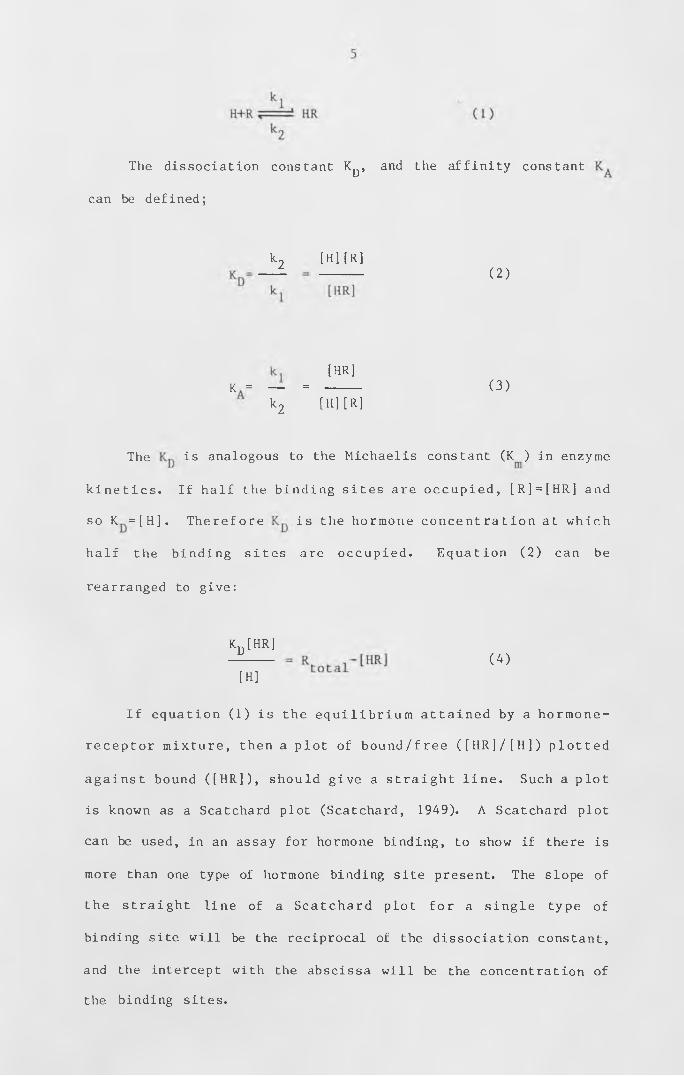
The dissociation constant K^, and the affinity constant
can be defined;
k2 [H][R]
( 2 )
[HR]
(3)K = _ = ------
k2 [H][R]
The is analogous to the Michaelis constant (K ) in enzyme
k i n e t i c s . If h alf the b ind ing s ites are o cc u pie d , [R]=[HR] and
so K = [H ] . Therefore is the hormone c o n c e ntrat io n at which
half the binding s ites are occupied . Equation (2) can be
rearranged to give:
I f equation (1) is the e q u i l i b r i u m a t t a in e d by a hormone-
receptor mixture , then a plot of b o u n d / f r e e ( [ H R ] / [ H ] ) plotted
a g a in s t bound ( [H R ] ) , should give a st ra ig h t l i n e . Such a plot
is known as a Scatchard plot (Scatchard, 1949). A Scatchard plot
can be used, in an assay for hormone binding, to show if there is
more than one type of hormone binding site present. The slope of
the straigh t line of a Scatchard plot for a s ingle type of
binding site will be the reciprocal of the dissociation constant,
and the intercept with the abscissa w ill be the concentration of
the binding sites.
k d [h r ]
(4)
[H]
6
The Scatchard plot w i l l only give a st ra ig h t line with a
slope of the reciprocal of the dissociation constant if there is
only one type of binding site present. If there are two types of
b ind ing s i t e , the Scatchard plot w i l l give a curve which is the
sum of the two st r a ig h t l ine s which would be obtained from the
two binding sites on their own. The intercept with the abscissa
w i l l give the total number of bind ing s i t e s . The two st r a ig h t
l i n e s , and hence the binding parameters from such a Scatchard
plot can be c a lcu lat e d (Weder et al . 1974) . The parameters
obtained from a Scatchard plot may be inaccurate and this is made
clear by plotting moles of bound ligand against the logarithm of
the concentrat io n of free l igand (K l o t z , 1982) . Such a plot
should give an S-shaped curve with the inflection point at half
the number of binding s i t e s . Klotz shows that the data from
p u b l is he d Scatchard p lo t s , when plotted on a sem i- logarithm i c
graph, do not reach an inflection point, and that even if the
last point is assumed to be the inflection point, it gives a much
larger number of binding sites than was originally obtained from
the o r i g i n a l Scatchard plot . I f (a) the b ind ing s ites for a
particular ligand are being assayed in a biological extract, (b)
there is more than one type of s i t e , (c) there is more than one
s it e per binding molecule where the a f f i n i t y of one s ite is
dependent on whether any of the other sites are occupied, or (d)
there are a large number of non-specific sites, the determination
of the dissociation constant or the number of binding sites can
be u n r e l ia b l e from a Scatchard plot . T h e re fo re , before such
parameters can be determined, a hormone-binding component has to
be purif ied .
T h e r e are a n u m b e r of d i f f e r e n t m e t h o d s of m e a s u r i n g the
equilibrium of a small ligand that is bound .to large molecules,
such as proteins, and free ligand. The most direct method is to
use e q u i l i b r i u m d i a l y s i s . The macromolecule is confined by a
cell
membrane which a l lo w s the passage of the l igand . The / i s l e f t
for the amount of free l igand to e q u i l i b r a t e each side of the
membrane. I f the l igand binds to the macromolecule , at
equilibrium, there will be more ligand on the macromolecule side
of the membrane. This method was described by Klotz et a l .
(1946). For a hormone-receptor mixture, the amount of hormone on
each s i d e of the m e m b rane can be d e t e r m i n e d by u s i n g a
radioactive hormone. Other methods of measuring this equilibrium
between a l igand and a macromolecule have been devised . An
ultrafiltration method (Lever, 1972) can be used where the free
ligand is removed by collecting the macro-molecule on a nitro
cellulose filter . Alternatively, the free ligand can be removed
by gel filtration chromatography (Hummel and Dreyer, 1962; Tze-
Fei and Porter, 1983). If the macromolecule is in a particulate
f r a c t i o n , it can be re m o v e d from the f r e e l i g a n d by
centrifugation (Lembi et a l . , 1971).
An alternative method of detecting a hormone receptor is to
use a p h o t o a f f i n i t y la b e l . The synthesis of a var ie ty of
different photoaffinity labels for detecting cytokinin-binding is
d e s c r ib e d by Mornet et a l . ( 1 9 7 9 ) , (see also Colman, 1983 for
general review on purine nucleotide photoaffinity labels). These
are azidopurines that have cytokinin activity, but when exposed
to ultraviolet light a reactive in te rm e diat e is produced which
then covalently binds to surrounding molecules. Therefore, such
a photoaffinity label can be added to a biological system, where
7
8
i t s h o u l d b in d to any s p e c i f i c b i n d i n g s i t e s . I t can
subsequently be ac t ivat e d and should bind covalently to the
m olecules that s p e c i f i c a l l y bind to c y to k in in . The label can
then be used to locate the cytokin in- binding m olecules , during
purification. Any molecules to which the photoaffinity label is
found to be bound could then be tested as a possible receptor.
Plant hormone binding
Most plant hormone binding assays have been performed using
e it h e r e q u i l ib r iu m d i a l y s i s or c e n t r i f u g a t i o n . The plant
hormones most studied with respect to receptors are auxin and
cytokinin. One of the first reports of active auxin binding was
in a p a r t i c u l a t e fr a c t io n of corn c o le o p t i le s (Hertel et a l . ,
1972). They showed that radiolabelled NAA is displaced from the
p a r t i c u l a t e f r a c t io n by 1-NAA and IAA, but not by ina c t iv e
yl-3~
analogues such as 2-NAA or indol propionic ac id . More prec iseA
kinetics of auxin binding in corn coleoptile membranes are given
by Batt et al. (1976). They showed evidence, by Scatchard plots,
of two types of auxin b in ding s i t e s . They showed also that the
b in d in g s i te s could be s o l u b i l i s e d by t rea ting the pe l le t w ith
T r i t o n X100 . The two s ites were studied for t h e ir i n t e r a c t i o n
w ith various auxin analogues . B inding of NAA to s i t e 1 was
c o m p e t it iv e w ith ina c t iv e auxin analogues as w e l l as ac tive
auxins and ant i- auxins . These two binding s ites have been
s epa rated on a sucrose gradient (Batt and V e n is , 1976) . They
report that site 1 appears to be associated with the dictyosomes
or endoplasm ic reticulum , and that s ite 2, which is the s i te
reported by Hertel ( 1 9 7 2 ) , is asso c iat ed with the plasma
membrane. They conclude that this site is the stronger candidate
for an auxin receptor as only active auxins and anti-auxins
compete with NAA for binding sites.
The auxin binding s ites from corn c o le o p t i le s have been
s t u d i e d by u s i n g t h e a u x i n a n a l o g u e s 2 - c h l o r o - 4 -
a minophenoxyace t ic acid (CAPA) and 2 ,5-dichloro-3-aminobenzoic
acid and by using p-mercuribenzoate (PMB), (Venis, 1977a). From
these s t u d ie s , Venis has suggested that the amino acids in the
b i n d i n g s i t e c o u l d be h i s t i d i n e , a s p a r t a t e / g l u t a m a t e ,
tyrosine/lysine and cysteine. The auxin binding site 2, reported
by Batt e t a l . ( 1976 ) , has been p u r i f i e d further (V e n is , 1977b) .
Venis states that further purification could be achieved if the
binding fractions were solubilised by acetone. Venis showed that
the b indin g a c t i v i t y was confined to a s ingle peak on a DEAE-
c e l l u l o s e column. The b ind in g a c t i v i t y was heat l a b i l e , non-
d i a l y s a b l e and p r e c i p i t a t e d with 70% ammonium sulphate . The
binding activity appeared to be associated with proteins with
molecular weights of 40 ,000 and 47,300.
A supernatant factor (SF) which reduces the amount of auxin
binding to corn coleoptiles has been reported (Ray et al ., 1977a
and b). This supernatant factor can be removed by washing the
p e l l e t in b u f f e r . Ray et a 1 . r e p o r t e d that the SF was
resp o n s ib le for the small amount of b in d in g reported by He rte l
(1972). This SF was not in itially found in the binding fraction
of Batt et a l . (1976). The SF appears to be present only in some
varieties of Zea mays. Ray et a l . show that the SF can be added
back to reduce the auxin-binding a c t i v i t y . This reductio n in
binding activity is a reduction in affinity , not a reduction in
the number of b ind ing s i t e s . They showed that the s p e c i f i c
9
10
binding is heat labile and is removed by treating with detergent.
As the assay was by c e n t r i f u g a t i o n , it was not p o ssib le to
distiguish between denaturation or solubilisation of the binding
a c t i v i t y by the dete rge nt . The amount of s p e c i f i c b in d in g was
reduced by reducing agents such as d i t h i o n i t e . I n h i b i t i o n by
d i t h i o n i t e was p a r t i a l l y reversed by ferricyanide. Dithionite
reduced the number of binding sites, but not the affinity for NAA
of the unaffected sites.
A very d i f f e r e n t auxin binding s i t e has been reported in
tobacco pith c a l lu s (Maan et a l . , 1983) . Auxin binding to a
particulate fraction did not occur at 0°c, and was slower at 25°C
than auxin binding to corn coleoptiles at 0°C. Auxin binding was
measured by a non-equilibrium filtration method. As the rate of
binding was slow, the time course of the binding reaction could
be measured. Maan et al. showed that the binding was not due to
a s in g le s i t e or due to take-up of auxin into v e s ic l e s . From
their data of the kinetics of the reaction, they propose a model
where there are four b ind ing s i t es and that b inding can cause a
co n fo r m a t io n a l change to form a stable complex. This was the
simplest model that fitted the data.
The first report of specific binding of cytokinin in vitro
was to Chinese cabbage leaf ribosomes (Berridge et a l ., 1970 ) .
They showed that the b ind ing a f f i n i t y to the 83S ribosomes was
c o rr e la te d w ith the the cyto kin in a c t i v i t y of a number of
compounds. They in f e r r e d from the b in d in g a c t i v i t y on the 83S
ribosomes that c y to k in in control of growth was at the le ve l of
the ribosomes . Takegami and Yoshida (1 9 7 5 ) p u r i f i e d a 4000
m olecular w eight prote in from tobacco leaves by a f f i n i t y
chromatography which bound benzyl amino purine . This protein
also s p e c i f i c a l l y bound k i n e t i n , and with lower a f f i n i t y ,
ad en ine , but not adenosine . Fox and Erion (19 7 5 ) reported 2
c y to k in in- bind ing s ites on wheat germ and tobacco ribosomes.
From Scatchard plots, they show that there is one high affinity
s i t e and m ultiple low a f f i n i t y s ites per ribosome. The high
affinity sites can be washed off the ribosomes with 0.5M KC1 and
st i l l show high affinity binding.
A s s o c ia t io n of the cyto kin in- bind in g protein with the
ribosomes has also been reported by Takegami and Yoshida (1977).
The c y to kin in- bind ing prote in was r a d i o l a b e l l e d by in c u b a t in g
tobacco leaf discs on a so lution c o n t a in in g H-4, 5-leucine and
the cyto kin in- bind ing protein i s o la t ed . The cytokinin-binding
protein was found to bind to KCl-washed ribosomes in the presence
of BAP. The amount of binding was reduced to 10% with unwashed
ribosomes , which they claim was due to the ribosomes being
already saturated with cytokinin-binding protein. They show that
the cyto kin in- bind ing prote in only binds to the 40S subunit of
the ribosome.
The first study of cytokinin-binding using a radiolabel with
high a f f i n i t y was in tobacco (Sussman and Kende, 1978) . The
tritiated BAP was sythesised by catalytically dehalogenating a p-
bromo benzyl amino purine which was s y t h e s is e d by Sussman and
F i r n ( 1 9 7 6 ) . By the use of t h is h ig h s p e c i f i c a c t i v i t y
r a d i o l a b e l , two c y to k in in- bin d ing s i t es were detected in a
particulate fraction, one of high affinity for BAP, the other of
low a f f i n i t y . These s i t es appeared to be d i f f e r e n t frora the
11
cytokinin-binding sites associated with the ribosomes, reported
by previous authors . Host of the cytokinin-binding was to the
low affinity sites which were heat stable. The number of these
low a f f i n i t y s it e s increased by heating to 10 0 °c . The reason
stated for this increase is that denaturation of proteins could
expose more non- specif ic bind ing s i t e s . The few high a f f i n i t y
s i t e s were only detected using the t r i t i a t e d . BAP. This h igh
a f f i n i t y b inding was grea tly reduced by heat and was shown to
have a high s p e c i f i c i t y towards b i o l o g i c a l l y active c y to kin in
analogues. Two inactive analogues, p-chloro BAP and p-bromo BAP,
which showed the greatest non-specific binding, did not bind to
the high affinity site.
Kinetin binding to a soluble glycoprotein with a molecular
weight of 1 8 0 , 0 0 0 has been reported in wheat germ (Polya and
Davis, 1978). Kinetin binding to this protein could be displaced
by low concentrations of biologically active cytokinins but not
by inactive adenine derivatives. One exception was that zeatin
was l e s s e f f e c t i v e than N - d i m e t h y l a 1 1 y l a d e n i n e . The
c on c e ntrat io n of this cytokin in-binding prote in is r e l a t i v e l y
high for a receptor , 27 ;j M, compared with animal hormone
receptors.
Using the same t r i t i a t e d BAP as Sussman and Kende ( 1 9 7 8 ) ,
G a r d n e r et a l . ( 1 9 7 8 ) d e t e c t e d c y t o k i n i n - b i n d i n g in a
p a r t i c u l a t e f r a c t i o n of the moss F u n a r i a h y g r o m e t r i c a .
Biologically active cytokinins, including BAP, zeatin, IPA, and
kinetin, competed with the tritiated BAP. Adenine, 9-methy1-BAP,
and the ribosides of BAP, IPA and zeatin did not compete with the
radiolabel. As well as the specificity of the binding for active
1 2
1 3
cytokinins, Gardner et a l. showed that over half of the binding
activity was removed by heat and that it was greatly reduced by
t rea ting the pellet w ith T r i t o n . To see if the T r i t o n was
solubilising the binding fraction rather than denaturing it, they
tested the b ind ing of the supernatant by gel f i l t r a t i o n .
However, they reported that there was an interaction between the
BAP and the Triton. The binding activity was small even with the
high specific activity H-BAP which could have been due to there
being only 0 .5-1.0 g fresh weight of t issue per assay. They
stated that a Scatchard plot gave a curve which in d ic a te s that
there is more than one type of binding site.
Polya and Bowman (19 7 9 ) show that the c y to kin in- bind ing
fraction in wheat, described earlier (Polya and Davis, 1978), has
a high affinity for non-purine cytokinins, such as DCMU and CMU,
and for hydrophobic derivatives of urea, triazine, carbamate and
tryptophan. As this c y tokin in- bind ing f r a c t io n also has an
unusually low a f f i n i t y for z e a t i n , Polya and Bowman suggested
that this prote in may not be a receptor , but could have a
c y to k in in " b u f f e r i n g " or se q u e st e r in g fu n ct io n . A s i m i l a r
protein from wheat germ has been reported by Moore (1979). This
also shows an unusually low a f f i n i t y for z e a t in , but has a
molecular weight of only 122,000. However, Moore suggests that
the c y to k in in a c t i v i t y reported by a l l three groups could be
derived from the same in vivo moiety.
Erion and Fox ( 1 9 8 1 ) report that the c y t o k in in - b in d in g
protein from wheat germ has a molecular weight of 183,000, made
up of subunits with molecular weights of 34,000, 39 ,000 , 53,000
and 5 9 , 0 0 0 . They claim that the subunit of 1 5 , 0 0 0 reported by
Moore ( 1 9 7 9 ) could be obtained by ageing for se vera l weeks, and
that the 5 bands obtained by Polya and Davis (1979) could be due
their own stated lack of homogeneity. The high concentration of
the cytokinin-binding protein, 27 nmol/g fresh weight reported by
Polya and Davis and 15 nmol/g fresh weight reported by Erion and
Fox, in wheat germ, is only found in the germinating seed and in
female reproductive t is s ue , according to Erion and Fox ( 1 9 8 1 )
using antibodies to the cytokinin-binding protein.
In summary, auxin binding in corn coleoptiles is relatively
well characterised. Two separate auxin high affinity sites have
been located in corn membranes, one of which appears to have the
same specificity as auxin activity. This receptor has been shown
to be a protein, and some of the amino acids in the binding site
have been tentatively assigned. The auxin binding fraction from
tobacco was also membrane bound but showed a much slower rate of
b i n d i n g . T h i s b i n d i n g f r a c t i o n has not been so w e l l
characterised and a model for auxin binding is suggested on the
k i n e t i c s of the re a c t io n . The i n i t i a l reports of a cytokinin-
binding fraction were of a soluble fraction which was associated
with the ribosomes. The soluble protein which binds cytokinin to
ribosomes in wheat germ and tobacco has been reported by a number
of groups. The d i f f e r e n t groups report d i f f e r e n t m olec ular
weights ranging from 122,000 to 183,000, and different molecular
w e ig h t s and number of subunits . From the data g iv e n , it is not
c e r t a in that these groups are looking at the same prote ins or
whether only some of the subunits are required for b in d in g of
c y to k in in . Polya and Bowman have reported a broad l ig a nd
specificity and so suggest that this protein may be a cytokinin
sequestering agent rather than a receptor.
14
The p a r t ic u l a t e c y to kin in- bind ing frac t io ns reported in
tobacco and moss have so far not been greatly studied. Sussman
and Kende reported two s ites in tobacco, one of which showed
specificity towards active cytokinins, was heat labile and, due
to its low concentration, was only detectable with a radiolabel
of high s p e c i f i c a c t i v i t y . A s im i l a r binding frac t io n was
reported by Gardner et al. from moss, which also appeared to be
s p e c i f i c for active cyto kin ins and was heat l a b i l e . However,
this binding activity was not purified further to give a straight
line on a Scatchard plot.
Detecting high affinity binding sites
Most plant hormone binding assays are performed using
commercially available radiolabelled plant hormones, which have a
f a i r l y low s p e c i f i c a c t i v i t y . U s i n g t h e s e r a d i o l a b e 1 led
compounds one is able to detect binding frac t io ns which are
relatively abundant, such as the auxin binding proteins in corn
c o l e o p t i l e m e m b ra n e s and the c y t o k i n i n - b i n d i n g proteins
a sso c ia t e d with the ribosomes in wheat germ and tobacco.
However , to detect plant hormone b inding s it e s which are less
abundant, such as the p a r t i c u l a t e s i te s in tobacco and moss, a
plant hormone of much h igher s p e c i f i c a c t i v i t y is re qu ired . A
major problem in trying to detect a small number of high affinity
s i t e s is that the high background of less s p e c i f i c or non
specific binding sites produces Scatchard plots which are curved
rather than l in e a r . This makes the i d e n t i f i c a t i o n of high
affinity sites diff icult and means that the amount of unlabelled
ligand required to displace the radiolabel is much greater than
would be required for a purified binding site.
1 5
16
Even d e t e c t in g and p u r i fy in g a bind ing s i t e which has a
high affinity for the active hormones only, does not prove that
the b ind ing s ite is a receptor . For such a b ind ing s i t e to be
identified as a receptor, binding of the plant hormone has to be
shown to initiate a biochemical process which brings about the
responses assigned to that particular plant hormone. Few of the
reports of plant hormone binding have shown a link . Roy and
Biswas (1977) reported an auxin^binding protein in coconut which
promoted t r a n s c r ip t io n . Linde et a 1. ( 1984) have shown that a
soluble auxin binding protein, from tobacco c a l l u s , s t im u la t e s
IAA-dependent RNA sythesis in v itro. The reports of cytokinin-
b ind ing to a protein which binds to a subunit of the ribosome
have suggested that this then d i r e c t l y a f f e c t s t r a n s l a t io n of
mRNA.
One method of showing the relevance of a plant hormone
binding site is to use a mutant which is defective with respect
to the receptor . The most u s e fu l mutant would be one that did
not possess a receptor and so would not show hormone binding and
which lacked the response to that plant hormone. Mutants that
have a defective receptor could also be used, provided there was
a loss of b in d ing a c t i v i t y and a loss of the response to the
hormone. Other mutants that did not respond to a p a r t i c u l a r
plant hormone but had a defect a fte r the hormone-receptor
i n t e r a c t i o n c o u l d be u s e f u l in i d e n t i f y i n g part of the
b io c h e m ic a l process involved in the response to the plant
hormone.
17
Physcomitrella patens
The moss P h y sc o m it re l la patens is p o t e n t i a l l y a good
organism to use for the identification of a hormone receptor as
it is a relatively simple green plant with three distinct types
of tissue, the differentiation of which is under the control of
a u x i n an d c y t o k i n i n . Th e l i f e c y c l e of the m oss
Physcomitrella patens has been described in detail (Engel, 1968;
Ashton , 1974 ; Ashton and Cove, 1977) , and so w i l l only be
described briefly here.
The haploid spore of Physcomitrella patens, which is about
40jum in d iam eter , w i l l germinate in 2-3 days at 24 °C , under
- 2continuous white light of 15Wm . The filaments produced, the
primary chloronemata, consist of highly-branched filaments which are
20pm in diameter and 120pm in length. These c e l ls contain a
large number of plump chloroplasts and have perpendicular cross
w a l l s . A fter about 7 days , some of the cel ls at the tips of the
filaments grow with fewer, spindle-shaped chloroplasts, and have
o bl iqu e cross w a l l s . These cel ls are c a l le d the caulonemata .
Older caulonemal cells can sometimes produce a brown pigment and
can lack obvious chloroplasts. Side branching from a caulonemal
f i la m e n t can give rise to three types of t is s u e . Most of the
s ide branches develop into f i la m e n t s that are m o r p h o lo g ic a l ly
i d e n t i c a l to the primary chloronemata. These are c a l le d the
secondary chloronemata. A few of the side branches give rise to
more caulonemal filaments. The other side branches develop into
a leafy shoot called the gametophore. There is usually only one
gametophore per caulonemal filament. If a 4-week-old culture is
transferred to 15oC, antheridia and archegonia are produced
on the gametophore. Fertilization is facilitated by irrigating
the culture with d i s t i l l e d water . A fter 4 to 5 weeks, the
diploid sporophyte is produced. The capsules contain about 4,000
s p o r e s . P h y s c o m i t r e l l a p a t e n s can be maintained in the
gametophytic stage by removing a small piece of protonema and
subculturing this on a fresh plate of medium. This w ill undergo
regeneration through primary chloronemata.
Plant hormones and development of Physcomitrella patens
Cytokinin has been known to induce bud formation in mosses
for some time (Gorton and E ak in , 1957) . The f i r s t report of a
cytokinin being identified from moss tissue was the isolation of
6 2N -(A -isopentenyl)adenine from culture medium of callus cells of
the moss hybrid Funaria hygrometrica x Physcomitricum piriforme
( B e u t e l m a n n and B a u e r , 1 9 7 7 ) . One u s e f u l f e a t u r e of
P h y sc o m it re l la patens is that the i s o l a t i o n of mutants is
relatively easy and can be achieved by treating either spores or
tis sue with a chemical mutagen (Ashton and Cove, 1977) . As
mosses are haploid there is no masking of recessive mutant
alleles . Mutants of Physcomitrella patens have been isolated and
used to study the role of the two plant hormones, auxin and
cytokinin, in the development of the moss (Ashton et a l ., 1979b).
Ashton et a l . describe the e f fect of auxin and c y to k in in on
P h y s c o m it r e l la patens and the i s o l a t io n of mutants that are
resistant to exogenous auxin and cytokinin. Exogenous auxin is
s l i g h t l y i n h i b i t o r y at 2.5-5.0jaM. At this c o n c e nt rat io n , auxin
causes inhibition of chloronemata but promotion of cauloneniata.
Auxin also slightly inhibits gametophore production but increases
the number of rh izo ids produced by any one gametophore.
18
Cytokinins have the effect of suppressing the,production of both
chloronemata and caulonemata. At 50-500nM, the gametophores are
abnormal , the leaves are sm aller and ar ise d i r e c t ly from a
callus-like bud. At higher concentrations, almost every branch
site develops into an abnormal bud.
Mutants that have an abnormal response to exogenous auxin
and c y to k in in are also described by Ashton et a l . ( 1979b) .
Category 1 mutants produce only tightly packed chloronemata, are
u n a ffe c t e d by exogenous auxin and w i l l only produce a few
abnormal buds if treated with cytokinin. These buds arise from
the primary chloronemata . Category 2 mutants produce both
chloronem ata and caulonemata , bud do not produce gametophores.
They can be repaired by 5nM-50pM BAP. Category 3 mutants are
m o r p h o lo g ic a l ly normal and respond to exogenous BAP. However,
they are resistant, or less sensitive than wild type to exogenous
NAA. Category 4 mutants produce mainly chloronemata and a few
caulonemal type f i la m e n ts but no gametophores. Ashton et a l .
suggest from these findings that category 1 mutants are blocked
in the d i f f e r e n t i a t i o n from c h l o r o n e m a t a to c a u l o n e m a t a .
Category 2 mutants, which are repaired by cytokinin, appear to be
unable to produce normal levels of cytokinin. Category 3 mutants
appear to be blocked in the uptake of a ux in , w hile category 4
umutants appear to be a f f e c t e d in the synthesis of auxin . When a
w i l d type culture is grown under dr ip-feed , a l l the t is s u e is
chloronem al (Ashton et a l ., 1979a ) . The morphology of such a
culture resembles that of category 1 mutants. Wild^type cultures
can be induced to d i f f e r e n t i a t e under drip-feed by supp ly ing
auxin and cytokinin in the drip-feed medium (Cove, 1984). When
the drip-feed medium contains lOpM NAA, caulonemata are produced.
19
This is the phenotype of category 2 mutants. .When supplied with
IOjjM BAP, a w ild—type culture is unaffected. This concentration
of BAP causes the production of a large amounts of abnormal buds
on so l id medium. When both NAA and BAP are supplied at IOjjM,
over-production of buds occurs. From the e f fe ct of auxin and
c y t o k in in on the morphology of category 1 to A mutants , and the
effect on wild type under drip-feed, it has been concluded that
auxin is required for the differentiation of primary chloronemata
into caulonemata, and that the target cells for cytokinin action
are the c a u lo n e m a t a ,c y t o k in in inducing the production of the
gametophores. This is further supported by some mutants which
have the morphology of the wild type grown on cytokinin. Under
these conditions the wild type over-produces buds and hence the
mutants have been termed ove's (Ashton et a l . , 1979a). There are
several mechanisms that could give rise to these ove mutants
(Ashton et a l . 1979a):
a) an increase in synthesis of cytokinin by the normal or a
de novo pathway;
b) a decrease in the degradation of cytokinin;
c) the p r o d u c t i o n of a c y t o k i n i n of i n c r e a s e d
biological activity;
d) an increase in sensitivity to cytokinin;
e) a decrease in a cytokinin antagonist;
f ) the loss of requirement for cytokinin for bud induction.
A l l of the ove mutants studied so far have been shown to
o v e r - p r o d u c e c y t o k i n i n (Vang e t a 1 . , 1 9 8 0 ; 1 9 8 1 b ) . Two
c y to k in in s have been i d e n t i f i e d . These are N -(a -isopentenyl)
adenine and zeatin.
20
From the results mentioned above, Physcomitrella patens is a
good organism for the study of plant hormone action^as both auxin
and c y to k in in appear to cause s p e c i f i c responses rather than
wtl ichhelp ing to promote a variety of d i f f e r e n t responses, is the
case for many of higher plants (Skoog and Armstrong, 1970; Dodds
and H a l l , 1980) . The gametophore production in the response to
c y t o k in in occurs in a s p e c i f i c target t is s ue . I f c y t o k in in is
only required for the production of the gametophore in mosses ,
then any c y t o k in in receptor is l i k e l y to be present only in the
target c e l l s , the caulonemata. However, if cyto kin in is also
required for another process such as c e l l d i v i s i o n , a receptor
would be required in a l l d iv id in g c e l l s . This receptor could be
the same or a d i f f e r e n t receptor from the one for gametophore
production . Th e refo re any binding a c t i v i t y detected in moss
t is s ue can be in v e s t ig a t e d by the use of mutants, such as
category 1 mutants , to see if it is only present in the target
t is su e for bud in d u c t io n or if it is also in other t i s s u e . If
more than one b ind ing s it e is detected , they can be s tudied to
see if they are both present in the target cells.
If cytokinin is only required for bud production, or if bud
production has i t s own receptor , then it should be p o s s ib l e to
obtain mutants that do not have this receptor, or have a receptor
that is unable to bind cytokin in . Such mutants would be a
subclass of mutants that would be able to produce caulonem al
f i l a m e n t s , but would not produce any gametophores , even in the
presence of exogenous c yto kin in . Other such mutants would be
ones that had a normal cytokinin receptor but were blocked in a
b i o c h e m i c a l p r o c e s s i n v o l v e d in the i n i t i a t i o n of the
gametophore.
2 1
22
Once a hormone has bound to a receptor , a b iochem ical
process has to be i n i t i a t e d which results in the response
a t t r ib u t e d to that hormone. When the receptor in a s e n s i t i v e
moss caulonemal c ell binds c y to k in in , it has to induce a
biochemical process to produce a gametophore. There have been a
number of reports of calcium being involved with the actio n of
cytokinin. The level of calcium has been reported to enhance the
c y to k in in response in de lay ing senescence in corn leaf discs
(Poovaiah and Leopold, 1973), stimulation of ethylene production
in mung bean (Lau and Yang, 1975) and bud induct io n in the moss
Funaria hygrometrica (Saunders and Hepler, 1982).
Calcium gradients can be visualised with chlorotetracycline-
fluorescence (CTC). Using'CTC, Reiss and Herth (1979) have shown
that there is a calcium gradient in the caulonemal tip cells of
Funaria hygrometrica. This polarity is only present in the tip
cell of caulonemata (Reiss and Herth, 1979; Saunders and Hepler,
1981) . Saunders and Hepler ( 19 8 1 ) also showed that there is
bright fluorescence 12 hours after treating a filament with BAP.
This f luoresc e nce remains in the d iv id in g bud. Saunders and
Hepler (1982) used the calcium ionophore A23187 (Reed and Lardy,
1 972) to induce the i n i t i a l asym m etrical d i v i s i o n of bud
indu ct io n on caulonemata in the absence of c y to k in in . They
suggested that one action of cytokinin was to increase the level
of i n t r a c e l l u l a r calcium which may be a second messenger for
cyto kin in- induced bud i n i t i a t i o n . I f calcium levels play an
important role in the mechanism of c yto kin in a c t io n , then
calmodulin is likely to be involved in the regulation of the free
calcium in the cell. This increase of intracellular calcium may
involve the d i s r u p t i o n of m icrotub ules in the early stages of
23
bud form atio n (Doonan, 1983) . I n h i b i t i o n of cytokin in-induced
bud production in Funaria hygrometrica has been demonstrated by
calmodulin inhibitors (Saunders and Hepler, 1984), supporting the
role of c a lc iu m in bud induction . One report ( E l l i o t , 1983)
shows inhibition of the cytokinin-regulated response ,^induction
of betacyanin in Amaranthus tricolor in the dark, by calmodulin-
b ind in g compounds. Using the moss Funaria hygro m etrica , Bopp
(1984) reported that an isolated caulonemal cell can be induced
by k i n e t i n to undergo morphogenesis (from f i lam e nt to bud and
reverse) without undergoing cell division. Bopp also showed that
the antiauxin, parachlorophenoxyisobutyric acid (PCIB), inhibits
kinetin-accelerated cell division, but not bud formation. Due to
these two observations, Bopp (1984) suggested that the asymmetric
c e l l d i v i s i o n reported by Saunders and Hepler ( 1 9 8 2 ) is not a
replacement for c y to k in in and that the conclusion drawn by
Saunders and H e p ler that calcium i n f l u x is a primary e f f e c t of
cytokinin cannot be made from their experiments.
Light and moss development
Physcomitrella patens is a good model organism with which to
study other aspects in the mode of action of cytokinins. Robbins
( 1 9 1 8 ) r e p o r t e d t h a t the moss C e r a t o d o n p u r p u r e us c o u l d
a s s i m i l a t e organic carbon in the dark. Robbins showed that if
su p p l ie d w ith glu c os e , the moss would produce reddish-brown
tdprotonemaj, but no buds were produced. Robbins concluded that
light was necessary for bud production. Fries (1945) showed that
bud primordia initiated in the light w ill continue to develop in
the dark. M itra et a l . ( 1959) showed that the q u a l it y of l ig h t
a f f e c t e d development in P ohlia n u t a n s . A bud would develop in
white l ight or red l ight (580-700nm). With blue light (400-
530nm), protonemal growth was normal, but no buds developed.
With green l igh t (460-600nm) there was no d i f f e r e n t i a t i o n : a l l
the filaments were caulonemal in appearance, similar to growth in
the d a r k . The poor g r o w t h is p r o b a b l y due to poor
photosynthesis under green light.
Un l ike P h y s c o m it r e l la patens , a caulonemal f i lam ent of
Physcomitrium turbinatum does not undergo dedifferentiation when
i s o l a t e d (Nebel and N aylo r , 1968a ) . Nebel and Naylor (1968b )
showed that temperature and light intensity both played a role in
Othe i n i t i a t i o n of buds in Phy s comi t ricum turbinatum. At 16 or
26 °C caulonemal f i la m e n ts take 19 days before a bud starts to
develop w ith 30ft-c red l ig h t . As the l ight i n te n s i t y is
in c re a s ed , the time taken before the bud starts to develop is
reduced . In c r e a s in g the l ight i n t e n s i t y above 60ft-c does not
reduce the time taken (13 days) before a bud starts to develop .
At 26°c, the time taken for a bud to start to develop is reduced
to 6 days as the light intensity is increased to 240ft-c. Higher
light intensities do not reduce the time any further. Nebel and
Naylor also showed that at 16°C the total dur<lti07i of red l igh t
required over the 13 days was only 144 hours which is the same as
that required at 26 °C . This i n d ic a te s that there are two
processes involved . One process is temperature-dependent but
occurs in the dark , the other process is dependent on the total
amount of l ig h t received . Nebel and Naylor ( 1 9 6 4 ) have also
d e s c r i b e d a s i m i l a r p h o t o p e r i o d r e s p o n s e in the moss
Physcomitrium pyriforme. With this moss, bud induction can be
prevented by exposing the tissue to far-red light before it has
been exposed to a total of 150 hours of red l ig h t at 240ft-c .
24
25
This indicates that phytochrome is involved in the initiation of
gametophores.
E vidence of the role of phytochrome in bud i n i t i a t i o n has
also been shown in the moss Funaria hygrometrica (Simon and Neaf,
1981) . They show that bud induct io n in the presence of k i n e t i n
is dependent on the exposure to red light, and that the effect is
p a r t i a l l y reversed by far-red l ig h t . Simon and Neaf show that
growth , measured as increase in dry w eight , in blue light and
red l igh t is the same as in w hite l ig h t , but that bud indu ct io n
does not occur in blue l ig h t . This supports the evidence given
by Jahn (19 6 4 ) that under blue light an in h ib it o r of bud
initiation is produced and that buds can be produced under blue
light if the culture is transferred onto fresh medium to prevent
the build up of the i n h i b i t o r . Bud induct io n in the absence of
light in the moss Funaria hygrometrica has been reported (Chopra
and Gupta, 1967 ) . Buds were induced by k i n e t i n and sucrose .
Chopra and Gupta concluded that the non-photosynthetic l ight
requirement was replaced by kinetin.
In the fo rm ation of a bud by cyto kin in in P h y s c o m it r e l l a
p a t e n s , there is a l igh t requirem ent . In the absence of l i g h t ,
even in the presence of exogenous cytokinin, no buds w ill develop
(Cove et a l . , 1 9 7 8 ) . The l i g h t is r e q u i r e d on ly fo r the
i n i t i a t i o n of the bud. Once the bud has been i n i t i a t e d in the
light, it w ill continue to grow in the dark but the stem w ill be
e>f- exposure fco
e t i o l a t e d and the leaves grow as small scales . One hour^red
_ o _ 1light (6mmol quanta m s of 660nm) causes normal gametophore
development. However, if the red light is followed by 15 minutes
of far-red light (715nm), the gametophores develop as though they
26
are in the dark. Cove et a l . ( 1978) , argued that this in d ic a t e d
that phytochrome is involved in the mode of action of cytokinin.
The i n t e r a c t i o n of l ig h t , p a r t ic u l arly red and far-red l ight
and cytokinin,can be studied in more detail in the initiation of
buds.
In the f o l l o w i n g work, both the mode and the mechanism of
action of cytokinin are investigated in the moss Physcomitrella
patens. The mechanism of action is studied by competition assays
using a radiolabel to detect a possible receptor in various crude
prep arat io ns of wild-type moss t is s ue . C h a r a c t e r is t ic s of the
cytokinin-binding a c t i v i t y are in v e s t ig a t e d . The cytokinin-
binding activity is also studied in various mutants affected in
bud production. The mode of action is investigated by studying
the quantity and quality of light required for bud induction on
cultures treated with cytokinin. The role of phytochrome is also
investigated in the initiation of the gametophore. The cytokinin
production of a number of mutants was investigated, in particular
ove 409, which was found to be temperature-sensitive with respect
to c y t o k i n i n o v e r - p r o d u c t i o n . T h i s m utant was s t u d i e d
genetically by protoplast fusion.
27
CHAPTER II
Materials and Methods
1. Culture Conditions
P h y s c o m it r e l la patens cultures were grown on a sol id or
(lA/A’ng', person al Cow i t 1011)
liquid modified Knops mediunij. The standard medium contained the
following :
Ca(N03 ) 2 .4H20 59mg/l
MgS04 .7H20 250mg/l
kh2 po4 250mg/l
F e S O . . 7H„0 4 I
10mg/l
kno3 1 .036g / l
TES 1ml/I
Final concentration of trace elements (TES)
H3 BO3 614pg/l
MnCl24H20 389pg/l
NiCl 26 H20 59pg/l
A12 (S03 ) 3K2S04 24H20 55pg/l
Co C126H20 55»ig/l
CuSo, 5H„0 4 L
55Hg/l
ZnS047H20 55pg/l
KBr 28yg/l
LiCl 28pg/l
SnCl2 2H20 28yig/1
KI 28pg/l
28
Modifications
a) Nitrogen source
Unless tissue was being grown for caulonemata or gametophore
production , ammonium tartrate ( 9 2 0 m g / l ) was included in the
medium to promote the production of chloronemata.
b) Carbon source
When cultures were grown in the dark, the medium was
supplemented w it h 0.5% ( w / v ) sucrose as a carbon source. Other
carbon sources at various concentrations were also tried. These
were glucose, myo-inositol and acetate.
c) Supplements
These were u sual ly added to a l l media, except where
prototrophs were being selected. Adenine was only added to the
m e d iu m of th o se s t r a i n s w h i c h were s p e c i f i c a l l y adenine-
requiring.
p-amino benzoic acid
nicotinic acid
thiamine-HCl
adenine
250Mg/l
lmg/1
100,jg/l
6 7 . 5mg/l
d) Mannitol medium
Mannitol was included in the medium as an osmoticum for
the regenerat ion of p r o t o p last s . The concentrat io n of the
mannitol was 60g/l .
29
e) "Starvation" medium
If the level of phosphate is reduced , with or without
reducing the nitrate, in the standard Knops medium chloronemal
growth is g r e a t ly reduced or stopped. This was done to e nr ich
for caulonemata. Three starvation media were used:
( i ) Ki^PO^ was omitted and replaced by 137mg/l KC1
( i i ) Kl^PO^ was reduced to 12.5mg/l
( i i i ) KH^PO^ was reduced to 1 2 .5m g / l and the KNO^ was
omitted.
f) Solid medium
The medium was solidified with 12g/l oxoid agar no. 1 when
solid medium was required.
The medium was a d ju ste d to pH 6.5 w i t h NaOH, so l id medium
was dispensed into bottles in 400ml lots and autoclaved at 15 psi
\/esse Is
for 20 minutes. Ten and 15 litre batch culture j^were autoclaved
at 15 psi for i hour.
Axenic cultures were maintained as spot inocula on minimal
Knops medium containing supplements. Larger amounts of t is s ue
were obtained by growing the moss as an homogenate on medium
overlaid with cellophane so as to prevent the filaments growing
into the medium. Tissue was homogenised in an M.S.E. homogeniser
f o r 10-15 s e c o n d s in s t e r i l e d i s t i l l e d w a t e r , and t h r e e
m i l l i l i t r e s of the suspension put on each plate which had been
o v e r la id w ith a cellophane disc . This method of culture for
tha(- of
P h y s c o m it r e l la patens is a m o d i f ic a t io n o f ^G r im s le y et a l . ,
( 1 9 7 7 a ) . T issu e was grown in l iq u id culture e ith e r in c o n ic al
f l a s k s or large batch cultures that were aerated w ith air
30
sterilised by two miniature in-line f i l t e r s (IF 32, M ic r o f lo w ,
Dent and H e l l y e r , W alsorth Road, Andover , Hampshire) . Flasks
containing from 15ml to 1 litre of medium were kept on an orbital
shaker rotating at between 60-100 rpm. Batch cultures of 10 and
15 litres were used for the production of large amounts of tissue
or for c y to k in in production . The 10 l i t r e vessels were f i t t e d
with a condenser (to reduce water loss) and a collecting vessel
so that samples could be taken to examine and measure growth
rate. Growth rate was measured by weighing a Whatman 2.5cm GF/A
f i l te r wrapped in foil that had been dried in an oven at 80°C for
at least 2 days. A known volume of culture was collected on the
f i l t e r , which was then reweighed a f t e r being dr ied in the oven
for 2 days (Wang e t a l . , 1981b) .
__2
Cultures were routinely grown at 24°c under 10-15Wm white
light provided by fluorescent tubes (Ashton and Cove, 1977). For
the induction of gametogenesis, cultures were transferred to 15°c.
When required, a temperature gradient was obtained by heating one
end of an a l u m i n i u m b l o c k ( 2 0 x 5 Ox 1 0 0 0 mm) u s i n g a s m a l l
thermostatically controlled refrigerator heater. This was placed
in a constant temperature room at 10°c. Cultures were placed in
60ml specimen containers containing 20ml of Knops medium. The
ridge on the base of the jar was removed with glass paper so that
there was direct contact of the jar with the aluminium bar. The
jars were placed on the bar and left for 5 weeks for the culture
to grow. Just before the jars were removed, the temperature of
the agar near the culture was measured w ith a thermocouple .
Using this bar a temperature range of 26 to 16°C was obtained.
31
2. Spore production
Spores can be produced either by selfing or by crossing two
strains. The strains to be crossed were inoculated next to each
other in a 60ml specimen jar containing 40ml of Knops medium with
the appropiate v itam ins . S trains to be se l fe d were treated in
the same way but only one in o c u lat ion was done. The cultures
were grown at 24°C for 4 weeks. They were then t r a n sfe rre d to
15°C for 4 weeks to induce the production of the a n t h e r i d i a and
archegonia. The cultures were then irrigated with 10ml of a 20-
fold concentrated solution of the vitamins that were required by
those s t r a i n s , as they w i l l not produce v iable capsules when
i r r i g a t e d w ith s t e r i l e d i s t i l l e d water (Courtice et a l . , 1978).
For most s t ra in s being s e l fe d , capsules were produced about 6
weeks a f t e r i r r i g a t i o n . The crosses were re- irr ig ated i f no
capsules were visible. When capsules were produced, they were
removed from the culture, squashed in l/2ml of sterile dist illed
water per capsule and stored at 4°C until required.
3. Protoplast isolation
Pr o to p la s ts were normally i so la t e d e n z y m a t i c a l l y , as
d e sc r ib e d by G r im sle y et a l . ( 1977a ) . T issue was grown as an
homogenate for 5-7 days on ammonium tartrate plates overlaid with
c e l lo p h a n e s . Up to 5 plates of t issue were added to 10ml of
2 % (w /v ) D r i s e l a s e (Sigma) . The D r i s e l a s e s o l u t io n was made by
d i s s o l v i n g 0 .4g of D r is e la s e in 20ml of protoplast wash (PW) ,
which contained 80g /l mannitol. This was then acidified to pH5.5
and, if necessary , c en t r i fu g e d at 3 , 0 0 0 g ( 4 5 0 0 rpm in an M.S.E.
Minor) for 5 minutes . The supernatant was . . f i l t er - st e r il i ze d
through a Millipore nitrocellulose 0.45 pm filter (Millipore U.K.
Ltd . , M id d le sex ) and added to the t is s u e . The mixture was
incubated for 30 to 45 minutes at 20°C and then debris removed by
passing the suspension through a 50)im mesh. The D r is e l a s e
so lu t io n was removed by decanting a ft e r c e n t r i fu g in g the
protoplasts at lOOg (700 rpm in an M.S.E. minor) for 3-5 minutes.
The protoplasts were rinsed twice by resuspending in protoplast
wash and recentrifuging.
Other enzymes were used when attempts were made to obtain
protoplasts from dark-grown caulonemal filaments,as Driselase was
found to be ineffective. These were:
(a) cellulase and pectinase;
(b) cellulase, pectinase and rhomant P;
(c) meiselase;
(d) driselase and chitinase .
These enzymes were made up in a 5mM m orpholinoethane
sulphonic acid buffer at the optimum pH for the enzyme mixture.
4. Protoplast regeneration
Pro to plast regenerat ion was on the normal Knops medium
containing 60g/l mannitol, plus' vitamins if required (see Chapter
I I sect ion 1), and cellophane overlays . 1ml of protoplast
suspension was added to 9ml of Knops medium co n t a in in g 8 0 g / l
mannitol and 0 . 6 % ( w / v ) agar . The soft agar was poured onto the
cellophaned mannitol plates. Protoplasts were then regenerated
under high intensity white light for 5 days. The cellophane and
top layer were transferred to ammonium tartrate medium without
32
mannitol (as mannitol reduces the rate of' growth) once the
protoplasts had regenerated . The regenerated plants were then
picked off when they had grown to about the ten-cell stage.
5. Protoplast fusion
The method of protoplast fusion was based on the method
described by G r im s le y et a 1 . ( 1977b) . The two st ra ins to be
fused had to have d i f f e r e n t v itam in requirements so that
prototrophic hybrids could be selected. Protoplasts of the two
st r a in s to be fused were prepared as described above and
resuspended in calcium protoplast wash (CaPW) containing 50 mM
C a C ^ and 8 0 g / l mannitol . A proportion of each protoplast
p r ep arat ion was mixed together , c e n tr i fu g e d at lOOg for 3-5
minutes and the supernatant removed. The protoplasts were left
in 250jil CaPW. P rotoplast fusion was induced by adding 750jul
poly-ethylene glycol ( 6 0 0 g / l PEG in 50mM C a C ^ ) . Fusion was
a l lo w e d to take place for 40 minutes . A fter this time 1.5ml of
CaPW was added. Ten minutes l a t e r , 10ml CaPW was added and
another 10ml 10 minutes after this. After a further 10 minutes,
the protoplasts were harvested by centrifugation at lOOg for 3-5
minutes and resuspended in CaPW. The protoplasts were plated out
to r e g e n e rat e , as d esc r ibe d above, on m annitol medium w ithout
vitamins to select for hybrids.
33
34
6. Hormone treatments and bud counts
Hormone treatments were performed on both protoplasts and
homogenates. Protoplasts were prepared as described above and
regenerated in a 5x5 repli-dish with auxin and cytokinin included
in the medium. The concentration of the hormones, NAA and BAP,
were 0, 10 10 10 and 10 ^ M. The con c e ntrat io n of the
agar was only 0.4% (w /v ) . This al lowed the protoplasts to sink
to the bottom of the w e l ls . The dish could then be inverted so
that protoplasts could be examined under the microscope.
For the treatments using homogenate t is s u e , 3 ml of wild-
type homogenate was added to 17ml of Knops medium in a 100ml
c o n ic a l f lask and grown for 7 days on an o r b i t a l shaker . The
t is sue was c ollec ted on a s t e r i l e 2 .5 cm glass f ib r e f i l t e r
(Whatman G/F-A). The tissue was then added to a flask containing
20 ml of fresh medium with 1 BAP. After the time of exposure
to cyto kin in the medium was again c o l le c t e d on a glass f ib r e
f i lter , washed with 600ml sterile distilled water and added to
fre sh medium w ithout c y to k in in . A fter 2 weeks , the number of
buds in each flask was chrome-counted. The chrome-counting was
performed using 1ml of t is s u e , adding 3ml of 10% chromic a c id ,
homogenising for 10 seconds in an M.S.E. homogeniser and adding
another 3ml of water. The homogenate was used to f i l l slides to
a depth of 1.4mm. Ten slides were made from each flask and buds
were counted in 10 f i e l d s of view under the low power of a
microscope. The diameter of each field of view was 1.75mm.
35
7. Mutant Isolation
The mutagen used was N-methyl-N'-nitro-N-nitrosoguan/dine
(NTG). Two methods of mutagenesis were used.
a) Spore mutagenesis
Spores were harvested by picking off mature
capsules which were then crushed with a glass rod in 1 / 2 ml of
sterile distilled water per capsule. The spores were centrifuged
and resuspended in 10ml T r i s - m a l e a t e b u f f e r ( 6 g / l T r i s -
(hydroxymethyl)-aminomethane and 6 g / l maleic a c id ) pH 6 .0 . To
this was added lmg NTG, and the mixture incubated for 45 minutes
with intermittent shaking. The spores were washed three times in
sterile distilled water. The spore suspension was stored at 4°C
and plated o u t ,w h e n r e q u i r e d , t o produce about 200 s u r v iv in g
spores per 9cm petri dish^after testing the survival rate.
b) Somatic mutagenesis
This method was used for strains that were sterile
and so could not produce spores. Tissue from up to 5 homogenate
plates was added to 10ml Tris-maleate buffer containing lmg NTG
as in spore mutagenesis but only incubated for 30 minutes as
arcprotonema is more susceptible to NTG than^, spores. The tissue was
then collected on a 50^im mesh and washed with about 200ml sterile
distilled water. Protoplasts were prepared from the mutagenised
tissue and a l lo w ed to regenerate as described in Chapter I I ,
sections 3 and 4 above. For sc re e n in g , the plants were then
36
picked off and placed on Knops medium contain ing ammonium
tartrate and vitamins.
8. Mutant screening
Young plants derived from NTG treated spores or protoplasts,
as described above, were picked off and tested for the ir
i n a b i l i t y to produce buds even in the presence of exogenous
cytokinin. The young plants were inoculated onto Knops medium
supplemented with vitamins and sucrose. Each 9cm petri dish was
inoculated with 16 mutants for testing. These were grown for 7
_2days under continuous white light of 10-15 Wm . The plates were
then t r a n sfe rre d to the dark and placed on their edges for 14
days. Th is produced caulonemal f i la m e n ts which had grown
n e g a t iv e ly ge o t r o p ic a l ly along the surface of the agar. The
plates were then returned to white l ig h t , w ith the plate
h o r i z o n t a l , and 2.5 ml of 20 ;uM BAP added to each plate so as to
give a final concentration of 1 juM. After three days the plates
were screened for mutants that produced dark-grown caulonemata
but did not have any abnormal buds on the filaments.
9. Extraction of endogenous cytokinins
a) Isolation
A l l solvents used in the e x t r a c t io n of cyto k in in s were
c ulture medium from a 10-litre batch c ulture . The t is s ue was
separated from the medium by f i l t r a t i o n . The medium was then
d r ie d down in a rotary evaporator to about 200- 300m l . It was
befor<? useCytokinins were usually extracted from the liquid
then adjusted to pH8 with NaOH and partitioned. 5 times against an
equal volume of w a t e r s a t u r a t e d n-butanol. The bulked butanol
phases were dried down and red isso lve d in 2ml of 35% ethanol .
Undissolved salts were removed by centrifugation. The sample was
in je c t e d onto a Sephadex LH20 column (Pharmacia Ltd . , M i lto n
Keynes) , (Wang et a l . , 1980) that had been e q u i l i b r a t e d for 2
days us ing 35% ethanol ( v /v ) as the solvent . The column was
eluted with 35% ethanol at a flow rate of 3 0 m l / h and 50x30ml
fractions were collected for assay.
A portion of each f r a c t io n was then used in a b ioassay for
cytokinin before the relevant fractions were combined for further
analysis by HPLC and GCMS. If the medium was clean enough, such
fr o mas^the one-litre cultures described in Chapter I I I section 3, the
LH20 step was omitted and the samples were furth e r p u r i f i e d on
HPLC before being analysed by mass spectrometry.
b) Bioassay
Two bioassays were used for the assay of cytokinin. These
were the Amaranthus bioassay as described by B idd ington and
Thomas ( 1973) and the soya b ioassay as described by Wang et a l .
( 1 980 ) .
( i ) Amaranthus bioassay
Aliquots of the fractions to be assayed were dried down onto
a Whatman no. 1 f i l t e r paper in a 20 ml s c i n t i l l a t i o n v ia l .
Amaranthus caudatus (ca ta log ue no 1657 , Samuel Dobie and Son
Ltd., Llangollen) seeds were germinated for three days in total
.UNIVERSITY LIBRARY LEEDS
37
darkness on a f i l t e r paper in a 9cm petri dish w it h 4ml of
d i s t i l l e d water . The buffer for the assay contained 0 . 7 1 g /1
Na2HP04 and 0 .68g / l KH^PO^ (pH6.8). Before use, lmg/ml tyrosine
was dissolved in the buffer by heating to 100°c for 15-20 minutes
In dim l i g h t , 200pl of buffer was added to each v ia l and 10
Amaranthus seedlings with seed coat and root removed, were placed
on the f i l t e r paper. Controls were also set up us ing 0 , 1, 10,
100, and lOOOng BAP dried onto the f i l t e r s in place of a l iq u o t s
of the Sephadex f r a c t io n s . The vials were placed in a glass
casserole dish on moist paper towels. The dish was incubated in
total darkness at 25°C for 24 hours. Four ml of distilled water
was then added to each v ia l and the v ial freeze-thawed three
h, e x t r a c t tfc* b e t a c y x n l n s
timesj. The absorbance of the solution in the vial was measured
at 5 4 2 a n d 6 2 0 nm on a P ye U n i c a m S P 8 - 5 0 0 U V / V 1 S
spectrophotometer. The difference of these two readings was then
c a lc u la t e d and values in c yto kin in- equ iv alents c a lc u la t e d for
each fraction by reference to the BAP standard curve.
38
39
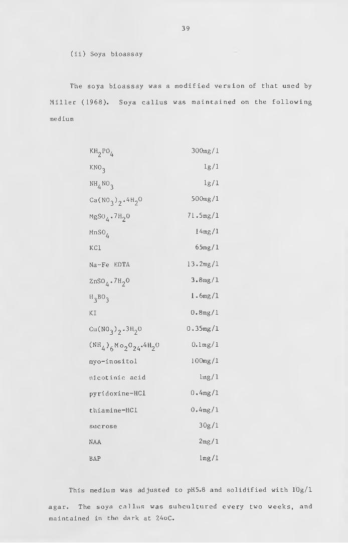
( i i ) Soya bioassay
The soya bioassay was a m odif ied version of that used by
M i l l e r ( 1 9 6 8 ) . Soya callus was m ainta ined on the fo l l o w in g
medium
KH„PO, 2 4
300mg/l
k n o 3 lg/1
NH.NCL 4 3 lg/1
Ca(N03 ) 2 .4H20 500mg/l
MgS04 .7H20 7 1 . 5mg/1
MnSO,4
14mg/1
KC1 65mg/l
Na-Fe EDTA 1 3 . 2mg/l
ZnS04 -7H20 3.8mg/l
h 3b o 3 1 . 6mg/l
KI 0 . 8mg/1
Cu (N03 ) 2 -3H20 0 . 35mg/l
(NH4 ) 6 Mo 20 24.4H20 0.1mg/l
myo-inositol 100mg/l
nicotinic acid lmg / 1
pyridoxine-HCl 0 . 4mg/1
thiamine-HCl 0 .4mg/l
sucrose 30g /l
NAA 2mg/l
BAP lmg/I
This medium was adjusted to pH5.8 and solidified with lOg/1
agar . The soya c a l lu s was subcultured every two weeks , and
maintained in the dark at 24oC.
40
For bioassay, the portion of the LH20 fraction to be assayed
was dried down in a 5cm deep, 7cm diam eter Pyrex c r y s t a l l i z i n g
d ish with l id . Standard dishes contained 0, 10, 20 , 50 , and
lOOjig/1 BAP. To this was added 40ml of the above medium with the
BAP omitted . The dishes were then autoclaved at 15 psi for 20
minutes . When cool , 10 x ca. lOmg pieces of soya callus were
placed in each dish and incubated in dim light at 2 4 ° c for 3
weeks. The p ieces were then weighed on an e le c t r o n ic balance
connected to an Apple micro-computer. The mean weight in mg was
calculated and plotted against fraction number.
c) High Performance Liquid Chromatography
HPLC was c arr ie d out using a LDC dual pump chromatograph
(Laboratory Data Control, Stone, Staffs.) fitted with a Gradient
M a s t e r c o n t r o l l e r . G r a d i e n t s b e t w e e n w a t e r (pH 7 w i t h
t r iethy lammonium bic arb o n a te ) and a c e t o n i t r i l e were used for
s e p a ra t in g c y to kin ins on columns of H y p e r s i l ODS (Shandon
Southern L t d . , Runcorn, Cheshire ) . Fractions were collected as
appropriate and used for analysis by mass spectrometry.
d) Mass Spectrometry
Mass spectrometry was c a rr ie d out in c o l l a b o r a t i o n w ith
Dr. T.L. Wang and Mr. J. Eagles (Food Research I n s t i t u t e ,
N o rw ic h ) u s in g e i t h e r an AEI MS 30 dual beam or a Kratos MS 80
mass spectrom eter . Low re so lut io n spectra were produced by
direct probe insertion of the samples.
Combined gas chromatography-mass spectrometry multiple-ion-

monitoring (GCMS MIM) was performed using permethyl derivatives
as pr ev io usly described (Wang et a l . , 1981b) . The column
temperature used was 225 °C for 2iP and 230°C for ze a t in . For
q u a n t i f i c a t i o n , ions at m/e 262 ( trimethy 1-^ N - z e a t i n ), 266
(trimethyl-^N-zeatin) , 232 (dimethyl-^N-2iP) and 236 (dimethyl-
l^N-2iP) were monitored . Levels of endogenous c ytokin ins were
c a lcu lat e d by d e te rm in ing the ^ N / ^ N ratio and r e fe r r in g to a
standard curve (Scott and Horgan, 1980) .
42
10. Binding assays
The methods for the assay of a cyto kin in receptor were
derived from the method of Gardner et a l . (1978). The tissue for
c y t o k in in work was grown on medium contain ing calcium , and
potassium nitrate as the sole nitrogen source, so as to promote
the production of caulonemal filaments, the target cells for the
cytokinin in development. Tissue was grown from an homogenate
either on cellophaned plates or in 15 litre liquid culture. All
t issue was grown for 14 days before use. Some l iqu id cultures
were grown on low levels of phospate or low phosphate and nitrate
so as to increase the percentage of caulonemal filaments which
had side branch initials .
a) Extraction procedure
Throughout the e x t r a c t io n procedure and the
c y tokin in b inding^the extract was m aintained at 0
extraction methods were used.
( i ) Homogenisation using fresh material
The t is sue was added to 4 volumes ( w /v ) of MES1 b u f f e r , pH
6.4 (25raM 2-(N-morpholino) ethane sulphonic acid, 0.25M sucrose,
ImM Na2S20^ or K2S20,-). In earlier experiments, the tissue was
homogenised in an M.S.E. homogeniser and then hand homogenised in
a glass homogeniser or a motorised teflon and glass homogeniser.
In later e x p e r im e n ts , the t i s s u e , a fter being ground in the
M.S.E. homogeniser, was homogenised in a glass bead homogeniser
assay of
-4°C. Two
(B. Braun, Melsungen AG), or a Bead-Beater (Biospec Products ,
P.O. Box 722 , B a r t l e s v i l l e , Oklahoma 74003 ) . The glass bead
homogenisers enabled more tissue to be used. The homogenate was
then filtered through 1 layer of Miracloth or 8 layers of cheese
cloth . If the Bead-beater had been used, the residue was re
homogenised in the Braun homogeniser and refiltered.
The f i l t r a t e was c en tr ifuge d at 4 , 0 0 0 g ( 3 , 0 0 0 rpm in an
M.S.E. 6 x 300ml rotor ) , or at 1 3 ,000g ( 9 , 0 0 0 rpm in a Sorval GSA
rotor or 1 0 , 0 0 0 rpm in an M.S.E. 8 x 50ml rotor) for 30 minutes.
The supernatant was c en t r i fu ge d at 8 0 , 0 0 0 g ( 2 6 , 0 0 0 rpm in a
Beckman type 30 rotor or an M.S.E. 8 x 50ml rotor) for 30
minutes. The pellet was removed and resuspended in a small glass
homogeniser in about 30ml of MES2 buffer pH 6.4 (25mM MES, 0.25M
sucrose). This was then recentrifuged at 80,000g for 30 minutes
and resuspended, in an appropiate volume for the bind ing assay
(6-12ml), in MES2 buffer.
( i i ) Homogenisation using frozen material
The tissue was frozen in liquid nitrogen and ground with a
mortar and pestle . The t issue was kept fro zen throughout the
g r in d in g . It was then taken up in MES1. Unless the 1 3 ,000g
supernatant was going to be used , the t is su e was suspended in 4
volumes (w/v) of MES1; otherwise it was suspended in only a small
volume of MES1. The extract was the centrifuged in the same way
as for the homogenised tissue.
43
b) Solubilisation of the cytokinin binding fraction
44
If the pe lle t was to be s o l u b i l i s e d , it was treated for
solubilisation without being resuspended first. Two methods were
used to try to solubilise the cytokinin-binding fraction.
( i ) Acetone method
This was the method described by Venis (1977a). The pellet
was resuspended in 0.1 volume (based on original fresh weight of
tissue) in MES2. The suspension was then added to 20-fold volume
of Analar acetone at 4°C. The clumped precipitate was collected
by low speed c e n t r i f u g a t i o n ,w a s h e d with acetone and dried in a
stream of n itro g en . This pellet was re-extracted in 12ml of
MES2, and then c e n t r i fu g e d at 3 8 , 0 0 0 g ( 1 7 , 0 0 0 rpm in an M.S.E. 8
x 50 ml rotor) for 15 minutes. The pellet was resuspended in 5ml
of MES2 and both the p e l le t and supernatant were used to assay
for cytokinin-binding activity.
( i i ) Triton method
This was the method of Thomas et a l . ( 1 9 8 4 ) . The f i n a l
pellet was resuspended by adding 1ml of 20% Triton X-100 in MES2.
The r e s u s p e n d e d p e l l e t was h o m o g e n i s e d in a g l a s s hand-
homogeniser and then mixed w it h 10ml of MES2. This was then
centrifuged again at 80,000g for 30 minutes and the supernatant
used in the cytokinin-binding assay.
c) Assay for cytokinin binding
Assays for cytokinin-binding activity were carried out using
a r a d i o l a b e l l e d b e n zy la m in o p u r ine (BAP). E a r l i e r experim ents
14 14were done with a 13 .4mCi/mmo 1 C-BAP or a 57mCi/mmol C-BAP
(Amersham I n t e r n a t i o n a l p ic , Amersham, Buckinghamshire , UK.)*
Later experiments were performed with a tritiated cytokinin of a
much higher specific activity, 12Ci/mmol, which was prepared as
de sc ribe d in appendix 1. Displacement of the ra d io la b e l was
usually by BAP, but some assays were performed using zeatin and
adenine as the displacement ligand. Two assay methods were used:
a centrifugation assay, which could only be used, on a particulate
f r a c t i o n , and an e q u i l ib r iu m d i a l y s i s method for p a r t ic u la t e
fractions and soluble fractions.
( i ) Centrifugation assay
The assay was performed in 10ml polycarbonate c e n t r i fu g e
tubes . Each tube had 1ml of resuspended p e l l e t , 1ml of MES2
containing the radiolabel and 1ml of MES2 containing the required
amount of displacement ligand. For replicates, where possible,
the mixture was made up in bulk and then d iv ided into the
r e p l i c a t e tubes. The tubes were then incubated on ice for
between 0 and 24 hours. Most of the incubations were for 1
hour. The particulate fraction was recovered by centrifugation
at 8 0 , 0 0 0 g ( 3 1 , 0 0 0 rpm in a Beckman type 50 rotor or 3 2 , 0 0 0 rpm
in an M.S.E. 10 x 10 ml rotor) for 30 minutes . The supernatant
was then removed and the sides of the tube washed w ith 1ml of
d i s t i l l e d water . A fter this treatm ent , the tube was the le ft
upside down on a paper towel to drain for 10 minutes. The pellet
was then resuspended for liquid scintillation counting.
45
46
( i i ) Equilibrium dialysis
Equilibrium dialysis was performed in Teflon Dianorm cells,
type Makrozellen GD IS (M.S.E. Crawley, UK.). The membranes were
e i t h e r V is k in g tubing or Spectropor 2 (M.S.E. Crawley, UK.),
which had a molecular weight cut-off of 10,000. Membrane discs
of 46mm diameter were cut from the tubing. The membranes were
soaked three times in d i s t i l l e d water for at least 30 minutes
each, and then in MES2 three times for 15-30 minutes each.
Stoppers were put into all the emptying holes of the cells. The
membranes were then drained and streched across the l id of the
dialysis cell. The base of the dialysis cell was subs equently
placed on top, the cell turned over and placed in the carrier. 1
ml of the extract to be assayed was added to the left half (lid)
of the c e l l , and l / 2 m l of MES2 c o n t a in in g the r a d io la b e l and
l / 2 m l of MES2 c o n ta in in g the displacem ent l igand in the right
hand (base) half cell. All stoppers were replaced and the cells
rotated at 12 rpm at 4°C for 5 hours. The contents of each ha l f
c e l l were then drained into glass v ia ls and a measured amount
removed for scintillation counting.
d) Scintillation counting
Due to the ch lo ro p h y ll content , the samples could not be
counted d i r e c t l y . In e a r l i e r e xpe rim e nts , the green colour of
the c hlo roph yll was removed by b le a c h in g w ith 25;ul of bleach
(Boots sterilising solution, Boots PLC, Nottingham, or Domestos,
Lever Bros.) . B leaching took 10-20 minutes . A fter this , a
measured volume of the sample was put in a s c i n t i l l a t i o n v ial
with 10ml of scintillant (1 litre toluene, 0.5 l itre Triton X100,
4g PPO and 250mg POPOP). The bleach caused eh em ilum inescence ,
and the samples were left at 4°c overnight before counting.
In later experiments, the samples were oxidized in a Harvey
b io l o g ic a l o x i d i z e r 0X 300 (R .J . Harvey Instruments Corporation,
H i l l s d a l e , New J e r s e y ) b e f o r e c o u n t i n g . I n i t i a l l y the
centrifugation pellets were transferred to the quartz boats by
resuspending the pellets in 1ml of distilled water or a solution
of 2% T r ito n X100. However, it was difficult to resuspend them
in the water and the T r i t o n was too v o l a t i l e for the o x i d i z e r .
The most reliable method was to transfer the whole of the pellet
in 3 x 1/2 ml aliquots of distilled water. This volume of water
was too large for the oxidizer, and so the quartz boats were left
in the fume cupboard overnight , for the samples to dry down,
before being o x i d i z e d . Samples were o x id i z e d for 3 minutes .
The s c i n t i l l a n t used in the o x i d i z e r was t r i t iu m Co cktail (R.J.
Harvey 0X162) or Quickscint 1 (Zinsser Analytic (UK) Ltd.).
The samples were counted for 10 minutes in a Beckman LS 7500
or an LKB 1215 Rackbeta s c i n t i l l a t i o n counter . Both machines
were c a l ib r a t e d for the s c i n t i l l a n t being used and for quench
using the external standard ratio method.
11. Microscopy
Scanning electron microscopy was performed in collaboration
w it h Dr. T.L. Wang and Mr.R. Turner (Food Research I n s t i t i u t e ,
Norwich) us ing a P h i l i p s scanning electron microscope equipped
with a cryostage. Tissue was grown on Knops medium supplemented
with v itam ins for 4 weeks at the required temperature and
47
48
prepared for microscopy by f r e e z in g in a l iq u id n itrogen slush
and coating with gold.
12. Light treatments
A l l treatments were performed using monochromatic l igh t
produced by interference filters (DAL type; Schott, Mainz. FRG).
For earlier experiments, the monochromatic light apparatus was
the same as described by Jenkins and Cove (1983). Illumination
was provided by Aldis Tutor 2 p r ojec to rs (Rank Audio V is u a l
Lt d. ,B ren t f o r d , UK), which were f i t t e d with thyristor dimmer
controls and 250W tungsten halogen bulbs (type M36; Thorn
Lighting, London, UK). For later experiments the projectors were
supplied by a regulated D.C. supply so that they were unaffected
by mains fluctuations.
Deep (2cm), 5cm petri dishes, f il led with 20ml Knops medium
supplemented with sucrose, were inoculated with a small piece of
w ild-type protonema 1cm from the edge of the d ish . These were
grown under white light (10-15Wm for 8 days. The plates were
then t ran sferre d to the dark, on the ir edges, w ith the plates
o r ien t a t e d so that the inoculum was at the lowest point . The
plates were incubated for either 14 or 21 days. This caused the
growth of unbranched filaments that resembled caulonemata. These
grew negatively geotropically. After this period the tips of the
dark-grown f i la m e n ts had grown into the centre of the dish .
Under green (542nm monochromatic) "safe" light of about 2mWm f
hormone solutions were added to the p la te s . One ml of a 20jiM
solution was added, to give a final concentration of 1/uM.
49
The plates were then put into in d iv id u a l cardboard boxes
that had a 1 x 2 cm gap in the middle of one edge. The plates
were orientated so that the dark-grown caulonemal filaments were
perpendicular to the gap in the box. The plates were left for at
le ast 1 hour for the s o lu t io n to be absorbed by the agar . The
first experiments were performed with h o r izo n ta l i l l u m i n a t i o n
but when low l igh t i n t e n s i t i e s were used the f i l a m e n ts did not
bend so it was difficult to know which bud sites to count. The
boxes were therefore placed on their edges under v e r t ic a l
i l l u m i n a t i o n so that the dark-grown caulonemal f i lam e nts were
perpendicular to the light direction. This caused the filaments
to bend upwards, due either to a positive phototropic response,
or, at lower light intensities, to a negative geotropic response.
The light intensity was measured with a radiometer (type J16 with
a J 6 5 1 2 probe; Te ktro nic U.K. Ltd . , London,U .K . ) . The opening of
the box was put into the p o s it io n of the sensor of the probe,
after the reading had been taken.
The plates were left under the monochromatic light for three
days. They were scored by counting the number of buds produced
at f ive c e l l s before the bend, the last part of the caulonemal
f i la m e n t to grow in the dark before being transfe rre d to the
monochromatic light. Twenty filaments were counted in this way
so as to give a total number of buds from 100 cells.
CHAPTER I I I
Isolation and analysis of mutants
Results and discussion
In the search for a cytokinin receptor, any fraction that is
isolated from the wild-type moss which appears to bind cytokinin
s p e c i f i c a l l y has to be shown to be develop mentally re levant .
One method of achieving this is to study mutants that do not have
a specific response to applied cytokinin. Some of these mutants
may not possess a receptor that is able to bind c y t o k in in ,a n d
thus cannot respond. These mutants can be used to show that the
loss of the specific binding of cytokinin is associated with the
loss of the cytokinin response by comparison with the wild type.
The moss, Physcomitrella patens, is useful in this respect,
as the indu ct io n and screen ing of mutants is r e l a t i v e l y easy.
A lso , c y to k in in plays an important role in the development of
P h y s c o m it r e l la p a te n s . Cytokinin induces the production of the
gametophore from a caulonemal side branch. Therefore, it may be
p o s s i b l e to i s o l a t e m u t a n t s that are u n a b l e to p r o d u c e
gametophores in the presence of exogenous c y to k in in . Some of
these mutants may have a defective receptor, some no receptor at
all and some may be blocked at a later stage in the production of
the gametophore.
51
1« Isolation of cytokinin non-responding mutants
(i ) Spore mutagenesis
Spores from pab A3 and thi Al were treated with NTG and
screened as described in Material and Methods. Of the 25 ,000
mutants picked off and tested in this way, none that produced
dark-grown caulonemata failed to produce buds in the presence of
1 pM exogenous cytokinin. Typical colonies that have responded
to the BAP are shown in figure 1. There are a number of
explanations for not detecting any cytokinin non-responding
mutants, viz:
a) insufficient mutants have been screened for the rate of
occurrence of such mutants;
b) two genes need to be altered to make such mutants;
c) the receptor for cytokinin in bud induction is also
required for an essential process such as cell division;
d) mutants that are unable to respond to cytokinin in the
process of bud formation are also unable to produce dark-
grown caulonemata.
From other mutation screening procedures, mutants are
detected at a rate of about one in a thousand (Ashton et a l . ,
1979b). Therefore screening 25 ,000 plants should produce some
mutants of the type being s o u g h t . The lack of non
responding mutants is unlikely to be due to insuff icient
treatment with mutagen as only about 1 0 % of the spores germinated
and other types of mutant were isolated in the screening* If
more than one gene has to be altered,due to there being replicate
genes, then the rate of occurrence of such mutants would be
poss i'bis.
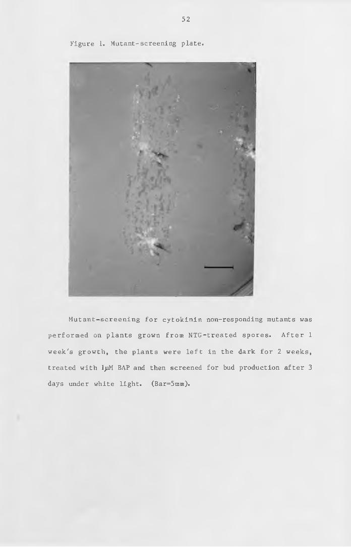
52
Figure 1. Mutant-screening plate.
Mutant-screening for cytokinin non-responding mutants was
performed on plants grown from NTG-treated spores. After 1
week's growth, the plants were left in the dark for 2 weeks,
treated with IjjM BAP and then screened for bud production after 3
days under white light. (Bar=5mm).
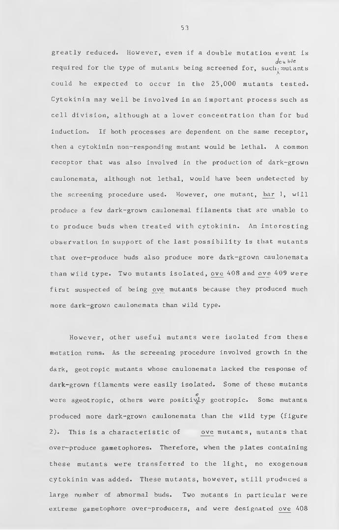
53
greatly reduced. However, even if a double mutation event is
<J0 u_ ble
required for the type of mutants being screened for, such|mutants
could be expected to occur in the 25 ,000 mutants tested.
Cytokinin may well be involved in an important process such as
cell d ivision , although at a lower concentration than for bud
induction. If both processes are dependent on the same receptor,
then a cytokinin non-responding mutant would be lethal. A common
receptor that was also involved in the production of dark-grown
caulonemata, although not lethal, would have been undetected by
the screening procedure used. However, one mutant, bar 1, will
produce a few dark-grown caulonemal filaments that are unable to
to produce buds when treated with cytokinin. An interesting
observation in support of the last p ossibil ity is that mutants
that over-produce buds also produce more dark-grown caulonemata
than wild type. Two mutants isolated , ove 408 and ove 409 were
first suspected of being ove mutants because they produced much
more dark-grown caulonemata than wild type.
However, other useful mutants were isolated from these
mutation runs. As the screening procedure involved growth in the
dark, geotropic mutants whose caulonemata lacked the response of
dark-grown filaments were easily isolated. Some of these mutants
e
were ageotropic, others were positively geotropic. Some mutants
produced more dark-grown caulonemata than the wild type (figure
2). This is a characteristic of ove mutants, mutants that
over-produce gametophores. Therefore, when the plates containing
these mutants were transferred to the light , no exogenous
cytokinin was added. These mutants, however, s t i l l produced a
large number of abnormal buds. Two mutants in particular were
extreme gametophore over-producers, and were designated ove 408
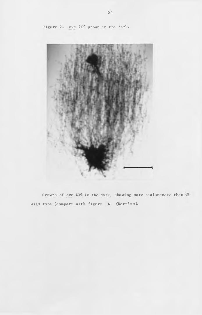
54
Figure 2. ove 409 grown in the dark.
Growth of ove 409 in the dark, showing more caulonemata than
wild type (compare with figure 1). (Bar=5mm).
55
and ove 409. These were held as stocks and as such were
maintained at 15°C. After one month, the stock tube of ove 409
was observed to have produced some normal leafy gametophores.
When this mutant was subcultured 24°c , it again started to
over-produce gametophores and so appeared to have a temperature-
sensitive phenotype. This mutant, as with other ove mutants, may
have arisen due to a number of factors which resulted in
either too much cytokinin being produced or in the tissues being
over-sensitive to normal levels of cytokinin. The levels of
cytokinin in this mutant were therefore studied (see this
chapter, section 3). ove 409 was also studied genetically (see
this chapter, section 2 ).
( i i ) Somatic mutagenesis
Somatic mutagenesis was attempted with ade 5, which requires
adenine. This strain was used,in particular, since an ove mutant
in an ade background would be useful in the study of cytokinin
synthesis ffo m radiolabelled adenine. About 10^ protoplasts
were prepared from NTG-treated tissue. None of the protoplasts
regenerated. This was most likely due to the combination of high
k i l l by NTG and to the fact that the level of adenine required
for this strain is near the level that is toxic in the wild type.
As this strain was difficult to regenerate from protoplasts, it
was not used to isolate any further mutants.
2. Genetic analysis of ove 409
ove 409 appeared to be a mutant which was temperature-
sensitive (see this Chapter, section 3) for bud production and
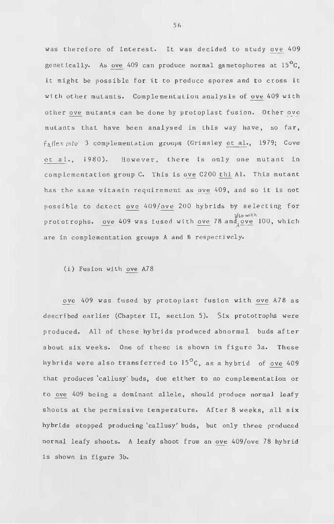
56
was therefore of interest. It was decided to study ove 409
genetically. As ove 409 can produce normal gametophores at 15°C,
it might be possible for it to produce spores and to cross it
with other mutants. Complementation analysis of ove 409 with
other ove mutants can be done by protoplast fusion. Other ove
mutants that have been analysed in this way have, so far,
fallen into 3 complementation groups (Grimsley et al ., 1979; Cove
et a 1 ., 1 98 0 ) . Ho w ever , there is only one mutant in
complementation group C. This is ove C200 thi Al. This mutant
has the same vitamin requirement as ove 409, and so it is not
possible to detect ove 409/ove 200 hybrids by selecting for
j/so witKprototrophs. ove 409 was fused with ove 78 and^ove 100, which
are in complementation groups A and B respectively.
(i ) Fusion with ove A78
ove 409 was fused by protoplast fusion with ove A78 as
described earlier (Chapter II, section 5). Six prototrophs were
produced. All of these hybrids produced abnormal buds after
about six weeks. One of these is shown in figure 3a. These
hybrids were also transferred to 15°C, as a hybrid of ove 409
that produces 'callusy' buds, due either to no complementation or
to ove 409 being a dominant allele, should produce normal leafy
shoots at the permissive temperature. After 8 weeks, all six
hybrids stopped producing 'callusy' buds, but only three produced
normal leafy shoots. A leafy shoot from an ove 409/ove 78 hybrid
is shown in figure 3b.
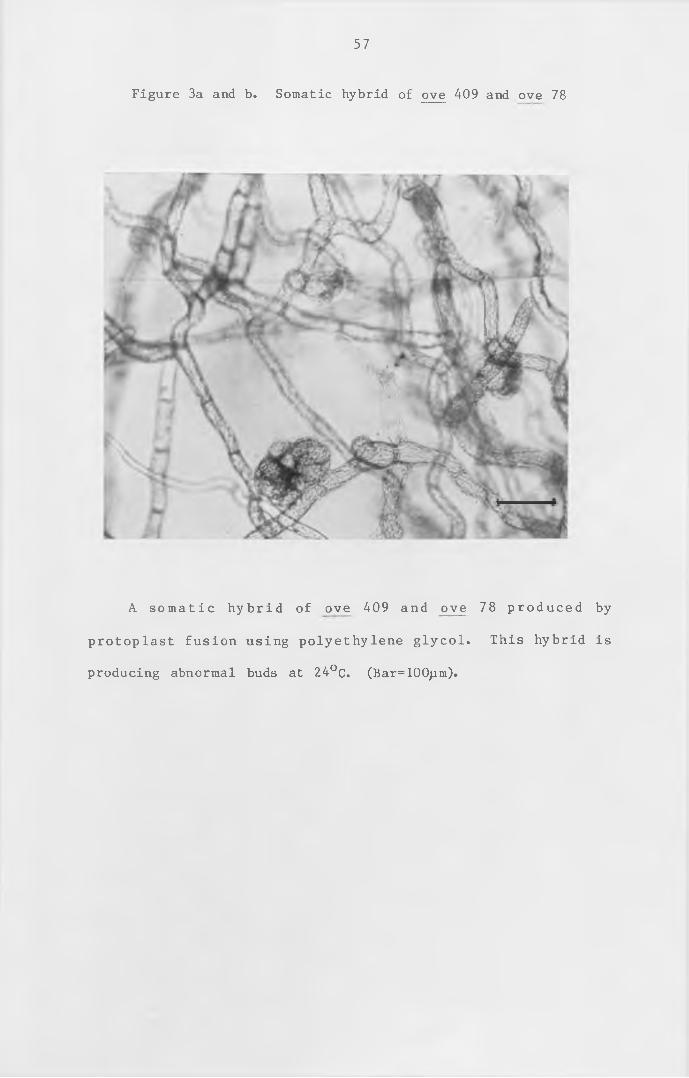
57
Figure 3a and b. Somatic hybrid of ove 409 and ove 78
A som atic h ybr id of ove 409 and ove 78 produced by
protoplast fusion using polyethylene glycol. This hybrid is
producing abnormal buds at 24°C. (Bar= 100pm).
58
A somatic hybrid of ove 409 and ove 78 induced with
polyethylene glycol. This hybrid is producing normal buds at
15°C. (Bar=500jim).
( i i ) Fusion with pab A3 and pab A5
also withove 409 was fused with pab A3 and^ pab A5 to see if the ove
409 phenotype was due to a dominant allele . However, neither
fusion produced any prototrophs.
( i i i ) Fusion with ove 100
ove 409 was fused with ove 100
one prototroph was produced. This
shoots within three weeks at 24°c .
4a and 4b.
(iv) Crossing of ove 409 with self-sterile mutants
Cultures for selfing ove 409 and for crossing ove 409 with
auxotrophic mutants to obtain ove 409 in a different background
were set up as described in Materials and Methods (section 2).
Even after 5 months, with the cultures being irrigated every
month, no capsules were produced.
The attempts to cross ove 409 with other auxotrophic mutants,
to isolate ove 409 in a different background so that it could be
fused with ove 200, were unsuccessful. Also, the fusions of ove
409 with pab 3 and pab 5, to test for dominance, did not produce
any hybrids. However, from the fusions with ove 78 and ove 100,
the ove 409 phenotype is highly likely to be due to a recessive
allele, and can be assigned to complementation group A, which is
the group containing ove 78. Because ove 409 is temperature-
sensitive for the production of cytokinin , it is potentially a
59
by protoplast
produced some
These are sho
fusion. Only
normal leafy
wn in figures
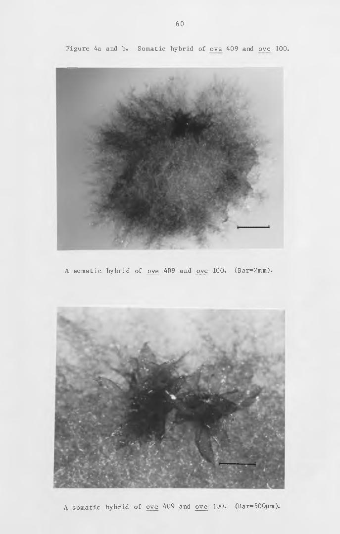
60
Figure 4a and b. Somatic hybrid of ove 409 and ove 100.
A somatic hybrid of ove 409 and ove 100. (Bar=2mm).
A somatic hybrid of ove 409 and ove 100. (Bar=500jam).
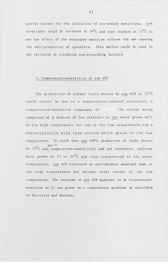
61
useful mutant for the isolation of secondary mutations. ove
revertants could be isolated at 24°c and then studied at 15°C to
see the effect of the secondary mutation without the one causing
the over-production of cytokinin. This method could be used in
the isolation of cytokinin non-responding mutants.
3. Temperature-sensitivity of ove 409
The production of normal leafy shoots by ove 409 at 15°C
could either be due to a temperature-induced reversion, a
temperature-sensitive response, or the colony being
comprised of a mixture of two strains:- an ove which grows well
at the high temperature but not at the low temperature, and a
p h e n o t y p i c a l l y w i ld type s t r a i n which grows at the low
temperature. To check that ove 409's production of leafy shoots
due to
at 15 c was^ temperature-sensitivity and not reversion, cultures
were grown at 15 or 24°C and then transferred to the other
temperature. ove 409 continued to over-produce abnormal buds at
the high temperature but normal leafy shoots at the low
temperature. The response of ove 409 appeared to be temperature-
sensitive so it was grown on a temperature gradient as described
in Materials and Methods.
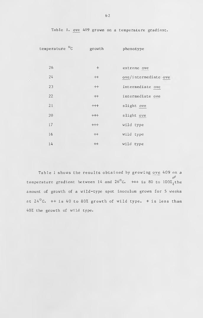
62
Table 1. ove 409 grown on a temperature gradient.
temperature °C growth phenotype
26 + extreme ove
24 ++ ove/intermediate
23 ++ intermediate ove
2 2 ++ intermediate ove
2 1 +++ slight ove
2 0 +++ slight ove
17 +++ wild type
16 ++ wild type
14 ++ wild type
Table 1 shows the results obtained by growing ove 409 on a
of
temperature gradient between 14 and 26°C. -H-+ is 80 to 100%^the
amount of growth of a wild-type spot inoculum grown for 5 weeks
at 24°C. ++ is 40 to 80% growth of wild type. + is less than
40% the growth of wild type.
63
Table 1 gives the results obtained when ove 409 was grown at
a range of temperatures from 26°C to 17°C; there is a gradual
change in phenotype of ove 409 from an extreme ove to wild type.
The extreme ove phenotype of ove 409 is shown in figure 5a.
The intermediate ove phenotype is shown in figure 5b. ove 409
grown at 15°C is shown in figure 5c and wild type in figure 6 .
To check that ove 409 was not a mixture of two strains , it was
grown at 15°C and protoplasts were isolated from the protonema.
50 regenerants were replica plated and cultured at the permissive
and n o n- p e rm issive tem perature . A ll 50 re g en e r an t s
phenotypically resembled ove's at the non-permissive temperature,
but were wild type at the permissive temperature. Furthermore,
ove mutants are usually more d if f icu lt to protoplast and
regenerate than wild type. Thus, if ove 409 was a mixture, most
of the isolated protoplasts would have been the wild type strain
and therefore it would have been expected that most of the
regenerating protoplasts would be wild type at both temperatures.
As all 50 regenerants changed phenotype with temperature, ove 409
does appear to be a temperature-sensitive mutant for gametophore
overproduction.
Figure 5a, b and c. The phenotype of ove 409 at different
temperatures.
64
ove 409 grown for 5 weeks at 2 6 ° C . (Bar-1mm).
65
An SEM of ove 409 grown for 5 weeks at 23°C. (Bar=100)iin).
66
An SEM of ove 409 grown for 5 weeks at 15°C. (Bar= lOOjim).
67
Figure 6. Wild type at 24°C.
An SEM of wild type grown for 5 weeks at 24°C. (Bar= 100/um).
68
As mentioned in section 1 a (i) above, ove 409's ove
phenotype could be due to ove 409 over-producing cytokinin or
being over-sensitive to normal levels of cytokinin. These two
possibilities can be subdivided (Ashton et al . , 1979a; Wang et al . ,
1983) as follows:
increase in cytokinin activity
a) increased synthesis of cytokinin
b) decreased metabolism of cytokinin
c) production of a more active cytokinin
d) decrease in production of a cytokinin antagonist
change in sensitivity to cytokinin
a) loss of requirement of cytokinin for bud production
b) increase in sensitivity to normal levels of cytokinin
All the ove's that have been studied so far overproduce buds
and overproduce cytokinin (Wang et a l . 1981a). ove 409 was
therefore studied for production of cytokinin. Cytokinin
production was measured at both 15 and 24°C. This was carried
out by growing ove 409, ove 78 and wild type in 1 litre of liquid
medium in a 2 litre conical flask on an orbital shaker at the two
temperatures for 6 weeks. The tissue was collected , weighed and
freeze dried. The medium from these cultures was labelled with
cytokinin as an internal standard (a gift from Dr. R. Horgan,
Aberystwyth). To each flask was added lOOng of ^N - ze a t in and
lOOng ^N-isopentenyl adenine. The medium was then extracted for
cytokinin as described in materials and methods. After butanol
extraction, the samples were dried down into dram vials . They
were then p u r i f i e d on HPLC us ing a g r ad ie n t from 5-*25%
acetonitrile with an exponent of 0.5 in 15 minutes. Zeatin
eluted after 5.5 minutes and 2iP after 13 minutes. The relevant
69
fractions for each sample were collected, dried down and
permethylated. Quantification was carried out as described in
Materials and Methods (section 8 d) using GCMS under chemical
ionisation conditions. The results of isopentenyl adenine are
shown in Table 2. The zeatin samples could not be measured
accurately as so little was present in the samples, and other ions
interfered at similar m/e values.
In all three strains, the amount of isopentenyladenine per
mg dry weight is higher at 24°C than at 15°C. ove 409's
phenotype at 24°c is highly likely to be due to the over
production of isopentenyladenine. However, the amount of over
production of 2 iP by ove 409 is much less than the over
production of 2iP by ove 78. The drop in production of ove 409
with the drop in temperature is enough so that at 15°C the amount
of 2iP produced is within the range produced by wild type. This
alteration in the level of 2iP would account for the change of
phenotype of ove 409 from an extreme ove to wild type^ Thus, ove
409's phenotype could be due to an increased synthesis of
cytokinin by the normal or a de novo pathway, or a decreased
metabolism of cytokinin. With wild type and ove 78, there is
only a 5-6 fold change in 2iP production between 15 and 24°C. In
the case of ove 409 there is a 14-fold change in 2iP production.
This change in cytokinin production with temperature, especially
the 5-6 fold change with wild type and ove 7 8 ,could be due to
less cytokinin-producing tissue being present at 15°C. This
would result in slower regeneration and dif f erent iation, and a
delay in the production of cytokinin. Temperature may also have
an effect in d i f fe r e n t ia t io n and so at the lower temperature
there is more primary chloronemata, and less cytokinin and auxin.
Tab
le
2.
Pr
od
uc
tio
n
of
is
op
en
te
ny
la
de
nin
e
at
15
and
24
70
in
te
rn
al
st
an
da
rd
71
In the case of the 14-fold change of cytokinin of ove 409, there
could be a temperature-sensitive mutation which is contributing
to the change in cytokinin production.
4. Growth on starvation medium
When wild type is grown on phosphate-free agar medium, it
forms mainly radially-growing caulonemata with the normal number
of buds, about 1 per filament. Side-branch initials form at most
of the potential sites, but these fail to develop further. When
such a culture is supplied with 1 /iM BAP, the side branch
in it ia ls are induced to form callus-like buds. This type of
tissue is therefore a good source of target tissue for cytokinin
and hence potentially a good source of any cytokinin receptor
(see chapter 5). Wild type was grown in a 1-litre phosphate-free
liquid culture and produced long caulonemal filaments with
normal gametophores (figure 7), but no side-branch i n it ia l s . A
possible explanation of the difference on solid and in liquid
phosphate-free medium is that the agar could contain low levels
of phosphate. Side-branch in it ia ls can be produced in liquid
culture on a low phosphate and low nitrate medium. Both
phosphate-freehand low phosphate with low nitrate, media were used
to grow batch cultures for the assay of cytokinin binding (see
chapter 5 section 2) . bar 1 was grown in 1 litre of phosphate-
free medium. After about a week it appeared to stop growing.
bar 1 did not turn brown even after 2 months. Therefore, bar 1
could not be enriched for cytokinin target cells by this method.
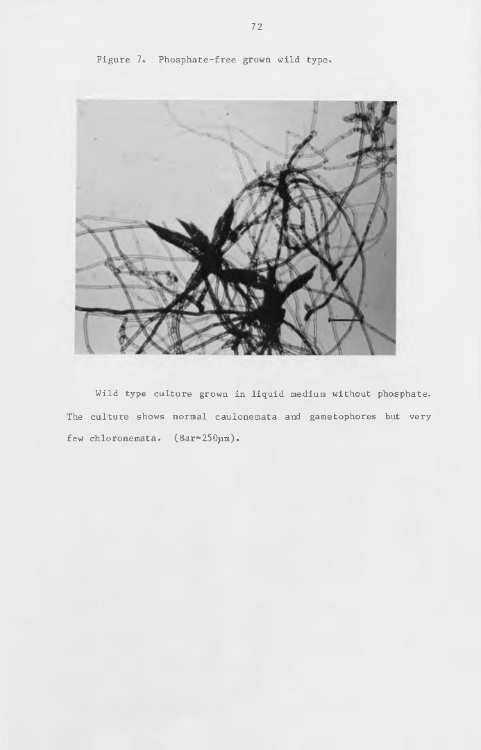
72
Figure 7. Phosphate-free grown wild type.
Wild type culture grown in liquid medium without phosphate.
The culture shows normal caulonemata and gametophores but very
few chloronemata. (Bar=250pm).
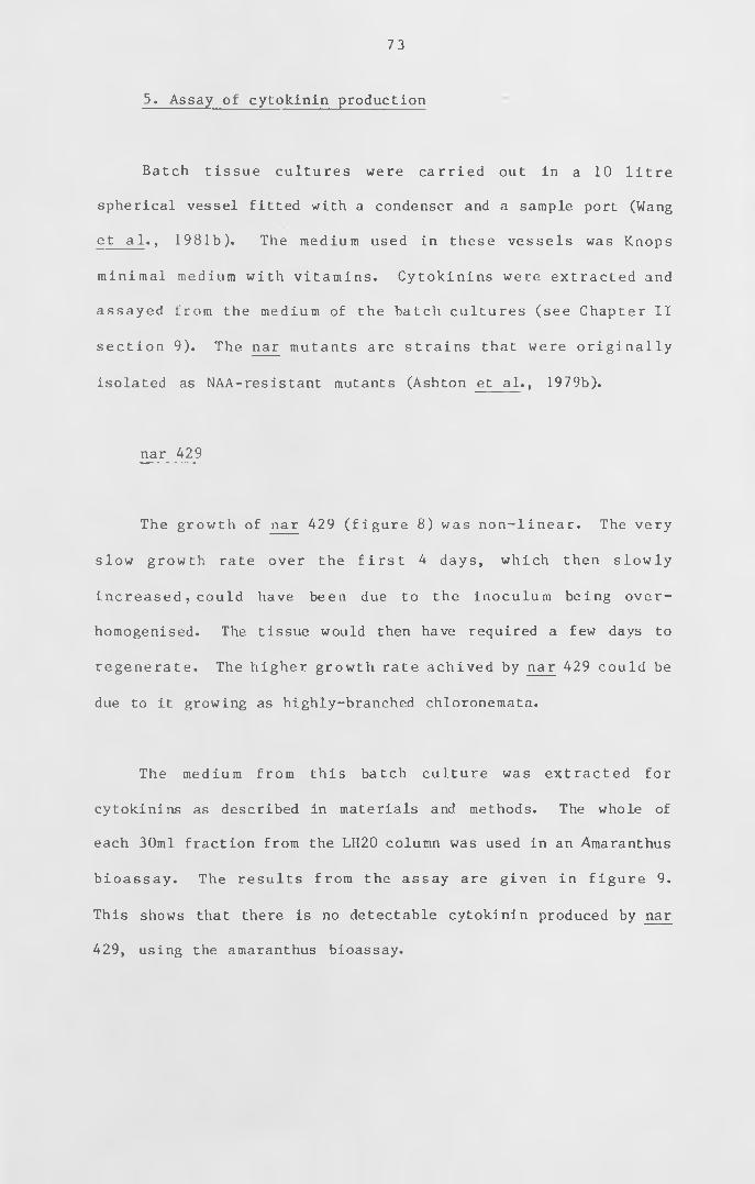
73
5. Assay of cytokinin production
Batch tissue cultures were carried out in a 10 l itre
spherical vessel fitted with a condenser and a sample port (Wang
et a l ., 1981b). The medium used in these vessels was Knops
minimal medium with vitamins. Cytokinins were extracted and
assayed from the medium of the batch cultures (see Chapter I I
section 9). The nar mutants are strains that were originally
isolated as NAA-resistant mutants (Ashton et a l . , 1979b).
nar 429
The growth of nar 429 (figure 8 ) was non-linear. The very
slow growth rate over the f irst 4 days, which then slowly
increased, could have been due to the inoculum being over
homogenised. The tissue would then have required a few days to
regenerate. The higher growth rate achived by nar 429 could be
due to it growing as highly-branched chloronemata.
The medium from this batch culture was extracted for
cytokinins as described in materials and methods. The whole of
each 30ml fraction from the LH20 column was used in an Amaranthus
bioassay. The results from the assay are given in figure 9.
This shows that there is no detectable cytokinin produced by nar
429, using the amaranthus bioassay.
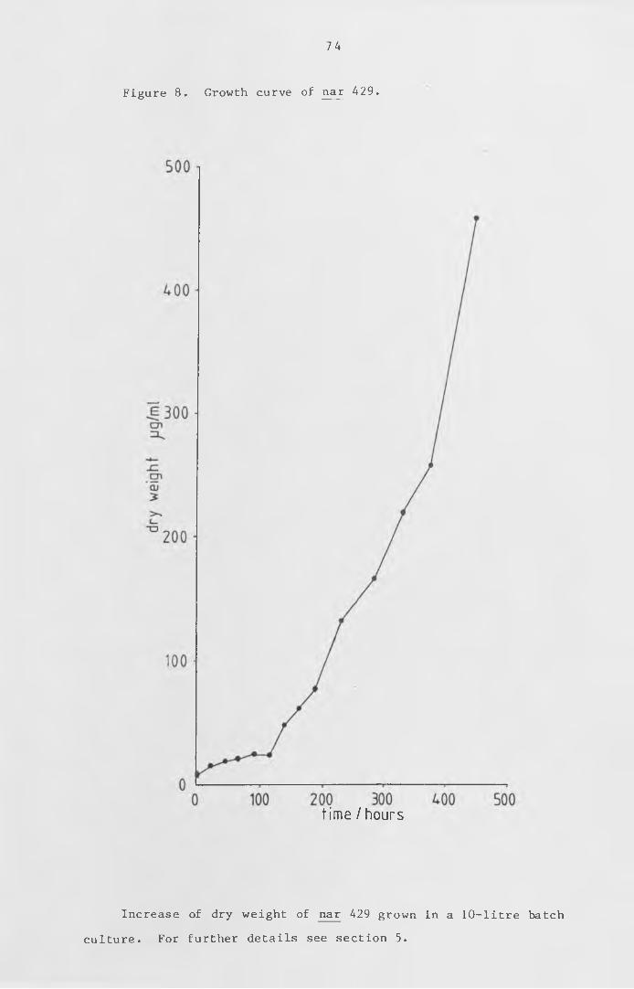
74
Figure 8 . Growth curve of nar 429.
time I hours
Increase of dry weight of nar 429 grown in a 10-litre batch
culture. For further details see section 5.
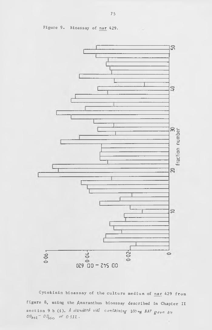
75
Figure 9 . Bioassay of nar 429.
CDln
X—VOcpO
o
—r^ Cvjo
0Z9 a o - z v s ao
Cytokinin bioassay of the culture medium of nar 429 fro m
figure 8 , using the Amaranthus bioassay described in Chapter II
section 9 b ( i ) . A standard viAl containing- [00 ■ng BAP g a v e a.'n
0DS *l~ 0D6Sv * 0 111 >
frac
tion
num
ber
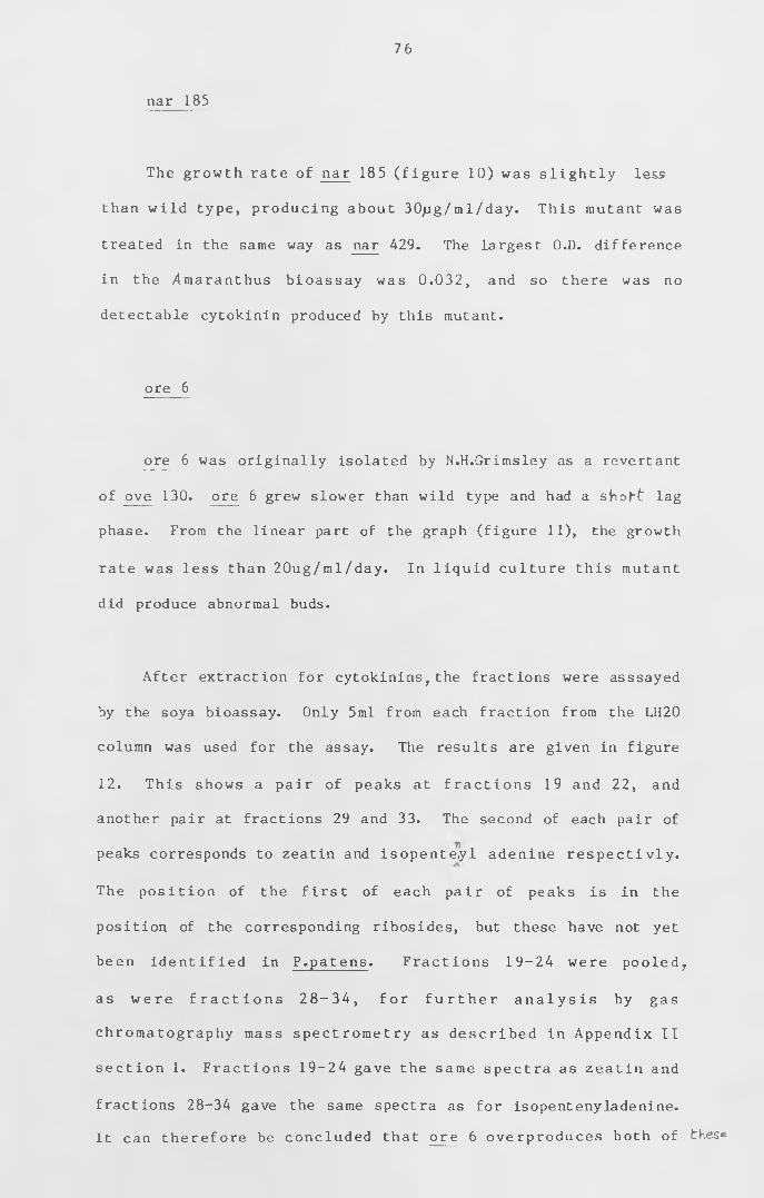
76
The growth rate of nar 185 (figure 10) was slightly less
than wild type, producing about 30jag/ml/day. This mutant was
treated in the same way as nar 429. The largest O.D. difference
in the /maranthus bioassay was 0 .032, and so there was no
detectable cytokinin produced by this mutant.
ore 6
ore 6 was originally isolated by N.H.Grimsley as a revertant
of ove 130. ore 6 grew slower than wild type and had a sjnof-t lag
phase. From the linear part of the graph (figure 11), the growth
rate was less than 20ug/ml/day. In liquid culture this mutant
did produce abnormal buds.
After extraction for cytokinins; the fractions were asssayed
by the soya bioassay. Only 5ml from each fraction from the LH20
column was used for the assay. The results are given in figure
12. This shows a pair of peaks at fractions 19 and 22, and
another pair at fractions 29 and 33. The second of each pair of
peaks corresponds to zeatin and isopenteyl adenine respectivly.
The position of the f irst of each pair of peaks is in the
position of the corresponding ribosides, but these have not yet
been identified in P.patens. Fractions 19-24 were pooled,
as were f r a c t i o n s 28-34, for f u r t h e r a n a l y s i s by gas
chromatography mass spectrometry as described in Appendix II
section 1. Fractions 19-24 gave the same spectra as zeatin and
fractions 28-34 gave the same spectra as for isopentenyladenine.
It can therefore be concluded that ore 6 overproduces both of
nar 185
these.
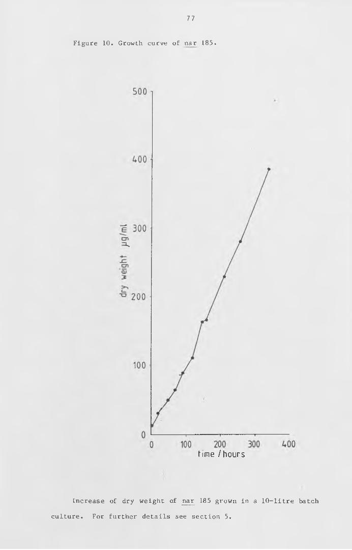
77
Figure 10. Growth curve of nar 185.
time /hours
Increase of dry weight of nar 185 grown in a 10-litre batch
culture. For further details see section 5.
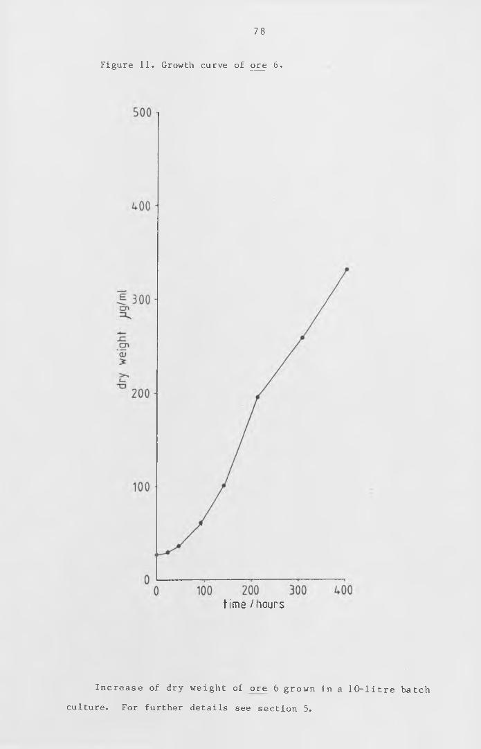
78
Figure 11. Growth curve of ore 6.
time /hours
In cre a se of dry w eight of ore 6 grown in a 10- litre batch
culture. For further details see section 5.
79
Figure 12. Bioassay of ore 6.
G > O C > O OO OO ^ < t OnI
6lu/ 4qBia/A ueaw
Cytokinin bioassay of the culture medium of the batch
culture of ore 6 shown in figure 1 1 , using the soya bioassay
described in Chapter I I section 9 b. ( i i ) .
Mean wdMght of standards-. « P, ^ 50/jg/f BLP*24-2*g.
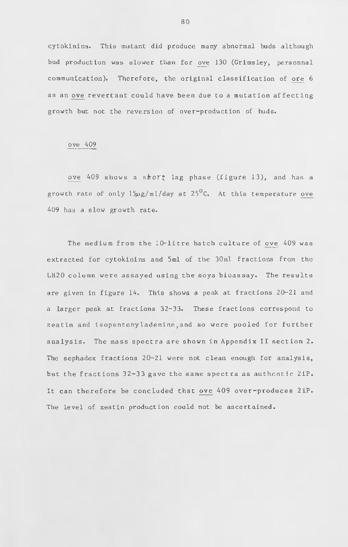
80
cytokinins. This mutant did produce many abnormal buds although
bud production was slower than for ove 130 (Grimsley, personnal
communication). Therefore, the original classification of ore 6
as an ove revertant could have been due to a mutation affecting
growth but not the reversion of over-production of buds.
ove 409
ove 409 shows a sltort iag phase (figure 13), and has a
growth rate of only 15;ug/ml/day at 25°C. At this temperature ove
409 has a slow growth rate.
The medium from the 10-litre batch culture of ove 409 was
extracted for cytokinins and 5ml of the 30ml fractions from the
LH20 column were assayed using the soya bioassay. The results
are given in figure 14. This shows a peak at fractions 20-21 and
a larger peak at fractions 32-33. These fractions correspond to
zeatin and isopentenyladenine and so were pooled for further
analysis . The mass spectra are shown in Appendix I I section 2.
The sephadex fractions 20-21 were not clean enough for analysis,
but the fractions 32-33 gave the same spectra as authentic 2iP.
It can therefore be concluded that ove 409 over-produces 2iP.
The level of zeatin production could not be ascertained.
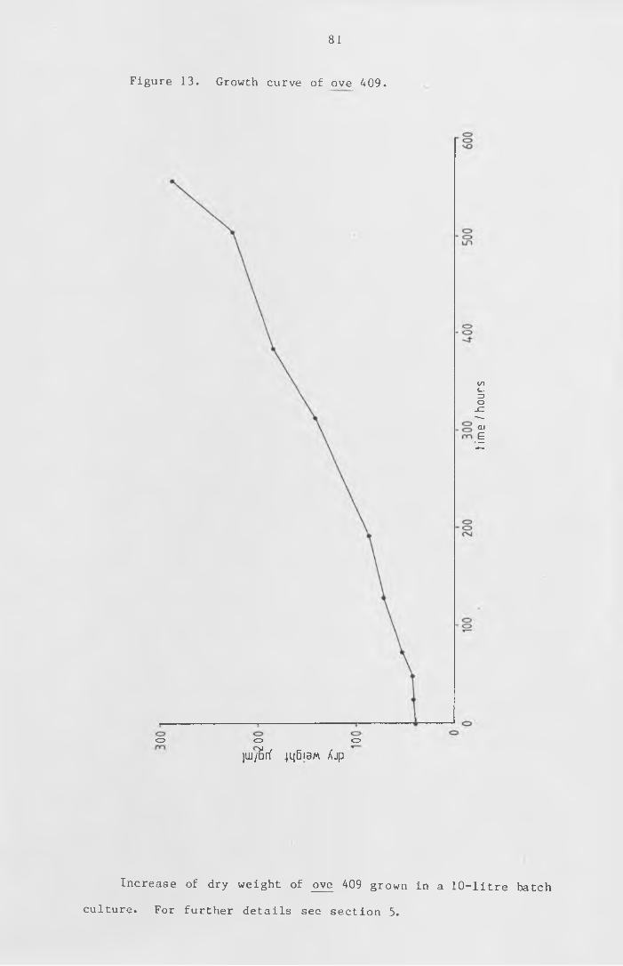
81
Figure 13. Growth curve of ove 409.
<=> o c>
ju j/ S r f 4 q B i 3 M X j p
Increase of dry weight of ove 409 grown in a 10-litre batch
culture. For further details see section 5.
time
I hou
rs
82
Figure 14. Bioassay of ove 409.
' o . m
. CVJ
—i—oOJm
—,— cd oo cvj
<o
Ovl
CDCDCM
ONO
oOsJ
CDCO
Buj/ jq6ia/A ueaw
Cytokinin bioasay of the culture medium of the batch culture
of ove 409 shown in figure 13, by the soya bioassay described in
Chapter II section 9 b. ( i i ) .
frac
tion
num
ber
CHAPTER IV
Characteristics of cytokinin action
Results and discussion
1. Time of exposure to BAP for bud induction
Brandes and Kende (1968) showed that bud induction could be
reversed by removal of the cytokinin in the moss Funaria
hygrometrica. Brandes and Kende concluded that cytokinin does
not act as a trigger for bud induction but is required for a
critical time period to allow the differentiation to stabilize.
This effect was investigated in Physcomitrella patens. Wild-type
cultures were exposed to cytokinin as described in Materials and
Methods for 1 to 7 days, after growth in liquid culture for 7
days. The tissue was collected by f i ltrat ion , added to liquid
medium containing 1 pM BAP and, after set intervals, removed by
f i l tr a t io n , washed with sterile d ist il led water and placed in
fresh medium without BAP. The number of buds produced was
counted 14 days after the transfer to the medium containing BAP.
The results are shown in figure 15. There is very l i t t le
increase in the number of buds with exposures of up to 48 hours.
With longer exposures to BAP, there is an increase in the number
of buds produced. As buds can be seen in a culture exposed to
cytokinin for less than 72 hours, the effect of the cytokinin is
removed by washing the culture. The increase of the number of
buds after 72 hours could be due to bud induction on new tissue.
83
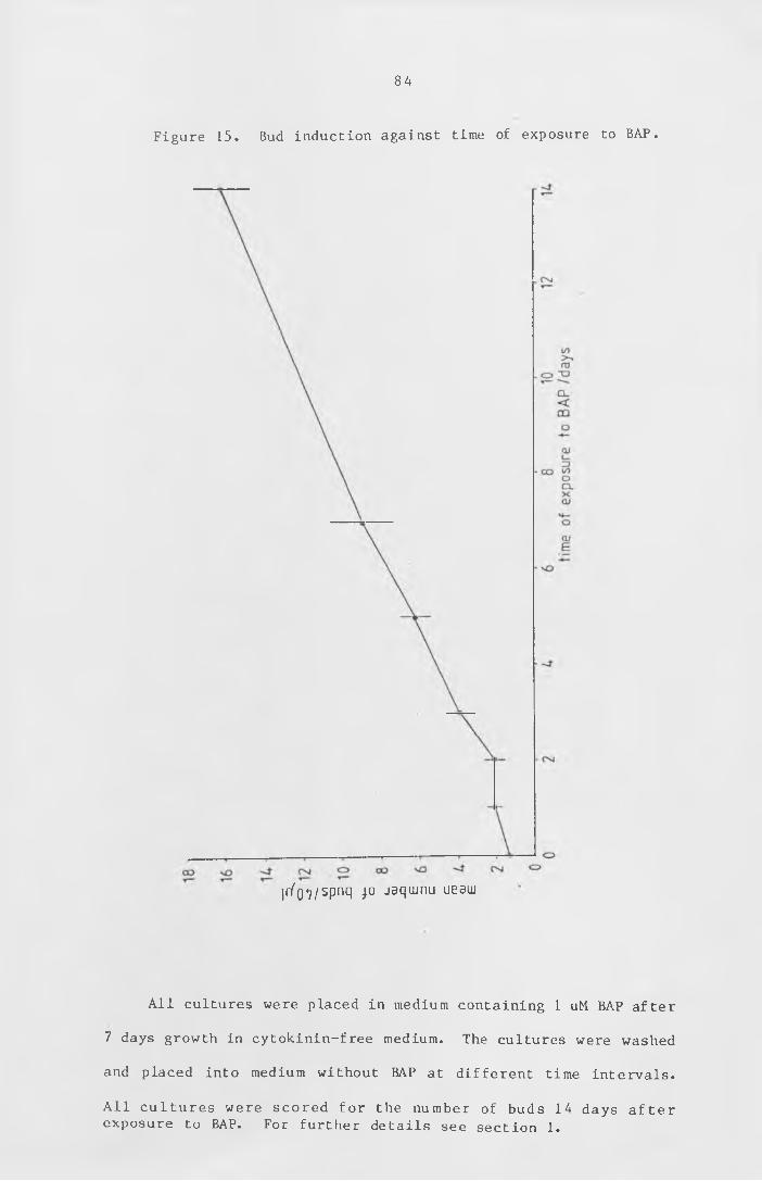
84
Figure 15. Bud induction against time of exposure to BAP.
ir/o^/spnq jo jaqiunu ueaw
All cultures were placed in medium containing 1 uM BAP after
7 days growth in cytokinin-free medium. The cultures were washed
and placed into medium without BAP at different time intervals.
All cultures were scored for the number of buds 14 days after
exposure to BAP. For further details see section 1.
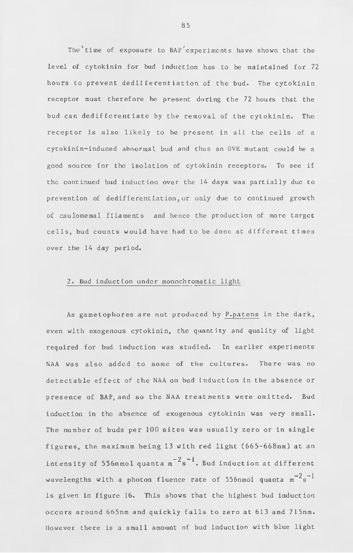
The' time of exposure to BAP'"experiments have shown that the
level of cytokinin for bud induction has to be maintained for 72
hours to prevent dedifferentiation of the bud. The cytokinin
receptor must therefore be present during the 72 hours that the
bud can dedifferentiate by the removal of the cytokinin. The
receptor is also likely to be present in all the cells of a
cytokinin-induced abnormal bud and thus an OVE mutant could be a
good source for the isolation of cytokinin receptors. To see if
the continued bud induction over the 14 days was partially due to
prevention of dedifferentiation , or only due to continued growth
of caulonemal filaments and hence the production of more target
cells , bud counts would have had to be done at different times
over the 14 day period.
2. Bud induction under monochromatic light
As gametophores are not produced by P.patens in the dark,
even with exogenous cytokinin, the quantity and quality of light
required for bud induction was studied. In earlier experiments
NAA was also added to some of the cultures. There was no
detectable effect of the NAA on bud induction in the absence or
presence of BAP, and so the NAA treatments were omitted. Bud
induction in the absence of exogenous cytokinin was very small.
The number of buds per 100 sites was usually zero or in single
figures , the maximum being 13 with red light (665-668nm) at an
- 2 - 1intensity of 556nmol quanta m s . Bud induction at different
- 2 - 1wavelengths with a photon fluence rate of 556nmol quanta m s
is given in figure 16. This shows that the highest bud induction
occurs around 665nm and quickly falls to zero at 613 and 715nm.
However there is a small amount of bud induction with blue light
85
86
(442nm). This indicates that phytochrome could be involved in
the induction of gametophores, as phytochrome-regulated responses
are activated with red light of around 665nm. Bud induction by
red light (665-668nm) in the presence of l^M BAP is shown on
figure 17. Each point on the graph is the average of all
measurements at that light intensity. This appears to show a
linear response of bud production to light intensity, and can be
investigated further by exposing the plates to different light
intensities for only part of the three-day period.
In order to see if phytochrome might be involved in light
perception, attempts were made to reduce the amount of bud
induction under 665nm light by simultaneous exposure to far-red
light of 730 or 760nm. The results from these experiments were
too variable for any conclusion to be drawn. Experiments were
also performed to try to find out how much exposure to red light
was required to cause bud induction during the three days'
incubation with cytokinin, and whether this could be reversed by
short exposures to far-red light. These results were also too
inconsistent for any conclusions to be drawn, apart from the fact
- 2 - 1that at least 6 hours of red light at 556nmol quanta m s
appear to be required to cause bud induction.
87
Figure 16. Graph of wavelength of light on bud induction.
Ec
cu>ru3:
spnq jo 'ou
Cultures with dark-grown caulonemata were supplied with BAP
- 2 - 1and exposed to light of 556nM quanta m s of d ifferent
wavelengths. After three days exposure to the monochromatic
light the cultures were scored for bud induction. For further
details see section 2 .
Figure 17. Graph of effect of red-light intensity on bud
induction.
88
spnq jo 'ou
Cultures with dark-grown caulonemata were supplied with ljiM
BAP and exposed to different light intensities of red lightt c.c. c — \
Cultures were also exposed to 556nmol. quanta ra s red
light (665-668nm) before the addition of 1 ;jM BAP. Plates were
exposed to the red light for 24, 30 and 48 hours. Some of the
plates exposed for 24 hours were then exposed to 1/2 hour far-red
-2 -Ilight (760nm) at an intensity of 556nmol quanta m s . The
plates were left in the dark for 0, 2, 4 and 24 hours before the
addition of the BAP. The results are shown in figure 18. In all
cases there is only a small amount of bud induction when the BAP
is added immediately after the exposure to the light. There is
much more bud induction with a period of two hours in the dark
before the addition of the BAP with the plates exposed only to
the red light. This response is reduced after 4 hours in the
dark and almost gone after 24 hours in the dark. Therefore this
response to red light appears to be slowly lost if the plates are
left in the dark before the addition of the BAP. The exposure
to the far-red light after the red light appears to have
prevented the bud induction with the subsequent addition of BAP.
These results indicate that phytochrome could be involved as the
Aconversion of P r to P occurs slowly in the dark and quickly in
fr r
far-red light. The reduction of bud induction by far-red light
and darkness before treating the cultures with BAP can be
investigated further by reducing the intensity or the amount of
red light exposure and altering the time of the dark period.
Such experiments would indicate that the rate of bud induction is
reduced by the dark period. The shorter dark periods before
adding the BAP would show at which stage the amount of bud
induction was at a maximum. This increase in bud induction when
there is a dark period between the exposure to the red light and
the addition of the cytokinin implies that there is another
f a c to r in v o lv ed in the i n d u c t io n of the bud as w e l l as
89
-2 -1
90
Figure 18. Cytokinin bud induction after light exposure.
spnq jo -ou
Cultures with dark-grown caulonemata were exposed to 24, 30
“ 2 — 1
and 48 hours red light (556 nM quanta m s , 665nm). Some
cultures were then exposed to 1 /2 hour far-red light (556 nM
- 2 - 1quanta m s , 760nm). After this, cultures were placed in the
dark, and at different time intervals were supplied with l iM BAP.
All cultures were scored for bud induction 3 days after adding
the BAP. ( *-2-4-h, , *-40h ted.l{g ht , o-24-A fa l~-hecL light-)
91
phytochrome and cytokinin.
In summary, these results show that light is required for
the induction of buds in P.patens and that this light requirement
is in addition to the cytokinin requirement. The most effective
w i ' t h t h e e x c e p t i o n
wavelength of light is 665nm. Wavelengths below 578nm^and above
715nm do not cause bud induction even with exogenous cytokinin.
This red-light response may mean that bud induction is under the
control of phytochrome. If this is the case it should be
possible to reverse the effect with far-red light. However, due
to the variation between experiments, it has not been possible to
demonstrate the reduction of bud induction by simultaneous red
light with far-red light. In some of these experiments there was
no greater reduction of bud induction with the far-red light than
with the red light on its own. Exposure to far-red light after
the exposure to red light, but before the addition of BAP has
shown that the amount of bud induction is reduced. This shows
the possible involvement of phytochrome in the induction of buds
in moss. The result also shows that the amount of bud induction
appears to be greater if there is a 2 -hour dark period after the
exposure to red l ight , before the addition of the BAP. Far-red
reversibility might be easier to demonstrate by changing the red
filter for a far-red filter every few minutes and seeing if this
produces fewer buds than a regime of red light and darkness.
The monochromatic light experiments suffer from a high
degree of variation. There are a number of possible causes of
this variation. The main causes are probably the positioning of
the box after taking the light reading (as the light intensity
changed as the light meter was moved around under the projector)
and the fact that some of the filaments were, growing under the
agar. Thus there would be a greater light loss from the gap in
the box to these filaments than to those on the surface of the
agar. It may be possible to improve this by not using the boxes
and placing the dish horizontally under the vertical light. This
would allow the light intensity at the surface of the agar to be
measured and there would be only a small reduction in light
intensity for the filaments growing just under the surface.
However, fewer plates could be put under each projector in this
orientation and it might be more difficult to see which part of
the filament had grown under the monochromatic light. One
possible solution may be to use polarised light at 45 ° to the
filaments. This may cause the filaments to bend either parallel
or perpendicular to the direction of the polarised light.
3. Auxin and cytokinin effects on protoplast regeneration
Protoplasts were regenerated in the presence of cytokinin
and auxin to see if they could be induced to regenerate to form
caulonemata or gametophores. If hormones can affect protoplast
regeneration in this way, it would show that the protoplasts
contained the receptors for these hormones. This possibility was
investigated by preparing protoplasts as described in Materials
and Methods. They were then regenerated in a 5x5 repli-dish with
auxin against cytokinin. The concentrations of the hormones were
0, 10 10 10 ^ , and 10 M. The concentration of the agar
was only 0.4% (w/v). This allowed the protoplasts to sink to the
bottom of the wells and for the dish to be inverted so that the
protoplasts could be examined under the microscope.
92
93
There was good protoplast regeneration , about 60 to 80%,
w ith in 7 days in all wells . However, all regenerants were
chloronemal. The only observed effect of the hormones was
inh ibit ion at the higher concentrations. Both NAA and BAP, at
the higher concentrations, caused the cells to be shorter and
wider than the chloronemal cells with less or no exogenous
hormones . Th is lack of a developmental response of the
protoplasts to exogenous cytokinin and auxin may Ise due to the
cells having lost their cell walls, losing also the appropriate
hormone receptors. Alternatively the procedure for preparing
protoplasts may have selected for protoplasts from young
chloronemal cells which have not yet developed the abil ity to
respond develop mentally to cytokinin. Attempts were therefore
made to isolate caulonemal protoplasts, which should i n i t ia l l y
contain the receptor for cytokinin. These protoplasts should be
able to respond to exogenous cytokinin unless they have to
undergo a regeneration sequence through primary chloronemata.
A good source of caulonemal tissue without chloronemata is
dark-grown filaments. These were obtained by growing spot
inocula of wild type, on Knops medium supplemented with sucrose
and overlaid with cellophane^ for 1 week. The plates were
transferred to the dark on their edges for 3 weeks. The dark-
grown filaments were easily removed from the tissue that had
grown in the light. The dark-grown filaments were put into a
number of d ifferent protoplast preparations as described in
Chapter I I section 3. None of these enzyme mixtures had any
visible effect on these dark-grown filaments. The filaments were
left in the mixture of cellulase , pectinase and rhomant P for 7
days with no observable effect. To check that the enzyme mixture
94
had not killed the cells , the filaments were rinsed and placed
onto Knops medium. Within 2 days, regeneration into highly-
branched chloronema had occurred at almost every cell division,
so it appears that these filaments have some component which
prevents the enzymes digesting the cell wall. This could be due
to a cell wall component of all caulonemata or of older or dark-
grown caulonemata. If it is only dark-grown filaments that are
resistant to these enzymes, it could be due to a complex
saccharide formed when grown on sucrose. Therefore other carbon
sources were tested for dark-grown filaments.
Four different carbon sources were tried at three different
concentrations. They were sucrose, glucose, myo-inositol, and
sodium acetate, at 5, 10, and 15mM. Wild-type spot inocula, 4
per plate, were grown,on Knops medium with these carbon sources
overlaid with cellophane,for 1 week in the light followed by 3
weeks in the dark with the plates on their edges. In darkness
there was good growth with all three concentrations of sucrose
and glucose, only 5-10 filaments produced per inoculum on myo
inositol and no growth on sodium acetate. The filaments produced
on glucose were also resistant to the protoplast enzymes used
above.
The regeneration of protoplasts in the presence of NAA and
BAP did not e f f e c t d i f f e r e n t i a t i o n , show ing e i t h e r that
protoplasts lose the ability to respond developmentally to
cytokinin and auxin before they can regenerate, or that the
protoplast isolation procedure selects for young chloronemal
protoplasts which are unable to respond to cytokinin and auxin.
As attempts to isolate protoplasts from caulonemal filaments

95
produced in the dark, to obtain filaments free of chloronemal
ce l ls , were unsuccessful with a variety of enzymes, it was not
possible to determine whether caulonemal protoplasts, if they are
able to regenerate, could respond to auxin and cytokinin or
whether they have to d e d i f f e r e n t i a t e to c h loron e m ata .
Alternati vely, sucrose or glucose may cause the production of
complex saccharides in the cell wall, making it resistant to
attack by the protoplast enzymes.
CHAPTER V
Development of the cytokinin binding assay
Results and discussion
Assays for cytokinin binding were performed using either a
centrifugation method or equilibrium dialysis as described in
Materials and Methods. Each set of results presented is
tabulated with the variables for that experiment. In each table,
the " f o l d excess BAP" is the r at io of u n l a b e l l e d BAP to
radioactive BAP. In tables of centrifugation assays, "DPM" is
the amount of radioactivity in the pellet. "Mean" is the mean
of replicate samples. In the tables for equilibrium d ia lys is ,
"DPM sample" is the amount of radioactivity in the dialysis half
cell containing the moss tissue. "DPM other" is the amount of
radioactivity in the dialysis half cell not containing the moss
tissue. The "difference" is the difference in radioactivity in
the two half cells. The "displacement", in both types of table,
is the difference between that sample and the sample with the
largest excess of unlabelled BAP. This figure is the number of
counts in that sample which are displaced in the presence of
excess unlabelled BAP.
The first experiments were carried out using the method of
Gardner et al . (1978) , except the samples were chemically
bleached rather than oxidised before s c in t i l la t io n counting.
These results gave apparent displaceable binding using wild type
and NAR 87 (a typical result is shown in Table 3). This mutant
was used as it appears to produce less or no cytokinin compared
97
to wild type and therefore, if cytokinin binding is irreversible,
may show high cyokinin binding activity. The apparent binding
was also obtained when W-thet zeatin o r adenine w^s used as the
diplacement ligand. As adenine is a purine that does not have
cytokinin or anti-cytokinin activity, it might be expected not to
bind to a cytokinin receptor.
To check the reproducibility with the dialysis cells, all
the cells were used with the same amount of radiolabelled
cytokinin and no unlabelled cytokinin. These results showed a
similar apparent displaceable binding (Table 4). The samples
were then left at 4°C overnight and recounted. These results
(Table 5) show that there is only a small variation between
r e p l i c a t e s . The apparent d is p la ce m e n t in a l l e a r l i e r
experiments, e.g. Tables 3 and 4, was due to chemiluminescence
caused by the bleach, which was gradually dying down while the
samples were being counted. In all further experiments where the
samples were chemically bleached, they were left in the fridge
overnight before counting for the chemiluminescence to die away.
The bleach also reduced the counting efficiency and so was added
to all samples, except some of the controls. When samples were
left overnight before counting, the proportion of counts that
could be displaced was greatly reduced and did not show a
reduction of counts with an increase in excess unlabelled BAP
(Table 6 ).
One method employed to homogenise the tissue was to freeze
it in liquid nitrogen and then grind with a mortar and pestle.
Using this method, after the low speed centrifugation , the
supernatant was a straw yellow colour instead of green. The
98
samples could therefore be counted without the need to bleach
them. When the tissue was ground in liquid nitrogen, the 13,000g
supernatant was used so as to detect any binding that might be
present in the soluble fraction . Results from a binding assay
after grinding in liquid nitrogen are given in Table 7. This
shows that there is less binding to the protein side and that the
small amount of displacement does not match the amount of excess
unlabelled BAP.
All these (Tables 3-7) and other experiments, using 13.4 or
57mCi/mmol ^C-BAP, showed that specific cytokinin binding could
1 4not be detected. These C-BAP labels do not appear to have a
high enough specific activity for specific binding to be
detected. A tritiated BAP was therefore sythesised as described
in Appendix 1. This tritiated BAP had a specific activity of
12Ci/mmol and was used in all further cytokinin binding studies.
As the tritiated cytokinin has a specific activity of over
14200 times that of the highest C-BAP, a much smaller amount was
required in each assay and therefore the ratio of excess
u n l a b e l l e d BAP used could be h ig h e r and s t i l l remain
physiologically relevant. I n it ia l results using the tritiated
BAP did not show any more binding (Table 8 ). The Braun glass
bead homogeniser was used as this made it possible to grind up
more tissue for each assay. A tissue oxidiser became available
and this was used to oxidise the samples before counting;
bleaching was therefore unnecessary. A typical result is given
in Table 9. This shows only a small amount of binding which is
displaced by unlabelled BAP. To test the reproducibility of the
centrifugation assay with the tritiated BAP and the oxidiser, 5
99
replicates were done. As only 10 assays could be carried out at
once, only 0 and 10,000-fold excess BAP were used. In case any
binding activity was being removed by the 13,000g centrifugation,
the low speed spin was reduced to 2 ,000g. These results are
given in Table 10. Although this experiment shows very l ittle
cytokinin binding that can be competed with by unlabelled BAP, it
shows good reproducibility.
The incubation of the assay mixtures was increased from 15
minutes to 1 hour. Results of a long incubation are given in
Tables 11 and 12. These show a much higher amount of radiolabel
bound to the pellet which can be displaced with excess unlabelled
BAP. An assay was,therefore carried out with incubations at 4°C
for 1 and 24 hours (see Table 13). This demonstrates that the
displaceable binding that is present after 1 hour's incubation
has been greatly reduced after 24 hours. The assay was repeated
with 0, 1 and 2 hour incubations (see Table 14). The 0 hour
incubation was, in fact, about 1 0 minutes - the time taken to set
up the tubes and start the centrifuge. The highest displaceable
binding is at 1 hour and is greatly reduced, to below the 0
incubation, at 2 hours.
As a higher amount of displaceable binding was obtained with
a 1 hour incubation, different particulate fractions were tested
(see Table 15). There were slightly more counts in the 13,000-
80,000g pellet than in the 2 ,000-13 ,000g pellet . As the 2,000-
13,000g pellet was larger and more d i f f ic u l t to resuspend than
the 13,000-80,OOOg pellet and did not contain a higher cytokinin
binding activity , all further assays were performed with the
13,000-80,OOOg pellet.
100
In developing the assay, once the chemiluminescence had been
14eliminated, it was shown that commercially available C-BAP does
not have a high enough specific activity to detect cytokinin
binding activity in Physcomitrella patens, as it has to be used
at relatively high concentrations with unlabelled cytokinin
already present. The amount of tissue that can be used per assay
has been greatly increased by the use of glass bead grinders to
homogenise the tissue.
The reproducibility of both the centrifugation assay and the
equilibrium dialysis assay appears to be good with one batch of
tissue. However, there is variation in the amount of label
binding to the pellet between assays. This could be accounted
for by the degreee of homogenising of the tissue for each assay.
The homogenisation could also account for the binding activity
being present in both the 2 ,000-13 ,000g pellet and the 13,000-
80,000g pellet. For the amount of homogenisation to be optimised
so as to release as much of the cytokinin-binding activity into
the smallest d if fe r e n t ia l centrifugation fraction , without
denaturing it, more assays have to be done with the same tissue.
The d i f f e r e n t i n c u b a t i o n times have shown that the
displaceable binding is relatively slow, taking an hour at 4°C to
-m3y b e
reach a maximum. Much of this activity^ lost by 2 hours. The
loss of cytokinin-binding activity could be due to the "receptor"
being unstable, or if it is a protein, being attacked by a
protease in the extract. Alternatively, the radiolabel might be
broken down by a cytokinin-metabolising enzyme. The optimum time
for the dialysis cells needs to be determined. However, because
of the variation between assays, the different assays would have
to be done at the same time.
The best assay conditions so far achieved were:
( i ) using about lOg fresh weight of tissue per assay,
( i i ) grinding in a glass bead homogeniser,
( i i i ) using the 13,000-80,OOOg particulate fraction,
(iv) using the tritiated BAP of high specific activity
and
(v) using the centrifugation assay with a 1 -hour
incubation on ice.
10 1
\
102
Table 3 Cytokinin-14
-binding activity with C-
Strain wild type
Tissue/assay 6.6g fresh weight.
Extract ion glass homogenised
Fraction used 13,000-80,OOOg pellet
Radiolabel 13.4 mCi/mmol 14C-BAP
Amount/assay 0.056yig ( O'll/j-i^
Ligand BAP
Assay method Equilibrium dialysis
Membrane Visking tubing
Bleaching 20)il Domestos
Fold .excess ^C /^C-BAP DPM DPM difference
BAP sample other
0 4 8969 6131 2838
0 .1 4 .5 7651 6032 1619
1 9 7736 6192 1544
10 54 7573 6164 1409
100 504 7472 6345 1127
1711
492
417
282
The results of a cytokinin-binding assay performed as
described in Chapter I I section 10 are shown. "Fold excess BAP"
is the ratio of unlabelled BAP to radiolabelled BAP added to the
mixture. The ratio of unlabelled to radiolabelled BAP present in
the mixture is dependent on the specific activity of the
12 14radiolabel used. This is listed in the table under " C/ C-
BAP". "DPM sample" is the number of disintegrations per minute
in the dialysis half cell containing the moss sample. "DPM
other" is the number of disintegrations per minute in the
d i a l y s i s half c e l l not c o n t a i n i n g the moss sample. The
"d if ference " is the difference in counts between the two half
cells, which is the amount of the radiolabel bound by the sample.
The "displacement" is the number of counts bound to the sample
minus the number of counts bound to the sample with the highest
ratio of unlabelled BAP to labelled BAP.
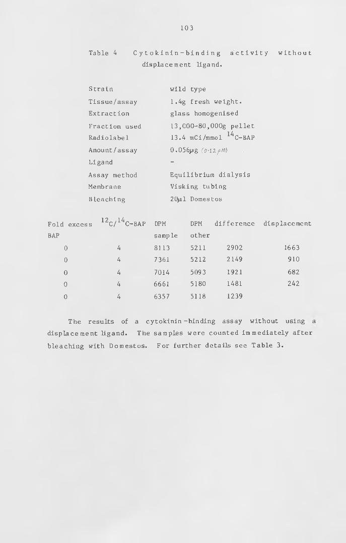
103
Table 4 C y t o k i n i n - b i n d i n g aictivity wi thout
displacement ligand.
S train wild type
Tissue/assay 1.4g fresh weight.
Extract ion glass homogenised
Fraction used 13 ,000-80,000g pellet
Radiolabel 13.4 mCi/mmol ^C-BAP
Amount/assay 0 .056pg (o-lljtM)
Ligand -
Assay method Equilibrium dialysis
Membrane Visking tubing
Bleaching 20/il Domestos
Fold excess ^C /^C - B A P DPM DPM difference displacement
BAP sample other
0 4 8113 5211 2902 1663
0 4 7361 5212 2149 910
0 4 7014 5093 1921 682
0 4 6661 5180 1481 242
0 4 6357 5118 1239
The results of a cytokinin-binding assay without using ;
displace ment ligand. The samples were counted immediately after
bleaching with Domestos. For further details see Table 3.
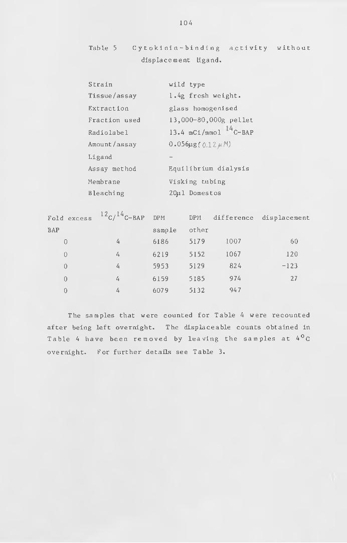
104
Table 5 C y t o k i n i n - b i n d i n g a c t i v i t y wi thout
displacement ligand.
Strain wild type
Tissue/assay 1.4g fresh weight.
Extraction glass homogenised
Fraction used 13,000-80,000g pellet
Radiolabel 13.4 mCi/mmol C-BAP
Amount/assay 0.056jjgf 0.1Zjj.M)
Ligand -
Assay method Equilibrium dialysis
Membrane Visking tubing
Bleaching 20pl Domestos
Fold excess ^C /^C - BA P DPM DPM difference displacement
BAP sample other
0 4 6186 5179 1007 60
0 4 6219 5152 1067 120
0 4 5953 5129 824 -123
0 4 6159 5185 974 27
0 4 6079 5132 947
The samples that were counted for Table 4 were recounted
after being left overnight. The displaceable counts obtained in
Table 4 have been removed by leaving the samples at 4 ° C
overnight. For further details see Table 3.

105
Table 6 Displacement of cytokinin-binding by BAP.
Strain wild type
Tissue/assay 7g fresh weight.
Extraction glass homogenised
Fraction used 13,000-80,OOOg pellet
Radiolabel 57 mCi/mmol ^C-BAP
Amount/assay 0.033^ig (0-07/tM)
Ligand BAP
Assay method Equilibrium dialysis
Membrane Visking tubing
Bleaching 20ji 1 Domes t os
Fold excess ^C /^C - B A P DPM DPM difference displaceme
BAP sample other
0 0.2 6895 5395 1500 388
0 .1 0 .3 5615 4445 1170 58
1 1.2 7380 5742 1638 526
10 10 7229 5797 1432 320
100 100 7096 5984 1112
The cytokinin-binding activity displaced by BAP after
samples were left overnight before counting to allow the
chemiluminescence to die away. For further details see Table 3.
106
Table 7 Cytokinin-- binding activity in a soluble fraction
Strain wild type
Tissue/assay 4 .6g fresh weight.
Extraction ground in liquid nitrogen
Fraction used 13,OOOg supernatant
Radiolabel 57mCi/mmol14
C-BAP
Amount/assay 0.033jjg 1 0 .0 7 /M)
Ligand BAP
Assay method Equilibrium dialysis
Membrane Spect rapor 2
Bleaching -
Fold excess ^C /^C - BA P DPM DPM difference displacemei
BAP sample other
0 0.2 8418 7697 721 26 7
0 .1 0 .3 8418 7852 566 112
1 1.2 8416 7893 523 69
10 10 8375 7818 557 103
100 100 8342 7888 454
Cytokinin-binding activity in the 13,OOOg supernatant. For
further details see Table 3.
107
3Table 8 Cytokinin-binding activity detected using a H-BAP.
Strain wild type
Tissue/assay 5.1g fresh weight.
Extraction motorised teflon homogenised
Fraction used 13,000-80,000g pellet
Radiolabel 12Ci/mmol ^H-BAP
Amount/assay 0.08ng (0-
Ligand BAP
Assay method centrifugation
Bleaching 20^1 sodium hypochlorite
1 3Fold excess H/ H-BAP DPM mean displacement
BAP
0 1.5 1389 1287 366
0 1.5 1185
1 2.5 1368 1257 336
1 2 .5 1145
10 11.5 1289 1315 394
10 11.5 1341
100 102 1184 1137 216
100 102 1089
1000 1002 928 921
1000 1002 914
Cytokinin-binding activity using the tritiated BAP. The
assay mixtures were incubated on ice for 15 minutes before
1 3centrifugation. " H / H - B A P " is the ratio of unlabelled to
tritiated BAP present in the mixture. " D P M " is the number of
disitegrations per minute in the 80,000g pellet. "Mean" is the
average DPM of the replicates. For further details see Table 3.
108
Table 9 Cytokinin-binding activity detected using a H-BAP.
Strain
Tissue/assay
Extraction
Fraction used
Radiolabel
Amount/assay
Ligand
Assay method
Bleaching
wild type
4.5g fresh weight.
glass bead
13 ,000-80,OOOg pellet
12Ci/mmol 3H-BAP
0 . 08ng
BAP
centrifugation
oxidiser
Fold excess
BAP
0
0
1
1
10
10
100
100
1000
1000
1h /3h-bap
1.5
1 .5
2.5
2.5
11.5
11.5
102
102
1002
1002
DPM
2238
2249
1943
2247
2030
2081
2208
2016
1976
1733
mean
2244
2095
2056
2112
1855
displacement
389
240
201
257
The assay for the above results was performed in the same
way as in Table 8 except that the tissue was ground in a glass
bead homogeniser.
109
Table 10 Reproducibility of the centrifugation assay.
Strain
Tissue/assay
Extraction
Fraction used
Radiolabel
Amount/assay
Ligand
Assay method
Bleaching
wild type
10 .6g fresh weight,
glass bead
2,000-80,OOOg pellet
12Ci/mmol 3H-BAP
0.08ng (0 .18*
BAP
centrifugation
oxidiser
Fold excess
BAP
0
1H /3H-BAP
1.5
DPM mean
2282
displacement
3932260
2105
2570
2295
2178
10000 10002 1718 1889
1975
1949
1806
1999
T h e results ab ov e show the
centrifugation assay and the tissue oxidiser using the tritiated
BAP. For further details see Table 3.
reproducibility of the
1 10
Fold
BAP
Table 11 Binding activity with a 1-hour incubation.
Strain
Tissue/assay
Extraction
Fraction used
Radiolabel
Amount/assay
Ligand
Assay method
Bleaching
wild type
8.5g fresh weight.
glass bead
2,000-80,OOOg pellet
12Ci/mmol ^H-BAP
0.08ng (0.l$~H)
BAP
centrifugation
oxidiser
excess 1H / 3H-BAP DPM displacement
0
0
1
1
10
10
100
100
1000
1000
1.5
1 .5
2.5
2.5
11.5
11.5
102
102
1002
1002
6691
6711
6364
6295
5973
6550
6029
6491
5701
5534
6701
6330
6262
6260
5618
1083
712
644
642
For the results above, the assay mixtures were incubated at
4°C for 1 hour before centrifugation. For further details see
Table 3.
111
Table 12 Binding activity with a 1-hour incubation.
Strain
Tissue/assay
Extraction
Fraction used
Radiolabel
Amount/ assay
Ligand
Assay method
Bleaching
wild type
11 . lg fresh weight,
glass bead
2,000-80,OOOg pellet
12Ci/mmol 3H-BAP
0.08ng C0 .(8 «m)
BAP
centrifugation
oxidiser
Fold excess
BAP
0
0
1
1
10
10
1000
1000
10000
10000
1H /3H-BAP
1.5
1.5
2.5
2.5
11.5
11.5
1002
1002
10002
10002
DPM
6414
6472
6464
6864
6335
6540
5833
5854
5220
5556
mean
6443
6664
6438
5844
5388
displacement
1055
127 6
1050
456
The assay for the above results was a repeat of the assay
for Table 11.
1 1 2
Table 13 Effect of time on binding activity.
Strain
Tissue/assay
Extract ion
Fraction used
Radiolabel
Amount/assay
Ligand
Assay method
Bleaching
wild type
9 .2g fresh weight.
glass bead
2 ,000- 80 ,000g pellet
12Ci/mmol 3H-BAP
0 .08ng (0.ig~M)
BAP
centrifugation
oxidiser
Fold excess
BAP
0
1000
10000
1000
10000
1 h / 3 h-bap
1.5
1002
10002
1.5
1002
10002
time
hours
1
24
24
24
DPM
4360
4510
3868
3789
3535
3751
3141
3237
2985
3194
3049
2984
mean
4435
3829
3643
3189
3090
3017
displacement
792
186
172
73
The above results show the cytokinin-binding activity with
incubations of 1 and 2 4 hours at 4 ° C before centrifugation. For
further details see Table 3.
113
Table 14 Effect of time on binding activity.
Strain
Tissue/assay
Extraction
Fraction used
Radiolabel
Amount/assay
Ligand
Assay method
Bleaching
wild type
8.5g fresh weight.
glass bead
2 ,000- 80 ,OOOg pellet
12Ci/mmol 3 H-BAP
0 .08ng (0■ l*«*)
BAP
centrifugation
oxidiser
Fold excess
BAP
0
H/ H-BAP time
hours
0
DPM mean displacement
3269 6291.5 0 3273
3264
10000 10002 0 2655 2640
2624
0 1.5 1 3836 3854 1093
3871
10000 10002 1 2658 2761
2863
0 1 .5 2 3988 3277 373
2565
10000 10002 2 2918 2904
2889
The above results show the binding activity when the assay
mixture is incubated on ice for 0 , 1 and 2 hours. For further
details see T a b l e 3.
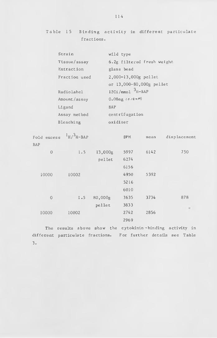
114
T a b l e 15 B i n d i n g a c t i v i t y in d ifferent particulate
fractions .
Strain
Tissue/assay
Extraction
Fraction used
Radiolabel
Amount/assay
Ligand
Assay method
Bleaching
wild type
6 . 2 g filtered fresh weight
glass bead
2 ,000-13 ,OOOg pellet
or 13 ,000-80,OOOg pellet
12Ci/mmol 3 H-BAP
0.08ng ( o. i8 « m)
BAP
centrifugation
oxidiser
Fold excess
BAP
0
10000
0
10000
!H / 3H-BAP
1.5
DPM mean displacement
13 ,OOOg
pellet
6142 750
10002
5997
6274
6156
4950
5216
6010
3635
3833
2742
2969
The results above show the cytokinin -binding activity in
different particulate fractions. For further details see Table
3.
1.5 80, OOOg
pellet
10002
5392
3734
2856
878
115
CHAPTER VI
The mechanism of action of cytokinin
Results and discussion
1. Using the cytokinin binding assay
The assay developed in the previous chapter has been used to
determ ine the nature of the b inding a c t i v i t y , to purify it
further and to determine the type of t issue contain ing the
activity. If the binding is due to a specific site on a protein
it is likely to be lost by heat treatment. Non-specific binding
is more l i k e l y to be heat —stable or even increase by heat
treatment due to exposure of non-specific sites as proteins are
denatured (Sussman and K e n d e , 1978) . The heat-stability of the
binding f r a c t io n was tested by b o i l i n g the extract for two
minutes . The b in ding a c t i v i t y was grea tly reduced by heat
treatment (T ab le 16). If the b inding a c t i v i t y that has been
detected in the particulate fraction is mainly due to a single or
a few s p e c i f i c b ind ing s i t e s , further study of these b indin g
s i t e s would be a ided by s o l u b i l i s i n g them out of the membrane.
Venis (1977a) has solubilised auxin binding fractions from corn
membranes by treating them with acetone. This method (described
in Chapter I I s e c t i o n 5 b.) was used on a 1 3 , 0 0 0 - 8 0 , OOOg
particulate fraction from moss. As the acetone treatment did not
appear to s o l u b i l i s e much of the p e l l e t , both the re-extracted
p e l l e t and s u p e r n a t a n t w er e tested for cyto kin in - bind ing
activity. Very little of the binding to either the pellet or the
supernatant was displaced by excess unlabelled BAP (Table 17).
116
An alternative method is to solubilise membrane components
with a detergent. This was achieved by using the Triton method
of Thomas et al . ( 1 9 8 4 ) . The supernatant was obtained by
t r e a t i n g the 1 3 , 0 0 0 - 8 0 , 0 0 0 g p e l l e t w i t h T r i t o n and then
c e n t r i f u g i n g at 8 0 , 0 0 0 g . The p e l l e t , i n this instance , that was
tested for c y to k in in - b in d in g a c t iv i t y was the 1 3 , 0 0 0 - 8 0 ,000g
p e l l e t which had not been treated with Trito n . 85% of the
c y to k in in - b in d in g a c t i v i t y of t h is p e l l e t a p p e a r s to be
solubilised by this Triton treatment (Table 18). If facilities
had a l lo w e d , the pellet obtained a fte r treatment with T r it o n
could also have been tested for cytokinin-binding activity. If
all three fractions could have been tested, a useful check would
have been to see if the total bind ing a c t i v i t y in the Tr iton^
treated pellet and supernatant was equal to the binding activity
in the untreated pellet.
As shown in Chapter I I I section 4, moss t issue grown in
phosphate-free medium p r o d u c e s m a i n l y c a u l o n e m a t a . The
c a u l o n e m a l f i l a m e n t s p r o d u c e s i d e b r a n c h i n i t i a l s and
gametophores . There is u s u a l ly only one gametophore per
filament. The side-branch initials do not develop further, but
w i l l d i f f e r e n t i a t e into buds if supplied with c y tokin in . Such
tissue may be enriched for a cytokinin receptor. The results in
Table 19 are from a 15-litre liquid culture of wild type grown in
phosphate-free medium (See Chapter I I section 1 e.). The amount
of t issue produced in this culture was much less than would be
expected for a normal 15-litre culture. The tissue was tougher
than usual and so was ground in 8 volumes of MES1 instead 6 f 4
volumes. A relatively high amount of cytokinin-binding activity
was obtained for this amount of tissue (activity with phosphate-
free culture is 630 DPM/g fresh w eig h t , a c t iv i t y with t issue
grown on phosphate is 100-140 DPM/g fresh weight).
As phosphate-free-grown culture of wild type appears to be a
good source of cytokinin-binding activity, an attempt was made to
solubilise the binding a c t i v i t y from a phosphate-free culture .
Th is result is shown in Table 20. The pe l le t was the 8 0 , 0 0 0 g
pe l le t a ft e r treating with T r i t o n . Most of the a c t i v i t y has
remained in the pellet. This could be due to the phosphate-free
t is su e being tougher and not being broken up by the T r i t o n so
e a s i l y as normal t is sue . However, the very small amount of
counts bound to the sample in d ic a t e that the T r i t o n is not
responsible for the binding ShoWn in Table 18. A Triton-BAP
in t e r a c t i o n was reported by Gardner et a l . ( 1978)3 using a gel
f i ltration method to assay for cytokinin-binding activity. This
interaction prevented Gardner et al. detecting whether the Tr iton
denatured or solubilized the binding activity they reported in a
moss particulate fraction.
The culture grown without phosphate in Table 19 gave a
h igher amount of d i s p la c e a b le b ind ing per gram of t issue than
t is s ue on normal Knops medium, but was very tough. On a low
p h o s p h a t e and ' low n i t r a t e m e d iu m , w i l d type gr ow s as
caulonemata with side branch initials , but these do not develop
further. However, these side branch initials , and therefore this
type of t i s s u e , i s p o t e n t i a l l y su i ta b le for seeking c y to k in in
receptors. Such a culture was used to obtain the results shown
in Table 21. This gave d i s p l a c e a b l e b in d in g . The amount of
cytokinin-binding activity was similar to that of a normal-grown
wild type culture.
1 1 7
1 1 8
In the identification of a biologically meaningful cytokinin
receptor, it is useful to compare cytokinin binding activity in
mutants that do not respond to cytokinin. Showing that a mutant
that does not have the cytokinin binding activity also does not
t h e h y p o t h e s i s
have the ability to respond to cytokinin would support^that such
binding was involved in the hormone response. bar mutants were
originally isolated as benzyl-adenine resistant mutants. bar 1
produces some caulonemata but does not produce buds even with
exogenous cytokinin. This mutant therefore has the phenotype of
a c yto kin in non-responding mutant that was screened for as
described in section 8 of chapter I I . bar l's phenotype is that
of a mutant that has no functional cytokinin receptor, but there
are other p o s s i b i l i t i e s for this phenotype, such as a block in
the response to cytokinin after the receptor, bar 1 was therefore
used to see if it lacked the cytokinin-binding activity that has
been obtained in the w ild type. The results in Table 22 show
that bar 1 does have a s i m i l a r b ind ing a c t i v i t y as w i l d type in
the 13,000-80,OOOg pellet (activity is 330 DPM/g fresh weight).
The results in Table 23 were also obtained with bar 1, but
included a dilution of the pellet to see if the binding activity
of the pellet was limiting. This binding activity is similar to
that obtained in the previous experiment (220 DPM/g fresh weight)
and is reduced to almost half by the dilution of the resuspended
pellet by a half before being used for the assay. This indicates
that the extract is limiting.
W i l d type has to be exposed to cyto kin in for about three
days to prevent a bud d e d i f f e r e n t i a t i n g into protonemata. I f
each cell of a bud has receptors for cytokinin, then mutants that
1 1 9
overproduce buds may have more receptors than wild type. ove 78
was used in a cytokinin-binding assay as ove's produce abnormal
buds which may be a good source for cytokinin receptors. ove 78
showed d i s p l a c e a b l e c yto kin in binding (see Table 24) . This
b i n d i n g is r e d u c e d when the p e l l e t is d i l u t e d , a g a i n
demonstrating that the displaceable cytokinin-binding fraction is
l i m i t i n g . ove 78 appears to have s im i la r cyto kin in- bind ing
activity as wild type (250 DPM/g fresh weight).
Using the cytokinin-binding assay developed in the previous
se c t io n , the b ind in g a c t i v i t y in the 13,000-80,OOOg particulate
f r a c t io n in P h y s c o m it r e l la patens appears to be heat l a b i l e ,
which suggests that the d is p la c e a b le binding could be specific
b ind ing to prote in . The binding a c t i v i t y is lost from both the
p e l l e t and the supernatant when treated w it h acetone . I f the
b ind ing a c t i v i t y is due to an i n t r i n s i c membrane pro t e in ,
removing the lipids with acetone could denature the protein and
so result in no b ind ing a c t i v i t y in the pe l le t or supernatant .
When a membrane f r a c t io n is s o l u b i l i s e d w ith a de te rge nt ,
hydrophobic parts of a protein are less likely to be exposed and
so are less l i k e l y to be denatured . The T r i t o n method used
appeared to s o l u b i l i s e most of the b in ding a c t i v i t y that was
present in the p a r t i c u l a t e f r a c t io n . Although it was not
p o s s ib l e to q u a n t i fy the degree of b ind ing for the amount of
t is s ue used , the T r i t o n treatment does appear to have enriched
for binding activity as some of the particulate fraction was not
solubilised by the Triton.
The phosphate-free culture had more b ind ing a c t i v i t y per
gram t is sue than t is sue grown on normal Knops medium. As this
120
t issue was mainly caulonem al , which is the target t issue for
c y t o k in in w ith respect to bud d i f f e r e n t i a t i o n , it is l ik e ly to
have more cytokinin receptors than the chloronemata . However,
this bind ing a c t i v i t y in the phosphate-free cultu re was not
solubilised with Triton. So, either the binding activity in the
phosphate-free culture is different from that of a culture grown
in the presence of phosphate or is more difficult to solubilise
from the tougher caulonemal t is sue . The low phosphate and
nitrate culture, which contained more side branch initials than
the phosphate-free c ulture , did not show any more binding
activity than a normal wild type culture and so did not appear to
enrich for a possible cytokinin receptor.
bar 1 was tested for cytokinin-binding a c t i v i t y as it does
not respond to exogenous c y to k in in and so is a c andidate for a
mutant that does not have a functional receptor. This mutant has
been shown to have s i m i l a r d i s p la c e a b le c y to k in in - b in d in g
activity as the wild type and therefore if this binding is due to
a receptor , bar 1 must be blocked at a later stage in the
initiation of the gametophore. When the pellet is diluted by a
h a l f , the number of d i s p l a c e a b l e counts in the p e l le t are a lso
reduced by about a h a l f . This in d ic a te s that the d i s p l a c e a b l e
b in d in g in the p e l le t is l i m i t i n g . The d i l u t i o n e f f e c t is also
shown w it h ove 78, which appears to have more d i s p l a c e a b l e
b i n d in g per gram of t is sue than w ild type. ove mutants could
well have more cytokinin receptors than wild type as a bud needs
cytokinin to be present for the first three days after initiation
to prevent de d if f e r e n t i a t i o n , and so each c e l l of the abnormal
buds of the ove mutants may contain cytokinin receptors. If this
is the case , then ove mutants would be a good source for the

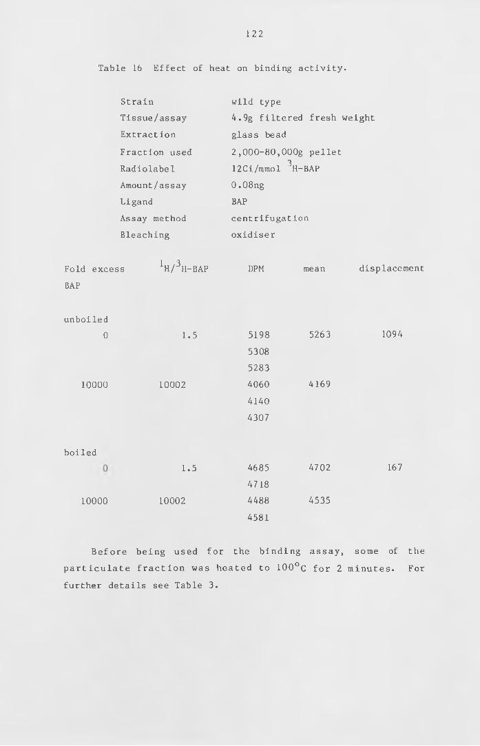
1 2 2
Table 16 Effect of heat on binding activity.
Strain
Tissue/assay
Extract ion
Fraction used
Radiolabel
Amount/assay
Ligand
Assay method
Bleaching
wild type
4.9g filtered fresh weight
glass bead
2 ,000-80,OOOg pellet
12Ci/mmol 3 H-BAP
0 .08ng
BAP
centrifugation
oxidiser
Fold excess
BAP
^/■^H-BAP DPM mean displacement
unboiled
0
10000
1.5
10002
5198
5308
5283
4060
4140
4307
5263
4169
1094
boiled
10000
1.5
10002
4685
4718
4488
4581
4702
4535
167
Before being used for the b ind ing assay , some of the
p a r t i c u l a t e f r a c t io n was heated to 1 0 0 ° c for 2 m inutes . For
further details see Table 3.
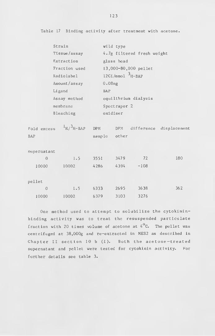
123
Table 17 Binding activity after treatment with acetone.
Strain
Tissue/assay
Extraction
Fraction used
Radiolabel
Amount/assay
Ligand
Assay method
membrane
Bleaching
wild type
4.7g filtered fresh weight
glass bead
13 ,000-80,000 pellet
12Ci/mmol 3 H-BAP
0 .08ng
BAP
equilibrium dialysis
Spectrapor 2
oxidiser
Fold excess
BAP
1 H / 3 H-BAP DPM DPM difference displacement
sample other
supernatant
0
10000
1.5
10002
3551
4286
3479
4394
72
-108
180
pellet
0
10000
1.5
10002
6333
6379
2695
3103
3638
3276
362
One method used to attempt to s o l u b i l i z e the cytokinin-
b ind ing a c t iv i t y was to treat the resuspended p a r t ic u la t e
fraction with 20 times volume of acetone at 4°C. The pellet was
centrifuged at 38 ,OOOg and re-extracted in MES2 as described in
C h a p t e r I I s e c t i o n 10 b ( i ) . Both the a c e t o n e —t r e a t e d
supernatant and pellet were tested for cytokinin activity. For
further details see table 3.
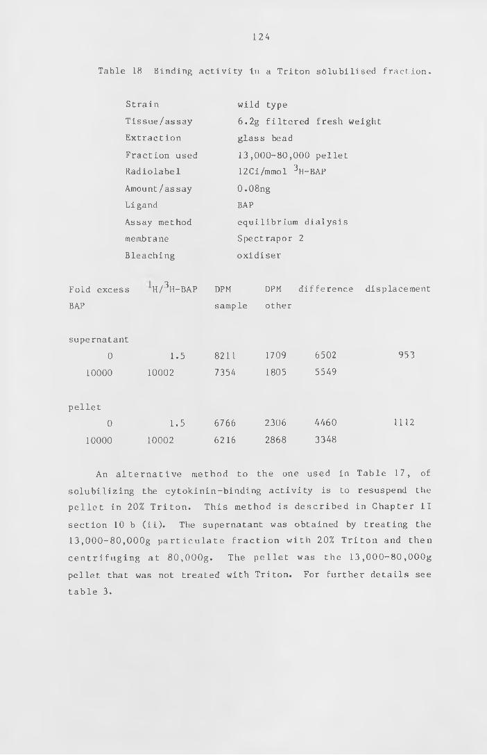
124
Table 18 Binding activity in a Triton solubilised fraction.
Strain
Tissue/assay
Extraction
Fraction used
Radiolabel
Amount/assay
Ligand
Assay method
membrane
Bleaching
wild type
6 . 2 g filtered fresh weight
glass bead
13,000-80,000 pellet
12Ci/mmol ^H-BAP
0 .08ng
BAP
equilibrium dialysis
Spectrapor 2
oxidiser
Fold excess
BAP
H / 3 H-BAP DPM DPM difference displacement
sample other
supernatant
0 1.5 8211 1709 6502 953
10000 10002 7354 1805 5549
pellet
0 1.5 6766 2306 4460 1112
10000 10002 6216 2868 3348
An a l t e r n a t iv e method to the one used in Table 17, of
solubilizing the cytokinin-binding activity is to resuspend the
pe l le t in 20% T r i t o n . This method is described in Chapter I I
section 10 b ( i i ) . The supernatant was obtained by treating the
1 3 , 0 0 0 - 8 0 ,OOOg p a r t i c u l a t e f r a c t io n with 20% T r ito n and then
c e n t r i f u g in g at 8 0 , OOOg. The pellet was the 1 3 , 0 0 0 - 8 0 ,OOOg
pellet that was not treated with Triton. For further details see
table 3.
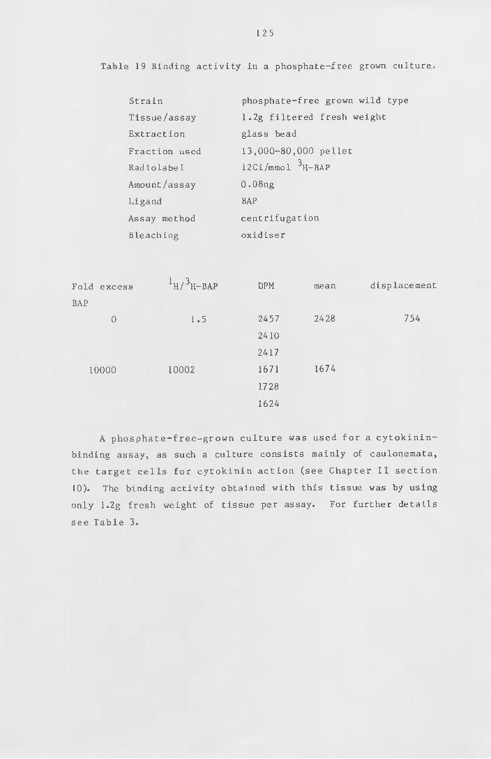
1 2 5
Table 19 Binding activity in a phosphate-free grown culture.
Strain
Tissue/assay
Extraction
Fraction used
Radiolabel
Amount/assay
Ligand
Assay method
Bleaching
phosphate-free grown wild type
1 . 2 g filtered fresh weight
glass bead
13,000-80,000 pellet
12Ci/mmol 3 H-BAP
0 .08ng
BAP
centrifugation
oxidiser
Fold excess 1 H / 3 H-BAP DPM mean displacement
BAP
0 1.5 2457 2428 754
2410
2417
10000 10002 1671 1674
1728
1624
A phosphate-free-grown culture was used for a cytokinin-
binding assay, as such a culture consists mainly of caulonemata,
the target cel ls for c y t o k in in a c tio n (see Chapter I I se ct io n
10). The binding activity obtained with this tissue was by using
only 1.2g fresh weight of tissue per assay. For further details
see Table 3.
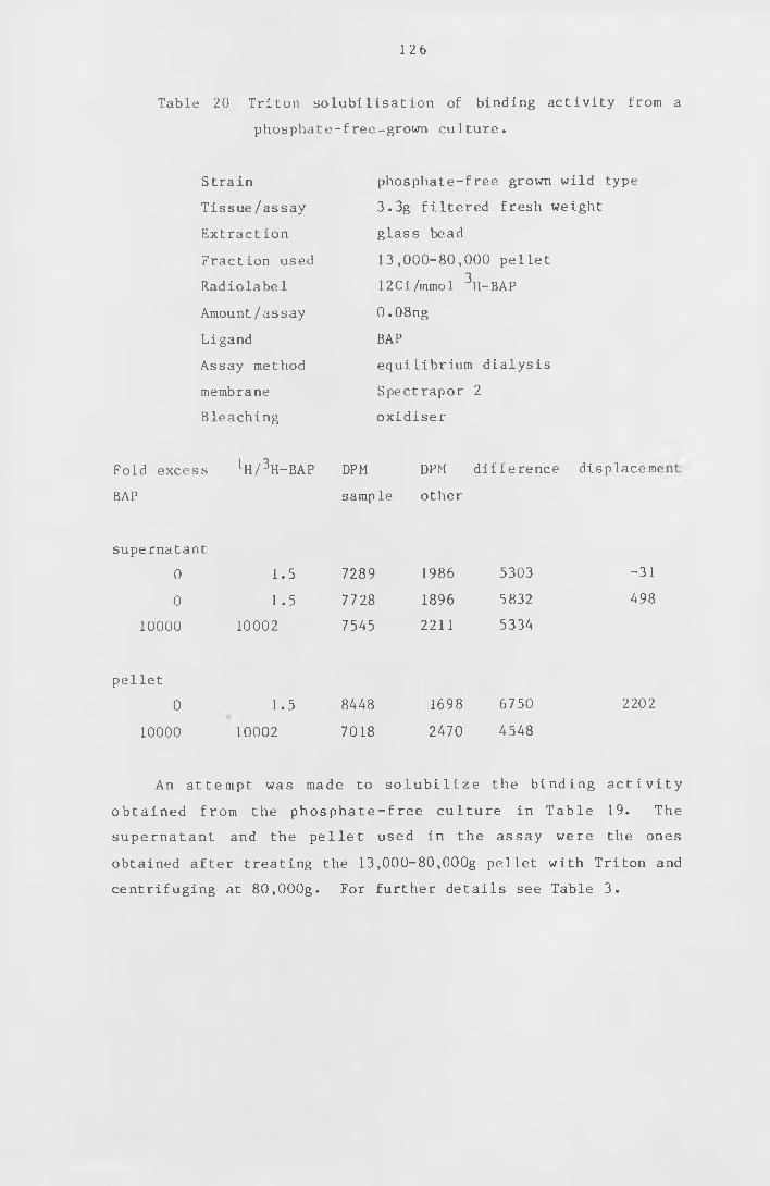
126
Table 20 Triton solubilisation of binding activity from a
phosphate-free-grown culture.
Strain
Tissue/ assay
Extraction
Fraction used
Radiolabel
Amount/assay
Ligand
Assay method
membrane
Bleaching
phosphate-free grown wild type
3.3g filtered fresh weight
glass bead
13 ,000-80,000 pellet
12Ci/mmol \-BAP
0 . 08ng
BAP
equilibrium dialysis
Spectrapor 2
oxidiser
Fold excess 1H /3H-BAP DPM DPM difference displaceme
BAP sample other
supernatant
0 1.5 7289 1986 5303 -31
0 1 .5 7728 1896 5832 498
1 0 0 0 0 1 0 0 0 2 7545 2 2 1 1 5334
pellet
0 1.5 8448 1698 6750 2 2 0 2
1 0 0 0 0 1 0 0 0 2 7018 2470 4548
An attempt was made to s o l u b i l i z e the binding a c t i v i t y
obtained from the phosphate-free culture in T ab le 19. The
supernatant and the pellet used in the assay were the ones
obtained after treating the 13,000-80,000g pellet with Triton and
centrifuging at 8 0 , OOOg. For further details see Table 3.
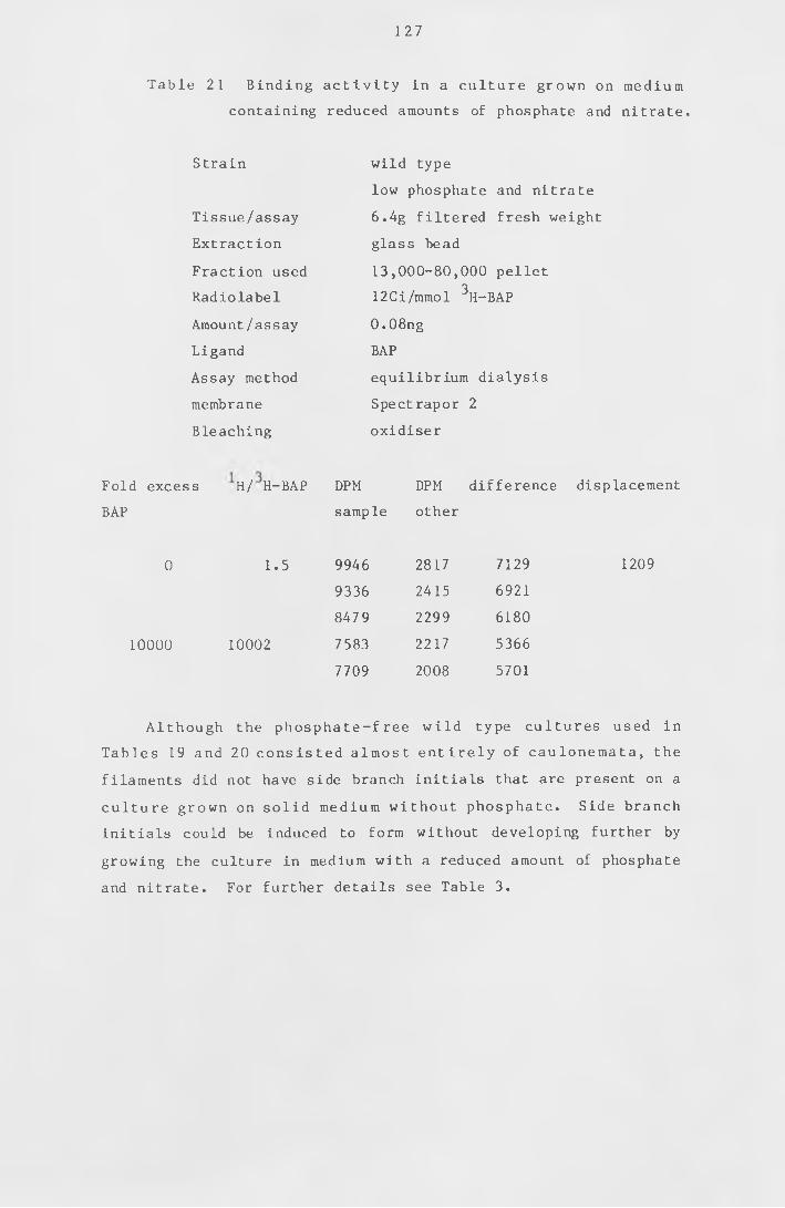
127
Table 21 B inding a c t i v i t y in a culture grown on medium
containing reduced amounts of phosphate and nitrate.
Strain
Tissue/assay
Extraction
Fraction used
Radiolabel
Amount/assay
Ligand
Assay method
membrane
Bleaching
wild type
low phosphate and nitrate
6 .4g filtered fresh weight
glass bead
13,000-80,000 pellet
12Ci/mmol 3 H-BAP
0 . 08ng
BAP
equilibrium dialysis
Spectrapor 2
oxidiser
Fold excess H / H-BAP DPM DPM difference displacement
BAP sample other
0 1.5 9946 2817 7129 1209
9336 2415 6921
847 9 2299 6180
1 0 0 0 0 1 0 0 0 2 7583 2217 5366
7709 2008 5701
Although the phosphate-free w i ld type cultures used in
Tables 19 and 20 c ons iste d almost e n t i r e l y of caulonemata , the
filaments did not have side branch initials that are present on a
c ult ure grown on so l id medium w ithout phosphate. Side branch
initials could be induced to form without developing further by
growing the culture in medium with a reduced amount of phosphate
and nitrate . For further details see Table 3.
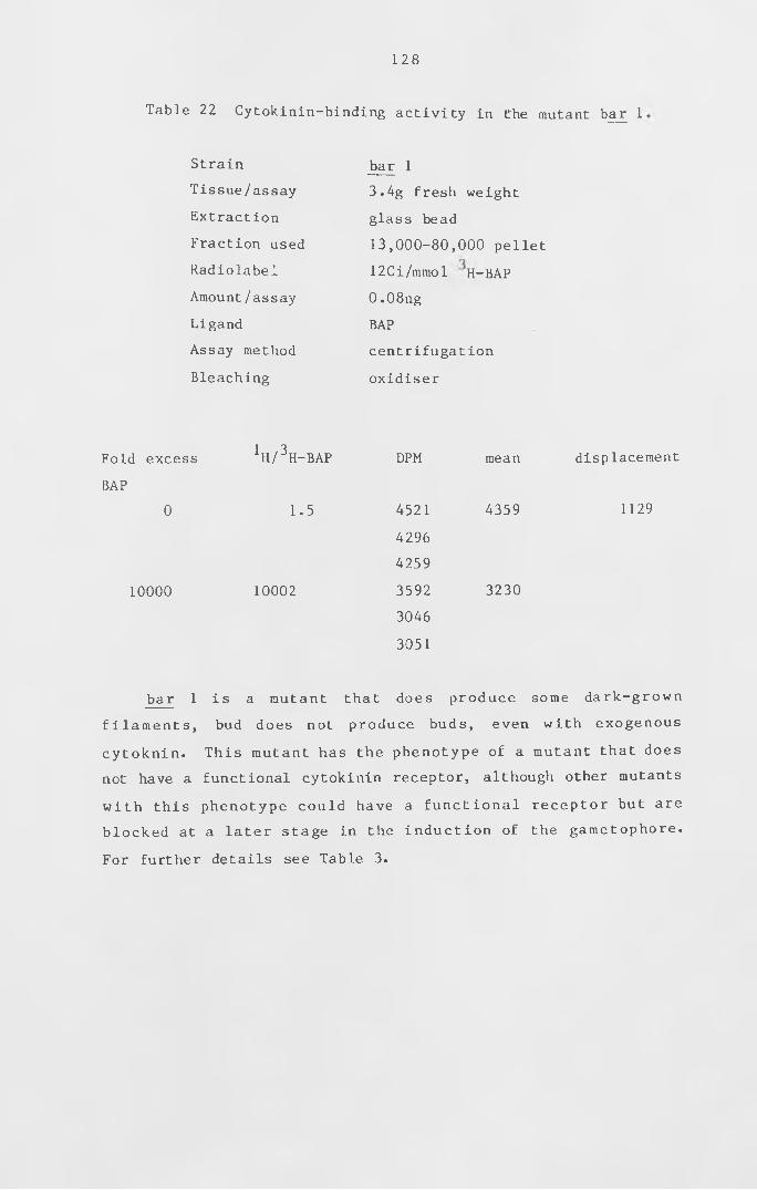
128
Table 22 Cytokinin-binding activity in the mutant bar 1
Strain
Tissue/assay
Extraction
Fraction used
Radiolabel
Amount/assay
Ligand
Assay method
Bleaching
bar 1
3.4g fresh weight
glass bead
13 ,000-80,000 pellet
12Ci/mmol H-BAP
0 .08ng
BAP
centrifugation
oxidiser
Fold excess
BAP
0
10000
1 H / 3 H-BAP
1.5
10002
DPM
4521
4296
4259
3592
3046
3051
mean
4359
3230
displacement
1129
bar 1 is a mutant that does produce some dark-grown
f i l a m e n t s , bud does not produce buds, even with exogenous
cytoknin . This mutant has the phenotype of a mutant that does
not have a functional cytokinin receptor, although other mutants
w i t h this phenotype could have a f u n c t io n a l receptor but are
blocked at a later stage in the in d u c t io n of the gametophore.
For further details see Table 3.
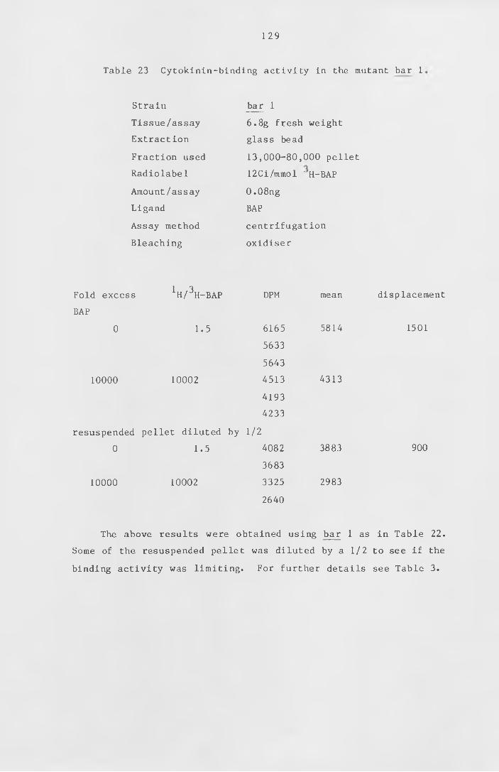
129
Table 23 Cytokinin-binding activity in the mutant bar 1,
Strain
Tissue/assay
Extract ion
Fraction used
Radiolabe1
Amount/assay
Ligand
Assay method
Bleaching
bar 1
6 . 8 g fresh weight
glass bead
13,000-80,000 pellet
12Ci/mmol "H-BAP
0 .08ng
BAP
centrifugation
oxidiser
Fold excess
BAP
0
IH / 3 I1-BAP DPM
1.5 6165
5633
5643
10000 10002 4513
4193
4233
resuspended pellet diluted by 1 / 2
0 1.5 4082
3683
10000 10002 3325
2640
mean
5814
4313
3883
2983
displacement
1501
900
The above results were obtained using bar 1 as in Table 22.
Some of the resuspended pellet was diluted by a 1 /2 to see if the
binding activity was limiting. For further details see Table 3.
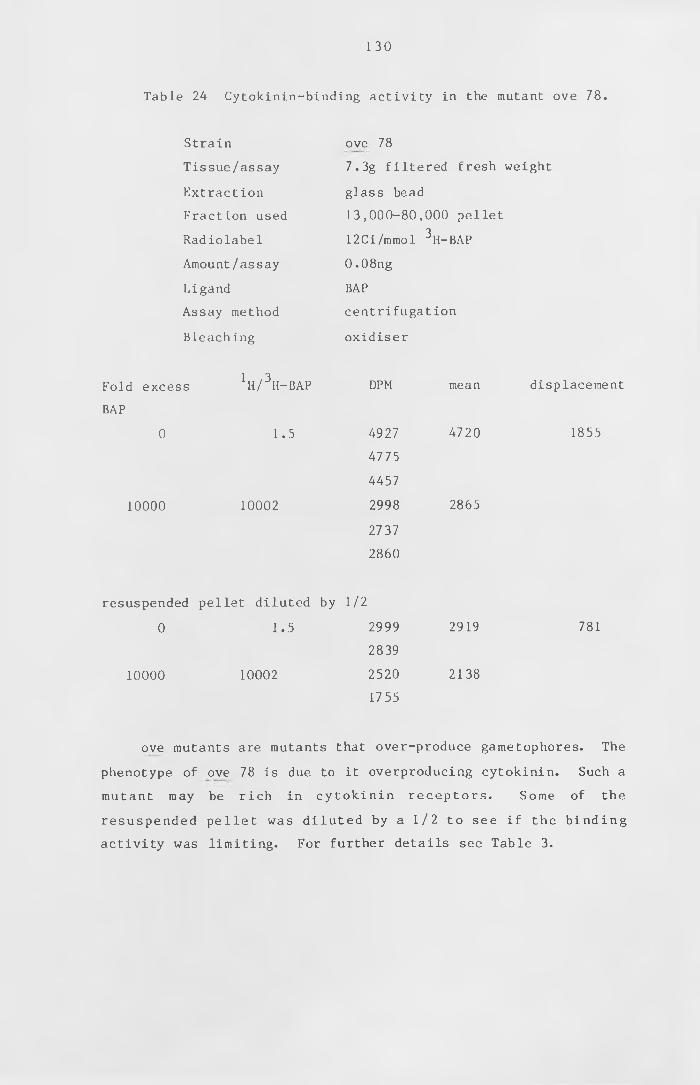
130
Table 24 Cytokinin-binding activity in the mutant ove 78.
Strain
Tissue/assay
Extraction
Fraction used
Radiolabel
Amount/assay
Ligand
Assay method
Bleaching
ove 78
7.3g filtered fresh weight
glass bead
13,000-80,000 pellet
12Ci/mmol 3 H-BAP
0.08ng
BAP
centrifugation
oxidiser
Fold excess
BAP
0
10000
1 h / 3 h-bap
1.5
10002
DPM
4927
4775
4457
2998
2737
2860
mean
4720
2865
displacement
1855
resuspended pellet diluted by 1 / 2
0 1.5 2999 2919 781
2839
10000 10002 2520 2138
1755
ove mutants are mutants that over-produce gametophores. The
phenotype of ove 78 is due to it overproducing cytokinin. Such a
mutant may be rich in c y to kin in receptors . Some of the
resuspended pe l le t was d i lu t ed by a 1 / 2 to see if the b i n d in g
activity was limiting. For further details see Table 3.
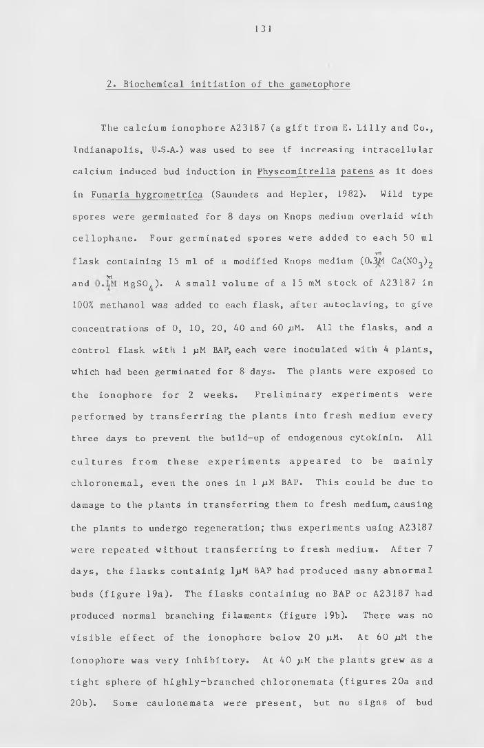
131
The ca lc ium ionophore A23187 (a g i f t from E. L i l l y and Co.,
Indianapolis, U.S.A.) was used to see if increasing intracellular
calcium induced bud induction in Physcomitrella patens as it does
in Funaria hygrometrica (Saunders and Hepler, 1982). Wild type
spores were germinated for 8 days on Knops medium overlaid with
c el lo p h ane . Four germinated spores were added to each 50 ml
yYlflask containing 15 ml of a modified Knops medium (0.3jM CaCNO^^
Trland MgSO^) . A small volume of a 15 mM stock of A23187 in
1 0 0 % methanol was added to each flask, after autoclaving, to give
concentrations of 0, 10, 20, 40 and 60 ;uM. All the flasks, and a
control flask with 1 >iM BAP, each were inoculated with 4 plants,
which had been germinated for 8 days. The plants were exposed to
the ionophore for 2 weeks. P r e l im in a r y experiments were
performed by t r a n s f e r r i n g the plants into fresh medium every
three days to prevent the build-up of endogenous cytokinin. All
c u l t u r e s from t h e s e e x p e r i m e n t s a p p e a r e d to be m a i n l y
chlo ro nem al , even the ones in 1 /iM BAP. This could be due to
damage to the plants in transferring them to fresh medium, causing
the plants to undergo regeneration; thus experiments using A23187
were repeated w ithout t r a n s f e r r i n g to fresh medium. A f t e r 7
days, the f l a s k s c o n t a in ig ljiM BAP had produced many abnormal
buds ( f i g u r e 19a) . The f lasks c o n t a in in g no BAP or A23187 had
produced normal branching filaments (figure 19b). There was no
v i s i b l e e f f e c t of the ionophore below 20 >iM. At 60 juM the
ionophore was very i n h i b i t o r y . At 40 pM the plants grew as a
t ight sphere of highly-branched chloronemata ( f i g u r e s 2 0 a and
20b) . Some caulonemata were present , but no signs of bud
2. Biochemical initiation of the gametophore
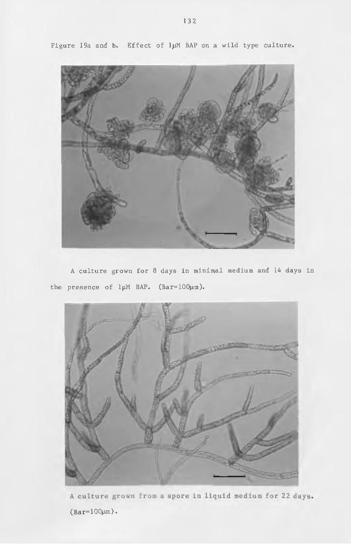
132
Figure 19a and b. Effect of IjiM BAP on a wild type culture.
A culture grown for 8 days in minimal medium and 14 days in
the presence of l^M BAP. (Bar= 100pm).
(Bar=100jLim).
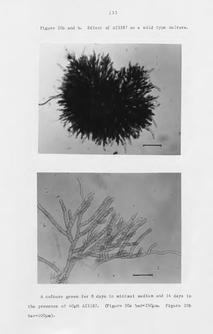
133
Figure 20a and b. Effect of A23187 on a wild type culture.
A culture grown for 8 days in minimal medium and 14 days in
the presence of 40pM A23187. (Figure 20a bar=250jjm. Figure 20b
bar= 1 0 0 pm).
induction. The plants resembled nar mutants' with more primary
chloronemata than a normal wild type culture.
T h e e f f e c t of the c a l c i u m i o n o p h o r e A 2 3 1 8 7 on
P h y s c o m it r e 1 la patens is very d i f f e r e n t from the e f fect on
Funaria hygrometrica reported by Saunders and Hepler (1982). The
calcium ionophore appeared to induce side —branching but not the
i n i t i a t i o n of buds. In P h y s c o m i t r e l l a p a t e n s i n c r e a s i n g
i n t r a c e l l u l a r calcium appears to induce side branching but may
not be a second messenger for cytokinin-induced bud production.
Bud production can be prevented in P h y s c o m it r e l la patens by
concentrations of calcium ions above lOmM (McClelland, personal
com m unication ) . These high levels of c a lc ium produce mainly
highly-branched primary chloronemata. This is very s i m i l a r to
■mthe effect of low levels of calcium (O.^M) in the presence of the
calcium ionophore A23187. T h erefo re , in c r e a s in g i n t r a c e l l u l a r
calcium by a calcium ionophore or by high levels of extracellular
calcium appears to induce branching of primary chloronemata. The
p r eve nting of bud i n d u c t io n by calcium could be due to e i t h e r a
d irect or an i n d i r e c t e f fe c t by promoting branching of primary
chloronemata. There would then be only a small amount of target
tissue for bud induction and so the role of calcium in the action
of cytokinin in Physcomitrella patens cannot be discounted. The
results of the calcium ionophore on Physcomitrella patens support
the o bse rvat ions of Bopp (1 9 8 4 ) on Funar ia h y g r o m e t r i c a , that
although an increase in calcium may be an important step in the
induction of the bud, it is not a replacement for cytokinin.
134
135
General discussion
The results of this investigation have shown that the mode
of actio n of c y to k in in is d i f f i c u l t to e l u c i d a t e , even in the
simple model organism P h y s c o m it r e l la p a t e n s , which has a
c l e a r l y d e f ine d target t issue w it h a s p e c i f i c morphogenetic
response to c y t o k in in . A useful method of showing that the
iS J o / i c a I (y nieiv'ingfdl
binding of a particular hormone to a sub-cellular fraction^is to
demonstrate the lack of the same type of b ind ing with a mutant
that is unable to respond to endogenous or exogenous hormone.
The i s o l a t i o n of a c y t o k i n i n n o n - r e s p o n d i n g m ut ant in
Physcomitrella patens has been shown to be more difficult than
would be expected if such a phenotype could ar ise by a s ingle
mutational event. Some of the possible reasons for cytokinin non
responding mutants not being isolated were suggested in Chapter
I I I section 1. One possibility was that insufficient mutants had
been screened. However, other mutants, such as cytokinin over
producing mutants, are o btained at a frequency of about 1 in
every 1000 clones screened. Therefore it is unlikely that non
responding mutants can be obtained by a single mutational event.
This could be due to there being d u p l ic a t e genes coding for a
cytokinin receptor. Alternatively, the same cytokinin receptor
might be essential for some process such as cell division, or for
the production of the f i l a m e n t s which develop in the dark and
which were used as target cells for the screening procedure. If
cytokinin non-responding mutants are not lethal, then screening
for such mutants in ove revertants may be more successful. The
CHAPTER VII
136
screening procedure could be done in two stages. The majority of
mutants could be eliminated by their continued over-production of
buds. Only a few mutants would have to be tested for a lack of
bud production in the presence of supplied cytokinin. Screening
ove revertants for cytokinin non-responding mutants would also
enable the isolation of cytokinin non-responding mutants even if
such mutants were unable to produce caulonemal filaments in the
dark.
One mutant isolated in the mutation runs for cytokinin non
responding mutants, ove 409, could be useful in the isolation of
ove revertants. As this mutant is temperature-sensitive, having
an ove phenotype above 23°C and a wild type phenotype below 17°C,
o v e r e v e r t a n t s c o u l d be s e l e c t e d at the n o n - p e r m i s s i v e
temperature. Some of the mutants that do not produce buds at the
non—permissive temperature may not produce buds at the permissive
temperature when supplied with c y to kin in . And mutants that do
not respond to supplied cytokinin may be defective in a cytokinin
receptor. The non-responding phenotype could be studied at 17°C,
in the absence of the ove phenotype . By the use of p roto plast
fusion, ove 409 has been shown to be in complementation group A
and is recessive to wild type.
The phenotype of ove 409 has been shown to be due to the
over-production of i s o p e n te n y la d e n in e at the non-perm issive
temperature* The two other s t r a in s s tudied for c y t o k in in
production at 24 and 15 °C , ove 78 and w i ld type, also produced
more i s o p e n te n y la d e n in e per mg dry weight at the h igher
tem perature . However, there was only about a 6 -fold change in
cytokinin production for these strains whereas ove 409 produced
14 times more isopentenyladenine at the higher temperature. The
phenotype change of ove 409 with temperature is due to a change
in c y to k in in production ,from s i m i l a r levels as those found in
w i l d type to h ig he r l e v e l s , w i t h a change from 15 to 24 °c . This
change in cytokinin production may be partially due to the same
change in c y to k in in production found in w ild type and ove 78.
The temperature-dependence of a l l three strains on c y to kin in
production may be due to slower growth, and hence delayed
differentiation, at the lower temperature . The cultures grown
at 15 °c were more chloronemal than the cultures at 24 °C ,and so
temperature may have a more direct e f fe ct on i n h i b i t i n g
d i f f e r e n t i a t i o n . A s i m i l a r t e m p e r a t u r e e f fect could be
investigated with respect to auxin production. Slower or delayed
auxin production or response would result in a culture that was
more chloronemal at the lower temperature. Such a culture may
produce less c y to k in in p a r t i c u l a r l y if cyto kin in production
occurs in the caulonemata. A tem p erature- sensit ive ove mutant
could be obtained i f the le ve l of over-production of c y t o k in in
was at such a level that the six-fold decrease was sufficent to
drop the level of c y to k in in prod uct io n to levels found in w ild
type. The change in production of 2iP by ove 409 between these
two temperatures is larger than with ove 78 and w ild type.
Therefore ove 409 may have an additional mechanism resulting in
i t s t e m p e r a t u r e - s e n s i t i v e phenotype . I f the temperature
s e n s i t i v i t y is due to a pr ot e in lo s ing a c t i v i t y at the non-
pe rm iss iv e tem perature , it is most l i k e l y to be a re gulato r of
c y t o k i n i n s y n t h e s i s or an en zy m e i n v o l v e d in c y t o k i n i n
metabolism.
The induction of gametophores in Physcomitrella patens, even
138
following treatment with high levels of cytokinin, does not occur
in the dark. There appears to be a s p e c i f i c l igh t requirement
for the differentiation of the caulonemal side branch into a bud.
When dark-grown caulonemal filaments are supplied with cytokinin
— 2 — 1
and exposed to 556nM quanta m s for three days, bud induction
is strong with red light of around 665nm. Bud induction does not
occur with far-red l ight or with light below about 600nm,apart
from the small amount of bud induction with blue light of 442nm.
This response to red light in d ic a te s that the photoreceptor
i n v o l v e d c o u l d be p h y t o c h r o m e . E v i d e n c e to s u p p o r t the
involvement of phytochrome has come from experiments where
c u lt u re s were exposed to red light before the a d d it io n of
c y to k in in . Some plates were exposed to half-an-hour far-red
l ig h t im m e d ia te ly a f t e r the red l ig h t . All cultures were then
placed in the dark,and a f t e r d i f f e r e n t time in te r v a ls supplied
with cytokinin and left for three days for buds to develop in the
dark. The bud in d u c t io n that occurred due to the red l igh t
exposure was reduced by exposure to far-red light. The amount of
bud induct io n was also reduced if the red light exposure was
separated from the addition of cytokinin by more than four hours.
Both types of reduction of bud induct io n support the role of
phytochrome, as the conversion of P ^ to P^ occurs rapidly under
far-red light and slowly in the dark. Therefore the response to
c y t o k i n i n to induce the gametophore may only occur when the
equilibrium of phytochrome is towards P fr* The involvement of
phytochrome in bud in d u c t io n could be i n v e s t i g a t e d fu rth e r by
expo sing cultures to a l t e r n a t in g red and far-red light of
different intensities and duration.
One unexpected feature of bud induction, when the cytokinin
139
is supp lied a f t e r the exposure to red l ig h t , is that the amount
of bud induction was greater if the light exposure was separated
from the a d d it io n of c y to k in in by a two-hour dark period than
when the cytokinin was added immediately after the light period.
This occurred when the length of the l ight period was 24 , 30 or
48 hours. This phenomenon in d ic ate s that there is a dark
reaction involved in the response to c y to kin in . This reaction
may be inhibited when cytokinin is applied. Bud induction with
cultures where the cytokinin is applied after the dark period but
before exposure to the red light was about 40%. This was similar
to the bud induction of cultures where the red light exposure and
the a d d i t i o n of c y t o k i n i n w ere s e p a r a t e d by two h o u r s .
Th e refo re , it may be p o ss ib le for this dark reaction to occur
before or a f t e r the exposure to l ig h t . The induct io n of a
gametophore on a target c e l l appears to require red light and a
short dark period before the a d d i t io n of cyto kin in if bud
indu ct io n is going to occur in the dark. Bud indu ct io n is not
reduced by the absence of a dark period i f the c y to kin in and
l ight are present at the same time. This phenomenon could be
i n v e s t ig a t e d furth er to f ind out what length of dark period
between the light exposure and the a d d it io n of cyto kin in gives
the maximum bud indu ct io n . The l igh t period could also be
in tersp e rsed with dark periods to see if the amount of bud
indu ct io n could be in c re a s e d . The length and in t e n s i t y of the
red light exposure could also be in v e s t ig a t e d to see if they
affect the requirement for a dark period before the addition of
c y t o k in in . It may be i n t e r e s t i n g to see if speed of bud
induction can be altered by putting a light-grown culture in the
dark before the addition of cytokinin.
140
Light could be involved in the synthesis or sensitivity of a
cytokinin receptor, or in a later process in the development of
the bud. I f red l igh t was required for the synthesis of the
receptor , far-red l ight would have to able to in a c t iv a t e the
receptor . I f red l igh t is involved in both the synthesis and
s e n s i t i v i t y of a cyto kin in receptor , the r e a c t iv a t io n of the
c y t o k in in response by red l ight a ft e r a far-red l ig h t exposure
should be less than the 6 hours o r i g i n a l l y required for bud
induction.
Studies on the e ffect of auxin and c y to k in in on the
regeneration of protoplasts were undertaken to see if i s o la t e d
pr ot o plast s had the a b i l i t y to respond to the hormones. This
would show that the c e l l s contained the receptors during the
process of r e g e n e rat io n . P rotoplasts of Physcomitrella patens
a re u n a f f e c t e d by the p r e s e n c e of NAA and BAP d u r i n g
regeneration. With respect to cytokinin, the lack of response to
the hormone could be due to the loss of ability to respond or due
to the r e ge ne rat ing protoplasts being derived from chloronemal
c e l l s . It was not po ss ib le to i n v e s t ig a t e this further as a l l
enzymes used to try to make protoplasts from dark-grown
caulonemal filaments, which would in itially contain receptors for
c y t o k in in , had no observable e f f e c t . I f p r oto plasts i s o la t e d
from caulonemal f i l a m e n ts could be shown not to respond to
c y t o k in in they might be a s u i t a b l e m a te r ia l for showing the
amount and type of c y to k in in b ind in g that was not due to a true
cytokinin receptor.
In developing the assays for a cytokinin receptor, four main
improvements have been made to the original assay. These were to
increase the amount of t issue used, the use of a t r i t i a t e d BAP,
the o x id a t io n of the samples before counting and in cu b a t io n of
the samples for 1 hour before centrifugation. Attempts to detect
c y t o k in in b ind in g a c t i v i t y in both p a r t ic u l a t e and soluble
fr a c t io n s w it h ^ C - B A P were u n suc c es s fu l . m-iodo-BAP was
t herefore s y t h e s is e d and sent to Amersham I n t e r n a t i o n a l to be
3tritiated. The H-BAP thus produced had a much higher specific
activity than the ^C-BAP. By the use of glass bead homogenisers
a larger amount of t issue could be prepared for each assay.
Using the increased amount of tissue and the tritiated cytokinin,
d i s p l a c e a b l e c y to k in in binding a c t i v i t y was detected . More
consistent results were obtained by the oxidation of the samples
rather than b le ac h in g them. With the c e n t r i f u g a t i o n assay ,
maximum binding activity was found by incubating the samples on
ice for 1 hour before i s o l a t in g the p a r t ic u la te frac t io n by
c e n t r i f u g a t i o n . The b in d ing a c t i v i t y was reduced w ith longer
in c u b a t io n s . The assay may be improved further by a h igher
degree of p u r i f i c a t i o n or by incuba ting the assay mixture at
different temperatures. The equilibrium dialysis binding assay
could be performed for longer or shorter times than the 5 hours
that have been used.
14 1
The centrifugation and equilibrium dialysis binding assays
that have been developed here have been used to characterise the
type of c y t o k i n i n b in d in g that has been detected in P .patens to
see if it has the c h a r a c t e r i s t i c s expected for a c yto kin in
receptor . The c y to k in in b ind ing a c t i v i t y , the b ind ing of the
ra d io a c t iv e c y to k in in that is prevented from bind ing in the
presence of excess u n la b e l le d c y t o k i n i n , has been shown to be
142
greatly reduced by b o i l in g the sample before the assay. The
c y t o k in in b inding that is not d isplaced with excess unlabelled
cytokinin is not reduced after heat treatment. This demonstrates
that the binding activity is heat labile, as would be expected for
a receptor if it was a protein . Heat treatment causes many
proteins to be denatured and so any s p e c i f i c b ind ing s i t es are
likely to be lost. The background binding, the cytokinin binding
that is not d is p la c e d by excess u n l a b e l l e d c y to k in in , was not
reduced by heat treatment . This is the result expected if the
b a c k g r o u n d b i n d i n g is due to n o n - s p e c i f i c b i n d i n g to
macromolecules and lipids.
The study of the binding f r a c t io n in the p a r t ic u la t e
fraction would be aided if the cytokinin-binding component could
be isolated from the particulate fraction. This can be achieved
by solubilising the binding component. The acetone treatment ,
which has been used to s o l u b i l i s e an auxin-binding particulate
f r a c t io n in maize c o l e o p t i l e s ( V e n is , 1977a ) , greatly reduced
c y to k in in b inding a c t i v i t y in both the supernatant and the
pellet. Acetone dissolves the lipids and so either the cytokinin-
binding component is sensitive to acetone or has to be associated
w ith membrane to remain a c t iv e . This would be the case for an
i n t r i n s i c prote in . The T r i t o n m ethod ,used to s o l u b i l i s e the
c y t o k i n i n - b i n d i n g a c t i v i t y , r e c o v e r e d a b o u t 85% of the
displaceable cytokinin-binding activity. Triton appears to have
solubilised the cytokinin-binding component. If the cytokinin-
b in d in g component that has been s o l u b i l i s e d is an i n t r i n s i c
p r o t e in , the Triton presumably has not denatured it due to
s o l u b i l i s i n g it w ith some of the membrane or by p rote ct ing the
hydrophobic domains . Gardner et a l . ( 1978) reported that
143
treatment with Triton removed the cytokinin-binding activity in
the particulate fraction. The loss of cytokinin-binding activity
could be due to solubilisation or denaturation of the cytokinin-
binding component. Gardner et al . were unable to d i s t i n g u i s h
between the two p o s s i b i l i t i e s as the Tr iton appears to cause
b ind ing in the s o lu b le f r a c t i o n ,u s i n g a gel f i l t r a t i o n b ind ing
assay. The Triton interfering in the cytokinin binding, reported
by Gardner et a l . ( 1 978 ) , does not appear to be responsible for
the cytokinin-binding activity obtained by equilibrium dialysis .
This is dem onstrated by the attem pts to s o l u b i l i s e the binding
activity from phosphate-starved cultures (see below). In such a
culture almost all the cytokinin-binding activity remained in the
Triton treated pellet.
C y t o k i n i n b i n d i n g a s s a y s have been p e r f o r m e d u s i n g
phosphate- and n i t r a te - s t a r v e d t is s ue . T issue grown under
c o n d i t i o n s of p h o s p h a t e s t a r v a t i o n c o n s i s t s m a i n l y of
caulonemata, the target tissue for cytokinin action. For a much
sm a lle r amount of t is s u e per assay , the phosphate-free-grown
cultures gave almost as much cytok in in - b in d in g a c t i v i t y w ith
about a third the amount of background binding. This indicates
that the phosphate-starved t i s s u e , which may be r icher in
cytokinin receptors, is richer for displaceable cytokinin-binding
a c t i v i t y . The b in d in g - a c t iv i t y present in a culture grown in
phosphate-free medium was not s o l u b i l i s e d by treatment with
Triton, but was st i l l present in the Triton-treated pellet. The
i n a b i l i t y to s o l u b i l i s e the c y to k in in binding component in the
phosphate-starved culture may be due to other components in this
type of t issue which prevent s o l u b i l i s a t i o n of the
cytokinin-binding component. The phosphate-starved caulonem al
144
filaments are tougher than tissue grown on complete medium, and
so may not release membrane components so readily . These
filaments are similar in appearance and toughness to dark-grown
caulonemal filaments. The difficulty in releasing the cytokinin-
binding component of phosphate-starved tissue may be related to
the d i f f i c u l t y in i s o l a t i n g p r o t o p l a s t s from dark-grown
caulonemal filaments.
As mentioned previously, in the identification of a hormone
receptor it is useful to have mutants which are altered in their
ability to bind the hormone. Mutants that are unable to bind
the hormone are particularly useful in showing that a mutant that
lacks the ability to respond to a particular hormone also lacks
the s p e c i f i c hormone b inding that is present in the w ild type.
Although no c y to k in in non-responding mutants were i s o l a t e d , a
mutant that was o r i g i n a l l y i s o l a t e d as a BAP re s ist an t mutant,
bar 1 , does produce a few dark-grown caulonemal f i l a m e n t s , but
does not produce buds when supplied with BAP. This mutant was
used in a cytokinin-binding assay as it has the phenotype of a
mutant that does not have a receptor . bar 1 showed s i m i l a r
cytokinin binding activity. If this binding activity is due to
the same component as that in wild type, then this component is
not the cytokinin receptor for bud induction, or bar 1 is blocked
at a later stage in the production of the gametophore.
Other types of mutants that could be useful in the isolation
of a cytokinin receptor are mutants that contain more receptors
than wild type. As cytokinin has to be present for more than 48
hours to prevent dedifferentiation of the bud, a young bud may be
rich in cytokinin receptors. Mutants that over-produce cytokinin
145
contain many abnormal buds and therefore may be a richer source
of c y to k in in receptors than w i l d type. ove 78 was found to
contain similar displaceable cytokinin-binding activity to wild
type. Although the assay cannot be used q u a n t i t a t i v e l y , the
results suggest that this mutant and bar 1 have almost twice the
amount of d i s p l a c e a b l e cyto kin in- bind ing a c t i v i t y per gram of
t issue than w ild type. Mutants should be use fu l in further
studies on cytokinin binding. If there is more cytokinin-binding
activity in ove mutants, as well as supporting that this binding
activity is due to a receptor, such mutants may be a good source
for the isolation and identification of the component responsible
for the c y t o k in in - b in d in g a c t i v i t y . ove mutants may also be
useful in the isolation of cytokinin non-responding mutants. As
mentioned earlier, the temperature-sensitive mutant, ove 409, may
be particularly useful in this respect.
Other improvements to the isolation of a cytokinin-binding
f r a c t io n as a p u t at iv e receptor may be to check for and reduce
any protease a c t i v i t y that may be present, as this could be
destro ying the c y t o k in in receptor before it can be assayed .
C y t o k in in - b ind ing components may be p u r i f ie d on an a f f i n i t y
column to which has been linked a cytokinin such as BAP. If such
a column could be used it might be a quick method to i s o l a t e
cytokinin-binding components from a crude tissue homogenate and
so reduce the possible reduction in activity due to the action of
proteases. If the cytokinin-binding fraction could be purified
fu r t h e r , it would the be worth assaying for prote in and
calculating the amount of c y to kin in- bind ing a c t i v i t y per mg of
protein. Further purification would also show whether the assay
technique is co n s iste n t enough to use the data for k i n e t i c s
c a l c u l a t i o n s . When c ons isten t data are o btained , these can be
used to c a lc u la t e d i s s o c i a t io n constants and the number of
binding sites by the use of Scatchard plots (Scatchard, 1949).
Work so far on the po ss ib le receptor for c y to k in in in the
moss P h y sc o m it re l la p a t e n s .d i s p l a c e a b l e c y t o k i n i n - b i n d i n g
a c t i v i t y has been i s o la t e d in a p a r t i c u l a t e f r a c t io n which is
denatured by heat, appears to be present at higher concentrations
in phosphate-starved wild type and in an ove mutant than in wild
type. It appears to be s o l u b i l i s e d by T r i t o n , but not acetone.
This b ind ing f r a c t io n therefo re s a t i s f i e s the c r i t e r i a for a
membrane protein which binds cytokinin and is richer in tissue
which is expected to be rich in a c y to k in in receptor . This
f r a c t io n could contain the c y to k in in receptor ; however, a
cytokinin carrier protein might also be expected to have similar
characteristics .
Without further k i n e t i c data , the resu lts of c y to k in in
binding in the moss Physcomitrella patens cannot be compared with
c y t o k in in binding a sso c ia te d with the ribosomes (Berridge
et a l . , 1 9 7 0 ; Fox and E r i o n , 1 9 7 5 ; Takegami and Y oshida , 1977) .
The cytokinin binding activity in the 13,000-80,OOOg particulate
f r a c t io n of the moss P h y s c o m it r e l la patens is s i m i l a r to that
r e p o r t e d by G a r d n e r et a_l« ( 1 9 7 8 ) f o r t h e m o s s
Funaria h y g ro m e tr ic a . The c y t o k in in - b in d in g a c t i v i t y in both
mosses has only been detected using a c y t o k in in w ith a high
specific activity, is in a 1 3 , 0 0 0 - 8 0 , OOOg p a r t i c u l a t e f r a c t io n ,
is heat l a b i le and is removed from the p e l l e t by Trito n . The
cytokinin-binding a c t i v i t y in Funaria hygrometr ica was only
reduced by a half by heating to 100oC for 5 minutes whereas that
146
147
in Physcomitrella patens was reduced by over 6 -fold after heating
to 100°C for 2 minutes. This binding activity also appears to be
solubilised by treatment with Triton. Gardner et al. were unable
to determine whether the loss of cytokinin-binding activity in
the p a r t i c u l a t e f r a c t io n of Funaria hygrometrica was due to
denaturation or solubilisation of the cytokinin-binding component
by the deterge nt . For the c y to kin in- bind ing component to be
isolated and identified it is useful to be able to solubilise it
from the m e m b r a n e . One o t h e r u s e f u l f e a t u r e of the
P h y s c o m it r e l la p a t e ns system is that there are developmental
mutants in the cytokinin response available which may be useful in
showing that the c y t o k in in b inding a c t iv i t y is b i o l o g i c a l l y
meaningful.
Once a c y to k in in - b in d in g component has been iso lated and
i d e n t i f i e d , it then has to be linked to the response before it
can be c alled the receptor . Saunders and Hepler (1982 ) have
reported that calcium ions may act as a second messenger in the
cytokinin-induced bud induction in the moss Funaria hygrometrica.
The claim by Saunders and Hepler is based on the in itial swelling
of a side branch in the presence of calcium ions and the calcium
ionophore A23187. The effect of this ionophore and calcium ions
on P h y s c o m i t r e l l a p a t e n s is very d i f f e r e n t . Under these
conditions highly-branched chloronemal f i la m e n ts are produced,
causing the plants to grow as tight spheres of protonemata. So
if calcium ions have a role in the response to c y to k in in , it
appears also to have a role in chloronemal branching . Calcium
ions could be involved in the branching of protonemata and so are
increased in the response to c y t o k i n i n , but there may also be
other facto rs which are r e s p o n s ib le for the side branch to
1 4 8
develop into a gametophore. There is s t i l l . a large amount of
work to be done on the mechanism of cytokinin action and this may
prove to be more difficult to elucidate than the identification
of possible receptors for cytokinin. However, once identified in
r e l a t i v e l y simple organisms as mosses , the e l u c id a t io n of
hormone mechanisms may be simplified in higher plants.
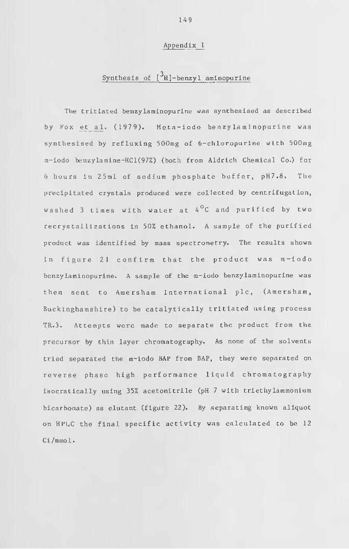
149
Appendix I
Synthesis of [ H]-benzyl aminopurine3
The tritiated benzylaminopurine was synthesised as described
by Fox et a 1 . ( 1 9 7 9 ) . M e t a - i o d o be n zy l a m i n o p u r i n e was
sy n th e s ise d by r e f l u x i n g 500mg of 6 -chloropurine w ith 500mg
m-iodo benzylamine-HCl(97%) (both from Aldrich Chemical Co.) for
6 h ou rs i n 25m l of s o d iu m p h o s p h a t e b u f f e r , p H 7 .8 . The
precipitated crystals produced were collected by centrifugation,
w a s h e d 3 t im e s w i t h w a t e r at 4 °C and p u r i f i e d by two
r e c r y s t a l l i z a t i o n s in 50% e t h a n o l . A sample of the p u r i f i e d
product was identified by mass spectrometry. The results shown
i n f i g u r e 2 1 c o n f i r m t h a t t he p r o d u c t w a s m - i o d o
benzylaminopurine. A sample of the m-iodo benzylaminopurine was
t h e n s e n t to A m ers h a m I n t e r n a t i o n a l p i c , ( A m e r s h a m ,
B uc king ha m sh ire ) to be c a t a l y t i c a l l y t r i t i a t e d using process
TR.3. Attempts were made to separate the product from the
precursor by thin layer chromatography. As none of the solvents
tried separated the m-iodo BAP from BAP, they were separated on
r e v e r s e p h ase h i g h p e r f o r m a n c e l i q u i d c h r o m a t o g r a p h y
isocratically using 35% acetonitrile (pH 7 with triethylammonium
bicarbonate) as elutant (figure 22). By separating known aliquot
on HPLC the f i n a l s p e c i f i c a c t i v i t y was c a lc u la te d to be 12
C i / mmo1.
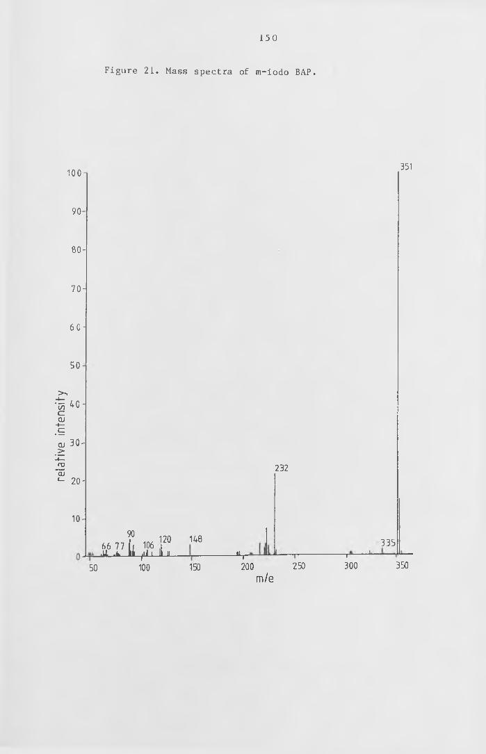
relat
ive
inte
nsify
150
Figure 21. Mass spectra of m-iodo BAP.
100
90-
6 0 -
7 0 -
6 0 -
50-
4 0 -
3 0 -
2 0 -
1 0 -
9066 77 ( i 106 ' iU. .l h.^iiii-i
120 148JL
50 100 150JL,
232
i ---- ’
200 2 50 3 00m/e
351
335
350
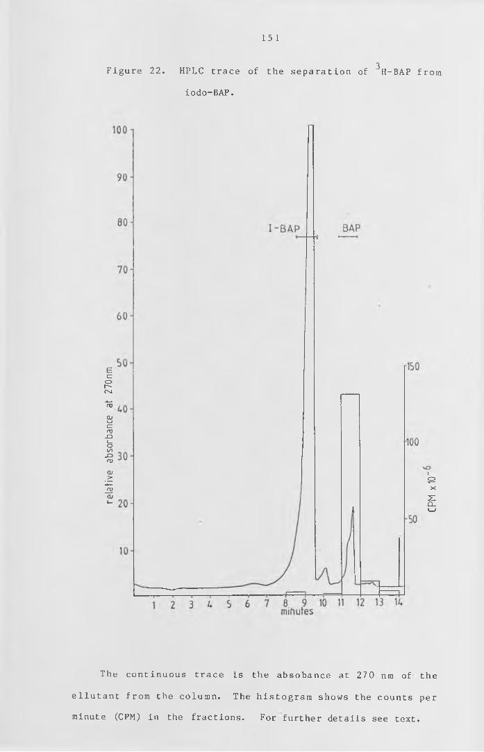
rela
tive
abso
rban
ce
at 27
0nm
Figure 22. HPLC trace of the se pa rat io n of H-BAP from
iodo-BAP.
15 1
3
The continuous trace is the absobance at 270 nm of the
e l lu t a n t from the column. The h istogram shows the counts per
minute (CPM) in the fractions. For further details see text.
CPM
x 10
“
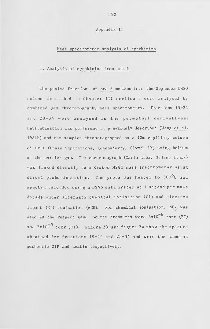
152
Appendix I I
Mass spectrometer analysis of cytokinins
1. Analysis of cytokinins from ore 6
The pooled fractions of ore 6 medium from the Sephadex LH20
column d esc r ibe d in Chapter I I I s e c t i o n 5 were analysed by
combined gas chromatography-mass spectrometry. Fractions 19-24
and 28- 34 w er e a n a l y s e d as the p e r m e t h y l d e r i v a t i v e s .
Derivatisation was performed as previously described (Wang et al.
1981b) and the samples chromatographed on a 12m capillary column
of OV-1 (Phase Separations, Queensferry, Clwyd, UK) using helium
as the carrier gas. The chromatograph (Carlo Erba, Milan, Italy)
was l inked d i r e c t l y to a Kratos MS80 mass spectrometer using
d irect probe i n s e r t i o n . The probe was heated to 300°C and
sp ectra recorded us in g a DS55 data system at 1 second per mass
decade under a l t e r n a t e chemical i o n i s a t i o n (Cl) and e le c t ro n
impact (El) ionisation (ACE). For chemical ionisation, NH^ was
used as the reagent gas. Source pressures were 4x10 ^ torr (El)
and 7x10 ^ torr (Cl) . Figure 23 and f i g u r e 24 show the spectra
obtained for f r a c t i o n s 19-24 and 28-34 and were the same as
authentic 2iP and zeatin respectively.
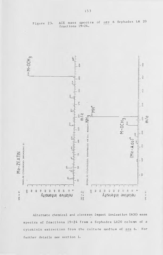
153
Figure 23. ACE mass spectra of ore 6 Sephadex LH 20
fractions 19-24.
<U JM
iai
m
O
i— i— i— i— r "i— i— r
+JC
ai
m3= :i_i >oi
ai
QJ v)tn "
< i
cu
A j IS U a jU l 0AI4B]3J U u£ <x c o
A4isua-|U| aA^ejaj
Alternate chemical and electron impact ionisation (ACE) mass
spectra of f r a c t io n s 19-24 from a Sephadex LH20 column of a
c y t o k i n i n e x t r a c t io n from the culture medium of ojre 6 . For
further details see section 1 .
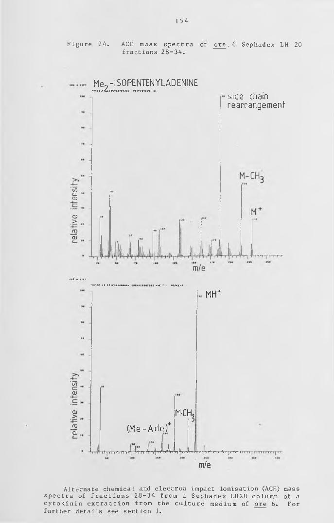
154
Figure 24 . ACE mass spectra of ore .. 6 Sephadex LH 20
fractions 28-34.
Mg9 -IS0PENT£NYLADENINEV K I I . k U l t C ' l l W l l i I H X ' I M I K ] Cl
(/)czaj
QJ>
qj
side chain re a rra n g e m e n t
UWC 4 firTYlicca.4* (TIC-»44»*««, «Vt Cl. RCACCHTI
MH+
r•
(/)czQJ
QJ>
'■£Z_ro
QJ(Me - A d e ) +
k X f U Ji**V|T i' rr r fV r r i »■' H «» »' iT 1 ‘ l^~ 'l ^ Tt ~r ^ r r n r r p Y i n t rV rj-rrV i*r
M-CH,
m/e
Alternate chemical and electron impact ionisation (ACE) mass
spectra of f r a c t io n s 28-34 from a Sephadex LH20 column of a
c y t o k in in e x t r a c t io n from the c u lt u re medium of ore 6 . For
further details see section 1 .
155
2. Analysis of cytokinins from ove 409
The putative 2iP sample from the medium of ove 409 described
in Chapter I I I se c t io n 5 was analysed by using d irect probe
insertion mass spectrometry (Kratos MS80) as described in section
1 above. Figure 25 shows the spectra obtained which contain the
same ions as authentic 2iP under the same conditions.
156
Figure 25. ACE mass spectra of ove 409.
ISOPENTENYLADENINE (PROBE) OVE 409
(/)czQJ
QJ>
ro
at
VDCCI.3* C T 1C“ I • IH X * I ***•■»» 11
L i
Ade
i1 *i*t iH V rrrl r.4W lr«
m/e
Pqrrf Wnl
side chain rearrangem ent-
M-CH-
M+
i»
GOczQJ
QJ>
QJ
Y K t l . M | I I O « H I ) 7 4 i *V* C l. RKRGCNTI HHj
>"■ ■ ■+
nf
MH
. - TJ ^ 1 mJrfTT,Jtr/Tr‘ -J[V WVrnr/l ......... 1111 n 111 11 i irrrnTrjr........n j
Alternate chemical and e l e c W & n impact ionisation (ACE) mass
spectra of f r a c t io n s 32-33 from a Sephadex LH20 column of a c y to k in in e x t r a c t io n from the c ultu r e medium of ove 409 . For
further details see section 2 .
157
References
Ashton,N.W. 1974
Genetic studies on auxotrophic and developmental mutants in the
moss, Physcomitrella patens.
Ph .D . Thesis, University of Cambridge.
A sh t o n ,N .W . , C o v e ,D . J . 1977
The isolation and preliminary characterisation of auxotrophic and
analogue resistant mutants of the moss, Physcomitrella patens.
Molecular and General Genetics 154,87-95
A sh to n ,N .W . ,C o v e ,D .J . ,Featherstone,D.R. 1979a
The isolation and physiological analysis of mutants of the moss,
Physcomitrella patens, which over-produce gametophores.
Planta 144,437-442
A sh to n ,N .W . ,G r im sle y ,N .H . ,Co ve , D .J . 19 79b
A n a l y s i s of g a m e t o p h y t i c d e v e l o p m e n t in the m o s s ,
P h y s c o m it r e l l a p a t e n s , using auxin and c y t o k in in r e s ist a n t
mutants.
Planta 144,427-435
B a t t , S . ,Venis ,M .A . 1976
Seperation and localization of two classes of auxin binding sites
in corn coleoptile membranes.
Planta 130,15-21
158
B a t t ,S . ,W i l k in s ,M .B . ,V e n i s ,M .A . 1976
Auxin b ind ing to corn c o le o p t i le membranes: k in e t ic s and
specificity .
Planta 130,7-13
Bengochea, T . , Dodds,J . H . , E v a n s ,D .E . , J e r i e , P .H . ,
N i e p e l ,B . , S h a a r i , A .R . , H a l l , M . A . 1980a
Studie s on ethylene b inding by c ell- free preperations from
cotyledons of Phaseolus vulgaris L.
Planta 148,397-406
B e n g o c h e a , ! . ,A c a s te r ,M .A . , D o d d s , J . H . , E v a n s ,D .E . ,
J e r i e , P . H . , H a l l , M . A . 1980b
Studie s on ethylene b inding by c ell free preperations from
cotyledons of Phaseolus vulgaris L.
Planta 148,407-411
Berridg e ,M .V . ,R a lph ,R .K . ,Le th am ,D .S . 1970
The binding of kinetin to plant ribosomes.
Biochemical Journal 119,75-84
Beutelmann,P . ,Bauer ,L . 1977
Purification and identification of a cytokinin from moss callus
c e l l s .
Planta 133,215-217
B id d in g t o n ,N .L . , Thomas,T.H. 1973
A m od if ied amaranthus b e t a c y a n i n b i o a s s a y for the r a p i d
determination of cytokinins in plant tissue.
Planta 111,183-186
159
Bopp,M. 1984
The hormonal regulation of protonema development in mosses
I I .The first steps of cytokinin action.
Zeitschrift fur Pflanzenphysiologie 113,435-444
Brandes,H. ,Kende,H. 1968
Studies on cytokinin-controlled bud formation in moss protenema.
Plant Physiology 43,827-837
Chopra, R . N . , Gupta,U . 1967
Dark-induction of buds in Funaria hygrometrica Hedw.
Bryologist 70,102-104
Colman,R.F . 1983
Affinity labeling of purine nucleotides in proteins.
Annual Review of Biochemistry 52,69-91
C o u rt ic e ,G .R .M . ,A sh to n ,N .W . ,C o ve ,D .J . 1978
Evidence for the r e s t r ic t e d passage of m etabolites into the
sporophyte of the moss Physcomitrella patens (Hedw.) Br. Eur .
Journal of Bryology 10,191-198
Cove ,D .J . 1984
The role of c y t o k in in and auxin in protonemal development in
Physcomitrella patens and Physcomitrium sphaericum.
Journal of the Hattori Botanical Laboratory 55,79-86
160
C o v e , D . J . ,A s h t o n ,N .W . , Feathers tone, D .R . ,W a n g ,T .L . 1980
The use of mutant s t r a in s in the study of hormone a c tio n and
metabolism in the moss Physcomitrella patens.
In 'The Proceedings of the Fourth John Innes Symposium,1979"
( D . R . D a v i e s an d D . A .H o p w o o d , ed s . ) , p p 23 1 - 2 4 1. Joh n I n n e s
Chari ty ,Norwich, England.
Cove ,D .J . , Schild , A . ,A sh ton ,N .W . , Hartmann,E. 1978
Genetic and physiological studies of the effect of light on the
development of the moss, Physcomitrella patens.
Photochemistry and Photobiology 27,249-254
C r o s s , J . W . , Briggs ,W .R . 1978
Properties of a solubilized microsomal auxin-binding protein from
coleoptiles and primary leaves of Zea mays.
Plant Physiology 62,152-157
C r o s s , J . W . , Briggs ,W .R . 1979
Solubilized auxin-binding protein.
Planta 146,263-270
Cuatrecasas ,P . 1972
P ro p e rt ie s of the i n s u l i n receptor i s o l a t e d from l iv er and fat
cell membranes.
Journal of Biological Chemistry 247,1980-1991
Cuatrecasas , P . 1974
Membrane receptors.
Annual Reveiw of Biochemistry 43,169-214
D o d d s , J . H . , Hall ,M .A . 1980
Plant Hormone Receptors.
Science Progress 66,513-535
Dohrmann.U. , Hertel , R . ,Kowalik ,H . 1978
P ro pe rtie s of auxin b ind ing s ites in d i f f e r e n t s u b c e l l u l a r
fractions from maize coleoptiles .
Planta 140,97-106
Doonan,J .H. 1983
Morphogenetic role of the cytoskeleton in Physcomitrella patens.
Ph .D . thesis, University of Leeds.
E l l io t ,D .C . 1983
Inhibition of cytokinin-regulated responses by calmodulin binding
compounds.
Plant Physiology 72,215-218
Engel ,P .P . 1968
The in d u c t io n of b io c h e m ical and m orphological mutants in the
moss Physcomitrella patens.
American Journal of Botany 55,438-446
E r i o n , J . L . , F o x , J . E . 1981
Purification and properties of a protein which binds cytokinin-
active 6 -substituted purines.
16 1
Plant Physiology 67,156-162
F o x , J . E . , E r i o n , J .L . iy/5
A cytokinin binding protein from higher plant ribosomes.
Biochemical and Biophysical Research Communications 64,694-700
F o x , J . E . , E r io n , J .L . .M c C h e s n e y ,J .D . 1979
A new intermediate in the synthesis of a tritiated cytokinin with
high specific activity .
Phytochemistry 18,1055-1056
Fries >N. 1945
Some experiments with mosses cultured in the dark.
Botaniska Notiser 98,417-424
G a r d n e r ,G . .Sussman ,M .R . ,Kende,H. 1978
it ro c y t o k i n i n b in d in g to a particulate cell fraction from
protnemata of Funaria hygrometrica.
Planta 143,67-73
G o r t o n ,B .S . ,E a k in ,R .E . 1957
Development of the gametophyte in the moss Tortella caespitosa.
Botanical Gazette (Chicago) 119,31-38
G r im s le y ,N .H . ,A sh ton ,N .W . ,C o ve ,D .J . 1977a
The pr o p er t ie s of somatic hybrids by pro t o p last fu s io n in the
moss, Physcomitrella patens .
Molecular and General Genetics 154,97-100
163
G r im sle y ,N .H . ,Ash to n ,N .W . ,Co ve ,D .J . 1977b
Complementation analysis of the auxotrophic mutants of the moss,
Physcomitrella patens, using protoplast fusion.
Molecular and General Genetics 155,103-107
Grimsley ,N .H . , Feathers t o n e , D . R . , C o u r t ic e ,G .R .M . ,
A sh to n ,N .W . , Cove ,D .J . 1979
Somatic hybridization following protoplast fusion as a tool for
g e n e t i c a n a l y s i s of d e v e l o p m e n t i n t h e m o s s
Physcomitrella patens.
Advances in Protoplast Research
Proceedings of the 5 Ctl International Protoplast Symposium pp363-
379
Hertel , R . , Thomson,K-st. , Russo,V .E .A . 1972
In-vitro auxin binding to p a r t ic u la t e c e l l f r a c t io n s from corn
coleoptiles .
Planta 107,325-340
Hocking, T . J . , C la p h a m ,J . ,C a tt e l l ,K . J . 1978
A b s c i s i c acid b in d in g to s u b c e l l u l a r f r a c t io n s from leaves of
Vicia fab a .
Planta 138,303-304
H o rnberg ,C . ,Weiler ,E .W . 1984
High-affinity binding sites for abscisic acid on the plasmalemma
of Vicia faba guard cells .
Nature 310,321-324
Hummel,J. P . .Dreyer ,W .J . 1962
Measurement of protein-binding phenomena by gel filtration.
Biochimica et Biophysica Acta 63,530-532
Ihl .M . 1976
Indole-acetic acid binding proteins in soyabean cotyledon.
Planta 131,223-228
Jaco b s ,M . ,H e rtel , R. 1978
Auxin binding to subcellular fractions from Cucurbita hypocotyls:
In vitro evidence for an auxin transport carrier.
Planta 142,1-10
Jacobsen,H-J. 1982
Soluble auxin-binding proteins in pea epicotyls.
Physiologia Plantarum 56,161-167
Jahn,H. 1964
D i e W i r k u n g v o n B l a u l i c h t u n d K i n e t i n a u f d i e
Protonemaentwicklung und Knospenbildung von Funaria hygrometrica.
Flora 155,10-29
J enkins ,G . I . , C o v e ,D . J . 1983
Light requirments for re ge ne rat ion of p rotoplasts of the moss
Physcomitrella patens.
i /Planta 157,39-45
164
Jense n ,E .V . , DeSombre, E .R . 1972
Mechanism of action of the female sex hormones.
Annual Review of Biochemistry 41,203-230
165
K e im .P . ,F o x , J .E . 1980
Interaction of a radiolabeled cytokinin photoaffinity probe with
a receptor protein.
Biochemical and Biophysical Research Communications 96,1325-1334
/
Kende ,H . .Gardner,G. 1976
Hormone binding in plants.
Annual Review of Plant Physiology 27,267-290
K lo t z , I .M . 1982
Numbers of receptor s ites from Scatchard graphs: Fact and
fantas i e s .
Science 217,1247-1249
K l o t z , I .M . ,W a l k e r ,F .M . ,P i v a n ,R .B . 1946
The binding of organic ions by proteins.
Journal of the American Chemical Society 68,1486-1490
L a u , 0 . L . , Y a n g , S . F . 1975
Interaction of kinetin and calcium in relation to their effect on
stimulation of ethylene production.
Plant Physiology 55,738-740
Lembi, C .A . ,M o r r e ,D . J . , Thomson,K., Hertel ,R . 1971
N-l-Napthylphthalamic-acid-binding activity of a plasma membrane-
rich fraction from maize coleoptiles.
Planta 99,37-45
166
L e v e r ,J .E . 1972
Quantitative assay of the binding of small molecules to protein.
Analytical Biochemistry 50,73-83
Linde,van d e r ,P .C .G . ,B o u m a n ,H . ,M e n ne s ,A .M . ,
Libbenga,K .R . 1984
A soluble auxin- b in ding protein from c ultured tobacco t issue
stimulates RNA sythesis in v itr o .
Planta 160,102-108
M a a n ,A .C . ,Vreugdenhil, D . ,Bogers, R . J . , Libbenga,K.R . 1983
Thecomplex kinetics of auxin-binding to a particulate fraction
from tobacco-pith callus.
Planta 158,10-15
M i l le r ,C .O . 1968
Biochemistry and Physiology of Plant Growth Substances.
Runge P ress , Ottawa.
M i t r a ,G .C . ,A l l s o p p ,A . ,W a r e i n g ,P .F . 1959
I.The effects of light of various qualities on the development of
the protonema and bud formation in Pohlia nutans (Hedw.) Lindb.
Phytomorphology 9,47-55
Mornet, R . , T h e i l e r ,J . B . , Leonard, N . J . , Schmitz, R . Y . ,
Moore III,F.H. ,Skoog,F. 1979
Active c y to k in in s . P h o t o a f f in i t y l a b e l l i n g agents to detect
binding.
Plant Physiology 64,600-610
167
A cytokinin-binding protein from wheat germ.
Plant Physiology 64,594-599
Nebel, B .J . , Naylor,A.W . 1964
Plant Physiology 39,S51
Nebel, B . J . , Naylor,A.W. 1968a
Initiation and development of shoot-buds from protonemata in the
moss Physcomitrium turbinatum.
American Journal of Botany 55,33-37
Nebel, B . J . , Naylor,A.W . 1968b
L ig h t , temperature and carbohydrate requirement for shoot-bud
initiation from protonemata in the moss Physcomitrium turbinatum.
American Journal of Botany 55,38-44
P o l y a ,G .M . , Bowman,J.A. 1979
Ligand specificity of a high affinity cytokinin-binding protein.
Plant Physiology 64,387-392
P o l y a ,G .M . ,D a v ies ,J .R . 1983
R e so lut io n and pr o p er t ie s of a prote in k in ase c a ta ly z in g the
phosphorylation of a wheat germ cytokinin-binding protein.
Plant Physiology 71,482-488
P o l y a ,G .M . , D a v is ,A.W. 1978
P ro perties of a high a f f i n i t y c y to k in in - bind ing protein from
wheat germ.
Planta 139,139-147
Moore III ,F .H . 1979
168
Poovaiah, B . W . , Leopold,A .C . 1.973
Deferral of leaf senescence with calcium.
Plant Physiology 52,236-239
Ray,P .M . 1977
Auxin - bind in g s i t es in maize c o le o p t i le s are l o c a l iz e d on
membranes of the endoplasmic reticulum.
Plant Physiology 59,594-599
R a y ,P .M . ,Dohrm ann ,U . , Hertel ,R . 1977
Characterization of naphthaleneacetic acid binding to receptor
sites on cellular membranes of maize coleoptile tissue.
Plant Physiology 59,357-364
Ray ,P .M . ,Dohrm ann ,U . ,Hertel ,R . 1977
Specificity of auxin-binding sites on maize coleoptile membranes
as possible receptor sites for auxin action.
Plant Physiology 60,585-591
R e e d , P .W . ,Lardy ,H .A . 1972
A23187 :A divalent calcium ionophore.
The Journal of Biological Chemistry 247,6970-6977
R e is s ,H .- D . , Herth,W. 1979
Calc ium g r a d ie n ts in tip growing plant cel ls v i s u a l i z e d by
chlorotetracycline fluorescence.
Planta 145,225-232
169
Robbins,W .J . 1918
Direct assimilation of organic carbon by Ceratodon purpureus.
Botanical Gazette 65,543-551
Roy, P . , B iswas ,B . B . 1977
A receptor protein for indoleacetic acid from plant chromatin and
its role in transcription.
Biochemical and Biophysical Research Communications 74,1597-1606
Saunders ,M .J . , Hepler, P .K . 1981
Localization of membrane-associated calcium following treatment
in Funaria using chlorotetracycline.
Planta 152,272-281
S a u n d e r s ,M .J . , Hepler, P .K . 1982
Calcium ionophore A23187 st im u la te s c y to kin in - like m itosis in
Funaria .
Science 217,943-945
S a un d e r s ,M .J . , Hepler ,P .K . 1983
Calcium antagonists and calmodulin i n h i b i t o r s block cytokinin-
induced bud formation in Funaria.
Developmental Biology 99,41-49
Scatchard,G. 1949
The attractions of proteins for small molecules and ions.
Annals New York Academy of Science 51,660-672
170
Sco tt , I .M . ,H o rg an ,R . 1980
Q u a n t i f i c a t i o n of c y to k in in s by s e le c t e d ion monitoring using
^N-labelled internal standards.
Biomedical Mass Spectrometry 7,446-449
S im o n ,P .E . ,N a e f , J .B . 1981
L ig ht dependency of the c ytokinin- induced bud i n i t i a t i o n in
protenemata of the moss Funaria hygrometrica.
Physiologia Plantarum 53,13-18
S is le r .E .C . 1979
Measurement of ethylene binding in plant tissue.
Plant Physiology 64,538-542
S k o o g ,F . ,Armstrong,D.J. 1970
Cytokinins
Annual Review of Plant Physiology 21,359-384
Stoddart, J . L . 1979a
oI n t e r a c t io n of [ H] g ibbe rel l i n Aj w ith a sub-cellular fraction
from lettuce ( Lactuca sativa L . ) hypocotyls.
Planta 146,353-361
Stoddart ,J .L . 1979b
3Interaction of [ H]gibberellin A^ w ith a sub- ce llu lar f r a c t io n
from lettuce (Lactuca sativa L . ) hypocotyls.
Planta 146,363-368
Sussman.M.R. ,F irn ,R . 1-976
The synthesis of a rad io a ct ive c y to k in in w ith high s p e c i f i c
activity .
Phytochemistry 15,153-155
Sussman.M .R . ,Kende,H. 1978
In vitro cytokinin binding to a particulate fraction of tobacco
c e l l s .
Planta 140,251-259
Takegam i ,T . ,Y osh ida ,K . 1975
I s o l a t i o n and p u r i f i c a t i o n of c yto kin in binding protein from
tobacco leaves by affin ity chromatography.
Biochemical and Biophysical Research Communications 67,782-789
Takegam i ,T . ,Yoshida,K . 1977
S p e c i f i c i n t e r a c t i o n of cyto kin in b ind ing protein w it h 40S
ribosomal subunits in the presence of cytokinin in vitro.
Plant and Cell Physiology 18,337-346
T h o m a s ,C . J .R . , S m ith ,A .R . ,H a l l ,M .A . 1984
The e f fe c t of s o l u b i l i s a t i o n on the character of an ethylene-
binding site from Phaseolus vulgaris L . cotyledons.
Planta 160,474-479
Tze-Fei Wong,J . , P o r t e r ,L . S . 1983
Ligand b in ding to prote in measured by gel f i l t r a t i o n on a
dispersion column.
Analytical Biochemistry 130,491-497
17 1
172
Venis ,M .A . 1973
Hormone receptor proteins.
Current Advances In Plant Science 2,21-28
Venis,M .A. 1977a
Solubilisation and partial purification of auxin-binding sites of
corn membranes.
Nature 266,268-269
Venis,M .A . 1977b
A f f i n i t y labels for auxin binding s i t e s in corn c o le o p t i le
membranes.
Planta 134,145-149
Venis,M .A . 1977c
Receptors for plant hormones.
Advances in Botanical Research 5,53-58
V e n i s ,M . A . ,Watson ,P .J . 1978
Naturally occurring modifiers of auxin-receptor intereaction in
corn: Identification as benzoxazolinones.
Planta 142,103-107
Vreugdenhil, D . , Burgers ,A . ,L ibbenga ,K .R . 1979
A particle-bound auxin receptor from tobacco pith callus.
Plant Science Letters 16,115-121
1 7 3
Wang ,T .L . ,B eutelm ann ,P . ,Cove ,D .J . |981a
C y t o k i n i n b i o s y n t h e s i s i n m u t a n t s of t h e m o s s
Physcomitrella patens.
Plant Physiology 68,739-744
W an g ,T .L . ,C o v e , D . J . , Beutelmann,P. , Hartmann,E. 1980
Isopentenyladenine from mutants of the moss Physcomitrella patens.
Phytochemistry 19,1103-1105
Wang, T . L . .Futers , T . S . ,McGeary, F . , C o v e ,D . J . 1984
Moss mutants and the analysis of cytokinin metabolism,
in "The biosynthesis and metabolism of plant hormones."
(Eds. A .Crozier and J .R .H i l lm a n p p . 135-164 Cambridge U n iv er s ity
Press)
Wang, T . L . , Horgan ,R . ,Cove , D . J . 1981b
Cytokinins from the moss Physcomitrella patens.
Plant Physiology 68,735-738
W a r d r o p ,A . J . , Polya,G .M. 1977
P ro p e rt ie s of a so luble auxin- binding prote in from dwarf bean
seedlings .
Plant Science Letters 8,155-163
W e d e r ,H .G . , S c h i ld k n e c h t , J . , L u t z ,R .A . ,
Kesselring ,P . 1974
Determination of binding parameters from Scatchard plots.
European Journal of Biochemistry 42,475-481

Williams, D . ,Gorski , S . 1972
Kinetic and equillbrum analysis of estradiol in uterus; a model of
binding site distribution in uterine cells .
Proceedings of the National Academy of Science USA 11,3464-3468
Yoshida,K . .Takegami,T . 1977
I s o l a t i o n of c y to k in in binding pro te in from tobacco leaves by
bioaffinity chromatography and its partial characterization.
Journal of Biochemistry 81,791-799
1 7 4


![[2 Timothy] Leader’s Guide - Cru at UNCcruatunc.com/hp_wordpress/wp-content/uploads/2013/08/2-Timothy... · [2 Timothy] Leader’s Guide ... Timothy Introduction Welcome to](https://img.pdfslide.us/doc/110x75/5a7906cc7f8b9a9d218d1646/2-timothy-leaders-guide-cru-at-2-timothy-leaders-guide-timothy-introduction.jpg)