Embed Size (px)
Citation preview
Page 1/34
Physiological signi�cance of ClpP in Enterococcusfaecalis and its regulating proteins were identi�edby Tandem Mass Tag Mass SpectrometryJinxin Zheng
Fudan University School of Basic Medical Sciences https://orcid.org/0000-0003-4064-2430Yang Wu
Fudan University School of Basic Medical SciencesZhiwei Lin
Shenzhen Sixth People's HospitalGuangfu Wang
Fudan University School of Basic Medical SciencesSibo Jiang
University of FloridaXiang Sun
Shenzhen Sixth People's HospitalHaopeng Tu
Shenzhen Sixth People's HospitalZhi-jian Yu ( [email protected] )
https://orcid.org/0000-0002-5677-2064Di Qu
Fudan University School of Basic Medical Sciences
Research article
Keywords: Enterococcus faecalis; ClpP; stress tolerance; bio�lm formation; virulence; antimicrobialstolerance
Posted Date: August 29th, 2019
DOI: https://doi.org/10.21203/rs.2.11200/v1
License: This work is licensed under a Creative Commons Attribution 4.0 International License. Read Full License

Page 2/34
Version of Record: A version of this preprint was published at BMC Microbiology on February 7th, 2020.See the published version at https://doi.org/10.1186/s12866-020-1719-9.
Page 3/34
AbstractBackground ClpP is important for bacterial growth and plays an indispensable role in cellular proteinquality control systems by refolding or degrading damaged proteins, but the physiological signi�cance ofClpP in Enterococcus faecalis is still obscure. Thus a clpP deletion mutant (△clpP) was constructed in E.faecalis OG1RF strain to elucidate a more comprehensive picture of the effect of ClpP on E. faecalis. Theglobal abundance of proteins was determined by a mass spectrometer with Tandem Mass Tags labeling.Results The ΔclpP mutant strain showed impaired growth at 20°C or 45°C, at 5% NaCl or 2 mM H2O2.The surviving bacteria of the ΔclpP mutant strain reduced after exposure to the high concentration (50 xMIC) of linezolid or minocycline for 96 h. The ΔclpP mutant strain also demonstrated decreased bio�lmformation but increased virulence in a Galleria mellonella model. The mass spectrometry proteomics dataindicated that the abundances of 135 proteins changed (111 proteins increased, 24 proteins decreased)in the ΔclpP mutant strain. Among those differential abundance proteins, the abundances of stressresponse or virulence relating proteins: FsrA response regulator, gelatinase GelE, regulatory protein Spx(spxA), heat-inducible transcription repressor HrcA, transcriptional regulator CtsR, ATPase/chaperoneClpC, acetyl esterase/lipase, and chaperonin GroEL increased in the ΔclpP mutant strain; however, theabundances of ribosomal protein L4/L1 family protein (rplD), ribosomal protein L7/L12 (rplL2), 50Sribosomal protein L13 (rplM), L18 (rplR), L20 (rplT), 30S ribosomal protein S14 (rpsN2) and S18 (rpsR) allreduced. The abundances of bio�lm formation related adapter protein MecA increased, while theabundances of dihydroorotase (pyrC), orotate phosphoribosyltransferase (pyrE) and orotidine-5'-phosphate decarboxylase (pyrF) all decreased in the ΔclpP mutant strain. Conclusion The present studydemonstrates that ClpP participates in stress tolerance, bio�lm formation, antimicrobials tolerance, andvirulence of E. faecalis.
BackgroundEnterococcus faecalis has emerged as a signi�cant cause of nosocomial infections resulting in urinarytract infections, bacteremia, prosthetic joint infection, abdominal-pelvic infections, and endocarditis in thelast two decades [1]. E. faecalis has resistance to many commonly used antimicrobial agents, particularlyvancomycin-resistant enterococci (VRE) which has emerged as a major cause of outbreaks ofnosocomial infection in recent years [2]. In addition to drug resistance, E. faecalis carries a high capacityfor bio�lm formation; more than 40% of clinical E. faecalis isolates can form bio�lms worldwide [3-7].Several virulence factors have been found associated with the bio�lm formation of E. faecalis. Forexample, the enterococcal surface protein (esp) was found to adhere to and colonize abiotic surfacesthat participated in the bio�lm formation of E. faecalis, and the gelatinase (gelE) which can hydrolyzegelatin, collagen and hemoglobin was also reported to be involved in the adherence and bio�lmformation of E. faecalis [6, 8-10]. However, the esp or gelE were found to have no association with thebio�lm formation of E. faecalis in other extensive collections of E. faecalis isolates [11-13]. Thus, thegenes involved in the E. faecalis bio�lm formation remain controversial and obscure. Other unknownfactors may also participate in the bio�lm formation of E. faecalis.

Page 4/34
The Hsp100/Clp family protein, ClpP is important for bacterial growth and plays an indispensable role incellular protein quality control systems by refolding or degrading damaged proteins in stressed cells [14].ClpP was also found associated with the bio�lm formation in some pathogenic species. For example, thebio�lms of S. mutans, S. epidermidis, P. aeruginosa, and A. pleuropneumoniae decreased when the clpPwas mutated [15-18]. However, the capacities to form bio�lms were enhanced when the clpP wasmutated in S. aureus, H. parasuis, and P. gingivalis [19-21]. The roles of clpP on the bacterial bio�lmformation have not been fully understood. The RNA levels of clpP of S. epidermidis was decreased by theagr quorum-sensing system, but in S. aureus Newman and USA300 strains, the RNA levels of agrA andagrC were signi�cantly reduced in the clpP mutants [16, 21]. The clpP affected the expression oftranscriptional regulators csrA, rpoD and a possible bio�lm repressor luxS to enhance the bio�lmformation of H. parasuis, and negatively adjusted the surface exposure of the minor �mbrial (Mfa)protein that promoted the bio�lm formation of P. gingivalis [19, 20]. The role of clpP on the bio�lmformationof E. faecalis remain unknown to date.
In addition to bacterial growth, stress response, and bio�lm formation, ClpP also in�uences the virulenceand antibacterial tolerance of several pathogenic organisms. The clpP mutation signi�cantly attenuatedthe virulence of S. pneumoniae in a murine intraperitoneal infection model. The virulence-relatedpneumolysin and pneumococcal surface antigen A was regulated by ClpP proteases [22]. Michel A et al.found that the abundance of the agr system and agr-dependent extracellular virulence factors wasdiminished in S. aureus 8325 △clpP strain [23]. In L. pneumophila, the clpP de�cient mutant strain wasunable to escape the endosome-lysosomal pathway in host cells [24]. The clpP deletion mutation alsocauses the attenuation of S. Typhimurium virulence through mis-regulation of RpoS and indirect controlof CsrA and the SPI genes [25]. In S. aureus, except for the stress response, bio�lm formation, andvirulence, the truncating mutation in clpP is responsible for the raised vancomycin resistance in VISAstrain LR5P1-V3 [26]. Bæk KT et al. found that the inactivation of the components of the ClpXP proteasesubstantially increased the β-lactam resistance level of the S. aureus USA300 strain, and found the clpPmutant strain displayed signi�cantly thicker cell walls, increased peptidoglycan cross-linking, and alteredcomposition of monomeric muropeptide species compared to wild type [27]. As mentioned above, E.faecalis shows resistance to many antimicrobial agents, however, whether the clpP is involved in E.faecalis resistance to antimicrobials, especially resistance to vancomycin (VRE) is still unclear.
Thus, in order to get a more comprehensive picture of the role of ClpP protease on the stress response,bio�lm formation, virulence, and antimicrobials tolerance of E. faecalis, a △clpP strain was constructedin E. faecalis strain OG1RF. The global abundance of proteins was detected by Orbitrap Q Exactive HF-Xmass spectrometer with tandem mass tags (TMT) labeling.
MethodsBacterial strains, plasmids, growth conditions, and chemicals
Page 5/34
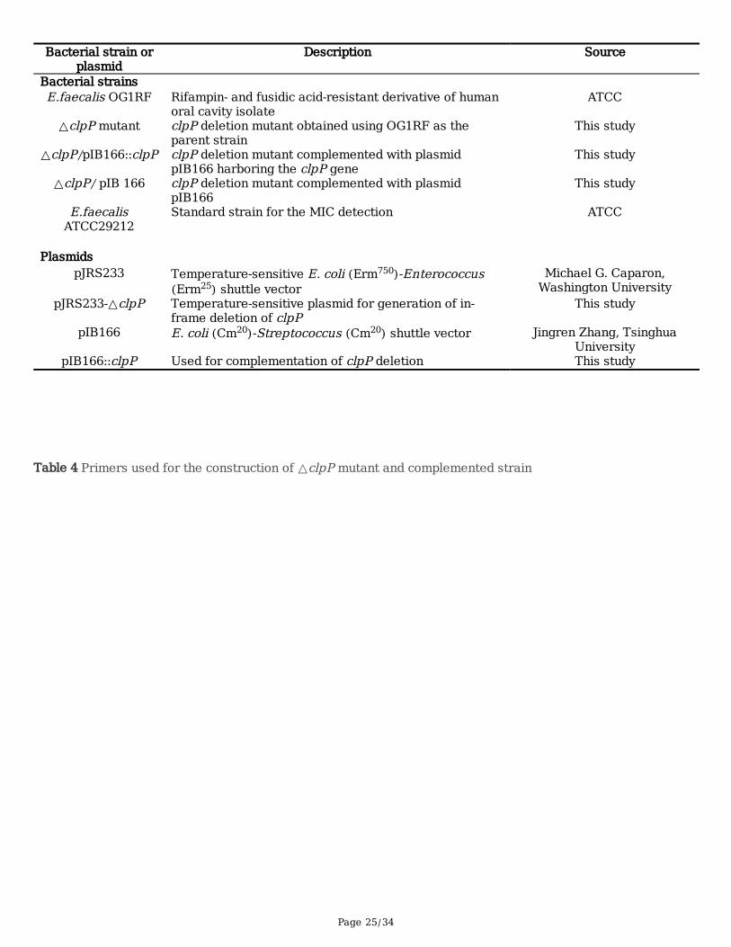
All of the bacterial strains and plasmids used in this study are shown in Table 3. The E. faecalis ATCC47077 (OG1RF; GenBank accession number CP002621.1) and ATCC 29212 were purchased from theAmerican Type Culture Collection (ATCC; Manassas, VA). The E. faecalis strains were cultured in trypticsoya broth (TSB; Oxoid, Basingstoke, UK) at 37℃ with shaking at 220 rpm. Glucose was added to theTSB medium at a concentration of 0.25% for the detection of bio�lm formation. Electroporation was usedfor plasmid transformation, and B2 medium (1% casein hydrolysate, 2.5% yeast extract, 0.5% glucose,2.5% NaCl, 0.1% K2HPO4, pH 7.5) was used for the recovery of bacteria. The antibiotics used in this studywere
purchased from Sigma Chemical Co. (Los Angeles, CA, USA) and used at concentrations of 20 mg/literfor chloramphenicol, 750 or 25 mg/liter for erythromycin.
Construction of △clpP mutants and complemented strains
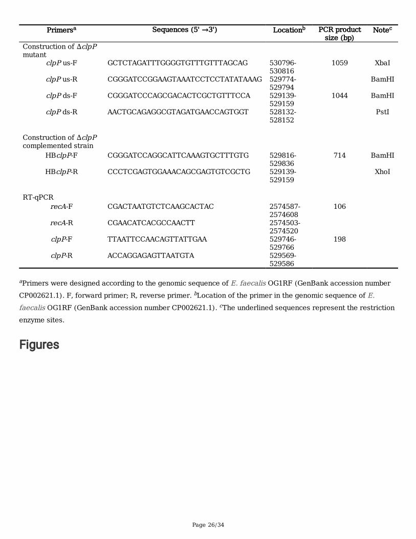
The clpP deletion mutant of the OG1RF strain was constructed by in-frame deletion using thetemperature-sensitive plasmid pJRS233 as previous described [49]. Brie�y, the upstream and downstreamfragments of OG1RF_10505 (gene: clpP; product: ATP-dependent Clp protease proteolytic subunit), whichhighly homologous (86.8%) to SA0723 (product as the ClpP protease) of S. aureus N315 strain [23], wereampli�ed from OG1RF by PCR and separately cloned into the pJRS233 vector, resulting in pJRS233-ΔclpP. The recombinant plasmid pJRS233-ΔclpP was successively transferred into E. coli DH5α andOG1RF by electroporation, and then the transformants selected at 30°C on Erm. Chromosomal integrantswere selected by growth at 42°C in the presence of Erm. Selection for excision of the integrated plasmidby homologous recombination was accomplished by growing the bacteria at 30°C in the absence of Erm.The complemented ΔclpP mutant strain was constructed using an E. coli -Streptococcus shuttle vectorpIB166. The clpP gene was ampli�ed by PCR and cloned into the pIB166 vector, resulting in pIB166:: clpP.The recombinant plasmid pIB166:: clpP was transformed by electroporation into the ΔclpP mutant strain,forming the complemented ΔclpP/pIB166:: clpP strain. The ΔclpP strain containing the empty vectorpIB166 was designated the ΔclpP/pIB166 mutant. The ΔclpP mutant and the complemented ΔclpPmutant strain were identi�ed by PCR, RT-qPCR, and direct sequencing. The primers used in this assay arelisted in Table 4.
Growth analysis of the △clpP mutant strain
The OG1RF, ΔclpP, ΔclpP/pIB166:: clpP and ΔclpP/pIB166 strains were cultured in TSB at 37°C withshaking for 12 h and diluted in the same medium to an OD600 value of 1.5, then 50 μL aliquot of thediluted suspension was inoculated into 100 mL fresh TSB and incubated at either 37°C, 45°C or 20 °Cwith circular agitation (220 rpm). The diluted suspension was also inoculated into fresh TSB with 5%NaCl pH5.5 or 2 mM H2O2 and incubated at 37°C with circular agitation (220 rpm). OD600 values for thecultures were determined using an Eppendorf Biospectrometer (Eppendorf, Hamburg, Germany) at one-hour intervals. Three independent experiments were performed.
The sensitivity of the △clpP mutant strain to sodium dodecyl sulphate (SDS)
Page 6/34
Overnight cultures of the E. faecalis strains were diluted 1:200 in fresh TSB medium and incubated at37°C for four hours until an OD600 of 1.0 was reached. After 10-fold serial dilution, 5 μL of the aliquotwas spotted onto a TSB agar plate
containing 0.008% SDS and incubated at 37°C for 24 h. The bacterial colonies on the plates werephotographed and counted [28]. Three independent experiments were performed, and the representativeresults were shown.
Microtiter plate assay of bio�lm formation
The bio�lm-forming ability of the E. faecalis isolates was detected as previously described withmodi�cations [50]. Overnight cultures were diluted 1:200 in 200 μl of TSBG (TSB with 0.25% glucose) andinoculated into 96 polystyrene microtiter plates. After 12, 24, or 48 h of static incubation at 37°C, thesupernatant was discarded, and plates were washed thrice with deionized water to remove unattachedcells, stained with 1% crystal violet (CV) for 20 min at room temperature, and rinsed with distilled water.Last, the CV was solubilized in ethanol-acetone (80:20, vol/vol) and optical density at 570 nm (OD570)was determined. Three independent experiments were performed.
Quanti�cation of extracellular DNA
Extracellular DNA (eDNA) was quanti�ed, as described previously [51]. Overnight cultures of the E.faecalis strains were diluted to OD600 = 0.001 in AB medium supplemented with 0.5% glucose, 0.05 mMpropidium iodide (PI) and 10% TSB. The diluted cultures were transferred to polystyrene microtitre plates(200 μL/well) and incubated for 24 h at 37°C. The cell density was measured at OD600 using a microtitreplate reader (BioRAD, United States). The �uorescence of PI-bound eDNA was measured by aVarioskanTM LUX multimode microplate reader (Thermo Fisher, United States) with theexcitation/emission wavelength at 535/610 nm. Relative amounts of eDNA per OD600 unit weredetermined. Three independent experiments were performed.
Determination of MIC and antimicrobials tolerance of strains
The minimal inhibitory concentrations (MICs) of the antimicrobials against the E. faecalis isolates weredetermined by the broth microdilution method according to Clinical and Laboratory Standards Institute(CLSI) guidelines CLSI-M100-S26, with CLSI-recommended MIC breakpoints. The antimicrobials-toleranceof strains was detected as described previously with modi�cations [28]. Antimicrobials (at 50 x MIC) wereadded to the stationary-phase cultures (16 h) of the E. faecalis strains, and then the cultures wereincubated at 37°C for 120 h without shaking. At the time points of every 24 h, one-milliliter aliquots weresampled and washed twice with ice-cold saline, ten-fold dilutions were then plated on Muller-Hinton agar,and the numbers of CFU were determined. Three independent experiments were performed.
Virulence of E. faecalis in Galleria mellonella
Page 7/34
Infection of G. mellonella larvae with E. faecalis strains was performed as described previously for otherpathogens [52]. G. mellonella larvae in groups of 40 were infected in the left posterior proleg with 20 μlinocula of E. faecalis strains containing 5 × 10⁶ CFU/mL. Survival of G. mellonella larvae was recorded at12 h intervals for 72 h p.i.. Every trial included a group of 20 G. mellonella larvae injected with saline as amethod control. Experiments were performed in at least three independent tests, and the representativeresults were shown.
Protein extraction and detected by a mass spectrometer with Tandem Mass Tags (TMT) labeling
The E. faecalis strains OG1RF and the ΔclpP mutant were inoculated into TSB and cultured at 37°C for 4h to logarithmic phase or for 12 h to stationary phase. The cells were harvested at 4°C centrifugation andminced individually with liquid nitrogen and lysed in lysis buffer, followed by 5 min of ultrasonication onice. Protein concentration was determined again by the Bradford protein assay. The supernatant fromeach sample, containing precisely 0.1 mg of protein, was digested with Trypsin Gold (Promega) at 1:50enzyme-to-substrate ratio. After 16 h of digestion at 37°C, peptides were desalted with C18 cartridge toremove the high urea, and desalted peptides were dried by vacuum centrifugation. Desalted peptides werelabeled with TMT6/10-plex reagents (TMT6/10plex™ Isobaric Label Reagent Set, Thermo Fisher),following the manufacturer’s instructions. For 0.1 mg of the peptide, 1 unit of labeling reagent was used.Peptides were dissolved in 100 μL of 0.1 M TEAB, and the labeling reagent was dissolved in 41 μL ofacetonitrile. After incubation for 1 h, the reaction was stopped with ammonium hydroxide. Differentlylabeled peptides were mixed equally and then desalted by peptide desalting spin columns (Thermo Fisher,89852). TMT-labeled peptide mix was fractionated using a C18 column (Waters BEH C18 4.6×250 mm, 5μm) on a Rigol L3000 HPLC operating at 1 mL/min, and the column oven was set at 50°C. Shotgunproteomics analyses were performed using an EASY-nLCTM 1200 UHPLC system (Thermo Fisher)coupled with an Orbitrap Q Exactive HF-X mass spectrometer (Thermo Fisher) operated in the data-dependent acquisition (DDA) mode. Q Exactive HF-X mass spectrometer was operated in positive polaritymode with a spray voltage of 2.3 kV and capillary temperature of 320°C. Two independent experimentswere performed.
Global protein abundance analysis
The resulting spectra from each fraction were searched separately against NCBI E. faecalis strainsOG1RF (CP002621.1) database (https://www.ncbi.nlm.nih.gov/nuccore/CP002621.1) by the searchengines: Proteome Discoverer 2.2 (PD 2.2, Thermo). The searched parameters as follows: A masstolerance of 10 ppm for precursor ion scans and a mass tolerance of 0.02 Da for the production scanswere used. Carbamidomethyl was speci�ed in PD 2.2 as �xed modi�cations. Oxidation of methionine,acetylation of the N-terminus and TMT of lysine were speci�ed in PD 2.2 as variable modi�cations. Amaximum of 2 miscleavage sites was allowed. For protein identi�cation, a protein with at least oneunique peptide was identi�ed at FDR less than 1.0% on peptide and protein level, respectively. Proteinscontaining similar peptides and could not be distinguished based on MS/MS analysis were groupedseparately as protein groups. Reporter Quanti�cation (TMT) was used for TMT quanti�cation. The
Page 8/34
protein quantitation results were statistically analyzed by Mann-Whitney Test, the signi�cant ratios,de�ned as p < 0.05 and ratio > 1.2 or ratio < 0.83 [fold change, FC], were used to screen the differentialabundance proteins (DAPs). Gene Ontology (GO) and InterPro (IPR) analysis were conducted using theinterproscan-5 program against the non-redundant protein database (including Pfam, PRINTS, ProDom,SMART, ProSitePro�les, PANTHER), and the databases COG (Clusters of Orthologous Groups) and KEGG(Kyoto Encyclopedia of Genes and Genomes) were used to analyze the protein family and pathway. Theprobable interacting partners were predicted using the STRING-db server (http://string.embl.de/) based onthe related species. STRING is a database of both known and predicted protein-protein interactions. Theenrichment pipeline was used to perform the enrichment analysis of GO, IPR, and KEGG, respectively.
RNA isolation and RT-qPCR
The RNA isolation of E. faecalis strains were performed as described previously with some modi�cations[28]. The E. faecalis strains OG1RF and the ΔclpP mutant were inoculated into TSB and cultured at 37°Cfor 4 h to logarithmic phase or for 12 h to stationary phase, the following operations were performed at4°C centrifugation or on ice: bacterial cultures were centrifuged at 5,000 x g for 5 min, and then the pelletswere washed twice with 0.9% saline; the culture was homogenized 5 times using 0.1-mm zirconia-silicabeads in a mini-BeadBeater (Biospec, Bartlesville, OK) at 5,000 rpm for 60 s at 1-min intervals; thesamples were centrifuged at 15,000 rpm and the bacterial RNA in the supernatant was puri�ed using anRNeasy minikit (Qiagen, Hilden, Germany) and quanti�ed using an ND-1000 spectrophotometer(NanoDrop Technologies, Wilmington, USA). RNA samples that had a 260/280 ratio between 2.0 and 2.2were used for the RT-qPCR.
Total RNA extracted from strains OG1RF and the ΔclpP mutant were reverse transcribed with thePrimeScript RT Reagent Kit (TaKaRa Biotechnology, Dalian, China) and RT-qPCR was performed with theSYBR Premix Ex Taq II Kit (TaKaRa Biotechnology, Dalian, China) on the Mastercycler ep realplex system(Eppendorf), with an initial incubation at 95 °C for 2 min, followed by 40 cycles of 15 s at 95 °C, and 60 sat 60 °C. Each sample was analyzed in triplicate. For all samples, the internal control gene recA was usedto normalize the abundance of E. faecalis strains OG1RFgenes [53]. The threshold cycle (Ct) numberswere con�rmed by the detection system software, and the data were analyzed based on the 2−△△Ct
method. The primers used for the RT-qPCR are listed in Table S2.
Statistical analysis
Experimental data were analyzed with SPSS software (version 16.0; SPSS, Chicago, IL, USA) andcompared using the Student’s t test, one-way analysis of variance, Mann-Whitney test, or the log-rank test.Differences with a P value of <0.05 were considered statistically signi�cant.
ResultsConstruction of clpP deletion mutant and the complemented strain
Page 9/34
To explore the role of ClpP in E. faecalis, we constructed the clpP deletion mutant in the E. faecalisOG1RF strain using the temperature-sensitive plasmid pJRS233. The deletion mutant strain was veri�edby PCR and direct sequencing and was referred to as the OG1RF ΔclpP mutant strain. The complementedΔclpP strain was constructed using shuttle vector pIB166, which named OG1RF ΔclpP/pIB166::clpP andalso veri�ed by PCR and direct sequencing. The ΔclpP strain containing the empty vector pIB166 wasdesignated as OG1RF ΔclpP/pIB166. The RNA levels of the clpP gene of all the above four E. faecalisOG1RF strains were determined by RT-qPCR and shown in Fig. S1.
ΔclpP mutant strain showed impaired growth at 20℃, 45℃, 5%NaCl or 2 mM H2O2
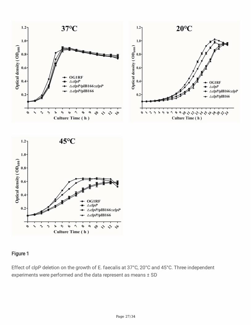
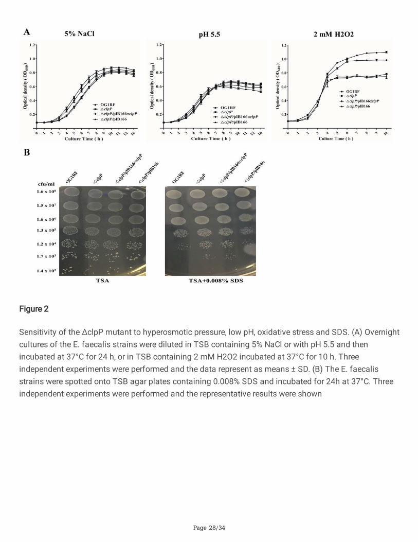
Previous research indicated that ClpP participated in the stress of low or high temperature and theoxidative stress response in S. aureus [23], however, these issues are still unknown in E. faecalis. Thus we�rst investigated the effects of clpP deletion on the growth of E. faecalis under the stress of low or hightemperature, hyperosmotic pressure, low pH, and oxidative stress. At 37℃, there were no signi�cantgrowth differences between the E. faecalis OG1RF parent strain and its ΔclpP mutant. However, under the20℃ or 45℃, the ΔclpP mutant strain showed a lower OD600 than was observed for the wild-type afterentering logarithmic phase growth (Fig.1). As Fig.2 indicated, the growth of ΔclpP mutant strain was alsoimpaired under 5%NaCl (logarithmic phase) or 2 mM H2O2 (later logarithmic phase or stationary phase).
Deletion of clpP leads to decreased bio�lm formation
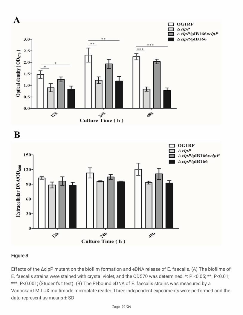
The polystyrene microtiter plate assay was performed to evaluate the role of clpP in the bio�lm formationof E. faecalis under static conditions. The bio�lm formation of E. faecalis OG1RF parent strain and itsΔclpP mutant was monitored at 12, 24, and 48 h on microtiter plates stained with crystal violet, and theOD570 were determined. The bio�lms of the ΔclpP mutant strain (OD570, 0.835 ± 0.091) were signi�cantlydecreased compared with that of the parent strain (OD570, 2.247 ± 0.138, P<0.001, student’s t test) afterincubation for 48 h, and this issue was also observed after incubation for 12 or 24 h (Fig.3A). We furtherinvestigated the eDNA release during the bio�lm formation of E. faecalis, but found no differencesbetween the ΔclpP mutant and its parent strain (Fig.3B).
Antimicrobials tolerance of the ΔclpP mutant strain
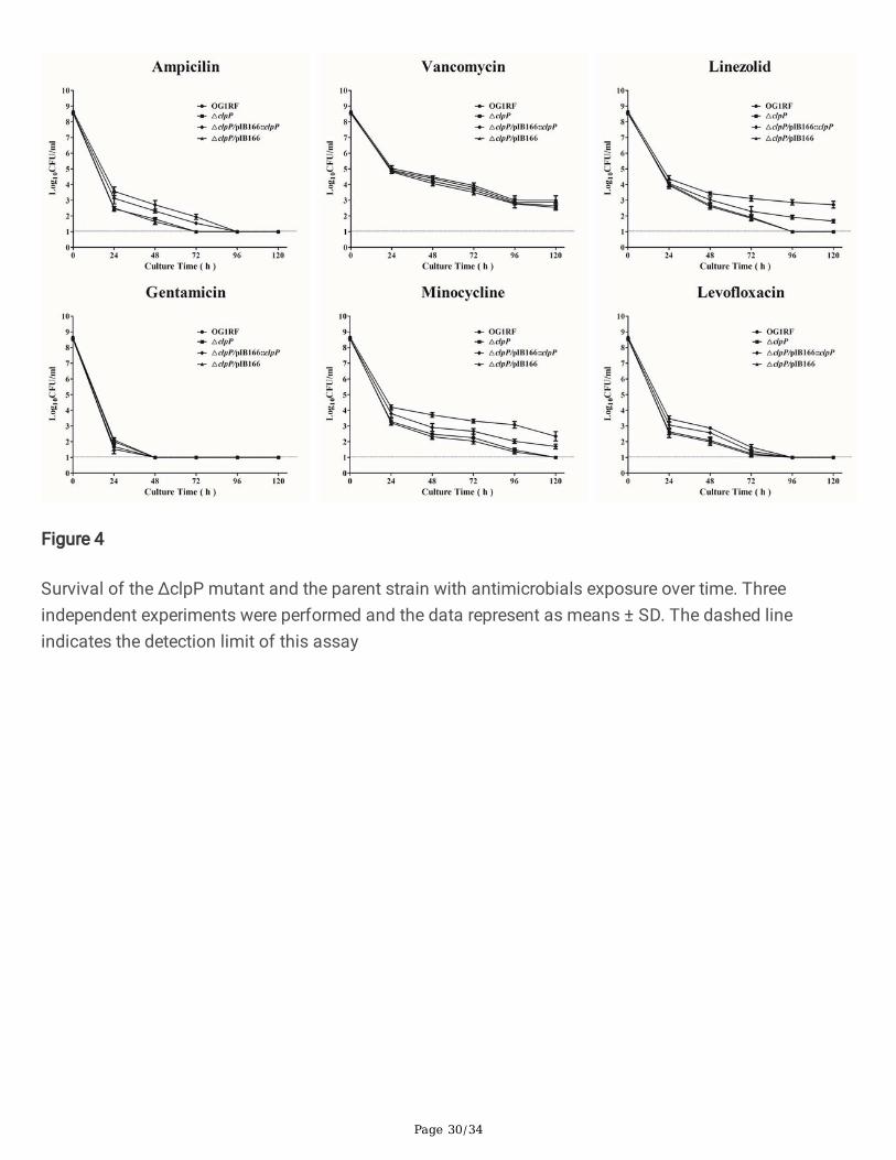
The minimum inhibitory concentrations (MICs) of eight antimicrobials for E. faecalis were detected bybroth microdilution method and the MICs for the ΔclpP mutant strain were found to be similar to those ofthe parent strain (Table S1). To explore the antimicrobial concentrations that ensured only drug-tolerantbacterial cells survived, we did the time-killing assays for six antimicrobials used in the present study.Based on the previous research [28] and our preliminary experimental results, the concentrations of sixantimicrobials were used at 50 x MIC in the present study. As Fig.4 indicated, the surviving bacteria of theΔclpP mutant strain (log10CFU/ml, under the detection limit) were signi�cantly decreased compared withthose of the parent strain (log10CFU/ml, 2.873 ± 0.243, P<0.001, student’s t test) after exposure to thelinezolid for 96 h. After exposure to the minocycline for 96 h, the surviving bacteria of the ΔclpP mutant
Page 10/34
strain (log10CFU/ml, 1.477 ± 0.171) were also decreased compared with those of the parent strain(log10CFU/ml, 3.078 ± 0.303, P<0.01, student’s t test).
ΔclpP mutant leads to increased virulence of E. faecalis
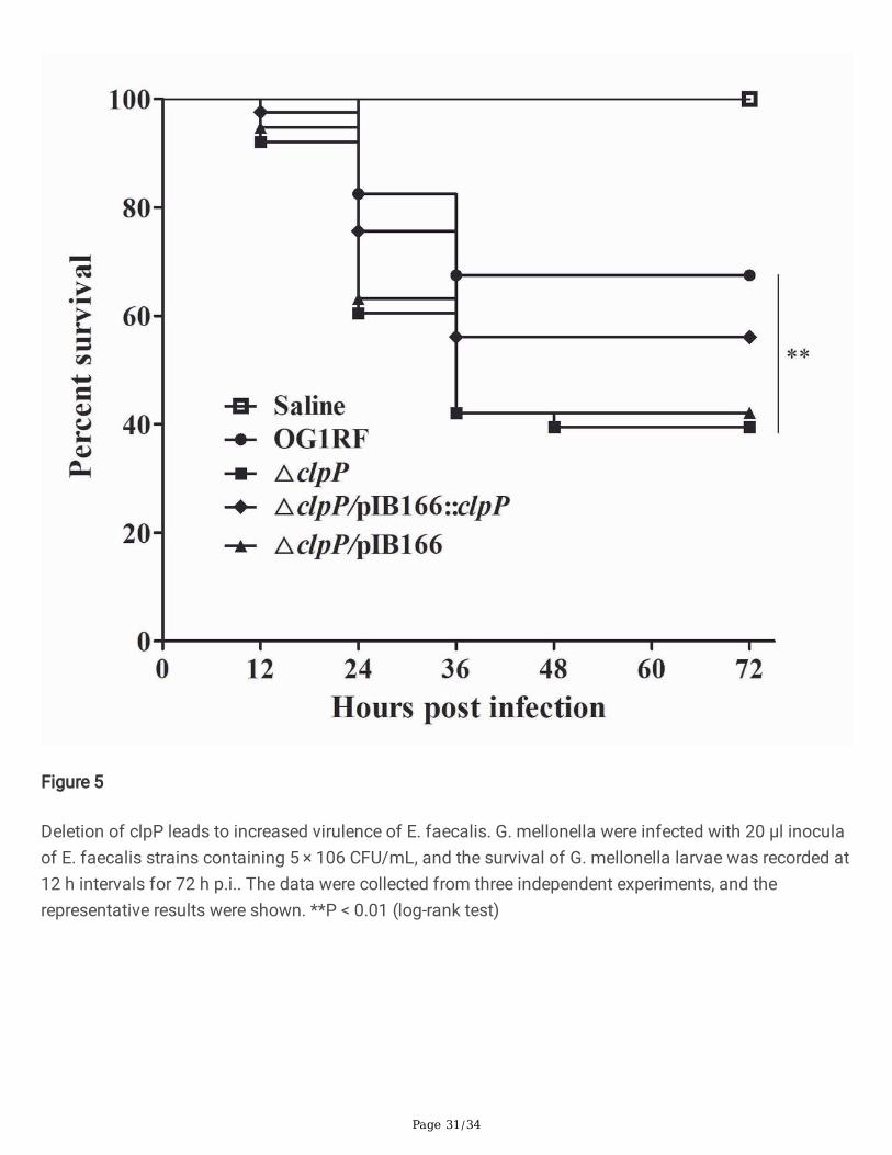
The virulence of E. faecalis strains was detected by the infection of G. mellonella larvae. The survival ofG. mellonella larvae infected with the ΔclpP mutant strain (15/40, 37.5%) signi�cantly decreasedcompared with the parent strain (28/40, 70.0%, P<0.01, log-rank test) after 72 h p.i (Fig.5). Thecomplemented △clpP/pIB166::clpP strain (23/40, 57.5%) showed a partially restored survival ability.
Comparison of the global protein abundances of the ΔclpP mutant and the parent strain
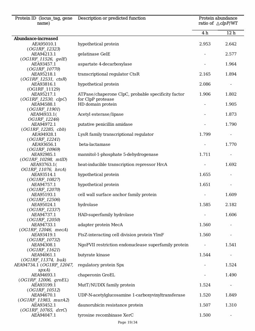
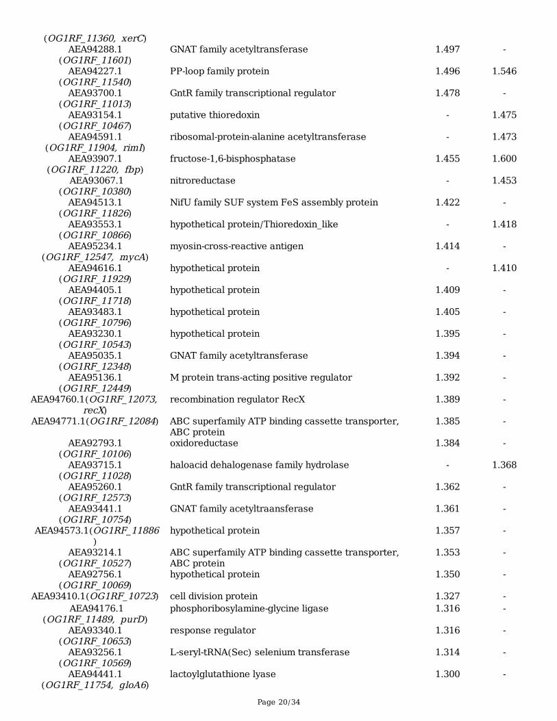
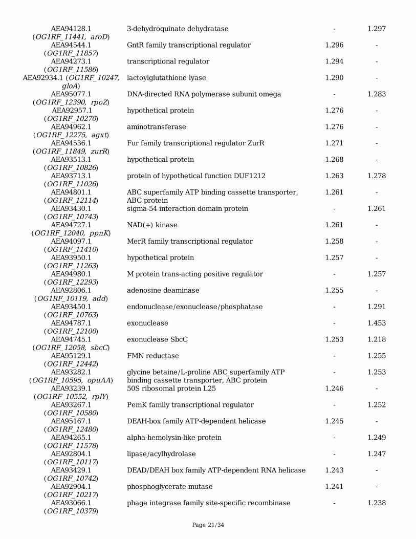
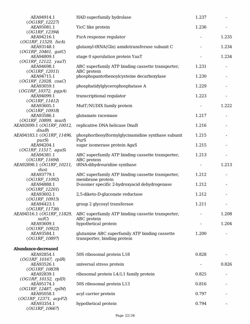
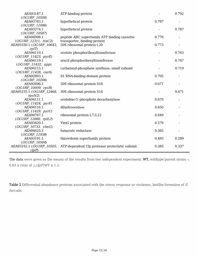
We compared the global protein abundance of the ΔclpP mutant with the parent strain. The total proteinswere extracted from logarithmic-phase (4 h), and stationary phase (12 h) bacteria and their abundanceswere determined by Q Exactive HF-X mass spectrometer with Tandem Mass Tags (TMT) labeling. Theprotein quantitation results were statistically analyzed by Mann-Whitney Test, the signi�cant ratios,de�ned as p < 0.05 and ratio > 1.2 or ratio < 0.83 [fold change, FC], were used to screen the differentialabundance proteins (DAPs).The protein quantitation results were given as the means from twoindependent experiments, the repeatability of the two independent experiments were evaluated by thecoe�cient of variation (CV), and as Fig. S2 indicated that the CV of the two independent experiments wasvery low. AlltheDAPsweresummarized in Table 1, the abundances of 135 proteins changed in the ΔclpPmutant strain, of which 111 proteins increased, while 24 proteins decreased.
Gene Ontology (GO) and KEGG analysis of DAPs
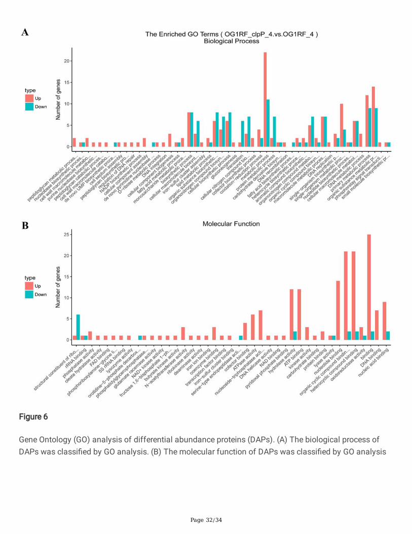
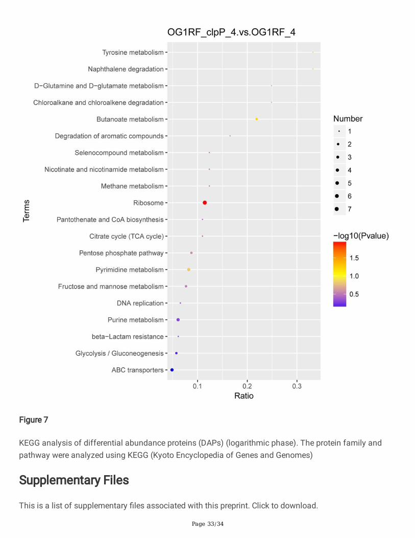
All the DAPs between the △clpP mutant and the parent strain were analyzed by the Gene Ontology (GO)and KEGG analysis. As Fig.6 GOanalysisshowed, the increased DAPs in the △clpP mutant strain(logarithmic phase) were mainly concentrated in the following molecular functions: N-acetyltransferaseactivity, coenzyme binding, cofactor binding, ATPase activity, nucleoside-triphosphatase activity,hydrolase activity, ATP binding, kinase activity, nucleotide binding, organic cyclic compound binding,heterocyclic compound binding, DNA binding and nucleic acid binding. However, the decreased DAPswere mainly included in the following molecular functions: structural constituent of ribosome, rRNAbinding, orotidine-5'-phosphate decarboxylase activity, hydrolase activity, organic cyclic compoundbinding, heterocyclic compound binding, and nucleic acid binding. The KEGG analysis demonstrated thatfunctions of most of these DAPs in the △clpP mutant (logarithmic phase) belonged to the ribosome,fructose and mannose metabolism, pyrimidine metabolism, purine metabolism, pentose phosphatepathway, glycolysis/gluconeogenesis, ABC transporters (Fig.7). The functions of DAPs in the stationaryphase of △clpP mutant strain were similar to that in the logarithmic phase (Fig.S3).
DAPs associated with the stress response or virulence, bio�lm formation of E. faecalis
Page 11/34
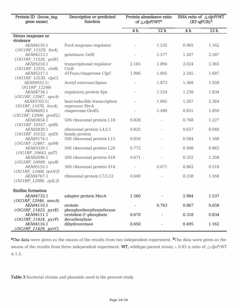
Based on the literatures, we selected the DAPs which may associate with stress response or virulence,bio�lm formation of E. faecalis for further analyzed. The abundance of DAPs which associated with thestress response or virulence of E. faecalis, including the FsrA response regulator and gelatinase GelE,ATPase/chaperone ClpC, chaperonin GroEL, acetyl esterase/lipase, and transcriptional regulator proteins,HrcA, CtsR, and Spx increased in the △clpP mutant strain (Table 2). However, the abundances ofribosomal proteins L4/L1, L7/L12, L13, L18, L20, S14, and S18 decreased in the △clpP mutant strain.The abundance of the bio�lm formation of E. faecalis associated DAPs, adapter protein MecA increasedin the △clpP mutant strain, while the abundances of orotate phosphoribosyltransferase, orotidine-5'-phosphate decarboxylase and dihydroorotase reduced (Table 2).The RNA levels of all the above DAPswhich may associate with stress response, virulence or bio�lm formation of E. faecalis, were veri�ed byRT-qPCR and were consistent with protein abundance changes in the △clpP mutant strain.
DiscussionClpP as a protease of Hsp100/Clp family is very important for bacterial growth and plays an irreplaceablerole in cellular protein quality control systems by refolding or degrading damaged proteins in stressedcells [14]. Up to now, ClpP has been found participating in many essential activities of bacteria, such asstress response, which including abnormal temperature stress, hyperosmotic pressure, low pH, andoxidative stress response, virulence, bio�lm formation and so on. However, the global abundance ofproteins affected by ClpP in bacteria is still little known for us. Feng J et al. found that the abundance oftranscriptional regulators CtsR and Spx, the ClpC adaptor proteins McsB and MecA, the cell divisionprotein FtsZ and so on, were clearly affected by ClpP in S. aureus strains NCTC8325-4, COL, SA564 andNewman by 2-D DIGE technique [29, 30]. However, the abundance of not more than 80 proteins changedin their studies, and it may be due to the low sensitivity of 2-D DIGE technique. In the present study, weexacted the total proteins from E. faecalis OG1RF and its △clpP mutant strains, digested and then all thepeptides labeled with Tandem Mass Tags (TMT, a new �uorescent dyes similar with iTRAQ), �nallydetected by Orbitrap Q Exactive HF-X mass spectrometer and found 135 differential abundance proteins(DAPs) in the △clpP mutant strain. Among those DAPs, we also found the transcriptional regulators CtsRand Spx, the ClpC adaptor proteins MecA and FtsZ-interacting cell division protein YlmF, which weresimilar to that in S. aureus strains, and interestingly we also found other new proteins, such as Acetylesterase/lipase, ribosomal protein, orotidine-5'-phosphate decarboxylase and so on.
The ClpP has been shown to participate in stress tolerance by refolding or degrading damaged proteinsduring the growth of bacteria, and several studies have indicated that the ΔclpP mutant strain showed agrowth defect over a broad range of temperatures, which included high (40, 42, 45°C) or low (20, 30°C)temperatures, even at the 37°C condition [19, 23, 31, 32]. However, this study only found the growth of E.faecalis OG1RF ΔclpP mutant strain to be impaired under the 45°C or 20°C temperatures, but notobserved at 37°C. Previous studies also demonstrated the ΔclpP strain was more vulnerable to oxidativestress, osmotic stress, acid, or sodium dodecyl sulfate [19, 33-35], this study found the growth of OG1RFΔclpP strain was impaired under the osmotic stress or oxidative stress conditions. The heat shock proteinheat-inducible transcription repressor HrcA and chaperonin GroEL were found associated with the
Page 12/34
temperature stress response [36]. However, the increased abundances of HrcA and GroEL may notcontribute to the impaired growth of the ΔclpP mutant strain under the 45°C or 20°C temperatures in thisstudy. As the increased abundances of HrcA and GroEL were in the stationary phase, not in thelogarithmic phase growth of the ΔclpP mutant strain, while the impaired growth of the ΔclpP mutantstrain mainly occurred in logarithmic phase growth under the 45°C or 20°C temperatures. The ribosomalprotein L9 was found played a signi�cant role in the E. coli response to starvation stress [37]. The presentstudy found that when the clpP was deleted in E. faecalis OG1RF, the abundance of many ribosomalproteins, including both 50S and 30S ribosomal proteins decreased in the ΔclpP mutant strain. Thus,ClpP may through affecting the abundance of ribosomal proteins participates in the stress response of E.faecalis.
Previous studies have found that ClpP can signi�cantly affect the bio�lm formation of bacteria, but theeffects of clpP on the bio�lm formation in different genera of bacteria were obviously different, or evenopposite [15, 16, 18, 19, 21]. This study �rst found that bio�lm formation decreased when the clpP ofOG1RF strain was deleted. The adapter protein MecA was found decreased the RNA level of eps, whichencodes synthesis of the bio�lm matrix exopolysaccharide, thus inhibited the bio�lm formation of B.subtilis [38]. The present study indicated that the abundance of MecA increased in the ΔclpP mutantstrain, and it may lead to the decreased bio�lm formation of the clpP deleted strain. Another reason forthe reduced bio�lm formation of the ΔclpP mutant strain may be the reduced abundances of orotatephosphoribosyltransferase (pyrE) and orotidine-5'-phosphate decarboxylase (pyrF), the two proteinswhich have been found promoted the bio�lm formation of S. sanguinis and E. faecalis respectively [39,40].
ClpP was found also participated in the bacterial virulence; the virulence of S. pneumoniae, S. aureus andL. pneumophila attenuated in the clpP mutation strains [22-24]. Recently, Liu Q et al. reported that in S.aureus, the clpP mutant strain showed increased bio�lm formation and reduced virulence [21]. However,this study found the ΔclpP mutant strain with the decreased bio�lm formation and increased virulence ina G. mellonella model. The previous study explored that the CtsR regulator controlled the expression ofclpC, clpE, and clpP, and was required for the virulence of E.faecalis V583, but the role of clpP in thevirulence of E.faecalis was still unclear [41]. The FsrABDC signal transduction system and the GelE weremajor virulence factors in E. faecalis [42, 43]. Thus it may be that the increased abundance of FsrA andGelE lead to the increased virulence of the ΔclpP mutant strain. The abundance of acetyl esterase/lipase,another virulence factor of E. faecalis, also was found increased in the ΔclpP mutant strain, this suggeststhat it may also cause the increased virulence of the ΔclpP mutant strain [44].
This study also found that the sensitivities to linezolid or minocycline of the ΔclpP mutant strainincreased. As we knew, linezolid as an inhibitor of bacterial protein synthesis which acts on the 50Sribosome subunit of gram-positive bacteria, and minocycline is a synthetic tetracycline derivative whichacts on the 30S ribosome subunit of gram-positive or gram-negative bacteria [45, 46]. The present studyindicated that the abundance of 50S ribosomal proteins L13, L18, L20, the 30S ribosomal proteins S14,
Page 13/34
S18, all reduced in the △clpP mutant strain, thus might lead to the increased sensitivities of the △clpPmutant strain to linezolid or minocycline.
In the B. subtilis, Spx plays a signi�cant role in the protection system against oxidative stresses [47].Recently Rojas-Tapias DF and Helmann JD found that the Spx was itself a regulator of the ctsR operon,and the ctsR operon was found regulated the expression of clpC and clpP [48]. The present studyindicated that when the clpP was deleted in E. faecalis OG1RF, the abundance of ClpC, CtsR, and Spx allincreased, this was similar with that observed in S. aureus [30]. In S. aureus, the RNA levels of the clpCoperon (ctsR-mcsA-mcsB-clpC), groE, and dnaK were induced in response to an accumulation ofmisfolded proteins, which supported the notion that ClpP proteases served to degrade misfolded proteins[30]. Our study found the abundances of ClpC, GroEL, and the DnaB, but not the DnaK, increased in the△clpP mutant strain, may also due to the accumulation of misfolded proteins.
It is easy to understand that ClpP, as a protease, has a signi�cant effect on the abundance of proteins, butnot on the RNA level of genes. In the present study, the abundance of many transcription regulationrelated proteins changed in the △clpP mutant strain, such as regulatory protein Spx (spxA), heat-inducibletranscription repressor HrcA, transcriptional regulator CtsR and so on, and this issue was also observed inother studies [29, 30]. As we knew, the transcriptional regulators usually control the transcription and RNAlevels of their functional genes. So the ClpP may through affecting the abundance of transcriptionalregulators to alter the RNA levels of other genes, and the RNA levels of many genes changed in the ΔclpPmutant strain in this study, and the similar results were also observed in other studies [23, 30]. Since ClpPwas a protease which involved in protein degradation, thus its absence should provoke the accumulationof proteins, and this was consistent with our result that the abundance of most of the DAPs increased inthe △clpP mutant strain. However, the abundance of some proteins and their corresponding RNA levels ofgenes decreased in the △clpP mutant strain in the present study, and the similar results were also foundin another study [30]. The reason for this issue may be that as mentioned above, ClpP reduced thetranscription and expression of those genes by regulating the abundance of transcriptional regulators.
ConclusionsThe present study indicates that ClpP may through affecting the abundance of ribosomal proteins L4/L1,L7/L12, L13, L18, L20, S14, and S18 participates in the stress response, and the linezolid or minocyclinetolerance of E. faecalis. ClpP participates in the bio�lm formation of E. faecalis may by affecting theabundances of adapter protein MecA, orotate phosphoribosyltransferase (pyrE), orotidine-5'-phosphatedecarboxylase (pyrF). Our results also suggest that ClpP may modulate the abundances of FsrA, GelE,and acetyl esterase/lipase participates in the virulence of E. faecalis.
AbbreviationsCV: crystal violet; DAPs: differential abundance proteins; eDNA: Extracellular DNA; MIC: minimal inhibitoryconcentration; TMT: Tandem Mass Tags; VRE: vancomycin-resistant enterococci;
Page 14/34
DeclarationsEthics approval and consent to participate Not applicable. Consent for publication Not applicable.Availability of data and materials All data generated or analysed during this study are included in thispublished article [and its supplementary information �les]. The mass spectrometry proteomics data havebeen deposited to the ProteomeXchange Consortium via the PRIDE partner repository with the datasetidenti�er PXD014211. Con�ict of Interest The authors declare that they have no competing interestsFunding This work was supported by the following grants: the Sanming Project of Medicine in Shenzhen(grant number SMGC201705029); Science , Technology and Innovation Commission of ShenzhenMunicipality of key funds (JCYJ20170412143551332; JCYJ20180508162403996) and basic researchfunds (JCYJ20180302144721183; JCYJ20180302144431923). Author’s contributions Jinxin Zhengparticipated in the design of the study, carried out the gene manipulation, bio�lm and eDNA assay,analyzed and interpreted the proteomic data, and drafted the manuscript. Yang Wu participated in thegene manipulation, RT-qPCR, and proteomic data. Zhiwei Lin conducted the RNA extraction and RT-qPCR.Guangfu Wang performed antibiotic-susceptibility testing, antimicrobials tolerance experiments andprotein extraction. Sibo Jiang, Xiang Sun and Haopeng Tu performed the gene manipulation, stresstolerance experiments, bio�lm and eDNA test, G.mellonella trials and RNA extraction and RT-qPCR. ZhijianYu and Di Qu designed the study, participated in the data analysis, and provided critical revisions of themanuscript for valuable intellectual content. Acknowledgments The authors thank Prof. Michael G.Caparon (Department of Molecular Microbiology, Washington University School of Medicine, Saint Louis,Missouri, USA ) for providing generously plasmid pJRS233, thank Prof. Jingren Zhang (Center forInfectious Disease Research, School of Medicine, Tsinghua University, Beijing, China ) for providinggenerously the plasmid pIB166. We also thank Ms. Cynthia Brast (University of Florida, Gainesville, FL,USA) for her review on this manuscript.
References1. Ali L, Goraya MU, Arafat Y, Ajmal M, Chen JL, Yu D. Molecular Mechanism of Quorum-Sensing in
Enterococcus faecalis: Its Role in Virulence and Therapeutic Approaches. Int J Mol Sci. 2017; 18(5).
2. Flokas ME, Karageorgos SA, Detsis M, Alevizakos M, Mylonakis E. Vancomycin-resistant enterococcicolonisation, risk factors and risk for infection among hospitalised paediatric patients: a systematicreview and meta-analysis. Int J Antimicrob Agents. 2017; 49(5):565-72.
3. Dupre I, Zanetti S, Schito AM, Fadda G, Sechi LA. Incidence of virulence determinants in clinicalEnterococcus faecium and Enterococcus faecalis isolates collected in Sardinia (Italy). J MedMicrobiol. 2003; 52(Pt 6):491-8.
4. Sandoe JA, Witherden IR, Cove JH, Heritage J, Wilcox MH. Correlation between enterococcal bio�lmformation in vitro and medical-device-related infection potential in vivo. J Med Microbiol. 2003; 52(Pt7):547-50.
5. Seno Y, Kariyama R, Mitsuhata R, Monden K, Kumon H. Clinical implications of bio�lm formation byEnterococcus faecalis in the urinary tract. Acta Med Okayama. 2005; 59(3):79-87.
Page 15/34
�. Toledo-Arana A, Valle J, Solano C, Arrizubieta MJ, Cucarella C, Lamata M et al. The enterococcalsurface protein, Esp, is involved in Enterococcus faecalis bio�lm formation. Appl Environ Microbiol.2001; 67(10):4538-45.
7. Zheng JX, Wu Y, Lin ZW, Pu ZY, Yao WM, Chen Z et al. Characteristics of and Virulence FactorsAssociated with Bio�lm Formation in Clinical Enterococcus faecalis Isolates in China. FrontMicrobiol. 2017; 8:2338.
�. Paganelli FL, Willems RJ, Leavis HL. Optimizing future treatment of enterococcal infections:attacking the bio�lm? Trends Microbiol. 2012; 20(1):40-9.
9. Kayaoglu G, Orstavik D. Virulence factors of Enterococcus faecalis: relationship to endodonticdisease. Crit Rev Oral Biol Med. 2004; 15(5):308-20.
10. Park SY, Kim KM, Lee JH, Seo SJ, Lee IH. Extracellular gelatinase of Enterococcus faecalis destroys adefense system in insect hemolymph and human serum. Infect Immun. 2007; 75(4):1861-9.
11. Kristich CJ, Li YH, Cvitkovitch DG, Dunny GM. Esp-independent bio�lm formation by Enterococcusfaecalis. J Bacteriol. 2004; 186(1):154-63.
12. Mohamed JA, Murray BE. Lack of correlation of gelatinase production and bio�lm formation in alarge collection of Enterococcus faecalis isolates. J Clin Microbiol. 2005; 43(10):5405-7.
13. Anderson AC, Jonas D, Huber I, Karygianni L, Wolber J, Hellwig E et al. Enterococcus faecalis fromFood, Clinical Specimens, and Oral Sites: Prevalence of Virulence Factors in Association with Bio�lmFormation. Front Microbiol. 2015; 6:1534.
14. Frees D, Savijoki K, Varmanen P, Ingmer H. Clp ATPases and ClpP proteolytic complexes regulate vitalbiological processes in low GC, Gram-positive bacteria. Mol Microbiol. 2007; 63(5):1285-95.
15. Lemos JA, Burne RA. Regulation and Physiological Signi�cance of ClpC and ClpP in Streptococcusmutans. J Bacteriol. 2002; 184(22):6357-66.
1�. Wang C, Li M, Dong D, Wang J, Ren J, Otto M et al. Role of ClpP in bio�lm formation and virulence ofStaphylococcus epidermidis. Microbes Infect. 2007; 9(11):1376-83.
17. Fernandez L, Breidenstein EB, Song D, Hancock RE. Role of intracellular proteases in the antibioticresistance, motility, and bio�lm formation of Pseudomonas aeruginosa. Antimicrob AgentsChemother. 2012; 56(2):1128-32.
1�. Xie F, Zhang Y, Li G, Zhou L, Liu S, Wang C. The ClpP protease is required for the stress tolerance andbio�lm formation in Actinobacillus pleuropneumoniae. PLoS One. 2013; 8(1):e53600.
19. Huang J, Wang X, Cao Q, Feng F, Xu X, Cai X. ClpP participates in stress tolerance and negativelyregulates bio�lm formation in Haemophilus parasuis. Vet Microbiol. 2016; 182:141-9.
20. Capestany CA, Tribble GD, Maeda K, Demuth DR, Lamont RJ. Role of the Clp system in stresstolerance, bio�lm formation, and intracellular invasion in Porphyromonas gingivalis. J Bacteriol.2008; 190(4):1436-46.
21. Liu Q, Wang X, Qin J, Cheng S, Yeo WS, He L et al. The ATP-Dependent Protease ClpP Inhibits Bio�lmFormation by Regulating Agr and Cell Wall Hydrolase Sle1 in Staphylococcus aureus. Front Cell
Page 16/34
Infect Microbiol. 2017; 7:181.
22. Kwon HY, Kim SW, Choi MH, Ogunniyi AD, Paton JC, Park SH et al. Effect of heat shock andmutations in ClpL and ClpP on virulence gene expression in Streptococcus pneumoniae. InfectImmun. 2003; 71(7):3757-65.
23. Michel A, Agerer F, Hauck CR, Herrmann M, Ullrich J, Hacker J et al. Global regulatory impact of ClpPprotease of Staphylococcus aureus on regulons involved in virulence, oxidative stress response,autolysis, and DNA repair. J Bacteriol. 2006; 188(16):5783-96.
24. Zhao BB, Li XH, Zeng YL, Lu YJ. ClpP-deletion impairs the virulence of Legionella pneumophila andthe optimal translocation of effector proteins. BMC Microbiol. 2016; 16(1):174.
25. Knudsen GM, Olsen JE, Aabo S, Barrow P, Rychlik I, Thomsen LE. ClpP deletion causes attenuation ofSalmonella Typhimurium virulence through mis-regulation of RpoS and indirect control of CsrA andthe SPI genes. Microbiology. 2013; 159(Pt 7):1497-509.
2�. Shoji M, Cui L, Iizuka R, Komoto A, Neoh HM, Watanabe Y et al. walK and clpP mutations conferreduced vancomycin susceptibility in Staphylococcus aureus. Antimicrob Agents Chemother. 2011;55(8):3870-81.
27. Baek KT, Grundling A, Mogensen RG, Thogersen L, Petersen A, Paulander W et al. beta-Lactamresistance in methicillin-resistant Staphylococcus aureus USA300 is increased by inactivation of theClpXP protease. Antimicrob Agents Chemother. 2014; 58(8):4593-603.
2�. Wang X, Han H, Lv Z, Lin Z, Shang Y, Xu T et al. PhoU2 but Not PhoU1 as an Important Regulator ofBio�lm Formation and Tolerance to Multiple Stresses by Participating in Various FundamentalMetabolic Processes in Staphylococcus epidermidis. J Bacteriol. 2017; 199(24).
29. Feng J, Michalik S, Varming AN, Andersen JH, Albrecht D, Jelsbak L et al. Trapping and proteomicidenti�cation of cellular substrates of the ClpP protease in Staphylococcus aureus. J Proteome Res.2013; 12(2):547-58.
30. Frees D, Andersen JH, Hemmingsen L, Koskenniemi K, Baek KT, Muhammed MK et al. New insightsinto Staphylococcus aureus stress tolerance and virulence regulation from an analysis of the role ofthe ClpP protease in the strains Newman, COL, and SA564. J Proteome Res. 2012; 11(1):95-108.
31. Gaillot O, Pellegrini E, Bregenholt S, Nair S, Berche P. The ClpP serine protease is essential for theintracellular parasitism and virulence of Listeria monocytogenes. Mol Microbiol. 2000; 35(6):1286-94.
32. Frees D, Qazi SN, Hill PJ, Ingmer H. Alternative roles of ClpX and ClpP in Staphylococcus aureusstress tolerance and virulence. Mol Microbiol. 2003; 48(6):1565-78.
33. Hou XH, Zhang JQ, Song XY, Ma XB, Zhang SY. Contribution of ClpP to stress tolerance and virulenceproperties of Streptococcus mutans. J Basic Microbiol. 2014; 54(11):1222-32.
34. Rajagopal S, Sudarsan N, Nickerson KW. Sodium dodecyl sulfate hypersensitivity of clpP and clpBmutants of Escherichia coli. Appl Environ Microbiol. 2002; 68(8):4117-21.
35. Park CY, Kim EH, Choi SY, Tran TD, Kim IH, Kim SN et al. Virulence attenuation of Streptococcuspneumoniae clpP mutant by sensitivity to oxidative stress in macrophages via an NO-mediated
Page 17/34
pathway. J Microbiol. 2010; 48(2):229-35.
3�. Laport MS, Dos Santos LL, Lemos JA, do Carmo FBM, Burne RA, Giambiagi-Demarval M.Organization of heat shock dnaK and groE operons of the nosocomial pathogen Enterococcusfaecium. Res Microbiol. 2006; 157(2):162-8.
37. Pei H, Han S, Yang S, Lei Z, Zheng J, Jia Z. Phosphorylation of bacterial L9 and its functionalimplication in response to starvation stress. FEBS Lett. 2017; 591(20):3421-30.
3�. Prepiak P, Defrancesco M, Spadavecchia S, Mirouze N, Albano M, Persuh M et al. MecA dampenstransitions to spore, bio�lm exopolysaccharide and competence expression by two differentmechanisms. Mol Microbiol. 2011; 80(4):1014-30.
39. Ge X, Kitten T, Chen Z, Lee SP, Munro CL, Xu P. Identi�cation of Streptococcus sanguinis genesrequired for bio�lm formation and examination of their role in endocarditis virulence. Infect Immun.2008; 76(6):2551-9.
40. Suriyanarayanan T, Qingsong L, Kwang LT, Mun LY, Truong T, Seneviratne CJ. QuantitativeProteomics of Strong and Weak Bio�lm Formers of Enterococcus faecalis Reveals Novel Regulatorsof Bio�lm Formation. Mol Cell Proteomics. 2018; 17(4):643-54.
41. Cassenego AP, de Oliveira NE, Laport MS, Abranches J, Lemos JA, Giambiagi-deMarval M. The CtsRregulator controls the expression of clpC, clpE and clpP and is required for the virulence ofEnterococcus faecalis in an invertebrate model. Antonie Van Leeuwenhoek. 2016; 109(9):1253-9.
42. Del Papa MF, Perego M. Enterococcus faecalis virulence regulator FsrA binding to target promoters. JBacteriol. 2011; 193(7):1527-32.
43. Armin S, Fallah F, Karimi A, Rashidan M, Shirdust M, Azimi L. Genotyping, antimicrobial resistanceand virulence factor gene pro�les of vancomycin resistance Enterococcus faecalis isolated fromblood culture. Microb Pathog. 2017; 109:300-4.
44. Elsner HA, Sobottka I, Mack D, Claussen M, Laufs R, Wirth R. Virulence factors of Enterococcusfaecalis and Enterococcus faecium blood culture isolates. Eur J Clin Microbiol Infect Dis. 2000;19(1):39-42.
45. Zahedi Bialvaei A, Rahbar M, Youse� M, Asgharzadeh M, Samadi Ka�l H. Linezolid: a promisingoption in the treatment of Gram-positives. J Antimicrob Chemother. 2017; 72(2):354-64.
4�. Rosenblat JD, McIntyre RS. E�cacy and tolerability of minocycline for depression: A systematicreview and meta-analysis of clinical trials. J Affect Disord. 2018; 227:219-25.
47. Shiwa Y, Yoshikawa H, Tanaka T, Ogura M. Bacillus subtilis degSU operon is regulated by the ClpXP-Spx regulated proteolysis system. J Biochem. 2015; 157(5):321-30.
4�. Rojas-Tapias DF, Helmann JD. Identi�cation of novel Spx regulatory pathways in Bacillus subtilisuncovers a close relationship between the CtsR and Spx regulons. J Bacteriol. 2019;
49. Kline KA, Kau AL, Chen SL, Lim A, Pinkner JS, Rosch J et al. Mechanism for sortase localization andthe role of sortase localization in e�cient pilus assembly in Enterococcus faecalis. J Bacteriol. 2009;191(10):3237-47.
Page 18/34
50. Mohamed JA, Huang W, Nallapareddy SR, Teng F, Murray BE. In�uence of origin of isolates,especially endocarditis isolates, and various genes on bio�lm formation by Enterococcus faecalis.Infect Immun. 2004; 72(6):3658-63.
51. Dai L, Yang L, Parsons C, Findlay VJ, Molin S, Qin Z. Staphylococcus epidermidis recovered fromindwelling catheters exhibit enhanced bio�lm dispersal and "self-renewal" through downregulation ofagr. BMC Microbiol. 2012; 12:102.
52. Velikova N, Kavanagh K, Wells JM. Evaluation of Galleria mellonella larvae for studying the virulenceof Streptococcus suis. BMC Microbiol. 2016; 16(1):291.
53. Ruiz-Cruz S, Espinosa M, Goldmann O, Bravo A. Global Regulation of Gene Expression by the MafRProtein of Enterococcus faecalis. Front Microbiol. 2015; 6:1521.
TablesTable 1 Global differential abundance of proteins between the ΔclpP mutant and its parent strains
Page 19/34
Protein ID (locus_tag, genename)
Description or predicted function Protein abundanceratio of △clpP/WT
4 h 12 hAbundance-increased
AEA95010.1(OG1RF_12323)
hypothetical protein 2.953 2.642
AEA94213.1(OG1RF_11526, gelE)
gelatinase GelE - 2.577
AEA93457.1(OG1RF_10770)
aspartate 4-decarboxylase - 1.964
AEA95218.1(OG1RF_12531, ctsR)
transcriptional regulator CtsR 2.165 1.894
AEA93816.1(OG1RF_11129)
hypothetical protein 2.086 -
AEA95217.1(OG1RF_12530, clpC)
ATPase/chaperone ClpC, probable specificity factorfor ClpP protease
1.906 1.802
AEA94588.1(OG1RF_11901)
HD domain protein - 1.905
AEA94933.1(OG1RF_12246)
Acetyl esterase/lipase - 1.873
AEA94972.1(OG1RF_12285, cbh)
putative penicillin amidase - 1.790
AEA94928.1(OG1RF_12241)
LysR family transcriptional regulator 1.799 -
AEA93656.1(OG1RF_10969)
beta-lactamase - 1.770
AEA92985.1(OG1RF_10298, mtlD)
mannitol-1-phosphate 5-dehydrogenase 1.711 -
AEA93763.1(OG1RF_11076, hrcA)
heat-inducible transcription repressor HrcA - 1.692
AEA93514.1(OG1RF_10827)
hypothetical protein 1.655 -
AEA94757.1(OG1RF_12070)
hypothetical protein 1.651 -
AEA95193.1(OG1RF_12506)
cell wall surface anchor family protein - 1.609
AEA95024.1(OG1RF_12337)
hydrolase 1.585 2.182
AEA94737.1(OG1RF_12050)
HAD-superfamily hydrolase - 1.606
AEA94733.1(OG1RF_12046, mecA)
adapter protein MecA 1.560 -
AEA93419.1(OG1RF_10732)
FtsZ-interacting cell division protein YlmF 1.560 -
AEA94308.1(OG1RF_11621)
NgoFVII restriction endonuclease superfamily protein - 1.541
AEA94061.1(OG1RF_11374, buk)
butyrate kinase 1.544 -
AEA94734.1 (OG1RF_12047, spxA)
regulatory protein Spx - 1.524
AEA94693.1(OG1RF_12006, groEL)
chaperonin GroEL - 1.490
AEA93199.1(OG1RF_10512)
MutT/NUDIX family protein 1.524 -
AEA94670.1(OG1RF_11983, murA2)
UDP-N-acetylglucosamine 1-carboxyvinyltransferase 1.520 1.849
AEA93452.1(OG1RF_10765, drrC)
daunorubicin resistance protein 1.507 1.310
AEA94047.1 tyrosine recombinase XerC 1.500 -
Page 20/34
(OG1RF_11360, xerC)AEA94288.1
(OG1RF_11601)GNAT family acetyltransferase 1.497 -
AEA94227.1(OG1RF_11540)
PP-loop family protein 1.496 1.546
AEA93700.1(OG1RF_11013)
GntR family transcriptional regulator 1.478 -
AEA93154.1(OG1RF_10467)
putative thioredoxin - 1.475
AEA94591.1(OG1RF_11904, rimI)
ribosomal-protein-alanine acetyltransferase - 1.473
AEA93907.1(OG1RF_11220, fbp)
fructose-1,6-bisphosphatase 1.455 1.600
AEA93067.1(OG1RF_10380)
nitroreductase - 1.453
AEA94513.1(OG1RF_11826)
NifU family SUF system FeS assembly protein 1.422 -
AEA93553.1(OG1RF_10866)
hypothetical protein/Thioredoxin_like - 1.418
AEA95234.1(OG1RF_12547, mycA)
myosin-cross-reactive antigen 1.414 -
AEA94616.1(OG1RF_11929)
hypothetical protein - 1.410
AEA94405.1(OG1RF_11718)
hypothetical protein 1.409 -
AEA93483.1(OG1RF_10796)
hypothetical protein 1.405 -
AEA93230.1(OG1RF_10543)
hypothetical protein 1.395 -
AEA95035.1(OG1RF_12348)
GNAT family acetyltransferase 1.394 -
AEA95136.1(OG1RF_12449)
M protein trans-acting positive regulator 1.392 -
AEA94760.1(OG1RF_12073, recX)
recombination regulator RecX 1.389 -
AEA94771.1(OG1RF_12084) ABC superfamily ATP binding cassette transporter,ABC protein
1.385 -
AEA92793.1(OG1RF_10106)
oxidoreductase 1.384 -
AEA93715.1(OG1RF_11028)
haloacid dehalogenase family hydrolase - 1.368
AEA95260.1(OG1RF_12573)
GntR family transcriptional regulator 1.362 -
AEA93441.1(OG1RF_10754)
GNAT family acetyltraansferase 1.361 -
AEA94573.1(OG1RF_11886)
hypothetical protein 1.357 -
AEA93214.1(OG1RF_10527)
ABC superfamily ATP binding cassette transporter,ABC protein
1.353 -
AEA92756.1(OG1RF_10069)
hypothetical protein 1.350 -
AEA93410.1(OG1RF_10723) cell division protein 1.327 - AEA94176.1
(OG1RF_11489, purD)phosphoribosylamine-glycine ligase 1.316 -
AEA93340.1(OG1RF_10653)
response regulator 1.316 -
AEA93256.1(OG1RF_10569)
L-seryl-tRNA(Sec) selenium transferase 1.314 -
AEA94441.1(OG1RF_11754, gloA6)
lactoylglutathione lyase 1.300 -
Page 21/34
AEA94128.1(OG1RF_11441, aroD)
3-dehydroquinate dehydratase - 1.297
AEA94544.1(OG1RF_11857)
GntR family transcriptional regulator 1.296 -
AEA94273.1(OG1RF_11586)
transcriptional regulator 1.294 -
AEA92934.1 (OG1RF_10247,gloA)
lactoylglutathione lyase 1.290 -
AEA95077.1(OG1RF_12390, rpoZ)
DNA-directed RNA polymerase subunit omega - 1.283
AEA92957.1(OG1RF_10270)
hypothetical protein 1.276 -
AEA94962.1(OG1RF_12275, agxt)
aminotransferase 1.276 -
AEA94536.1(OG1RF_11849, zurR)
Fur family transcriptional regulator ZurR 1.271 -
AEA93513.1(OG1RF_10826)
hypothetical protein 1.268 -
AEA93713.1(OG1RF_11026)
protein of hypothetical function DUF1212 1.263 1.278
AEA94801.1(OG1RF_12114)
ABC superfamily ATP binding cassette transporter,ABC protein
1.261 -
AEA93430.1(OG1RF_10743)
sigma-54 interaction domain protein - 1.261
AEA94727.1(OG1RF_12040, ppnK)
NAD(+) kinase 1.261 -
AEA94097.1(OG1RF_11410)
MerR family transcriptional regulator 1.258 -
AEA93950.1(OG1RF_11263)
hypothetical protein 1.257 -
AEA94980.1(OG1RF_12293)
M protein trans-acting positive regulator - 1.257
AEA92806.1(OG1RF_10119, add)
adenosine deaminase 1.255 -
AEA93450.1(OG1RF_10763)
endonuclease/exonuclease/phosphatase - 1.291
AEA94787.1(OG1RF_12100)
exonuclease - 1.453
AEA94745.1(OG1RF_12058, sbcC)
exonuclease SbcC 1.253 1.218
AEA95129.1(OG1RF_12442)
FMN reductase - 1.255
AEA93282.1(OG1RF_10595, opuAA)
glycine betaine/L-proline ABC superfamily ATPbinding cassette transporter, ABC protein
- 1.253
AEA93239.1(OG1RF_10552, rplY)
50S ribosomal protein L25 1.246 -
AEA93267.1(OG1RF_10580)
PemK family transcriptional regulator - 1.252
AEA95167.1(OG1RF_12480)
DEAH-box family ATP-dependent helicase 1.245 -
AEA94265.1(OG1RF_11578)
alpha-hemolysin-like protein - 1.249
AEA92804.1(OG1RF_10117)
lipase/acylhydrolase - 1.247
AEA93429.1(OG1RF_10742)
DEAD/DEAH box family ATP-dependent RNA helicase 1.243 -
AEA92904.1(OG1RF_10217)
phosphoglycerate mutase 1.241 -
AEA93066.1(OG1RF_10379)
phage integrase family site-specific recombinase - 1.238
Page 22/34
AEA94914.1(OG1RF_12227)
HAD superfamily hydrolase 1.237 -
AEA95081.1(OG1RF_12394)
YicC like protein 1.236 -
AEA94216.1(OG1RF_11529, fsrA)
FsrA response regulator - 1.235
AEA93148.1(OG1RF_10461, gatC)
glutamyl-tRNA(Gln) amidotransferase subunit C - 1.234
AEA94809.1(OG1RF_12122, yaaT)
stage 0 sporulation protein YaaT - 1.234
AEA94698.1(OG1RF_12011)
ABC superfamily ATP binding cassette transporter,ABC protein
1.231 -
AEA94715.1(OG1RF_12028, coaC)
phosphopantothenoylcysteine decarboxylase 1.230 -
AEA93059.1(OG1RF_10372, pgpA)
phosphatidylglycerophosphatase A 1.229 -
AEA94099.1(OG1RF_11412)
transcriptional regulator 1.223 -
AEA93605.1(OG1RF_10918)
MutT/NUDIX family protein - 1.222
AEA93586.1(OG1RF_10899, murI)
glutamate racemase 1.217 -
AEA92699.1 (OG1RF_10012,dnaB)
replicative DNA helicase DnaB 1.216 -
AEA94183.1 (OG1RF_11496,purS)
phosphoribosylformylglycinamidine synthase subunitPurS
1.215 -
AEA94204.1(OG1RF_11517, agaS)
sugar isomerase protein AgaS 1.215 -
AEA94381.1(OG1RF_11694)
ABC superfamily ATP binding cassette transporter,ABC protein
1.213 -
AEA92898.1 (OG1RF_10211,dus)
tRNA-dihydrouridine synthase - 1.213
AEA93779.1(OG1RF_11092)
ABC superfamily ATP binding cassette transporter,membrane protein
1.212 -
AEA94888.1(OG1RF_12201)
D-isomer specific 2-hydroxyacid dehydrogenase 1.212 -
AEA93602.1(OG1RF_10915)
2,5-diketo-D-gluconate reductase 1.212 -
AEA94423.1(OG1RF_11736)
group 2 glycosyl transferase 1.211 -
AEA94516.1 (OG1RF_11829, sufC)
ABC superfamily ATP binding cassette transporter,ABC protein
- 1.208
AEA93609.1(OG1RF_10922)
hypothetical protein - 1.204
AEA93584.1(OG1RF_10897)
glutamine ABC superfamily ATP binding cassettetransporter, binding protein
1.200 -
Abundance-decreased
AEA92854.1(OG1RF_10167, rplR)
50S ribosomal protein L18 0.828 -
AEA93526.1(OG1RF_10839)
universal stress protein - 0.826
AEA92839.1(OG1RF_10152, rplD)
ribosomal protein L4/L1 family protein 0.825 -
AEA95174.1(OG1RF_12487, rplM)
50S ribosomal protein L13 0.816 -
AEA95058.1(OG1RF_12371, acpP2)
acyl carrier protein 0.797 -
AEA93354.1(OG1RF_10667)
hypothetical protein 0.794 -
Page 23/34
AEA93187.1(OG1RF_10500)
ATP-binding protein - 0.792
AEA93783.1(OG1RF_11096)
hypothetical protein 0.787 -
AEA93274.1(OG1RF_10587)
hypothetical protein - 0.787
AEA94998.1(OG1RF_12311, traC2)
peptide ABC superfamily ATP binding cassettetransporter, binding protein
0.776 -
AEA93330.1 (OG1RF_10643,rplT)
50S ribosomal protein L20 0.773 -
AEA94110.1(OG1RF_11423, pyrE)
orotate phosphoribosyltransferase - 0.763
AEA94119.1(OG1RF_11432, upp)
uracil phosphoribosyltransferase - 0.747
AEA94115.1(OG1RF_11428, carA)
carbamoyl-phosphate synthase, small subunit - 0.719
AEA92893.1(OG1RF_10206)
S1 RNA-binding domain protein 0.705 -
AEA92696.1(OG1RF_10009, rpsR)
30S ribosomal protein S18 0.671 -
AEA95155.1 (OG1RF_12468,rpsN2)
30S ribosomal protein S14 - 0.671
AEA94111.1(OG1RF_11424, pyrF)
orotidine-5'-phosphate decarboxylase 0.670 -
AEA94116.1(OG1RF_11429, pyrC)
dihydroorotase 0.650 -
AEA94767.1(OG1RF_12080, rplL2)
ribosomal protein L7/L12 0.649 -
AEA93420.1(OG1RF_10733, ylmG)
YlmG protein 0.579 -
AEA94625.1(OG1RF_11938)
fumarate reductase 0.565 -
AEA93191.1(OG1RF_10504)
thioredoxin superfamily protein 0.493 0.289
AEA93192.1 (OG1RF_10505, clpP)
ATP-dependent Clp protease proteolytic subunit 0.385 0.337
The data were given as the means of the results from two independent experiment. WT, wildtype/parent strain; -,0.83 ≤ ratio of △clpP/WT ≤ 1.2.
Table 2 Differential abundance proteins associated with the stress response or virulence, biofilm formation of E.faecalis
Page 24/34
Protein ID (locus_tag,gene name)
Description or predictedfunction
Protein abundance ratioof △clpP/WTa
RNA ratio of △clpP/WT(RT-qPCR)b
4 h 12 h 4 h 12 hStress response orvirulence
AEA94216.1(OG1RF_11529, fsrA)
FsrA response regulator - 1.235 0.965 1.162
AEA94213.1(OG1RF_11526, gelE)
gelatinase GelE - 2.577 1.267 2.587
AEA95218.1(OG1RF_12531, ctsR)
transcriptional regulatorCtsR
2.165 1.894 2.024 2.365
AEA95217.1(OG1RF_12530, clpC)
ATPase/chaperone ClpC 1.906 1.802 2.345 1.687
AEA94933.1(OG1RF_12246)
Acetyl esterase/lipase - 1.873 1.368 1.928
AEA94734.1(OG1RF_12047, spxA)
regulatory protein Spx - 1.524 1.258 1.834
AEA93763.1(OG1RF_11076, hrcA)
heat-inducible transcriptionrepressor HrcA
- 1.692 1.267 2.364
AEA94693.1(OG1RF_12006, groEL)
chaperonin GroEL - 1.490 0.831 1.859
AEA92854.1(OG1RF_10167, rplR)
50S ribosomal protein L18 0.828 - 0.768 1.227
AEA92839.1(OG1RF_10152, rplD)
ribosomal protein L4/L1family protein
0.825 - 0.657 0.948
AEA95174.1(OG1RF_12487, rplM)
50S ribosomal protein L13 0.816 - 0.584 1.168
AEA93330.1(OG1RF_10643, rplT)
50S ribosomal protein L20 0.773 - 0.408 0.862
AEA92696.1(OG1RF_10009, rpsR)
30S ribosomal protein S18 0.671 - 0.352 1.358
AEA95155.1(OG1RF_12468, rpsN2)
30S ribosomal protein S14 - 0.671 0.862 0.518
AEA94767.1(OG1RF_12080, rplL2)
ribosomal protein L7/L12 0.649 - 0.338 1.168
Biofilm formation
AEA94733.1(OG1RF_12046, mecA)
adapter protein MecA 1.560 - 2.984 1.537
AEA94110.1(OG1RF_11423, pyrE)
orotatephosphoribosyltransferase
- 0.763 0.867 0.658
AEA94111.1(OG1RF_11424, pyrF)
orotidine-5'-phosphatedecarboxylase
0.670 - 0.318 0.834
AEA94116.1(OG1RF_11429, pyrC)
dihydroorotase 0.650 - 0.495 1.162
aThe data were given as the means of the results from two independent experiment. bThe data were given as themeans of the results from three independent experiment. WT, wildtype/parent strain; -, 0.83 ≤ ratio of △clpP/WT≤ 1.2.
Table 3 Bacterial strains and plasmids used in the present study
Page 25/34
Bacterial strain orplasmid
Description Source
Bacterial strains E.faecalis OG1RF Rifampin- and fusidic acid-resistant derivative of human
oral cavity isolateATCC
△clpP mutant clpP deletion mutant obtained using OG1RF as theparent strain
This study
△clpP/pIB166::clpP clpP deletion mutant complemented with plasmidpIB166 harboring the clpP gene
This study
△clpP/ pIB 166 clpP deletion mutant complemented with plasmidpIB166
This study
E.faecalisATCC29212
Standard strain for the MIC detection ATCC
Plasmids
pJRS233 Temperature-sensitive E. coli (Erm750)-Enterococcus(Erm25) shuttle vector
Michael G. Caparon,Washington University
pJRS233-△clpP Temperature-sensitive plasmid for generation of in-frame deletion of clpP
This study
pIB166 E. coli (Cm20)-Streptococcus (Cm20) shuttle vector Jingren Zhang, TsinghuaUniversity
pIB166::clpP Used for complementation of clpP deletion This study
Table 4 Primers used for the construction of △clpP mutant and complemented strain
Page 26/34
Primersa Sequences (5' →3') Locationb PCR productsize (bp)
Notec
Construction of ΔclpPmutant
clpP us-F GCTCTAGATTTGGGGTGTTTGTTTAGCAG 530796-530816
1059 XbaI
clpP us-R CGGGATCCGGAAGTAAATCCTCCTATATAAAG 529774-529794
BamHI
clpP ds-F CGGGATCCCAGCGACACTCGCTGTTTCCA 529139-529159
1044 BamHI
clpP ds-R AACTGCAGAGGCGTAGATGAACCAGTGGT 528132-528152
PstI
Construction of ΔclpP complemented strain
HBclpP-F CGGGATCCAGGCATTCAAAGTGCTTTGTG 529816-529836
714 BamHI
HBclpP-R CCCTCGAGTGGAAACAGCGAGTGTCGCTG 529139-529159
XhoI
RT-qPCR
recA-F CGACTAATGTCTCAAGCACTAC 2574587-2574608
106
recA-R CGAACATCACGCCAACTT 2574503-2574520
clpP-F TTAATTCCAACAGTTATTGAA 529746-529766
198
clpP-R ACCAGGAGAGTTAATGTA 529569-529586
aPrimers were designed according to the genomic sequence of E. faecalis OG1RF (GenBank accession numberCP002621.1). F, forward primer; R, reverse primer. bLocation of the primer in the genomic sequence of E.faecalis OG1RF (GenBank accession number CP002621.1). cThe underlined sequences represent the restrictionenzyme sites.
Figures
Page 27/34
Figure 1
Effect of clpP deletion on the growth of E. faecalis at 37°C, 20°C and 45°C. Three independentexperiments were performed and the data represent as means ± SD
Page 28/34
Figure 2
Sensitivity of the ΔclpP mutant to hyperosmotic pressure, low pH, oxidative stress and SDS. (A) Overnightcultures of the E. faecalis strains were diluted in TSB containing 5% NaCl or with pH 5.5 and thenincubated at 37°C for 24 h, or in TSB containing 2 mM H2O2 incubated at 37°C for 10 h. Threeindependent experiments were performed and the data represent as means ± SD. (B) The E. faecalisstrains were spotted onto TSB agar plates containing 0.008% SDS and incubated for 24h at 37°C. Threeindependent experiments were performed and the representative results were shown
Page 29/34
Figure 3
Effects of the ΔclpP mutant on the bio�lm formation and eDNA release of E. faecalis. (A) The bio�lms ofE. faecalis strains were stained with crystal violet, and the OD570 was determined. *: P <0.05; **: P<0.01;***: P<0.001; (Student’s t test). (B) The PI-bound eDNA of E. faecalis strains was measured by aVarioskanTM LUX multimode microplate reader. Three independent experiments were performed and thedata represent as means ± SD
Page 30/34
Figure 4
Survival of the ΔclpP mutant and the parent strain with antimicrobials exposure over time. Threeindependent experiments were performed and the data represent as means ± SD. The dashed lineindicates the detection limit of this assay
Page 31/34
Figure 5
Deletion of clpP leads to increased virulence of E. faecalis. G. mellonella were infected with 20 μl inoculaof E. faecalis strains containing 5 × 10⁶ CFU/mL, and the survival of G. mellonella larvae was recorded at12 h intervals for 72 h p.i.. The data were collected from three independent experiments, and therepresentative results were shown. **P < 0.01 (log-rank test)
Page 32/34
Figure 6
Gene Ontology (GO) analysis of differential abundance proteins (DAPs). (A) The biological process ofDAPs was classi�ed by GO analysis. (B) The molecular function of DAPs was classi�ed by GO analysis
Page 33/34
Figure 7
KEGG analysis of differential abundance proteins (DAPs) (logarithmic phase). The protein family andpathway were analyzed using KEGG (Kyoto Encyclopedia of Genes and Genomes)
Supplementary Files
This is a list of supplementary �les associated with this preprint. Click to download.

Page 34/34
supplement1.pdf
supplement2.tif
supplement3.tif
supplement4.tif