Embed Size (px)
Citation preview
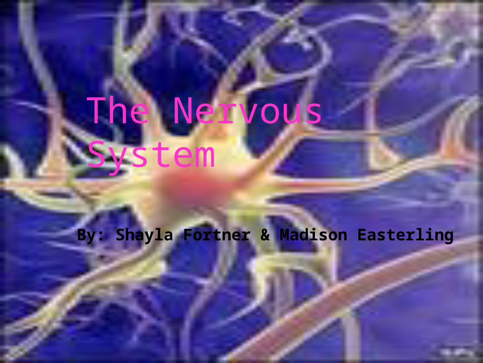
By: Shayla Fortner & Madison Easterling
The Nervous System
The nervous system controls and coordinates functions throughout the body and responds to internal and external stimuli.
The messages carried by the nervous system are electrical signals called impulses. The cells that transmit these impulses are called neurons.
Neurons can be classified into 3 types according to the direction in which an impulse travels.
Motor neurons- carry impulses from the brain and spinal cord to muscles and glands.
Interneurons- connect sensory and motor neruons.
Sensory neurons- carry impulses from sense organs to spinal cord and brain.
Dendrites are short, branched extensions from the cell body.
The largest part of a typical neuron is the cell body.
Axon- a long fiber that carries impulses away from cell body.
In some neurons the axon is surrounded by an insulating membrane know as the myelin sheath.
The electrical charge across the cell membrane of a neuron in its resting state is known as the resting potential.
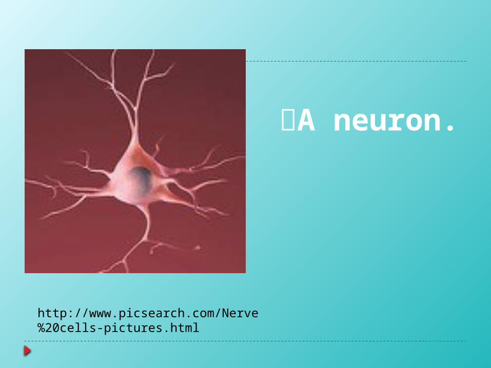
A neuron.
http://www.picsearch.com/Nerve%20cells-pictures.html
Reversal of charges from negative to positive, is called a nerve impulse or an action potential.
The location at which a neuron can transfer an impulse to another cell is called a synapse.
Neurotransmitters- chemicals used by a neuron to transmit an impulse across a synapse to another cell.
Meninges- both the brain and spinal cord are wrapped in 3 layers of connective tissue.
Cerebrospinal fluid- bathes the brain and spinal cord and acts as a shock absorber that protects the central nervous system.
The central nervous system relays messages processes information and analyze information.
Cerebrum- the largest and most prominent region of the human body.
Cerebellum- the second largest region of the brain.
Brain stem- connects the brain and spinal cord.
Thalamus- receives messages from all of the sensory receptors throughout the body and then relays the information to the proper region of the cerebrum for further processing.
Hypothalamus- the control center for recognition and analysis of hunger, thirst, fatigue, anger and body temperature.
Reflex- a quick automatic response to a stimulus.
The sensory division of the peripheral nervous system transmits impulses from sense organs to the central nervous system. The motor division transmits impulses from the central nervous systems to the muscles and glands.
Reflex Arc- the pathway that an impulse travels from your foot back to your leg; a reflex arc includes a sensory neuron, motor neuron, and effecter.
Sensory receptors- react to a specific stimulus such as light or sound by sending impulses to other neurons and eventually to be the central nervous system.
There are five categories of sensory receptors: pain receptors, thermo receptors, mechano-receptors, chemoreceptor's, and photo receptors.
In the middle of the Iris is a small opening called the pupil.
Behind the Iris is the lens. The lens focuses light onto the retina. Rods are extremely sensitive to light, but they
do not distinguish color. Cones- less sensitive than rods, but they do
respond to light of different colors, producing color vision.
Vibrations of the oval window create pressure waves in the fluid-filled cochlea.
The three canals are called semicircular canals because they each form a half circle.
The sense organs that detect taste are taste buds.
A drug is any substance, other than food, that changes the structure or function of the body.
A number of drugs, called stimulants, increase the actions regulated by nervous system.
Depressants decrease the rate of function regulate by brain.
Fetal alcohol syndrome- a group of birth defects caused by the effects of alcohol on the fetus.
Drug abuse- defined as the intentional misuse of any drug for nonmedical purposes.
Addiction- uncontrollable dependence on a drug.
All information came from the book.
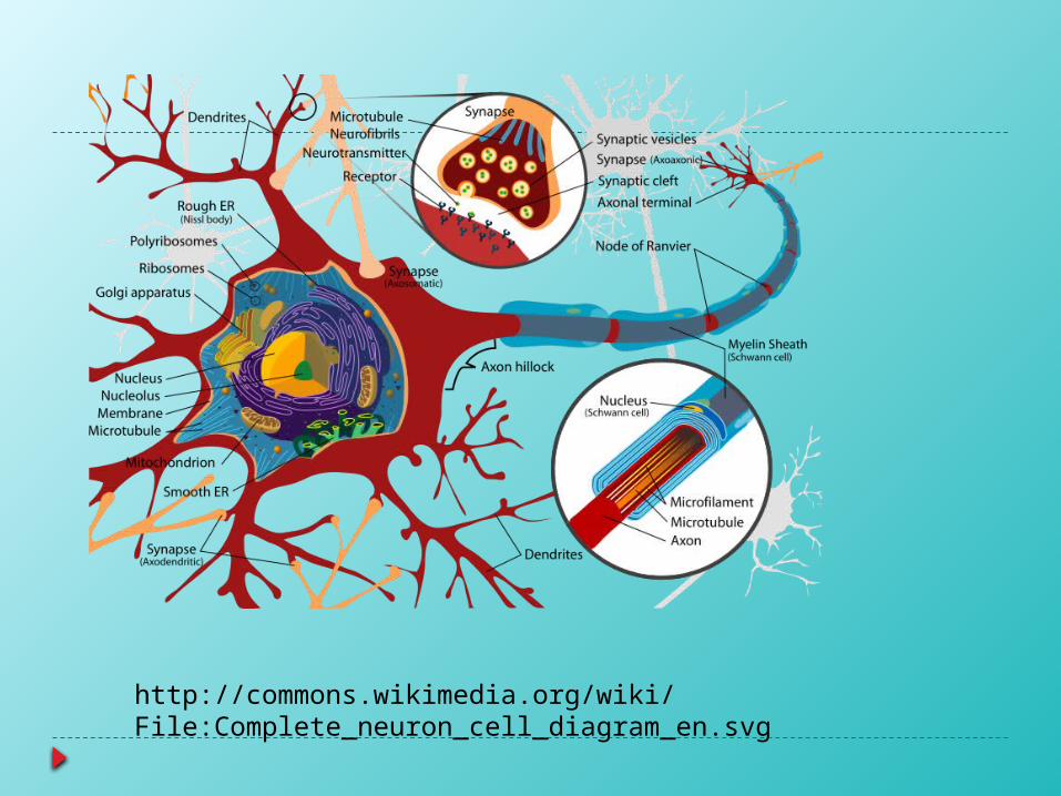
http://commons.wikimedia.org/wiki/File:Complete_neuron_cell_diagram_en.svg