Embed Size (px)
Citation preview
('
(
Quantitative Analysis of Functj onal Knee
Appliances in Controlling Anterior
Cruciate Ligament Deficient Knees
By
Monica Kosiuk
Research Project Submi tted ta the
Faculty of Graduate Studies in Partial
Fulfillment of the Requirements for
The Degree of Masters of Science (Education)
Department of Physical Education
Di V is1011 of Graduate Studies and Research
Facul ty of Education
McGill University
Montreal, Quebec.
September 1990
----------------
(
(
(
ABSTRACT
The purpose of this investigation was to evaluate and
compare the efficacy of three functional knee braces in
stabilizing anterior cruciate ligament (ACL) deficient
knees. Brace effectiveness was established as the ability
to control joint range of motion during active and dynamic
activity. The subject sample consisted of eighteen males
and females with a unilateral ACL deficiency.
This study consisted of two parts; the first involving
twenty-four braced knees. A completely randomized design
involving one independent variable with three leveis was
used. The second part consisted of three subjects, eâch
having aIl three types of braces. A randomized block design
with repeated measures was used, with each subject acting ao
a block.
The criterion variables consisted of the ability of
each brace in controlling internaI rotation and knee
extension during active movernent and knee extension during a
high velocity activity (dynamic task). Finally, total
displacement of the knee brace during a running test was
also evaluated.
Analysis consisted of a one-way Anova for each of the
criterion variables. The results of the first part of the
study demonstrated significant differences (p<.OS) between
the efficacy of the three braces for control of knee
extension during active movernent , knee extension during a
dynamic task and brace migration during a running task.
i

1 There was no significant difference between the efficacy of
the three braces in controlling internaI rotation during
active movement. However, the second part of the study
demonstrated significant differences among braces in brace
migration only (p<.05).
ii
r
RESUME
Cette etude a pour but d'evaluer et de comparer l'efficacite de
trois ortheses fonctionelles pour le genou offrant plus de stabilite aux
dechirures de ligament croise anterieur (LCAl. L'efficacite d'une orthe se
se definit par sa capacite a controler l'amplitude articulaire durant 11&1
mouvement actif et dynamique. L'echantillonnage de sujets c.ompte
dix-huit hommes et femmes avec une difference unilaterale du croise
anterieur (LeA).
Cette etude se divise en deux parties, la premiere implique
vingt-quatre genoux avec orthese. Une situation entierement au
hasard utilisant une variable Independante avec trois niveaux. En
deuxieme lieu, chacun des trois sujets portera trois ortheses differenteE.
liA randomized block design" avec mesures repetees est utilise avec
c.haque sujet agissant comme un bloc.
Les variables de criteres consistent en la capacite de chaque
orthe se a controler la rotation interne, et l'extension du genou durant
un mouvement act-if et extension du genou pendant une activite a haute
velocite (manœuvre dynamique). Finalement, le deplacement total
du genou a aussi ete evalue lors de la course.
iii
RESUME (continued)
L'analyse consiste en un sens "anova" pour chaque variable
choisie. Les resultats de la premiere section de l'etude demontre une
difference significative (p < .05) entre l'efficaclte de trois ortheses pour
chacun des criteres evalues a l'exception de la rotation interne.
Toutefois la deuxieme partie de l'etude demontre des differences
significatives parmi les orthe ses a l'epreuve, concernant la migration
de l 'orthe se , seulement (p < . 0 5) •
lv
, f
ACKNOWLEDGEMENTS
This project would never have reached a successful
completion without the help and support of several
individuals.
First, 1 would like to thank J. E. Hangers, Medicus and
B.B.G. trace companies for their co-operation in providing
both the knee braces, as weIl as the subjects for this
study. 1 would also like to thank Dr. E. Lenczner for his
co-operation in recruiting subjects as weIl as offering his
expert advice during the early stages of this project.
To my co-workers w~o were always patient when l became
preoccupied with this study and more than understanding when
l developed SUMS disease (Sudden Ugly Mood Swings) when my
rescarch ran into sudden snags.
To my friend Mary Ann Dalzell who first suggested that
l embark on this project (I do forgive her), for her moral
support and understanding when l could not give 100% of my
attention to our business. AIso, for her constant example
of perfection and over achieving, l will do weIl to try to
follow in her footsteps.
To my parents and brothers for their support and
understanding especially when 1 was forced to abandon
certain family duties. To my cousin David and Uncle Warren
for their help in the final typing and printing of this
manuscript.
Most importantly, l am grateful to my co-graduate
students who were always there when things seemed almost
v
insurmountable. To Ron Tycherniak for his help in typing
thi.s manuscript. To Andrew Mullins for his technical advice
and friendship. Also, tu Michael Simmmonds and Sonya
Mathews, long after the pages of this thesis have worn away
and yellowed around the edges, l will be left with the fond
memories of this time spent together.
Finally, l would like to thank Dr. T. B. Hoshizaki for
his support and advice throughout this project. Not only
was he patient with my lack of literary genius but also with
my somewhat feeble attempts at athletic endeavours. He
taught me not only about biomechanics but a1so about life
and for this l am forever grateful.
vi
1
\
TABLE OF CONTENTS
Page ABSTRACT . . . ... · . . . ..... · .. .• 1 RESUME .•.••.. ACKNOWLEDGEMENTS TABLE OF CONTENTS
... · . . . . . . . . ~ . · ........... . ii1 ....... · . · .... • v . . . . · ..... . . . .. · ............ . vii
LIST OF TABLES LIST OF FIGURES
..... .ix
Chapter 1 ...•.. . . . -..... . · ........ . ...
· ... •• ' x
. •• 1
INTRODUCTION • • f' ••••• · ..... . · . . · . . . . . . . . . . . . ~
• • • • • • • •• .a.
1.1 Nat.ure and Scope of the Problem · ... · ....... ., 2
1.2
1.3
1.4
1.5
1.6
'Jbjectives .•...•••••••.• • • • • · . . · ..... . • • • • • • • •• 7
Criterion Variables .•...
H~·pothes~s • · .... Limi tat.ions ...•••••••.
Delimi~ations ..•••••••
· . . · .. · .. • •••••
· . . · . .
• • · ... · . . . ... · ...
· .. · ..... · ...... . • •• · ..... . · . . . . . . .
Chapter 2 .. ~ ............ . · ..... . · . . · ..... . · .. • • • • • REVIEW OF LITERATURE ........ · . . . .. . . .
2.1 Introduction ....••. · . . · . . · .... • • • • • • • • 2.2 Characteristics of an ACL Deficient Knee · . . . . . . .
2.2.1 Anterior Translation 2.2.2 Internal Rotation .••• 2.2.3 Extens ~.on •••.••..••••••.
· . . . · .......... . · ..... . · . . . · ..... . · .. . ..
2.3 Characteristics of a Functional Knee Orthosis •••
2.3.1 Control of Ar.terior Translation ..•••••••••
2.4
Internal Rotation •••••••••• 2.3.2 Limitations of 2.3.3 Limitations of Knee Extension .•• · . . . . . . . 2.3.4 Review of Three Functional 2.3.5 Brace Fit and Design
Knee Braces
· .. · .. 3race 2.4.1 2.4.2
2.4.3
Studies .................................. .
Static Testing ..••••• · . . · . . . . . . . . . . . . . . . . . . . . . . . . · .. . . · . . . · . Dynamic Testing
Brace Fit Tests . . . . · ... . . . . . · ..... . 2.4.4 Surnmary · .. · ..... . · . . . . .
Chapter 3 .•... .. . . . . . · . METHODOLOGY . . . . . . . . • • . . . . . · ... . .. · ..... . · .
3.1 Introduction ......................
vii
9
10
11
12
13
13
13
14
14 15
16
18
19
19 20
21
22
24
24
26
29
31
33
33
33
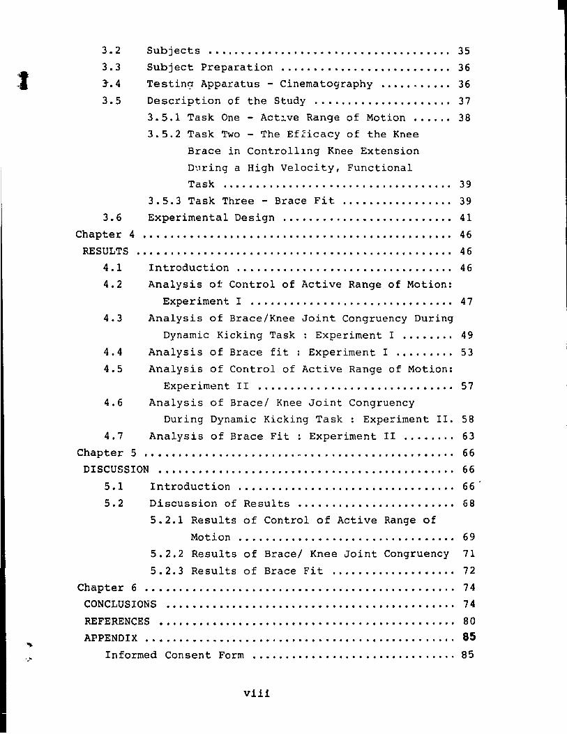
t 3.2
3.3
3".4
Subjects ......... Il ..................................................... 35
Subject Preparation •....•••......•..•.••••.•• 36
Testin~ Apparatus - Cinernatography •.••• " •.••. 36
3.5 Description of the Study .•....••.•.••.•••.... 37
3.5.1 Task One - Act~ve Range of Motion .••... 38
3.5.2 Task Two - The EfZicacy of the Knee
3.6
Chapter 4
RESULTS
4.1
4.2
4.3
4.4
4.5
4.6
4.7
Chapter 5
Brace in Control11ng Knee Extension
During a High Velocity, Functional Task ..•.••......•...•...•.....•.•••.... 39
3.5.3 Task Three - Brace Fit .•••••.•......... 39
Experimental Design .•.....••..••.•...••.••...
.............................................................................................. ................................................................................................
Introduction ................................................................ ..
Analysis ot Control of Active Range of Motion:
41
46
46
46
Experiment l ............................... "........................... 47
Analysis of Brace/Knee Joint Congruency During
Dynamic Kicking Task : Experirnent l •..••..• 49
Analysis of Brace fit: Experirnent l .•...•... 53
Analysis of Control of Active Range of Motion:
Experirnent II ............................... 57
Analysis of Brace/ Knee Joint Congruency
During Dynarnic Kicking Task : Experirnent II. SB
Analysis of Brace Fit: Experirnent II ..••.... 63
.............................................. ," ............................................ .. DISCUSSION ...................... Il ................................................................ ..
66
66
5.1 Introduction. . . . . . . . . . . . . . . . . . . . . . . . . . . . . . . .. 66
5.2 Discussion of Results •••••....••...••••••...• 68
Chapter 6
5.2.1 Results of Control of Active Range of
Motion ...................... *' • • • • • • • • •• 69
5.2.2 Results of Brace/ Knee Joint Congruency
5.2.3 Results of Brace Fit ....•.••..•••.••..•
...................................... " ....... .
71
72
74
CONCLUSIONS •••••••• ~ . . . • . . • . • . . . . • • • . . . • • . • • . • . . • . • . . .. 74
REFERENCES ••••••••••••••• *' • • • • • • • • • • • • • • • • • • • • • • • • • • • •• 80
APPENDIX ..•...............• ft • • • • • • • • • • • • • • • • • • • • • • • • • •• 85 Inforrned Consent Forrn •.•....•.•.•....••...•.••...... 85
viii
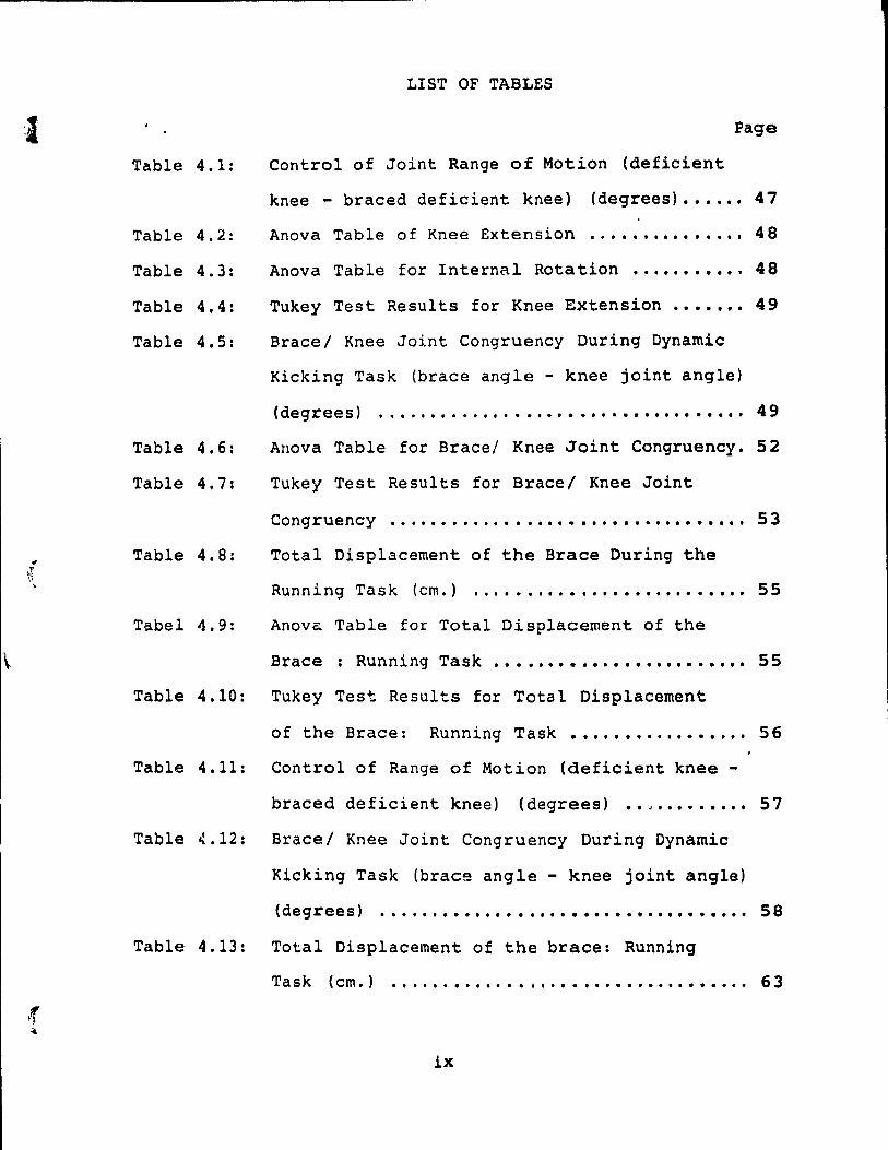
Table 4.1:
Table 4.2:
Table 4.3:
Table 4.4:
Table 4.5:
Table 4.6:
Table 4.7:
Table 4.8:
Tabel 4.9:
LIST OF TABLES
Page
Control of Joint Range of Motion (deficient
knee - braced deficient knee) (degrees) •••••• 47
Anova Table of Knee Extension ............... 48
Anova Table for InternaI Rotation .......... " 48
Tukey Test Results for Knee Extension ....... 49
Bracel Knee Joint Congruency During Dynamic
Kicking Task (brace angle - knee joint angle)
(degrees) .................................... 49
Auova Table for Brace/ Knee Joint Congruency. 52
Tukey Test Results for Brace/ Knee Joint
Congruency .................................. 53
Total Displacement of the Brace During the
Running Task (cm.) ........ ~ ................ . 55
Anova Table for Total Displacement of the
Brace : Running Task .••••••••••••••••••••••• 55
Table 4.10: Tukey Test Results for Total Displacement
of the Brace: Running Task ••••••••••••••••• 56
Table 4.11: Control of Range of Motion (deficient knee -
braced deficient knee) (degrees) •• , ••••••••• 57
Table 4.12: Brace/ Knee Joint Congruency During Dynamic
Kicking Task (brace angle - knee joint angle)
(degrees) ..................•...•......•.•... 58
Table 4.13: Total Displacement of the brace: Running
Task (cm.) ......•....••...••.•.••••••.••••.• 63
lx
1
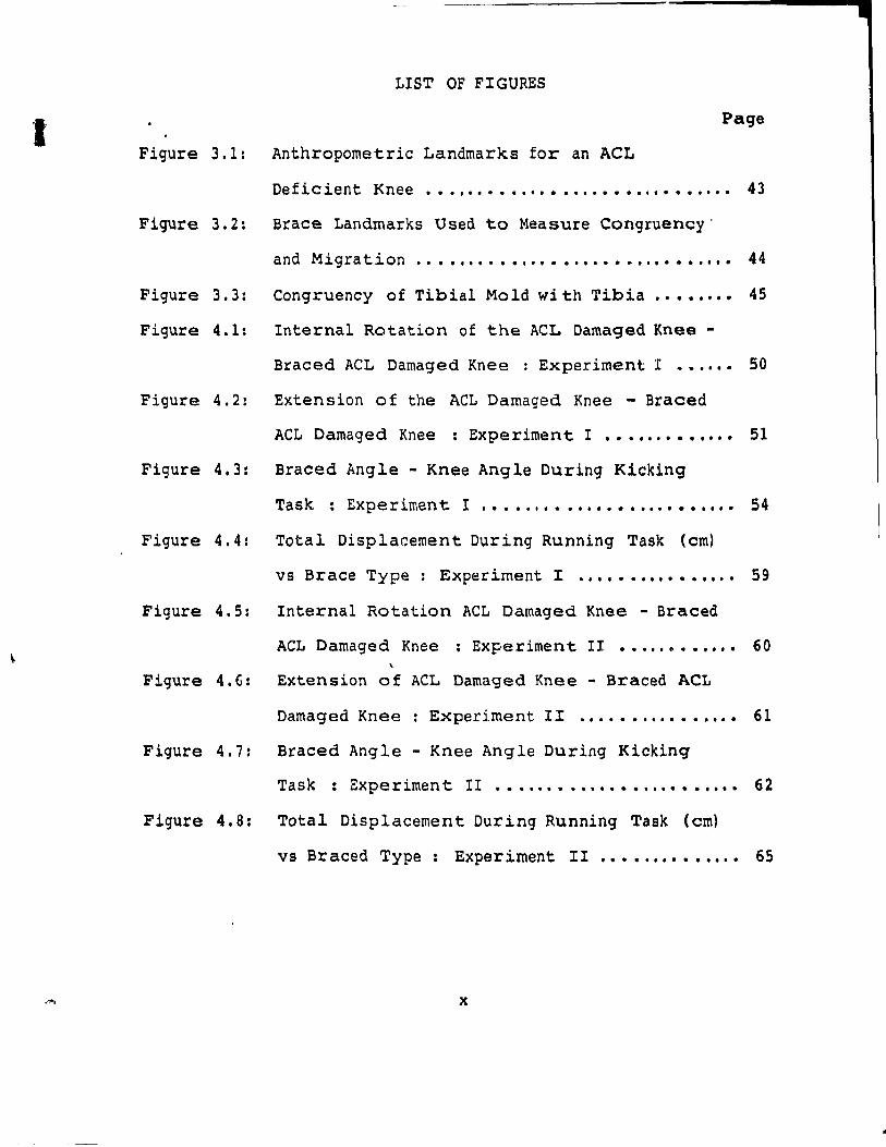
LIST OF FIGURES
Page
Figure 3.1: Anthropometrie Landmarks for an ACL
Deficient Knee .•. ,.......................... 43
Figure 3.2: Srace Landmarks Used to Measure Congruency'
and Migration .. . . . . . . . . . . . • . . . . . • • . . . . • . . . .. 44
Figure 3.3: Congruency of Tibial Mold wi th Tibia •• • • •••• 45
Figure 4.1: Interna1 Rotation of the ACL Damaged Knee -
Braced ACL Damaged Knee : Experiment l ••..•• 50
Figure 4.2: Extension of the ACL Damaged Knee - Braced
ACL Damaged Knee : Experiment l ••••••••••••• 51
Figure 4.3: Braced Angle - Knee Ang le During Kicking
Task : Experiment l .•••.•••••••••••••••••••• 54
Figure 4.4: Total Displacement During Running Task (cm)
vs Brace Type : Experiment l .•••••••••••••.• 59
Figure 4.5: Internal Rotation ACL Damaged Knee - Braced
ACL Damaged Knee ; Experimen t II ••••.••••••• 60
Figure 4. G: Extension of ACL Damaged Knee - Braced ACt
Damaged Knee : Experiment II ...............• 61
Figure 4.7: Braced Angle - Knee Angle Duritlg Kicking
Task : Experiment II •••••••••••••••••••••••• 62
Figure 4.8: Total Displacement During Running Task (cm)
vs Braced Type : Experiment II •••••••••••••• 65
x
Chapter 1
INTRODUCTION
One of the most common injuries in sports today is a
lear of the anterior cruciate ligament of the knee joint.
Not only is this considered a serious in jury but it can also
be a long term debilitating in jury ( Noyes, McGinniss, & Mooar,
1984). Athletes who sustain this type of injury are faced
with the choice of either having the injury surgically repaired
or following a conservative method of treatment. The non-
-surgical approach involves rehabilitative therapy and often
the use of a functiona1 knee orthosis ls recommended in hopes
that the athlete can return to their respective sport with
less risk of re-in jury. However, controversy exists concerning
the usefulness and effectiveness of knee braces. In 1987,
the American Academy of Orthopaedic Surgeons ( Akeson, Frank,
Amiel, & Woo, 1985) issued a policy statement indicatinq that
functiona1 knee braces aid in the control of unstable knees
by 1imiting anterior translation of the tibia on the femur.
It was aiso indicated that under high forces this mechanism
of control was no longer valid Akeson et al., 1985). And
yet, others~ldies suqgest that knee braces do play a role
in stabilizinq ACL damaged knees during dynamic activity
( Coughlin, Oliver, & Berretta 19867 Colville, Lee, Ciullo,
19867 Nocholas, 1983).
To date very little research has been reported
concerning the evaluation of the efficacy of a knee orthosis
during dynamic activity. The absence of an objective method
1
2
of identifying whether a brace is in fact effective has . pr~ved a dilemma to physicians, trainers and athletea who
are le ft questioning whether the brace they are using 1a
, going to protect the knee from further in jury. Research in
this area is necessary in order to improve existing orthotic
technology and design. Therefore, the purpose of thia
research was to determine which characteristics of the brace
design were important 1n providing knee stability dur1ng
active and dynamic movement and how th!s mechanism of
control was established. Once the criteria for measuring
eff!cacy was determined it was then used as a basis for
comparison of three commerc!ally available functional knee
braces.
1.1 Nature and Scope of the Problem
Functional knee braces are frequently prescribed with
the goal to improve stability of an anterior cruciate
ligament (ACL) defic!ent knee and to prevent re-in jury of
that sarne knee. Knee braces have been evaluated under
conditions of low passive loads (Bassett & Flemming, 1983;
Hoffman, Wyatt, Bourne, & Daniels, 1984; Knutzen, Bates, ,
Hamill, 1984) w~ere they have been shown to be effective in
controlling anterior translation of the tibia on the femur
and internal rotation, two rnovements which render the ACI.
deficient knee unstable. The (Akeson et al., 1985) has
stated that th!s control mechanism does in fact help
stabilize the knee joint during passive, low load activity.
However, funct!onal knee braces were designed to work during
•
(
(
3
dynamic activity. It is under these conditions that the
efficacy of the knee brace must be evaluated. The AAOS
seriously questions the ability of the knee brace to
stabilize the knee joint by controlling anterior translation
during dynamic activity. And yet, several studies indicate
that there is indeed a mechanism functioning during dynamic
activity which helps to stabilize the ACL deficient knee
(Colville, & Ciullo1986; Kennedy, Weinberg, & Wilson, 1974;
Lysholm, & Gillquist, 1982). This study postulates that the
key element for effective stabilization during dynamic
activity ls control of joint range of motion and that the
effectiveness of this mechanism is dependent on the actual
fit of the brace.
The proposed theory of gaining knee stability by
controlling range of motion is based on the fact that the
ACL deficient knee is rendered more uns table when placed in
positions of extreme internal rotation where excessive
stretch is placed on the ligament and knee extension where
the tibia is more prone to subluxate anteriorly and then
relocate ( Knutzen et al., 1984; Hoffman et al., 1984;
Bassett & Flemming, 1983). This later phenomenon is known
as the pivot shift and is prominent in the last few degrees
of knee extension due to the strong pull of the quadriceps
muscle coupled with the poor counter pull of the hamstrings
muscle group (Noye~ et al., 1984; Solomonow, Baralta, & D'
Ambrosia, 1989; Gurtler,Stine,& Torg, 1987; Fetto &
Marshall, 1979).
.'
4
Since dynamic activity often involves the transfer of
high magnitude forces through the knee joint (Vardaxis,
1988) the mechanism of controlling anterior translation
directly becomes ineffective (Beek, Drez, Young, Cannon, &
Stone, 1986; Bassett & Fleming, 1983). However, by limiting
the extent of ~nternal rotation in the knee joint and by
preventing full knee extension, a secondary method of
stabilizing the knee joint can be established and this
mechanism can provide much needed stability during dynamic
activity.
This study investigated the functional characteristics
of three knee braces using activ~ and dynamic tests. This
research was based on the following two premises:
1) In order to determine and evaluate the function of the
knee brace the characteristics of an ACL deficient knee must
be understood. The following are characteristics of a
damaged ACL:
a) an increase in anterior translation of the
tibia on the femur.
b) an increase in internaI rotation of the tibia
on the femur.
c) an increase in knee instability as the knee
joint approaches full knee extension ( Ahmed, Hyder,
Burke, & Chan ,1987; Lipke, Janecki, Nelson, McLeod,
T!lompson, Thompson, & Haynes, 1981; Daniel, Malcolm,
Loose, Stone, Sachs, & Burks, 1985; Markolf , Shapiro,
Gorek,& Kalo, 1990).
5
In vitro studies by Ahmed (1987), Lipke (1981) and
Fukubayashi (1982) aIl demonstrate that the primary function
of the ACL is to control anterior translation and internaI
rotation of the tibia on the femur. Subsequently, damage to
the ACL results in an increase in anterior translation and
internaI rotation. The increased laxity renders the knee
joint uns table resulting in an Interference of the transfer
of energy from the tibia to the femur during dynamic
activity (Marquette, 1988).
Ahmed (1987) and Markolf (1976) both stated that the
combinat ion of translation and rotation seen in ACL
deficient knees result in a medial shift in the transverse
axis of the knee joint causing the lateral tibial plateau to
subluxate. This produces the commonly described sensation
of the knee " giving way " Both Markolf (1976, 1990) and
Ahmed (1987) stated that this mechanism of instability is
most prevalent in the last degrees of knee extension.
2) Functional knee braces were designed to limit the
deficiencies of an ACL injured knee using the following
structural devices:
a) a four point pressure system formed by the
straps and moldings of the brace to limit anterior
translation (Marquette, 1988).
b) a tibial strap, bar or mold which by nature
of its shape and approximation to the tibia limits
internaI rotation of the tibia on the femur (Coughlin
et al., 1986iNicholas, 1983).
6
c) an extension stopper mechanism in the brace
hinges and a posterior popliteal strap to limit full
knee extension (Marquette, 1988).
In summary the efficacy of the functional knee brace in
limiting ACL deficiencies is dependent on two proposed
factors:
a) The ability of the brace to control range of
motion of the knee joint (specifically internaI
rotation and extension) during active and dynamic
movement.
b) The general fit of the brace as measured by
the amount of displacement of the brace during dynamic
activity (Lew, Patrnchak, Lewis, &Scmidt, 1982;
Coughlin et al., 1986; Walker, Kurosawa, Rovick, &
Zimmerman, 1985; Shafer, Russ,Patrnchak 1 & Tarr ,
1988).
The orthosis must be anchored to the skin adequately 50
that the limitations imposed on the dynamics of the brace as
designed by the manufacturers ( ego extension stopper) can
be transmitted to the skeletal system resulting in control
of the femur on the tibia. Lew (1982) suggested that the
displacement of the knee brace cou Id disrupt the normal
phase of knee motion and could impede the actual function of
the knee brace which is, to stabilize the knee joint. Lew
also stated that the optimum position of the hinge of the
brace is when it is aligned as weIl as possible with the
axis of rotation of tha knee joint. Displacement of the
L
hinge from this position will impede the function of the
brace. Walker (1985) and Shafer. (1988) supported these
findings and concluded that the function of the brace hinge
was to account for axial rotation and anterior/posterior
translation of the knee joint. A~y migration of the hinge
would render the brace less effective.
7
Thus, it can be postulated, knee appliances are in part
designed to provide protection to the knee joint by
controlling the range of motion of the knee. In doing 50
the brace prevents the joint from applying excessive forces
to the deficient ligament by preventing the knee from
attaining a weak or unstable posture. Furthermore, the
effectiveness of the brace is enhanced by proper fit.
The purpose, therefore, of this investigation was te
establish whether three different yet very popular
functional knee braces were effective in controlling the
range of motion of the knee joint under active and dynamic
conditions and if this was related to the general fit of the
brace.
1.2 Objectives
Criterion variables most likely to accurately evaluate
the efficacy of the knee orthosis can be determined by
considering the factors which reflect the function of the
ACL as weIl as the factors which characterize knee orthoses.
The knee brace is designed to control increased internaI
rotation characteristics of ACL deficient knees. Therefore
the difference between internaI rotation of the ACL
1 8
deficient knee and actIve internaI rotation of the braced
ACL deficient knee refiect the efficacy of the knee orthosis
in controlling range of motion. Secondly, ~~e knee braces
being evaluated in this study aIl have extension stoppers
built into their hinge joints to prevent the knee from going
into full extension. In ACL deficient knees this is a
position of extreme vulnerability since the tibia is more
likely to subluxate or shift during this part of the range
and thus the knee is more unstable and more prone to ln jury.
The difference between active extension of the ACL deficient
knee and active extension of the braced ACL deficient knee
was used as a criterion variable to evaluate the efficacy of
the knee orthosis in controll~ng range of motion. In
addition, a functional evaluation of this important
characteristic of knee braces was performed using a dynamic
task. The subject was asked to perform an instep straight
soccer kick with the ACL braced deficient knee. The
difference between the knee joint angle and the brace angle
(brace/ joint congruency) was measured to evaluate the
efficacy of the orthosis in preventing knee extension during
high velocity dynamic activity.
Finally, the precise fit of the knee brace in relation
to the actual knee joint was evaluated. The literature
strongly supports the fact that the fit of the bracp. has a
direct effect on the function of the orthosis ( Shafer et
al., 1988; Walker et al., 1985). The evaluation of the
function of the knee brace determines the efficacy of that
----------------------------------------------------.
9
knee brace in ultimately providing stability to the ACL
deficient knee. Therfore, no matter how weIl designeg a
brace is, no matter how effective it is in controlling the
range of motion, if it does not fit properly it will prove
to be ineffective during dynamic activity.
Two different measurements were used to evaluate brace
fit. First, the total amount of displacement of the brace
hinge during a ten minute run on the treadmill was evaluated
and used as a criterion variable. Second, the distance
between the tibial mold and the tibia was measured. The
purpose of the plastic mold on the tibia is to control
internaI rotation and tibial translation and thus it must be
in close contact with the tibia throughout the range of
{ motion of the knee joint. In order to evaluate this as a
component of brace fit and use it as a criterion variable
the following three measurements were taken: the distance
between the brace and the center of the medial tibial
plateau, the distance between the brace and the center of
the patellar tendon and the distance between the b~ace and
the center of the lateral tibial plateau. These measurements
were taken at four different flexicn angles (15, 30, 60 & 90
degrees) in order to reflect the fit of the brace throughout
the range of motion of the knee joint.
1.3 Criterion Variables
The following criterion variables were chosen to
r determine the efficacy of a functional knee brace during
'. dynamic activity:
---------------------~ ~-----
,.. ..
10
Brace function / control of range of motion:
1) Control of internal rotation = (active
internal rotation of the ACL deficient knee) - ( active
internal rotation of the braced knee).
2) Control of extension = (active extension of
the ACL deficient knee) - ( active extension of the
braced knee).
3) Brace/ knee joiI,;" congruency = relative brace
angle - relative knee angle during dynamic activity.
Brace fit:
1) Total brace displacement during the running
task = (vertical displacement of the hinge) +
(horizontal displacement of the hinge).
2) Approximation of the tibial mold = (medj~l
distance) + (central distance) + (lateral distance).
1.4 Hypotheses
The hypotheses for this study are as follows:
1) There will be no significant differences
among the values for internal rotation control for all
three braces.
2) There will be no significant difference among
the values for extension control for all three braces.
3) Tnere will be no significant difference among
the values for Brace/ knee joint congruency for all
three braces.
~- --------------------------------------
1
(
Il
4) There will be no significant difference arnong
the values for brace displacement during the running
task for aIl three braces.
1. 5 Limitations
Even though aIl precautions were followed to decrease
error in this study certain limitations still exist.
1) Inherent errors due to the use of
cinematography data (camera movement, lens distortion).
2) The effects of muscular forces on knee
stability and brace function were not taken into
account.
3) The braces were neither fitted by, nor
manufactured by the same individual for aIl cases.
4) Isolated tears of the ACL do not occur
frequently and are usually associated with damage to
secondary structures which may affect the function and
characteristics of the knee joint (Noyes et al., 1984).
The effect of muscular activity during the testing
procedure was not measured. Although, recent research by
Howel (1990) indicates that muscular activity plays an
important role in stabiliziag the knee joint, it was not th~
purpose of this study to evaluate joint stability due to
muscle activity but instead to evaluate brace function and
how this related to joint stability. Intra- subject
differences in muscular activity did not effect the
measurements taken during the testing procedure. Although
braces were not fitted by the same individual or
12
manufactured by the same person, a licensed orthetist teok
the measurements thus previdlng standardization and
consistency. Since this entire procedure occurred randomly
and was standardized there was no need to be concerned about
this variable confounding the results.
Finally, the extent of a knee in jury should not effect
the values of the criterion variables being measured in this
study sinee the controlling factor of the brace ls not
dependent on the level of in jury for the measurements ta ken
in this study. AlI subjects in this study had complete
ruptures of the ACL.
Further control of aIl the above limitations was
achieved by the fact that sorne subjects were tested with aIl
three hraees and their results were compared with the
individuai brace groups.
1.6 Delimitations
The following delimitations are found in this study:
1) Subjeets were between the ages of eighteen and thirty
five.
2) only subjects with ACL deficient knees were studied.
3) AIl injuries were a minimum of 6 months oid.
4) Only three functional knee braces were evaluated.
13
Chapter ~
REVIEW OF LITERATURE
2.1 Introduction
This chapter examines the deficiencies of an ACL
injured knee and determines how ta best control for the
dysfunction in arder ta improve joint stability.
Functional knee braces have been designed in an attempt
to 1essen th~ degree of disability found in ACL deficient
knees. The components and design of the knee orthosis act
to control the factors which cause instability in the knee
joint and which ultimately increase the potential for re-
in jury. This chapter examines the logistics behind knee
brace designs and why their control of knee range of motions
is an important determinant of brace efficacy.
Another important factor to be considered when studying
knee braces is how weIl does the brace fit the anatomical
contours of the specifie knee joint. This chapter examines
the work of several authors (Lew et al., 1982; Walker et
al., 1985; Shafer et al., 1988) who have indicated the
importance of minimizing brace migration in order ta
optimize brace function and joint stability.
Finally, a review of previous research in the areas of
static and dynamic testing are presented in order ta
establish the relevance of evaluating brace efficacy based
on joint range of motion control and brace fit.
,",
14
2.2 Characteristics of an ACL Deficient Knee
In order to truly comprehend the purpose and function
of a knee orthosis it is important to have a clear
understanding of the role of the ArL in knee stability.
Many in-vitro studies attempt to ascertain the function of
the ACL while under static or qua~i-static loading. These
studies are designed to demonstrate the biomechanical
functional characteristics of joint structures ( ligaments,
menisci and capsule) by removing the effect of each
structure one at a time and measuring the impact on joint
stability.
The ACL is a fibrous band originating from the
posterior medial surface of the lateral femoral condyle and
inserting into a wide depressed area in front of and lateral
to the anterior tibial spine. When the knee is extended the
ACL is fIat and when the knee is flexed the ACL twists on
itself and the posterior cruciate ligament (peL). More
specifically the ACL is comprised of three separate
sections: the anterior fibres which are taut in extension,
the medial fibres which are taut in internaI rotation and
the posterior fibres which are taut in flexion ( Wang,
Walker, & Wolf, 1973; Smillie, 1970; Girgis, Marshall &
Monajem, 1975).
2.2.1 Anterior Translation
One of the functions of the ACL is to prevent anterior
translation of the tibia on the femur. Butler et al.,
(1980) demonstrated that the ACL was the primary restraint
.----------------------------------------------
{ .
against anterior translation, providing 86 % of the total
resisting force. Studies by Ahmed (1987), Lypke (1981)
Daniel (198~), and Fukubayashi (1982), aIl further
demonstrated that the primary function of the ACL is to
control anterior translation of the tibia on the femur.
Marko1f, Mensch, & Amstutz (1976) and Fetto et al., (1979)
concluded that a damaged ACL resulted in an increase in
anterior displacement especially in extension. This
15
dJsplacement in turn causes a disruption of the alignment of
the femur and tibia which Interferes with proper transfer of
energy through the knee joint resulting in further
instability, especia1ly dnring high intensity dynamic
activities ( Butler, Noyes, & Grood, 1980; Crownshield,
Pope, & Johnson, 1976). Therefore, the ability of the brace
to control or limit extension results in improved stability
and more effective function during dynamic activity.
Control of knee extension by the orthosis improves the
efficacy of that particular brace and reduces the risk of
re-in jury (Blacharski & Somerset, 1975).
2.2.2 InternaI Rotation
The second function of the ACL involves limitation of
excess internaI rotation. Pizia1i, Rastegar, Nagel, &
Schurman (1980) reported that the cruciate ligaments
resisted internaI rotation of the tibia by winding around
themselves. Rotatory motion can be related to the axis of
rotation between the femur and the tibia and the movements
generated about this axis by the 1igamentous structures.
16
Gollehon , Torzilli, & Warren (1987) reported that the
ligaments in an intact knee acted to maintain the
equilibrium in the joint. When a ligament is damaged, the
balancing forces exerted by the remaining structures will
change as the knee moves to a new position of equilibrium to
compensate for the loss. Lipke et al., (1981) and Kennedy &
Fowler (1971) support the idea that the function of the ACL
is to prevent internaI rotation of the tibia on tne femur.
Lipke et al., (1981) stated that the role of the ACL is to
maintain the position of the center of rotation of the tibia
in the transverse plane. If there is damage to the ACL the
axis will shift medially, and under these conditions,
excessive internaI rotation will result. A medial shift of
the axis will provide a longer lever arm for the applied
rotary forces from the lateral collateral ligament resulting
in anterior lateral subluxation of the lateral tibial
plateau and increased internaI rotation. Noyes et al.,
(1984) described the phenomenon as a ("pivot shift")
subluxation of _he tibia which can occur during static
testing (pivot shift test) and is often reproduced in
dynamic setting during movements involving jumping, cutting
and deceleration.
!.. 2 .3 Extension
The position of full knee extension becomes a position
of increased instability in an ACL deficient knee. Ahmed et
al., (1986) stated that with an increase in knee extension
there is an increase in anterior displacement of the tibia
{
1 _~
17
on the femur. Markolf et al., (1976) also demonstrated that
a damaged ACL resulted in an increase in anterior
displacement especially in extension. Cabaud & Rodkey
(1985) stated that the ACL has a unique function in
providing stability as the knee joint extends, allowing the
tibial plateau to track on the longer medial femoral
condyle, thus forcing the tibia into external rotation with
a screw-home mechanism. A deficient ACL knee will alter the
posi tion of the tibial plateau resulting in increased
internaI rotation and decreased stabili ty.
The previously described phenomenon of the "pi vot
shift" has been biomechanically explained by the fact that
upon heel strike (knee extension) during a strong quadriceps
contraction with the knee in extension, the quadriceps
muscle pulls the tibia forward so that i t rests in a
subluxated position and when the knee is flexed between 20
and 40 degrees the iliotibial band tightens over the lateral
tibial plateau causing a reduction of the tibia. The
posl tion of knee extension 15 further compromised by the
fact that the hamstring muscles, which functlonally are
capable of Hmi ting anterior drawer, are at a rnechanical
disadvantage and "hus have little effect on the joint
stability at this point (Noyes, et al., 1984; Solomonow et
al., 1989; Gurtler et al., 1987 & Fetto et al., 1979). The
AAOS has stated that knee braces under dynamic conditions
are incapable of controlling anterior translation and thus
J, the best means of controlling the excessive movernent is by
avoiding positions where it is most prevalent.
2.3 Characteristics of a Functional Knee Orthosis
18
The purpose of a functional knee orthosis is to control
laxi ty in an uns table knee and to protect the knee from
further damage or in jury. Al though i t is imposs ible for a
brace ta mechanically substi tu te for the function of the ACL
it must compensate for the deficiencies in an ACL injured
knee ( Marquette, 1988).
During dynamic acti vi ty the purpose of the ACL is to
maintain femoral and tibial articular surfaces in correct
alignment so that joint configuration can provide stability
when loads are applied (Lipke et al., 1981; Hsieh & Walker,
1976). In an ACL deficient knee, the joint surfaces are not
aligned due to increased laxi ty and therefore a knee
orthosis which can control anterior translation and internaI
rotation of the tibia on the femur will increase the
stability of an ACL deficient knee by improving joint
alignment. Butler et al., (1980) concluded that in ACL
deficient knees secondary restraints may block clinical
laxi ty tests but in Ume they stretch out and cannot provide
stability under higher functional activity forces.
Therefore, the knee orthosis must control the laxity or
instability present in the knee for the joint to accommodate
the large forces found during dynamic acti vi ty. Passive
testing of knee laxity in a braced knee may result in the
conclusion that. a brace is effective when in fact it ls the
(
(
(
19
secondary structures of the knee that are improving the
sti ffness and not necessarily the brace. By testing the
brace under high load functional activi ties the compensation
from secondary structures can be reduced (Buttler et al. ,
1980) •
2.3. 1 Control of Anter ior Translation
The difficulty with controlling anterior translation of
the tibia on the femur arises from the fact that a small
displacement (8-10 mm) must be controlled while the knee
joint is subjected to hundreds of newtons of force
(Marquette, 1988). Brace manufacturers use a four-point
pressure system in an attempt to limit anterior translation.
This involves placing the pressure through elastic or non
elastic straps on the distal posterior femur and the
proximal anterior femur to produce one lever system and
applying pressure on the anterior proximal tibia and the
posterior distal tibia for a second lever system. The
result of this lever system is to promote the normal
geometric alignment of the tibia and femur so that forces
can be t:l.'ansferred from the tibia to the femur. In heal thy
knees this trans fer occurs in part through the function of
the ACL (Marquette, 1988; Butler et al.,; 1980).
2.3.2 Limi tations of Intarnal Rotation
A further means of improving knee stability is to
control the amount of internaI rotation occurring at the
knee joint. One of the characteristics of an ACL deficient
knee is an increase of internaI rotation of the tibia on the
t
20
femur since anatomically the ACL acts as a restraint to
intêrnal rotation (Gollehon et al., 1987). The resultinq
increase in internaI rotation of ACL deficient knees leads
to an anterior subluxation of the lateral tibial plateau
often presented as a 'pivot shi ft , or " giving way "
sensation (Lipke et al., 1981; Noyes et al., 1984) resultinq
in an increase in knee instability. By controlling internaI
rotation of the tibia on the femur, the knee brace provides
improved stability of the joint. Rotational control was
maintained in the braces evaluated in this study by close
fittinq plastic pre-tibial shell or bar (Couqhlin et al.,
1986), lateral le9 pads and derotation straps (Nicholas,
1983). FinaIly, the sliding axis of motion found in the
hinge of each brace, theoretically, corresponds tO the axis
of rotation of the knee and helps control rota tory
instabili ties.
2.3.3 Limitations of Knee Extension
The final means of improving knee stability is by
Iimiting knee extension. As previously discussed, when th~
ACL deficient knee approaches full extension the amount of
anterior translation or anterior laxity increases resulting
in joint mal-aIignment and thus further instability
(Markolf et al., 1976; Ahmed et al., 1986). The position of
knee extension is aiso the starting point for the
development of the phenomenon of the pivot shift which once
again renders the knee unstable and uitimately leads to a
Iess functional joint. Therefore, it 15 extremely important
21
( for a knee brace to prevent complete knee extension during
dynamic activity otherwise, the joint is placed in a
vulnerable, uns table position which could result in re-
in jury (Hughston & Norwood, 1980). This limitation of knee
extension is achieved first with an extension stopper which
prevents the knee from going heyond 10- 15 degrees of
extension and second, by a posterior strap which maintains
the subject's leg in the orthosis and thus prevents the
joint from fully extending.
2.3.4 Review of Three Functional Knee Braces
In this study three commercially available functional
knee braces, which are representative of the existing
market, were evaluated. These included the following: the
( A-C brace, the Lennox Hill brace and the Medicus brace.
coughlin et al., (1986) and Beretta, Charuest, Berretta, &
Berretta (1985) described the A-C brace as being a
polycentric joint with an extension stopper and a !ateral
tibia! mold to decrease abnormal tibial rotation and lateral
tibial subluxation. The A-C orthosis is made from a plaster
cast taken of the flexed leg at approximately 30 degrees and
the foot completely dorsiflexed. AlI orthoses have a 15
degree extension stopper which prevents the knee from
extending completely. The plastic pre-tibial shel! helps to
suspend the orthosis and provides a distribution of pressure
over the anterior tibia as the orthosis reaches its
( extension stop and thus prevents possible pain from tibial
impingement. Rotational control is obtained by the shape
22
and close fit of the plastic pre-tibial shell. The
posterior strap maintains the subject's leg inside the
orthosis and pre~ents the joint from extending beyond 10-15
degrees of flexion.
The Lennox Hill brace was described by Nicholas (1983)
as being designed to restrict anterior lateral instability.
If there is an increase in translatory or rota tory motion
causing the knee axis to shift into an uns table position the
brace will act to restrain the shift. Anterior translation
of the tibia is controlled in the Lennox-Hill brace by
forces created by the pre-tibial bar, the derotation strap,
the distal knee loop and the circumferential rubber band. A
hyper-extension stop prevents movement into the uns table
position of full extension. Rotatory instability is
restrained by the contours and placement of the lateral leg
pads, the medial knee disc, the circumferential rubber band
above and below the knee and the de-rotation straps.
The Medicus brace consists of an extension stopper (10-
15 degrees) to prevent full knee extension and a close
fitting tibial mold to control internaI rotation of the
tibia. A pressure system consisting of a femoral and tibial
mold as weIl as four non-elastic straps help to control
anterior translation of the tibia on the femur. The hinge
joint provides further control of rotation and extension.
2.3.5 Brace Fi~ and Design
Walker et al., (1985) and Shafer et al., (1988) stated
that one of the major problems with knee braces was that the
(
(
(
23
joints of the brace follow kinematic pathways which are
simpler than those of the natura1 knee joint. As a result
the fixed hinges do not account for important axial rotaion
and anterior-posterior translation. Thus, optimal stability
is not achieved. Polycentric hinges more clearly simu1ate
complex rolling and gliding of the knee during flexion and
extension by providing a changing center of rotation.
However, in order for the hinge to function properly it must
be in line with the center of the joint 1ine (Berretta et
al., 1985). Thus any displacement of the hinge during
dynamic activity renders the brace less effective.
Poor alignment between the orthosis and the natural
knee joint motions may cause disp1acement of the brace
during dynamic activity. Lew et al., (1982) stated that
when an orthosis ls applied to a joint conflict occurs as
the orthosis attempts to force the knee to follow a
simplified motion. Since this is impeded by joint
structures, unwanted constraint forces develop in the
suspension points of the orthosis causlng the interface
components to migrate over the 1imb segments. Sorne
constraints are beneficial when they compensate for 1acking
stability but they can become detrimenta1 if they disrupt
the normal phases of knee motion. Also, disp1acement of the
brace will Interfere with its ability to control joint range
of motion and thus make it less effective.
Not only is it important that the brace not be
displaced in a vertical or horizontal direction but also
t
, .
that the brace conform to the specifie anatomical contours
of the knee joint. In a weIl fitted brace the tibial mold
will be in close contact with the actual tibia in order to
prevent translation and rotation and the brace hinges will
align with the c~nter of rotation of the knee joint.
2.4 Brace Studies
24
There have been many studies published involving the
results of static and clinical testing of functional knee
braces but there are only a few studies which employ dynamic
testing. Although it is important that a brace functions in
a static setting it does not necessarily imply that it is
effecti,re under dynamic conditions. Functional activities
apply much greater stress on both the knee joint and the
brace (Vardaxis, 1988). In dynamic activities components
such as muscular force and joint compression loading act on
the knee joint to stabilize it. A major critic!sm of both
the clinical and in- vitro studies that have been published
is their lack of generalizability to dynamic situations
because the criterion variables used in a static situation
do not measure efficacy in a dynamic situation. Therefore,
it is important to first of aIl establish a method of
testing functional braces during dynamic activity and
secondly evaluate aIl braces both statically and
dynamically.
2.4.1 Static Testing
Knutzen et al., (1984) and Hoffman et al., (1984) both
demonstrated the ability of functional knee braces to
(
(
25
control translation of the tibia on the femur under low °
stress loads. The forces used in these studies were of the
magnitu~e of fifteen ta twenty pounds of force which is
substantially lower than the hundreds of pounds of force
which are applied to the knee joint during dynamic activity
(Marquette, 1988).
In a study by Coughlin et al., (1985) the A-C
functional knee brace was tested under static conditions
using the Genucom Knee Analysis system. The authors
reported an average of 46 % decrease in internaI rotation
with the knee brace on. Measurements were taken while
passive load was being applied to the joint.
In a study by Colville et al., (1986) the
effectiveness of the Lennox-Hill brace in treating knee
instability was both objectively and subjectively evaluated.
The brace fai1ed to significant1y reduce maximal anterior
subluxation of the tibia but did increase stiffness in the
knee joint. Rotatory instability was improved an avera~e of
one grade on the measurement scale. Patients reported a
significant decrease in episodes of "giving way" and
athletic performance was improved by using the brace in 69 ,
of the patients. The subjective results for this study were
obtained using a questionnaire while objective results for
this study were evaluated by an apparatus that measured
tibial subluxation. The authors concluded that a decrease
in symptoms of instabiloi ty when wearing the brace may be due
,....
to the ability of the brace to improve relative knee
stiffness even though maximal laxity remained unchanged.
26
In a study by Bassett and Fleming, 1983 the efficacy of
the Lennox-Hill brace in controlling anterior lateral
rotatory instability of the knee was reported. A comparison
was made of the degree of instability with and with out the
brace applied during clinical tests. The results showed
that 89 % of the knees with grade one instability and 45 %
of those with grade two instability improved. However, this
was static testing only. In the dynamic situation 70 % of
the subjects complained of episodes of "giving way" while
wearing the brace although this was not objectively
measured. It is clear that tlere 15 need for further
research ta study the adaptation of the knee brace to
functional activities in a controlled objective dynamic
situation.
2.4.2 Dynamic Testing
A review of the literature presented few studies that
address the topie of the analysis of knee braces during
dynamic activity. Knutzen, Bates, Schot, & Hamill (1987)
stated that the application of a knee orthosis significantly
alters the kinematic characteristics of the knee joint. The
influence of knee bracing was evaluated during the activity
of running by examining ground reaction forces and knee
joint parameters obtained using an electrogoniometer. Both
knee brace conditions were shown to significantly reduce
knee flexion during s~ing and support phases, as well as
(
(
(
total rotation of the tibia on the femur and total varus!
valgus movement parameters of the experimental knee joint.
The important question is whether this deviation helps to
improve the stability of the knee or whether it hinders
joint function stability.
27
Shiavi, Lirnbird, Frazer, Stivers, strauss, & Abramovitz
(1986) undertook a study designed to establish the dynamic
kinematics of ACL deficient knees during walking at
different speeds and during a pivoting movement. He found
that there was a significant tendency towards adduction and
external rotation during certain periods of the stride. If
these kinematics represent an attempt by the subject to
stabilize the ACL deficient braced knee then perhaps the
orthosis should aiso reflect ~he same limitations and
kinematics. That is, by limiting internaI rotation, the
brace can improve knee stability.
In a study by Knutzen, Bates and Hamill (1983), an
e1ectrogoniometer was used to measure dynamic range of
motion of the affected or damaged knee while using a support
knee brace and a functional derotation brace (Lennox-Hill).
The results showed a general reduction in knee flexion
during swing and support phase when the derotation brace was
app1ied. The Lennox-Hill brace also limited both internaI
and external rotation when compared with the no brace
condition and the contralateral intact 1imb. The amoun"~ of
restriction during knee extension was not studied.
• .. 28
Knutzen et al., (1984) studied the influence of support
braces and derotation braces on tibial rotation in post
surgical knees. The results suggested that the knee orthosis
tested showed a trend towards limi ting external rotation
more than internaI rotation. However, this study was
completed in a statie setting with the hip and knee at 90
degrees of flexion and thus the results though important can
not be gp.neralized to include dynamic activities. Al though
both of Knutzen 1 s studies show a trend toward limiting
external rotation it must be remembered that these studies
were do ne on post-surgical ACL deficient knees.
Van Horn, MacKinnon, Witt, & Hooker (1988) presented a
study concerning the kinematic analysis of gai t patterns
among subjects wearing the Anderson Knee Stabilizer brace,
McDavid Knee Guards and no brace. Although the braces
tested are prophylactic and not functional orthoses the
resul ts are worth mentioning. Fourteen gait variables were
measured for each brace and speed condition. Thp.re was an
increase in hip and knee flexion and knee angular velocity
wi th and wi thout braces at 8 mph as compared ta 4 mph, a
decrease in knee extens ion when ei ther brace was worn and a
minimal gai t pattern di fference wi th the Anderson Knee
Stabilizer as compared with the McDavid Knee Guard. The
results of this study demonstrated that no clear superiori ty
exists between the braces 1 effect on gai t characteristics
measured. The au thors s11gges t that other parameters should
be considered when evaluating braces. In this present study
(
(
(
29
the intention was to evaluate restrictions on knee extension
during dynamic activities.
Inoue, McGunk-Burleson, Hollis, & Woo (1987)
demonstrated that restriction to the joint motion can have a
significant influence on which ligaments are being put under
stress. The results of his study showed that when knee
motion is limited to varus-valgus rotation the medial
collateral ligament is the primary restraint ta valgus
stress but when tibial rotation and translation are allowed
the medial collateral ligament has less effect on valgus
stability and the ACL becomes more of a stabilizing factor
in the knee. It is thus possible that by limiting the
tibial rotation and anterior translation in the knee joint
by using an orthosis, the ACL deficient knee can be better
stabilized by the medial collateral ligament playing a
greater role. This would reduce the function of the ACL
resulting in less stress being placed on the already damaged
ligament.
2.4.3 Brace Fit Tests
In a dynamic situation the forces being transmitted
through the knee joi,t prevent the orthosis from providing
subtle control of anterior translation of the tibia on the
femur (Beek et al., 1986; Bassett & Fleming, 1983).
Therefore, co~trol of the range of motion of the knee must
occur if the orthosis is ta provide joint stability. The
orthosis must be anchored to the soft tissue adequately so
that the limitations imposed on the dynamics of the brace as
.;
30
designed by the manufacturers (eg. extension stopper) can be
transmitted to the skeletal system resulting in cQn~rol of
the femur and tibia interaction.
Functional instability in an ACL deficient knee usually
manifests itself as lateral tibial plateau rotatory
translation produced by the movements of extension and
internaI rotation (Lipke et al., 1981). Therefore a knee
orthosis must limit extension and internaI rotation of the
tibia on the femur if it is going to provide stability to
the damaged knee. Certain factors will influence the
effectiveness of controlling knee joint range of motion.
One such factor is the amount of movement which occurs
between the brace and the knee joint. If the brace is not
secure it will migrate during dynamic movement. This will
result in a change in position of the various components of
the brace which are necessary for the overall effective
function of the brace. This shift May impede the mechanics
of these components making the brace less effective. For
example, the purpose of the tibial mold is to control tibial
rotation. The mold is effective because of its close fit to
the tibia and because it restricts rotation at the proximal
end of the tibia where the movement originates. If the
tibial mold migrated distally it would not function at an
optimum level. Displacement of the brace May also change
the position of the popiiteai strap making it a less
effective restrictor of knee extension. Lew et al., (1982)
developed a method to quantitatively measure the relative
(
31
efficiency of knee orthoses by comparing migration during
motion. The pistoning tendency was quantified by using a
transducer which measured the portion of the orthotic
constraint force which was directed parallel to the side
bars which attached the joints to the orthotic interface
components. The results showed that there was no
significant difference in migration for different types of
knee orthoses but there was a difference between the types
of activity. The study did not establish what influence the
migration of the brace had on joint kinematics.
Van Horne et al., (1988) stated that they found no
displacement of the brace in relation to the knee joint
during the activity of running. This may explain why most
subjects when completing scoring scales for functional
activity levels state that they have little difficulty
during straight running activities (Kettlekamp & Thompson,
1975; Lysolm & Gillquist, 1982). However, manyathletes
complain of instability with the brace during high force,
high impact pivoting movements. The proposed study will use
brace fit and brace displacement as criterion variables for
comparing brace efficacy of the three braces being studied.
2.4.4 Summary
The literature supporta the concept that functional
knee braces are in part designed to protect the knee joint
and provide increased stability by controlling the range of
motion of the knee joint. Specifically, by limiting
internaI rotation and knee extension the functional
-.
32
instability seen in ACL deficient knees (je. 'giving way')
can be controlled. Thus, control of internaI rotation and
extension are used as criterion variables in this study to
evaluate the efficacy of functional knee braces.
Previous research indicates that the effectiveness of
the knee orthosis is al 50 determined by the fit of the
brace. A brace that does not fit properly is unable to
effectively,control knee joint range of motion and as a
result the stability and mechanics of the joint are
compromised. In this study brace fit was evaluated by the
criterion variables of approximation of the tibial mold and
total displacement of the brace after running.
Chapter 3
METijODOLOGY
3.1 Introduction
In llght of the literature review it is clear that
control of knee joint range of motion by the knee orthosis
is a very important factor for evaluating the efficacy of
knee braces in stabilizing the knee joint during dynamic
activity.
33
There are several factors which may affect the ability
of the knee orthosis to control knee joint motion. These
include improper fit of the orthosis as displayed by
horizontal and vertical displacement of the brace in
relation to the knee joint and excess movement of the brace
on the tibia and femur due to compression and wobbling of
soft tissue. In this study the first two factors were
evaluated. As described in section 2.3.5, as an external
support the function of a knee brace is impaired if the
brace does not fit properly. Displacement of the brace, in
either a horizontal or vertical direction changes the
alignment of the components of the brace with the knee joint
and thus May diminish the efficacy of that brace. For
example brace migration changes the position of the tibial
mold and popliteal strap rendering them less effective in
performing their functions as restrictors of joint range of
motion.
Three separate dynamic tasks were chosen as a means of
determining the functional characteristics of the three
34
braces being evaluated. From the results of these tests the
efficacy of the braces during dynamic activity were
determined.
The first task involved measuring the active range of
motion (flexion, extension, internal rotation and external
rotation) of the ACL deficient knee and the braced ACL
deficient knee. The purpose of this test was ta determine
whether the functional knee brace controls internaI rotation
and extension, two movements which can cause increased
instability in the knee joint during dynamic activity (Noyes
et al., 1984, Lipke et al. / 198!). Although this test does
not excessively load the knee it does give a clear
indication of the restrictions which the brace applies to
the joint.
In order to simulate a more dynamic task, subjects were
instructed ta perform an instep straight soccer kick
representing a high velocity, high force activity. The
purpose of the test was to determine how effective the brace
was in limiting knee extension. This was determined by
measuring the brace angle at the end of the kick just before
the knee started to flex and comparing this to the joint
angle at the same point in time (brace/knee joint
congruency). It is important to the stability of the knee
joint that the knee is limited from extending completely,
otherwise, the joint becomes malaligned (pivot shift) and
upon landing on the kicking leg there would be a greater
tendency for the knee to collapse since the force could not
35
( be properly transmitted from the tibia to the femur
(Marquette, 1988).
The final dynamic task consisted of having the subjects
jog at 4-6 MPH on a treadmill for ten minutes. The purpose
of this task was to determine brace fit in terms of the
amount of displacement occurring in the brace during dynamic
activity. Since most functional tasks involve sorne degree
of running this test was a good indicator of possible brace
migration during a running activity. As was previously
discussed, a brace can function effectively only if its
components are held in position so that they can affect the
appropriate anatomical areas of the knee joint (Lew et al.,
1982).
( A further test to determine brace fit consisted of
measuring the distance of the tibial mold to the tibia
during various points of knee flexion. This test howcver,
could only be applied to two of the braces since the third
brace, the Lennox-Hill does not have a tibial mold. A close
fitting mold is effective in controlling tibial rotations.
3.2 Subjects
The subject population for this study consisted of
eighteen individuals with a unilateral chronic isolated ACL
deficient knee as diagnosed clinically via arthroscopie
surgery. Subjects were excluded if they had one of the
following conditions: meniscal in jury, medial collateral
ligament in jury, lateral collateral ligament in jury or ACL
reconstructive surgery. Subjects were between the ages of
36
eighteen and thirty-five in order to avoid complications due
to possible arthritic conditions which may be found in an
older age group. Fifteen of the subjects had one of the
previously described functional knee braces prescribed in
order to improve knee stability. In addition, a group of
three subjects were fitted for aIl three braces and each
subject evaluated with each brace.
3.3 Subject Preparation
Each subject was required to wear shorts, a t-shirt and
running shoes during the experimental session to allow easy
viewing of the anthropometric landmarks. In order to aid in
the collection of kinematic data from the film the following
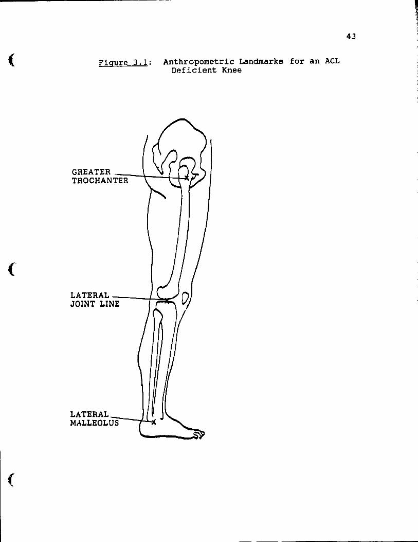
reference points of the ACL deficient knee were marked with
fluorescent tape; the greater trochanter, the lateral joint
line of the knee and the lateral malleolus (figure 3.1).
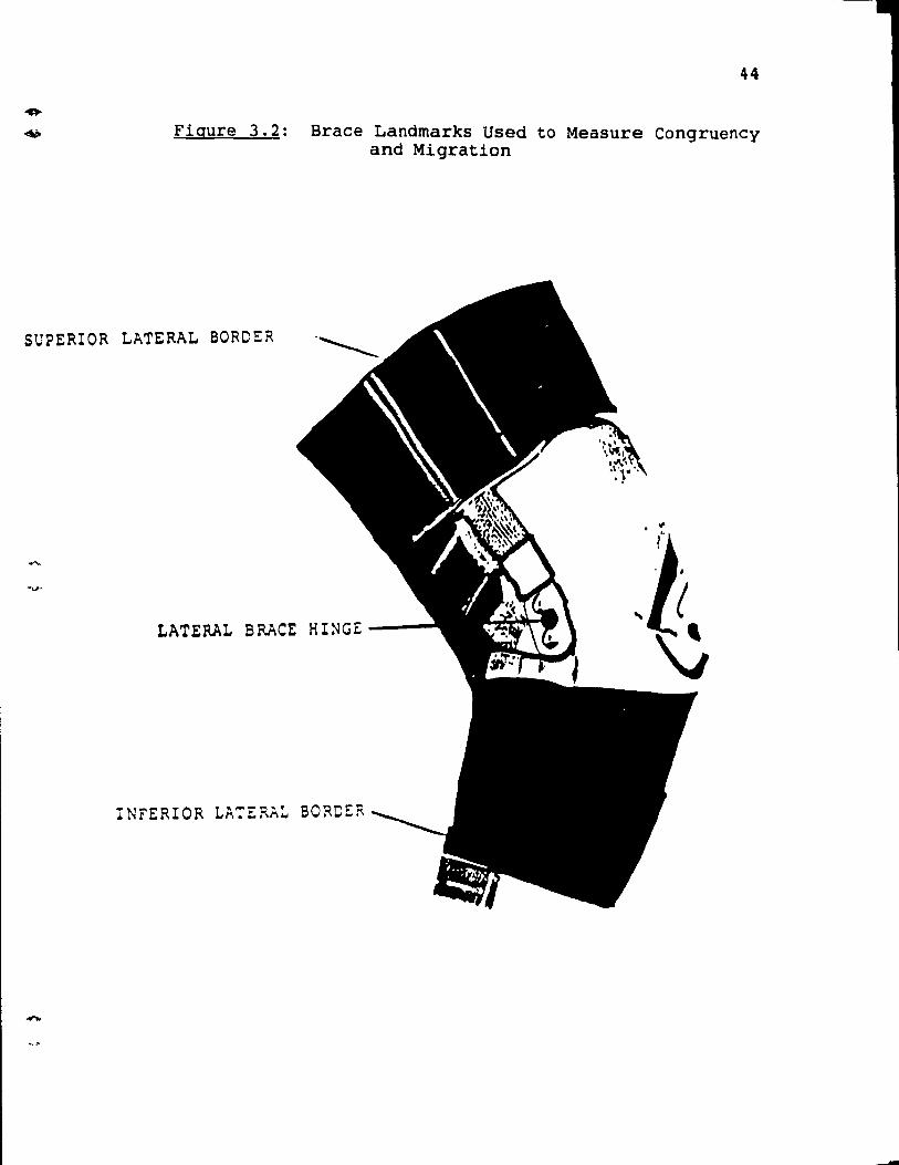
Markers wore also placed on the orthosis at the following
points: the superior lateral border, the lateral hinge and
the lateral inferior border (figure 3.2 ).
3.4 Testing Apparatus - Cinematography
The data for this study was collected using a high
speed camera set at a frequency of 30 Hz. The location of
the camera depended on two factors: the plane of action and
the area of the subject which was to be viewed. In order to
be able to digitize the movement, the filming was do ne at a
perpendicular angle to the plane of movement. For aIl the
testing procedures in this study the filming was do ne from a
side view. Once the film was processed it was displayed
(
37
frame by frame on a video digitizer so that the appropria te
relative angles could be determined.
Given the co-ordinated data from the anatomical
landmarks at either end of a limb segment the absolute
segment and joint angles were determined. In this study the
relative angle of the knee joint equaled the absolute angle
of the shank minus the absolute angle of the thigh. The
relative angle of the brace was calculated by subtracting
the absolute angle of the proximal brace segment from the
absolute angle of the distal segment.
3.5 Description of the study
The purpose of this study was to design a quantitative
method of evaluating the efficacy of functional knee braces
during active and dynamic activity. As previously
mentioned, it was established that brace efficacy can be
measured by control of joint range of motion and brace
migration. Two movements, which, when limited, help control
joint instability are, knee extension and internaI rotation.
By 1imiting these movements the brace prevents the pivot
shift phenomenon or joint malalignment from occurring and
thus improves stability and protects the joint from possible
re-in jury. It is through the actual design of the brace
that the limitation or control of joint movement is
achieved. A poor fitting brace or a brace that becomes
displaced during activity can not provide adequate support
or protection, Lew et al., (1982) alluded to the fact that
' ..
in order for a knee orthosis to be effective it must " act
as one" with the knee joint.
38
The criterion variables that were used in this study to
evaluate the efficacy of a knee orthosis were determined in
the previous two chapters. The following discussion will
explain how these variables were measured.
3.5.1 Task One = Active Range of Motion
a) Subjects were asked to actively flex and extend, as
far as possible, their ACL deficient knee with and without
the knee brace.
b) Angle measurements were obtained using a standard
goniometer where the proximal arm was aligned with the
greater trochanter, the distal arm with the lateral
malleolus and the center axis with the center of the lateral
joint line of the knee.
c) Subjects were then seated in a chair, with the hips
and knees at 90 degrees and each foot was placed on a
rotating platform one at a time. The neutral zone was
established by placing the foot in a position which was
parallel to the coronal plane of the body (Osternig, Bates,
& James, 1980). Subjects were then asked to first
internally and then externally rotate the tibia on the femur
as far as possible. Angles of rotation were measured by a
goniometer resting on the rotating plat.'=orm.
l
39
3.5.2 Task Two ~ The Efficacy of the Knee Brace in
Controlling Knee Extension During ~ High Velocity,
Functional Task.
a) Subjects were asked to perform a straight toe instep
soccer kick. This action was repeated three times.
b) The high speed video camere was placed so that it
was perpendicular to the saggital plane. The distance from
the camera to the kicking leg was recorded. The movement
from contact to follow through was filmed.
c) The relative knee angle and the relative brace angle
at the end point of follow through was calculated from the
data. This point was defined as the frame just prior to the
point where the knee begins to descend and flex. The
ability of the knee brace to limit full knee extension and
thus prevent 1 knee kickback' from occurring during high
acceleration activities such as kicking is important because
it aids in avoiding the possibility of greater instability
upon landing, due to malalignment of the knee joint as a
result of being forced into the last degree of extension.
This test evaluates the restraining characteristic for knee
extension of the brace under high load activity without
placing the subject at risk of possible re-in jury.
3.5.3 Task Three ~ Brace Fit
Lew et al., (1982) suggested that displacement of the
knee brace during dynamic activity could disrupt the normal
phases of knee motion and could impede the actual function
of the knee brace which is to stabilize the knee joint.
......
......
40
Malalignrnent of the brace with the knee joint can affect the
efficacy of the orthosis (Walker et al., 1985; Shafer et
al.,1988).
Therefore, this testing procedure was specifically
designed to rneasure the amount of vertical and horizontal
displacement of the brace during strenuous repetitive
dynamic activity.
a) Subjects were asked to jog at 4-6 MPH for ten
minutes.
~) The total displacement of the brace in relation to
the knee joint was calculated by measuring the position of
the center of the lateral hinge in relation to the center of
the lateral knee joint line with the knee flexed at 15 and
90 degrees both before and after running. If the lateral
plateau was not visible another origin such as the inferior
pole of the patella was used.
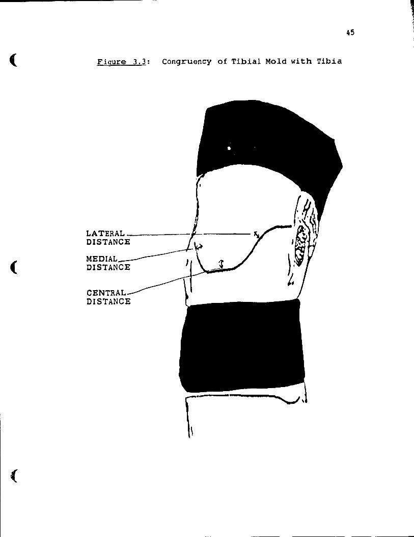
c) As a further investigation of brace fit subjects
placed their ACL deficient braced knee at angles of 15, 30,
60, and 90 degrees of knee flexion ( as measured by a
goniometer). The distance from the knee to the tibial mold
was measured. The following three measurements were taken:
1. center of the medial lip of the brace to the
center of the medial tibial plateau
2. center of the central lip of the brace to the
center of the patellar tendon
3 . center of the lateral lip of the brace to the
center of the lateral tibial plateau (figure 3.3).
--------------------
{
41
3.6 Experimental Design
Task one: Active range of motion (ACL deficient knee - braced
ACL deficient knee) (Degrees)
Brace 1
Extension
InternaI rotation
Brace 2 Brace 3
This ls a completely randomized design with one
independent variable with three levels.
Criterion Variables:
1) extension control (degrees) = extension of ACL
deficient - extension of braced ACL deficient
2) internaI rotation control (degrees) = internaI
rotation of ACL deficient - internaI rotation of braced ACL
Deficient.
Task two: Kicking task (relative brace angle .- relative
knee angle)
Brace/Joint Congruency
Brace 1 Brace 2 Brace 3
This is a completely randomized design with one
independent variable (braces) with three levels.
-
.....
--
42
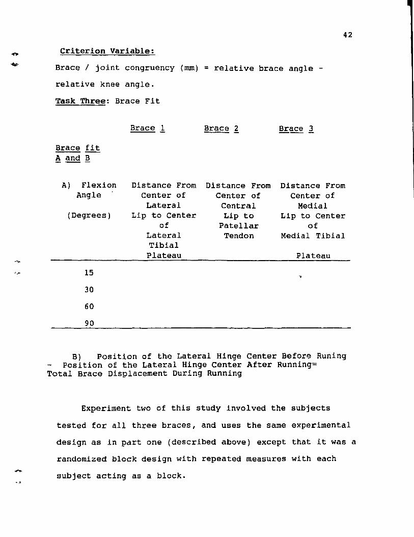
Criterion Variable:
Brace / joint congruency (mm) = relative brace angle -
relative knee angle.
Task Three: Brace Fit
Brace fit A and B - ---
A) Flexion Angle
(Degrees)
15
30
60
90
Brace 1
Distance From Center of Lateral
Lip to Center of
Lateral Tibial Plateau
Brace 2 Brace 3 --
Distance From Distance From Center of Center of
Central Medial Lip to Lip to Center
Patellar of Tendon Medial Tibial
Plateau
B) Position of the Lateral Hinge Center Before Runing Position of the Lateral Hinge Center After Running=
Total Brace Displacement During Running
Experiment two of this study involved the subjects
tested for aIl three braces, and uses the same experimental
design as in pdrt one (described above) except that it was a
randomized b10ck design with repeated measures with each
subject acting as a block.
(
(
Figure 3.1: Anthropometrie Landmarks for an ACL Deficient Knee
GREATER TROCHANT~E~R~~~~~
LATERAL JOINT LIN=E~--I-~",;'
LATERAL MALLEOL U:-;O;S::---..il
43
44
Figure 3.2: Brace Landmarks Used to Measure Congruency and Migration
SUPERIOR LATERAL BORDER
LATERAL BRACE HINGE ---
INFERIOR LA:ERA~ BORDER
-
(
(
(
Figure 3.3: Congruency of Tibial Mold wi th Tibia
LATERAL _---DISTANCE
45
-
-
4. l Introduction
Chapter 4
RESULTS
The purpose of this study was to evaluate and compare
the efficacy of three commercial functional knee braces
based on the following cri terion variables:
46
1) range of motion of the deficient knee - range
of motion of the braced def icient knee.
2) relative brace angle - relative knee angle (
brace / knee joint congruency).
3) total displacement of the brace dur ing a
running task.
4) approximation of the tibial mold.
Var iables one and two reflect the abili ty of the knee
brace to control range of motion, particularly, movements
such as internaI rotation and extension which render the
knee joint uns table . Variables three and four were used to
determine the general fit of the brace or how weIl the brace
des ign approximated the contours of the knee joint. This
factor was deerned extremely important since the beneficial
restrictions imposed by the brace in order to stabilize the
knee joint could only be transmi tted to the skeletal system
if the brace was weIl aligned wi th the knee joint (Lew et
al., 1982).
The subject sample consisted of three groups of eight
subjects each. Subjects were designated to the groups
according to which of the three knee orthoses they had been
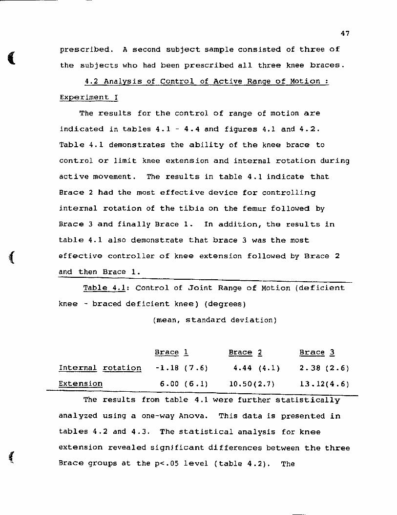
(
(
(
47
prescribed. A second subject sample consisted of three of
the subjects who had been prescribed aIl three knee braces.
4.2 Analysis of Control of Active Range of Motion:
Experiment I
The results for the control of range of motion are
indicated in tables 4. 1 - 4.4 and figures 4.1 and 4.2.
Table 4. l demonstrates the abili ty of the knee brace to
control or limi t knee extension and internal rotation during
acti ve movement. The resul ts in table 4.1 indicate that
Brace 2 had the most effective device for controlling
internaI rotation of the tibia on the femur followed by
Brace 3 and finally Brace 1. In addition, the resul ts in
table 4.1 also demonstrate that brace 3 was the most
effective control 1er of knee extension foilowed by Brace 2
and then Brace 1.
Table 4.1: Control of Joint Range of Motion (deficient
knee - braced deficient knee) (degrees)
InternaI rotation
Extension
(mean, standard deviation)
Brace 1
-1.18 (7.6)
6.00 (6.1)
Brace 2
4.44 (4.1)
10.50(2.7)
Brace 3
2.38 (2.6)
13.12(4.6)
The results from table 4.1 were further statistically
analyzed using a one-way Anova. This data is presented in
tables 4.2 and 4.3. The statistical analysis for knee
extension revealed signj ficant differences between the three
Brace groups at the p<.05 level (table 4.2). The
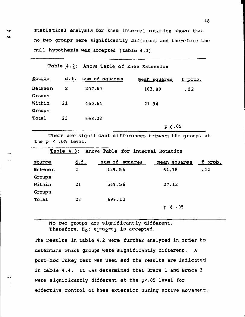
....
48
statistical analysis for knee internaI rotation shows that
no two groups were significantly different and therefore the
nuli hypothesis was accepted (table 4.3)
Table 4.2: Anova Table of Knee Extension
source d. f. sum of squares mean squares f probe
Between 2 207.60 103.80 .02 Groups
Within 21 460.64 21. 94 Groups
Total 23 668.23
p <.05
There are significant differences between the groups at the p < .05 level.
Table 4.3: Anova Table for Internal Rotation
source d.f. sum of squares Mean sguare s -Between 2 129.56 64.78 Groups
Within 21 569.56 27.12
Groups Total 23 699.13
P < .05
No two groups are signiflcantly different. Therefore, Ho: ul=u2=u3 ls accepted.
f probe
.12
The results in table 4.2 were further analyzed in order to
determine which groups were slgnificantly different. A
post-hoc Tukey test was used and the results are indlcated
in table 4.4. It was determined that Brace 1 and Brace 3
were significantly different at the p<.05 level for
effective control of knee extension during active movement.
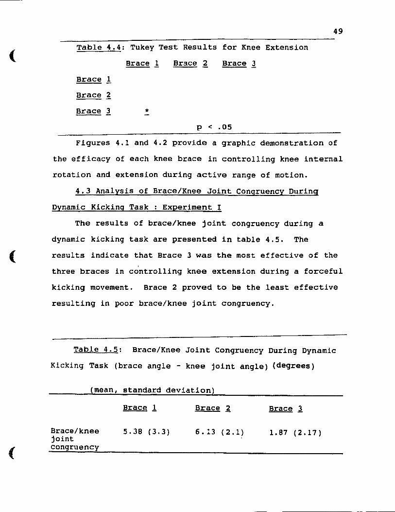
(
(
(
49
Table 4.4: Tukey Test Results for Knee Extension
Brace 1 Brace 2 Brace 3
Brace 1
Brace 2
Brace 3 * p < .05
Figures 4.1 and 4.2 provide a graphic demonstration of
the efficacy of each knee brace in controlling knee internaI
rotation and extension during active range of motion.
4.3 Analysis of Brace/Knee Joint Congruency During
Dynamic Kickinq Task : Experiment l
The results of brace/knee joint congruency during a
dynamic kicking task are presented in table 4.5. The
results indicate that Brace 3 was the most effective of the
three braces in controlling knee extension during a forceful
kicking movement. Brace 2 proved to be the least effective
resulting in po or brace/knee joint congruency.
Table 4.5: Brace/Knee Joint Congruency During Dynamic
Kicking Task (brace angle - knee joint angle) (deqrees)
(mean, standard deviation)
Brace/knee joint congruency
Brace l
5.38 (3.3)
Brace ~
6.13 (2.1)
Brace 3
1. 87 (2.17)
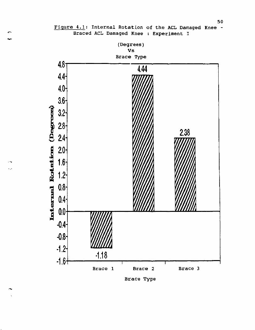
50 Figure 4.1: Internal Rotation of the ACL Damaged Knee -
Braced ACL Damaged Knee : Experiment l
(Degrees) Vs
Brace Type
4.8----------4.4-4-----.---,
4.4 4.0 3.6
e 2.4
,r~O ~ 1.6
a 1.2
1 :a 0.0
-0.4 -0.8 -1.2
-1.18 -1.64--------,...-----,-------1 Brace 1 Brace 2 Brace 3
Brace Type
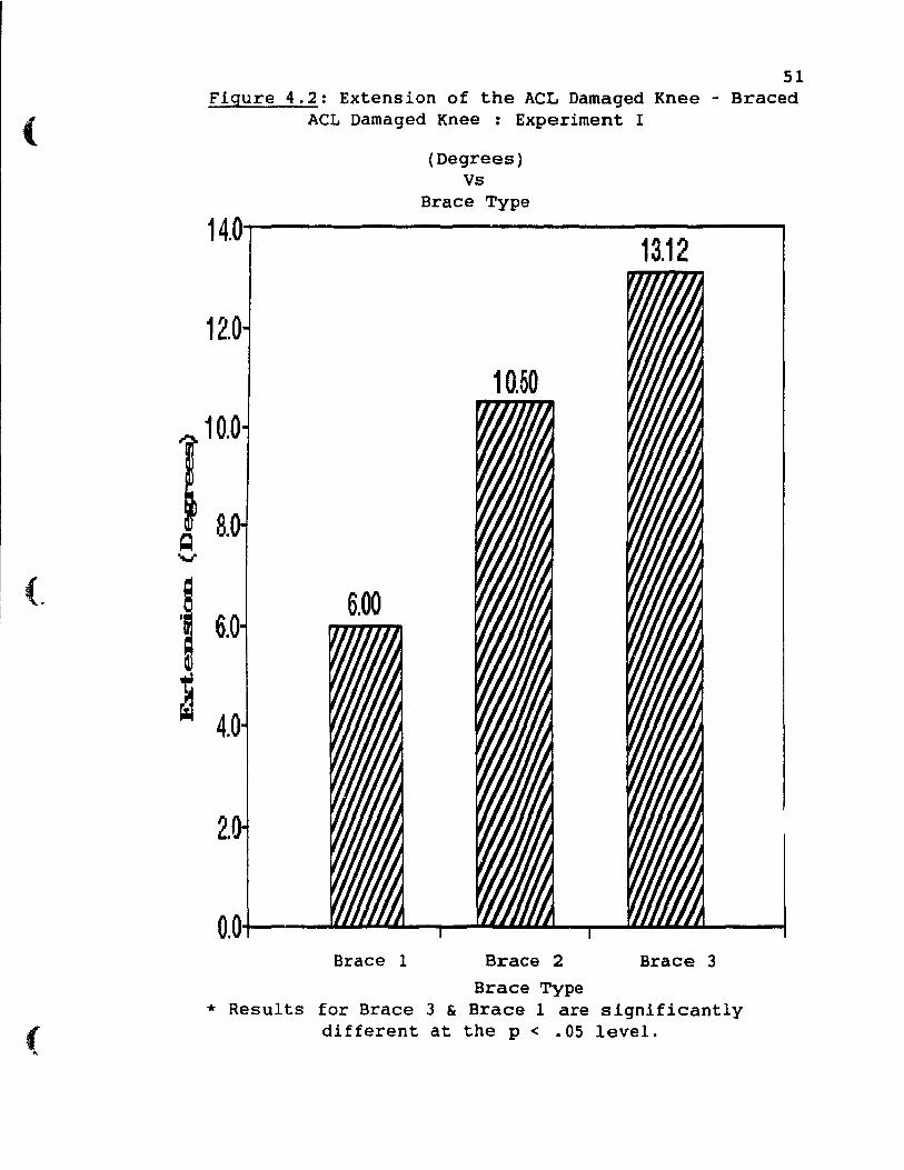
(
(
51 Figure 4.2: Extension of the ACL Damaged Knee - Braced
ACL Damaged Knee : Experiment l
(Degrees) Vs
Brace Type
14.0..,....---------1-3.1-2-----...,
12.0
10.0
~ 8.0 ---
6.0
4.0
2.0
Brace 1 Brace 2 Brace 3
Brace Type * Results for Brace 3 & Brace 1 are significantly
different at the p < .05 level.
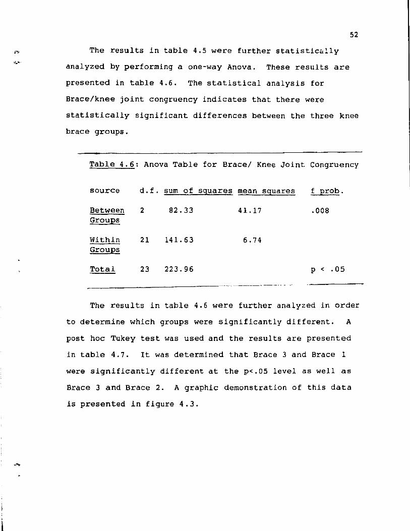
52
The results in table 4.5 were further statistic~~ly
analyzed by performing a one-way Anova. These results are
presented in table 4.6. The statistical analysis for
Brace/knee joint congrueney indieates that there were
statistieally significant differences between the three knee
braee groups.
Table 4.6: Anova Table for Bracel Knee Joint Congruency
source
Between Groups
Within Groups
Total
d.f. sum of squares mean squares f prob.
2 82.33 41.17 .008
21 141.63 6.74
23 223.96 p < .05
The results in table 4.6 were further analyzed in order
to determine which groups were significantly different. A
post hoc Tukey test was used and the results are presented
in table 4.7. It was determined that Brace 3 and Brace l
were signifieantly different at the p<.OS level as weIl as
Brace 3 and Brace 2. A graphie demonstration of this data
is presented in figure 4.3.
(
53
Table 4.7: Tukey Test Results for Brace/ Knee Joint
Congruency
Brace l Brace 2 Brace 3
Brace l * Brace 2 * p < .05
Brace 3
---------------------------------------4.4 Analysis of Brace fit: Experiment l
The final criterion variable used to evaluate the
efficacy of the three orthoses consisted of brace fit. This
was measured first by the displacement of the hinge of the
brace during a ten minute running task and secondly by the
approximation of the tibial mold to the tibia. The results
of the displacement values are listed in tables
4.8 - 4.10 and figure 4.4.
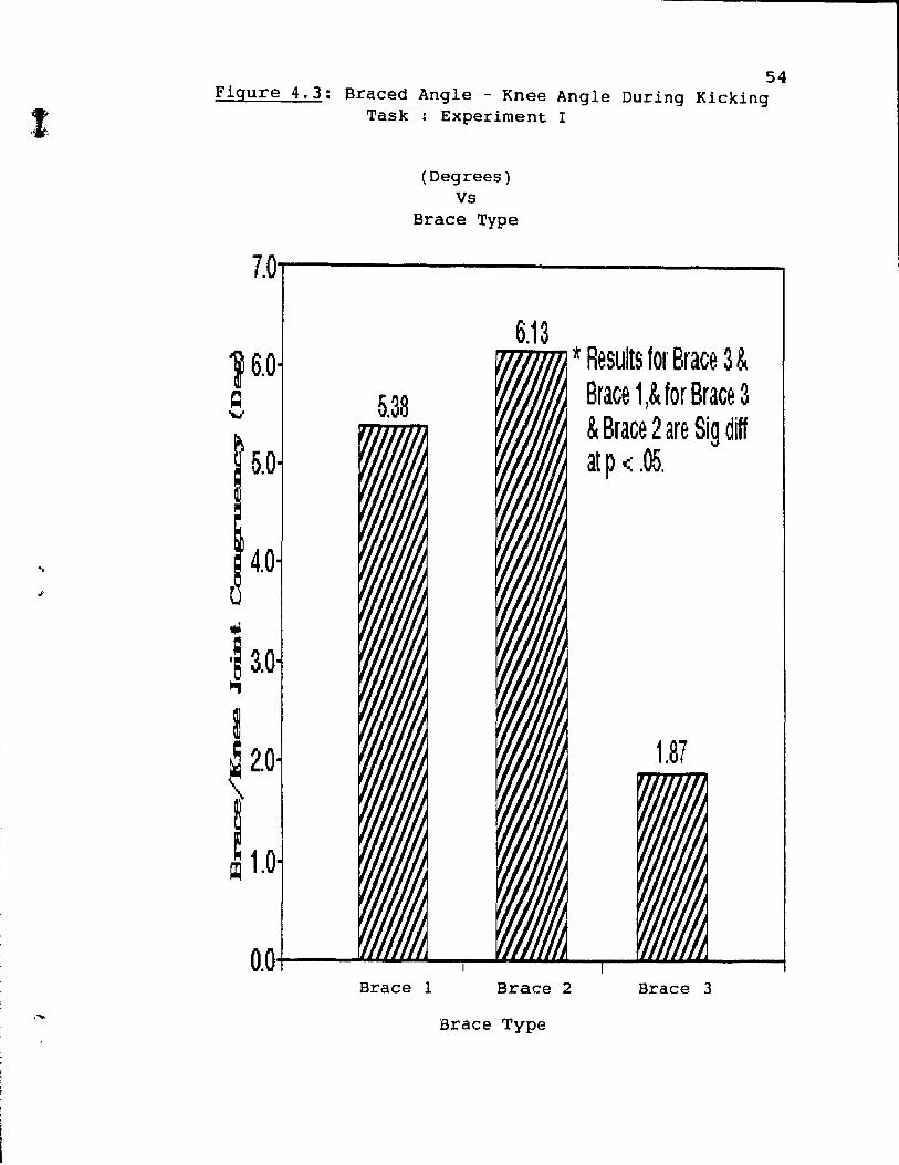
54 Figure 4.3: Braced Angle - Knee Angle During Kicking
Task : Experiment l
(Degrees) Vs
Brace Type
7.0'f-------------~
5.0
4.0
... ,~ 3.0 III
2.0
~ ~ 1.0
Brace 1
6.13
Brace 2
Brace Type
* Results for Brace 3 & Brace 1,& for Brace 3 & Brace 2 are Sig diff at p < .05.
Brace 3
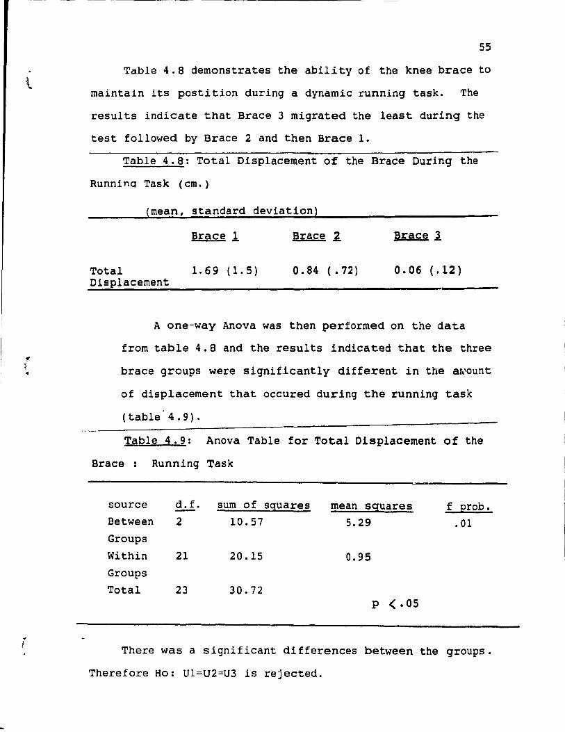
55
Table 4.8 demonstrates the ability of the knee brace to
maintain its postition during adynamie running task. The
results indicate that Brace 3 migrated the least during the
test followed by Brace 2 and then Brace 1.
Table 4.8: Total Displacement of the Brace During the
Runnina Task (cm.)
(mean, standard deviation)
Total Displacement
Brace l
1.69 (1.5)
Brace ~
0.84 (. 72)
Brace l
0.06 (.:12)
A one-way Anova was then performed on the data
from table 4.8 and the results indicated that the three
brace groups were significantly different in the a~ount
of displacement that occured during the running task
( table' 4 . 9) .
Table 4.9: Anova Table for Total Displacement of the
Brace: Running Task
source d. f. sum of sguares mean sguares f Erob. Between 2 10.57 5.29 .01 Groups
Within 21 20.15 0.95 Groups
Total 23 30.72 p < .05
There was a significant differences between the groups.
Therefore Ho: Ul=U2=U3 ls rejected.
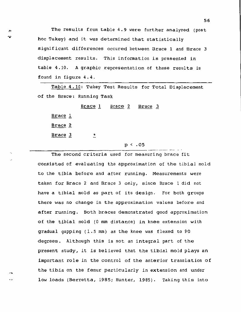
56
The results from table 4.9 were further analyzed (post
hoc Tukey) and it was determined that statistically
signif icant differenees oecured between Braee 1 and Braee 3
displacement resul ts. This in formation is presented in
table 4.10. A graphie representation of these resul ts is
found in figure 4.4.
Table 4.10: Tukey Test Results for Total Displacement
of the Braee: Running Task
Brace 1 Brace 2 Braee 3
Braee 1
Braee 2
Braee 3 * p < .05 ._----------- - ---
The second er i teria used for measuring brace fi t
consisted of eval uating the approximat ion of the tibial mold
to the tibia before and after running. Measurements were
taken for Brace 2 and Braee 3 on1y, sinee Braee 1 did not
have a tibial mold as part of its design. For both groups
there was no change in the approximation values before and
after running. Bath braces demonstrated good approximation
of the tibial mold (0 mm distance) in knee extension wi th
graduaI gapping (l. 5 mm) as the knee was f lexed to 90
degrees. Al though this is not an integral part of the
present study, it is believed that the tibial mold plays an
important role in the control of the anterior translation of
the tibia on the femur particularly in extension and under
low loads (Berretta, 1985; Hunter, 1985). Taking this into
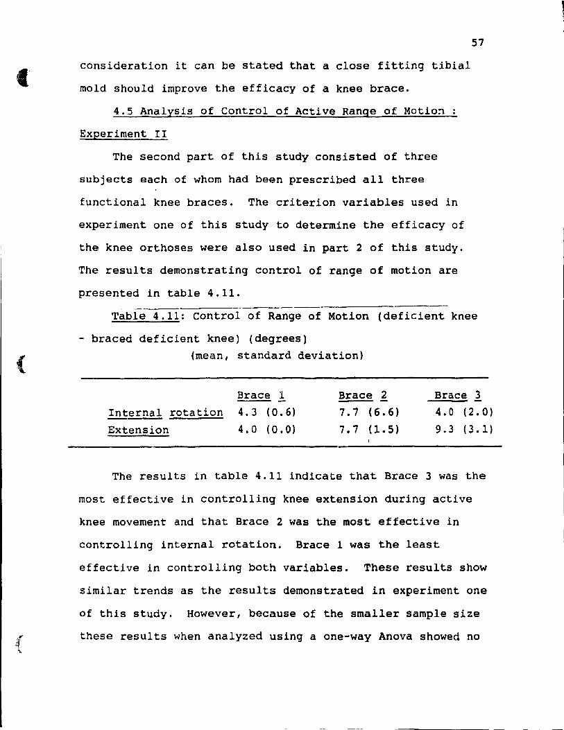
(
{
consideration it can be stated that a close fitting tibial
mold should improve the efficacy of a knee brace.
4.5 Analysis of Control of Active Range of Motio~
Experiment II
The second part of this study consisted of three
subjects each of whom had been prescribed aIl three
functional knee braces. The criterion variables used in
experiment one of this study to determine the efficacy of
the knee orthoses were also used in part 2 of this study.
The results demonstrating control of range of motion are
presented in table 4.11. ---- -----
57
Table 4.11: Control of Range of Motion (deficient knee
- braced deficient knee) (degrees) (mean, standard deviation)
Brace 1
InternaI rotation 4.3 (0.6)
Extension 4.0 (0.0)
Brace 2 7.7 (6.6)
7.7 (1.5)
Brace l 4.0 (2.0)
9.3 (3.1)
The results in table 4.11 indicate that Brace 3 was the
most effective in controlling knee extension during active
knee movement and that Brace 2 was the Most effective in
controlling internaI rotation. Brace 1 was the 1east
effective in controlling both variables. These results show
similar trends as the results demonstrated in experiment one
of this study. However, because of the smaller sample size
these results when analyzed using a one-way Anova showed no
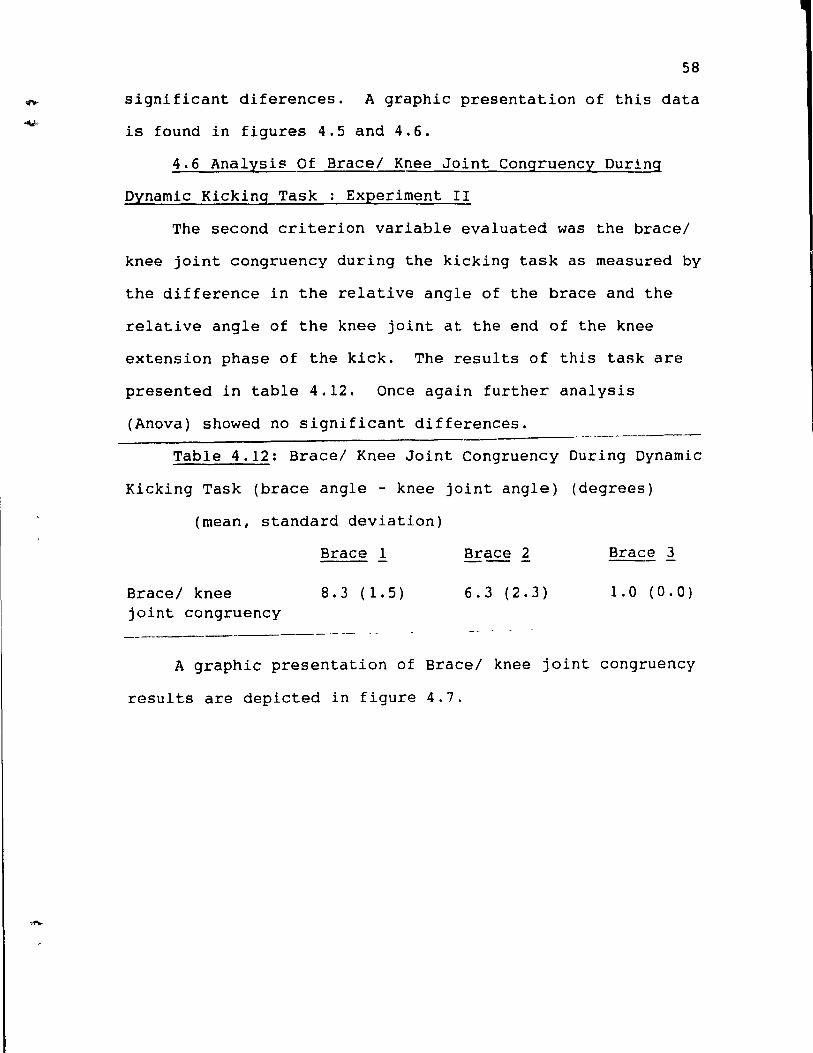
58
significant diferences. A graphie presentation of this data
is found in figures 4.5 and 4.6.
4.6 Analysis Of Brace/ Knee Joint Congruency During
Dynamic Kicking Task : Experiment II
The second criterion variable evaluated was the brace/
knee joint congruency during the kicking task as measured by
the difference in the relative angle of the brace and the
relative angle of the knee joint at the end of the knee
extension phase of the kick. The results of this task are
presented in table 4.12. Once again further analysis
(Anova) showed no significant differences.
Table 4.12: Brace/ Knee Joint Congruency During Dynamic
Kicking Task (brace angle - knee joint angle) (degrees)
(mean, standard deviation)
Brace/ knee joint congruency ----------
Brace 1
8.3 (1.5)
Brace 2 Brace 3
6.3 (2.3) 1.0 (0.0)
A graphie presentation of Brace/ knee joint congruency
results are depicted in figure 4.7.
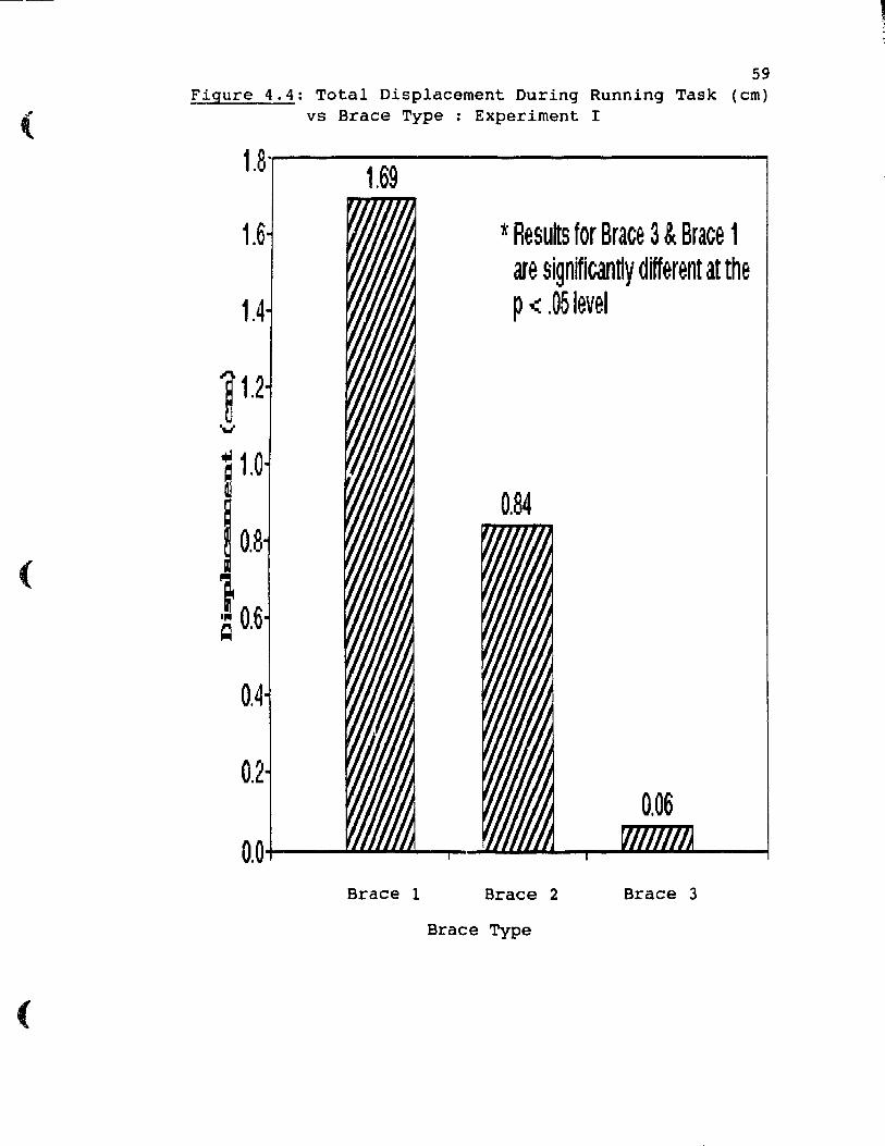
(
(
(
59 Figure 4.4: Total Displacement During Running Task (cm)
vs Brace Type ; Experiment l
1.8",,...----------------. 1.69
1.6
1.4
0.8
lO.6
Brace 1
t Results for Brace 3 & Brace 1 are significantly different at the p < .05level
Brace 2 Brace 3
Brace Type
1 , 1
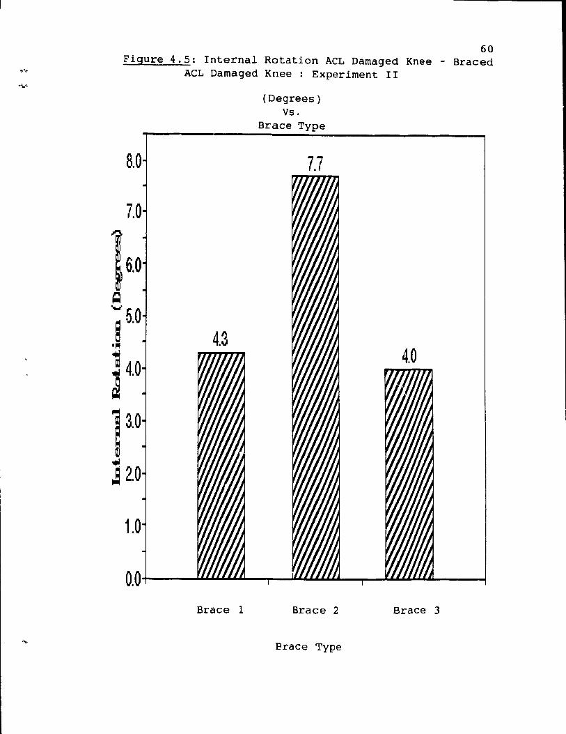
60 Figure 4.5: Internal Rotation ACL Damaged Knee - Braced
8.0
1.0
ACL Damaged Knee : Experiment II
Brace 1
(Degrees) Vs.
Brace Type
7.7
Brace 2
Erace Type
Brace 3
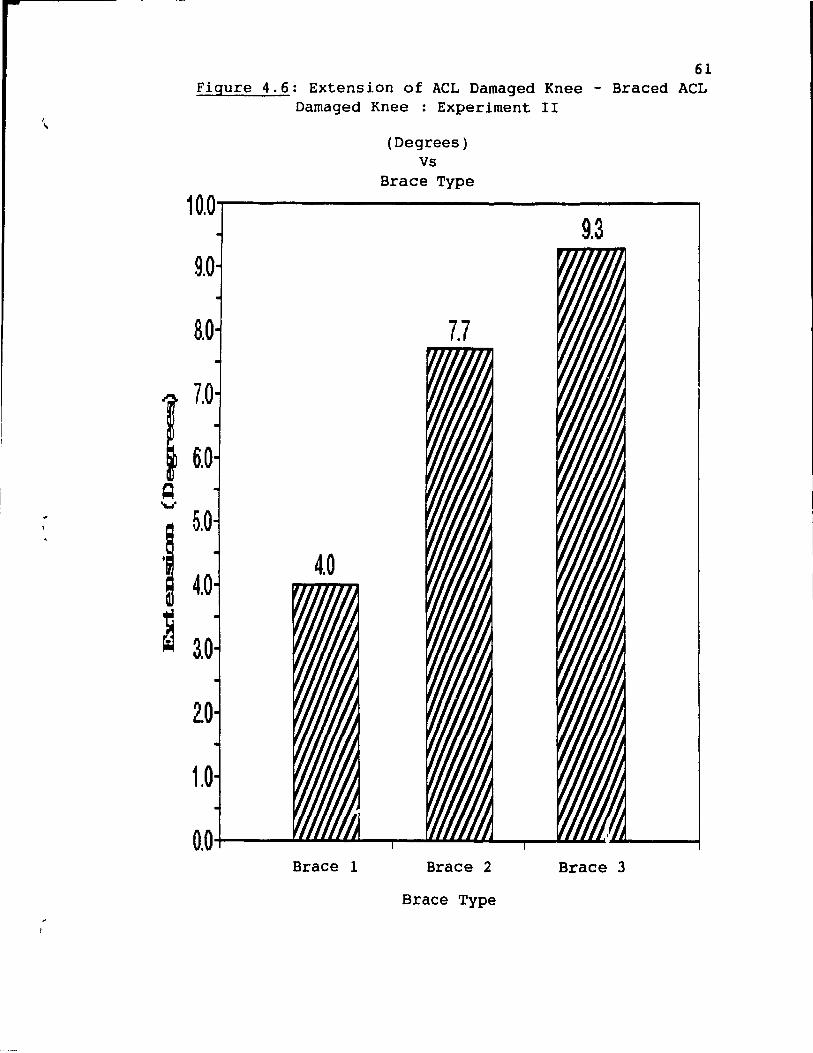
61 Figure 4.6: Extension of ACL Damaged Knee - Braced ACL
Damaged Knee : Experiment II
(Degrees) Vs
Brace Type
10.0~--------------'
9.3 9.0
5.0
Brace 1 Brace 2 Brace 3
Brace Type
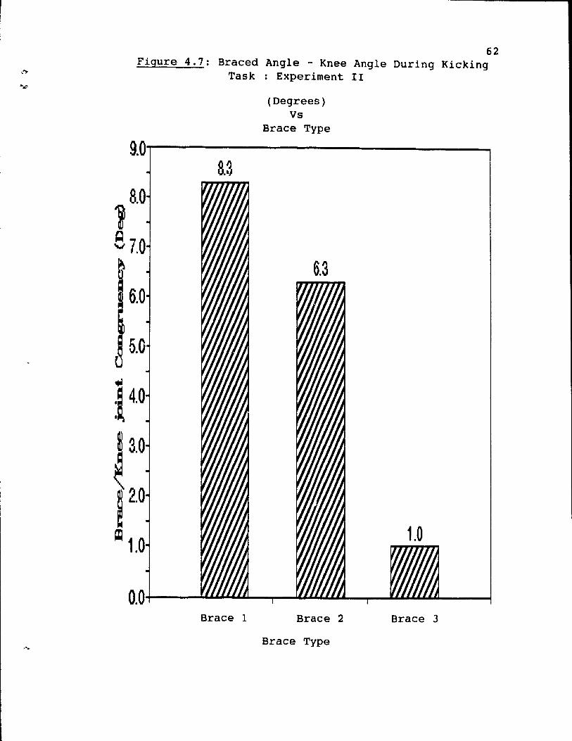
,.".
1
62 Figure 4.7: Braced Angle - Knee Angle During Kicking
Task : Experiment II
(Degrees) Vs
Brace Type
9.0---------------. 8.3
8.0
87.0
~ 2.0
~ 1.0
Brace 1 Brace 2 Brace 3
Brace Type
------------------- - -------
(
(
63
These results indicate that Brace 3 was the most
effective in controlllng knee extension during high velocity
movement (kicking task). This result indicates that the
congruency between Brace 3 and the knee joint is the
highest. Brace 2 is the second best controller of knee
extension during dynamic activity with Brace 1 demonstrating
the least control. These results are not entirely
concurrent with those from experiment one of this study, but
this slight discrepancy (Brace 2 scoring better) may be due
to the small sample size in this part of the study.
4.7 Analysis of Brace Fit: Experiment II
The final measurement involved the total migration of
the brace during a ten minute jog. This was determined by
calculating the difference between the initial position and
the final position of the lateral hinge of the brace. The
results of the test are presented in table 4.13 and figure
4.8. ------------_. -- - ----- --- --- -.
Table 4.13: Total Displacement of the brace: Running
Task (cm.)
(Mean, standard deviation)
Total Displacement
Brace 1
.83 (.76)
Brace 2 Brace 3
.33 (.29) .17 (.lS)
---------- --The results in table 4.13 are similar to those
demonstrated in the first part of this study. Brace 3 shows
the least amount of migration during the running task
1
,o.
64
followed by Brace 2 and finally Brace 1. Further analysis
using a one-way Anova and a post hoc Tukey test revealed
that there were significant differences between Brace 3 and
Brace 1 at the p<.OS level. The resu1ts in table 4.13 are
also presented graphically in figure 4.8.
The results of part two of this study ref1ect the
results discussed in part one of this study for aIl tests.
It can thus be concluded that for this particu1ar study the
subjects themselves have little influence on the results and
that in fact the results are primarily governed by the type
of brace and how closely the brace fits the contours of the
knee joint.
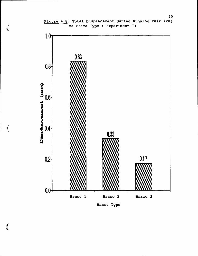
r .. 1
~
65 Figure 4.8: Total Displacement During Running Task (cm)
vs Brace Type : Experiment II
1.0~-----------------'
0.83 0.8
0.4
0.2
Brace 1 Brace 2 }Jrace 3
Brace Type
5.1 Introduction
Chapter 5
DISCUSSION
The factors representing the underlying premises for
this research were as follows:
1) ACL damaged knees are characterized by specifie
physiological and anatomical deficiencies (Ahmed et al.,
1987; Lipke et al., 1981; Markolf et al., 1990).
2) These deficiencies are responsible for the
functional limitations and high incidence of re-in jury
amongst ACL injured athletes (Cabaud & ROdkey, 1985).
3) Functional knee orthoses were designed to minimize
the anatomical deficiencies and thus prevent re-in jury
(Marquette, 1988).
4) Two factors that best quantify the efficacy of the
66
knee orthosis in minimizing ACL anatomical and physiological
deficiencies are: control of range of motion of the knee
joint and the general fit of the knee brace (Lew et al.,
1982; Walker et al., 1985; Shafer et al., 1988).
For the purposes of this research the most notable
characteristic deficiencies of an ACL damaged knee were
anterior translation of the tibia on the femur especially in
the final 10-15 degrees of knee extension (Markolf et al.,
1990; Ahmed et al., 1987) and internaI rotation of the tibia
on the femur (Kennedy & Fowler, 1971; Piziali et al., 1980;
Lipke et al., 1981). These deficiencies result in a highly
unstable knee joint since they allow for excessive movement
(
(~
67
occurring between the femur and the tibia. Unchecked, this
instability can result in a high risk of re-in jury to the
knee joint since an uns table knee does not provide a solid
base for high intensity, high force generating dynamic
activity. Knee instability inhibits the transmission of
energy along the kinetic chain resulting in less effective
performance of aIl functional activities (McLeod, 1985).
The functional knee orthosis was designed to minimize
the effects of the deficiencies of ACL damaged knees and in
doing acts to prevent further in jury. The question is ' how
does one determine the efficacy of a knee orthos~s?' This
research project used two factors to establish the efficacy
of the knee orthosis. The first factor involved control of
joint range of motion. The literature clearly states that
an ACL damaged knee demonstrates greater instability,
reflected in increased movement between the tibia and the
femur, in positions of knee extension and internaI rotation
(Markolf et al., 1990; Ahmed et al., 1987). Therefore, an
effective knee orthosis would be one which restricted the
amount of available knee extension and internaI rotation of
the damaged knee. In doing 50, it should protpct the
ligament from further damage and provide a more stable joint
necessary for dynamic activity and high loads. For this
reason two specifie measurements were analyzed. First, the
amount of active ranqe of motion (specifically, internaI
rotation and extension) present in the braced knee in
comparison to the unbraced ACL damaged knee. Second, the
amount of active knee extension present during a straight
toe soccer kick with the braced knee was measured using a
high speed video set on regular speed 30 fps.
68
One factor that affects the ability of the brace to
control range of motion of the knee is how weIl the brace
fits the contours of the knee joint .. A brace may be very
effective in controlling range of motion in a static
environment however, if the orthosis shifts position during
dynamic activity due to a less than perfect fit, the
efficacy of joint control will be lost and ultimately the
function of the orthosis compromised. In order to evaluate
this factor of brace fit a series of measurements were taken
to establish the total migration of the knee brace during a
specifie running task.
It was the purpose of this research project to
determine how effective each of the three braces were in
controlling internaI rotation ot the tibia on the femur and
knee extension. Secondly, the effectiveness of this control
during dynamic activitv was determined based on brace fit
and how weIl it maintained its position during a dynamic
running task.
5.2 Discussion of Results
The results from this study indicate that there is a
significant difference between the three commercial braces
in their control of range of motion and their capacity ta
maintain their position in relation to the knee joint during
dynamic activity.
5.2.1 Results of Control of Active Range of Motion
The results for the active range of motion tests
indicate that Brace 2 was the most effective controller ot
internaI rotation while Brace 3 was most effective in
69
controlling knee extension. Brace 1 was the 1east effective
of the three braces in control1ing joint range of motion.
The components of a knee brace that aid in the control
of internaI rotation include the following: 1) a close
fitting tibial mold which cornes into contact with the tibia
and thus restricts rotation and 2) a tight fitting
circumferential strap to hold the tibial mold and the brace
in place (Berretta et al., 1985; Hunter, 1985). The results
of partlof this study indicate (although not significant) a
trend where Brace 2 and Brace 3 were most effective in
controlling internaI rotation, because their tibial molds
allowed for greater restriction of the tibia in relation to
the femur. Brace 1 on the other hand did not have a tibial
mold and most likely for this reason, the brace was less
effective in controlling tibial rotation.
The results for control of knee extension indicate that
during active range of motion Brace 3 was most effective in
controlling full knee extension., Brace 2 was the sec~nd
most effective controller of knee extension with Brace 1
being the least effective.
The components of a brace to provide control for knee
extension includes the following: 1) a knee extension
stopper, 2) non-elastic posterior popliteal strap and 3) a
l
70
weIl fitting tibial mold. A knee extension stopper is a
device that is built into the hinges of a knee orthosis
preventing the orthosis from obtaining a position of knee
extension past 15 degrees of flexion ie. it prevents full
knee extension. This device is most effective when the
brace fits weIl and thus the limited or blocked movement in
the hinge can be transrnitted to the actual knee joint 50
that the movement is also blocked at this level. The
posterior popliteal strap and the weIl fitting tibial rnold
ensure that the brace is in constant contact with the
specifie nnatomical contours of the knee joint so that the
restrictions built into the brace can be transrnitted to the
knee joint itself (Coughlin et ~l., 1985; Berretta et al.,
1985; Hunter, 1905).
Brace 3 included aIl the above mentioned components and
therefore was able to restrict full knee extension most
effectively. Brace 2 lacked a posterior popliteal strap
although it does have a superior and inferior popliteal
strap, however, these are not as effective in controlling
knee extension. Brace 1 was the least effective in
controlling knee extension because sorne subjects braces
lacked an extension stopper and because the posterior straps
were cornprised of a very elastic component which allowed the
knee to move away from the confines of the actual brace
(Berretta et al., 1985; Hunter, 1985).
l' t
71
5.2.2 Results of Brace/ Knee Joint Congruency
The l~terature supports the idea that a functional knee
brace must not only work under low force passive or active
movements but also during high velocity dynamic tasks (Beck
et al., 1985; Knutzen et al., 1983). Taking this fact into
consideration, the kicking task was ubed as a test to
determine the efficacy of the three knee braces in
controlling knee extension during dynamic activity. The
results indicate that Brace 3 was the Most effective
controller of knee extension during high velocity activity.
Brace 1 was the second Most effective while brace 2
demonstrated the least control of knee extension during this
task.
The components of the orthosis that provide restriction
of knee extension under dynamic loading conditions are the
extension stopper and the posterior popliteal strap
(Berretta et al., 1985; Hunter, 1985). As the knee joint
progresses through the kicking motion the brace follows the
same movement until it reaches 15 degrees of knee flexion
where the extension stopper pre vents the brace from going
further. Theoretically the knee joint and the knee brace
should move anu stop as one, however the momentum of the
tibia causes the knee joint to forcefully extend past the 15
degree fl~xion point. This movement of the tibia is
restricted anteriorly by the knee brace and sa the knee
joint is forced ta drop posteriorly through the brace
allowing for further knee extension. The popliteal non-
---------- ---- -------------------------
elastic posterior strap of Brace 3 helped to limit this
posterior disengageruent of the knee joint from the knee
brace. In doing 50, there is a greater and more effective
bracel joint eongrueney which allows for improved efficacy
of brace function.
5.2.3 Results of Brace Fit
Sorne miyr~tion oecurs in all th~ee braces during the
running task. However, che amount is insignificant for
braces 2 and 3 and mueh more pronounced for
72
Brace 1. This resu1t indicates that Brace 2 and Brace 3 fit
their respecttve subjects more ùccurately resulting in a
better bracel joint congruency. This is an important factor
to consider since it is well documented in the literature
that a brace can only be effective if it maintains its
alignment with the knee joint (Lew et al., 1982).
The devices in Braces 2 and 3 which limit the migration
of the brace during the running task are the close fitting
tibial mold and the tight circumferential straps (Berretta
et al., 1985). Brace 1 employs a circumferent~al strap but
does not have a tibial mold to prcvide adequate grip on the
tibia and dS a resuit demonstrates the highest leveis of
migration during the running task.
In summary, Braces 2 and 3 proved ta have the greatest
efficacy in stabilizing the ACL deficlent knee joint. In
this case effieacy was determined by the ability ta control
internal rotation and knee extension while maintaining good
anatamical alignment during active and dynamic activity.
----------- -
From a functional perspective Brace 3 may be considered
slightly more effective b~cause of its better control of
knee extension during the d) "lamie kicking task. Braee l
scored low for a11 categories and may be designated the
least effective of the three braees studied.
73
Chapter 6
CONCLUSIONS
74
The purpose of this research was two- fold. F irst, the
characteristics of an ACL deficient knee as weIl as the
fundamentals of functional knee braces were determined using
an extensive li terature review. Then this information was
used to design a testing procedure to determine and compare
the efficacy of three functional knee braces. Previous
studies have shown that under low passive loads functional
knee braees are effective in controlling anterior
translation. This is considered an important mechanism
because often it is under low loads (walking and pivoting,
stair climbing) that the ACL damaged knee 'gives way'
(Coleville et aL, 1986). However, as the AAOS stated (Akeson
, et al., 1985) under higher loads this effective cc.ntrol of
anterior translation is lost (Beek et al. 1 1986; Bassett et
al., 1983). There,fore, in order for the brace to remain
effective another means of control must be implemented
during higher load, dynamic activity. "
One of the ways to limit anterlor translation ls to
avoid ranges of knee motion which are deemed more unstab1e
and are characterized by an increase in translation of the
tibia on the femur. In the case of the ACL damaged knee,
previous studies by Markolf et al., (1990, 1976), Ahmad et
al.,(1987) and Fetto et al., (1979) have shown that
instability is more pronounced during the last degrees of
knee extension and that this instabill ty Is further
75
exaggerated by a coinciding increase in internaI rotation.
Therefore, it can be concluded that a functional knee brace
will be more effective if it i5 able to control or limit
knee extension and internaI rotation during active or
dynamic movernent.
In the present study the criterion variable of active
range of motion between the ACL damaged knee and the braced
ACL damaged knee (for extension and internaI rotati0n) was
used as one measure of the efficacy of the knee orthosis.
It was decided that a brace that limited full knee extension
and internaI rotation wouid provide more stability to the
ACL damaged knee. The results of this study indicate that
Brace 3 and Brace 2 were the most effective in fulfilling
these requirements.
In order ta further investigate this mechanism of range
of motion control, a dynamic task was designed to measure
the control of knee extension during high velocity mcvement.
Subj ects were asked to perfor:., a straight toe c:;occer kick
and the difference between the brace angle and the knee
angle was measured. It was felt that increased instability
during this movement due to a position of full knee
extension could result in a high risk of re-in jury since
under normal circumstances (ie. during sports activities~
the athlete would land on a joint which was not weIl aligned
(anterior translated) resulting in a 'giving way' episode.
The results in this study indicate that Brace 3 was most
1
1
76
effective in controlling the extensor mechanism during high
velocity activity.
The final criterion variable used in this research to
determine the efficacy of functional knee braces was the
measurement of 'brace fit' or the approximation of the brace
to the contours of the knee joint. In this study a brace
was designated as weIl fitting if it did not change position
or migrate during a dynamic activity over a set period of
time. The task of running for a ten minute period on a
treadmill set at 6 MPH was used as a testing procedure for
the measurement of brace displacement and ultimately brace
fit. The results in this study indicate that Brace 3 and 2
migrated minimally during the running task.
This research protocol used the described criterion
variables as a means of determining the efficacy of three
commercial functional knee braces. This study differs from
previous research in that it does not use the criterion
variable of anterior translation as a measurement of brace
effectiveness but rather uses measurements more appropriate
for active and dynamic movements.
The results of this study indicate that there are
significant differences in how the three braces being
evaluated respond to the testing procedures. Initially, it
was felt that there would be no significant difference
between the three braces. As a result of this, the null
hypothesis was proposed for each of the criterion variables.
However, results indicated that there was a significant
(
r
difference between the three braces for the following
measurements:
1) knee extension of the ACL damaged knee - extension
of the braced ACL damaged knee during active movement.
2) relative brace angle - relative knee angle during
the kicking task.
3) total displacement of the brace during the running
task.
The only measurement for which there was no significant
difference between the groups was for the control of
internaI rotation. The fact that there was significant
difference in the measurements of efficacy for the three
braces indicates that for the criterion variables being
77
evaluated, brace design plays an important role in how weIl
a particular brace scores. The different characteristics of
the braces are reflected in their results on the efficacy
tests.
From this study it can be concluded that important
components of a knee brace for dynamic control include: an
extension stopper of 15 degrees, a non-elastic popliteal
strap, a weIl fitting tibial mold and an appropriate
suspension system for the brace ( i.e circumferential
straps). Brace manufacturers are faced with the dilemma of
producing a brace which is light weight, comfortable and
protective while at the sarne time able ta withstand large
forces being placed on the knee joint during high level
dynamic activity. In addition, this brace must adhere ta
'Y
78
the non- uniform contours of the knee joint and surrounding
musculature as this very surface undergoes a constant,
repetitive jostling force during various dynamic activities.
Even a weIl designed brace will prove to be ineffective if
the alignment of the brace with the actual knee joint is not
maintained ( Lew et al., 1982).
In summary, this study determined a means of evaluating
the efficacy of functional knee braces during active and
dynamic movement. Previous studies have established the
importance of controlling anterior translation under low
passive loads (Hoffman, 1984; Beek, 1986) but few studies
have indicated how a functional brace can be effective under
active and dynamic loads. The control of anterior
translation remains one of the key factors in effective
functional knee bracing and further studies are needed to
improve the mechanisms of controlling this instability.
However, if researchers in orthopedies and sports rnedicine
are going to improve design of functional knee braces
further research must be carried out in the dynarnic setting.
Research designs are needed to evaluate the efficacy of knee
braces during more progressive activities such as basketball
and football. Further studies are needed to correlate
efficacy measurements with knee stability, subjective rating
scales and activity levels. As more people bec orne involved
in sporting activities and the risk of in jury and subsequent
re-in jury increases, greater demands will be placed on
researchers and sports medicine professionals to develop
79
( effective means of protecting uns table knee joints while
allowing return to p~evious activity levels.
{
( ,
REFERENCES
Ahmed, A., Hyder, A., Burke, D. & Chan, R. (1987). In-vitro ligament tension in the f lexed knee in passive loading. Journal of Orthopaedic Research, 5, 217-230.
Akeson, W.H., Frank, C.B., Amiel,D.,& Woo, S.L. (1985) AAOS Symposium on Sports Medicine "The Rnee". C.V. Mosby, 111-152.
Baldwin, J. & Cunningham, K. (1974). attack: a olinical study involving
Goniometry under physiotherapiats.
Physiotherapy Canada, 26, 74-76.
Bassett, G., & Fleming, B. ( 1983 ) . anterolateral rotary instabili ty. Sports Medicine, 11, 345-348.
The Lennox-Hill brace in The Amerioan Journal of
Beck, C., Drez, D., Young, J., C'3nnon, W., & Stone, M. ( 1986). Instrumented testing of fur,otional knee braces. ~ Amerioan Journal of Sports Medioin~, 14, 253-256.
Berretta, G. , Charuest, G., Berretta, G. 1 & Berretta, C. (1985). The A.C. Knee Orthosis. Unpublished manuscript.
Blacharski, P.A.,& Somerset, J.H. (1975). A ThreeDimensional Study of the Rinematics of the Human Rnee. ~ Biomechanics, Vol 8, 375-384.
Butler, D. 1 Noyes, F., & Grood, E. ( 1980) . Ligamentous restraints ta anterior-posterior drawer in the human knee. The Journal of Bane and Joint Surgery, 62-A, 259-270.
Cabaud, H.E., & Rodkey, W. (1985). Philosophy and Rationale for the Management of Anterior Cruciate Injuries and the Resultant Deficiencies. Clinics in Sports Medicine, vol 4, No. 2., 313-323.
Colville, M., Lee, C. & Ciullo, J. (1986). The Lennox-Hill brace. The American Journal of Sports Medicine, 14, 257-261.
Coughlin, L., Oliver, J., & Berretta, G. (1986). Uns table knee wi th Antero Lateral Instabili ty. manuscript.
Bracing the Unpubliahed
Crowninshield, R. , Pope, M. , analytical model of the knee. 397-405.
& Johnson, R. (1976). An .2" Journal of Biomechanics,
Daniel, D. M., Malcom, L. L., Loose, G., ~tüne, M. L., Sachs, R., & Burks, R. ( 1985) . Instrumented measurement of anterior laxi ty of the knee. Journal of Bane and Joint Surgery, 97-1\, 720-726.
80
1 .
Fe~to, J., & MarE hall, D. ( 1979) . Inj ury to the Anterior Cruciate Ligament Producing the Pivot Shift Signe The Journal of BQne and Joint Surgery, 710-713.
Frankel, V.,& Nordin, M. (1980). Basic biomechanics Qf the ~eletal system. Philadelphia: Lea & Febiger.
FUkubayashi, T., Torzilli, P.A., Sherman, M.F.,& Warren, R.F. ( 1982) . An in-vitro biomechanical evaluat ion Qf anteriorposterior motion of the knee. The Journal of Bone and Joint Surgery. 64-A (2), 258-264.
Girgis, F., Marshall, J., & Monajem, A. (1975). The cruciate ligaments of the knee joint. Clinical Orthopaedics and Relat~d Research, 106, 216-230.
Gollehon, D., Torzilli, P. & Warren, R. (1987). The raIe of the posterolateral and cruciate ligaments in the stability of the human knee. The Journal of Bone and Joint Surgery, ~, 233-242.
81
Gurtler, R., Stine~ R.,& Torg, J. (1987). Lachman Test Evaluated. Clinical Orthopaedics and Related Research, (216), 141-150.
Hofmann, A., Wyatt, R., Bourne, M. & Daniels '\. (1984). Knee stability in orthotic knee braces. The American Journal of Sports Medicine, 12, 371-374.
Hoppenfeld, S. ( 1976). Physica1 examination of the spl.ne and extremities. New York: Prentice Hall.
Howel, S.M. (1990). Anterior Cruciate Ligament Load During Maximal Quadriceps Contraction: ls i t Clinically Significant? AAOS Proceedings, 147.
Hsieh, H., & Walker, P. (1976). Stabilizing Mechanism of the loaded and unloaded knee joint. The Journal of Bone and JQint Surgery, 58-A, 87-93.
Hughston, J.C.,& NorwQod, L.A. (1980). The PQsterolatera~ Drawer Test and External Rotational RecurvatiQn Test for PQsterolateral Rotary Instability of the Knee. Clinica~ Orthopaedics and Related Research, (147l, 82-87.
Hunter, L. (1985). Braces and taping. Medicine, 4, 39-454.
Clinics in SpQrts
Inoue, M. 1 McGunk-Burleson, E., Hollis, M., & Woo, S. ( 1987) • Treatment of the media1 collateral ligament injury. ~ American Journal of Sports Medicine, lâ, 15-21.
82
Kennedy, J.C.,& Fow1er, P.J. (1971). Medial and Anterior Instabili ty of the Knee. The Journal of Bone and Joint Surgery, 53-A (7), 1257-1270.
Kennedy, J.C., weinberg, H.W., & Wilson, A.S. (1974). The Anatomy and Function of the Anterior Cruciate Ligament as Determined by Clinical and Morphological Studies. The Journal of Bone and Joint Surgery, 56-A, 223-235.
Kettlekamp, D.B.,& Thompson, C. (1975). Development of a knee scoring scale~ Clinica1 Orthopaedics and Related Research, 107, 93-99.
Knutzen, K., Bates, B., & Hamill, J. (1983). goniometry of post-surgical knee braci og in running. Journal of Physical Medicine, 62, 172-181.
ElectroAmerica!)
Knutzen, K., B_:ltes, B. & Hami11, J. (1984). Knee brace influences on the tibial rotation and torque patterns of the surgical limbe The Journal of Orthopaedic and Sports Physical Therapy, §, 116-121. -
Knutzen, K., Bates, B., Schot, P., & Hamill, J. (1987). A biomechanical analysis of two functiona1 knee braces. Medicine and Science in Sports and Exercise, 303-309.
Lafortune, M ~ , & Cavanagh, P. , ( 1987 ) . Three-dimensional kinematics of the pate11a during walking. R.C. Nelson & C.A. Morehouse (Eds), Biomechanics VII. Baltimore: University Park Press.
Lew, W., Patrnchak, C., Lewis, J., & Scmidt, J. (1982). A Comparison of pistoning forces in orthotic knee joints. Journal of 8iomechanical Engineering. 101, 86-95.
Lipke, J., Janecki, C., Nelson, C., McLeod, P., Thompson, C., Thompson, J., & Haynes, D. (1981). The role of incompetence cf the anterior cruciate and 1ateral ligaments. The Journal of Bone and Joint Surgery, 63-A, 954-959.
Lysho1m, J., & Gillquist, J. (1982). Evaluation of knee ligament surgery resu1ts with special emphasis on use of a scoring scale. The American Journal of Sports Medicine, lQ, 150-154.
Magee, D. (1982). Orthopaedics conditions, assessment and treatment (4th ed. )
Markolf, K., Mensch, J., & Amstu tz, H. ( 1976) . Stiffness and 1axi ty of the knee- the contributions of the supporting structures. The Journal of Bone and Joint Surgery, 58-A, 583--593.
(
<.
(
83
Markolf, K., Shapiro, M., Gorek, J.,& Kalo, J. (1990). Mechanisms of Anterior Cruciate Ligament Force Generation: A New In Vitro Study of Normal and Median Cruciate Ligament Deficient Knees. AAOS proceedings, 147.
Marquette, S.H. (1988). Stabilizing Anterior Cruoiate Ligament Injuries: Biomechanical Requirements of Orthot1c Design. Orthotics and Prosthetics, 41 (4), 18-28.
McLeod, W. D. ( 1985 ) . Biomechanics and function of the secondary restraints to the anterior cruciate ligament. Orthopaedic Clinics of North America, 16 (2), 165-170.
Moffroid, M., Whippie, R" Hofkosh, J., Lowman, E. & Thistle, H. (1969). Physical Therapy, 49, 735-747.
Nicholas, J. (1983). Bracing the anterior cruciate ligament deficient knee using the Lennox-Hill derotation brace. Clinical Orthopaedics and Related Research, 172, 137-142.
Noyes, F., Disability Syndrome.
McGinniss, G.,& Mooar, L. in the Anterior Cruciate
Sports Medicine 1: 278-302.
( 1984 ) . Functional Insufficient Knee
0' Donaghue, D. (1976). Treatment of injuries to athletes (3rd ed.). W.8. Saunders Company.
O'Riain, M., Sibelle, J., & Balmer, S. (1983). A new goniometer for physiotherapy. Physiotherapy Canaàa, 35, 280-281.
Osternig, L.R., Bates, B.T., & James, S.L. (1980). Patterns of Tibial Rotary Torque in Knees of Heal thy Subjects. Medicine and Science in Sports and Exercise, Vol. 12, No. 3, 195-199.
Piziali, R., Rastegar, J., Nagel, D., & Schurman, D. (1980). 'l'he contribution of the cruciate ligaments to the load displacement characteristics of the hum an knee joint. Journal of Biomechanical Engineering, 102, 277-283.
Richards, C. (1981). Dynamic strength characteristics during isokinetic knee movements in heal thy women. Physiotherapy Canada, 33, 141-150.
Shafer, M., Russ, J., Patrnchak, C. M., & Tarr, R. (1988). The Northwestern University Rnee Orthotic System, Part 1: The N.U.K.O knee joint. Orthotics and Prosthetics, 41 (4), 29-36.
Shapiro, R. (1978). Direct linear transformation method for three-dimensional cinematography. The Rese~rch Quarterly, .i2., 199-205.
,
84
Shiavi, R., Limbird, T., Frazer, M., Stivers, 1<., Strauss, A., & Abramovitz, J. (1986). Helical motion analysis of the knee- Il. ;1ournal of Biomechanics, 12., 653-665.
Smillie, I. S. (1970). Injuries of the Knee Joint. Ed. 4 Edinburgh and London, E. and S. Livingstone.
Solomonow, M., BaraIta, R., & D'Ambrosia, R. (1989). The Role of the Hamstrings in the Rehabilitation of the Anterior Cruciate Ligament-Deficient Knee in Athletes. Sports Medicine 1: 42-48.
Townsend, M., Izak, M., & Jackson, R. (1976). Total motion knee goniometry. ~ourna1 of Biomechanics, 10, 183-193.
Van Gheluive, B. (1974). A new three-dimensional filming technique involving simpli fied alignment and measurement procedures. R. C. Nelson & C. A. Morehouse (Eds), Biomechanics IV. (476-481) Baltimore: University Park Press.
Van Horn, O., MacKinnon, J., Witt, P., & Hooker, D. (1988). Comparison of the effects of the Anderson Knee Stabler and McDavid knee guard on the kinematics of the lower extremity ga1 t. The Journal of Orthopaedic and Sports Physical TheraRY, 2" 254-260.
Vardaxis, V., & Hoshizaki 1 T. 8. (1988). The Use of Mechanical Power Analysis in Understanding and Coaching Sport Skills: Sprinting. Unpublished manuscript.
Walker, P. S., Kurosawa, H., Rovick, J. S., Go Zimmerman, R. A. (1985). Journal of Rehabilitation Research, 22(l}, 9-22.
Wang, C., Walker, P., & Wolf, B. (1973). The effects of flexion and rotation on the length patterns of the ligaments of the knee. JO.!,g'nal of Biomechanics, 2, 587-596.
Winter, D. (1985). Biomechanics of human movement with application to the study of human locomotion. CRe Cri tical Revues in Biomedical Engineering, .2" 287 -308.
APPENDIX
85
Biomeehanics Laboratory HcGill University
Informed Consent Form
The study you have volunteered to partieipate in i5 designed to evaluate anterior cruciate ligament defieieneies. There are three distinct elements to the study. Two of these will involve the evaluation of functional knee braees, the third will evaluate the biomeehanieal eharaeteristics of crueiate dysfunction.
The Genueom Knee Analyzer ls a non-invasive researeh too 1 wh ieh wi Il be used to ça the r a portion 0 f the data required for analysis. During the Genucom assessment various forces will be applied to both knees by the examiner in a serie~ of clinical tests. A maximum force of 33 lbs will be applied to the joint and the thigh muscle will be restrained by the device. Further tests involving measuring the amount of movement in the knee joint with and without the brace and the arnount of movement during a kicking task will also be evaluated. Finally, you will be asked to run on a tre admi Il for ten minute s ln order to de t.e rmine how much displaeement oceurs between the brace and knee joint.
It is important to appreciat~ that any one or series of these proposed tests may cause sorne minor discomfort to vou. Therefore you~ partlclpatlon in this study can be di scontinued at any time throughout the protocol by s imply commun i e a tinq y(.~ur inten t ion to the techni c ian. As such 1
you may refuse to complete one or aIl of the proposed tests.
AIl results obtained in this study become the property of the MeGill Biomechanics Laboratory. Confidentiality will be respected for aIl sub)ects involved in the study. The results and interpretation of the evaluation will be available to you at the eompletion of the stuoy.
(
(
86
l have read and understand this informed CO'1sen t form. My signature below reflects my consent to be a participant in th 1s study.
Signature' ____________________________ ___
Datel __________________________________ ___
Address' ______________________________________________________ ___
Telephone 1 _______________ _
, -,