Embed Size (px)
Citation preview
DNA MicroarrayBy
Moayed al SuleimanSuleiman al borican
Ahmad al Ahmadi
It is widely believed that thousands of genes and their products (i.e., RNA and proteins) in a given living organism function in a complicated way that creates the mystery of life.
However, traditional methods in molecular biology generally work on a "one gene in one experiment" basis, which means that the throughput is very limited and the "whole picture" of gene function is hard to obtain.
Introduction
In the past several years, a new technology, called DNA microarray, has attracted tremendous interests among biologists.
This technology promises to monitor the whole genome on a single chip so that researchers can have a better picture of the interactions among thousands of genes simultaneously.
So DNA microarray is teqnique for studying gene expression .
Introduction
A single strand DNA molecule are attached onto the slide using a computer- controlled high speed robotic arm called an arrayer.
Fitted with a number of tiny pins. Each pin is immersed in a small amount of solution containing millions of copies of different DNA molecules(such as cDNA ) and the arrayer fix DNA onto the slide at specific location that recorded by the computer.
Principle
Extract mRNA from tissue of interest. Then tagged with florescent dye. And
incubated overnight with microarray(cDNA). Scanned by a laser that causes the mRNA
hybridized to the microarray to fluoresce. These fluorescents spots which reveal
genes are expressed in the tissue of interest.
Principle

1. Buffer solution.2. Columns with oligo dt _beds.3. Solvent solution.4. Green & red labeling.5. Microarray washing solution.6. Micro centrifuge.7. Vortex8. Sample tubes.9. Pipettes' ,tips.10. Microarray scanner & workstation.
Equipment we need to start…
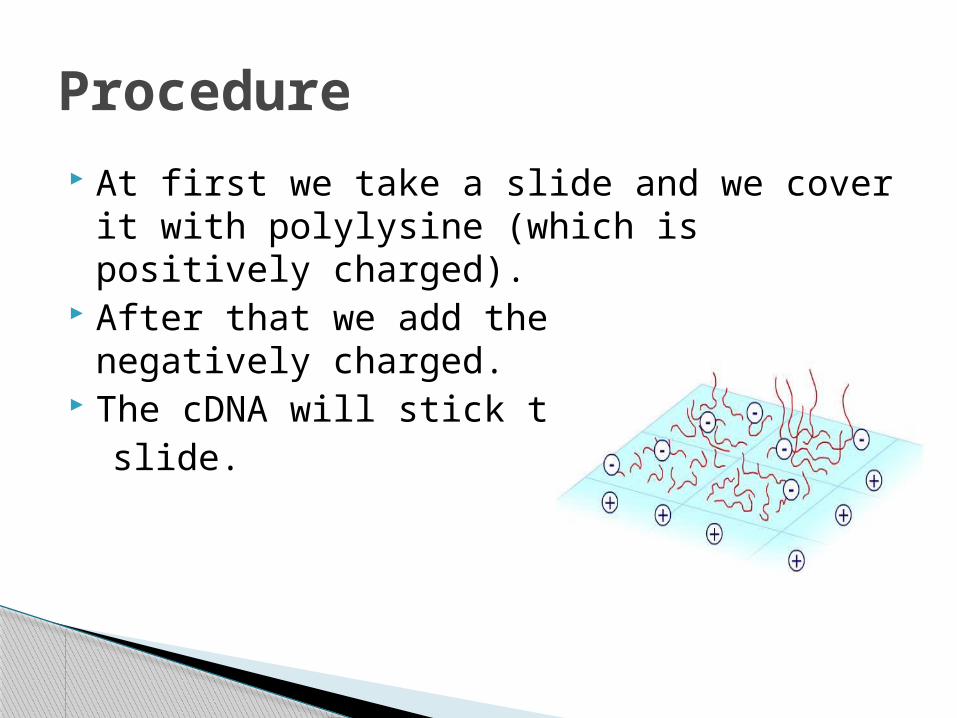
At first we take a slide and we cover it with polylysine (which is positively charged).
After that we add the cDNA which is negatively charged.
The cDNA will stick to the slide.
Procedure
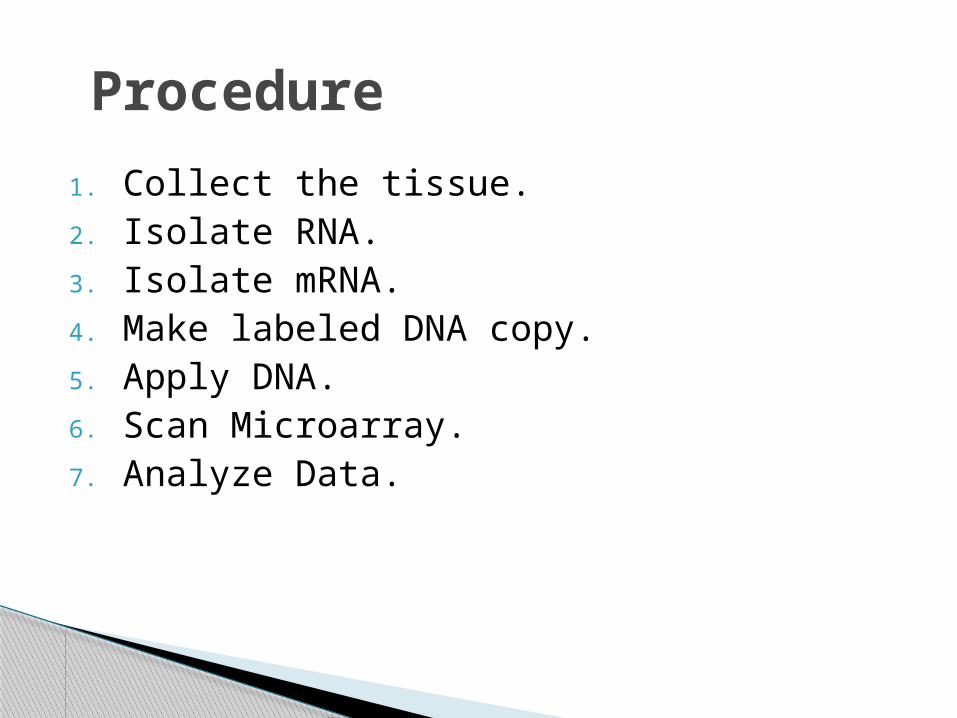
1. Collect the tissue.2. Isolate RNA.3. Isolate mRNA.4. Make labeled DNA copy.5. Apply DNA.6. Scan Microarray.7. Analyze Data.
Procedure
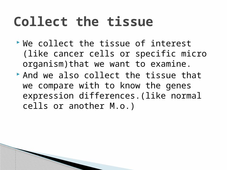
We collect the tissue of interest (like cancer cells or specific micro organism)that we want to examine.
And we also collect the tissue that we compare with to know the genes expression differences.(like normal cells or another M.o.)
Collect the tissue
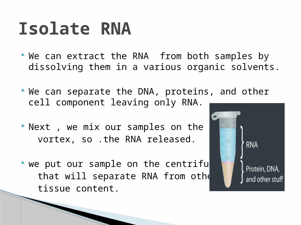
We can extract the RNA from both samples by dissolving them in a various organic solvents.
We can separate the DNA, proteins, and other cell component leaving only RNA.
Next , we mix our samples on the vortex, so .the RNA released.
we put our sample on the centrifuge that will separate RNA from other tissue content.
Isolate RNA
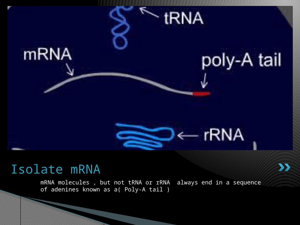
mRNA molecules , but not tRNA or rRNA always end in a sequence of adenines known as a( Poly-A tail )
Isolate mRNA
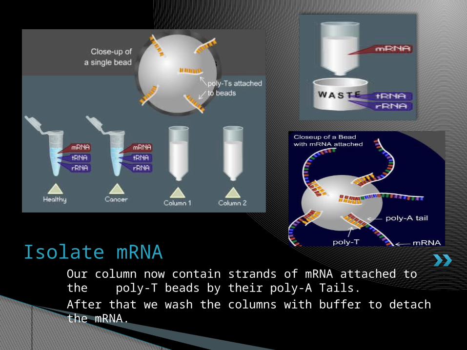
Our column now contain strands of mRNA attached to the poly-T beads by their poly-A Tails.After that we wash the columns with buffer to detach the mRNA.
Isolate mRNA
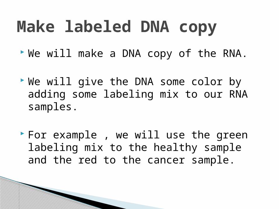
We will make a DNA copy of the RNA.
We will give the DNA some color by adding some labeling mix to our RNA samples.
For example , we will use the green labeling mix to the healthy sample and the red to the cancer sample.
Make labeled DNA copy
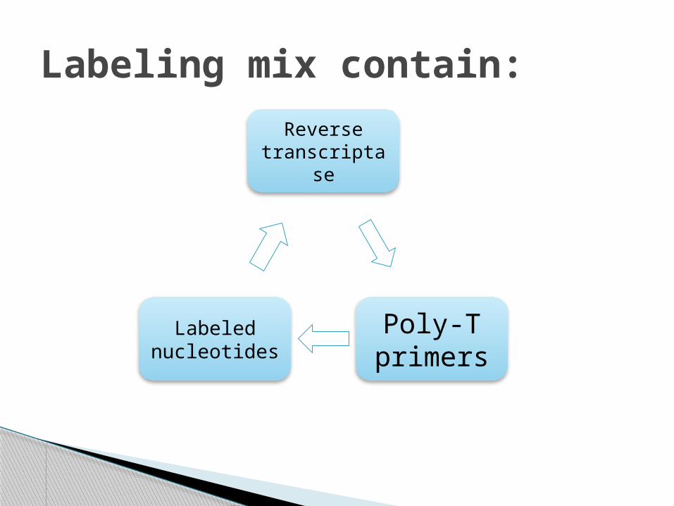
Reverse transcriptas
e
Poly-T primers
Labeled nucleotides
:Labeling mix contain
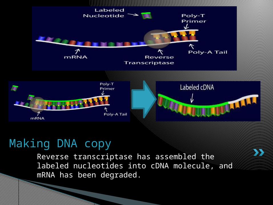
Reverse transcriptase has assembled the labeled nucleotides into cDNA molecule, and mRNA has been degraded.
Making DNA copy
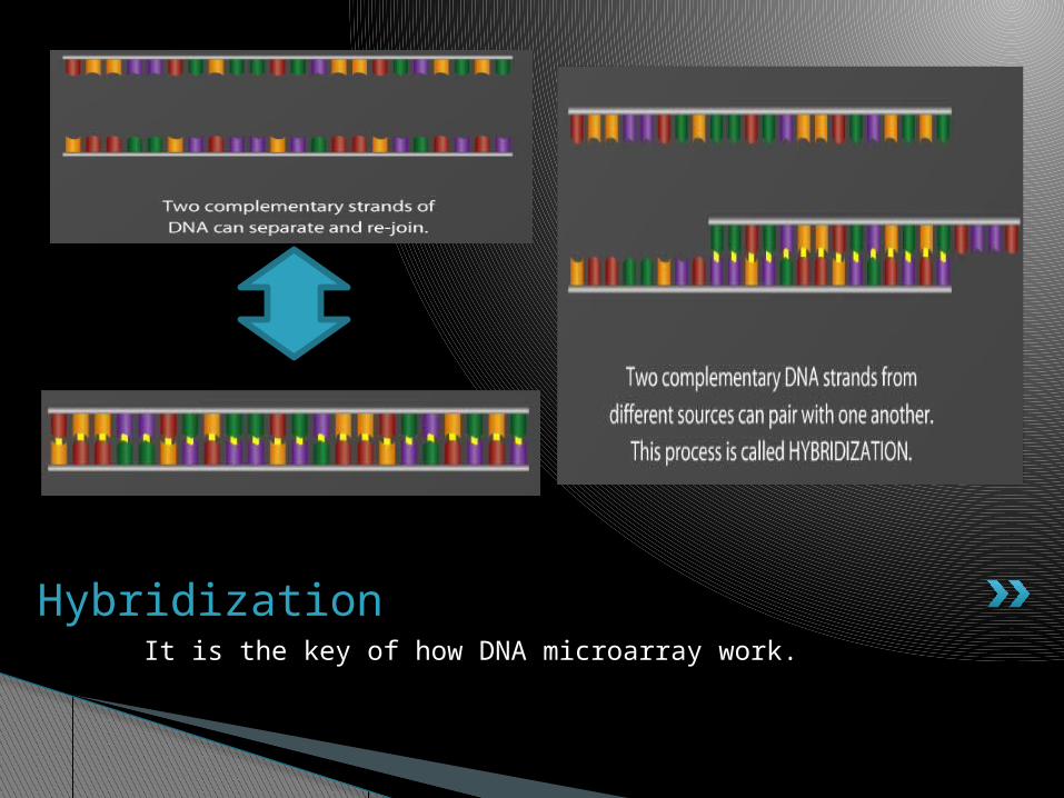
It is the key of how DNA microarray work.
Hybridization
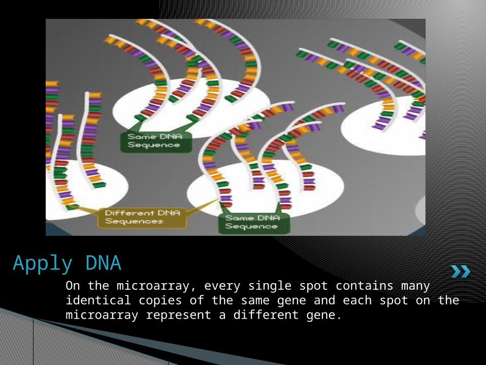
On the microarray, every single spot contains many identical copies of the same gene and each spot on the microarray represent a different gene.
Apply DNA
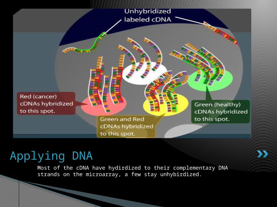
Most of the cDNA have hydirdized to their complementary DNA strands on the microarray, a few stay unhybirdized.
Applying DNA
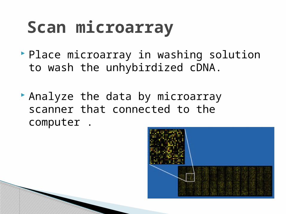
Place microarray in washing solution to wash the unhybirdized cDNA.
Analyze the data by microarray scanner that connected to the computer .
Scan microarray
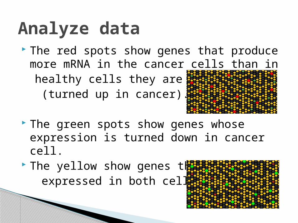
The red spots show genes that produce more mRNA in the cancer cells than in
healthy cells they are (turned up in cancer).
The green spots show genes whose expression is turned down in cancer cell.
The yellow show genes that expressed in both cells.
Analyze data
Determination of DNA sequence and mutation detection.
Genome scale determination gene expression.
Application

Thank you