Embed Size (px)
Citation preview

Burn Injuries in Children and the Use ofBiological Dressings
Bonnie Hartstein, MD, FACEP,* Marianne Gausche-Hill, MD, FACEP, FAAP,Þand Leopoldo C. Cancio, MD, FACSþ
Abstract: Burns represent a significant cause of morbidity and mortalityin children. In this article, a case discussion will serve as a platform fordiscussing the evaluation and treatment of burns in children. Use of var-ious burn dressings such as hydrocolloids, polyurethane films, hydrogels,biosynthetic skin dressing, and biological dressings will be discussed.
Key Words: burns, biological dressings, hydrocolloids
(Pediatr Emer Care 2013;29: 939Y948)
TARGET AUDIENCEThis CME activity is intended for physicians who care for
children. Pediatricians, emergency physicians, pediatric emer-gency physicians, and surgeons will find this information espe-cially useful.
LEARNING OBJECTIVESAfter completion of this article, the reader should be able to:
1. Manage burn injuries in children in the acute phase includingidentification of burn severity, estimation of body surface areainvolvement, and fluid resuscitation calculation.
2. Facilitate the rejuvenation of new skin and promote woundhealing using biologic burn dressings.
CASE 1A 3-year-old boy found hiding under a couch during a house
fire is brought into the emergency department (ED) by EMS. Heis awake but appears sleepy and has a soft hoarse cry.
Vital SignsTemperature is 38-C; blood pressure is 72/45 mm Hg; heart
rate is 132 beats/min; respiratory rate is 37 breaths/min, and pulseoximetry reads 95% on room air.
Physical ExaminationHis face is covered in soot; there is black-crusted material in
his nostrils, and his eyebrows are singed. His lung sounds areclear. The heart examination reveals tachycardia but is otherwisenormal, and he demonstrates no abdominal tenderness. Expo-sure of his body reveals red confluent burns with thick blistersintermixed with whitish patches on his left arm, and his left legand left foot are pale, appearing white and waxy. There are noobvious bony deformities and no active bleeding. Total bodysurface area (TBSA) with partial- and full-thickness burns isestimated at 25%.
MedicationsHe takes no medications and is fully immunized. He has no
known allergies.1. How do you quantify the severity of burns in children?2. What are the initial steps in management of burn injuries?3. Which patients require referral to a burn center?4. What dressings are available to treat burns in the ED?
PEDIATRIC BURN INJURIESBurns represent the second most frequent cause of trau-
matic death in children younger than 5 years resulting in 1000 to5000 pediatric deaths and approximately 30,000 hospitalizationsper year.1 Younger children are particularly vulnerable because oftheir physiologic limitations in handling the stress of fluid shiftsresulting from burn injuries, and burns exceeding 20%TBSA carrya higher mortality rate (9.9%) in children younger than 3 years.2
Although burns can be the results of contact with hot objects,electricity, and chemical or radiation exposure, flame (as in ahouse fire) or flash burns account for nearly half of all burn in-juries. Scalds are the most common etiology of burns in patientsyounger than 5 years and the second most common type of burnin all age groups.3
Duration of exposure and heat exchange contribute to depthof burn. Scalding by grease or liquid heated in a microwave isparticularly dangerous as it may cause third-degree burns. Waterheaters should be adjusted in families with young children, asthe time to cause burns from exposure to hot water from sinksand bathtubs can be seconds at temperatures greater than 56-C(133-F).3 Although the US Consumer Safety Commission rec-ommends that residential water heaters be set at no more than120-F (48-C), an infant or child may sustain second-degree burnsif left in water at this temperature for more than a minute.Therefore, a safe temperature for bathing a baby is a temperatureof 100-F (Table 1).4
CME REVIEW ARTICLE
Pediatric Emergency Care & Volume 29, Number 8, August 2013 www.pec-online.com 939
Lieutenant Colonel, Medical Corps, US Army, and Assistant Professor of Pe-diatrics (Hartstein), *Uniformed Services University of the Health Sciences,Department of Emergency Medicine, Brooke Army Medical Center, SanAntonio, TX; Professor, †Clinical Medicine, David Geffen School of Medicineat UCLA; Vice Chair and Chief, Division of Pediatric EmergencyMedicine, andDirector (Gausche-Hill), Pediatric Emergency Medicine and EMS Fellowships,Department of Emergency Medicine, Harbor-UCLA Medical Center,Torrance, CA; and Colonel (Cancio), ‡Medical Corps, US Army, US ArmyInstitute of Surgical Research, Fort Sam Houston, TX.The authors and staff in a position to control the content of this CME activity
and their spouses/life partners (if any) have disclosed that they have nofinancial relationships with, or financial interest in, any commercial or-ganizations pertaining to this educational activity.
The opinions or assertions contained herein are the private views of theauthors and are not to be construed as official or as reflecting the views ofthe Department of the Army or the Department of Defense.
Reprints: Marianne Gausche-Hill, MD, FACEP, FAAP, Pediatric EmergencyMedicine and EMS Fellowships, Department of Emergency Medicine,Harbor-UCLA Medical Center, 1000 W Carson St, Box 21,Torrance,CA 90509 (e-mail: [email protected]).
Copyright * 2013 by Lippincott Williams & WilkinsISSN: 0749-5161
Copyright © 2013 Lippincott Williams & Wilkins. Unauthorized reproduction of this article is prohibited.
Report Documentation Page Form ApprovedOMB No. 0704-0188
Public reporting burden for the collection of information is estimated to average 1 hour per response, including the time for reviewing instructions, searching existing data sources, gathering andmaintaining the data needed, and completing and reviewing the collection of information. Send comments regarding this burden estimate or any other aspect of this collection of information,including suggestions for reducing this burden, to Washington Headquarters Services, Directorate for Information Operations and Reports, 1215 Jefferson Davis Highway, Suite 1204, ArlingtonVA 22202-4302. Respondents should be aware that notwithstanding any other provision of law, no person shall be subject to a penalty for failing to comply with a collection of information if itdoes not display a currently valid OMB control number.
1. REPORT DATE 01 AUG 2013
2. REPORT TYPE N/A
3. DATES COVERED -
4. TITLE AND SUBTITLE Burn injuries in children and the use of biological dressings
5a. CONTRACT NUMBER
5b. GRANT NUMBER
5c. PROGRAM ELEMENT NUMBER
6. AUTHOR(S) Hartstein B., Gausche-Hill M., Cancio L. C.,
5d. PROJECT NUMBER
5e. TASK NUMBER
5f. WORK UNIT NUMBER
7. PERFORMING ORGANIZATION NAME(S) AND ADDRESS(ES) United States Army Institute of Surgical Research, JBSA Fort SamHouston, TX
8. PERFORMING ORGANIZATIONREPORT NUMBER
9. SPONSORING/MONITORING AGENCY NAME(S) AND ADDRESS(ES) 10. SPONSOR/MONITOR’S ACRONYM(S)
11. SPONSOR/MONITOR’S REPORT NUMBER(S)
12. DISTRIBUTION/AVAILABILITY STATEMENT Approved for public release, distribution unlimited
13. SUPPLEMENTARY NOTES
14. ABSTRACT
15. SUBJECT TERMS
16. SECURITY CLASSIFICATION OF: 17. LIMITATION OF ABSTRACT
UU
18. NUMBEROF PAGES
10
19a. NAME OFRESPONSIBLE PERSON
a. REPORT unclassified
b. ABSTRACT unclassified
c. THIS PAGE unclassified
Standard Form 298 (Rev. 8-98) Prescribed by ANSI Std Z39-18
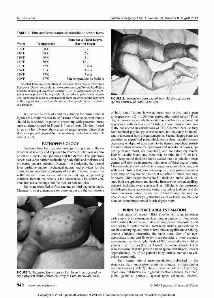
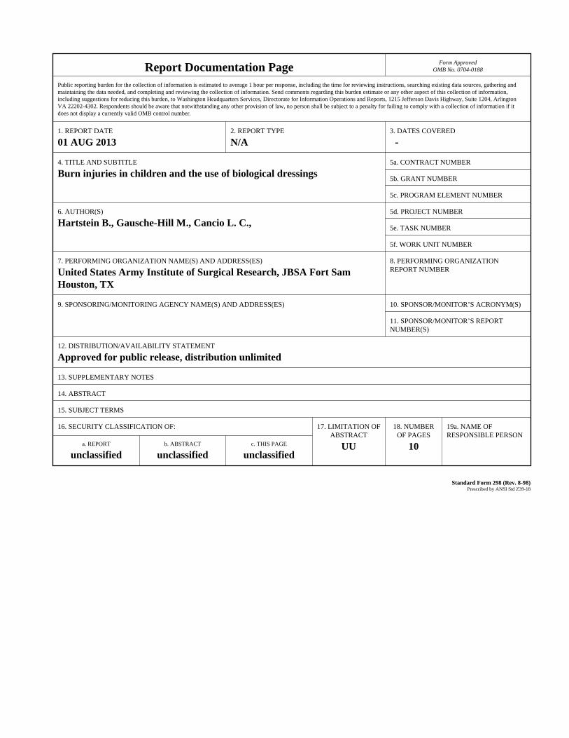
Ten percent to 20% of children admitted for burns sufferedinjuries as a result of child abuse.5 Burns of nonaccidental traumashould be suspected in patients presenting with patterned burnssuch as demonstrated in Figure 1 from an iron. Children forcedto sit in a hot tub may show areas of sacral sparing where theirskin was pressed against by the relatively protective cooler tubbase (Fig. 2).
PATHOPHYSIOLOGYUnderstanding burn pathophysiology is important to the es-
timation of severity and approach to treatment. The skin is com-posed of 2 layers, the epidermis and the dermis. The epidermisserves as a vapor barrier, maintaining body fluid and moisture andprotecting against infection. Beneath the epidermis, the dermallayer cushions against mechanical trauma and provides for theelasticity and mechanical integrity of the skin.6 Blood vessels runwithin the dermis and extend into the dermal papillae, providingnutrition. Beneath the dermis is the subcutaneous fat, which con-tains free nerve endings and the roots of hair follicles.
Burns are classified as first, second, or third degree in depth.Changes in skin appearance on presentation are the cornerstone
of burn identification; however, burns may evolve and appearto deepen over a 24- to 36-hour period after initial injury.4 First-degree burns involve only the epidermis and have a confluent redappearance with an absence of blisters. These burns are not nor-mally considered in calculations of TBSA burned because theyhave minimal physiologic consequences, but they may be impor-tant to document from a legal standpoint. Second-degree burns areclassified as superficial partial-thickness or deep partial-thicknessdepending on depth of extension into the dermis. Superficial partial-thickness burns involve the epidermis and superficial dermis, ap-pear pink and moist, are blanching, and are extremely tender.Hair is usually intact, and there may be thin, fluid-filled blis-ters. Deep partial-thickness burns extend into the reticular (deep)dermis and may be intermixed with areas of third-degree burns.Characteristically red and white in appearance, nonblanching, andwith thick blisters that commonly rupture, deep partial-thicknessburns may or may not be painful; if sensation is intact, pain maybe severe. Third-degree burns are full-thickness burns, which de-stroy both the epidermis and dermis. Because the dermal capillarynetwork, including sweat glands and hair follicles, is also destroyed,third-degree burns appear dry, white, charred, or leathery, and thetissue has no sensation. Burns that extend through the subcuta-neous tissue into underlying structures such as fascia, muscle, andbone are sometimes termed fourth-degree burns.
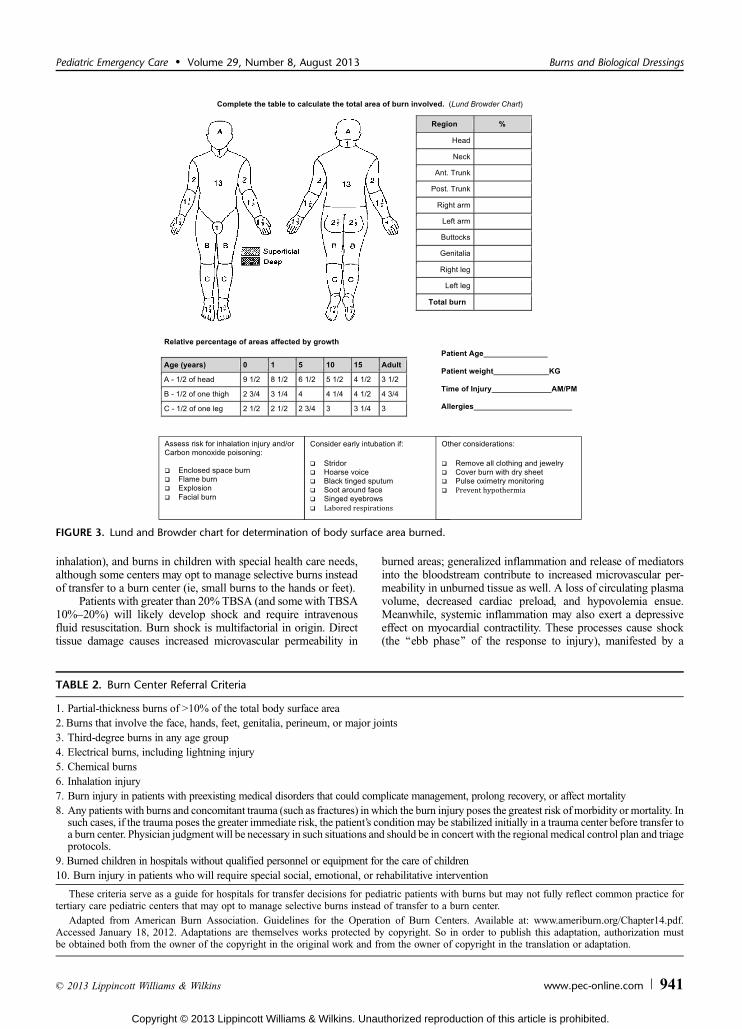
BURN SURFACE AREA ESTIMATIONEstimation of percent TBSA involvement is an important
early step in burn management, serving as a guide for fluid needsand assisting the clinician in determining patient disposition andneed for burn center referral. Total body surface area estimationcan be challenging, and studies have shown significant variabilityamong clinicians measuring the same burn. Use of an age-appropriate Lund and Browder chart provides a more accurateassessment than the simpler ‘‘rule of 9’s,’’ especially for childrenyounger than 10 years (Fig. 3). A quick method to estimate TBSAis to recognize that the patient’s hand (palm and fingers) coversapproximately 1% of the patient’s body surface area and to cal-culate accordingly.
Burn center referral recommendations published by theAmerican Burn Association assist the clinician in determiningneed to transfer (Table 2). These criteria include TBSA (910%),depth (any full thickness), high-risk locations (hands, feet, face,joints, genitalia, perineal), special types (chemical, electric,
TABLE 1. Time and Temperature Relationship to Severe Burns
Water TemperatureTime for a Third-Degree
Burn to Occur
155-F 68-C 1 s148-F 64-C 2 s140-F 60-C 3 s133-F 56-C 15 s127-F 52-C 1 min124-F 51-C 3 min120-F 48-C 5 min100-F 37-C Safe temperature for bathing
Adapted from American Burn Association. Scald Injury PreventionEducator’s Guide. Available at: www.ameriburn.org/Preven/ScaldInjuryEducator’sGuide.pdf. Accessed January 2, 2012. Adaptations are them-selves works protected by copyright. So in order to publish this adapta-tion, authorization must be obtained both from the owner of the copyrightin the original work and from the owner of copyright in the translationor adaptation.
FIGURE 1. Patterned burn from an iron in an infant caused bychild physical abuse (photo courtesy of Carol Berkowitz, MD).
FIGURE 2. Immersion burn caused by child physical abuse(photo courtesy of EMSC Slide Set).
Hartstein et al Pediatric Emergency Care & Volume 29, Number 8, August 2013
940 www.pec-online.com * 2013 Lippincott Williams & Wilkins
Copyright © 2013 Lippincott Williams & Wilkins. Unauthorized reproduction of this article is prohibited.
inhalation), and burns in children with special health care needs,although some centers may opt to manage selective burns insteadof transfer to a burn center (ie, small burns to the hands or feet).
Patients with greater than 20% TBSA (and somewith TBSA10%Y20%) will likely develop shock and require intravenousfluid resuscitation. Burn shock is multifactorial in origin. Directtissue damage causes increased microvascular permeability in
burned areas; generalized inflammation and release of mediatorsinto the bloodstream contribute to increased microvascular per-meability in unburned tissue as well. A loss of circulating plasmavolume, decreased cardiac preload, and hypovolemia ensue.Meanwhile, systemic inflammation may also exert a depressiveeffect on myocardial contractility. These processes cause shock(the ‘‘ebb phase’’ of the response to injury), manifested by a
FIGURE 3. Lund and Browder chart for determination of body surface area burned.
TABLE 2. Burn Center Referral Criteria
1. Partial-thickness burns of 910% of the total body surface area2. Burns that involve the face, hands, feet, genitalia, perineum, or major joints3. Third-degree burns in any age group4. Electrical burns, including lightning injury5. Chemical burns6. Inhalation injury7. Burn injury in patients with preexisting medical disorders that could complicate management, prolong recovery, or affect mortality8. Any patients with burns and concomitant trauma (such as fractures) in which the burn injury poses the greatest risk of morbidity or mortality. In
such cases, if the trauma poses the greater immediate risk, the patient’s condition may be stabilized initially in a trauma center before transfer toa burn center. Physician judgment will be necessary in such situations and should be in concert with the regional medical control plan and triageprotocols.
9. Burned children in hospitals without qualified personnel or equipment for the care of children10. Burn injury in patients who will require special social, emotional, or rehabilitative intervention
These criteria serve as a guide for hospitals for transfer decisions for pediatric patients with burns but may not fully reflect common practice fortertiary care pediatric centers that may opt to manage selective burns instead of transfer to a burn center.
Adapted from American Burn Association. Guidelines for the Operation of Burn Centers. Available at: www.ameriburn.org/Chapter14.pdf.Accessed January 18, 2012. Adaptations are themselves works protected by copyright. So in order to publish this adaptation, authorization mustbe obtained both from the owner of the copyright in the original work and from the owner of copyright in the translation or adaptation.
Pediatric Emergency Care & Volume 29, Number 8, August 2013 Burns and Biological Dressings
* 2013 Lippincott Williams & Wilkins www.pec-online.com 941
Copyright © 2013 Lippincott Williams & Wilkins. Unauthorized reproduction of this article is prohibited.
reduction in organ perfusion that lasts about 48 hours after burndespite adequate fluid resuscitation. After resuscitation is com-plete, an increased cardiac output, increased metabolic rate, andcatabolic state characterize the ‘‘flow’’ phase of the response toinjury. Hypermetabolic responses result in increased body tem-perature, increasing oxygen requirements and glucose consump-tion, lipolysis, and proteolysis.7 This phase lasts until the woundsare permanently closedVand even months after discharge in pa-tients with massive burns. Global immunosuppression, loss of thenatural barrier to bacteria normally provided by intact skin, inva-sive lines, and smoke-injured lungs all predispose to death in theintensive care unit from infectious complications.
RESUSCITATIONThere are currently several fluid resuscitation formulas
available for use in children; the Parkland, modified Brooke, andGalveston formulas all serve as acceptable approaches to guideinitial fluid rates (Table 3). These formulas currently use lactatedRinger’s (LR) solution for resuscitation. Conservative fluid man-agement is essential, as overresuscitation will lead to third spacingand the consequences of edema formation, such as abdominal andextremity compartment syndromes, airway edema and respiratorydistress, and/or progression of the depth of injury. Hourly moni-toring of the urine output and adjustments (titration) of the LR in-fusion rate up or down to maintain tight control of the urine outputin the range 1 to 2 mL/kg per hour (0.5Y1.0 mL/kg per hour in bigchildren) are the primary task during burn resuscitation.
Children are especially vulnerable to the body’s systemicresponses to burn injury. Hypoglycemia, hypothermia, and airwaycompromise are deadly complications that warrant close moni-toring. Children need additional glucose during resuscitation,most conveniently provided as a continuous and constant-rateinfusion of 5% dextrose in one-half-normal saline at the mainte-nance rate, in addition to resuscitation fluids. The resuscitationfluids, for example, LR, are titrated based on urine output; themaintenance fluids, for example, 5% dextrose in one-half-normalsaline, are not titrated. (Note, however, that the Galveston formuladoes not call for the routine inclusion of glucose in the mainte-nance intravenous line but rather recommends close monitoringof glucose levels in children with burn shock.) Extra care mustbe taken to protect against hypothermia in young, exposedpatients, especially during transport. Dry, sterile dressings andthe avoidance of wet dressings are imperative. Finally, patientswith circumferential deep burns of the extremities are at risk forextremity eschar syndrome. The tight, inelastic eschar exerts atourniquet-like effect, which worsens as edema formation takesplace. This may progress to arterial occlusion. Elevation of such
extremities above the heart, hourly Doppler flow-meter moni-toring of distal pulses, prompt burn center consultation, andconsideration of escharotomy by a qualified operator are keysto management.
Airway compromise after burns is a real threat, particularlyin very young children whose small tracheal dimensions arenarrowed significantly by mucosal swelling. Children who re-ceive more than 180 mL/kg of fluid resuscitation are particularlyat risk of airway edema. Warning signs of airway issues on pre-sentation in the ED include stridor, hoarseness, drooling, gagging,retractions, and brassy cough, and those with soot staining thenose or mouth are at risk of losing their natural airway. Use ofhumidified oxygen and racemic epinephrine may help in man-agement before urgently securing the airway with intubation.
Children caught in fires are at risk of carbon monoxide (CO)and cyanide (CN) poisoning produced by partial combustionof cellulosics and synthetic chemicals, respectively. Children aremore likely to hide in confined places where oxygen is replacedby dangerous gases. Carbon monoxide binds the hemoglobinmolecule in red blood cells more strongly than does oxygen andcreates a systemic hypoxemia manifested by neurologic andmyocardial dysfunction.8 A high index of suspicion for childrenexposed to smoke with neurologic changes, regardless of burnstatus, should lead clinicians to suspect CO poisoning and to checkcarboxyhemoglobin levels. Treatment is with 100% oxygen untilthe carboxyhemoglobin level is less than 5%. In severe cases,hyperbaric oxygen may be needed.9,10 Diagnosis of CN toxicitymay be difficult because (1) symptoms are similar to those of COpoisoning; (2) CN and CO poisoning frequently occur together infire death victims; and (3) there is no rapid CN assay available.Because CN binds to the terminal cytochrome on the electrontransport chain, it interferes with oxygen utilization at the mito-chondrial level. Thus, CN patients classically have a high mixed-venous saturation of oxygen (SvO2) and lactic acidosis, whichdoes not improve with fluid resuscitation. Hydroxocobalamin(high-dose intravenous vitamin B12), a CN chelator, is a rapid andeffective antidote. Amyl nitrite and sodium nitrite have the dis-advantage of causing methemoglobinemia. Sodium thiosulfate isa catalyst for hepatic degradation of CN, thus has a longer onsetof action.
Just as circumferential burns of the extremities can act like atourniquet, anterolateral burns of the torso can act like a straight-jacket and impair respiration. If not rapidly corrected, this thoraciceschar syndrome may cause cardiopulmonary arrest. Treatmentinvolves rapid bedside thoracic escharotomy: bilateral incisionsthrough the burned skin into underlying fat, extending from theclavicles to the anterior axillary lines, down to the epigastrium,and across the midline.
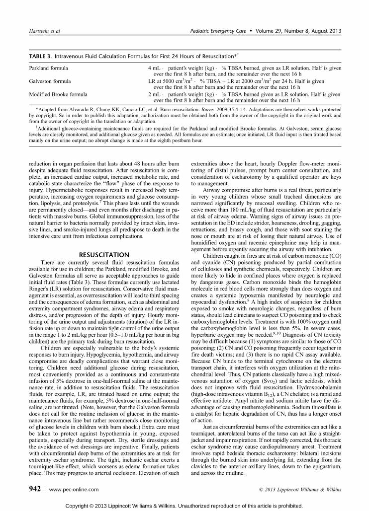
TABLE 3. Intravenous Fluid Calculation Formulas for First 24 Hours of Resuscitation*†
Parkland formula 4 mL � patient’s weight (kg) � % TBSA burned, given as LR solution. Half is givenover the first 8 h after burn, and the remainder over the next 16 h
Galveston formula LR at 5000 cm3/m2 � % TBSA + LR at 2000 cm3/m2 per 24 h. Half is givenover the first 8 h after burn and the remainder over the next 16 h
Modified Brooke formula 2 mL � patient’s weight (kg) � % TBSA burned given as LR solution. Half is givenover the first 8 h after burn and the remainder over the next 16 h
*Adapted from Alvarado R, Chung KK, Cancio LC, et al. Burn resuscitation. Burns. 2009;35:4Y14. Adaptations are themselves works protectedby copyright. So in order to publish this adaptation, authorization must be obtained both from the owner of the copyright in the original work andfrom the owner of copyright in the translation or adaptation.
†Additional glucose-containing maintenance fluids are required for the Parkland and modified Brooke formulas. At Galveston, serum glucoselevels are closely monitored, and additional glucose given as needed. All formulas are an estimate; once initiated, LR fluid input is then titrated basedmainly on the urine output; no abrupt change is made at the eighth postburn hour.
Hartstein et al Pediatric Emergency Care & Volume 29, Number 8, August 2013
942 www.pec-online.com * 2013 Lippincott Williams & Wilkins
Copyright © 2013 Lippincott Williams & Wilkins. Unauthorized reproduction of this article is prohibited.
WOUND MANAGEMENT ANDBIOLOGIC DRESSINGS
Minor BurnsThe last 2 decades have seen an explosion of options for
the treatment of burn wounds. This section will outline a logicalapproach to selection of topical treatments. Treatment of minorburns (G5% TBSA) can usually be accomplished in the ED withoutpatient referral to a primary care physician or surgeon forsubsequent care. The mainstay of treatment is cleansing and de-bridement of sloughing tissue. Broken blisters may be removedwith warm water, soap, and coarse-mesh gauze. Intact blistersmay be broken or left in place to protect against infection; a ruleof thumb described by some authors is to debride large (92-cmdiameter) blisters. Pain management is essential. Minor burnsshould be treated with a topical antimicrobial, daily dressingchanges, and dry nonadherent gauze. Inability to tolerate dailyhome wound care may necessitate admission. Topical antibiotics
such as bacitracin, polymyxin, or silver sulfadiazine can be usedon burns of limited extent.11 An alternative approach is to use aproduct that can contribute a moist, protective, wound-healingenvironment. A list of such products is provided in Table 4.
Extensive BurnsWith extensive burns, the risk of infection increases, and
the focus of topical care shifts to prevention of infection. Thetraditional topical antimicrobial burn creams are silver sulfa-diazine and mafenide acetate. Although bactericidal and relativelypainless on application, silver sulfadiazine has some limitations. Itpromotes the accumulation of proteinaceous exudate on thewound surface and can retard keratinocyte migration and theepithelialization process.12 Mafenide acetate offers better gram-negative coverage than any other topical agent and has theadded benefit of penetration into eschar and cartilage. Thus, itremains the drug of choice for bacterial burn wound infections.It has the disadvantages of pain on application, no antifungal
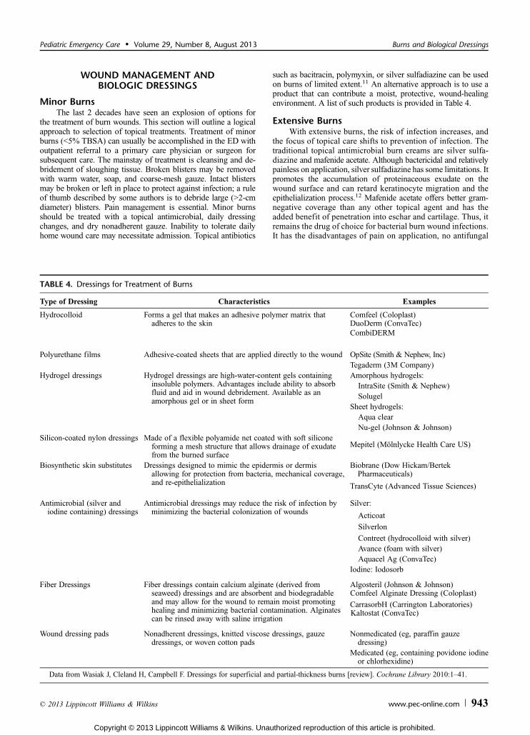
TABLE 4. Dressings for Treatment of Burns
Type of Dressing Characteristics Examples
Hydrocolloid Forms a gel that makes an adhesive polymer matrix thatadheres to the skin
Comfeel (Coloplast)DuoDerm (ConvaTec)CombiDERM
Polyurethane films Adhesive-coated sheets that are applied directly to the wound OpSite (Smith & Nephew, Inc)Tegaderm (3M Company)
Hydrogel dressings Hydrogel dressings are high-water-content gels containinginsoluble polymers. Advantages include ability to absorbfluid and aid in wound debridement. Available as anamorphous gel or in sheet form
Amorphous hydrogels:IntraSite (Smith & Nephew)Solugel
Sheet hydrogels:Aqua clearNu-gel (Johnson & Johnson)
Silicon-coated nylon dressings Made of a flexible polyamide net coated with soft siliconeforming a mesh structure that allows drainage of exudatefrom the burned surface
Mepitel (Molnlycke Health Care US)
Biosynthetic skin substitutes Dressings designed to mimic the epidermis or dermisallowing for protection from bacteria, mechanical coverage,and re-epithelialization
Biobrane (Dow Hickam/BertekPharmaceuticals)
TransCyte (Advanced Tissue Sciences)
Antimicrobial (silver andiodine containing) dressings
Antimicrobial dressings may reduce the risk of infection byminimizing the bacterial colonization of wounds
Silver:
Acticoat
Silverlon
Contreet (hydrocolloid with silver)Avance (foam with silver)Aquacel Ag (ConvaTec)
Iodine: Iodosorb
Fiber Dressings Fiber dressings contain calcium alginate (derived fromseaweed) dressings and are absorbent and biodegradableand may allow for the wound to remain moist promotinghealing and minimizing bacterial contamination. Alginatescan be rinsed away with saline irrigation
Algosteril (Johnson & Johnson)Comfeel Alginate Dressing (Coloplast)
CarrasorbH (Carrington Laboratories)Kaltostat (ConvaTec)
Wound dressing pads Nonadherent dressings, knitted viscose dressings, gauzedressings, or woven cotton pads
Nonmedicated (eg, paraffin gauzedressing)
Medicated (eg, containing povidone iodineor chlorhexidine)
Data from Wasiak J, Cleland H, Campbell F. Dressings for superficial and partial-thickness burns [review]. Cochrane Library 2010:1Y41.
Pediatric Emergency Care & Volume 29, Number 8, August 2013 Burns and Biological Dressings
* 2013 Lippincott Williams & Wilkins www.pec-online.com 943
Copyright © 2013 Lippincott Williams & Wilkins. Unauthorized reproduction of this article is prohibited.
activity, and a risk of metabolic acidosis if used twice daily onextensive wounds (due to absorption of a metabolite, whichinhibits carbonic anhydrase in the kidneys). To minimize theadverse effects and maximize the advantages of both agents,the usual practice at several burn centers has been ‘‘alternatingagents’’: that is, (1) to perform twice-daily wound care to in-clude thorough cleansing with chlorhexidine gluconate and (2)to alternate mafenide acetate in the morning with silver sulfa-diazine in the evening.
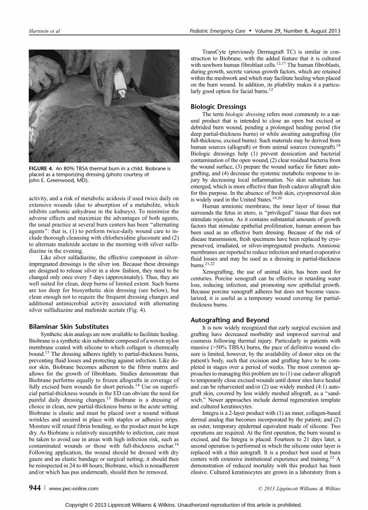
Like silver sulfadiazine, the effective component in silver-impregnated dressings is the silver ion. Because these dressingsare designed to release silver in a slow fashion, they need to bechanged only once every 5 days (approximately). Thus, they arewell suited for clean, deep burns of limited extent. Such burnsare too deep for biosynthetic skin dressing (see below), butclean enough not to require the frequent dressing changes andadditional antimicrobial activity associated with alternatingsilver sulfadiazine and mafenide acetate (Fig. 4).
Bilaminar Skin SubstitutesSynthetic skin analogs are now available to facilitate healing.
Biobrane is a synthetic skin substitute composed of awoven nylonmembrane coated with silicone to which collagen is chemicallybound.13 The dressing adheres tightly to partial-thickness burns,preventing fluid losses and protecting against infection. Like do-nor skin, Biobrane becomes adherent to the fibrin matrix andallows for the growth of fibroblasts. Studies demonstrate thatBiobrane performs equally to frozen allografts in coverage offully excised burn wounds for short periods.14 Use on superfi-cial partial-thickness wounds in the ED can obviate the need forpainful daily dressing changes.15 Biobrane is a dressing ofchoice in clean, new partial-thickness burns in the acute setting.Biobrane is elastic and must be placed over a wound withoutwrinkles and secured in place with staples or adhesive strips.Moisture will retard fibrin bonding, so the product must be keptdry. As Biobrane is relatively susceptible to infection, care mustbe taken to avoid use in areas with high infection risk, such ascontaminated wounds or those with full-thickness eschar.16
Following application, the wound should be dressed with drygauze and an elastic bandage or surgical netting; it should thenbe reinspected in 24 to 48 hours; Biobrane, which is nonadherentand/or which has pus underneath, should then be removed.
TransCyte (previously Dermagraft TC) is similar in con-struction to Biobrane, with the added feature that it is culturedwith newborn human fibroblast cells.12,17 The human fibroblasts,during growth, secrete various growth factors, which are retainedwithin the meshwork and which may facilitate healing when placedon the burn wound. In addition, its pliability makes it a particu-larly good option for facial burns.12
Biologic DressingsThe term biologic dressing refers most commonly to a nat-
ural product that is intended to close an open but excised ordebrided burn wound, pending a prolonged healing period (fordeep partial-thickness burns) or while awaiting autografting (forfull-thickness, excised burns). Such materials may be derived fromhuman sources (allograft) or from animal sources (xenograft).18
Biologic dressings help (1) prevent dessication and bacterialcontamination of the open wound, (2) clear residual bacteria fromthe wound surface, (3) prepare the wound surface for future auto-grafting, and (4) decrease the systemic metabolic response to in-jury by decreasing local inflammation. No skin substitute hasemerged, which is more effective than fresh cadaver allograft skinfor this purpose. In the absence of fresh skin, cryopreserved skinis widely used in the United States.19,20
Human amnionic membrane, the inner layer of tissue thatsurrounds the fetus in utero, is ‘‘privileged’’ tissue that does notstimulate rejection. As it contains substantial amounts of growthfactors that stimulate epithelial proliferation, human amnion hasbeen used as an effective burn dressing. Because of the risk ofdisease transmission, fresh specimens have been replaced by cryo-preserved, irradiated, or silver-impregnated products. Amnionicmembranes are reported to reduce infection and retard evaporativefluid losses and may be used as a dressing in partial-thicknessburns.21,22
Xenografting, the use of animal skin, has been used forcenturies. Porcine xenograft can be effective in retarding waterloss, reducing infection, and promoting new epithelial growth.Because porcine xenograft adheres but does not become vascu-larized, it is useful as a temporary wound covering for partial-thickness burns.
Autografting and BeyondIt is now widely recognized that early surgical excision and
grafting have decreased morbidity and improved survival andcosmesis following thermal injury. Particularly in patients withmassive (950% TBSA) burns, the pace of definitive wound clo-sure is limited, however, by the availability of donor sites on thepatient’s body, such that excision and grafting have to be com-pleted in stages over a period of weeks. The most common ap-proaches to managing this problem are to (1) use cadaver allograftto temporarily close excised wounds until donor sites have healedand can be reharvested and/or (2) use widely meshed (4:1) auto-graft skin, covered by less widely meshed allograft, as a ‘‘sand-wich.’’ Newer approaches include dermal regeneration templateand cultured keratinocytes.
Integra is a 2-layer product with (1) an inner, collagen-baseddermal analog that becomes incorporated by the patient; and (2)an outer, temporary epidermal equivalent made of silicone. Twooperations are required. At the first operation, the burn wound isexcised, and the Integra is placed. Fourteen to 21 days later, asecond operation is performed in which the silicone outer layer isreplaced with a thin autograft. It is a product best used at burncenters with extensive institutional experience and training.22 Ademonstration of reduced mortality with this product has beenelusive. Cultured keratinocytes are grown in a laboratory from a
FIGURE 4. An 80% TBSA thermal burn in a child. Biobrane isplaced as a temporizing dressing (photo courtesy ofJohn E. Greenwood, MD).
Hartstein et al Pediatric Emergency Care & Volume 29, Number 8, August 2013
944 www.pec-online.com * 2013 Lippincott Williams & Wilkins
Copyright © 2013 Lippincott Williams & Wilkins. Unauthorized reproduction of this article is prohibited.
biopsy obtained from the patient after injury. Although they havea low rate of permanent engraftment, these cells may be lifesavingfor selected patients with the most extensive burns.
CASE 1 SUMMARY OF CLINICAL COURSEThe patient was electively intubated. Laboratory testing re-
vealed a carboxyhemoglobin level of 15%, and the patient wasplaced on 100% oxygen. Total body surface area burn estimation(partial and full thickness) was 25% and arrangement for transferto a burn center was made. While awaiting transport, the burnswere treated with dry sterile dressing, and pain medication wasadministered as needed.
His weight is estimated at 16 kg. His maintenance fluidswould be 52 mL/h (40 mL for the first 10 kg plus 2 mL/kg foradditional 6 kg). Using the modified Brooke formula for chil-dren, his calculated fluid resuscitation requirements are 75 mL/h.That is, 3 mL � 16 kg � 25% = 1200 mL for the first 24 hours,with half given over the first 8 hours. Thus, for the first 8 hours,total fluids to be infused are estimated as 75 mL/h plus 52 mL/hfor a total of 127 mL/h. After LR is initiated at the 75-mL/h rate,it is adjusted hourly (up or down) to achieve a target urine out-put of 1 to 2 mL/kg per hour.
In this article, the authors have presented strategies formanaging burn injuries in children in the acute phase includingidentification of burn severity, estimation of body surface areainvolvement, and fluid resuscitation calculation and discussedhow biologic burn dressings can facilitate the growth of newskin and promote wound healing.
REFERENCES
1. Armour AD, Billmire DA. Pediatric thermal injury: acute care andreconstruction update. Plast Reconstr Surg. 2009;124:117eY127e.
2. Barrow RE, Spies M, Barrow LN, et al. Influence of demographicsand inhalation injury on burn mortality in children. Burns.2004;30:72Y77.
3. D’Souza AL, Nelson NG, McKenzie LB. Pediatric burn injuries treatedin US emergency departments between 1990 and 2006. Pediatrics.2009;124:1424Y1430.
4. American Burn Association. Scald Injury Prevention Educator’sGuide. Available at: www.ameriburn.org/Preven/ScaldInjuryEducator’sGuide.pdf. Accessed January 2, 2012.
5. Klein GL, Herndon DN. Burns. Pediatr Rev. 2004;25:411Y417.
6. Grunwald TB, Garner WL. Acute burns. Plast Reconstr Surg. 2008;121:311eY319e.
7. Tredget EE, Yu YM. The metabolic effects of thermal injury. World J
Surg. 1992;16:68Y79.
8. Kao LW, Nanagas KA. Toxicity associated with carbon monoxide.Clin Lab Med. 2006;26:99Y125.
9. Weaver LK, Howe S, Hopkins R, et al. Carboxyhemoglobin half-lifein carbon monoxideYpoisoned patients treated with 100% oxygen atatmospheric pressure. Chest. 2000;117:801Y808.
10. Crocker PJ, Walker JS. Pediatric carbon monoxide toxicity. J EmergMed. 1985;3:443Y448.
11. Dunn K, Edwards-Jones V. The role of Acticoat with nanocrystallinesilver in the management of burns. Burns. 2004;30(suppl 1):S1YS9.
12. Noordenbos J, Dore C, Hansbrough JF. Safety and efficacy of TransCytefor the treatment of partial-thickness burns. J Burn Care Rehabil.1999;20:275Y281.
13. Hansbrough JF, Morgan J, Greenleaf G, et al. Development of atemporary living skin replacement composed of human neonatalfibroblasts cultured in Biobrane, a synthetic dressing material. Surgery.1994;115:633Y644.
14. Purdue GF, Hunt JL, Gillespie RW, et al. Biosynthetic skin substituteversus frozen human cadaver allograft for temporary coverage ofexcised burn wounds. J Trauma. 1987;27:155Y157.
15. Whitaker IS, Prowse S, Potokar TS. A critical evaluation of the use ofBiobrane as a biologic skin substitute: a versatile tool for the plasticand reconstructive surgeon. Ann Plast Surg. 2008;60:333Y337.
16. Lesher AP, Curry RH, Evans J, et al. Effectiveness of Biobrane fortreatment of partial-thickness burns in children. J Pediatr Surg.2011;46:1759Y1763.
17. Hansen SL, Voigt DW, Wiebelhaus P, et al. Using skin replacementproducts to treat burns and wounds. Adv Skin Wound Care. 2001;14:37Y44; quiz 5Y6.
18. Stafford P. Burn management in pediatric patients. Pediatr Emerg Med
Rep. 2010;15:213Y224.
19. Landeen LK, Ziegler FC, Halberstadt C. Characterization of humandermal replacement. Wounds. 1992;4:167Y175.
20. Pham C, Greenwood J, Cleland H, et al. Bioengineered skin substitutesfor the management of burns: a systematic review. Burns. 2007;33:946Y957.
21. Greenwood JE. A randomized, prospective study of the treatment ofsuperficial partial-thickness burns: AWBAT-S versus Biobrane.Eplasty. 11:e10.
22. Saffle JR. Closure of the excised burn wound: temporary skinsubstitutes. Clin Plast Surg. 2009;36:627Y641.
Pediatric Emergency Care & Volume 29, Number 8, August 2013 Burns and Biological Dressings
* 2013 Lippincott Williams & Wilkins www.pec-online.com 945
Copyright © 2013 Lippincott Williams & Wilkins. Unauthorized reproduction of this article is prohibited.
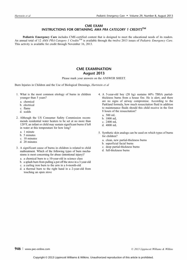
CME EXAMINSTRUCTIONS FOR OBTAINING AMA PRA CATEGORY 1 CREDITSTM
Pediatric Emergency Care includes CME-certified content that is designed to meet the educational needs of its readers.An annual total of 12 AMA PRA Category 1 CreditsTM is available through the twelve 2013 issues of Pediatric Emergency Care.This activity is available for credit through November 16, 2013.
CME EXAMINATIONAugust 2013
Please mark your answers on the ANSWER SHEET.
Burn Injuries in Children and the Use of Biological Dressings, Hartstein et al
1. What is the most common etiology of burns in childrenyounger than 5 years?a. chemicalb. electricalc. flamed. scalds
2. Although the US Consumer Safety Commission recom-mends residential water heaters to be set at no more than120-F, an infant or child may sustain significant burns if leftin water at this temperature for how long?a. 1 minuteb. 5 minutesc. 10 minutesd. 20 minutes
3. A significant cause of burns in children is related to childmaltreatment. Which of the following types of burn mecha-nisms is most concerning for abuse (intentional injury)?a. a chemical burn to a 10-year-old in science classb. a splash burn from pulling a pot off the stove in a 3-year-oldc. a curling iron burn to the arm in a 6-month-oldd. a thermal burn to the right hand in a 2-year-old from
touching an open stove
4. A 5-year-old boy (20 kg) sustains 60% TBSA partial-thickness burns from a house fire. He is alert, and thereare no signs of airway compromise. According to theParkland formula, how much resuscitation fluid in additionto maintenance fluids should this child receive in the first8 hours of the resuscitation?a. 500 mLb. 1000 mLc. 2400 mLd. 4800 mL
5. Synthetic skin analogs can be used on which types of burnsfor children?a. clean, new partial-thickness burnsb. superficial facial burnsc. deep partial-thickness burnsd. full-thickness burns
Hartstein et al Pediatric Emergency Care & Volume 29, Number 8, August 2013
946 www.pec-online.com * 2013 Lippincott Williams & Wilkins
Copyright © 2013 Lippincott Williams & Wilkins. Unauthorized reproduction of this article is prohibited.
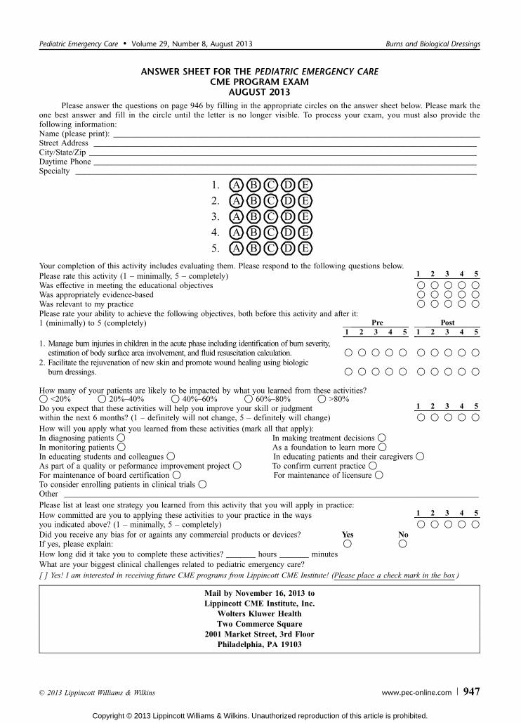
ANSWER SHEET FOR THE PEDIATRIC EMERGENCY CARECME PROGRAM EXAM
AUGUST 2013Please answer the questions on page 946 by filling in the appropriate circles on the answer sheet below. Please mark the
one best answer and fill in the circle until the letter is no longer visible. To process your exam, you must also provide thefollowing information:Name (please print): _______________________________________________________________________________Street Address ____________________________________________________________________________________City/State/Zip _____________________________________________________________________________________Daytime Phone ____________________________________________________________________________________Specialty ________________________________________________________________________________________
1. A B C D E
2. A B C D E
3. A B C D E
4. A B C D E
5. A B C D E
Your completion of this activity includes evaluating them. Please respond to the following questions below.Please rate this activity (1 Y minimally, 5 Y completely) 1 2 3 4 5
Was effective in meeting the educational objectives ° ° ° ° °Was appropriately evidence-based ° ° ° ° °Was relevant to my practice ° ° ° ° °Please rate your ability to achieve the following objectives, both before this activity and after it:1 (minimally) to 5 (completely) Pre Post
1 2 3 4 5 1 2 3 4 5
1. Manage burn injuries in children in the acute phase including identification of burn severity,estimation of body surface area involvement, and fluid resuscitation calculation. ° ° ° ° ° ° ° ° ° °
2. Facilitate the rejuvenation of new skin and promote wound healing using biologicburn dressings. ° ° ° ° ° ° ° ° ° °
How many of your patients are likely to be impacted by what you learned from these activities?° G20% ° 20%Y40% ° 40%Y60% ° 60%Y80% ° 980%Do you expect that these activities will help you improve your skill or judgment 1 2 3 4 5
within the next 6 months? (1 Y definitely will not change, 5 Y definitely will change) ° ° ° ° °
How will you apply what you learned from these activities (mark all that apply):In diagnosing patients ° In making treatment decisions °In monitoring patients ° As a foundation to learn more °In educating students and colleagues ° In educating patients and their caregivers °As part of a quality or peformance improvement project ° To confirm current practice °For maintenance of board certification ° For maintenance of licensure °To consider enrolling patients in clinical trials °Other ___________________________________________________________________________________________________Please list at least one strategy you learned from this activity that you will apply in practice:How committed are you to applying these activities to your practice in the ways 1 2 3 4 5
you indicated above? (1 Y minimally, 5 Y completely) ° ° ° ° °Did you receive any bias for or againts any commercial products or devices? Yes NoIf yes, please explain: ° °How long did it take you to complete these activities? _______ hours _______ minutesWhat are your biggest clinical challenges related to pediatric emergency care?[ ] Yes! I am interested in receiving future CME programs from Lippincott CME Institute! (Please place a check mark in the box )
Mail by November 16, 2013 toLippincott CME Institute, Inc.
Wolters Kluwer HealthTwo Commerce Square
2001 Market Street, 3rd FloorPhiladelphia, PA 19103
Pediatric Emergency Care & Volume 29, Number 8, August 2013 Burns and Biological Dressings
* 2013 Lippincott Williams & Wilkins www.pec-online.com 947
Copyright © 2013 Lippincott Williams & Wilkins. Unauthorized reproduction of this article is prohibited.
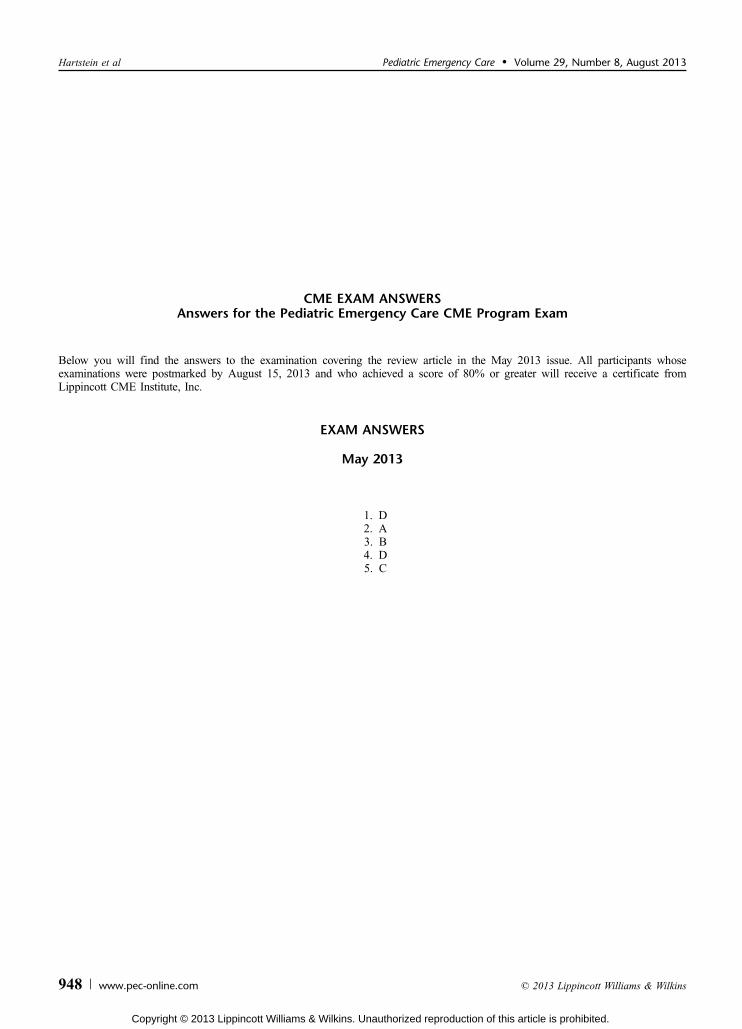
CME EXAM ANSWERSAnswers for the Pediatric Emergency Care CME Program Exam
Below you will find the answers to the examination covering the review article in the May 2013 issue. All participants whoseexaminations were postmarked by August 15, 2013 and who achieved a score of 80% or greater will receive a certificate fromLippincott CME Institute, Inc.
EXAM ANSWERS
May 2013
1. D2. A3. B4. D5. C
Hartstein et al Pediatric Emergency Care & Volume 29, Number 8, August 2013
948 www.pec-online.com * 2013 Lippincott Williams & Wilkins
Copyright © 2013 Lippincott Williams & Wilkins. Unauthorized reproduction of this article is prohibited.