Embed Size (px)
Citation preview

PRIMER
Budding Yeast for Budding Geneticists: A Primer onthe Saccharomyces cerevisiae Model System
Andrea A. Duina,* Mary E. Miller,† and Jill B. Keeney‡,1
*Biology Department, Hendrix College, Conway, Arkansas 72032, †Department of Biology, Rhodes College, Memphis, Tennessee38112, and ‡Department of Biology, Juniata College, Huntingdon, Pennsylvania 16652
SUMMARY The budding yeast Saccharomyces cerevisiae is a powerful model organism for studying fundamental aspects of eukary-otic cell biology. This Primer article presents a brief historical perspective on the emergence of this organism as a premier experimentalsystem over the course of the past century. An overview of the central features of the S. cerevisiae genome, including the nature of itsgenetic elements and general organization, is also provided. Some of the most common experimental tools and resources available toyeast geneticists are presented in a way designed to engage and challenge undergraduate and graduate students eager to learn moreabout the experimental amenability of budding yeast. Finally, a discussion of several major discoveries derived from yeast studieshighlights the far-reaching impact that the yeast system has had and will continue to have on our understanding of a variety of cellularprocesses relevant to all eukaryotes, including humans.
THE model organism Saccharomyces cerevisiae is com-monly known as baker’s yeast or brewer’s yeast. Indeed,
the scientific name cerevisiae derives from old world termi-nology for beer (Mortimer 2000). To scientists, S. cerevisiae isclassified as a fungus or mold, although those who enjoyartisan breads or a great microbrew would probably rathernot think of it that way! S. cerevisiae is a single-celled eukary-ote, and thus it contains membrane-bound organelles, such asa nucleus, endomembrane system, and mitochondria. Yeastcells divide as rapidly as once every 90 min under optimallaboratory conditions, through a process of budding in whichsmaller daughter cells pinch, or bud, off the mother cell (seeFigure 1). The common name “budding yeast” derives fromthis notable feature of cell division and distinguishes S. cer-evisiae from the fission yeast, Schizosaccharomyces pombe, alsoa powerful model organism. S. cerevisiae cells in nature switchreadily between two mating types: haploid a cells mate withhaploid a cells to form diploids. Under nutrient-poor condi-tions, diploids can be induced to undergo meiosis and spor-ulation, forming four haploid spores, two of each mating type(Figure 2). Due to their microscopic size and simple growthrequirements, yeast cells are inexpensive and easy to grow inthe laboratory. Unbudded yeast cells are �5 mm in diameter,
between bacteria and human cells in size. They form colonieson agar plates in the laboratory in a few days with no specialincubators required. Yeast stocks are maintained by freezingat 280� in glycerol or can be freeze-dried and stored at roomtemperature for years. In nature, yeasts are found in abun-dance in vineyards, but can also be found associated with oaktrees and in other natural habitats (Greig and Leu 2009).However, it is unclear whether S. cerevisiae as a species occursnaturally or exists solely as a domesticated species. We notethat recent genomic analyses suggest that wild populations ofS. cerevisiae exist (Mortimer 2000; Greig and Leu 2009).
The countless domesticated strains of S. cerevisiae are rec-ognized the world over for their ability to ferment sugars toethanol and carbon dioxide, producing a variety of beveragesenjoyed by all cultures. Human use of yeast is thought to havebegun.7000 years ago with the discovery that crushed grapesin water would ferment, as yeast is found in high abundanceon the fruit (Mortimer 2000). Yeast can be purposefully addedto other sources of sugars, such as grains, malts, or other plantmaterials, to produce other alcoholic beverages. Although a rel-atively “newer” discovery, the carbon dioxide released by yeasthas been used for centuries to raise bread dough, resulting inthe light texture of leavened bread. For most of history, winemakers relied solely on the yeast present on the harvestedgrapes, but brewers and bakers would lovingly transfer theirstarter cultures from batch to batch as S. cerevisiae workedunknown and unseen to ferment the various sugars provided
Copyright © 2014 by the Genetics Society of Americadoi: 10.1534/genetics.114.1631881Corresponding author: Department of Biology, Juniata College, 1700 Moore St.,Huntingdon, PA 16652. E-mail: [email protected]
Genetics, Vol. 197, 33–48 May 2014 33

by their owners. In the late 1800s, the Carlsberg Laboratoryintroduced the brewing industry to the process of science,and Emil Christian Hansen discovered how to purify yeastfrom mixed starter cultures (Greig and Leu 2009). In thepast 60 years, a rather short time evolutionarily speaking,the strains of S. cerevisiae used in modern baking, brewing,fermenting, and wine-making have been carefully cultured,selected, and purified by these respective industries; eachstrain has its own particular characteristics and they arenot interchangeable (Mortimer 2000). However, a few win-eries continue the practice of indigenous yeast fermentation,relying on the naturally provided yeasts of the grapes.
Yeast research has certainly established for itself a place inhistory, beginning with the famed first demonstrations ofenzymatic activities outside of a living cell by EdwardBuchner and continued with work with yeast extractsthrough the early 1900s, which laid the groundwork for bio-chemical analysis of metabolic pathways (Bohley and Fröhlich1997). In the 1930s, Øjvind Winge and Carl Lindegrenbegan the first work on yeast as an experimental organ-ism (Mortimer and Johnston 1986; Mortimer 2000). Wingestarted with strains isolated by Hansen at the Carlsberg brew-ery laboratory, while Lindegren used strain EM93 (isolated byEmil Mrak from rotting figs in California). Strain EM93 wasamenable to genetic crosses, a key development in modernyeast genetics. Multiple generations of genetic crosses al-lowed researchers to isolate specific traits and eventually tocharacterize the genes that encode those traits. One of themost widely used experimental strains, S288C, was con-structed in the early 1950s by Robert Mortimer (primarily fromEM93) and was subsequently used as the parental strain for theisolation of biochemical mutants (Mortimer and Johnston1986). Whereas the yeast system was beginning to attractattention from the scientific community, most notablythrough brilliant genetic and biochemical experiments byFred Sherman in the 1960s and 1970s (nicely reviewed inLiebman and Haber 2013), there was little to distinguish itfrom the dozens of experimental organisms being developedat the time. Then in 1978 S. cerevisiae was successfully trans-formed with a plasmid that had been replicated in the bacterium
Escherichia coli. The publication of the “Transformation ofyeast” launched the stellar research career of this simpleorganism (Hinnen et al. 1978).
What ultimately has distinguished yeast as a premiermodel organism is the ease with which researchers can movegenes in and out of yeast cells, either on plasmids or withinthe yeast chromosomes. Thus, geneticists can easily mutate ormanipulate yeast gene expression and study the resultingphenotypic effects. Following the discovery of transformation,the subsequent rapid development of a veritable menu ofreplicating plasmids and selectable markers set in place the“awesome power of yeast genetics.” Naturally occurring yeastreplication origins were adapted to create plasmids that rep-licate within the yeast cell. The cloning of selectable markersinto these plasmids, in the form of auxotrophic marker genesthat provide essential enzymes needed for growth or drugresistance genes, led to the development of large sets of stan-dardized plasmids. Moreover, in the absence of a replicationsignal, transformed DNA integrates readily into the yeast ge-nome via homologous recombination, allowing for targetedgene sequences to be disrupted. The advent of polymerasechain reaction (PCR), along with the efficiency of homologousrecombination in yeast, has led to the development of “de-signer deletion strains,” which, combined with the compre-hensively annotated Saccharomyces Genome Database (SGD;http://www.yeastgenome.org/). gives yeast geneticists flexi-bility in experimental design that is the envy of investigatorsworking with other model organisms (Goffeau et al. 1996;Brachmann et al. 1998; Cherry et al. 2012).
S. cerevisiae Genome Organization
The genes
In 1996, the S. cerevisiae genome became the first fully se-quenced eukaryotic genome (Goffeau et al. 1996). In additionto representing a landmark achievement in the history ofeukaryotic biology, the sequencing of the S. cerevisiae genomehas provided a wealth of information on various aspects ofgenome organization and evolution. In a typical haploidbudding yeast cell, the �12,000 kb of genomic DNA are
Figure 1 Budding yeast cells. (A) Confocal fluores-cence microscopy of haploid yeast expressing Spc42-GFP (green: spindle pole body marker) and HistoneH2-mCherry (red: nuclear marker). The yeast strainwas constructed by Shawnecca Burke, an undergradu-ate in M.E.M.’s research laboratory by crossing strainskindly provided by Jan Skotheim (Stanford University)and Mark Winey (University of Colorado at Boulder)(photo by Mary Miller). (B) Epifluorescence microscopyof diploid yeast cells expressing Spt16-GFP, a nuclearprotein. The yeast strain was generated by students inA.A.D.’s Spring 2005 Advanced Cell Biology class(photo by Andrea Duina). (C) Electron microscopy ofdividing yeast cells (photo by Christine Walls). Note thatbuds are visible emerging from some of the cells in eachof the panels. Unbudded cells are �5 mm in diameter.
34 A. A. Duina, M. E. Miller, and J. B. Keeney

subdivided into 16 chromosomes, thought to have arisen fol-lowing an ancient whole-genome duplication event from anancestral set of 8 distinct chromosomes (Kellis et al. 2004). Aswould be expected for any eukaryotic genome, the buddingyeast genome is studded with a large number of genes thatcan be broadly grouped into those that encode proteins (pro-tein-coding genes) and those that do not (noncoding genes).According to the SGD (http://www.yeastgenome.org/), as ofFebruary 3, 2014, the number of “verified open readingframes (ORFs)” in the reference strain S288C stood at5076. Perhaps surprisingly for such a well-characterizedand extensively studied model system, there are still a rela-tively large number of ORFs—745 as of the same date—likelyto contain information for the synthesis of proteins but forwhich experimental data are lacking as to whether the pro-teins are in fact expressed in cells and, if so, what their func-tions might be. The density of protein-encoding genes in thebudding yeast genome is quite high (one gene every �2 kb),�50-fold higher than the gene density in the human genome.High gene density is partly explained by the relatively lownumber of intron-containing genes in S. cerevisiae, estimatedto be at �4% of all genes (Goffeau et al. 1996; Spingola et al.1999). The S. cerevisiae genome also harbors 786 so-called“dubious ORFs,” which, while technically being ORFs, areunlikely to encode proteins.
The noncoding genes present in the budding yeast genomeinclude those transcribed into RNA molecules involved in thetranslation process [transfer RNA (tRNA) genes, close to 300 innumber, and ribosomal DNA (rDNA) genes, present in a tandemarray of 100–200 copies on chromosome XII, encoding fourdistinct rRNA molecules]; those involved in pre-messengerRNA splicing (small nuclear RNA genes, 5 in total); andthose that facilitate chemical modifications of a variety ofRNA molecules (small nucleolar RNA genes, 76 genes in total)(Piekna-Przybylska et al. 2007; Kavanaugh and Dietrich 2009).
Additional types of noncoding genes include SCR1 and TLC1,whose RNA products participate in protein targeting to theendoplasmic reticulum and in synthesis of telomeric DNA, re-spectively (Singer and Gottschling 1994; Van Nues and Brown2004), and the regulatory genes SRG1, ICR1, and PWR1,whose transcription directly regulates the expression of adja-cent genes through a phenomenon referred to as transcriptioninterference (Martens et al. 2004; Bumgarner et al. 2009).Additional genomic elements have been discovered that areable to regulate transcription of nearby genes through thegeneration of noncoding RNAs (e.g., see Hongay et al. 2006;Houseley et al. 2008; Pinskaya et al. 2009; Gelfand et al. 2011;van Werven et al. 2012; Castelnuovo et al. 2013), and, basedon the finding that noncoding RNA synthesis appears to bea widespread phenomenon in budding yeast (David et al.2006; Neil et al. 2009; Xu et al. 2009), many more such reg-ulatory examples are likely to be identified in the future.
Genomic regions involved in chromosome maintenance
Several features of the yeast genome ensure that chromo-somes are properly replicated, maintained, and eventuallysegregated to cells following either mitosis or meiosis.Origins of replication are located at �20- to 40-kb intervalson each chromosome. Many of these elements were origi-nally identified as sequences of a few hundred base pairsthat confer plasmids (otherwise devoid of their own replica-tion origin) the ability to replicate in budding yeast cellsfollowing transformation experiments and were thus coinedas autonomously replicating sequences (ARSs). Key func-tional aspects of ARS elements include the ability to recruita variety of factors involved in triggering DNA replication(with the origin recognition complex being a critical one)and the intrinsic propensity to easily unwind to facilitate theDNA replication process (for a review, see Dhar et al. 2012).Whereas many ARSs are bona fide origins of replication in
Figure 2 A simplified life cycle diagram of laboratorybudding yeast. Haploid yeast cells can be one of twomating types: MATa (a cell) or MATa (a cell). Thesecells can undergo mitotic cell division through bud-ding, producing daughter cells. In laboratory strains,the mating type of haploid cells is stable due to theabsence of a functional HO endonuclease. The twocell types release pheromones, initiating the forma-tion of schmoos and subsequent mating, resultingultimately in a stable diploid MATa/MATa (a/a cell).Diploid cells also divide mitotically by budding to pro-duce genetically identical daughter cells. Under nitrogen-poor conditions, diploids are induced to undergomeiosis,forming four haploid spores, which can germinate intotwo MATa cells and two MATa cells.
Primer 35
their natural chromosomal contexts, some are not, thus ne-cessitating the use of sophisticated techniques to unequivo-cally designate a specific ARS as a true chromosomal originof replication. The latest information on ARS designations inthe S. cerevisiae genome comes in the form of a website calledOriDB (http://cerevisiae.oridb.org/; also see Nieduszynskiet al. 2007; Siow et al. 2012).
Proper chromosome segregation during mitosis andmeiosis relies on the ability of kinetochore microtubules tomake specific contacts with chromosomes. Each of the 16S. cerevisiae chromosomes contains a centromere to directassembly of the kinetochore, itself responsible for makingdirect contacts with microtubules. These “point” centromeresare unusual among eukaryotes in that they are very short(�125 bp) and are not surrounded by heterochromatin.Nevertheless, because of their simplicity and their conservedfunctions, budding yeast centromeres have served as animportant model for understanding eukaryotic centromerebiology (for review, see Mehta et al. 2010). Combining cen-tromere (CEN) and ARS sequences in artificial plasmidsallows researchers an additional level of control of plasmidcopy number in the yeast system.
The ends of chromosomes are capped by telomeres,composed of specialized DNA sequences and associatedproteins. A typical S. cerevisiae telomeric region includesa heterogeneous stretch of �300 bp of the C1-3A /TG1-3 re-peat and an adjacent subtelomeric region referred to as thetelomere-associated sequence. Telomeres are one of threeregions in the budding yeast genome that form heterochro-matin-like environments—the others being the rDNA locusand the silent mating-type cassettes—and as such they havebeen used extensively as a model for understanding hetero-chromatin structure and function (for reviews, see Buhlerand Gasser 2009; Wellinger and Zakian 2012).
Additional genetic elements found in theS. cerevisiae genome
The S. cerevisiae genome houses a large number of so-calledlong terminal repeat (LTR) retrotransposons, called Ty ele-ments, scattered across all 16 chromosomes. The active formsof these transposable elements can transpose within thegenome through a cycle that, similar to mammalian retrovi-ruses, includes transcription and translation of the elementand subsequent assembly of viral-like particles (VLPs). Reversetranscription of the retrotransposons’ RNA into complementaryDNA (cDNA) by reverse transcriptase occurs within the VLPsand is followed by insertion of the cDNA into the genome. Arecent survey of the S288C genome identified 483 Ty elements,427 of which are inactive solo LTRs (Carr et al. 2012). Inter-estingly, Ty elements have evolved mechanisms of targeted in-tegration, favoring integration near Pol III-transcribed genes (e.g., tRNA genes) or telomeres and silent mating-type cassettes—likely as a strategy to maximize their chances of survival sincethe targeted regions are gene-poor and are therefore morelikely to withstand integration events without detrimentaleffects to the host cell (for review, see Boeke and Devine 1998).
Chromosome III in S. cerevisiae harbors genetic informa-tion that determines the mating-type identity of cells. Hap-loid yeast cells can be either mating type a or a (MATa andMATa, respectively), which, under the appropriate condi-tions, can mate with each other to generate MATa/MATadiploids. These diploid cells cannot mate but can reproducemitotically or can undergo meiosis to produce haploidspores (Figure 2). The mating behavior of yeast cells is de-termined by the MAT locus housed on the right arm of chro-mosome III (Figure 3). This locus can harbor one of twononhomologous alleles called MATa and MATa: MATa cellscontain the MATa allele, MATa cells contain the MATa al-lele, and, in most cases, diploid cells contain both alleles(each located on one of the two homologous chromosomeIIIs). The two MAT alleles express different sets of proteins,which, through a rather complex mechanism, regulateproper mating-type behavior of cells (reviewed in Haber2012). Chromosome III also carries an additional copy ofeach MAT allele at two additional loci: the HMRa locus lo-cated near the end of the right arm of the chromosomecontains the MATa allele and the HMLa locus located nearthe end of the left arm of the chromosome contains theMATa allele. Unlike the MAT locus, however, HMRa andHMLa (also known as silent mating-type cassettes) are em-bedded in heterochromatin and are therefore transcriptionally
Figure 3 Cartoon representation of the S. cerevisiae chromosome III andsimplified view of the change that it undergoes following a mating-typeswitching event. In a haploid MATa cell, the MAT locus on chromosomeIII houses the MATa allele (top). During a mating-type switching event,the genetic information at HMLa is used to replace theMATa allele at theMAT locus with the MATa allele. The resulting chromosome III (bottom)expresses MATa information, which causes the cell to become phenotyp-ically MATa. A similar mating-type switching mechanism operates duringthe switch of MATa cells into MATa cells. The genetic elements shown inthe diagram (HMLa, CEN, MATa, MATa, and HMRa) and their relativepositions across the chromosome are depicted roughly to scale (note thatchromosome III is 316,620 bp in length). The asterisks next to MATa andMATa highlight the fact that these alleles are actively expressed, as op-posed to the alleles present at the HMLa and HMRa loci that are tran-scriptionally silent.
36 A. A. Duina, M. E. Miller, and J. B. Keeney

silenced, but can be used by haploid cells to switch fromone mating type to the other. During mating-type switch-ing, a particular allele found at the MAT locus is replacedby the opposite MAT allele using the information locatedat either the HMRa or the HMLa locus (Figure 3). Com-monly used laboratory strains are unable to switch mat-ing types due to the lack of a functional form of anenzyme, the HO endonuclease, required to initiate theprocess, thus allowing for the generation and mainte-nance of genetically stable cell populations. The buddingyeast mating-type system has served as a remarkablemodel system for understanding a wide array of basiccellular processes, including heterochromatin formationand maintenance, transcriptional silencing, and homologousrecombination.
Chromosome organization within the buddingyeast nucleus
The availability of powerful genetic and cell microscopytools, combined with the development of the chromo-some conformation capture technique, which allowsinvestigators to assess the conformation of chromosomesin live cells (Dekker et al. 2002), has made S. cerevisiaea prime model system for examining the relationship be-tween chromosomal spatial arrangement and the regula-tion of processes that occur across chromosomes. Keyprinciples obtained from current work include the notionthat chromosomes adopt nonrandom positions withinbudding yeast nuclei [we now have a map showing howall 16 chromosomes are arranged within budding yeastnuclei (Duan et al. 2010)] and that specific subnuclearlocations are associated with specific chromosomal pro-cesses—for example, certain inducible genes are seen toassociate with the nuclear pores following their activa-tion (reviewed in Taddei and Gasser 2012). Yet anotherbreakthrough that features yeast is the development ofa genome-wide map of nucleosome positions at base-pairresolution, representing an important step in investigat-ing how nucleosomes drive the folding of chromosomesin living cells (Brogaard et al. 2012). Given its experimen-tal tractability, S. cerevisiae will continue to be a majorworkhorse in investigations of the relationship betweenhigher-order chromosome configurations and chromosome-based processes.
Experimental Toolkit and Related Resources
One of the authors often jokes with students that the ease ofexperimental manipulation offered by S. cerevisiae couldmake one think that it is not a naturally occurring organismbut that it exists only for the pleasure of those interested inunderstanding the intricate workings of eukaryotic cells. Al-though this comment may draw an occasional perplexedlook, it reflects a sentiment felt by many yeast geneticists.To illustrate many of the tools and resources available toresearchers who use budding yeast as an experimental organ-ism, below we describe a hypothetical scenario and a series ofexperimental approaches that a budding yeast researcher (thereader in this example) might use to conduct a scientific in-vestigation. To further promote the learning process, we haveincluded an activity at the end of this section in which weinvite readers to synthesize what they have learned to artic-ulate a possible model describing the biological process underinvestigation and to envision possible future research avenues(refer to Table 1).
Imagine that, after several years of intense work asa graduate student, you have identified a new chemicalcompound, which we shall call “Kill-It,” that appears to behighly toxic to yeast cells but that does not harm mamma-lian cells. You are very excited about this discovery since,given its apparent specificity toward yeast cells, Kill-It mayrepresent a promising new antifungal drug. However, inother experiments, you have discovered that yeast cellscan develop resistance to this drug. You now wish to de-termine the molecular mechanism that underlies this resis-tance. In defining the basis of resistance, you will further theunderstanding of the mechanism of action of the drug, in-fluence treatment design aimed at potentially synergisticdrugs, and possibly influence target-based drug design ofantifungal agents in the future. The experimental strategiesand hypothetical results for this project are presented below.
Isolation of mutants
As a way to determine the mechanism by which yeast candevelop resistance to Kill-It, you start by isolating mutantS. cerevisiae cells that can withstand exposure to the drug.There are several tools at your disposal to isolate such mutants.You can carry out a genetic selection experiment and transferbillions of mitotically dividing cells to solid growth medium
Table 1 Summary of Kill-It study
Experimental goal Technique/resource used Result
Isolate yeast mutants resistant to Kill-It Selection of spontaneous mutantsIdentify complementation groups Mating of MATa and MATa recessive mutantsClone wild-type version of the KIR1 gene Functional complementationDetermine the sequence of KIR1 and its chromosomal location Sequence the library clone; BLASTDetermine effect of deleting the KIR1 gene Targeted disruption to construct a kir1D strainDetermine cellular location of Kir1 protein Tag Kir1 at N and C termini with GFPDetermine which cellular components interact with Kir1 SGD interactions resourcesDetermine if and where Kir1 interacts with chromatin ChIP-seqDetermine impact of a KIR1 deletion on the expression of other genes RNA-seq
Primer 37

containing Kill-It and select for cells able to grow. Such cellswould have acquired one or more spontaneous mutations atsome point during their growth prior to exposure to Kill-Itthat rendered them resistant to Kill-It. If desired, the rate ofmutagenesis can be increased dramatically through the useof mutagens, such as ethyl methanesulfonate. Alternatively,you could identify mutants of interest through the use ofgenetic screens. For these experiments, colonies or patchesof haploid cells, each derived from a single cell carrying anindependent pre-existing mutation, can be grown on permis-sive solid medium and then transferred to Kill-It-containingmedium to screen for those mutants able to grow in thepresence of the drug. To facilitate your studies, you couldtake advantage of one of the several budding yeast deletionlibraries generated by the Saccharomyces Genome Deletion Pro-ject consortium (http://www-sequence.stanford.edu/group/yeast_deletion_project/deletions3.html), a collection of con-ditional mutants (Li et al. 2011), or a set of mutants generatedby insertional mutagenesis (see Vidan and Snyder 2001).
Classical genetic analyses of mutants
Using a spontaneous selection experiment, you isolate 40mutants that form colonies in the presence of Kill-It; you callthese mutants “kir” for Kill-It resistance. You can now takeadvantage of aspects of S. cerevisiae that make it such aneffective experimental organism. First, you can easily set upgenetic crosses between the kir mutants and wild-type cellsto obtain diploids heterozygous for the kir mutations to de-termine if the mutations are recessive or dominant. Thisinformation will be important in your efforts to identify thegene in question and also provides perspective on potential
mechanisms of the drug resistance that you observed in youroriginal mutant. For example, a dominant mutation mightsomehow inactivate the effect of a drug while a recessivemutation might represent a target of the drug. Next, sinceyou cleverly set up the original selection experiment usingboth MATa and MATa cells, you have some MATa kirmutants and some MATa kir mutants. By analyzing resultsof genetic crosses between recessive kir mutants, you cancarry out complementation tests that will help you deter-mine how many different genes are represented in yourmutant strains. You learn that all of the 40 original mutantsare recessive and that they can be grouped into a total of fivecomplementation groups. In other experiments, you estab-lish that in each case a single genetic mutation is responsiblefor the Kill-It-resistance phenotype. Thus, the mutants youhave isolated define mutations in five distinct genes, whichyou temporarily name KIR1-KIR5 (refer to Table 2 for genenomenclature in S. cerevisiae).
Tools for gene identification and initialfunctional characterization
You decide to focus your questions on KIR1. For example,what is the identity of the KIR1 gene? Has KIR1 already beenstudied by others, or are you the first person to identify it? Isthere any information about the possible function of KIR1?On which chromosome does KIR1 reside? To start to addressthese questions, you investigate whether KIR1 is involved inmorphological or cell cycle processes in the cell. You are ableto make use of simple microscopic analysis of yeast contain-ing the kir1 mutation, making note of any changes in cellshape or cell cycle distribution (budding is coincident with
Table 2 Gene and protein nomenclature for S. cerevisiae
Name Description Nomenclature
YGL264W Systematic ORF designation for the gene Each ORF in the yeast genome is assigned a systematic nameusing the following conventions: Y stands for yeast; G representsthe chromosome number (where A corresponds to chromosomeI; B to chromosome II, etc.). This ORF is therefore located onchromosome VII; L indicates that the ORF is on the left arm of thechromosome; 264 indicates that the ORF is the 264th from thecentromere, and W denotes that the coding strand of the ORF ison the Watson strand, which is the strand whose 59 end is locatedat the left telomere (the complementary strand is referred to asthe Crick strand, or C).
KIR1+ Wild-type gene Three italicized uppercase letters and number followed by asuperscripted + sign
KIR1 Dominant allele of the gene. This nomenclature isalso often used to refer to the wild-type gene.
Three italicized uppercase letters and number
KIR1-1, KIR1-2, etc. Distinct dominant alleles of the gene Dominant mutant allele designation followed by a hyphen and anumber to indicate specific allele
kir1 Recessive allele of the gene Three italicized lowercase letters and numberkir1-1, kir1-2, etc. Distinct recessive alleles of the gene Recessive mutant allele designation followed by a hyphen and a
number to indicate specific allelekir1Δ Deletion allele of the gene Recessive mutant allele designation followed by a Δ symbolKir1p Protein product of the gene Three letters, with the first being uppercase, followed by a number
and lower case p; all in roman fontKir1 Alternative nomenclature for protein product of the
geneThree letters, with the first being uppercase, followed by a number;all in roman font
The gene identified in Experimental Toolkit and Related Resources is used as an example.
38 A. A. Duina, M. E. Miller, and J. B. Keeney

cell cycle progression, and therefore defects in cell cycleregulation can be initially characterized by this observation).You find that the kir1 mutation does not impact cellularmorphology since kir1 mutant cells look similar to wild-typecells.
You then design an experiment to identify the KIR1 geneusing a functional complementation approach of the recessivekir1 allele. For these experiments, you obtain a genomic DNAlibrary derived from a wild-type strain. These libraries nor-mally contain random pieces of wild-type yeast genomic DNAinserted into yeast centromeric plasmids, which carry a cen-tromere and an ARS element and thus function as mini-chromosomes. Each plasmid in the library contains a piece ofwild-type DNA that is large enough to carry several genes,and the collection of all library plasmids generally covers theentire genome multiple times. You introduce the plasmidsinto kir1 cells using one of several transformation protocols,which in budding yeast are highly efficient (for examples, seeKawai et al. 2010). To select for kir1 cells that contain a li-brary plasmid following transformation, you plate the cellsonto medium on which only cells that contain a plasmidcan grow. The resulting transformants are then replica-platedto medium containing Kill-It to identify those that have beenrestored to a normal phenotype, i.e., are now Kill-It sensitive,as these will likely contain a plasmid containing the wild-typeKIR1 gene. The library plasmids from Kill-it-sensitive coloniescan then be easily retrieved using one of several plasmid re-covery techniques (Robzyk and Kassir 1992) and sequenced.Following analysis of the genomic library fragment, you ob-tain the DNA sequence corresponding to the wild-type versionof the KIR1 gene. With decreasing genomic sequencing costs,it is becoming more common to bypass functional comple-mentation and instead locate the site of a mutation by directlysequencing the genome of the mutant strain. A drawback ofthis genome-sequencing approach, however, is that in manyinstances it may be difficult to identify the mutation respon-sible for the phenotype under investigation if additional un-related mutations are also present in the genome of themutant strain.
With the DNA sequence of KIR1 in hand, you now move tothe treasure trove of information in the SGD (http://www.yeastgenome.org/; also see Cherry et al. 2012). As a repositoryfor the S. cerevisiae genome sequence (specifically, the S288Creference strain), SGD contains an enormous amount of infor-mation on gene and protein function, and it disseminates newsrelevant to budding yeast researchers. You begin by using theBLAST function, which compares the KIR1 sequence that youobtained to the entire yeast genome sequence. The BLASTanalysis reveals that KIR1 corresponds to the gene with thesystematic name YGL264W. [Each gene, whether previouslystudied or not, is assigned a systematic name following certainguidelines (see Table 2)]. When you read about YGL264W onSGD, you are excited to find that its function has not yet beendiscovered! You might be the first investigator to discovera function associated with this gene. Using the Gene Registryfunction at SGD you are now able to “reserve” KIR1 as the
standard name for YGL264W, which will become its officialstandard name once you publish it in a scientific journal.(Note: YGL264W does not exist; it has been created justfor this example.)
Computational approaches for investigatingprotein function
As a way to explore the possible function of the Kir1 protein incells (see Table 2 for protein nomenclature), you use theBLAST function available at the National Center for Biotech-nology Information website (http://blast.ncbi.nlm.nih.gov/) tocompare the Kir1 protein sequence against protein databasesfrom other organisms to see if a protein similar in sequence toKir1 has been previously characterized by others. To your de-light, you find that Kir1 has substantial sequence similarities toa protein in the fruit fly Drosophila melanogaster that is knownto be a transcription factor. SGD also enables cross-organismsequence comparisons of either proteins or DNA to identifysequences similar to KIR1 in different S. cerevisiae strains(http://www.yeastgenome.org/cgi-bin/FUNGI/alignment.pl)or in other fungi (http://www.yeastgenome.org/cgi-bin/FUNGI/showAlign). Although such comparisons are not likely to pro-vide insight into the function of Kir1, they can highlight evo-lutionarily conserved features of a gene or protein and thushelp in the characterization of the gene and protein underinvestigation.
Genetic approaches for investigating protein function
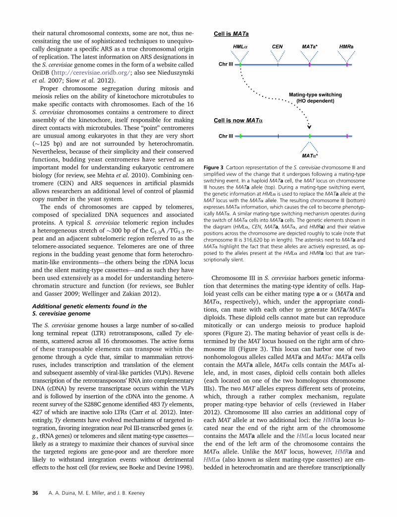
Many genetic tools are available in yeast to help youinvestigate the function of a previously uncharacterizedprotein. You decide to start by determining the effects ofa complete knockout of KIR1 on cell function using a one-step gene replacement approach (Figure 4A). To do this, youtransform a PCR product harboring a selectable markerflanked by DNA sequences homologous to genomic regionsflanking KIR1 into wild-type diploid cells and plate the cellsonto selective medium using a standard protocol (for anexample, see Brachmann et al. 1998). The transformationis done in diploid cells so that the resulting cells remain aliveeven if a deletion of KIR1 (kir1Δ) were to be lethal. Toexamine the effects of kir1D, you then induce the diploidcells to undergo meiosis, resulting in the generation of fourmeiotic products (two wild type and two kir1Δ) per originaldiploid cell, forming what yeast researchers call a “tetrad”(Figure 4B). The resulting tetrads can then be dissected ontosolid growth medium using a light microscope equippedwith a micromanipulator and the spores allowed to germi-nate into visible colonies (Figure 4B), which can be subse-quently analyzed for specific phenotypes using a procedurecommonly referred to as tetrad analysis. Based on your tet-rad analysis, you find that haploid kir1Δ cells are able togrow and, similarly to the mutants you isolated in the initialselection experiment, they are also resistant to Kill-It (Figure4B). Thus, KIR1 is not a gene that is essential for the life ofa yeast cell. In addition, you have ruled out the possibilitythat resistance to Kill-It is caused by a cryptic, secondary
Primer 39
mutation in your original studies instead of by your kir1mutation. A plethora of additional phenotypes can also beeasily scored to gain insight into the nature of the kir1Δmutants (see Hampsey 1997). These and some of the experi-ments described in the earlier sections underscore how theability to easily interconvert S. cerevisiae cells between thehaploid and diploid states can be extremely helpful whencarrying out genetic analyses.
The budding yeast system is also particularly well suited fora battery of genetic experiments that can uncover geneticinteractions between a gene of interest and other genes. Thesegenetic interactions often reflect physical and/or functionalinteractions between proteins and hence can be criticallyimportant when investigating the function of an uncharacter-ized protein. Examples of such genetic approaches include theisolation of spontaneous suppressor mutations and high-copysuppressors (for an extended discussion on suppressionanalysis in yeast, see Prelich 1999) and the implementation ofsynthetic lethal screens. Suppression and synthetic lethal inter-actions, as well as other types of genetic interactions, can alsobe unveiled through the use of a high-throughput methodologyknown as Synthetic Gene Array (SGA) analysis (for a detaileddescription, see Tong and Boone 2006). In an example of anSGA experiment, a query haploid strain harboring a null mu-tation in a gene of interest is crossed to a haploid deletionlibrary (consisting of �5000 haploid strains, each with a de-letion of a single nonessential gene) of the opposite mating
type. Using a series of replica-plating steps, haploid cells car-rying the original null mutation and one of each of the �5000gene deletions represented in the library can be selected andassayed for suppression or synthetic-lethal interactions. Whereasthese experiments can be carried out manually, investigatorsoften make use of robots to handle the hundreds of platesrequired for analysis of thousands of mutants. The phenotypicdata are then assembled into gene interaction networks thatcan be accessed as interactive maps. You instead opt to pro-ceed using the approaches described below.
Determining the cellular localization of a buddingyeast protein
The function of a protein in the cell is intimately related toits subcellular localization. To investigate the localization ofa protein within the cell, yeast geneticists often rely on theefficient homologous recombination system of S. cerevisiaeto generate gene fusions between a gene of interest and thegene encoding green fluorescent protein (GFP) from thejellyfish Aequorea victoria. The resulting engineered genewould then be expected to express a protein consisting ofthe protein of interest fused to GFP, which can be visualizedin live cells using fluorescence microscopy (see Figure 1,panels A and B). You may decide to use this approach togenerate two Kir1-GFP fusion proteins—one in which GFP isfused to the N terminus of Kir1 and another in which GFP isfused to the C terminus—to guard against the possibility
Figure 4 One-step gene replacement and analysis ofmeiotic products through tetrad analysis. (A) Deletionof KIR1. Step 1: A KIR1 homozygous diploid cell(KIR1/KIR1: only the cell nucleus is shown here) istransformed with a linear DNA molecule (usually gen-erated by PCR) containing a selectable marker geneflanked by regions identical in sequence to those thatflank the endogenous KIR1 gene (red regions in thecenter panel). Step 2: Following homologous recom-bination between the introduced DNA fragment andone of the two KIR1 genes, the transformed cell isheterozygous for the KIR1 gene and its genotype isKIR1/kir1Δ. (B) Generation of spores, tetrad manipu-lation, and tetrad analysis. The KIR1/kir1Δ cell from Ais then triggered to undergo meiosis through nitro-gen starvation to produce a tetrad—a set of fourspores encased in an ascus sac. Step 1: The ascusmembrane is partially digested and, through theuse of a light microscope equipped with a microma-nipulator, the four spores are released and placed ina row onto permissive solid growth medium andallowed to germinate; note that no cells are visibleto the naked eye immediately after this manipulationon the growth medium (the dark rectangle is a pho-tograph of a section of a growth plate as it wouldlook after the tetrad dissection). Step 2: Following�3days of incubation at 30�, the germinated spores give
rise to visible colonies on the growth medium (the photograph shows actual yeast colonies derived from germinated spores). Step 3: The colonies arethen replica-plated to solid growth medium containing Kill-It and allowed to incubate at 30� for 2 days. The 2:2 growth pattern of the colonies on thedrug plate (two alive and two unable to grow) is consistent with classic Mendelian segregation of a heterozygous trait and can be used to infer thegenotypes of the cells in each colony (and, by extension, of the original spores) as indicated to the right of the photograph. (Given the hypotheticalnature of the experiment, it should be noted that the actual genotypes of the cells photographed in this figure are not as indicated in the figure and thatthe medium in the last photograph does not contain Kill-It.)
40 A. A. Duina, M. E. Miller, and J. B. Keeney

that one fusion protein may misfold and thus be potentiallydegraded (Longtine et al. 1998). However, it would be wisefor you to first visit the Yeast GFP Fusion Localization Data-base (http://yeastgfp.yeastgenome.org/), which containssearchable protein localization data from a genome-widestudy in which all budding yeast ORFs have been fused toGFP (Huh et al. 2003). You may be able to answer yourquestion without doing a single experiment! Your resultssuggest that Kir1 is a nuclear protein, a finding consistentwith its orthology to a fly transcription factor. However, theKir1-GFP fusion protein may not function like the wild-typeKir1 protein and may not localize to its normal subcellularlocation. Thus, it is critical to determine whether or not theGFP-tagged version of your protein remains functional. Totest the functionality of Kir1-GFP, you check haploid cellsexpressing the fusion protein for Kill-It sensitivity and youbreathe a big sigh of relief when you see that the cells arestill sensitive to Kill-It. Thus, the Kir1-GFP fusion protein hasnormal function, at least in relation to growth on Kill-It, andyour characterized localization pattern is likely to be physi-ologically relevant to your studies.
Tools available to yeast researchers to detectprotein–protein interactions
Knowing which other proteins physically interact with Kir1can shed light on its function. Yeast biologists have severalexperimental techniques at their disposal to investigateprotein–protein interactions. One powerful approach is pro-vided by the yeast two-hybrid system, which allows for theidentification and the analyses of protein–protein interactionsin an in vivo setting (Figure 5 and Chien et al. 1991). Global
yeast two-hybrid analyses have been carried out for thebudding yeast proteome and have provided a wealth of in-formation regarding protein–protein interaction networks ata global scale (Uetz et al. 2000; Ito et al. 2001; Yu et al.2008). Standard biochemical coprecipitation experiments inwhich investigators generate fusions between a protein ofinterest and an affinity tag (again taking advantage of effi-cient homologous recombination in yeast) and analyze inter-acting proteins using affinity purification followed by massspectrometry are also widely used approaches for studyingprotein–protein interactions. A popular affinity tag used forsuch experiments is the so-called Tandem Affinity Purification(TAP) tag (Puig et al. 2001), and libraries containing full setsof budding yeast proteins fused to this tag have been gen-erated (e.g., Ghaemmaghami et al. 2003). These techniqueshave been applied to the yeast proteome using high-throughput technologies, generating complex networks ofputative protein, RNA, and genetic interactions. Becausesummary results of these studies can be accessed at SGD,you (again) may not have to do any experiments yourself tofind more information about Kir1! Sure enough, you checkthe interactions data summary at SGD and find that Kir1appears to interact with components of Swi/Snf, a chromatin-remodeling complex often involved in activation of transcrip-tion, once again pointing to the possibility that Kir1 is involvedin transcriptional regulation.
Wrapping-up your project using additional techniquescommonly used by yeast geneticists
The results you have obtained so far suggest that Kir1 maybe a transcription factor involved in regulation of gene
Figure 5 The yeast two-hybrid system. (A) (Top) Repre-sentation of the yeast Gal4 transcription activator with theDNA-binding and transcription activation domains coloredin different shades of blue, as indicated. (Bottom) Repre-sentations of two hypothetical hybrid proteins. The baitconsists of a fusion of the Gal4 DNA-binding domain andprotein X (orange), and the prey consists of the Gal4activation domain fused to protein Y (green). (B) (Left)Hypothetical scenario in which proteins X and Y do notinteract with each other. In this case, the bait protein isrecruited to the regulatory region of a reporter gene (lacZ)but is unable to activate transcription without an activa-tion domain. Yeast colonies derived from such cells re-main white when grown in the presence of X-gal,a substrate for the lacZ product. (Right) Hypothetical sce-nario in which proteins X and Y physically interact witheach other. The bait, bound to the regulatory region ofthe reporter gene, recruits the prey through an interactionbetween proteins X and Y, which in turn activates lacZtranscription through its activation domain. Colonies de-rived from these cells will turn blue when grown in thepresence of X-gal. Thus, interaction between proteins Xand Y can easily be tested by monitoring yeast colonycolor. Note that interactions between bait and prey pro-teins may not necessarily be direct but may be mediatedby bridging proteins. Since proteins X and Y can be de-rived from any source, interactions between proteins fromany species may be assessed using this system.
Primer 41
expression. To explore this possibility further, you decide touse a high-throughput version of the chromatin immunopre-cipitation (ChIP) technique (known as ChIP-Seq) to interrogateif and where Kir1 physically interacts with chromosomes andthe RNA-Seq technique to determine which genes, if any, areeither repressed or activated by Kir1. Using these approaches,you find that Kir1 interacts with the regulatory region of a geneencoding a previously studied amino acid transporter and thatthe expression of this gene is greatly reduced in cells deletedfor KIR1.
What have you learned about the ability of buddingyeast cells to develop resistance to Kill-It?
The hypothetical project on Kill-It has served as a convenientplatform to discuss many of the tools and resources(summarized in Table 3) available to the yeast geneticist.We now invite students of yeast genetics to trace your wayback to the beginning of this discussion and contemplate whatyou have learned about Kill-It using the different experimentalapproaches described. After filling in the “Result” column inTable 1, can you come up with a model that can explain therelationship you have uncovered between Kir1 function andKill-It toxicity? What types of testable hypotheses can be formu-lated based on your model? What additional experiments couldbe designed to deepen your understanding of Kir1 function inthe cell? How would you go about investigating the functions ofthe other Kir proteins you identified? Imagine Kir2 is localized tothe plasma membrane; how could this fit in your model? Thereare other genetic tools to use to investigate gene and protein
function in yeast. Can you think of how you could incorporatean experimental procedure not discussed here in your quest tounderstand the biology of Kill-It and the Kir proteins?
Notable Advances from Research on Budding Yeast
Understanding the genetic, biochemical, cytological, andgenomic approaches available in the budding yeast systemis exciting, but how has this type of work advanced ourunderstanding of living systems more generally? A quickglance at both the Lasker Awards and Nobel Prizes of thepast 15 years gives examples of some especially notablework in budding yeast that has advanced our understandingof highly conserved basic cellular processes. In a brilliantseries of experiments, Lee Hartwell blended basic pheno-typic observations with classic genetic approaches to de-scribe the foundations of regulated cell division in buddingyeast and was awarded a Lasker Award in 1998 and a NobelPrize in 2001. Hartwell combined morphological observationswith the use of temperature-sensitive mutants to distinguishmutations that impact cellular growth from mutations ingenes that regulate cell division. He was able to order eventsin critical pathways and initiated decades of work thatestablished the ordered cell division cycle in both buddingyeast and other eukaryotes, including humans (Hartwell et al.1970; Hartwell 1992; Weinert and Hartwell 1993; Hartwelland Kastan 1994; Weinert et al. 1994; Paulovich and Hartwell1995). Budding yeast continues to propel our understanding ofregulated cell division: critical studies in the structure and
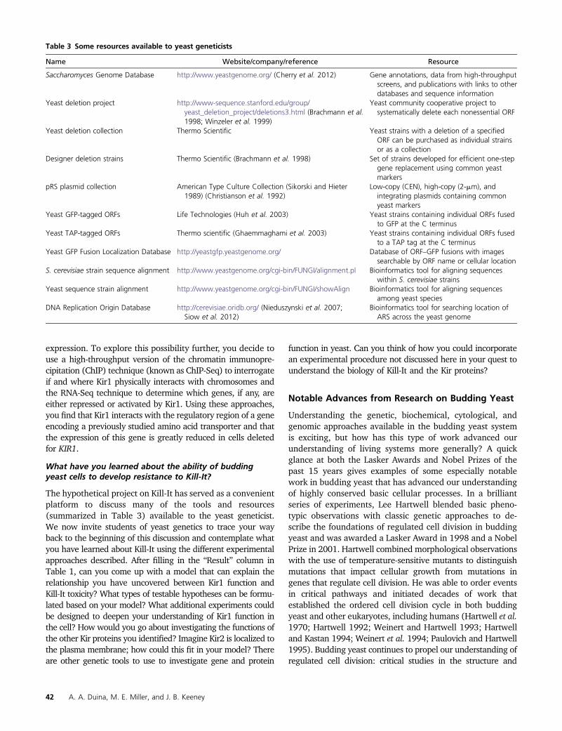
Table 3 Some resources available to yeast geneticists
Name Website/company/reference Resource
Saccharomyces Genome Database http://www.yeastgenome.org/ (Cherry et al. 2012) Gene annotations, data from high-throughputscreens, and publications with links to otherdatabases and sequence information
Yeast deletion project http://www-sequence.stanford.edu/group/yeast_deletion_project/deletions3.html (Brachmann et al.1998; Winzeler et al. 1999)
Yeast community cooperative project tosystematically delete each nonessential ORF
Yeast deletion collection Thermo Scientific Yeast strains with a deletion of a specifiedORF can be purchased as individual strainsor as a collection
Designer deletion strains Thermo Scientific (Brachmann et al. 1998) Set of strains developed for efficient one-stepgene replacement using common yeastmarkers
pRS plasmid collection American Type Culture Collection (Sikorski and Hieter1989) (Christianson et al. 1992)
Low-copy (CEN), high-copy (2-mm), andintegrating plasmids containing commonyeast markers
Yeast GFP-tagged ORFs Life Technologies (Huh et al. 2003) Yeast strains containing individual ORFs fusedto GFP at the C terminus
Yeast TAP-tagged ORFs Thermo scientific (Ghaemmaghami et al. 2003) Yeast strains containing individual ORFs fusedto a TAP tag at the C terminus
Yeast GFP Fusion Localization Database http://yeastgfp.yeastgenome.org/ Database of ORF–GFP fusions with imagessearchable by ORF name or cellular location
S. cerevisiae strain sequence alignment http://www.yeastgenome.org/cgi-bin/FUNGI/alignment.pl Bioinformatics tool for aligning sequenceswithin S. cerevisiae strains
Yeast sequence strain alignment http://www.yeastgenome.org/cgi-bin/FUNGI/showAlign Bioinformatics tool for aligning sequencesamong yeast species
DNA Replication Origin Database http://cerevisiae.oridb.org/ (Nieduszynski et al. 2007;Siow et al. 2012)
Bioinformatics tool for searching location ofARS across the yeast genome
42 A. A. Duina, M. E. Miller, and J. B. Keeney
regulation of signal transduction pathways (Mok et al.2011), subcellular organization (Taddei and Gasser 2012),movement of key regulators (D’Amours and Amon 2004),checkpoint regulation [of DNA repair, spindle function, cel-lular growth, nutrient response, and stress (Yasutis andKozminski 2013)], and aging (Longo et al. 2012) all touchon our understanding of regulated cell division and makeheavy use of budding yeast as a model for more complexeukaryotic systems. This work requires integration of themany components of the yeast toolkit from genetic interac-tion analysis to genome-wide protein localization studies.
Another excellent set of discoveries in budding yeast thattook full advantage of the yeast toolkit is Randy Schekman’swork on eukaryotic vesicle trafficking (Lasker Award in 2002and Nobel Prize in 2013). Schekman established an orderedsecretory system and clarified the striking underlying mecha-nism in this system (Novick and Schekman 1979; Novick et al.1980; Deshaies and Schekman 1987; Baker et al. 1988;Barlowe et al. 1994; Matsuoka et al. 1998). This work servesas the foundation of applications that allow production andsecretion of medically significant molecules such as insulinfrom yeast and has impacted a wide variety of fields rangingfrom neurobiology to virology (Hou et al. 2012).
Our understanding of other basic cellular processes hasalso been significantly shaped by the budding yeast toolkit.Roger Kornberg (awarded a Nobel Prize in 2006) decipheredthe structure of the components critical for transcription(Kornberg 1974; Kornberg and Thomas 1974; Lue and Kornberg1987; Sayre et al. 1992; Chasman et al. 1993; Asturias et al.1999; Gnatt et al. 2001; Bushnell et al. 2002, 2004) whileArthur Horwich’s work (awarded the 2011 Lasker award)established the action and mechanism of chaperones (Chenget al. 1989; Ostermann et al. 1989; Braig et al. 1994; Fentonet al. 1994; Rye et al. 1997). Given the critical importance ofprotein synthesis, folding, and aggregation to significant hu-man disorders, such as Huntington’s or Alzheimer’s disease,the conserved nature of these basic cellular processes hasproven that work in budding yeast is significant in anothermajor area of biomedical research. More recently, work in-volving budding yeast by Jack Szostak, Elizabeth Blackburn,and Carol Greider on eukaryotic telomere structure (Szostakand Blackburn 1982; Murray and Szostak 1983; Blackburn1984; Dunn et al. 1984; Shampay et al. 1984; Lundblad andSzostak 1989; Blackburn and Gall 1978; Greider and Blackburn1985, 1989; Yu et al. 1990; McEachern and Blackburn 1995;
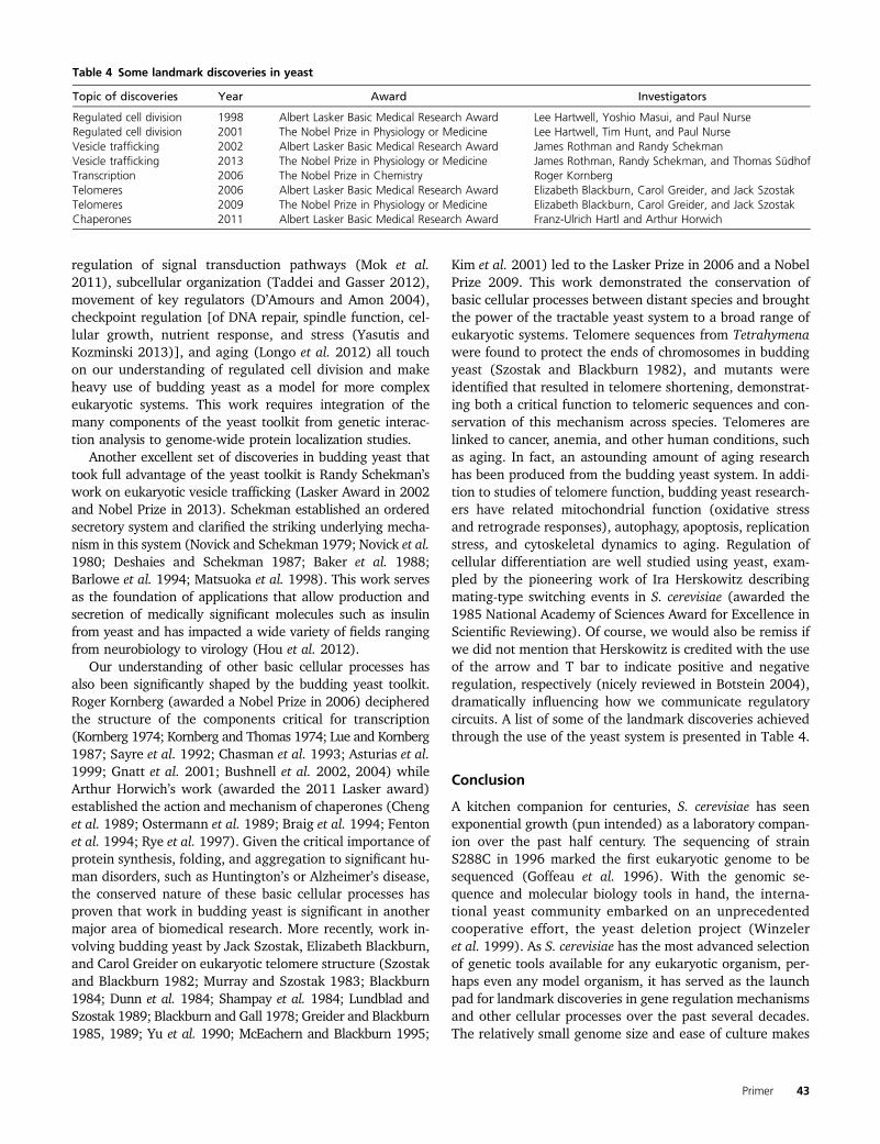
Kim et al. 2001) led to the Lasker Prize in 2006 and a NobelPrize 2009. This work demonstrated the conservation ofbasic cellular processes between distant species and broughtthe power of the tractable yeast system to a broad range ofeukaryotic systems. Telomere sequences from Tetrahymenawere found to protect the ends of chromosomes in buddingyeast (Szostak and Blackburn 1982), and mutants wereidentified that resulted in telomere shortening, demonstrat-ing both a critical function to telomeric sequences and con-servation of this mechanism across species. Telomeres arelinked to cancer, anemia, and other human conditions, suchas aging. In fact, an astounding amount of aging researchhas been produced from the budding yeast system. In addi-tion to studies of telomere function, budding yeast research-ers have related mitochondrial function (oxidative stressand retrograde responses), autophagy, apoptosis, replicationstress, and cytoskeletal dynamics to aging. Regulation ofcellular differentiation are well studied using yeast, exam-pled by the pioneering work of Ira Herskowitz describingmating-type switching events in S. cerevisiae (awarded the1985 National Academy of Sciences Award for Excellence inScientific Reviewing). Of course, we would also be remiss ifwe did not mention that Herskowitz is credited with the useof the arrow and T bar to indicate positive and negativeregulation, respectively (nicely reviewed in Botstein 2004),dramatically influencing how we communicate regulatorycircuits. A list of some of the landmark discoveries achievedthrough the use of the yeast system is presented in Table 4.
Conclusion
A kitchen companion for centuries, S. cerevisiae has seenexponential growth (pun intended) as a laboratory compan-ion over the past half century. The sequencing of strainS288C in 1996 marked the first eukaryotic genome to besequenced (Goffeau et al. 1996). With the genomic se-quence and molecular biology tools in hand, the interna-tional yeast community embarked on an unprecedentedcooperative effort, the yeast deletion project (Winzeleret al. 1999). As S. cerevisiae has the most advanced selectionof genetic tools available for any eukaryotic organism, per-haps even any model organism, it has served as the launchpad for landmark discoveries in gene regulation mechanismsand other cellular processes over the past several decades.The relatively small genome size and ease of culture makes
Table 4 Some landmark discoveries in yeast
Topic of discoveries Year Award Investigators
Regulated cell division 1998 Albert Lasker Basic Medical Research Award Lee Hartwell, Yoshio Masui, and Paul NurseRegulated cell division 2001 The Nobel Prize in Physiology or Medicine Lee Hartwell, Tim Hunt, and Paul NurseVesicle trafficking 2002 Albert Lasker Basic Medical Research Award James Rothman and Randy SchekmanVesicle trafficking 2013 The Nobel Prize in Physiology or Medicine James Rothman, Randy Schekman, and Thomas SüdhofTranscription 2006 The Nobel Prize in Chemistry Roger KornbergTelomeres 2006 Albert Lasker Basic Medical Research Award Elizabeth Blackburn, Carol Greider, and Jack SzostakTelomeres 2009 The Nobel Prize in Physiology or Medicine Elizabeth Blackburn, Carol Greider, and Jack SzostakChaperones 2011 Albert Lasker Basic Medical Research Award Franz-Ulrich Hartl and Arthur Horwich
Primer 43

yeast amenable to high-throughput screening. The data out-put of high-throughput technologies has led to the develop-ment of new fields of computational biology necessary tounderstand biology at the systems level and work towardbuilding a comprehensive model of the functioning ofa eukaryotic cell. The scientific community has access tothe compiled genetic and biological information at the ex-cellent online resource, the SGD (http://www.yeastgenome.org/) (Cherry et al. 2012). The database includes geneannotations, data from high-throughput screens, and publi-cations with links to other databases and sequence informa-tion. In keeping with the tradition of global cooperativity,one of the latest endeavors of the yeast community is thesynthetic yeast project, which has the goal of building a com-pletely synthetic strain of S. cerevisiae (Dymond et al. 2011).Through the rapid development of yeast as a model organ-ism, a delightful discovery has been the surprisingly highlevel of protein amino acid sequence and functional conser-vation between yeast and larger eukaryotic species. Theconservation of protein amino acid sequence and function,combined with the flexibility of genetic tools, make S. cer-evisiae a powerful model organism for studying the cellularworkings and diseases of larger eukaryotes. In fact, the yeastdeletion collection has been used to screen for human dis-eases (Steinmetz et al. 2002). Budding yeast continues to bea most versatile, powerful, and tasty model organism.
Acknowledgments
We thank Shawnecca Burke, Chris Walls, and the studentsin A.A.D.’s Spring 2005 Advanced Cell Biology class for theircontributions to the generation of Figure 1 and Reine Pro-tacio and Fred Winston for helpful comments on the manu-script. The authors apologize to yeast researchers who havegreatly contributed to the development of yeast as a modelsystem but who have not been highlighted in this article dueto space limitations. A.A.D. acknowledges the National Sci-ence Foundation (grant no. 1243680) for funding and M.E.M.thanks J. T. and V. B. Robertson and Patricia and CharlesRobertson for their support.
Glossary of experimental approaches and tools
BLAST (Basic Local Alignment Search Tool): A computa-tional resource whereby a specific nucleotide or amino acidsequence is compared to and aligned with sequences presentin one or more of the several available databases usingparameters defined by the user. BLAST is a powerful tool thatcan uncover functional and/or evolutionary relationships be-tween different genes or proteins.
Chromatin immunoprecipitation (ChIP): A powerful bio-chemical technique that allows investigators to determinethe level of occupancy of a protein of interest to a specificlocation across a genome. In a typical experiment using theyeast model system, cells are grown to logarithmic phase
and exposed to the cross-linking agent formaldehyde. Chro-matin is then isolated, sheered, and subjected to an immu-noprecipitation step using an antibody specific for a proteinof interest. The recovered chromatin is then further pro-cessed and quantified using quantitative PCR. Assessmentsof protein occupancy are determined by comparing the lev-els of precipitation of the regions of interest to those ofgenomic regions known to not interact with the proteinbeing investigated.
ChIP-Seq: A high-throughput version of the ChIP assay inwhich all of the genomic regions bound by a specific proteinof interest are identified using next-generation sequencingtechnology.
Complementation tests: In yeast genetics, a technique usedto determine if two independent haploid yeast populationsthat share a common recessive mutant phenotype do so asa result of mutations in the same gene or in different genes.The technique involves mating the two populations under in-vestigation and testing the resulting diploids for the sharedphenotype: presence of the mutant phenotype indicates lackof complementation (i.e., the two populations likely carry muta-tions in the same gene) and absence of the mutant phenotypeindicates complementation (i.e., the two populations likelycarry mutations in different genes).
Functional complementation: In yeast genetics, a termused to describe the ability of a gene to restore normalfunction to cells that otherwise display one or more mutantphenotypes due to a genetic mutation. Functional comple-mentation approaches are often used by yeast geneticists astools to isolate wild-type versions of mutant genes isolatedin genetic experiments.
Genetic crosses: In yeast genetics, an experimental strategyin which haploid yeast cells of opposite mating types andwith different genetic backgrounds are allowed to mate andsubsequently stimulated to produce haploid progenies withrecombined genomes. Genetic crosses are often used as a wayto generate yeast strains with specific combinations of geneticmutations.
Genetic screens: In yeast genetics, a technique that allowsfor independent clonal populations of mutant yeast cells—normally grown as independent colonies or patches on solidmedium—to be tested for specific phenotypes to identifymutations in genes of interest.
Genetic selection: In yeast genetics, a technique in whichyeast cells harboring specific mutations of interest can beidentified among large populations of genetically diversecells based on their ability to grow under conditions that areotherwise detrimental to cells not carrying the mutations.
Green fluorescent protein (GFP): A naturally occurringprotein expressed in the jellyfish Aequorea victoria withgreen fluorescence properties. The gene encoding GFP canbe fused to genes in other organisms, including yeast, using
44 A. A. Duina, M. E. Miller, and J. B. Keeney

molecular biology tools, and the resulting fusion proteins canoften be visualized in live cells using fluorescence microscopy.Localization of proteins through the use of GFP fusions hasbecome a powerful tool available to cell biologists, but cannotbe universally used as fusions of GFP to certain proteins canlead to their degradation or mislocalization in cells.
High-copy suppressors: A term used to describe thosegenes that, when present in cells at abnormally high copynumbers, can partially or completely mask (i.e., suppress)a mutant phenotype conferred by a mutation in a specificgene. The identity of the high-copy suppressor genes canoften provide insights into functional aspects of the mutantgene that they suppress.
Insertional mutagenesis: A general term to describe a setof techniques used to cause genetic mutations through theinsertion of foreign DNA fragments into the genome of a hostcell. Investigators can design the insertions to occur at ran-dom locations throughout the genome or to be targeted tospecific genomic locations.
One-step gene replacement: A term used to describeexperiments in which a specific gene is replaced by anothergene in situ as a result of a single recombination event. Thisexperimental approach, which is highly efficient in the yeastsystem, relies on the ability of engineered DNA fragments tointegrate at specific genomic locations through homologousrecombination. In a common application of this technique,a gene under investigation is replaced—and thus knocked-out—by an integrating DNA fragment that carries a nutri-tional or drug-resistance selectable marker.
Plasmid recovery: A general term to describe experimentaltools by which one or more plasmids of interest are recov-ered and purified from cells. Normally, plasmid recoveryfrom yeast cells involves lysing of cells carrying the plasmidsand transformation of the released plasmids into E. coli,followed by standard plasmid preparation protocols forE. coli.
RNA-Seq: A high-throughput experimental tool that utilizesnext-generation sequencing technology to obtain sequencedata representing the entire set of RNA molecules tran-scribed within a particular cell population.
Spontaneous suppressor mutations: Genetic mutationsthat arise through spontaneous processes, such as througherrors during DNA replication, that partially or completelymask (i.e., suppress) a mutant phenotype caused by anothermutation. Suppressor mutations are generally categorized aseither intragenic (located within the same gene that causedthe initial mutant phenotype) or extragenic (located in ge-nomic regions other than the gene that caused the initialmutant phenotype) and can often provide insights into thefunctional characteristics of the original mutant gene.
Synthetic Gene Array (SGA) analysis: A high-throughputexperimental platform available to yeast geneticists in which
genetic interactions between query mutant strains and li-braries of yeast strains carrying deletions of all nonessentialgenes are screened in a systematic fashion. Types of geneticinteractions that can be uncovered using this tool includesynthetic-lethal and suppression interactions.
Synthetic lethal screens: In yeast genetics, a specific type ofgenetic screen designed to identify nonlethal mutations that,when combined with a nonlethal mutation in a gene of interest,result in a lethal phenotype. Synthetic-lethal interactions areoften indicative of a functional relationship between the wild-type versions of the two genes that partake in the interaction.
Tandem Affinity Purification (TAP) tag: An affinity tagwhose genetic information can be fused to the coding regionof a gene of interest to generate a fusion gene encoding a so-called TAP-tagged protein. TAP-tagged proteins can be bio-chemically purified from cell lysates using IgG moleculesimmobilized to a solid support (such as magnetic beads) thatspecifically interact with the Protein A component of the tag.If desired, a second purification step can be carried out usingthe calmodulin-binding peptide component of the tag, whichbinds tightly to calmodulin. Analysis of material that copuri-fies with TAP-tagged proteins can result in the identificationof factors that physically interact with the tagged proteins.
Tetrad analysis: An experimental approach used by yeastgeneticists to assess the phenotypes—and associated geno-types—of colonies formed by the four haploid products(spores) produced by a single diploid cell through the processof meiosis. This process, which normally involves dissection oftetrads using a microscope equipped with a micromanipulatorand subsequent replica-plating of the resulting spore coloniesonto a series of solid growth media plates, also allows for thevisualization of the segregation of certain traits followingmeiosis and facilitates assessment of possible genetic interac-tions between different genetic mutations.
Transformation: In yeast genetics, a term used to describea set of tools used to introduce DNA molecules—commonlyin the form of plasmids or linear fragments—into yeast cells.The presence of a newly introduced DNA molecule intoa host cell changes, or transforms, its genotype and com-monly its phenotype as well.
Yeast two-hybrid system: An experimental system used todetect protein–protein interactions in an in vivo setting. Theyeast two-hybrid system can be used to test if two specificproteins interact with each other or to screen or select forprotein–protein interactions between a protein of interestand proteins expressed from a genomic or cDNA library. Thissystem can be used to assess interactions among yeast pro-teins as well as among proteins from other species.
Literature Cited
Asturias, F. J., Y. W. Jiang, L. C. Myers, C. M. Gustafsson, and R. D.Kornberg, 1999 Conserved structures of mediator and RNApolymerase II holoenzyme. Science 283: 985–987.
Primer 45

Baker, D., L. Hicke, M. Rexach, M. Schleyer, and R. Schekman,1988 Reconstitution of SEC gene product-dependent inter-compartmental protein transport. Cell 54: 335–344.
Barlowe, C., L. Orci, T. Yeung, M. Hosobuchi, S. Hamamoto et al.,1994 COPII: a membrane coat formed by Sec proteins thatdrive vesicle budding from the endoplasmic reticulum. Cell77: 895–907.
Blackburn, E. H., 1984 The molecular structure of centromeresand telomeres. Annu. Rev. Biochem. 53: 163–194.
Blackburn, E. H., and J. G. Gall, 1978 A tandemly repeated se-quence at the termini of the extrachromosomal ribosomal RNAgenes in Tetrahymena. J. Mol. Biol. 120: 33–53.
Boeke, J. D., and S. E. Devine, 1998 Yeast retrotransposons: find-ing a nice quiet neighborhood. Cell 93: 1087–1089.
Bohley, P., and K.-U. Fröhlich, 1997 A prize-winning discovery of1896: Buchner provides evidence of cell-free fermentation, pp.51–60 in New Beer in an Old Bottle: Eduard Buchner and theGrowth of Biochemical Knowledge, edited by A. Cornish-Bowden.Universitat de València, Valencia, Spain.
Botstein, D., 2004 Ira Herskowitz: 1946–2003. Genetics 166:653–660.
Brachmann, C. B., A. Davies, G. J. Cost, E. Caputo, J. Li et al.,1998 Designer deletion strains derived from Saccharomycescerevisiae S288C: a useful set of strains and plasmids for PCR-mediated gene disruption and other applications. Yeast 14:115–132.
Braig, K., Z. Otwinowski, R. Hegde, D. C. Boisvert, A. Joachimiaket al., 1994 The crystal structure of the bacterial chaperoninGroEL at 2.8 A. Nature 371: 578–586.
Brogaard, K., L. Xi, J. P. Wang, and J. Widom, 2012 A map ofnucleosome positions in yeast at base-pair resolution. Nature486: 496–501.
Buhler, M., and S. M. Gasser, 2009 Silent chromatin at the middleand ends: lessons from yeasts. EMBO J. 28: 2149–2161.
Bumgarner, S. L., R. D. Dowell, P. Grisafi, D. K. Gifford, and G. R.Fink, 2009 Toggle involving cis-interfering noncoding RNAscontrols variegated gene expression in yeast. Proc. Natl. Acad.Sci. USA 106: 18321–18326.
Bushnell, D. A., P. Cramer, and R. D. Kornberg, 2002 Structuralbasis of transcription: alpha-amanitin-RNA polymerase II cocrys-tal at 2.8 A resolution. Proc. Natl. Acad. Sci. USA 99: 1218–1222.
Bushnell, D. A., K. D. Westover, R. E. Davis, and R. D. Kornberg,2004 Structural basis of transcription: an RNA polymerase II-TFIIB cocrystal at 4.5 angstroms. Science 303: 983–988.
Carr, M., D. Bensasson, and C. M. Bergman, 2012 Evolutionarygenomics of transposable elements in Saccharomyces cerevisiae.PLoS ONE 7: e50978.
Castelnuovo, M., S. Rahman, E. Guffanti, V. Infantino, F. Stutzet al., 2013 Bimodal expression of PHO84 is modulated byearly termination of antisense transcription. Nat. Struct. Mol.Biol. 20: 851–858.
Chasman, D. I., K. M. Flaherty, P. A. Sharp, and R. D. Kornberg,1993 Crystal structure of yeast TATA-binding protein andmodel for interaction with DNA. Proc. Natl. Acad. Sci. USA90: 8174–8178.
Cheng, M. Y., F. U. Hartl, J. Martin, R. A. Pollock, F. Kalousek et al.,1989 Mitochondrial heat-shock protein hsp60 is essential forassembly of proteins imported into yeast mitochondria. Nature337: 620–625.
Cherry, J. M., E. L. Hong, C. Amundsen, R. Balakrishnan, G. Binkleyet al., 2012 Saccharomyces Genome Database: the genomicsresource of budding yeast. Nucleic Acids Res. 40: D700–D705.
Chien, C. T., P. L. Bartel, R. Sternglanz, and S. Fields, 1991 Thetwo-hybrid system: a method to identify and clone genes forproteins that interact with a protein of interest. Proc. Natl. Acad.Sci. USA 88: 9578–9582.
Christianson, T. W., R. S. Sikorski, M. Dante, J. H. Shero, and P.Hieter, 1992 Multifunctional yeast high-copy-number shuttlevectors. Gene 110: 119–122.
D’Amours, D., and A. Amon, 2004 At the interface between sig-naling and executing anaphase: Cdc14 and the FEAR network.Genes Dev. 18: 2581–2595.
David, L., W. Huber, M. Granovskaia, J. Toedling, C. J. Palm et al.,2006 A high-resolution map of transcription in the yeast ge-nome. Proc. Natl. Acad. Sci. USA 103: 5320–5325.
Dekker, J., K. Rippe, M. Dekker, and N. Kleckner, 2002 Capturingchromosome conformation. Science 295: 1306–1311.
Deshaies, R. J., and R. Schekman, 1987 A yeast mutant defectiveat an early stage in import of secretory protein precursors intothe endoplasmic reticulum. J. Cell Biol. 105: 633–645.
Dhar, M. K., S. Sehgal, and S. Kaul, 2012 Structure, replicationefficiency and fragility of yeast ARS elements. Res. Microbiol.163: 243–253.
Duan, Z., M. Andronescu, K. Schutz, S. McIlwain, Y. J. Kim et al.,2010 A three-dimensional model of the yeast genome. Nature465: 363–367.
Dunn, B., P. Szauter, M. L. Pardue, and J. W. Szostak,1984 Transfer of yeast telomeres to linear plasmids by recom-bination. Cell 39: 191–201.
Dymond, J. S., S. M. Richardson, C. E. Coombes, T. Babatz, H.Muller et al., 2011 Synthetic chromosome arms function inyeast and generate phenotypic diversity by design. Nature477: 471–476.
Fenton, W. A., Y. Kashi, K. Furtak, and A. L. Horwich,1994 Residues in chaperonin GroEL required for polypeptidebinding and release. Nature 371: 614–619.
Gelfand, B., J. Mead, A. Bruning, N. Apostolopoulos, V. Tadigotlaet al., 2011 Regulated antisense transcription controls expres-sion of cell-type-specific genes in yeast. Mol. Cell. Biol. 31:1701–1709.
Ghaemmaghami, S., W. K. Huh, K. Bower, R. W. Howson, A. Belleet al., 2003 Global analysis of protein expression in yeast. Na-ture 425: 737–741.
Gnatt, A. L., P. Cramer, J. Fu, D. A. Bushnell, and R. D. Kornberg,2001 Structural basis of transcription: an RNA polymerase IIelongation complex at 3.3 A resolution. Science 292: 1876–1882.
Goffeau, A., B. G. Barrell, H. Bussey, R. W. Davis, B. Dujon et al.,1996 Life with 6000 genes. Science 274(5287): 546, 563–567.
Greider, C. W., and E. H. Blackburn, 1985 Identification of a spe-cific telomere terminal transferase activity in Tetrahymena ex-tracts. Cell 43: 405–413.
Greider, C. W., and E. H. Blackburn, 1989 A telomeric sequence inthe RNA of Tetrahymena telomerase required for telomere re-peat synthesis. Nature 337: 331–337.
Greig, D., and J. Y. Leu, 2009 Natural history of budding yeast.Curr. Biol. 19: R886–R890.
Haber, J. E., 2012 Mating-type genes and MAT switching in Sac-charomyces cerevisiae. Genetics 191: 33–64.
Hampsey, M., 1997 A review of phenotypes in Saccharomycescerevisiae. Yeast 13: 1099–1133.
Hartwell, L., 1992 Defects in a cell cycle checkpoint may be re-sponsible for the genomic instability of cancer cells. Cell 71:543–546.
Hartwell, L. H., and M. B. Kastan, 1994 Cell cycle control andcancer. Science 266: 1821–1828.
Hartwell, L. H., J. Culotti, and B. Reid, 1970 Genetic control ofthe cell-division cycle in yeast. I. Detection of mutants. Proc.Natl. Acad. Sci. USA 66: 352–359.
Hinnen, A., J. B. Hicks, and G. R. Fink, 1978 Transformation ofyeast. Proc. Natl. Acad. Sci. USA 75: 1929–1933.
Hongay, C. F., P. L. Grisafi, T. Galitski, and G. R. Fink,2006 Antisense transcription controls cell fate in Saccharomy-ces cerevisiae. Cell 127: 735–745.
46 A. A. Duina, M. E. Miller, and J. B. Keeney

Hou, J., K. E. Tyo, Z. Liu, D. Petranovic, and J. Nielsen,2012 Metabolic engineering of recombinant protein secretionby Saccharomyces cerevisiae. FEMS Yeast Res. 12: 491–510.
Houseley, J., L. Rubbi, M. Grunstein, D. Tollervey, and M. Vogelauer,2008 A ncRNA modulates histone modification andmRNA induction in the yeast GAL gene cluster. Mol. Cell32: 685–695.
Huh, W. K., J. V. Falvo, L. C. Gerke, A. S. Carroll, R. W. Howsonet al., 2003 Global analysis of protein localization in buddingyeast. Nature 425: 686–691.
Ito, T., T. Chiba, R. Ozawa, M. Yoshida, M. Hattori et al., 2001 Acomprehensive two-hybrid analysis to explore the yeast proteininteractome. Proc. Natl. Acad. Sci. USA 98: 4569–4574.
Kavanaugh, L. A., and F. S. Dietrich, 2009 Non-coding RNA pre-diction and verification in Saccharomyces cerevisiae. PLoSGenet. 5: e1000321.
Kawai, S., W. Hashimoto, and K. Murata, 2010 Transformation ofSaccharomyces cerevisiae and other fungi: methods and possi-ble underlying mechanism. Bioeng. Bugs 1: 395–403.
Kellis, M., B. W. Birren, and E. S. Lander, 2004 Proof and evolu-tionary analysis of ancient genome duplication in the yeast Sac-charomyces cerevisiae. Nature 428: 617–624.
Kim, M. M., M. A. Rivera, I. L. Botchkina, R. Shalaby, A. D. Thoret al., 2001 A low threshold level of expression of mutant-template telomerase RNA inhibits human tumor cell prolifera-tion. Proc. Natl. Acad. Sci. USA 98: 7982–7987.
Kornberg, R. D., 1974 Chromatin structure: a repeating unit ofhistones and DNA. Science 184: 868–871.
Kornberg, R. D., and J. O. Thomas, 1974 Chromatin structure:oligomers of the histones. Science 184: 865–868.
Liebman, S. W., and J. E. Haber, 2013 Retrospective. Fred Sherman(1932–2013). Science 342: 1059.
Li, Z., F. J. Vizeacoumar, S. Bahr, J. Li, J. Warringer et al.,2011 Systematic exploration of essential yeast gene functionwith temperature-sensitive mutants. Nat. Biotechnol. 29: 361–367.
Longo, V. D., G. S. Shadel, M. Kaeberlein, and B. Kennedy,2012 Replicative and chronological aging in Saccharomycescerevisiae. Cell Metab. 16: 18–31.
Longtine, M. S., A. McKenzie, III, D. J. Demarini, N. G. Shah, A.Wach et al., 1998 Additional modules for versatile and eco-nomical PCR-based gene deletion and modification in Saccha-romyces cerevisiae. Yeast 14: 953–961.
Lue, N. F., and R. D. Kornberg, 1987 Accurate initiation at RNApolymerase II promoters in extracts from Saccharomyces cere-visiae. Proc. Natl. Acad. Sci. USA 84: 8839–8843.
Lundblad, V., and J. W. Szostak, 1989 A mutant with a defect intelomere elongation leads to senescence in yeast. Cell 57: 633–643.
Martens, J. A., L. Laprade, and F. Winston, 2004 Intergenic tran-scription is required to repress the Saccharomyces cerevisiaeSER3 gene. Nature 429: 571–574.
Matsuoka, K., L. Orci, M. Amherdt, S. Y. Bednarek, S. Hamamotoet al., 1998 COPII-coated vesicle formation reconstituted withpurified coat proteins and chemically defined liposomes. Cell93: 263–275.
McEachern, M. J., and E. H. Blackburn, 1995 Runaway telomereelongation caused by telomerase RNA gene mutations. Nature376: 403–409.
Mehta, G. D., M. P. Agarwal, and S. K. Ghosh, 2010 Centromereidentity: a challenge to be faced. Mol. Genet. Genomics 284:75–94.
Mok, J., X. Zhu, and M. Snyder, 2011 Dissecting phosphorylationnetworks: lessons learned from yeast. Expert Rev. Proteomics 8:775–786.
Mortimer, R. K., 2000 Evolution and variation of the yeast (Sac-charomyces) genome. Genome Res. 10: 403–409.
Mortimer, R. K., and J. R. Johnston, 1986 Genealogy of principalstrains of the yeast genetic stock center. Genetics 113: 35–43.
Murray, A. W., and J. W. Szostak, 1983 Construction of artificialchromosomes in yeast. Nature 305: 189–193.
Neil, H., C. Malabat, Y. d’Aubenton-Carafa, Z. Xu, L. M. Steinmetzet al., 2009 Widespread bidirectional promoters are the majorsource of cryptic transcripts in yeast. Nature 457: 1038–1042.
Nieduszynski, C. A., S. Hiraga, P. Ak, C. J. Benham, and A. D.Donaldson, 2007 OriDB: a DNA replication origin database.Nucleic Acids Res. 35: D40–D46.
Novick, P., and R. Schekman, 1979 Secretion and cell-surfacegrowth are blocked in a temperature-sensitive mutant of Sac-charomyces cerevisiae. Proc. Natl. Acad. Sci. USA 76: 1858–1862.
Novick, P., C. Field, and R. Schekman, 1980 Identification of 23complementation groups required for post-translational eventsin the yeast secretory pathway. Cell 21: 205–215.
Ostermann, J., A. L. Horwich, W. Neupert, and F. U. Hartl,1989 Protein folding in mitochondria requires complex forma-tion with hsp60 and ATP hydrolysis. Nature 341: 125–130.
Paulovich, A. G., and L. H. Hartwell, 1995 A checkpoint regulatesthe rate of progression through S phase in S. cerevisiae in re-sponse to DNA damage. Cell 82: 841–847.
Piekna-Przybylska, D., W. A. Decatur, and M. J. Fournier,2007 New bioinformatic tools for analysis of nucleotide mod-ifications in eukaryotic rRNA. RNA 13: 305–312.
Pinskaya, M., S. Gourvennec, and A. Morillon, 2009 H3 lysine 4di- and tri-methylation deposited by cryptic transcription at-tenuates promoter activation. EMBO J. 28: 1697–1707.
Prelich, G., 1999 Suppression mechanisms: themes from varia-tions. Trends Genet. 15: 261–266.
Puig, O., F. Caspary, G. Rigaut, B. Rutz, E. Bouveret et al.,2001 The tandem affinity purification (TAP) method: a generalprocedure of protein complex purification. Methods 24: 218–229.
Robzyk, K., and Y. Kassir, 1992 A simple and highly efficient pro-cedure for rescuing autonomous plasmids from yeast. NucleicAcids Res. 20: 3790.
Rye, H. S., S. G. Burston, W. A. Fenton, J. M. Beechem, Z. Xu et al.,1997 Distinct actions of cis and trans ATP within the doublering of the chaperonin GroEL. Nature 388: 792–798.
Sayre, M. H., H. Tschochner, and R. D. Kornberg, 1992 Reconstitutionof transcription with five purified initiation factors and RNA poly-merase II from Saccharomyces cerevisiae. J. Biol. Chem. 267:23376–23382.
Shampay, J., J. W. Szostak, and E. H. Blackburn, 1984 DNA se-quences of telomeres maintained in yeast. Nature 310: 154–157.
Sikorski, R. S., and P. Hieter, 1989 A system of shuttle vectors andyeast host strains designed for efficient manipulation of DNA inSaccharomyces cerevisiae. Genetics 122: 19–27.
Singer, M. S., and D. E. Gottschling, 1994 TLC1: template RNAcomponent of Saccharomyces cerevisiae telomerase. Science266: 404–409.
Siow, C. C., S. R. Nieduszynska, C. A. Muller, and C. A. Nieduszyn-ski, 2012 OriDB, the DNA replication origin database updatedand extended. Nucleic Acids Res. 40: D682–D686.
Spingola, M., L. Grate, D. Haussler, and M. Ares, Jr.,1999 Genome-wide bioinformatic and molecular analysis ofintrons in Saccharomyces cerevisiae. RNA 5: 221–234.
Steinmetz, L. M., C. Scharfe, A. M. Deutschbauer, D. Mokranjac, Z.S. Herman et al., 2002 Systematic screen for human diseasegenes in yeast. Nat. Genet. 31: 400–404.
Szostak, J. W., and E. H. Blackburn, 1982 Cloning yeast telomereson linear plasmid vectors. Cell 29: 245–255.
Taddei, A., and S. M. Gasser, 2012 Structure and function in thebudding yeast nucleus. Genetics 192: 107–129.
Primer 47

Tong, A. H., and C. Boone, 2006 Synthetic genetic array analysisin Saccharomyces cerevisiae. Methods Mol. Biol. 313: 171–192.
Uetz, P., L. Giot, G. Cagney, T. A. Mansfield, R. S. Judson et al.,2000 A comprehensive analysis of protein-protein interactionsin Saccharomyces cerevisiae. Nature 403: 623–627.
Van Nues, R. W., and J. D. Brown, 2004 Saccharomyces SRP RNAsecondary structures: a conserved S-domain and extended Alu-domain. RNA 10: 75–89.
van Werven, F. J., G. Neuert, N. Hendrick, A. Lardenois, S. Buratowskiet al., 2012 Transcription of two long noncoding RNAs mediatesmating-type control of gametogenesis in budding yeast. Cell 150:1170–1181.
Vidan, S., and M. Snyder, 2001 Large-scale mutagenesis: yeastgenetics in the genome era. Curr. Opin. Biotechnol. 12: 28–34.
Weinert, T. A., and L. H. Hartwell, 1993 Cell cycle arrest of cdcmutants and specificity of the RAD9 checkpoint. Genetics 134:63–80.
Weinert, T. A., G. L. Kiser, and L. H. Hartwell, 1994 Mitotic check-point genes in budding yeast and the dependence of mitosis onDNA replication and repair. Genes Dev. 8: 652–665.
Wellinger, R. J., and V. A. Zakian, 2012 Everything you everwanted to know about Saccharomyces cerevisiae telomeres: be-ginning to end. Genetics 191: 1073–1105.
Winzeler, E. A., D. D. Shoemaker, A. Astromoff, H. Liang, K. Ander-son et al., 1999 Functional characterization of the S. cerevisiaegenome by gene deletion and parallel analysis. Science 285:901–906.
Xu, Z., W. Wei, J. Gagneur, F. Perocchi, S. Clauder-Munster et al.,2009 Bidirectional promoters generate pervasive transcriptionin yeast. Nature 457: 1033–1037.
Yasutis, K. M., and K. G. Kozminski, 2013 Cell cycle checkpointregulators reach a zillion. Cell Cycle 12: 1501–1509.
Yu, G. L., J. D. Bradley, L. D. Attardi, and E. H. Blackburn, 1990 Invivo alteration of telomere sequences and senescence caused bymutated Tetrahymena telomerase RNAs. Nature 344: 126–132.
Yu, H., P. Braun, M. A. Yildirim, I. Lemmens, K. Venkatesan et al.,2008 High-quality binary protein interaction map of the yeastinteractome network. Science 322: 104–110.
Communicating editor: E. A. De Stasio
48 A. A. Duina, M. E. Miller, and J. B. Keeney