Embed Size (px)
Citation preview
2014
SC
HE
DU
LE
Federal Institute for Risk Assessment
Brucellosis 2014 International Research Conference BERLIN | 9–12 September 2014
Imprint Abstracts Brucellosis 2014 International Research Conference All authors are responsible for the content of their respective abstracts. Federal Institute for Risk Assessment Communication and Public Relations Office Max-Dohrn-Straße 8–10 10589 Berlin Germany Berlin 2014 222 Pages Photo: flobox/Quelle: PHOTOCASE Printing: cover, content pages and bookbinding BfR-printing house

3 Brucellosis 2014 International Research Conference
Welcoming Addresses Andreas Hensel President of the Federal Institute for Risk Assessment (BfR)
Welcoming address by the President of the Federal Institute for Risk Assessment
More than 100 years after the first description of Micrococcus melitensis by Sir David Bruce, brucellosis is still of major public health concern both in endemic and non-endemic countries all over the world. The impact on animal and human health is tremendous and eradication and control of this zoonotic disease remains a global and interdisciplinary challenge. Alt-hough eradicated in German livestock brucellosis is still a matter of interest because it has emerged as a disease among immigrants associated with diagnostic delays, possibly result-ing in treatment failures, relapses, chronic courses, focal complications, and a high case-fatality rate. The identification of health risks is the guiding principle for our work in the field of food safety and consumer protection. A total of 800 employees spare no effort to prove that no risk is more fun. I am delighted to host the Brucellosis 2014 International Research Conference at the Federal Institute for Risk Assessment, here in Berlin, the capital city of Germany, which was home and domain of so many Nobel laureates. Emil von Behring got the first Nobel Prize in Medi-cine for his work on serum therapy, especially its application against diphtheria; Robert Koch first identified pathogenic microorganisms as the cause of infectious diseases such as tuber-culosis, anthrax and cholera; and Paul Ehrlich developed the first antibiotic drug to treat syphilis and is regarded as the founder of chemotherapy. By the way, Robert Koch is proba-bly the most famous staff member of the Imperial Health Agency which is the predecessor of our institution, the Federal Institute for Risk Assessment. Berlin is a breakthrough city combining tradition and modernity and you have to pass the Brandenburger Tor to enjoy its spirit of freedom and creativity. From 9 to 12 September 2014 my team and I will do everything for you to ensure that you can feel like ‘Ich bin ein Berliner’. It is a great honor for us to welcome the Brucella research community and I wish you an in-spiring meeting with fruitful discussions.

4 Abstracts
Karin Schwabenbauer Head of Directorate Animal Health, Animal Welfare of the Federal Ministry of Food and Agriculture (BMEL) President of the World Organisation for Animal Health (OIE)
Welcoming address by the German Federal Ministry of Food and Agriculture
The control of zoonotic diseases has been a focus of the Federal Ministry's scientific support for many years. For some time we have known that a one-sided view of animal populations, public health, food safety and the security of global trade cannot provide the large-scale suc-cess in the fight against zoonotic diseases which we wish to achieve, and which citizens ex-pect us to achieve. It is only if health authorities, veterinary authorities, the agricultural sector, the food industry, the scientific community and an informed public work together that we will succeed, in the sense of ‘One Health’, in attaining our goal of successfully controlling zoono-tic diseases. In Germany we therefore launched the ‘National Zoonotic Research’ platform in 2006 which incorporates all necessary scientific disciplines. One distinctive feature of this programme is that it only supports projects that are multidisciplinary. Germany is in the hap-py position of its cattle, sheep and goat stocks having been officially free of brucellosis since 2000 - only a few sporadic outbreaks have been reported since. This success has been the result of hard work. It is only possible to maintain this status through an ongoing, legally bind-ing national control procedure, substantial funds and a nationwide monitoring system that is still in place. This successful example of disease control can act as encouragement for, and a basis for control procedures in, those countries in which brucellosis still causes considera-ble difficulties for animals and people. However, account must be taken of the specific socio-economic and socio-cultural situation in each country. Germany, for instance, cannot be compared with countries in which the pastoralism of sheep and goats represents an im-portant means of livelihood. Or in which the health services do not have sufficient funds at their disposal. It is our duty as the international community to provide help and assistance in , one of the most prevalent food-related zoonotic diseases in the world, with the consumption of raw milk and raw-milk cheese representing the most common means of transmission. Alt-hough pasteurisation of milk is consequently a very effective measure for minimising cases of brucellosis in mankind, the best way of eradicating brucellosis in animals and mankind is to monitor animals stocks.Although Germany is deemed officially free of brucellosis, it is still particularly important, in developing more efficient methods of detection, and in particular in assessing individual animals, to achieve success at international level and thus contribute to containing these zoonotic diseases. As false positive diagnoses still lead to considerable losses in international trade, I hope - also in my capacity as President of the OIE - that this conference will provide corresponding proposals that can then be incorporated into the OIE Manual.
I wish you all the best for this conference; I hope that you will have stimulating discussions that result in new ideas for successfully fighting zoonotic diseases.
5 Brucellosis 2014 International Research Conference
Thomas Mettenleiter President of the Friedrich-Loeffler-Institut (FLI)
Welcoming address by the President of the Friedrich-Loeffler-Institut
Brucellosis accompanied mankind as a life threatening zoonosis and burden for livestock since the domestication of sheep, goats, cattle and pigs to improve quality of human life. Proof of high disease prevalence in humans can already be found in the skeletal remains of individuals from ancient Egypt or Roman Pompeii. The importance of brucellosis for modern human and veterinary public health is demonstrated by approx. 500 000 new human infec-tions worldwide every year, its rapid re-emergence in developing countries and countries struck by civil disruption, and its continuing presence around the Mediterranean basin and the Levant despite enormous efforts for control presently supported by the EU. Therefore, research on brucellosis epidemiology, prophylaxis and control is well within the mission of the Friedrich-Loeffler-Institut as Federal Research Institute for Animal Health and OIE Col-laborating Centre for Zoonoses in Europe. The OIE reference laboratory for brucellosis at FLI currently is engaged in projects in Africa, Southern America, Europe and Asia. With our new research center on the island of Riems, best equipped and modern P3 laboratories and ex-perimental animal facilities are available for cutting-edge research projects. The work of our brucellosis laboratory will also benefit from new facilities to be built at the FLI site in Jena. Personally, I am very glad that FLI can and will continue to make important contributions to worldwide control and eradication of brucellosis inspired by the idea of ‘One world – one health’. Eradication of brucellosis is feasible as demonstrated by the experiences of countries like Germany where brucellosis was eradicated successfully from domestic livestock during the last century by applying consequent veterinary measures. I am also very glad that we have the pleasure to host the Brucellosis 2014 International Research Conference in Ger-many for the first time. I wish you an inspiring meeting, a fruitful exchange of ideas and pleasant days in Berlin, the lively capital of Germany.


7 Brucellosis 2014 International Research Conference
Heinrich Neubauer, Co-Chair Friedrich-Loeffler-Institut
Sascha Al Dahouk, Chair Federal Institute for Risk Assessment
Herzlich Willkommen
Mirë se erdhët
أْھ�ً َوَ�ْ��ً
Bienvenido
բարի գալուստ
Xoş gəldiniz
እንክዋን ደህና መጡ
Bem-vindo
欢迎
Velkommen
Bienvenue Tervetuloa
მოგესალმებით
Καλώς ήλθατε
आपका �वागत है
دیآ�د �وش
Benvenuto
בברכה
Добро пожаловать
Қош келдіңіздер
Karibu
Тавтай морилно уу
�वागत छ!
Welkom
Sànnu dà zuwà
Velkommen til Witamy
에 오신 것을 환영합니다 Bine ai venit
Välkommen
Vitajte
Soo dhawaada
ยนิดีตอ้นรับคุณ
Ласкаво просимо
Üdvözöljük
Xush kelibsiz
Welcome

9 Brucellosis 2014 International Research Conference
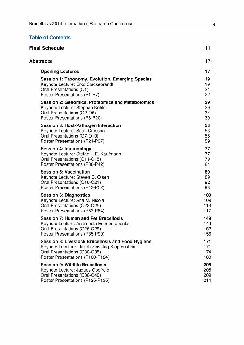
Table of Contents
Final Schedule 11
Abstracts 17
Opening Lectures 17
Session 1: Taxonomy, Evolution, Emerging Species 19 Keynote Lecture: Erko Stackebrandt 19 Oral Presentations (O1) 21 Poster Presentations (P1-P7) 22
Session 2: Genomics, Proteomics and Metabolomics 29 Keynote Lecture: Stephan Köhler 29 Oral Presentations (O2-O6) 34 Poster Presentations (P8-P20) 39
Session 3: Host-Pathogen Interaction 53 Keynote Lecture: Sean Crosson 53 Oral Presentations (O7-O10) 55 Poster Presentations (P21-P37) 59
Session 4: Immunology 77 Keynote Lecture: Stefan H.E. Kaufmann 77 Oral Presentations (O11-O15) 79 Poster Presentations (P38-P42) 84
Session 5: Vaccination 89 Keynote Lecture: Steven C. Olsen 89 Oral Presentations (O16-O21) 92 Poster Presentations (P43-P52) 98
Session 6: Diagnostics 109 Keynote Lecture: Ana M. Nicola 109 Oral Presentations (O22-O25) 113 Poster Presentations (P53-P84) 117
Session 7: Human and Pet Brucellosis 149 Keynote Lecture: Assimoula Economopoulou 149 Oral Presentations (O26-O29) 152 Poster Presentations (P85-P99) 156
Session 8: Livestock Brucellosis and Food Hygiene 171 Keynote Lecuture: Jakob Zinsstag-Klopfenstein 171 Oral Presentations (O30-O35) 174 Poster Presentations (P100-P124) 180
Session 9: Wildlife Brucellosis 205 Keynote Lecture: Jaques Godfroid 205 Oral Presentations (O36-O40) 209 Poster Presentations (P125-P135) 214


11 Brucellosis 2014 International Research Conference
Final Schedule
Tuesday, 9 September
05:00 p.m. – 08:00 p.m. Welcome Reception, ELLINGTON Hotel Berlin
Wednesday, 10 September
08:00 a.m. – 03:00 p.m. Registration
09:00 a.m. – 10:15 a.m. Opening Ceremony
Andreas Hensel Federal Institute for Risk Assessment, Berlin, Germany
Karin Schwabenbauer Federal Ministry of Food and Agriculture, Berlin, Germany
FAO works to curb the burden of brucellosis in endemic countries Ahmed El Idrissi
Global research on Brucella species 1950-2013 Mahmoud Abo-Shehada
10:15 a.m. – 11:00 a.m. Coffee Break 11:00 a.m. – 12:15 a.m. Session 1: Taxonomy, Evolution, Emerging Species
Chair: David O’Callaghan
Keynote Lecture: Erko Stackebrandt Leibniz Institute DSMZ, Braunschweig, Germany
Full taxonomic description of the proposed eleventh Brucella species, Brucella papionis sp. nov., isolated from baboons Adrian M. Whatmore
Round-Table
12:15 a.m. – 02:00 p.m. Lunch Break/Poster Session (Session 1-3) 02:00 p.m – 03:30 p.m. Session 2: Genomics, Proteomics and Metabolomics
Chair: Jean-Jaques Letesson
Keynote Lecture: Stephan Köhler Centre d’études d’agents Pathogènes et Biotechnologie pour la Santé, Montpellier, France
A robust WGS pipeline for identifying and genotyping Brucella species Christine R. Quance
Whole genome comparative study on Brucella suis biovar 2 Ricardo Dias
Revisiting the erythritol catabolic pathway 40 years later Thibault Barbier

12 Abstracts
sRNAs and Hfq regulation in Brucella abortus Félix J. Sangari
Role of the transcriptional regulator RegA in establishment of Brucella suis persistence in an original in vitro model Veronique Jubier-Maurin
03:30 p.m. – 04:30 p.m. Coffee Break/Poster Session (Session 4-6)
04:30 p.m. – 08:30 p.m. Sightseeing Tour
Thursday, 11 September 08:30 a.m. – 09:45 a.m. Session 3: Host-Pathogen-Interaction
Chair: Renato de Lima Santos
Keynote Lecture: Sean Crosson University of Chicago, Department of Biochemistry and Molecular Biology, Chicago (IL), USA
Marine mammal brucellae are attenuated in a mouse model of infection Ingebjørg H. Nymo
Defining the role of a LysR-type transcriptional regulator in the survival and pathogenesis of Brucella abortus 2308 Lauren M. Sheehan
Cell cycle progression of Brucella abortus in culture and inside HeLa cells and RAW264.7 macrophages Xavier de Bolle
Transcriptional profile of bovine chorioallantoic membrane explants in response to wild type, ∆virB2 or ∆btpB Brucella abortus infection Renato de Lima Santos
09:45 a.m. – 10:30 a.m. Coffee Break
10:30 a.m. – 12:00 a.m. Session 4: Immunology
Chair: Thomas A. Ficht
Keynote Lecture: Stefan H.E. Kaufmann Max Planck Institute for Infection Biology, Berlin, Germany
T cell responses to Brucella in humanized mice Beata Clapp
CXCR2 is a critical mediator of Brucella-induced articular inflammation Jerod A. Skyberg
Bovine γδ T cells‘ unique WC1 pattern recognition receptor (PRR) di-rects cellular immune responses to bacterial pathogens Cynthia L. Baldwin

13 Brucellosis 2014 International Research Conference
A Brucella spp. protease inhibitor is a useful adjuvant in oral vaccine formulations against infectious diseases Juliana Cassataro
12:00 a.m. – 01:45 p.m. Lunch Break/Poster Session (Session 7-9) 01:45 p.m – 03:30 p.m. Session 5: Vaccination
Chair: David W. Pascual
Keynote Lecture: Steven C. Olsen National Animal Disease Center, United States Department of Agriculture, Ames (IA), USA
Cloning, expression and immune response against protective antigens of M. bovis in B. abortus vaccine strain RB51 Nammalwar Sriranganathan
Vaccination with recombinant Brucella abortus RB51 strain engi-neered to express increased levels of O-polysaccharide provides en-hanced protection in murine brucellosis model Ramesh Vemulapalli
The road towards the development of a safe and efficacious live at-tenuated vaccine for animal and human brucellosis: from the bench to the non-human primate model Angela Arenas-Gamboa
Preliminary evaluation of a recombinant immunocontraceptive brucel-losis vaccine for swine Nammalwar Sriranganathan
Safety and protectiveness of a novel vector vaccine against Brucella abortus in first-calf heifers Kaissar Tabynov
Testing of immunogenic characteristics of the vaccine B. abortus strain S19 in application of reduced doses on small rumi-nants Khabibulo Khamdamov
03:30 p.m. – 04:15 p.m. Coffee Break 04:15 p.m. – 05:30 p.m. Session 6: Diagnostics
Chair: Adrian M. Whatmore
Keynote Lecture: Ana M. Nicola National Health Service and Agri-Food Quality (SENASA), Buenos Aires, Argentina
Oligosaccharide conjugates identify A and M epitopes and are superi-or ELISA antigens for presumptive diagnosis of brucellosis David R. Bundle
Application of synthetic oligosaccharide conjugates based on the structure of the native Brucella OPS to the serodiagnosis of bovine brucellosis John McGiven

14 Abstracts
Estimation of individual seroprevalence of brucellosis in large rumi-nants from bulk milk tank results
Wendy Beauvais
Use of Bionumerics and MALDI-TOF-MS data for the identification of Brucella species Rebekah Tiller
08:00 p.m. – 01:00 a.m. Soirée, Wasserwerk Berlin
Friday, 12 September 08:30 a.m. – 10:00 a.m. Session 7: Human and Pet Brucellosis
Chair: Rebekah Tiller
Keynote Lecture: Assimoula Economopoulou European Centre for Disease Prevention and Control (ECDC), Stockholm, Sweden
Human Brucella canis infection by contact with an asymptomatic in-fected dog Jorge C. Wallach
Linked human and livestock study on seroprevalence and risk factors for brucellosis in Kenya, 2012 Austine Bitek
Risk factors for human brucellosis in Kween District, Eastern Uganda: a case control study, 2011 Atuheire Emily
Epidemiological status of brucellosis in the Russian Federation Yury K. Kulakov
10:00 a.m. – 10:45 a.m. Coffee Break 10:45 a.m. – 12:30 a.m. Session 8: Lifestock Brucellosis and Food Hygiene
Chair: Falk Melzer
Keynote Lecture: Jakob Zinsstag-Klopfenstein Swiss Tropical and Public Health Institute (TPH), Basel, Switzerland
Seroprevalence survey of animal brucellosis in Afghanistan Abul Hussain
Descriptive epidemiology of bovine brucellosis in Gauteng Province, South Africa, 2009-2013 Krpasha Govindasamy
A nationwide cross-sectional study of ruminant brucellosis in Jordan Imadidden I. Musallam
The identification of Brucella strains isolated during mass vaccination campaign with B. melitensis Rev1 and B. abortus S19 vaccines in Turkey Sevil Erdenlig

15 Brucellosis 2014 International Research Conference
Temporal analysis and costs of ruminant brucellosis eradication pro-gram in Egypt between 1999 and 2011
Mahmoud El Tholth
Investigating strategies to reduce the risk of brucellosis: opinions of Albanian sheep farmers Mieghan Bruce
12:30 a.m. – 02:00 p.m. Lunch Break/Business Meeting 02:00 p.m. – 03:30 p.m. Session 9: Wildlife Brucellosis
Chair: Philip H. Elzer
Keynote Lecture: Jacques Godfroid University of Tromsø, Department of Arctic and Marine Biology, Tromsø, Norway
Brucella melitensis at the wildlife-livestock-human interface in the Emirate of Abu Dhabi Anne-Lise Chaber
Brucella suis biovar 2 in cattle in Europe: Results of an experimental infection David Fretin
Characterization of Brucella species from stranded cetaceans in the United States: 2010 to present Rebekah Tiller
First isolations of Brucella ceti from long-finned pilot whales (Globi-cephela melas) and a Sowerby‘s beaked whale (Mesoploden bidens) Geoffrey Foster
Atlantic cod (Gadus morhua): a potential transmission host for Brucel-la pinnipedialis hooded seal (Cystophora cristata) strain? Annett K. Larsen
03:30 p.m. – 04:00 p.m. Poster Award/Closing Remarks and Farewell

17 Brucellosis 2014 International Research Conference
Abstracts
Opening Lectures
FAO works to curb the burden of brucellosis in endemic countries Ahmed El Idrissi and Katinka de Balogh Food and Agriculture Organization of United Nations, Rome, Italy Brucellosis is recognized as a significant public health challenge, with major economic and financial burdens in countries where the disease remains endemic. To address the global threat of brucellosis for both animal health and public health, the Food and Agriculture Or-ganization of the United Nations (FAO) has been advancing practical knowledge and experi-ence of brucellosis in various countries and assisting the development of sound strategies and policies for sustainable control programmes. Technical support has been provided to selected countries where brucellosis has significant impacts on human health and livestock on which households depend for income and food security. The FAO brucellosis programme promotes capacity building; provides technical support, cutting-edge knowledge and practical experience in laboratory diagnostics and surveillance; and assists the development and implementation of sound strategies for sustainable control programmes against brucellosis in livestock. The control strategies promoted by FAO in en-demically infected countries aim to reduce prevalence and disease in susceptible species, therefore limiting spread within and among flocks and herds, using long-term vaccination as the main tool. The FAO brucellosis control programme in Tajikistan is one of the most suc-cessful in Central Asia and is a model for other countries in the region.As part of ongoing efforts to assist member countries in launching and improving programmes aimed at control-ling and eradicating brucellosis in animals and humans, FAO has developed a roadmap with a stepwise approach for progressive control of the disease. This roadmap is based on infor-mation from extensive research and practical experience in the field in many countries and over many years. The greatest challenge facing FAO’s programme is how to tackle the huge brucellosis disease burden in countries where the disease is endemic or re-emerging. Achieving effective control of brucellosis in these countries will require long-term commitment from all parties, with application of appropriate policies and control strategies. Country pro-grammes must be based on reliable, science-based data and information. Estimates of initial prevalence in both humans and livestock and of disease distribution are essential to ensure that the country strategies planned have a high likelihood of success. To accomplish this, realistic impact analyses and cost-benefit studies will be needed to support selection and planning. Pilot studies in selected regions would also be beneficial. E-mail of presenting author: [email protected]
18 Abstracts
Global research on Brucella species 1950-2013 Mahmoud Abo-Shehada Faculty of Epidemiology and Population Health, London School of Hygiene & Tropical Medicine, London, England Systematic search for Brucella research output produced worldwide (190 countries) between 1950 and 2003, and 2004 to 2013 was carried out using the Web of Science (WoS) database of Thomson Scientific’s Essential Science Indicators database. Brucella research was com-pared and ranked based on the number of articles, average citation per article and Hirsh-Index. For the time period of 63 years 12,036 articles on Brucella are listed in WoS database, 81 % were produced by 20 countries and 46 % in the last 10 years. During 1950 to 2003, 55.3 % (105 of 190) produced at least one article. The highest number of articles was 1,959, and the quartiles were Q1: 3, Q2: 7, Q3: 45. The maximum H-Index for the period was 87, Q1: 2, Q2: 4, Q3: 13. During the last ten years 60.5 % (115 of 190) of countries produced at least one article, maximum 1,072, Q1: 2, Q2: 9.5, Q3: 34.75. USA, Spain, France and India were consistently in the top 10. Turkey and Argentina improved their ranks from 10th to 2nd and from 8th to the 5th, respectively. China, Brazil, Germany and Italy joined the top ten. The maximum H-Index was 56, Q1: 1, Q2: 4, Q3: 10. USA, France and Spain topped the list of the H-Index for the two periods. Germany and Argentina improved their rank from 7th to 4th and from 9th to 6th,respectively. England, Belgium and Canada were consistently in the top ten, and Italy and Turkey joined. When normalized to population using growth domestic product per capita the output of most high income countries lagged behind those with middle income. The last 10 years witnessed 5 % increase in Brucella active research countries and an increase in the number of articles produced. In the developing countries, brucellosis re-search strengths and needs were identified. E-mail of presenting author: [email protected]
19 Brucellosis 2014 International Research Conference
Session 1: Taxonomy, Evolution, Emerging Species Keynote Lecture: Erko Stackebrandt Leibniz Institute DSMZ-German Collection of Microorganisms and Cell Cultures GmbH, Braunschweig, Germany Against the wind: The taxonomy of the genus Brucella The epoch-making work of Carl Woese on the discovery of the three domains of life and the use of ribosomal chronometers marked a paradigm shift in microbial taxonomy in that se-mantides instead of superficial phenotypic properties were used to position a microbial strain next to its phylogenetic neighbour. Most taxonomic schemes in use today include measures of evolutionary relationships based on gene sequence similarities (most notably the 16S rRNA gene and genes used in Multi-Locus Sequence Analysis [MLSA]), in order to deter-mine the phylogenetic position of an isolate. Knowing its place in the hierarchic scheme a strain can be either affiliated to a known species or described as a novel species by determi-nation of novelty by the so called polyphasic approach. Here, a broad range of chemotaxo-nomic, physiological and cultural properties are investigated in order to assess novelty and to measure genomic uniqueness by DNA-DNA hybridization (DDH) similarities between the new strain and type strains of neighbouring species which share higher than 98.5 % 16S rRNA similarities. Though only the naming of a prokaryotic taxon is governed by the Bacte-riological Code over time also the methods to be applied to describe a novel species and their interpretation became more and more regulated as laid down in individual minimal standards. This lead to the uncomfortable position of taxonomy to be recognized as an or-thodox field, meaning that the description of taxa (and here especially the species) follows fixed rules for defining taxonomic units which are artificial and arbitrary. Granted, the present application of such rules provided bacteriology with a hitherto unknown stability but prevent-ed progress made recently in ecology and methodological advances. A convincing species concept has not yet replaced the present species definition because due to intrinsic properties of the prokaryotic cell different major branches and twigs of the phylogenetic tree underwent different mode and tempo of evolution and were subject to dif-ferent degrees of horizontal gene transfer; hence, more than a single species concept may encompass the prokaryotic domain. As at present the pragmatic species definition cannot be replaced by a theory-based species concept, it continues to adopt novel approaches only hesitantly and so far ignores advances in ecology and genomic information. MLSA has the discriminating resolution power to look into the intraspecific structure (mainly of strains with clinical relevance) and has in many cases demonstrated the existence of discrete subspecific clusters which correlate with their ecological niche. On the other hand, ecologically distinct populations are able to coexist in the same region and in the absence of phenotypic differ-ences used in the classification of the traditional taxonomy the concept of the ecotypes was not further developed though some authors advocate the use of ecotypes as an alternative for describing microbial diversity. At the methodological level the substitution of the slow and ‘black box’ DDH approach by the average amino acid identity method has been proposed but in the absence of mandatory generation of at least draft genome sequences this worthy suggestion was ignored by taxon-omist. It was not considered timely as no minimal standard included this or other genomic based approaches, e.g. supertree analysis genome distance, genomic signatures codon us-age bias, metabolic pathway content, core and pan genome analysis, and in silico proteome analysis in the list of mandatory requirements for species description. One of the few examples for the deviation from the traditional circumscription of the taxon ‘species‘ is the genus Brucella (B.), class Alphaproteobacteria. The genus is the type genus of Brucellaceae, having Ochrobactrum, Pseudochrobactrum and Paenochrobactrum (www.arb-silva.de/projects/living-tree/) as neighbours. Six Brucella species were described until 2006, all of which exhibit distinct host preferences, though cross-species infections do occur and other hosts may be infected. However, the fact that type strains were highly relat-
20 Abstracts
ed among each other lead to the notion that they are all members of the same species for which B. melitensis has priority. The other species were considered synonyms and named biovars. This opinion was published in the subcommittee meeting in 1986, published by the International Committee on Systematic Bacteriology Subcommittee on the Taxonomy of Bru-cella (1988). Interestingly, the subspecies concept was never used. However, host specifici-ty, serology, phenotypic traits and biovar specific molecular identification techniques such as identifying IS711 and OMPs, and applying ERIC, APCR, MVNTR, MLST, MSLA, SNPs, PFGE, microarray, chromosome size allowed differentiation among the biovars, reinforcing the view that the biovars should be considered species. It was the Brucella Subcommittee that advocated a return to pre-1986 Brucella taxonomy with the consequence that six Brucel-la nomenspecies were re-approved. In contrast to current thinking, these authors and the Brucella subcommittee members stated that the DDH method is not discriminating enough to solve all problems of bacterial species delineation. It shows that the community of taxono-mists can change orthodox taxonomic thinking to make better sense of scientific knowledge. In July 2014, the Gold database of genome sequences lists 414 entries for draft, incomplete and complete Brucella genome sequences, sufficient material for discussing similarities and differences at the genetic and epigenetic level.
Co-Authors: Carmen Scheuner and Markus Göker
21 Brucellosis 2014 International Research Conference
Oral Presentations (O1) O1: Full taxonomic description of the proposed eleventh Brucella species, Brucella papionis sp. nov., isolated from baboons Adrian M. Whatmore1, Nicholas Davison2, Axel Cloeckaert3,4, Sascha Al Dahouk5, Michel S. Zygmunt3,4, Simon D. Brew1, Lorraine L. Perrett1, Mark S. Koylass1, Gilles Vergnaud6,7,8, Christine Quance9, Holger C. Scholz10, Edward J. Dick Jr11, Gene Hubbard12 and Natalia E. Schlabritz-Loutsevitch13
1OIE/WHO/FAO Brucellosis Reference Laboratory, Department of Bacteriology, Animal Health and Veterinary Laboratories Agency (AHVLA), Addlestone, United Kingdom; 2Animal Health and Veterinary Laboratories Agency (AHVLA), Polwhele, Truro, United Kingdom; 3INRA, UMR1282 Infectiologie et Santé Publique, Nouzilly, France; 4Université François Rabelais de Tours, UMR1282 Infectiologie et Santé Publique, Tours, France; 5Federal Institute for Risk Assessment (BfR), Berlin, Germany; 6Univ Paris-Sud, Institut de Génétique et Microbiologie, Orsay, France; 7CNRS, Orsay, France; 8DGA/MRIS, Mission pour la Recher-che et l'Innovation Scientifique, Bagneux, France; 9Mycobacteria and Brucella Section, National Veterinary Services Laborato-ries, USDA-APHIS, Ames, IA, USA; 10Bundeswehr Institute of Microbiology, Munich, Germany; 11Southwest National Primate Research Center, Texas Biomedical Research Institute, San Antonio, TX, USA; 12University of Texas Health Science Center at San Antonio, Department of Pathology, San Antonio, TX, USA; 13University of Texas Health Science Center at San Antonio (UTHSC), Department of Obstetrics and Gynaecology, San Antonio, TX, USA
Two Gram-negative, non-motile, non-spore-forming coccoid bacteria isolated from baboons (Papio spp.) that had delivered stillborn offspring were subjected to a polyphasic taxonomic study. On the basis of 16S rRNA sequence similarities both strains, which possessed identi-cal sequences, were assigned to the genus Brucella. This placement was confirmed by ex-tended multilocus sequence analysis (MLSA), where both strains possessed identical se-quences, and whole genome sequencing of a representative isolate. All the above analyses suggested that the two strains represent a novel lineage within the genus Brucella. The strains also possessed a unique profile when subjected to the phenotyping approach classi-cally used to separate Brucella species reacting only with Brucella A monospecific antiserum, being sensitive to the dyes thionin and fuchsin, being lysed by bacteriophage Wb, Bk2, and Fi phage at routine test dilution (RTD) but only partially sensitive to bacteriophage Tb and with no requirement for carbon dioxide, no production of hydrogen sulphide, but strong urease activity. Biochemical profiling revealed a pattern of enzyme activity and metabolic capabilities distinct from existing Brucella species. Further molecular analysis of the omp2 locus genes showed that both strains had a novel combination of two highly similar omp2b gene copies. Both of the strains shared a unique multiple copy IS711 fingerprint profile of this Brucella specific element. Like MLSA, a multilocus variable number of tandem repeat analysis (MLVA) showed that the isolates clustered very closely together, but represent a separate cluster within the genus Brucella. The two isolates could clearly be distinguished from all existing known Brucella species and their biovars by both phenotypic and molecular proper-ties. Therefore, by applying the Brucella species concept suggested by the ICSP Subcommit-tee on the Taxonomy of Brucella, we conclude that they represent a novel species within the genus Brucella for which the name Brucella papionis sp. nov. is proposed. E-mail of presenting author: [email protected]
22 Abstracts
Poster Presentations (P1-P7) P1: Characterisation of a novel Brucella from Litoria caerulea (White’s tree frog) in the United Kingdom Jakub Muchowski, Mark Koylass, Krishna Gopaul, Lorraine Perrett, Emma Jane Dale and Adrian Whatmore
OIE/WHO/FAO Brucellosis Reference Laboratory, Department of Bacteriology, Animal Health and Veterinary Laboratories Agency (AHVLA), Addlestone, United Kingdom
In the last ten years the genus Brucella (B.) has expanded thanks to several new isolations. These include newly recognized species such as B. microti, B. inopinata and isolations that are yet to be formally named from several sources. The application of multilocus sequence analysis (MLSA) has played a key role in characterisation of such isolates allowing rapid phy-logenetic placement relative to extant diversity of the genus. A pure bacterial isolation from a White’s Tree Frog Litoria caerulea was submitted to AHVLA for further characterisation following preliminary identification by MALDI-TOF as B. melitensis. Upon submission to AHVLA Weybridge, although this isolate did not agglu-tinate with A, M or R monospecific sera, and would be excluded as Brucella by conventional biotyping, the sample was identified as Brucella on the basis of positive reactions in real-time PCR assays targeting the genus specific bcsp31 and IS711 markers. Sequence analysis of 16S rDNA revealed over 99 % identity with other members of the ge-nus with closest match being to Brucella inopinata. To reveal further diversity and establish the position of the isolate within the genus, MLSA targeting 9 distinct loci was applied. Alt-hough divergent from both classical and atypical species, this isolate clearly belongs to the genus when compared to the closest phylogenetic neighbours, Ochrobactrum spp. The tree frog isolate generated a profile clearly distant from ‘core’ Brucella species by this assay, closely related to, but distinct from, two isolates recently described from amphibians in a quarantine facility in Germany and representing so called ‘atypical’ Brucella. This report represents the first isolation of Brucella spp. from amphibians in the UK and, to our knowledge, only the third globally and may indicate that these emerging organisms are widespread. E-mail of presenting author: [email protected]
23 Brucellosis 2014 International Research Conference
P2: Development of a pan-Brucellaceae multi-locus sequence analysis scheme (MLSA) facilitating accurate taxonomic and phylogenetic placement of emerging members of the group Mark S. Koylass1, Jakub K. Muchowski1, Holger C. Scholz2 and Adrian M. Whatmore1 1FAO/WHO Collaborating Centre for Brucellosis, OIE Brucellosis Reference Centre, Department of Bacteriology, Animal Health and Veterinary Laboratories Agency, Addlestone, United Kingdom; 2Bundeswehr Institute of Microbiology, Munich, Germany
Brucellosis is a disease caused by organisms of the genus Brucella, and is a zoonosis of great socio-economic importance. Historically, identification of Brucella isolates was based on a combination of host specificities and phenotypic characteristics. However, this method-ology requires expensive handling facilities requiring highly specialised and experienced staff. The results can sometimes be subjective which can lead to ambiguity between labora-tories. This has led to the development of a number of DNA based identification and charac-terisation tools. To overcome the problem of reliability and reproducibility associated with phenotypic ap-proaches we have developed and employed a multi-locus sequence analysis (MLSA) scheme as a molecular typing tool for the Brucella genus that has been applied to around 1,000 isolates in recent years. However, the scope of this method is limited when describing the increasing number of novel Brucella-like isolates that are emerging. This reflects the fact that some loci from non-Brucella members of the family cannot be amplified under the exist-ing scheme and thus isolates cannot be placed in context with the whole family including closely related genera such as Ochrobactrum, Paenochrobactrum, Pseudochrobactrum, and Falsochrobactrum. To address this problem we describe the development of a 6 locus pan-Brucellaceae MLSA scheme using degenerated primers to facilitate amplification of equivalent loci already in-cluded in the existing Brucella scheme. The scheme was tested successfully against 38 type strains representing the entire Brucellaceae family and used to place 52 Brucellaceae field strains. This pan-family MLST scheme will be a valuable tool for characterising novel/newly emerging Brucella organisms by placing them within the context of the wider Brucellaceae family with applications in diagnostics, taxonomy and understanding phylogeny and evolution of the group. Email of presenting author: [email protected]
24 Abstracts
P3: Extended multilocus sequence analysis and whole genome sequencing are con-sistent with the division of the current paraphyletic species Brucella ceti into two dis-tinct species Adrian M. Whatmore, Mark S. Koylass, Jakub Muchowski, Claire Dawson, Lorraine L. Perrett and Krishna Gopaul OIE/WHO/FAO Brucellosis Reference Laboratory, Department of Bacteriology, Animal Health and Veterinary Laboratories Agency (AHVLA), Addlestone, United Kingdom
Since the first isolation of Brucella (B.) from marine mammals in the early 1990s strains have been associated with a wide range of cetaceans and pinnipeds. While the isolates were for-mally described some as B. pinnipedialis (associated with pinnipeds) and B. ceti (associated with cetaceans) some years later, it has been suspected for some time that the taxonomic descriptions are not consistent with phylogenetic divisions. Evidence from a number of stud-ies based on a variety of molecular approaches has suggested that B. ceti may represent two distinct groups preferentially associated with dolphins and porpoises. Here we describe the application of extended multilocus sequence analysis to a large collection of marine mammal Brucella isolates, along with whole genome sequence analysis of a subset of iso-lates, to confirm the relationships within the group. In combination these analyses confirm the existence of three major groupings in the marine mammal Brucella two of which correspond to the existing B. ceti. We suggest that these data, in conjunction with a full polyphasic taxo-nomic study, may be consistent with a future proposal to subdivide the paraphyletic isolates currently described as B. ceti into B. ceti (isolates preferentially associated with porpoises) and B. delphini sp. nov. (preferentially associated with dolphins). E-mail of presenting author: [email protected]
25 Brucellosis 2014 International Research Conference
P4: Brucella ceti in the Mediterranean Sea shows a specific clade G. Garofolo, M. Ancora, M. Orsini, M. Marcacci, K. Zilli, E. Di Giannatale, M. Scacchia, F. De Massis and C. Cammà Instituto Zooprofilattico dell’Abruzzo e del Molise ‘G. Caporale’, National and OIE Reference Laboratory for Brucellosis, Teramo, Italy
In 1994 Brucella sp. was isolated for the first time from a marine mammal. Different studies have been conducted to elucidate the biology of the bacterium and now two species with different tropism are recognized: Brucella (B.) ceti (affecting cetaceans) and B. pinnipedialis (infecting pinnipeds). Extensive studies using MLVA-16 and MLST methodologies have been conducted to understand the interspecies evolution among the marine mammals. In Italy, surveillance activity on strandings has largely improved in the last years. During the period 2012-2013 three cases were reported from the Ionian Sea. The aim of this study was to clarify the genetic relationship among the three Ionian B. ceti strains and other B. ceti iso-lates by a whole genomics approach. Single-pair reads were generated using an Ion Torrent™ PGM (about 40x coverage) and de-novo assembled. A SNPs matrix was generated by a kmer approach for the three Mediterra-nean strains and for another five B. ceti genomes and one B. pinnipedialis available in Gen-bank including the B. abortus genome NC_006932, NC_006933 as outgroup. A Maximum Parsimony tree was constructed on the basis of core-SNPs only (approximately 2,719 in-formative SNPs). The Maximum Parsimony tree revealed three distinct clades, i.e. clade 1 associated with strains belonging to the sequence type 23 (ST23), clade 2 characterized by the B. ceti strain Cudo, and clade 3 comprising the strains of sequence type 26 (ST26). The Italian strains fell into the ST26 clade together with two Scottish isolates, but formed a distinct sister sub clade with 46 SNPs of distance from the Atlantic strains. Moreover, two Italian strains isolated in the same area but with a gap of eight months, showed nine common derived SNPs, suggest-ing that probably they were epidemiologically linked. On the contrary, the third strain was found to be ancestral for those SNPs, suggesting that this case is not strictly related to the previous ones. It is interesting to note that the MLVA profile was identical for the first two strains but diverged for two hyper-variable loci (bruce 07 and bruce 16) in the third one. These findings suggest that the analysis of WGS SNPs is a more robust phylogenetic meth-od than the MLVA assay, and that it is reliable in understanding the genetic relatedness. By using the MLVA assay, it is very difficult to measure the actual distance among different strains due to the homoplasy of the VNTRs. This study is based on the molecular characterization of B. ceti isolated in Italian seas and highlights the importance of the WGS approach to elucidate the genetic relationships among Brucella isolates. It is known that dolphins from the Mediterranean Sea rarely migrate to the ocean and vice versa, which is demonstrated by the genetic structure of the dolphin popula-tion. Further studies are needed to verify the presence of other B. ceti clones in the Mediter-ranean Sea. E-mail of presenting author: [email protected]
26 Abstracts
P5: MLVA-16 genotyping of Brucella ceti isolates from stranded striped dolphins on the Costa Rica Pacific coast Nazareth Ruiz-Villalobos1, Carla Murillo1, Elías Barquero-Calvo1, Gabriela Hernández-Mora1,2, Rocio González-Barrientos2, Alejandro Alfaro-Alarcón3, Olga Rivas4, Esteban Chaves-Olarte6, Edgardo Moreno1,5 and Caterina Guzmán-Verri1 1Programa de Investigación en Enfermedades Tropicales, Escuela de Medicina Veterinaria, Universidad Nacional, Heredia, Costa Rica; 2Servicio Nacional de Salud Animal, Ministerio de Agricultura y Ganadería, Heredia, Costa Rica; 3Laboratorio de Patología, Escuela de Medicina Veterinaria, Universidad Nacional, Heredia, Costa Rica; 4Centro de Investigación en Biotecnología. Escuela de Biología, Instituto Tecnológico de Costa Rica, Cartago, Costa Rica; 5Centro de Investigación en Enfermedades Tropicales, Facultad de Microbiología, Universidad de Costa Rica, San José, Costa Rica; 6Instituto Clodomiro Picado, Universidad de Costa Rica, San José, Costa Rica
Brucellosis in marine mammals was first described in 1994. Since then, the number of cases distributed in oceans worldwide has been increasing. Within Brucella (B.) ceti five different subgroups have been recognized according to their preferred host, bacteriological properties and genetic traits. Two subgroups named A1 and A2 represent B. ceti isolated from dolphins inhabiting the Atlantic. Another subgroup represents those isolated from dolphins of the Med-iterranean Sea. Cluster B comprises B. ceti isolated from porpoises. The B. ceti isolate origi-nating from a human case in New Zealand stands alone. Twenty-three isolates of stranded striped dolphins from the Pacific coast of Costa Rica had been collected from 2006 to 2013. Genotyping was carried out using multilocus variable number of tandem repeats analysis (MLVA-16) which showed that these isolates represent a new subgroup that we named B. ceti P1 (Pacific). This new subgroup confirms that spatial distribution of B. ceti genotypes is in agreement with its host’s geographical location and suggests close evolution of the host and the bacteria. E-mail of presenting author: [email protected]
27 Brucellosis 2014 International Research Conference
P6: Identification of Mongolian Brucella abortus isolates from various animals and humans by biochemical and molecular typing Ji-Yeon Kim, Sung-Il Kang, Jin Ju Lee, So-Ra Sung, Hyang-Keun Lee, Kichan Lee, Suk-Chan Jung, Yong Ho Park and Moon Her Animal and Plant Quarantine Agency, Anyang, Gyeonggi do, Republic of Korea
At present, Brucella (B.) abortus consists of seven biovars (bvs 1, 2, 3, 4, 5, 6 and 9). Gen-erally, biotyping and molecular detection methods are considered to discriminate most Bru-cella species and biovars. In a collaborative research project of South Korea and Mongolia, many Brucella isolates were obtained from humans and various animal sources such as sheep, goats, cattle and camel. In this study, we focused on and characterized the Mongoli-an B. abortus isolates, especially those which did not agree with any of the currently known biovars using well-established biochemical and molecular typing methods. A total of sixteen Mongolian Brucella isolates identified as B. abortus by species biotyping and PCR analysis were investigated. For biotyping, classical biochemical tests such as oxi-dase test, urea hydrolysis, CO2 requirement, H2S production, anti-A, -M, -R-monospecific sera agglutination test, growth on dyes, and phage lysis were applied. For molecular typing, BASS-PCR, omp2a-PCR and advanced multiplex PCR were performed to discriminate spe-cies and biovars. In addition, molecular epidemiological techniques such as MLVA (Multi-Locus Variable-number tandem repeat Analysis) were included to analyze the B. abortus isolates primarily identified as untypable. Putting biochemical and molecular typing results together, nine out of the sixteen B. abortus isolates from Mongolia revealed to be bv 3, whereas seven isolates remained untypable ac-cording to the existing scheme. The bv 3 isolates showed the specific traits of B. abortus bv 3: agglutination with monospecific sera A(+), M(-) and R(-), growth on dyes (thionin +, basic fuchsin +), and lysis for all phages except R/C. In contrast, the other seven isolates showed agglutination with the monospecific sera A(+), M(+) and R(-), growth on both dyes (thionin +, basic fuchsin +), and lysis by 3 phages, i.e. Tb, Wb, Iz. Biovars 1, 2, and 4 isolates were not identified using BASS-PCR and omp2a-PCR. Advanced multiplex PCR results revealed that all Mongolian isolates were Brucella spp. MLVA-16 grouped the bv 3 isolates closely togeth-er with Chinese B. abortus isolates, and the untypable isolates were grouped with the bv 7 Mongolian isolates from 1999. In summary, we assume that bv 3 is the most prevalent B. abortus type in Mongolia. For fur-ther characterization of the untypable B. abortus isolates, deeper analyses are required. E-mail of presenting author: [email protected]
28 Abstracts
P7: Identification of single nucleotide polymorphisms specific for Brucella strains iso-lated in the country of Georgia K. Sidamonidze1, M. Nikolich2, K. Drees3, J. Foster3, D. Birdsell3, E. Zhgenti1, N. Trapaidze1, G. Chanturia1, P. Keim3 and P. Imnadze1
1National Center for Disease Control and Public Health/R. Lugar Center; 2Walter Reed Army Institute of Research, Silver Spring, MD, USA; 3Center for Microbial Genetics and Genomics, Northern Arizona University, Flagstaff, AZ, USA
Brucellosis is one of the most globally widespread zoonotic diseases and is endemic in Georgia where it causes substantial human morbidity and significant agricultural economic loss. Because of its high infectivity in mammals, Brucella (B.) abortus and B. melitensis are classified as Category B biological threat agents. The lack of genetic resolution with available methods has made it challenging to understand how this pathogen has spread across the globe. Whole genome sequencing (WGS) allows for a deeper understanding of phylogenetic relationships among bacterial strains. In order to study Brucella genetic variation within Georgia, 10 Brucella strains, B. melitensis (n=3) and B. abortus (n=7), were chosen for 454 whole genome pyrosequencing and subsequent SNP discovery. These strains were chosen as representatives of major genetic clusters, determined by MLVA-15 as part of DTRA Co-operative Biological Research project GG-17. Whole genome sequences were assembled using the Newbler method. These assemblies, along with existing complete genomes of B. abortus and B. melitensis from GenBank were aligned using numeric MUMmer 0.7.5a to either the reference genomes of B. abortus 2308 or B. melitensis 16M, as appropriate. SNPs were called with MUMmer show-snps 0.7.5a, organized into a matrix and exported with cus-tom java scripts. Based on whole genome comparisons a phylogenetic tree was constructed using PAUP* 4.0 beta 10. A comparison of Georgian Brucella whole genome sequences to a worldwide collection of genomes showed that Georgian strains of B. abortus largely form a unique clade basal to the most common radiation of strains from biovars 1, 2, and 4, and are most similar to strains from Central Asia. Georgian B. melitensis isolates are less distinct and appear to mostly fall into the East Mediterranean lineage but also group with isolates found worldwide. Based on these WGS data, new SNPs specific for Georgian strains were discov-ered and incorporated in real-time PCR assays. These assays were added to the established SNP genotyping assay panel at the NCDC Lugar Center. This panel will allow the screening of not only archival, but also newly isolated Brucella strains in Georgia and neighbouring countries, allowing for a rapid understanding of their global phylogenetic context. E-mail of presenting author: [email protected]
29 Brucellosis 2014 International Research Conference
Session 2: Genomics, Proteomics and Metabolomics
Keynote Lecture: Stephan Köhler Centre d’études d’agents Pathogènes et Biotechnologie pour la Santé, Montpellier, France
Exploring the landscape of genomics, transcriptomics and proteomics in Brucella Brucella (B.) spp. are Gram-negative, facultative intracellular bacteria responsible for brucello-sis, a major bacterial zoonosis worldwide. Until 2007, six species were recognized within the genus: B. abortus, B. melitensis, B. suis, B. canis, B. ovis, and B. neotomae, of which the first four have been associated with human disease. Since 2007, four additional species have been added to the genus: B. ceti, B. pinnipedialis, B. microti, and B. inopinata. More Brucella species will likely be described in the near future, as additional strains have been isolated from a human patient, baboons, rodents and frogs, the first non-mammal host described for Brucella. Although it has been proposed in the past to group Brucella species as biovars of a single species based on DNA hybridization studies, the current classification in species according to differences in host preferences and in pathogenicity is preferred. Following the entry into humans or animals, the pathogens replicate in macrophages and den-dritic cells. Intracellular growth has also been reported in vitro in non-professional phagocytes and in trophoblasts. In fact, virulence of Brucella relies on its capacity to survive and to replicate in the Brucella-containing vacuoles of the phagocytes. In the past 15 years, lipopolysaccharide (LPS), the type-IV secretion system VirB, and the BvR/BvrS two-component system have been described as central in Brucella virulence, in addition to an important number of genes more generally involved in bacterial metabolism, transcriptional regulation or adaptation to stress. The age of ’Omics‘ for Brucella started with the publications of the genome sequences of B. melitensis and B. suis in the year 2002, representing the real ’kick-off‘ of genomics and system-atic approaches to the identification of virulence factors and mechanisms.
The first entire and assembled genomes of Brucella to be published were those of B. melitensis 16M and of B. suis 1330 in 2002, followed by B. abortus in 2005. The availability of these whole genome sequences allowed scientists to look deeper into genetic organization, genome com-parison between species and virulence mechanisms of the pathogens. However, there is a con-stant need to correct and re-annotate these first whole genome sequence versions; this is es-pecially true for B. melitensis. A high degree of similarity between Brucella species was noted at both the gene and nucleotide level, confirming previous DNA hybridization data. Publication of the B. suis genome revealed extensive homologies with plant pathogens (Agrobacterium tume-faciens) and symbionts (Mesorhizobium loti). On May 20, 2014 a total of 431 Brucella genomes at various levels of assembly, including 17 complete genome sequences of multiple species and strains, were accessible at the ‘Pathosystems Resource Integration Center’ (PATRIC; http://patricbrc.org/portal/portal/patric/Taxon?cType=taxon&cId=234), as compared to only 39 genomes in October 2011. Development of less expensive and much faster DNA-sequencing techniques such as pyrosequencing has contributed to this significant increase in the number of available Brucella genomes, resulting in part from a dedicated genome sequencing program at the Broad Institute (http://www.broadinstitute.org/). The possibility of extensive sequence com-parison between Brucella strains, together with the identification of strain-specific genetic mark-ers and single nucleotide polymorphisms (SNPs), will be a major asset in the development of new vaccines and of new strain identification methods, useful for diagnostics and in molecular epidemiology. The first complete genome sequences of Brucella species also made clear that this pathogen lacks classical bacterial virulence factors such as capsules, toxins or lysins. In parallel, the first large-scale transposon mutagenesis of Brucella, describing the intramacrophagic virulome of B. suis 1330, confirmed this finding and postulated the assembly of a pathogen-specific intracellu-lar niche, where the bacteria behaved furtively, resisted to stress conditions and low oxygen
30 Abstracts
tension, and synthesized all the metabolites necessary for survival and replication. The first ge-nome analysis of a non-zoonotic species was performed in 2009 for B. ovis, describing genome degradation by accumulation of pseudogenes and transposable elements, as well as host range narrowing, possibly due to inactivation of genes involved in nutrient acquisition and utilization, cell envelope structure and urease activity. More recently, whole genomes of a non-classical and of a marine mammals-associated Brucella species have been reported: B. microti and B. pinnipedialis B2/94. B. microti is the first representative of fast-growing Brucella and, as a bio-chemically highly active bacterium, shares more phenotypic traits with Ochrobactrum than with other brucellae. However, genome sequence comparisons showed little homology to Ochrobac-trum, but very high sequence similarity to B. pinnipedialis and to B. suis 1330. A more fine-tuned comparative analysis of the available genomes then led to the proposal of a branching order for the main groups of host-associated Brucella species, thus shedding light on the evolu-tionary history of the genus and the explosive radiation following adaptation to a host-associated lifestyle. Later, comparative genomics of 40 Brucella genomes with reference to Ochrobactrum split the genus into two groups: the recently identified, early-dividing ’atypical‘ strains, and a highly conserved core clade with the classical strains and major pathogenic spe-cies comprising B. microti as the most basal lineage. Lateral gene transfer differentiated Brucel-la strains from Ochrobactrum and resulted in the stepwise acquisition of known virulence factors such as the type IV secretion system and perosamine-based O-antigen. Subsequent radiation of the core clade resulted in adaptation to their preferred mammalian hosts and in virulence re-striction, allowing chronic infections. In addition, bioinformatic genome analysis of the new strains B. inopinata BO1, BO2, and Australian rodent strains, as well as experimental data, re-vealed the existence of atypical LPS molecules. It is noteworthy to state that in the ’early‘ period of Brucella genomics 10 years ago, scientists focused on the differences in DNA sequences of known species as possible explanations for distinct virulence mechanisms and host specificity. It has now become clear that vastly different phenotypes, such as those observed for B. suis and B. microti, cannot be easily explained at the genome level, shown to be often highly similar. To conclude, over the past decade, Brucella genomics contributed significantly to the identification and localization of virulence factors and strain-specific markers on the different genomes, and to a better characterization of the evolu-tionary relationship between Brucella species and strains. Based on the genome analysis and annotations performed for the three species B. melitensis, B. suis and B. abortus during the period 2002-2005, extensive work has been published since 2010 using techniques of transcriptomics for global gene expression studies. At least four dif-ferent approaches have been used: (1) Transcriptome analysis of brucellae under various con-ditions in liquid medium; (2) transcriptome analysis of intracellular brucellae; (3) transcriptome analysis of host cells infected by brucellae in vitro; (4) transcriptome analysis of host tissues infected by brucellae in vivo. Microarrays containing spotted oligonucleotides or PCR products of predicted Brucella ORFs in variable numbers were either ‘home-made’ or commercially avail-able. The power of transcriptomics applied to Brucella resides in the capacity (1) to identify the target genes of a given transcriptional regulator, hence opening additional perspectives on characterization of previously unrecognized factors in host-pathogen interaction, (2) to follow global adaptation to changing environments, such as extra- and intracellular localization, by differential gene expression in a given strain, and (3) to study the host side in the response to Brucella infection. Differential gene expression under the control of the transcriptional regulators BvrR, VjbR, BabR and MucR was studied in B. abortus and B. melitensis. These regulators are essential for viru-lence of the pathogen and participate in cell envelope modulation, quorum sensing, and adapta-tion to various stresses, LPS synthesis, and mutual cross-regulation. Hence, the powerful tool of microarray analysis using comparison between wild-type and regulator mutant strains allowed significant progress in the global characterization of the biological functions of these important regulators. The Brucella general stress response and the stringent response were also studied by transcriptome analysis under oxidative or nutrient stress conditions, focalized on the roles of rpoE1 and rsh, respectively, encoding sigma factor RpoE1 and the enzyme responsible for
31 Brucellosis 2014 International Research Conference
(p)ppGpp-synthesis. Microarray data for stringent response confirmed that known Brucella viru-lence determinants were among the regulator target genes. 12 % of the genome of B. suis were regulated in a stringent response-dependent manner, and transcriptomics contributed to a bet-ter understanding of the correlation between stringent response and Brucella virulence. Interest-ingly, (p)ppGpp-dependent cross-talk between at least three stress responses playing a central role in Brucella adaptation to the host, namely nutrient, oxidative, and low-oxygen stress, was evidenced. Finally, very recent work made advantage of transcriptome analysis in the character-ization of the biological roles of the two small regulatory RNAs (sRNAs) AbcR1 and AbcR2, and of Hfq, which is a RNA-binding protein mediating interactions between mRNAs and sRNAs and acting as as a key regulator in environmental stress adaptation and intracellular survival. In par-allel, global comparative gene expression studies were performed with intramacrophagic B. melitensis and B. canis, contributing to the elucidation of different intracellular survival strate-gies. On the host side of interactions with brucellae, also called the interactome, one of the first microarray analyses of murine macrophages gene expression confirmed previously observed inhibition of apoptosis and innate immune mechanisms. Systems biology approaches, linking transcriptomics with pathogenesis and host response by means of bioinformatics, have been developed for Brucella and are promising in the identification and modeling of the dynamics of host-pathogen interactions. Recent in vivo systems biology pioneer work on Brucella-infected bovine Peyer's Patches showed both subversion of the mucosal epithelial barrier function and immune response mechanisms during early infection. The availability of the first annotated Brucella genomes in 2002 opened up the way for global proteome analysis, together with the development of the corresponding techniques, in some cases coupled to transcriptome analysis. The first publications in the field applied traditional 2-D gel electrophoresis with normalization of quantification across replicate gels using a defined protein spot and its volume, followed by MS-based identification of picked protein spots. A later study used liquid-phase chromatography separation of peptides analyzed by quantitative, label-free tandem mass spectrometry (MS/MS) prior to searching of the obtained spectra against pro-tein databases to quantify the impact of BvrR/BvrS on cell envelope proteins. Our group de-scribed the first quantitative proteome analyses of intramacrophagic Brucella and of B. suis un-der oxygen deficiency, as well as under conditions of extreme long-term nutrient starvation ap-plying multiplexed 2-D DIGE technology and sample labeling with 3 different fluorescent dyes, including internal standard, coupled to MALDI-TOF for protein identification. The high reproduc-ibility and accuracy of this technique allowed efficient in-gel discrimination between host and bacterial proteins and reliable quantification. In the late stage of macrophage infection, we proved an adaptive response by quantitative reduction of bacterial energy processes, protein, and nucleic acid metabolism, whereas under restricted oxygenation, we showed a rather unex-pected variability of highly flexible respiration systems for this strictly aerobic pathogen. B. suis is also capable to adapt to long-term, severe nutrient deficiency by the combination of several strategies, allowing reduction of metabolism and of energy consumption to the strict minimum necessary for survival, and resulting in a state of persistence. A different approach was per-formed for a temporal analysis of the intramacrophagic proteomes of B. abortus 2308 and at-tenuated S19 at 3, 20 and 44h post infection, compared to extracellular bacteria in the late log phase. Bacterial proteins were digested and the peptide mix was directly analyzed by Liquid Chromatography (LC)-MS/MS, followed by protein identification with appropriate software and database screening. The virulent strain initially reduced most biosynthesis and adapted respira-tion, reverting to preinfection levels in the late phase. S19, in contrast, was unable to match the virulent strain’s level of metabolic adjustments, giving possible clues to the understanding of its intracellular attenuation. Combined transcriptomic and proteomic approaches using microarrays and 2-D DIGE coupled to MALDI-TOF or iTRAQ (isobaric tag for relative and absolute quantification) coupled to LC-MS/MS analysis were performed in another two studies, allowing to obtain complementary data and a more complete characterization of the VjbR/BabR or sRNAs regulator targets. Recently, our group started analysis of the role of the RegB/RegA-two-component-system regulator RegA in the establishment of the non-replicative state of B. suis, an important feature of chronic infec-
32 Abstracts
tion. To identify RegA-regulated genes, we developed an in vitro model of persistence. In this model, a regA mutant is strongly affected in the non-replicative phase, and a comparative mi-croarray-based transcriptome analysis of the wild-type versus a regA mutant was performed at the onset of persistence. A total of 12 % of the genome is under the control of RegA, with up-regulation of genes encoding transcriptional regulators, whereas genes coding for envelope biogenesis, the VirB secretion system, and energy metabolism, are down-regulated. In parallel, quantitative 2-D DIGE and multiplexed isotope-coded protein label (ICPL) analysis, allowing high-throughput proteome profiling, yielded data complementing transcriptomics. In summary, the different Brucella transcriptomics and proteomics approaches focused on: (1) Definition of the role of specific transcriptional regulators in global gene expression regulation (identification of target genes); (2) gobal adaptation of a strain to defined environmental condi-tions (growth phase, stress, intracellular state); (3) comparison between strains/species of glob-al adaptation to a given environmental condition; (4) host cell reaction to infection. Prospects and Conclusions. In the field of genomics, continuing increase in high-throughput genome sequencing efficiency is expected. This will allow further fine-tuning of the evolutionary history of the genus Brucella and of the relationship between species or specific genes/groups of genes. It must be stressed that the impressive accumulation of sequencing data, confirming very high identity at genome levels, strengthens arguments in favor of differential gene expres-sion as a major player in the origin of species-specific phenotypes (metabolism, virulence, ad-aptation to environmental stress) and host specificity profiles, and/or in favor of epigenetic modi-fications. In the ongoing next-generation sequencing era, increasing interest in the exploration of bacterial epigenomics reflects the awareness that DNA modifications such as methylation facilitate adaptation to varying environmental conditions. In the future, single molecule real time (SMRT) DNA sequencing technology, allowing simultaneous acquisition of genomic and epige-nomic data at the nucleotide level, coupled to analytical chemistry tools identifying the type of epigenetic modification, will most certainly contribute to breakthroughs in understanding mo-lecular mechanisms including variable events not encoded in the genome, and which may influ-ence on metabolic shifts, persistence, or host adaptation. In Brucella transcriptomics, a marked shift from microarray technology to high-throughput RNA-Seq approaches will certainly occur in the near future, and has already been observed in the study of other pathogens. RNA-Seq is different from hybridization techniques, since data are matched to genes by sequence alignment, and has several advantages: (1) unbiased study of all transcription, including non-coding RNAs (ncRNAs); (2) higher resolution by sequence data mapping; (3) much greater dynamic range for measuring variability in expression levels; making RNA-Seq one of the most powerful tools in analysis of differential gene expression. Saturation of sequence data by abundant transcripts such as rRNAs remains an issue, requiring efficient depletion prior to cDNA library construction, and the existing sequencing platforms offer differ-ent compromises between length of reads and depth of coverage. The use of strand-specific libraries resulting in directional RNA-Seq helps to define operons and ncRNAs, contributing late-ly to the highly interesting concept of the ‘excludon’, where asRNAs can inhibit expression of one operon, while functioning as mRNA for the adjacent operon. Analysis of a combined sense/antisense bacterial transcription under defined conditions will definitely contribute to a better understanding of the discrepancy between Brucella phenotypes, including host specifici-ty, and genome conservation. RNA-Seq costs still remain high, especially when subcontracting is necessary, and expert bioinformatic assistance is generally required for data analysis. After a decade of 2-D gels quantification techniques such as 2-D DIGE with spot-picking cou-pled to MS, the future of Brucella proteomics will be dedicated to the application of high-throughput proteomics based on LC separation of peptides, followed by peptide identification and quantification by mass spectrometry. Multiplexed quantitative proteomics with previously differentially labeled and digested bacterial or host cell protein samples can be performed using the stable isotope labeling techniques iTRAQ or ICPL. Despite their advantages and the more precise data generated, these methods are in the process of being outstripped by label-free approaches allowing identification of higher numbers of proteins. Furthermore, iTRAQ has been described to result in underestimation of protein fold changes. On the other hand, due to the
33 Brucellosis 2014 International Research Conference
impossibility to multiplex samples, the label-free approaches suffer from variability in the quanti-tative domain and the lack of reproducible quantification, especially for low-abundance proteins. Next-generation proteomics technology is expected to significantly increase the coverage of peptide detection in expressed proteins and to further develop ‘top-down’ approaches, where intact proteins may be accurately resolved by MS. The current explosion of genomic data and the constant progress in mass spectrometry tech-nologies for protein analysis allows shotgun proteomic approaches that are more rapid and less expensive than traditional transcriptomic approaches. Proteogenomics, consisting in mapping high-throughput proteomic data obtained by MS/MS onto genome sequences, is becoming a powerful strategy in correcting or refining automatic genome annotations, especially identifica-tion of translation initiation codons. In the field of Brucella research, one publication on proteo-genomics of B. abortus compiled 621 proteins from independent experiments, confirming ex-pression of proteins previously considered as putative and correcting several annotation errors. The most obvious current limitations of high-throughput ‘Omics’ are (1) yet underdeveloped bio-informatic tools dedicated to (very) large datasets, (2) imperfect protein databases used for the interpretation of MS/MS profiles, and (3) the dynamic range of mass spectrometers, which has to be improved to identify compounds of very low abundance. Additional restraints for the mi-crobiologists are the often limited competences in bioinformatics to analyze big data sets and the very time-intensive biological validation and interpretation of the obtained data. In addition, only few colleagues work on aspects of ‘Omics’, and strict application of laws regulating the handling of this ‘restricted agent’ in various European countries and the U.S. puts an additional brake on the study of this exciting pathogen.
34 Abstracts
Oral Presentations (O2-O6) O2: A robust WGS pipeline for identifying and genotyping Brucella species Christine R. Quance1, Tod P. Stuber1, Suelee Robbe-Austerman1, Patrick M. Camp1, David T. Farrell1, Jeffrey T. Foster2, Kevin P. Drees2, Rebekah Tiller3 and Matthew M. Erdman1 1United States Department of Agriculture, Animal and Plant Health Inspection Services, National Veterinary Services Laborato-ries, Ames, IA, USA; 2Northern Arizona University, Flagstaff, AZ, USA; 3Bacterial Special Pathogens Branch, Zoonoses and Select Agent Laboratory, Centers for Disease Control and Prevention, Atlanta, GA, USA
Whole genome sequencing (WGS) has become a more viable option in the diagnostic bacte-riology laboratory for routine identification as well as genotyping. Many laboratories rely on biochemical testing to augment molecular diagnostics for characterization of Brucella spp. and biovars. Here, we propose that a PCR assay to identify Brucella spp., followed by WGS with a flexible bioinformatics pipeline, can eliminate biochemical testing and allow for high resolution identification and genotyping directly from a primary culture. DNA from isolates identified as Brucella spp. is prepared using the Illumina Nextera XT li-brary prep kit and sequenced using the Illumina MiSeq. The resulting FASTQ files are used to screen for a 30-mer deletion unique to each species and/or biovar. If a match is found, the most closely related reference genome is used for resequencing. BWA, Picard and Broad Institute’s GATK program are used to output BAM and VCF files. If a match is not found, the reads are assembled de novo and contigs are used in a comparison that does not require a reference genome. BAM and VCF stats, along with unmapped reads assembled into contigs, are used to verify sequence contents. SNPs contained in VCFs are then placed through a pipeline which uses standard Unix programs to output easy to interpret SNP comparisons of isolates in the form of SNP tables and fasta files which are used to generate phylogenetic trees. Defining SNP positions, which identify clusters of closely related isolates, are used to automatically sort isolates into groups, subgroups and clades depending on the resolution needed. For sequences where no 30-mer deletion match is found, contigs are processed via kSNP, a phylogenetics analysis program that compares isolates using the k-mer concept. kSNP gen-erates phylogenetic trees and matrix tables which characterize the unknown isolate in rela-tion to the known Brucella spp. Using this pipeline, we are able to efficiently characterize Brucella spp. and generate high resolution genotyping reports quickly, providing timely information to assist field epidemiolog-ical investigations. Pipeline available at: https://github.com/stuber/SNP_analysis. E-mail of presenting author: [email protected]
35 Brucellosis 2014 International Research Conference
O3: Whole genome comparative study on Brucella suis biovar 2 Ricardo Dias1, Ana Cristina Ferreira1,2, Rogério Tenreiro1 and Maria Inácia Corrêa de Sá2 1Universidade de Lisboa, Faculdade de Ciências, Centro de Biodiversidade, Genómica Integrativa e Funcional (BioFIG), Cam-pus FCUL, Edificio TecLabs, Lisboa, Portugal; 2Instituto Nacional de Investigação Agrária e Veterinária, I.P. (INIAV, IP), Lisboa, Portugal
B. suis biovar 2 is the most commonly isolated biovar in Europe in pigs, wild boars (Sus scrofa) and hares (Lepus europeaeus). The prevalence in wild boars appears to be high throughout continental Europe and this species has been identified as the potential source of transmission of biovar 2 to extensively reared pigs. In Portugal and Spain, this biovar is the unique biovar isolated from domestic pigs and wild boars and the majority of strains share specific molecular characteristics establishing an Iberian clone (haplotype 2d, 2e). Though, in the North-East region of Spain (above Ebro river) strains have been isolated from wild boars similar to those isolated in various Central European countries, both in pigs and wild boars (haplotype 2c). In contrast to other B. suis biovars, biovar 2 has been rarely isolated from humans, and its zoonotic potential is doubtful. Currently, it is unknown which molecular or physiological mechanisms are responsible for the loss of virulence of this biovar. Whole-genome comparison, at greater resolution, of closely related Brucella spp. expressing differ-ent pathogenicity may provide insights into their lifestyle, virulence determinants and evolu-tion. This work aims to disclose the genomic and structural differences between B. suis biovar 2 Iberian and Central European strains and to better understand the low pathogenicity of biovar 2 for humans. We performed whole-genome sequencing of five B. suis biovar 2 strains isolated from wild boars, including three Iberian and two Central European strains of haplotypes 2c, 2d and 2e. Genomic sequences were obtained by Illumina HiSeq 2000 technology using a paired-end 35-bp protocol and reads were de novo assembled using de Bruijn graph method. Additional-ly, high-resolution single-molecule restriction maps of the whole genomes were produced through optical mapping technology, allowing high confidence and prompt disclose of typical sequence assembly problems. A comprehensive analysis and comparison of the genomic structure and allelic variation enabled a comparative functional and metabolic analysis of the five strains investigated with other Brucella spp. genomes available. The five B. suis biovar 2 genomes under study had similar sizes and were composed of two circular chromosomes with approximately 1.93 and 1.40 Mb, with an overall G+C content of 57.2 %. Three copies of 5S, 16S, and 23S rRNA genes were identified and a set of 54 copies of tRNA genes were predicted. The genome sequences of Iberian type strains PT09143, PT09172 and Bs143CITA presented 72 % and 71 % of similarity when compared with B. suis ATCC 23445 (biovar 2; CP000911, CP000912) and B. suis 1330 (biovar 1; AE014291, AE014292) reference genomes, respectively; the Central European strains Bs364CITA and Bs396CITA showed 99 % and 98 % similarity in comparison with those reference genomes. Extended synteny was evident; the relatively low level of similarity in Iberian clone is due to the presence of a 944 Kbp inversion in chromosome I carrying >88 % of all missense muta-tions in comparison to all other B. suis strains in this study. Functional, metabolic and evolu-tionary analyses of these genomic variants will be discussed. E-mail of presenting author: [email protected]
36 Abstracts
O4: Revisiting the erythritol catabolic pathway 40 years later Thibault Barbier1, François Collard2, Amaia Zúñiga-Ripa3, Judith Becker4, Christoph Witt-mann4, Ignacio Moriyón3, Emile Van Schaftingen2 and Jean-Jacques Letesson1 1Research Unit in Microorganisms Biology (URBM), NARILIS, University of Namur, Namur, Belgium; 2Welbio and de Duve Institute, Université catholique de Louvain, Bruxelles, Belgium; 3Departamento de Microbiología e Instituto de Salud Tropical Universidad de Navarra, Pamplona, Spain; 4Institute of Systems Biotechnology, Saarland University, Saarbrücken, Germany
Erythritol, a four carbon polyol, has been associated for more than half a century with the virulence of Brucella. This preferential carbon source is found in substantial quantities in the reproductive tract of natural hosts for which the bacteria have a particular tropism. It was therefore postulated that erythritol, at least partially, may account for this specific distribution and the local massive proliferation of the pathogen leading to the classical symptoms of ste-rility and abortion. Erythritol catabolism was first described about 40 years ago and involves 5 enzymatic reac-tions, three having been assigned to gene products from the Ery operon. It starts with a phosphorylation to L-erythritol-4-phosphate catalyzed by EryA, followed by an oxidation to L-3-tetrulose-4-phosphate by EryB. Further steps were not clearly defined, but are thought to involve oxidation and decarboxylation reactions yielding triose-phosphates, which may then enter the glycolytic/gluconeogenesis pathway. In contrast to this acknowledged pathway, we provide genetic, biochemical and isotopic evi-de nce that L-3-tetrulose-4-phosphate is converted to a metabolite of the pentose phosphate pathway, D-erythrose-4-phosphate, by three isomerisation reactions catalyzed by EryC, TpiA2 and RpiB. Contrary to previous assignments, EryC is a tetrulose-4-phosphate race-mase, whereas TpiA2 (renamed EryI) catalyzes the isomerisation of L-3-tetrulose-4-phosphate with D-erythrulose-4-phosphate. The latter is isomerized with D-erythrose-4-phosphate by RpiB (renamed EryJ). Erythrose-4-phosphate can then be converted to fruc-tose-6-P and glyceraldehyde-3-P by enzymes of the pentose phosphate pathway. This revision of the erythritol pathway may provide new clues about the preferential metabo-lism of Brucella and its role in pathogenicity. Interestingly, this pathway is not restricted to the pathogenic lifestyle of Brucella spp. because it can be found in phylogenetically close bacte-ria such as Rhizobium spp. (plant symbionts), Ochrobactrum anthropi (environmental bacte-ria). E-mail of presenting author: [email protected]
37 Brucellosis 2014 International Research Conference
O5: sRNAs and Hfq regulation in Brucella abortus Félix J. Sangari1,2, Candela González-Riancho1,2, Yelina Ortiz1,2, Iñigo Pariza1,2, Asunción Seoane1,2 and Juan M. García Lobo1,2 1Departamento de Biología Molecular, Universidad de Cantabria, Santander, Spain; 2Instituto de Biomedicina y Biotecnología de Cantabria (UC-CSIC-SODERCAN), Santander, Spain
Brucella spp. are intracellular pathogens able to survive and replicate in the hostile environ-ment of macrophages, and the etiological agent of brucellosis, the most prevalent zoonosis worldwide. Although there is an increasing knowledge of the virulence factors present in this genus, very little is known about the post-transcriptional regulation of these virulence factors at the RNA level. An hfq mutant in B. abortus is profoundly affected in its virulence and in several stress responses, suggesting that such a control exists. However, the targets for Hfq, either mRNAs or small noncoding RNAs (sRNAs), are largely unknown for Brucella, and only two sRNAs regulated by Hfq have been published so far. Aiming at sRNA discovery and identification of new regulatory mechanisms, directional mRNA enriched libraries have been constructed from strain B. abortus 2308 and an hfq iso-genic mutant derivative. The libraries were tagged and sequenced in an Illumina HiSeq2000 machine. Filtered reads were aligned to the reference B. abortus 2308 genome with bowtie2 and normalized read counts, expressed in RPKMs, were determined for specific features from the alignment sam or bam files with Artemis or Htseq. A strand specific plot of the reads starting at each genome position was also used to visualize the transcription along the ge-nome. From this plot non annotated regions producing transcripts were selected as putative sRNAs. Secondary structure analysis with RNAfold, promotor and terminator searches and comparison with previous results, were used to filter the candidates and to elaborate a list containing 170 strong sRNA candidates. A subset of them has been selected for further bio-chemical characterization by Northern blot, RT-PCR and cloning of their 5’ and 3’ ends. The analysis performed also identified genes regulated by Hfq, and several prediction programs were used to determine the putative targets of the sRNAs. Comparison of the target list with the regulated genes allowed the identification of sRNAs-target pairs. E-mail of presenting author: [email protected]
38 Abstracts
O6: Role of the transcriptional regulator RegA in establishment of Brucella suis per-sistence in an original in vitro model Elias Abdou2, Ignacio Martinez-Abadia1, Véronique Pantesco2, María Pilar Jiménez de Ba-güés3, Sascha Al Dahouk3 and Stephan Köhler1 and Veronique Jubier-Maurin1
1Centre d’études d’agents Pathogènes et Biotechnologies pour la Santé (CPBS), Université Montpellier 1, Université Montpellier 2, Montpellier, France; 2Institute for Research and Biotherapy, Montpellier, France; 3Federal Institute for Risk Assessment, Department of Biological Safety, Berlin, Germany
Low oxygen tension is one of the environmental conditions encountered by Brucella during intramacrophagic replication and chronic infection of the host. At chronic stage of brucellosis, the bacteria can be located in granulomas that tend to evolve into abscesses where anoxic conditions predominate. Therefore, tackling the capacity of this facultative aerobic microor-ganism to adapt to and persist under oxygen deficiency is of major interest. In the past few years, we demonstrated the high metabolic flexibility of Brucella (B.) suis with respect to oxy-gen deprivation (micro- and anaerobiosis). We proved the central role of the two-component system RegB/RegA in the coordinated control of oxidative respiration and denitrification res-piratory systems, which are crucial for virulence and/or persistence in vivo. More importantly, RegA was found to be essential for B. suis persistence in mice. Recently, we developed an original in vitro model, characterized by progressive oxygen deprivation in minimal medium, which allowed to show that RegA is essential for optimal long-lasting in vitro persistence. To identify RegA-dependent genes and proteins in this model, global transcriptomic analysis (microarrays) and whole proteome quantifications (2-D DIGE) were performed comparing wild-type B. suis to a ∆regA mutant strain. Analyses were conducted when anaerobic condi-tions had been established, corresponding to the cessation of wild-type strain multiplication. Genetic validation by quantitative PCR (RT-qPCR) indicated that RegA potentially regulates 12 % of the B. suis genes, and its role in bacterial adaptation to oxygen deficiency was con-firmed.The down-regulation of genes or proteins involved in cell envelope biogenesis and in cellular division suggests that RegA could be involved in the establishment of a non-replicative state. In addition, RegA-dependent repression of various genes involved in energy production may provide evidence of a RegA-associated slowing-down of central metabolism as it enters into the persistence phase. This was substantiated by the finding that two-thirds of the differentially produced proteins belonging to this functional class were also found re-pressed, amongst others isocitrate lyase, the first enzyme of the glyoxylate shunt. Several genes of the virB operon were also found repressed by RegA e.g. its regulator VjbR. In addi-tion, biological validations were performed to confirm results of the transcriptomic and prote-omic analyses. In conclusion, RegA was found to regulate genes that encode proteins of all functional groups. This makes the two-component system RegB/RegA a main regulatory system required for adaptation of B. suis to oxygen depletion, which can contribute to the constraint of bacterial growth, characteristic of chronic infection. E-mail of presenting author: [email protected]
39 Brucellosis 2014 International Research Conference
Poster Presentations (P8-P20) P8: Characterization of genetic loci which may contribute to the host specificity of diagnostic brucellaphages Jens A. Hammerl, Sascha Al Dahouk, Cornelia Göllner, Jochen Reetz, Karsten Nöckler and Stefan Hertwig
Federal Institute for Risk Assessment, Department of Biological Safety, Berlin, Germany
Brucellosis is one of the world’s most widespread bacterial zoonotic diseases and is of great public health concern. Members of the genus Brucella (B.) are facultative intracellular patho-gens which may cause reproductive failure or abortion in animals and a feverish multiorgan disease in humans. The bacteria are transmitted by wildlife and domestic animals. The histo-ry of brucellaphages and their use as a diagnostic tool for the identification and differentiation of brucellae began with the discovery of the phage Tb (Tbilisi) in the 1960s. On the basis of their host ranges, the currently known virulent brucellaphages are allocated to seven groups (prototype phages: Tb, Fi, Wb, Bk2, R/C, Iz and Np). Due to the high similarity (podoviral morphology, restriction profile, physicochemical properties) of the phages, it has been sug-gested to classify them as a single virus species comprising different host-range variants. Recently, whole genome sequencing and genomic comparison of several diagnostic brucel-laphages (Tb-Mexico, Pr, F1, Tb-Russia, Fz, Wb, S708, Bk and R/C) revealed high se-quence similarity but also distinct differences among the genomes. From these analyses, brucellaphages have been assigned to different genetic groups. Several genetic loci exhibit-ing high diversity have been identified which may contribute to a varying host specificity due to adaptive selection by phage/host-interaction. Here, we present the general characteristics of 30 diagnostic brucellaphages which are rou-tinely used for Brucella-typing in our laboratory. Electron micrographs revealed that virions of all currently known brucellaphages were composed of an isometric head and a short, non-contractile tail. Thus the phages belong to the virus family Podoviridae. According to their host specificity, these diagnostic brucellaphages were allocated to different host range groups. All phages were investigated in terms of their genetic diversity in selected regions of their genomes. Sequence alignments showed a high level of identity of these loci with only minor nucleotide exchanges. The identified divergences may contribute to the different host specificities supporting the hypothesis that all virulent brucellaphages are host range mutants originating from a common ancestor. E-mail of presenting author: [email protected]
40 Abstracts
P9: Differences in primary and tertiary structure of Gp21 protein of five brucellaphages Francisco Manuel Cuatepotzo-Burgos, Vanesa Madrigal-Carbajal, Karellen García-Méndez, Araceli Contreras-Rodríguez and Ahidé López-Merino Escuela Nacional de Ciencias Biologicas, Instituto Politecnico Nacional, México D.F., Mexico
The high specificity that brucellaphages (B-phages) display for their host is an important mat-ter. The analysis of putative proteins related to host attachment and adhesion is crucial to determine differential host specificity of B-phages. Recent studies comparing the whole ge-nome of B-phages Tbilisi (Tb) and Perote (Pr) have shown evidence that protein Gp21 is involved in the specific interaction between phages and brucellae. This study was focused on the analysis of nucleotide variations in the gp21 gene of B-phages BFM1, Izatnagar (Iz), Berkeley (Bk), Pr and Tb, and their effect on the putative protein. Initially, the gp21 gene was amplified from each B-phage for subsequent sequencing. Prediction of the 3D protein struc-ture and function were performed with the I-TASSER program; each model was evaluated with the programs PROSA and PROCHECK. Nucleotide differences were found in the gp21 gene sequence of the B-phages BFM1, Iz, Bk, Pr and Tb, which resulted in amino acid sub-stitutions affecting the tertiary structure of the Gp21 protein. It was observed that the primary structure of the Gp21 protein has similarities with the meso hypothetical proteins 0234 of Chelativorans sp. BNC1, the Rpdx 2528 of Rhodopseudomonas palistris DX-1 and Mesorhi-zobium ciceri biovar biserrulae WSM127. Furthermore, the tertiary structure resembles pro-teins with catalytic activity in the metabolism of xyloglucans reported in Geotrichum sp. M128 and Clostridium thermocellum. In conclusion, our comparative analysis of five B-phages led to the identification of a putative xyloglucanase with differences in its tertiary structure. E-mail of presenting author: [email protected]
41 Brucellosis 2014 International Research Conference
P10: Insights into the genetic lineages and current spread of B. abortus and B. melitensis in Italy G. Garofolo1, J.T. Foster2, K. Drees2, I. Platone1, K. Zilli1, M. Ancora1, F. De Massis1, M. Scacchia1 and E. Di Giannatale1
1Istituto Zooprofilattico Sperimentale dell’Abruzzo e Molise ‘‘G. Caporale’’, National and OIE Reference Laboratory for Brucello-sis, Teramo, Italy; 2Center for Microbial Genetics & Genomics, Northern Arizona University, Flagstaff, AZ, USA
Brucellosis is a recurring problem in animal husbandry that results in enormous economic losses and human infections. The disease has been endemic to the Mediterranean basin for centuries, but its phylogeography in Italy has not been extensively investigated. The burden of this zoonosis remains relevant in the southern part of the country and drawing the right risk maps is a priority for achieving its control. Determining which lineages are present throughout Italy is paramount for risk assessment, providing data to prevent the spread of the disease and protect public health. The aims of this study were to evaluate the genetic diversi-ty of Brucella (B.) melitensis and B. abortus populations in Italy, and to establish their phylo-genetic place in the global diversity of Brucella. Our previous MLVA analysis revealed sever-al clades, from which 12 representative B. abortus and 16 B. melitensis strains from different geographical regions in Italy were selected for whole genome sequencing. Libraries were sequenced with both paired-end Illumina and Ion Torrent sequencing; our analysis also in-cluded 450 B. abortus and 300 B. melitensis publicly available complete genomes. Single nucleotide polymorphisms (SNPs) were detected in read alignments and whole genomes, but SNPs from duplicated regions were removed from the analysis, as were any SNPs in-cluding missing or ambiguous base calls, read coverage less than 10x, and a proportion of SNP calls at a given position less than 90 %. Maximum parsimony was used to determine the SNP-based phylogeny. Approximately 22,000 putative SNPs were identified among the B. melitensis samples. The Italian isolates formed one clade with three sub-clades, which in some instances demonstrate a specific geographical spread. The B. abortus analysis re-vealed approximately 9,000 putative SNPs. The Italian strains fell into one of three clades. The first clustered with the biovar 1/2/4 lineage from northern Europe and the U.S., while the other two were part of a basal Eurasian lineage. Our data demonstrate that B. melitensis in Italy belongs to a specific clade suspected to be present in northern Africa and Italy (central Mediterranean), but distinct from all other global clades. B. abortus, however, belong to three different clades in Italy, and are most similar to Asian and northern European isolates. This study is a step forward in understanding of Brucella evolution within the Mediterranean area, and demonstrates the utility of WGS SNP analysis paired with extensive epidemiologic data for analyzing the distribution of Brucella isolates throughout endemic regions. E-mail of presenting author: [email protected]
42 Abstracts
P11: Scaffolding and validation of Brucella suis genome assemblies using whole-genome optical maps Ana Cristina Ferreira1,2, Rogério Tenreiro2, Maria Inácia Corrêa de Sá1 and Ricardo Dias2 1Instituto Nacional de Investigação Agrária e Veterinária, I.P. (INIAV, IP), Lisboa, Portugal; 2Universidade de Lisboa, Faculdade de Ciências, Centro de Biodiversidade, Genómica Integrativa e Funcional (BioFIG), Campus FCUL, Edificio TecLabs, Lisboa, Portugal
Optical mapping is a new tool that creates single molecule high-resolution, in situ ordered restriction maps of a bacterial genome. These maps are used to determine genomic organi-zation and perform comparative genomics to identify genomic rearrangements, such as in-sertions, deletions, duplications, and inversions. The main goal of this study was to assess the use of optical mapping, which had never been applied to Brucella, to complement and validate genome sequencing projects. We performed whole-genome sequencing (Illumina HiSeq 2000 technology) of five B. suis biovar 2 strains isolated from wild boars, including three Iberian (one of haplotype 2d and two of haplotype 2e) and two Central European (haplotype 2c) strains. Additionally, we con-structed the whole-genome optical maps of abovementioned strains as well as B. suis ATCC 23445 using the restriction enzyme BamHI (OpGen Technologies, Inc.), and later compared with those generated in silico from the sequence data. The whole-genome sequencing gen-erated between 8.408.102 and 12.172.794 high-quality reads (Phred score >30) that were de novo assembled into contigs using de Bruijn graph method (Velvet v. 1.2.09), resulting in depth coverage ranging from 89x to 129x. The assembly yielded between 55 to 116 contigs and the genome expected sizes ranged from 3.315.423 to 3.319.511 Mbp. Optical mapping produces a barcode-like genetic map of restriction sites with the depicted sites arranged in the order they occur in the genome. The consensus map was built within a minimal coverage of 50x. Overall, the six BamHI whole-genome optical maps were very similar, generating 228 to 232 fragments, with sizes ranging from 1.5 to 85 kb. The estimated chromosome sizes for the five sequenced strains ranged from 3,328,344 to 3,337,024 bp. The optical maps defined unique genome landmarks in each of the strains and demonstrated the ability of this method to determine the relative placement and orientation of sequence fragments produced during the assembly process. Chromosome I optical maps revealed the presence of one insertion event with approximately 3.5 kb in all strains, in relation to ATCC 23445 in silico and optical maps. Moreover, some inconsistencies in the cut sizes between the ATCC 23445 optical and in silico maps were found. Considering chromosome II optical maps, there were no relevant inserts/deletions or other genomic rearrangements. Unexpectedly, this technology permitted the identification and location of a genetic inversion of ~944 kb in chromosome I of the three Iberian strains in comparison to B. suis 1330 (biovar 1) and B. suis ATCC 23445 (biovar 2) reference strains and both Central European strains. The inversion was checked by PCRs at the suspected inversion junction sites on 113 Iberian and 50 Central European strains. In-deed, the inversion was exclusively present in the Iberian strains (haplotype 2d and 2e). E-mail of presenting author: [email protected]
43 Brucellosis 2014 International Research Conference
P12: Comparison of Brucella canis genomes isolated from different countries shows multiple variable regions Miryan Margot Sánchez-Jiménez1, Juan Pablo Isaza2,3, Juan F. Alzate2,3 and Martha Olivera-Angel1 1Grupo Biogénesis-Vericel, Escuela de Medicina Veterinaria, Facultad de Ciencias Agrarias, Universidad de Antioquia, Medellín, Colombia; 2Centro Nacional de Secuenciación Genómica-CNSG, Universidad de Antioquia, Medellín, Colombia; 3 Grupo de Parasitología, Facultad de Medicina, Universidad de Antioquia, Medellín, Colombia
Brucella (B.) canis is a pathogenic bacterium that mainly affects dogs but occasionally also infects humans. The aim of our study was to sequence, annotate and analyze the complete genome of the B. canis strain Oliveri isolated from a dog of a kennel in Medellín, Colombia, South America. Whole genome shotgun sequencing was carried out using the ROCHE 454 GS FLX TITANI-UM technology. The assembly procedure was performed using and comparing both a MIRA assembler 3.4 and a Newbler 2.6. In the genome annotation process, each contig was ana-lyzed independently. Brucella suis GenBank Accession NC_004310.3 was used as the refer-ence genome. Afterwards, annotation was manually carried out with the ARTEMIS software. The GC content of the genome was 57 %. A total of 2,132 and 1,158 coding sequences were annotated in chromosome 1 and 2, respectively. The genome of B. canis str. Oliveri showed perfect synteny with other B. canis and B. suis strains when compared with other genomes completely accessible from GenBank. Predicted peptide comparison showed that 82 % of the proteins were identical with the B. canis reference strain ATCC 23365, while only 63 % were identical with B. canis HSKA 52141 (isolated in Hwasung, Geyonggi, Republic of Korea), but 71 % when compared to B. suis 1330. The low protein identity in comparison to HSK A52141 is noteworthy. Single nucleotide polymorphisms between the genome of B. canis str. Oliveri and the other three genomes in our study were calculated. The sequences of chromosome I and II are de-posited in EMBL with accession number HG803175.1 and HG803176.1, respectively. The complete and annotated genome sequence of B. canis strain Oliveri shows unique ge-nomic characteristics indicating that there are differences in the genome structure of different isolates within a species depending on their geographic origin. This genome may be useful in the development of diagnostic tools and vaccines helping to reduce health complications of canine brucellosis in dogs and humans. E-mail of presenting author: [email protected]
44 Abstracts
P13: Differentially expressed host proteins in Brucella abortus-infected bovine chori-oallantoic membrane explants Juliana P. S. Mol1, Auricelio A. Macedo1, Simone F. Pires1, Alexander D. Chapeaurouge2, Jonas Perales2, Renato L. Santos1, Helida M. Andrade1 and Andrey P. Lage1 1Universidade Federal de Minas Gerais, Belo Horizonte, Minas Gerais, Brasil; 2Instituto Oswaldo Cruz, Rio de Janeiro, Brasil
Brucella (B.) abortus is the most important etiological agent of bovine brucellosis, which causes great economic losses worldwide especially due to abortion. B. abortus is also an important zoonotic agent. Proteomic studies of the interaction between Brucella spp. and their target cells have become more common in recent years mainly because of improve-ments in proteomics techniques and protein identification. However, studies about the inter-action of trophoblastic cells with B. abortus are scarce. Trophoblastic cells are the primary target for B. abortus infection in the bovine placenta. Thus, evaluation of the proteomic profile of bovine trophoblastic cells infected with B. abortus is extremely relevant, since the interac-tion of these cells with B. abortus is a key event in the pathogenesis and transmission of the disease. The aim of this study was to identify, by mass spectrometry, proteins differentially expressed in bovine trophoblastic cells infected with B. abortus 2308 (MOI 1:1000) using a previously described chorioallantoic membrane explant model. Protein spots that showed qualitative differences in the DIGE (Difference Gel Electrophoresis) analysis were analyzed by mass spectrometry. A total of 103 spots were analyzed and 74 (72 %, 74/103) could be identified, from which 73 (98.6 %, 73/74) belonged to Bos taurus, i.e. they were produced by the trophoblastic cells. Functional analysis indicated that the infected group had upregulated proteins with different functions when compared to the uninfected controls. Functionally, these proteins included maintenance of metabolic and structural homeostasis and also di-rected the host immune response to infection. Proteins related to recognition of PAMPs by TLR (HMGB1, TOLLIP), ROS production (AKR1B1, NDUFS8), intracellular trafficking (SCAMP2, RAB11A), and inflammation (AHCY, C1QBP, PDIA3, GALM) were identified only in B. abortus-infected trophoblastic cells. The role of some of these proteins during infection should be addressed in future studies about host-pathogen interaction during B. abortus in-fection. E-mail of presenting author: [email protected]
45 Brucellosis 2014 International Research Conference
P14: Differentiation of Brucella melitensis Rev1 and field strains based on periplasmic protein analysis Vassilios Sandalakis1,2, Evridiki Boukouvala3, Efstathios Katharopoulos3, Loukia V. Ekate-riniadou3, Anna Psaroulaki1 and Manuela Tittarelli4
1Laboratory of Clinical Bacteriology, Parasitology, Zoonoses, and Geographical Medicine, University of Crete, Greece; 2Regional Public Health Laboratory of Crete, Heraklion, Crete, Greece; 3Hellenic Agricultural Organization DIMITRA, Veterinary Research Institute, Thermi, Greece; 4Istituto Zooprofilattico Sperimentale dell’Abruzzo e Molise ‘G. Caporale’, Teramo, Italy
In our work we characterized candidate protein antigens useful for the differentiation of Bru-cella (B.) melitensis Rev1 from field strains. False-positive results of serologic testing for bru-cellosis can be attributed to cross-reactivity of antibodies directed towards Brucella spp., Yersinia enterocolitica O:9, E. coli O:157, Salmonella group N (O:30), Pseudomonas spp., Francisella tularensis, Pasteurella spp., Moraxella phenylpyruvica and Ochrobactrum anthro-pi. We tried to identify potential candidate antigens for serologic testing using bioinformatic analyses. After determining the cellular localization of 27.930 Brucella proteins, three sub-groups, representing a total of 1.397 proteins, were investigated to identify candidate anti-gens that discriminate Rev1 vaccinated from infected animals and cross-reactive bacteria; 170 of these proteins were localized extracellularly, 308 were outer membrane proteins and 889 proteins were periplasmic. Presence and homology within the species of the genus Bru-cella and throughout the cross-reactive species were investigated for selected proteins and in silico antigenicity testing was performed. The following proteins were identified: Porin omp2a, TonB-dependent receptor protein, Leu/Ile/Val-binding protein homolog 2, nitrous-oxide reductase, periplasmic dipeptide transport protein, outer membrane protein assembly factor (BamD), competence lipoprotein (Coml), sugar ABC transporter and a probable sugar-binding periplasmic protein. Two candidate proteins were detected that can discriminate Rev1 vaccinated from non-vaccinated animals and additionally do not cross-react with anti-gens of non-Brucella species: the D0B7Y2 sugar ABC transporter and the Q8YI0 periplasmic dipeptide transport protein. Two proteins were identified as targets to discriminate Brucella-infected from non-infected animals: the Omp2a and the Q8YCE2, sugar binding periplasmic protein. Finally, four additional candidate antigens were identified to discriminate Brucella-infected animals from carriers of bacteria displaying cross-reactive antibodies: the DNA-dependent RNA polymerase beta chain (encoded by rpoB), the copper/zinc superoxide di-smutase (sodC), the 50S ribomosal proteins L7/L12 (rpIL) and the ATP synthase subunit. Periplasmic proteins including the D0B7Y2 sugar ABC transporter, the Q8YI0 periplasmic dipeptide transport protein, the Q8YCE2 sugar binding periplasmic protein, Q8YJ22 Leu/Ile/Val-binding protein homolog 2, Q8YBC6 nitrous-oxide reductase, Q8YI58 Coml com-petence lipoprotein were analyzed using 2-D electrophoresis. Periplasmic proteins were extracted from B. melitensis cells using chloroform. 5-10µg of the extracted periplasmic proteins were analyzed by 2-D technique using the ZOOM® IPGRun-ner™ System (Life Technologies). First dimension IEF was performed on Zoom® Strip pH 3-10 and the second dimension was performed on a 10 % SDS PAGE gel. The 2-D gels were silver stained. Field strains of B. melitensis had a common unique pattern different from that of Rev1 strain. E-mail of presenting author: [email protected]
46 Abstracts
P15: Identification of novel immunodominant proteins from Brucella abortus Gamal Wareth1,2,3, Jayaseelan Murugaiyan2, Christoph Weise4, Falk Melzer1, Mandy C. El-schner1, Heinrich Neubauer1 and Uwe Rösler2
1Friedrich-Loeffler-Institut, Federal Institute for Animal Health, Institute of Bacterial Infections and Zoonoses, Jena, Germany; 2Freie Universität Berlin, Institute of Animal Health and Environmental Hygiene Centre for Infectious Medicine, Berlin, Germany; 3Benha University, Faculty of Veterinary Medicine, Department of Pathology, Qalyobia, Egypt; 4Freie Universität Berlin, Institute for Chemistry and Biochemistry, Berlin, Germany
Bovine brucellosis has been reported from almost all countries where cattle are farmed. The disease is usually caused by Brucella (B.) abortus. It leads to severe economic losses on livestock farms infected. Diagnosis of brucellosis is based mainly on serology and isolation of the bacteria. Serological tests basically rely on Brucella lipopolysaccharide (LPS) antigens which are of insufficient specificity due to serological cross-reactions with other Gram-negative bacteria, e.g. Yersinia enterocolitica. These false positive reactions reduce the effi-ciency of monitoring programs. Thus, there is an urgent need for immunogenic proteins to be used in more specific serological tests. In the present work 12 immunodominant proteins were identified from whole cell lysates of a B. abortus field strain isolated from cow by 2D-immunoblotting with serum samples collected from cattle and buffaloes naturally infected with B. abortus. The proteins were identified by subsequent MALDI-TOF mass spectrometry. The identified proteins are: metal-dependent hydrolase, ADP/ATP translocase, putative DNA processing protein, lysine-arginine-ornithine-binding periplasmic protein, sugar ABC trans-porter substrate-binding protein, dihydrodipicolinate synthase, glyceraldehyde-3-phosphate dehydrogenase, lactate-malate dehydrogenase, 2-hydroxyhepta-2,4-diene-1,7-dioate iso-merase, phosphopyruvate hydratase and a hypothetical protein. These proteins had no im-munogenicity towards negative sera. Hence, the present study opens a window to discover novel protein antigens suitable for the serodiagnosis of brucellosis. The identified immuno-genic proteins will be tested in serological assays to develop new sensitive and specific di-agnostic tools. E-mail of presenting author: [email protected] or [email protected]
47 Brucellosis 2014 International Research Conference
P16: Identification of immunogenic proteins of Brucella canis Miryan M. Sánchez-Jiménez, July Duque-Valencia and Juan Jacobo de la Cuesta-Zuluaga and Martha Olivera-Angel Grupo Biogénesis-Vericel, Escuela de Medicina Veterinaria, Facultad de Ciencias Agrarias, Universidad de Antioquia, Medellín, Colombia
The diagnosis of Brucella (B.) canis infections in humans and dogs is hampered because of the low awareness of healthcare professionals and the lack of laboratory techniques allowing the specific detection. The aim of our study was to identify B. canis immunogenic proteins which induce humoral immune response in humans and dogs. One and two-dimensional gels, Western blotting, MALDI TOF/TOF and LC MS/MS analyses were performed using the B. canis strain Oliveri (EMBL: HG803175-76) as well as human and dog sera which were seropositive for canine brucellosis in the 2ME-PARP assay to iden-tify immuno-reactive proteins. Human serum samples positive in the Rose Bengal test and sera negative by the abovementioned two tests were used as specificity controls; dog sam-ples were classified according to the results of 2ME-PARP, blood culture and PCR tests. Several proteins could be identified by means of mass spectrometry and bioinformatic analy-sis. Recombinant proteins were subsequently used as diagnostic antigens in ELISA and Western blots (WB). By 2-D gel analysis and immunoblotting, 20 immunogenic proteins were detected. Five of these have not yet been reported for Brucella spp. These proteins showed strong immuno-genicity: Inosine-5'-monophosphate dehydrogenase (52 kDa), pyruvate dehydrogenase sub-unit beta (49 kDa), elongation factor Tu (42.6 kDa), TetR family transcriptional regulator (27.4 kDa) and 50S ribosomal protein L10 (18 kDa). Currently, ELISA and WB assays are carried out to determine their usefulness as antigens in sensitive and specific diagnostic tests. The identified proteins may allow the detection of B. canis infections at an early stage and the differentiation of acute and chronic cases in humans and dogs. E-mail of presenting author: [email protected]
48 Abstracts
P17: Immunogenic antigens based on infection periods with Brucella abortus Jin Ju Lee1, Moon Her1, Suk Chan Jung1, Ji-Yeon Kim1, Sung-Il Kang1, So-Ra Sung1, Yeon Hee Kim1 and Suk Kim2 1Animal and Plant Quarantine Agency, Anyang, Republic of Korea; 2Institute of Animal Medicine, College of Veterinary Medi-cine, Gyeongsang National University, Republic of Korea
Brucellosis is one of the major zoonotic diseases caused by pathogens belonging to the ge-nus Brucella. The eradication of brucellosis in domestic animals can be supported by accu-rate diagnosis. Improvements in the eradication of animal brucellosis in stocks are directly related to the prevention of human infections. However, conventional serological tests may fail at diagnosing brucellosis during different stages of infection. The aim of this study is to determine reliable immunogenic proteins to detect a Brucella (B.) abortus infection according to the stage of infection which will help to manage brucellosis control. Whole-cell protein extracts of B. abortus 544 were separated by two-dimensional electropho-resis (2-DE) within the pI range 4-7. The separated proteins were visualized by silver-staining of 2-D gels; simultaneously, proteins were transferred to nitrocellulose membranes for im-munoblot analysis. A total of 25 mice were divided into 3 groups, i.e. B. abortus infected (n=10), Y. enterocoitica O:9 infected (n=10) and uninfected controls (n=5). Sera were collected at three different stages of infection (10, 30, and 60 days post infection) and used for immunoblotting. The spots of gels and immunoblots were collated. After excluding both non-specific and cross-reacting spots, immunoreactive proteins with remarkable intensity more than mean were chosen and identified by MALDI-TOF MS. On the silver-stained 2-D gels, a total of 1181 protein spots were found within the pI range 4-7. By immunoblotting, 13, 24, and 55 immunodominant proteins were identified by immunob-lot profiles with Brucella antisera taken at days 10, 30, and 60 p.i., respectively. Excluding non-specific spots cross-reacting with antisera from Y. enterocolitica O:9-infected or non-infected mice, a total of 67 immunoreactive proteins were selected. Although there was no common immunogenic antigen found at all three stages of infection, there was a variety of immunodominant proteins with common spots in two different infection periods; 1, 5, and 8 spots were matched to correspondent spots at days 10 and 30, 10 and 60, and 30 and 60, respectively. Finally, 17 protein spots with remarkable intensity were selected and identified by MALDI-TOF MS and subsequently a NCBI BLAST search was conducted. Thus, 7 pro-teins (41.2 %) were predicted to be cytoplasmic, 2 (11.7 %) were outer membrane-bound and located in the periplasmic space, and 1 (5.9 %) was ribosomal, while the remaining 7 (41.2 %) were unknown. Immunogenic proteins identified at different stages of infection may be helpful for the improvement of diagnostic methods and vaccines. E-mail of presenting author: [email protected]
49 Brucellosis 2014 International Research Conference
P18: Investigation on the carbon metabolism of the intracellular pathogen Brucella suis biovar 5 T. Barbier1, H. Plovier1, A. Zuniga Ripa2, M.-C. Steinhauser-Durand1, C. Nicolas1, C.J. Bol-ten3, C. Wittman3, I. Moriyon Uria2 and J.-J. Letesson1
1Laboratoire d’Immunologie et de Microbiologie – Unité de Recherche en Biologie des Microorganismes, Université de Namur, Belgium; 2Departamento de Microbiologia y Parasitologia de la Universidad de Navarra, Spain; 3Biochemical Engineering Insti-tute, Saarland University, Saarbrücken, Germany As an intracellular pathogen, Brucella spp. have to adapt their metabolism to the new growth medium encountered during the infection i.e. the host. Little is known though on its metabo-lism even if it is closely linked to the virulence of the bacteria. According to genomic data and biochemical information, Brucella possesses a complete pen-tose-phosphate pathway (PPP) and an incomplete classical glycolysis (EMP) as these bacte-ria seem to lack a phosphofructokinase. Moreover, brucellae have the two genes coding for the enzymes of the Entner-Doudoroff pathway (EDP) but no in vitro activity could be detected for the first enzyme. These information are useful to define the potential ‘architecture’ of the central metabolic network of brucellae but do not provide any evidence neither on how the network works nor on its adaptation potential. In order to investigate the functionality of the metabolism of Brucella, stable isotope labeling experiments were performed. Such experiments rely on the culture of bacteria with a labeled substrate. In our study 1_13C glucose was used, whose first C is 13C which can be detected by mass spectrometry. The biomass derived from this culture consequently labels itself de-pending on the way the substrate has been metabolized. We investigated the metabolism of the ‘fast growing’ B. suis bv 5 grown in a chemically defined medium including 1_13C glu-cose. The subsequent labeling of proteinogenic amino acids was analyzed by GC-MS. EDP turned out to be the main glycolytic route, unlike the model usually considered in the literature describing the PPP as the major pathway. The relative significance of EDP and PPP for glucose metabolism can be confirmed by mutants deleted for the gene encoding the first enzyme specific of each pathway (i.e. B. suis bv 5 ∆edd and B. suis bv 5 ∆gnd). Indeed, the growth of B. suis bv 5 ∆edd mutant with glucose as sole carbon source is strongly im-peded while the growth of B. suis bv 5 ∆gnd is also altered but to a lesser extent. The relevancy of each pathway was also determined in an in vivo context. The ability of the two mutants to multiply within a host cell was assessed in RAW 264.7 macrophages. No at-tenuation could be observed for both mutants. Similarly, another mutant which is unable to catabolize glucose (B. suis bv 5 ∆glk) was also not attenuated. These results suggest that the glucose catabolism pathways mainly found in Brucella are the EDP and to a lesser extent the PPP. These pathways and glucose, however, are not re-quired for the bacteria to be virulent in a RAW 264.7 murine macrophage model of infection. Thus, Brucella seems to rely on other hitherto unknown carbohydrates to multiply during in-tracellular infection. E-mail of presenting author: [email protected]
50 Abstracts
P19: Link between PTSNtr and carbon metabolism in Brucella abortus S. Moussa, T. Barbier, K. Willemart and J.-J. Letesson Laboratoire d’Immunologie et de Microbiologie – Unité de Recherche en Biologie des Microorganismes, Université de Namur, Belgium
Brucella is a Gram-negative intracellular bacterium belonging to α2-proteobacteria. It is the etiological agent of brucellosis which is one of the most widely spread zoonosis worldwide, causing abortion and infertility in animals. It can also infect humans causing a disease with flu-like symptoms called Malta fever. If not treated, it can lead to a chronic infection with se-vere focal complications. To proliferate, bacteria need carbon sources which can be acquired from the environment with the participation of the phosphotransferase system (PTS). First discovered in E. coli, it forms a phospho-relay, ending with the entry and concomitant phosphorylation of a sugar. It possesses functions regulating the carbon metabolism and has, for instance, a well-known implication in carbon catabolism repression. Brucella (B.) abortus exhibits a paralogous sys-tem called Nitrogen PTS (PTSNtr), acting the same way except for the absent sugar entry. This suggests that it is only implicated in the metabolism regulation process. Starting from phosphoenolpyruvate, the phosphoryl group is successively transferred on histidine residues of Enzyme I (EINtr) (encoded by the ptsP gene), then to the NPR protein (ptsO) and finally to EIIANtr (ptsN) or EIIAMAN (ptsM). Since we were interested in the identification of a link between central metabolism and the B. abortus PTSNtr system, we have generated knockout mutants of the 4 abovementioned protagonists and studied their bacterial growth behavior on media containing different single carbon sources. B. abortus was mainly able to grow on sugar entering the pentose phos-phate pathway. Interestingly, compared with the wild type strain, the ptsN (EIIAMAN) mutant revealed a growth defect on those sugars while the ptsM (EIIANtr) mutant showed better growth. These results suggest a potential role of the Brucella PTSNtr system in the regulation of central metabolism. E-mail of presenting author: [email protected]
51 Brucellosis 2014 International Research Conference
P20: Biosynthesis of inositol in Brucella: Exploring its contribution in the bacterial virulence Silvio L. Cravero and Marcos D. Trangoni Instituto de Biotecnología, CICVyA, Instituto Nacional de Tecnología Agropecuaria (INTA), Hurlingham, Argentina
The biosynthesis of phospholipids is an area of considerable research attention, both in terms of the knowledge about host-pathogen interactions, as well as the understanding of phospholipid metabolism which may help to identify new therapeutic targets. One area of particular interest is to understand how facultative intracellular pathogens acquire basic mol-ecules such as myo-inositol, serine, choline or ethanolamine for phospholipid synthesis. In Brucella, the inositol pathway and the implication of this pathway in virulence have not yet been studied. The predictive scheme of enzymes present in the genome of B. abortus 9-941 in the metabolic pathway of inositol phosphate from KEGG public database was used to ad-dress the study. These metabolic pathway enzymes predict two participants in inositol syn-thesis. The first enzyme is a phosphatase (EC 3.1.3.25) encoded by at least two genes (BruAb1_1696, BruAb2_0513), and the second enzyme is a dehydrogenase (EC 1.1.1.18) encoded by the gene BruAb2_0519. These genes were selected and mutated in B. abortus S2308 modified by integrative disruption (PTMm) generating the respective mutant B. abortus 1696, B. abortus 513 and B. abortus 519. Assessing the virulence in mice no at-tenuated phenotype was observed for the mutant B. abortus 519 with a disruption in the gene coding for the dehydrogenase. The other two mutants with disrupted genes encoding the phosphatase showed diametrically opposed forms of behaviour. The mutant B. abortus 513 was not attenuated, whereas B. abortus 1696 showed a highly attenuated phenotype in mice. Based on the attenuated phenotype observed in B. abortus 1696, we decided to generate mutants in other Brucella species in order to determine whether it is conserved in the genus. By applying the method PTMm, we were only able to obtain mutants from B. suis 1330 in BruAb1_1696 and BruAb2_513 orthologous genes, named B. suis 1696 and B. suis 513, respectively. The phenotype of these mutants was similar to the parental strain B. suis 1330. We also investigated whether the strain B. abortus 1696 was able to generate a protective immune response in the mouse model of infection. After exposure with the mutant the mice were challenged with the virulent parental strain B. abortus S2308 and the vaccine strain B. abortus S19; then splenomegaly and splenic infiltration were measured. The B. abortus 1696 was able to generate a protective response both for the challenge with B. abortus S2308 and similarly with S19. To the best of our knowledge, this is the first study analyzing the role of the inositol phos-phate metabolism in the virulence of Brucella spp. Further studies should also take into ac-count the possible implications of lipid molecules containing inositol (phosphatidylinositol, glycosylphosphatidylinositol) as well as inositol transporters or carriers with affinity to this compound, because reports on these topics are still missing. The constructed mutants may help to get a deeper insight in the pathogenicity of Brucella. In the future, this can improve the design of new attenuated live vaccines. E-mail of presenting author: [email protected]

53 Brucellosis 2014 International Research Conference
Session 3: Host-Pathogen Interaction
Keynote Lecture: Sean Crosson Department of Biochemistry and Molecular Biology, University of Chicago, Chicago, IL, USA
Molecular and genetic analyses of environmental response systems in Brucella abortus Bacteria often encounter stressful conditions over the course of their lifetime including nutrient deprivation, exposure to antibiotics and other toxic compounds, attack by bacteriophage, and changes in environmental conditions (e.g. temperature, osmolality, pH, etc.). To respond to these insults, numerous defense and adaptation mechanisms have evolved in the bacterial kingdom. Our group is investigating multiple classes of environmental response systems in Brucella (B.) abortus including type II toxin-antitoxin (TA) systems, two-component signal transduction systems, and alternative sigma factors. Our efforts and discoveries in these areas as they relate to Brucella physiology and infection are outlined briefly below. Type II TA systems are protein-encoding genetic modules that have been implicated in persistence, biofilm development, and resistance to phage. These systems typically consist of a stable protein toxin and a labile protein antitoxin that is susceptible to proteolysis. TA systems are highly abundant on many bacterial and archaeal chromosomes; some species encode 100 or more, which can constitute up ~1.5 % of open reading frames. In contrast there are bacterial genomes that as of yet, have no predicted TA systems. Using a combination of bioinformatic approaches, Makarova and colleagues predicted four type II TA systems in the B. abortus 2308 genome. Of these, two of the systems encoded toxins that were homologous to known type II toxins: RelE/ParE and Doc. Two other operons (bab2_0933-932 and bab1_0994-993) were predicted to encode novel, uncharacterized tox-ins that were classified as part of sequence clusters COG5654 and COG2929, respectively. The RelE/ParE homolog is encoded by the gene bab1_0436 and its cognate antitoxin is bab1_0437. The Doc toxin homolog is encoded by bab1_0581 and the cognate antitoxin is bab1_0582. We have experimentally confirmed that the RelE/ParE and Phd/Doc systems are, indeed, functional type II TA systems in Brucella. Under our assayed conditions, BAB2_0933-932 had no genetic, biochemical, or physiological features of a type II TA sys-tem. However, we have demonstrated that bab1_0994 encodes a novel ribonuclease toxin that is inhibited by a novel antitoxin, BAB1_0993. We have named this new toxin/anti-toxin system BrnT/BrnA and demonstrated that B. abortus BrnT is a ribonuclease. Brucella abortus encounters many different stressors throughout its life cycle in the host, including low pH, oxidative stress, and nutrient deprivation. To test whether these and other stressors have an effect on transcription of brnTA we subjected B. abortus to heat shock (44°C), chloramphenicol treatment, 5 mM H2O2, and acidic pH (pH 4.0) to wild-type B. abor-tus and quantified brnTA transcript levels by RT-qPCR. Chloramphenicol, H2O2 stress, and low pH stress all induce brnTA transcription by 9- to 80-fold. We presume these stressors lead to the degradation of BrnA through an unknown protease and this degradation results in derepression of the brnTA operon. However, we cannot rule out other mechanisms of brnTA transcriptional activation. Transcriptional activation of the brnTA during stress indicated that it may be playing a role in stress adaptation. To test this we measured growth of B. abortus 2308 or B. abortus 2308
∆brnTA in various stress and non-stress conditions. When grown in complex medium (Bru-
cella Broth), both wild-type and ∆brnTA grow at identical rates and reach comparable densi-ties in stationary phase. Similar results were obtained in Gerhardt’s defined medium. There is also no significant difference in survival of wild-type B. abortus or ∆brnTA upon exposure to
54 Abstracts
heat shock (44 °C), pH 4.0 (3 hour exposure), or 10 mM H2O2 (1 hour exposure). Although we did not observe a phenotype in the Brucella ∆brnTA strain when treated in vitro with vari-ous stressors, it was still possible that this strain may exhibit differential survival and replica-tion in macrophages. Peritoneal macrophages from BALB/c mice were activated with IFN-γ and infected with opsonized B. abortus 2308 or B. abortus 2308 ∆brnTA. Both strains showed equivalent invasion and replication within these cells. Thus, to date, we have not identified a cellular/in vivo phenotype that is associated with genetic perturbation of this novel TA system. We are currently investigating other conditions in vitro and in vivo that result in differential growth or survival of ∆brnTA. The capacity of Brucella spp. to survive the harsh environmental stressors encountered en route to, and within, their replicative niche, including reactive oxygen species, acidic pH, and nutrient deprivation is a key virulence determinant. In collaboration with the group of Marty Roop, we have shown that the alphaproteobacteria general stress response (GSR) system consisting of the protein PhyR, NepR, and σE1 is required for acute oxidative and acid stress in vitro and for maintenance of B. abortus within murine spleens during the chronic phase of infection (i.e. > 1 month post-infection).
Given our discovery that B. abortus strains lacking an intact GSR system are deficient in sur-vival under acute oxidative and acid stress in vitro, we initially predicted that there would be some defect in the acute phase of animal infection where the B. abortus cell is exposed to oxidative burst and acidification of the early phagosome. However, our data indicate that phyR and rpoE1 are dispensable in the context of initial colonization of both murine spleens and primary murine macrophages. These results provide evidence that other stress adapta-tion systems not under the control of PhyR-NepR-σE1 are sufficient to mediate bacterial cell survival in the face of stresses encountered during the first month of infection; it is only dur-ing the chronic phase of infection that we observe a difference in the rate of clearance of B. abortus ∆phyR and ∆rpoE1 strains relative to wild-type. We envision two models by which the B. abortus GSR system mediates long-term cell sur-vival in an animal host: 1) GSR is required for adaptation to a stress condition(s) that is unique to the chronic phase of infection or 2) the B. abortus GSR system regulates genes that are required for subversion or evasion of host immunity during the chronic stage. In re-gard to the latter model, our microarray analysis confirms that rpoE1 controls expression of genes required for flagellum biosynthesis. It is known that expression of this classical patho-gen-associated molecular pattern (PAMP) is required for B. melitensis host persistence and interacts with the host innate immune system. rpoE1 also regulates genes involved in syn-thesis of the LPS O-chain, which has known immunomodulatory properties. Of course, nei-ther of these models is mutually exclusive: regulation of chronic infection by the GSR system may involve changes in both Brucella stress physiology and modulation of its interactions with multiple arms of the host immune system, thus ensuring it remains weakly immunogenic throughout the course of infection. Future study of this regulatory system will provide insight into host side stresses and signals that are unique to the chronic phase of infection, and to which Brucella spp. must adapt in order to maintain long-term host colonization.
55 Brucellosis 2014 International Research Conference
Oral Presentations (O7-O10) O7: Marine mammal brucellae are attenuated in a mouse model of infection Ingebjørg H. Nymo1,2, Maykel A. Arias3,4,, Ana Alcaraz5, Jacques Godfroid1,2 and Maria Pilar Jiménez de Bagüés3 1University of Tromsø – the Arctic University of Norway, Research Group of Arctic Infection Biology, Tromsø, Norway; 2Member of the Fram Centre – High North Research Centre for Climate and the Environment, Tromsø, Norway; 3Centro de Investigación y Tecnología Agroalimentaria, Unidad de Sanidad Animal, Gobierno de Aragón, Zaragoza, Spain; 4Cell Immunity in Cancer, Inflammation and Infection Group, Dept. of Biochemistry and Molecular and Cell Biology, Biomedical Research Centre of Aragon, University of Zaragoza, Zaragoza, Spain; 5College of Veterinary Medicine, Western University of Health Sciences, Pomona, CA, USA
Brucella (B.) spp. were first isolated from marine mammals in 1994 and were validly pub-lished as the species B. pinnipedialis and B. ceti in 2007, with pinnipeds and cetaceans as preferred hosts, respectively. Marine mammal brucellae have, since then, been serologically proven in and isolated from pinnipeds and cetaceans around the world. Gross pathology as-sociated with Brucella infection in marine mammals is reported almost exclusively in ceta-ceans. The goal of this study was to evaluate the pathogenicity of B. ceti (NCTC 12891T) from a harbour porpoise (Phocoena phocoena) and B. pinnipedialis (NCTC 12890T) from a harbour seal (Phoca vitulina) in a BALB/c mouse model of infection. We also characterized the path-ogenicity of the B. pinnipedialis hooded seal (Cystophora cristata) strain 22F1 in the same model. B. suis bv 1 strain 1330 was included as a classical brucellae positive control. Spleen replication patterns showed that both B. pinnipedialis strains were strongly attenuated compared to B. suis. At day 35 p.i. approximately log 2 CFU were found in the spleen of the mice infected with B. pinnipedialis, and by day 84 p.i. the infection was cleared. The B. pinnipedialis reference strain yielded a slight spleen inflammation that peaked at day sev-en p.i., while the hooded seal strain yielded no increase in spleen weight. B. ceti was attenu-ated, but was still present in the spleen at day 84 p.i. (approximately log 2 CFU), and yielded a slight increase in spleen weight, peaking at day 14 p.i. By day 35 p.i. the spleen weights of the mice infected with the marine mammal brucellae were similar to that of the non-infected controls. The number of Brucella spp. in the liver and the liver weight showed the same kinet-ics as for the spleen. Histological findings were in accordance with these patterns of attenua-tion. B. suis and B. ceti were isolated from the brain until day 7 and 14 p.i., respectively. All the marine mammal brucellae were isolated from the blood, although not persistently, and the bacteraemia values were always less than those of B. suis. By day 21 p.i. B. pinnipedialis were not present in the blood, while B. ceti persisted until day 35 p.i. B. ceti was isolated from the faeces solely at day 14 p.i., while B. suis was isolated from the faeces until day 35 p.i. The minimal pathology induced by both B. pinnipedialis strains in the mouse model is con-sistent with the lack of Brucella-associated gross pathology reported when B. pinnipedialis is isolated from true seals. B. ceti was also attenuated, but showed a more chronic pattern which is in line with the pathological findings found in porpoises. Whether other strains of B. ceti, e.g. from striped dolphin (Stenella coeuleoalba) where B. ceti have been isolated numerous times in conjunction with abortions, lesions in the reproductive organs and neuro-brucellosis have a different pathogenic potential, warrants further investigation. E-mail of presenting author: [email protected]
56 Abstracts
O8: Defining the role of a LysR-type transcriptional regulator in the survival and path-ogenesis of Brucella abortus 2308 Lauren M. Sheehan1, Paul M. Dunman2 and Clayton C. Caswell1
1Virginia-Maryland Regional College of Veterinary Medicine, Virginia Tech, Blacksburg, VA, USA; 2University of Rochester Medical Center, Department of Microbiology and Immunology, Rochester, NY, USA
Small RNAs (sRNAs) play critical roles in both bacterial virulence and gene regulation. As a whole, bacterial sRNAs control the expression of a variety of genes by acting, most common-ly, at the post-transcriptional level. In Brucella abortus, two sRNAs in particular, AbcR1 and AbcR2, are crucial for bacterial survival and replication in a mouse model of chronic Brucella infection. Although these findings are central to understanding the pathogenesis of Brucella, it was not known until recently how sRNA expression is regulated. Upon examination of the Brucella chromosomes, a LysR-type transcriptional regulator (LTTR), which we named VtlR, was found flanking abcR2 on chromosome 1. It was demonstrated that VtlR controls the ex-pression of abcR2, but not abcR1. To clarify the role of VtlR as an activator of abcR2, elec-trophoretic mobility shift assays (EMSAs) were employed, and these experiments showed that VtlR binds directly to the abcR2 promoter, but not to the abcR1 promoter. Furthermore, virulence assays with both macrophage and mouse models revealed a crucial role for VtlR in Brucella pathogenesis. The vtlR mutant strain exhibits a significant defect in its capacity to survive and replicate within macrophages, and additionally, the vtlR mutant is attenuated in experimentally infected mice compared to the parental strain 2308. Microarray analysis of the vtlR mutant strain also revealed three genes putatively encoding small proteins under the control of this regulator in addition to abcR2. Although we have defined the importance of VtlR in both gene regulation and virulence, the next steps include elucidating the sig-nal/stimuli that VtlR is responding to within its host, and ultimately, to determine the mecha-nism by which VtlR is able to control gene expression in Brucella. Interestingly, orthologs of VtlR are found amongst a wide range of other α-proteobacteria including Sinorhizobium, Bartonella, and Agrobacterium. In the long-term, our laboratory is interested in characterizing the evolutionary aspects of VtlR and its orthologs in terms of how these transcriptional regulators impact host-bacterium interactions. E-mail of presenting author: [email protected]
57 Brucellosis 2014 International Research Conference
O9: Transcriptional profile of bovine chorioallantoic membrane explants in response to wild type, ∆virB2 or ∆btpB Brucella abortus infection Juliana P. S. Mol1, Erica A. Costa1, Alex F. Carvalho2, Reneé M. Tsolis3, and Tatiane A. Paixão4 and Renato L. Santos1
1Universidade Federal de Minas Gerais, Escola de Veterinária, Departamento de Clínica e Cirurgia Veterinárias, Belo Horizon-te, MG, Brazil; 2Universidade Federal de Minas Gerais, Escola de Veterinária, Departamento de Medicina Veterinária Preventi-va, Belo Horizonte, MG, Brazil; 3University of California at Davis, School of Medicine, Department of Medical Microbiology and Immunology, Davis, CA, USA; 4Universidade Federal de Minas Gerais, Instituto de Ciências Biológicas, Departamento de Pato-logia Geral, Belo Horizonte, MG, Brazil
Brucellosis is one of the most important zoonoses worldwide. The pathogenesis of the Bru-cella-induced inflammatory response in the bovine placenta is not completely understood. Therefore, studies on the interaction between bovine trophoblastic cells and Brucella (B.) are extremely important. In this study the transcription profile of bovine trophoblastic cells infect-ed with Brucella strains was evaluated. Transcription profiles of chorioallantoic membrane (CAM) explants inoculated with wild type (strain 2308), ∆virB2 or ∆btpB B. abortus were compared by microarray analysis at 4 hours post infection. Genes with significant variation in levels of transcripts (fold change > 2 and p<0.05) were functionally classified, and transcripts related to defense and inflammation were assessed by quantitative real-time RT-PCR. Infec-tion with wild type B. abortus resulted in more genes with decreased than increased tran-scription levels. Conversely, infection of trophoblastic cells with the ∆virB2 or the ∆btpB mu-tant strains, that lack a functional T4SS or that has impaired inhibition of TLR signaling, re-spectively, induced more upregulated than downregulated genes. Importantly, wild type B. abortus impaired the host transcription of genes related to immune response when com-pared to ∆virB and ∆btpB. CAM explants infected with the ∆virB2 or ∆btpB B. abortus had increased transcription of proinflammatory cytokines and chemokines, including chemokine (CC motif) ligand 5, chemokine (CC motif) receptor-like 2, chemokine (CXC motif) receptor 5, chemokine binding protein 2 (CCBP2) family with sequence similarity 19 (chemokine (CC motif)-like) member A4, IFN-alpha C, Alpha-omega interferon-like, interleukin 1 family mem-ber 6 (epsilon)-like, interleukin 15, interleukin 2 receptor alpha, tumor necrosis factor (ligand) superfamily member 10-like, tumor necrosis factor receptor superfamily member 13C, tumor necrosis factor receptor superfamily member 9, interferon alpha G and molecules of the complement cascade as complement component 3a receptor 1, complement factor H, com-plement factor I. Our findings suggest that proinflammatory genes can be negatively modu-lated in bovine trophoblastic cells at early stages of infection with B. abortus. The virB operon and btpB are directly or indirectly related to modulation of these host genes. E-mail of presenting author: [email protected]
58 Abstracts
O10: Cell cycle progression of Brucella abortus in culture and inside HeLa cells and RAW264.7 macrophages Michaël Deghelt, Caroline Mullier, Jean-François Sternon, Nayla Francis, Jean-Jacques Letesson and Xavier De Bolle University of Namur (UNamur), Namur, Belgium
Brucella (B.) abortus, like several intracellular pathogens, displays a biphasic infection pro-cess starting with a non-proliferative stage. Whether this stage results from bacterial death, division defects or cell cycle arrest is unknown. Here, we study the cell cycle of B. abortus at the single-cell level, in culture and during infection of HeLa cells and macrophages. In order to monitor cell growth, we took advantage of its unipolar growth. The duplication of replica-tion origins was detected by counting the number of fluorescent foci per cell, generated by the production of mCherry-ParB and YFP-RepB fusions. The insertion of plasmidic parS se-quences recognized by a cognate plasmidic ParB fused to YFP or CFP, near the predicted replication origins validate the use of the fusions to ParB and RepB to monitor the number of replication origins. Moreover, the tagging of replication termination regions suggests that both chromosomes are oriented in B. abortus. The localization of segregation and replication loci of the two bacterial chromosomes indicat-ed that, immediately after being engulfed by endocytic vacuoles in HeLa cells, most bacterial cells are newborn cells. These bacteria do not initiate DNA replication for the subsequent 6 hours, indicating a G1 arrest. Moreover, growth is completely stopped during that time, re-flecting a global cell cycle block. Growth and DNA replication resume later on although bac-teria still reside within endosomal-like compartments, identified by the presence of the Lamp1 marker. Interestingly, a ∆virB mutant also initiates growth in Lamp1 positive compartments, but it is unable to complete its cell cycle afterwards. The cell cycle block of wild type B. abortus in early stages post-infection was also observed in macrophages, with a different timing since growth was detected earlier compared to HeLa cells. E-mail of presenting author: [email protected]
59 Brucellosis 2014 International Research Conference
Poster Presentations (P21-P37) P21: Adhesion, invasion and intracellular replication of Brucella in polarized epithelial cells Cecilia Czibener, Fernando Merwaiss, Juan M. Spera, Francisco Guaimas and Juan E. Ugalde Instituto de Investigaciones Biotecnológicas ‘Dr. Rodolfo A. Ugalde’, Universidad Nacional de San Martín (UNSAM), CONICET, Buenos Aires, Argentina
Virulence of Brucella is completely dependent on the capacity of the bacteria to invade, sur-vive and replicate in host cells, particularly in professional phagocytes such as macrophages and dendritic cells. Because of this, much has been done in order to understand the intracel-lular life cycle of the bacteria in these cell types and a good body of information has been generated regarding invasion and trafficking. Albeit that little is known about the intracellular behavior of Brucella in polarized epithelial cells, the first cell type encountered by the bacteria upon infection of the host. We identified an adhesin (ilgA), conserved in all Brucella species that harbors an intimin-like domain and mediates the adhesion and invasion of Madin-Darby canine kidney (MDCK) cells and other epithelial cells. Our results indicate that Brucella is able to invade and replicate in MDCK cells and that invasion occurs through the cell-cell in-teraction membrane in a process that depends, in part, on ilgA. IlgA knock out significantly decreased adhesion to host cells and over-expression of the gene dramatically increased both adhesion and invasion, indicating that this gene plays a central role in the initial interac-tion with the cells. Treatment of different cell lines with the recombinant intimin-like domain of IlgA showed that it localized to focal adhesion sites and induced complete detachment of the cells suggesting that it targets proteins in these structures. Additionally, we have performed a detailed microscopic analysis of the intracellular life cycle in these cells which showed that the bacteria achieve a complete cycle including a programmed exiting to re-infect other cells. E-mail of presenting author: [email protected]
60 Abstracts
P22: Investigating Brucella abortus cell cycle regulation in a HeLa cell infection model Nayla Francis, Katy Poncin, Luca Rappez, Jean-Jacques Letesson and Xavier De Bolle University of Namur (UNamur), Namur, Belgium
The intracellular trafficking of the facultative intracellular mammalian pathogen Brucella (B.) abortus has been often studied in host cells or animal models. However, the control of the cell cycle both in culture and during infection is poorly understood. Recent results obtained in our lab showed that, during the early non proliferative phase of a HeLa cell infection, B. abortus growth is arrested and its cell cycle is blocked in G1 phase. The goal of the pre-sent project is to investigate this cell cycle blockage by studying the phosphorelay involving the essential response regulator DivK and its potential kinases/phosphatases (PdhS, PleC and DivJ). This pathway is thought to control the central transcription factor CtrA that poten-tially regulates B. abortus cell cycle. Phosphotransfer assays done on purified recombinant proteins showed that the essential histidine kinase PdhS can phosphorylate DivK. Further-more, we aimed to investigate CtrA regulon by fusing putative CtrA target promoters to a reporter gene coding for an unstable GFP. Hence, we were able to show that pleC promoter harbouring a CtrA binding box is not regulated in a cell cycle-dependent manner in culture; but its activity varies significantly during infection. Indeed, we detected a high activity of pleC promoter at 3h p.i. followed by a drastic drop in the fluorescence around 6-8h p.i., the time point at which the cell cycle is resumed in HeLa cells. These data suggest that the DivK-CtrA pathway could be differentially controlled along infection and that PleC might play a role in the cell cycle blockage of B. abortus at early time points p.i. We thus decided to monitor B. abortus growth during its intracellular trafficking by labelling the bacteria with TRSE (Tex-asRed succinimidyl ester). TRSE forms covalent bonds with amine groups present on the surface of bacteria and allows the staining of the bacterial envelop. Given the fact that B. abortus has a unipolar growth, the newly added material at the growing pole was not la-belled with TRSE. The results showed that a strain deleted for pleC started its growth 2 to 4 hours before the WT strain; however, its growth was delayed at later points in time (10-12h p.i.). This suggests that PleC might contribute to the cell cycle blockage and that this block-age is important to allow an optimal growth of the bacteria at later stages post infection. E-mail of presenting author: [email protected]
61 Brucellosis 2014 International Research Conference
P23: Brucella suis and Brucella abortus use distinct molecular components for adhe-sion to HeLa cells Jean-Francois Sternon, Mathilde Van der Henst, Jean-Jacques Letesson and Xavier De Bolle University of Namur (UNamur), Namur, Belgium
Little is known about the mechanisms allowing Brucella (B.) spp. to adhere to and invade host cells. So far, several studies have highlighted the importance of the interaction between Brucella and components of the host cell extracellular matrix for infection. More specifically, for B. suis it was shown that the deletion of two genes coding for the monomeric autotrans-porter bmaC and the trimeric autotransporter btaE had a severe impact on the adherence and thus infectiosity in HeLa cells. Here, we report that the deletion of bmaC and btaE, separately or combined in a double de-letion strain, does not alter the infectiosity of B. abortus 2308 in HeLa cells, thus implying that adherence mechanisms are not shared among these closely related bacterial species. We therefore assume that B. abortus 2308 uses other molecular components to stimulate adher-ence and/or invasion in HeLa cells. E-mail of presenting author: [email protected]
62 Abstracts
P24: The two-component system BvrRS of Brucella abortus senses intracellular acidic conditions and triggers signals required for replicationn Pamela Altamirano1, Jazmín Meza1, Carlos Chacón Díaz1, Edgardo Moreno2, Caterina Guzmán Verri2 and Esteban Chaves Olarte1,2 1Centro de Investigación en Enfermedades Tropicales, Universidad de Costa Rica, San José, Costa Rica; 2Programa de Inves-tigación en Enfermedades Tropicales, Universidad Nacional, Heredia, Costa Rica
The two-component system (TCS) BvrR/BvrS (BvrRS) is essential for the intracellular life-style of Brucella (B.) abortus and controls the expression of virulence determinants such as the transcriptional regulator VjbR and the Type Four Secretion System (T4SS) VirB. To un-derstand the role of BvrRS we have analyzed the expression and phosphorylation of the sys-tem in bacteriological cultures and in intracellularly grown bacteria in cultured mammal cells. The TCS was highly expressed at early hours of the bacterial growth curve, and steadily de-clined at a later point in time. Incubation of log phase bacteria in minimal medium at pH 5.5 induced the phosphorylation of BvrR and increased the expression of VjbR and VirB. In con-trast, stationary phase bacteria were unable to respond to these conditions. The BvrS was undetectable in the stationary phase. Consistently, cells infected with B. abortus at stationary phase achieved significantly lower levels of replication than those infected with bacteria at exponential phase. Bacteria extracted from infected cells demonstrated phosphorylated BvrR and an increase in VjbR and VirB8 proteins. Intracellular replication of B. abortus was affect-ed by inhibition of the endosomal acidification of cells. However, the intracellular replication under these conditions could be reestablished if the bacteria were extracellularly incubated in acidic minimal medium before infection. Our results support a model where the TCS BvrRS senses the transition from an extracellular milieu to an intracellular niche triggering a tran-scriptional response required to guide the bacteria during the first hours of its intracellular journey. E-mail of presenting author: [email protected]
63 Brucellosis 2014 International Research Conference
P25: The c-di-GMP phosphodiesterase BpdA regulates Brucella melitensis stealthy strategy Leah Armon1,2, Menachem Banai1, Linda Eskra3, Jerome Harms3, Marina Durward3, Mike Khan3, Judy Smith4 and Gary Splitter3 1Kimron Veterinary Institute, Department of Bacteriology, Bet Dagan, Israel; 2The Koret School of Veterinary Medicine, The Hebrew University of Jerusalem, Rehovot, Israel; 3University of Wisconsin-Madison School of Veterinary Medicine, Department of Pathobiological Sciences, Madison, WI, USA; 4University of Wisconsin-Madison School of Medicine and Public Health, De-partment of Pediatrics, Madison, WI, USA
Brucellosis is a worldwide zoonotic disease caused by facultative intracellular Gram-negative bacteria of Brucella (B.) species. A major hallmark of the infection process is the ability of the bacteria to evade both innate immune response and host apoptosis, thus leading to the tag-ging of Brucella spp. as ‘stealthy’ pathogens. Recently, B. melitensis deletion mutants for three of its c-di-GMP metabolizing enzymes [BpdA (BMEI1453), BpdB (BMEI1448) and CgsB (BMEI1520)] were found to affect the virulence of the bacterium in a mouse model of infection. Our aim was to use these mutants to study the c-di-GMP directed regulation of B. melitensis virulence and cognate stealthy strategy. We found that c-di-GMP phos-phodiesterases attenuated mutants (bpdA and bpdB) as well as c-di-GMP synthase hyper virulent mutant (cgsB) presented a smaller colony size than wild-type bacteria, while the growth rate in liquid broth was not changed. The bpdA mutant was found to be most affected by its capacity to influence the host among the studied mutants. In accordance with low pathogenicity it showed lower bacterial counts in the spleen of infected mice compared to wild type infection. Moreover, using real-time PCR array for analyzing expression of type I interferon related genes in mouse bone marrow derived macrophages, we identified a higher than wild-type induction of this pathway by the bpdA mutant, as early as 4 hours post infec-tion. For example, we showed increase in CCL4, IL10 and TYK2 expression. Nevertheless, 24 hours p.i. confocal analysis of the infected macrophages did not reveal differences be-tween the mutants and the WT. Our results suggest that BpdA plays a key role in regulation of B. melitensis ability to avoid the innate immune response of the host. We propose that this c-di-GMP phosphodiesterase resides upstream to major cellular processes in B. melitensis affecting both the physical properties of the bacterium, and its communication with the envi-ronment. This work was funded by BARD grant US-4378-11. E-mail of presenting author: [email protected]
64 Abstracts
P26: Complementation of a natural mutation in the NtrB histidine kinase locus re-stores Brucella suis biovar 2 virulence in vivo in the mouse model of infection Marcella Mori1, Ioanna Ramou1, Patrick Michel1, Christel De Smedt1, Jacques Godfroid2 and David Fretin1 1Bacterial Zoonoses of Livestock, Operational Directorate Bacterial Diseases, Veterinary and Agro-chemical Research Centre (VAR), Brussels, Belgium; 2Norwegian School of Veterinary Science, Department of Food Safety and Infection Biology, Tromsø, Norway
Brucella (B.) suis biovar 2 is rarely zoonotic in contrast to B. suis biovar 1, which is highly pathogenic to humans. To investigate genetic determinants responsible for the differential pathogenicity in humans between B. suis biovar 1 and biovar 2, we combined in silico ap-proaches with in vitro and in vivo (mouse) studies. Here, we showed that the ntrB gene of B. suis biovar 2, encoding for the NtrB histidine kinase of the NtrBC two-component systems (TCS) carries a frameshift mutation and a deletion of 69 bp which renders it inactive (pseudogenisation). The mutations were conserved in all the tested B. suis biovar 2 strains (5/5) isolated from Belgian wild boars (Sus scrofa) over a period of 20 years. B. suis biovar 2 displayed in vitro a reduced proliferative capacity and persistence inside HeLa cells and THP1 human-derived macrophages, in comparison to B. suis biovar 1. The observed attenu-ated virulence of B. suis biovar 2 was restored by complementing B. suis biovar 2 with the wild type B. suis biovar 1-derived ntrB gene, cloned in a pBBR1 plasmid vector. The pheno-type of the two biovars and the complemented strain was compared in the in vivo murine model of infection and it was found that the functional complementation of the ntrB gene in B. suis biovar 2 increased the persistence of the bacteria in spleen (5.1 log10 CFU at 8 week post-infection (8WPI)), inducing a phenotype similar to B. suis biovar 1 (4.9 log10 CFU at 8WPI), in comparison to B. suis biovar 2 wild type strain (3.4 log10 CFU at 8WPI). This is the first description of the functional complementation of a TCS pseudogene acquired through natural selection in wildtype B. suis biovar 2 and confirms that the NtrBC TCS proteins are necessary for displaying the full pathogenicity of B. suis as an intracellular pathogen. E-mail of presenting author: [email protected]
65 Brucellosis 2014 International Research Conference
P27: Characterization of regulatory regions recognized by the two-component system BvrR/BvrS in Brucella abortus Amanda Castillo Zeledón1, Esteban Chaves-Olarte1,2, Edgardo Moreno2,3 and Caterina Guzmán-Verri1 1Programa de Investigación en Enfermedades Tropicales (PIET), Escuela de Medicina Veterinaria, Universidad Nacional, Here-dia, Costa Rica; 2Centro de Investigación en Enfermedades Tropicales, Facultad de Microbiología, Universidad de Costa Rica, San José, Costa Rica; 3Instituto Clodomiro Picado, Universidad de Costa Rica, San José, Costa Rica
Two-component systems (TCS) are signal transduction systems commonly used by bacteria to regulate gene expression in response to environmental signals. The TCS BvrR/BvrS (TCS BvrRS) is one of the best characterized systems in Brucella (B.) abortus and has been relat-ed to Brucella virulence. Identification of genes under the control of TCS BvrRS will help to understand and control brucellosis. To date, many of these genes have been defined, none-theless it is unknown if the exerted regulation is a direct or indirect mechanism. Three B. abortus genes that have been previously linked to the TCS BvrRS regulon and Brucella virulence, i.e. virB, vjbr and BAB10046 (omp160) were selected for further analysis. Using electrophoretic mobility shift assays (EMSA) we have demonstrated that BvrR-P binds direct-ly to the regulatory regions of these genes, suggesting that the TCS BvrRS directly controls the expression of virB, vjbr and omp160. To narrow BvrR-P binding site at the virB regulatory region, 10 oligonucleotides of 40 bp each were submitted for EMSA analysis with BvrR-P. Only one of the 10 oligonucleotides bound to BvrR-P, confirming DNase footprinting analysis and denoting the specific binding site of BvrR-P within the virB regulatory region. Altogether, these data are contributing to dissect the intricate gene regulatory networks in Brucella and the role of TCS BvrRS in regulating virulence factors. E-mail of presenting author: [email protected]
66 Abstracts
P28: In vivo virulence and immunogenicity of the Brucella melitensis 133 invA-km strain. Suárez-Güemes Francisco1, Alva-Pérez Jorge1, Arellano-Reynoso Beatriz1 and Hernández-Castro Rigoberto2
1National Autonomous University of Mexico, College of Veterinary Medicine, Department of Microbiology and Immunology, Mexico DF, Mexico; 2National Health Department, General Hospital ‘Dr. Manuel Gea González’, Department of Ecology of Pathogen Agents, Mexico DF, Mexico
Brucellosis is a zoonotic disease distributed worldwide and caused by bacteria of the genus Brucella (B.). In animals, this disease is characterized by reproductive failure, and in hu-mans, the disease is known to produce febrile episodes, general malaise and prostration. Out of the 10 recognized species of the genus, mainly B. abortus, B. melitensis and B. suis cause human infections. The invasion and intracellular survival mechanisms of B. melitensis are not fully understood. The complete genome sequence of B. melitensis shows an ORF with NUDIX hydrolase characteristics. This enzyme has been described in other bacterial pathogens as an invasin. In the present work, we generated a mutation in the BMEI0215 gene (invA) of B. melitensis 133 strain. The objective was to elucidate the role of the invA in the murine infection model. Mice were inoculated intraperitoneally with 104 CFU. Bacterial infection was determined by spleen colonization. Bacterial counts were performed per gram of tissue. Also, spleen weight was measured in order to evaluate splenomegaly. Antibody isotype switching in serum and IFN-γ transcription in blood was determined. Results of in vivo infection demonstrated that mutant spleen colonization was significantly lower than that of the parental strain (p<0.001). The most significant difference was observed at day 30 p.i., when the parental strain concentration was 6-fold higher than that of the mutant strain (p<0.001). Splenomegaly was constant in the mice which were inoculated with the parental strain, in contrast to the group inoculated with the mutant strain. The immune response test-ed by IFN-γ transcription and antibody production was similar for the Rev1 vaccine strain and the mutant strain. This response was less protracted than the immune response induced by the parental strain. Our results show that mutation of invA results in attenuation in vivo. E-mail of presenting author: [email protected]
67 Brucellosis 2014 International Research Conference
P29: The Brucella suis effector protein BR1024 co-localizes with mitochondria in HeLa cells and interacts with the COP9 signalosome subunit CSN6 Christine Felix1,2, Angeline Reboul1,2, Nabil Hanna1,2, David O’Callaghan1,2 and Annette Vergunst1,2 1INSERM, U1047, UFR Médecine, Nîmes, France; 2Université Montpellier 1, EA4204, UFR Médecine, Nîmes, France
The Brucella (B.) suis protein BR1024 is an ortholog of the previously identified B. abortus protein BPE043, which was shown to be translocated into mouse macrophage J774.A1 cells in a VirB-dependent manner, suggesting a role for this protein in host cells during infection. BR1024 is annotated as a ‘kinesin-like’ protein in the B. suis genome, but its function re-mains unknown. BR1024 is a conserved hypothetical protein of 1557 amino acids, with two predicted transmembrane domains in its N-terminal region and four apolipoprotein domains in the C-terminal region. The C-terminal of the protein also shows homology with eukaryotic myosin and kinesin. By qRT-PCR we found that the gene was co-expressed with the virB operon under acidic conditions, in agreement with a role for this protein in host cells. We were unable, however, to obtain an unmarked deletion of the gene encoding BR1024 after many attempts, suggesting this protein has an essential role in the bacteria, or possibly that other essential regulatory elements, such as sRNAs, may be located in the gene sequence. A GFP-BR1024 fusion protein ectopically expressed in HeLa cells was seen to co-localize with mitochondria. In a yeast two-hybrid screen we identified the CSN6 subunit of the COP9 signalosome as a putative interacting host protein. We will discuss the possible roles for this protein during Brucella infection. E-mail of presenting author: [email protected]
68 Abstracts
P30: Identification of human alpha-enolase (ENO1) as an interaction partner of a Brucella abortus VirB substrate Inés Marchesini, Susana Morrone Seijo, Francisco Guaimas and Diego J. Comerci Instituto de Investigaciones Biotecnológicas ‘Dr. Rodolfo A. Ugalde’, Universidad Nacional de San Martín (UNSAM-CONICET), Buenos Aires, Argentina
Brucella (B.) abortus, the causative agent of bovine brucellosis, invades and replicates within cells inside a membrane-bound compartment known as the Brucella containing vacuole (BCV). After trafficking along the endocytic and secretory pathways, BCVs mature into endo-plasmic reticulum (ER)-derived compartments permissive for bacterial replication. Brucella Type IV Secretion System (VirB) is a major virulence factor essential for the biogenesis of the replicative organelle. VirB system translocated effector proteins are thought to modulate host cell signaling pathways to favor intracellular survival and replication. Recently, many VirB substrates have been identified. Uncovering the targets and functions of these translo-cated effectors is essential to understand the role of VirB in pathogenesis. BPE123 (YP_418361.1) is a B. abortus VirB-translocated effector protein identified by our group. It is a hypothetical protein whose function remains unknown. In an attempt to identify host cell proteins interacting with BPE123, a pull-down assay was performed and human alpha-enolase (ENO1) was identified by LC/MS-MS as a potential interaction partner of BPE123. These results were confirmed by immunoprecipitation assays. Microscopy studies further confirmed BPE123-ENO1 interaction: ENO1 relocalization was detected upon ectopic expression of BPE123 in HeLa cells, where both proteins localized to the ER. Furthermore, during macrophage infection we observed recruitment of ENO1 to the vicinities of BPE123 positive BCVs, indicating that interaction with translocated BPE123 might also occur during the intracellular phase of B. abortus. Taken together, these preliminary results suggest a direct interaction between BPE123 and ENO1, a multifunctional protein that has already been shown to be required for Brucella replication in host cells. Further experiments are un-derway to determine how BPE123-ENO1 interaction modulates the outcome of the infection process. E-mail of presenting author: [email protected]
69 Brucellosis 2014 International Research Conference
P31: Interaction network and localization of Brucella abortus membrane proteins involved in the biosynthesis, transport and succinylation of cyclic β-1,2-glucans Leticia S. Guidolin, Susana Morrone, Francisco F. Guaimas, Diego J. Comerci and Andrés E. Ciocchini Instituto de Investigaciones Biotecnológicas ‘Dr. Rodolfo A. Ugalde’, Universidad Nacional de San Martín (UNSAM-CONICET), Buenos Aires, Argentina
Cyclic β-1,2-glucans (CβG) are periplasmic homopolysaccharides that play an important role in several symbiotic and pathogenic relationships. Brucella (B.) abortus CβG synthase (Cgs) is a polytopic integral inner membrane (IIM) protein of 320 kDa (2,867 amino acid residues) with six transmembrane-spanning segments. Cgs itself acts as a protein intermediate and catalyzes four reactions, namely initiation, elongation, phosphorolysis and cyclization, re-quired for the synthesis and control of the degree of polymerization of CβG. Once synthe-sized in the cytoplasm, CβG are transported to the periplasm by the CβG transporter (Cgt) and succinylated by the CβG modifier enzyme (Cgm). Cgt and Cgm as well as Cgs are poly-topic IIM proteins. In this work, we used a bacterial two-hybrid system (BACTH) and co-immunoprecipitation techniques to study the interaction network between these three IIM proteins. Our results indicate that Cgs interacts with Cgt and Cgm, and that Cgt interacts with Cgm. We also observed that each one of these proteins form homotypic complexes. Anal-yses carried out with Cgs in-frame pentapeptide insertion mutants and Cgs deletion mutants revealed that a coiled-coil motif located in the N-terminal domain of the protein (Cgs-region 1 to 418) is required to sustain the interactions of Cgs with Cgt and Cgm as well as with itself, although other regions may also be involved. Finally, to analyze the subcellular localization of the protein complexes we performed fluorescence confocal microscopy studies fusing Cgs, Cgt and Cgm to mCherry, eYFP or eCFP and found that these proteins are localized at the cell poles in B. abortus. Taken together, these results demonstrate that Cgs, Cgt and Cgm interacts with itself (homotypic interactions) and with each other (heterotypic interactions) forming a membrane-associated biosynthetic complex located at the poles of the bacterial cell. Biosynthesis of CβG is predicted to require strict coordination of the initiation, elonga-tion, phosphorolysis and cyclization reactions as well of its export to the periplasm and suc-cinylation. We propose that the formation of the Cgs-Cgt-Cgm membrane complex at cell poles could serve as a mechanism to maintain the fidelity of cyclic β-1,2-glucans biosynthe-sis and coordinate the synthesis with its transport and modification. E-mail of presenting author: [email protected]
70 Abstracts
P32: Characterization of highly conserved hypothetical proteins in Brucella abortus James A. Budnick and Clayton C. Caswell
Virginia-Maryland Regional College of Veterinary Medicine, Virginia Tech, Blacksburg, VA, USA
Small RNAs (sRNAs) are important regulators of protein function and gene expression in bacteria. Their regulatory roles are crucial for a wide range of functions in bacteria, spanning from host-pathogen interactions in Agrobacterium tumefaciens to metabolism in E. coli. AbcR1 and AbcR2 are two sRNAs in Brucella (B.) abortus that are required for the survival and replication of the bacteria within macrophages and the host. Indeed, deletion of both abcR1 and abcR2 results in significant attenuation of B. abortus in experimentally infected mice. Recently, we have identified a LysR-type transcriptional regulator that controls the expres-sion of abcR2, but not abcR1. A B. abortus lysR mutant shows no transcription of AbcR2, and importantly, this mutant strain is attenuated in both macrophage and mouse models of Brucella infection. These experiments also revealed the transcriptional regulation of three genes putatively encoding hypothetical proteins in B. abortus by the LysR regulator. Genetic and amino acid analysis reveals that two of these hypothetical proteins share over 90 % pro-tein similarity signifying that they may be serving redundant functions. Interestingly, there are over 100 orthologs of these hypothetical proteins found in other organisms, specifically in a wide variety of host-associated α-proteobacteria. Why are these hypothetical proteins so well conserved among highly related bacteria? This is one question that our lab is trying to an-swer by characterizing these proteins in B. abortus. Our studies show that deletion of these hypothetical proteins does not affect the ability of B. abortus to survive and replicate in macrophages. Our on-going studies are assessing the capacity of these mutant strains to establish a chronic infection in the mouse model of brucel-losis. The goals of this project are to 1) identify whether these hypothetical proteins are en-coding proteins or sRNA, 2) determine the molecular function of each sRNA/hypothetical protein, and 3) assess the role of these hypothetical genes in the ability of Brucella to survive in and to colonize the host. E-mail of presenting author: [email protected]
71 Brucellosis 2014 International Research Conference
P33: Brucella abortus mutant deficient in a cell envelope associated protein overex-pressed during intracellular trafficking
César Jiménez1, Carlos Chacón Díaz2, Esteban Chaves Olarte2, Edgardo Moreno1,3 and
Caterina Guzmán-Verri1
1Programa de Investigación en Enfermedades Tropicales, Escuela de Medicina Veterinaria, Universidad Nacional, Heredia, Costa Rica; 2CIET, Facultad de Microbiología, Universidad de Costa Rica, San José, Costa Rica; 3Instituto Clodomiro Picado, Universidad de Costa Rica, San José, Costa Rica
Intracellular pathogens are able to modify their protein composition to cope with different extracellular and intracellular conditions. During macrophage infection with Brucella (B.) abortus 2308, several bacterial cell envelope-associated proteins increased in time, as re-vealed by proteomic analysis. One protein corresponds to the Bab1_0046 gene product. Af-ter three hours of infection, this protein already accumulates and persists in high concentra-tion up to 44 hours. It has been demonstrated that the inactivation of B. melitensis gene BMEI_1894 (orthologous to Bab1_0046) generates an attenuated phenotype. Bab1_0046 is supposed to be under control of the two component regulatory system BvrR/BvrS since BvrR binds to a sequence located upstream the promoter of this gene. Here, we report the genera-tion of a B. abortus ∆Bab1_0046 deletion mutant. E-mail of presenting author: [email protected]
72 Abstracts
P34: Differential contribution of OMP19 to the virulence in Brucella spp. Silvio L. Cravero1, Maria E. Cerón Cucchi2, Daiana Hollender1, Bernardo Sioya1, Andrea Gioffreé1 and Marcos Trangoni1 1Instituto de Biotecnología, CICVyA, Instituto Nacional de Tecnología Agropecuaria (INTA), Hurlingham, Argentina; 2Instituto de Patobiología, CICVyA, Instituto Nacional de Tecnología Agropecuaria (INTA), Hurlingham, Argentina
The omp19 gene of Brucella (B.) spp. encodes an outer membrane lipoprotein. The biologi-cal function of Omp19 remains undefined but its amino acid sequence shows conserved do-mains with inh, which is a family of protease inhibitors (Pfam accession no. PF02974). Omp19 orthologues can be identified in members of four families (Brucellaceae, Rhizobi-aceae, Bartonellaceae and Phyllobacteriaceae) of the order Rhizobiales, appearing both in free-living microorganisms as well as in bacteria symbiotically associated with eukaryotic cells. Several reports have shown that purified recombinant Omp19 induces the secretion of proinflammatory mediators from antigen presenting cells such as endothelial cells. The inac-tivation of omp19 in B. abortus S544 was postulated to alter their outer membrane proper-ties, which suggests that Omp19 is important in the maintenance of the outer membrane integrity. This omp19 null mutant was slightly attenuated in the model of splenic colonization in mouse, in comparison with their parental wild type S544. We studied the contribution of Omp19 to the pathogenicity of the three more virulent species of Brucella. Therefore, we constructed omp19 knock-out mutants of the reference strains B. abortus S2308, B. melitensis 16M and B. suis 1330. The mutants were evaluated in a mouse model of infection. The colony forming units (CFU) recovered from spleens at 4 weeks and 8 weeks p.i. were determined. We observed that the inactivation of omp19 in B. suis 1330 and B. melitensis 16M did not alter its virulence in mice, whereas in the S2308 ∆omp19 mutant the CFU counts obtained were almost 1 log lower in comparison to the pa-rental strain, in both sampling points and in agreement with the results reported for S544 ∆omp19. Comparable to all bacterial lipoproteins, the N-terminal cysteine residue of Omp19 is lipid modified. We investigated the role of Omp19 lipidation in the virulence of B. abortus S2308. We obtained an allele of omp19 where the cysteine was replaced by a glycine residue, thus encoding a non-lipidated version of Omp19 (U-OMP19). The wild type omp19 gene and the u-omp19 version were cloned into pBBRMCS1 and were introduced by electroporation in the mutant S2308 ∆omp19. The complemented transformants were evaluated in mice. The re-sults demonstrated that the lipidation of Omp19 was not essential to recover the full virulent phenotype of B. abortus S2308. E-mail of presenting author: [email protected]
73 Brucellosis 2014 International Research Conference
P35: Organotypic brain cultures as a model for neurobrucellosis research Félix J. Sangari1,2, Sara Remuzgo-Martinez3, José Ramos-Vivas3, Elsa M. Valdizán2,4, Beatriz Romero2,4, Asunción Seoane1,2 and Juan M. García Lobo1,2 1Universidad de Cantabria, Departamento de Biología Molecular, Santander, Spain; 2Instituto de Biomedicina y Biotecnología de Cantabria (UC-CSIC-SODERCAN), Santander, Spain; 3Servicio de Microbiología, Hospital Universitario Marqués de Valdecilla-IDIVAL, Santander, Spain; 4Universidad de Cantabria, Departamento de Fisiología y Farmacología, Santander, Spain
Brucellosis is a zoonotic disease caused in humans by infection with different species of the genus Brucella (B.). The most characteristic symptoms are undulant fever, sweating, and joint and muscle pain, the classic brucellosis triad. One of the life-threatening complications of brucellosis is neurobrucellosis that can be diagnosed in up to one-third of the patients with confirmed Brucella infection. Studies of the mechanisms of infectious diseases in the brain classically rely on the use of animal models and two-dimensional (2D) cell cultures. However, 2D monolayers lack the structural complexity and the physiological relevance of in vivo tissue preparations. Hence, they are limited predictors of the infection process. Contrariwise, the use of animal models is time-consuming and depends on the availability of costly animal fa-cilities. The introduction of the ex-vivo nervous system model (3D organotypic slice cultures) has introduced a fundamental experimental tool in this research field. Key features of the ex vivo model include well-defined cellular architecture, the presence of axonal projections, and the preservation of the in vivo 3D organization and long-term thickness of the preparation. Thus, the model mimics the morphological and functional features of the in vivo parental tis-sues. Brain slice cultures have been used to study infections of the CNS with several pathogens, and we have tested their ability to support Brucella infection and to imitate some of the in vivo findings. Organotypic explants were obtained from newborn (7-10-day old) Sprague Dawley rats, and infected with B. abortus 2308 and several avirulent mutant strains. The ki-netics of the bacterial infection was determined. E-mail of presenting author: [email protected]
74 Abstracts
P36: Brucella virulence in human trophoblasts
Suzana Pinto Salcedo1,2,3,4, Jean-Louis Mege5 and Jean-Pierre Gorvel1,2,3 1Aix-Marseille Univ UM 2, Centre d’Immunologie de Marseille-Luminy, Marseille, France; 2INSERM U 1104, Marseille, France; 3CNRS UMR 7280, Marseille, France; 4Institut de Biologie et Chimie des Protéines, Bases Moléculaires et Structurales des Systèmes Infectieux, CNRS UMR 5086, Université Lyon 1, Lyon, France; 5Unité de Recherche sur les Maladies Infectieuses Tropicales et Emergentes, CNRS-IRD UMR 7278, Aix-Marseille Univ, Marseille, France
The intracellular replication of brucellae has been extensively characterized in a variety of host cells. Brucella (B.) spp. have been shown to replicate in a vacuole derived from the en-doplasmic reticulum (ER) in epithelial cells, macrophages and dendritic cells as well as in animal trophoblasts. We have investigated the ability of Brucella spp. to infect human troph-oblasts, using immortalized and primary cultures of these specialized cells of the placenta. For the first time, we describe an atypical intracellular compartment where Brucella is able to survive and replicate. Furthermore, important differences were observed in the pathogenesis of B. melitensis, B. abortus and B. suis. These results will be presented and discussed, par-ticularly with respect to the nature of the replication niche and its impact on Brucella-associated abortion in infected women. E-mail of presenting author: [email protected]
75 Brucellosis 2014 International Research Conference
P37: Growth of B. abortus 2308 inside macrophages Ong Thi Anh Phuong, Jean-Jacques Letesson and Xavier De Bolle Research Unit in Biology of Microorganisms - University of Namur, Namur, Belgium
The characterization of the trafficking of intracellular Brucella (B.) strains inside host cells is incomplete yet and many interesting mechanisms still need to be studied and discovered. It is possible to follow the growth of single bacteria inside infected cells by labelling bacteria with Texas-Red succinimidyl ester (TRSE) since the unipolar growth generates new non-labelled parts on the surface of Brucella. In addition, the replication and segregation of replication origins of both chromosomes, named oriI and oriII, could be observed by using fluorescent fusions to ParB and RepB, putatively binding to parS and repS sequences located close to oriI and oriII, respectively. Besides, it was revealed that replication and segregation of oriI always occurs before oriII in tested conditions. Hence, a ParB fusion is sufficient to monitor G1 phase or the initiation of S phase. By using these methods and tools, in previous studies we have found that 80 % of the intracellular B. abortus block their growth and their cell cycle at the G1 stage during the first 6 hours p.i. and resume their growth and replication at 8 hours p.i. in HeLa cells. It would be important to continue to expand the discoveries of intracellular growth lifestyle of B. abortus in other cell types such as murine RAW264.7 macrophages, bone marrow-derived macrophages (BMDM) and trophoblasts which are more relevant to the natural host cell infections. Our results in RAW264.7 macrophages and BMDM show that about 70 % of the intracellular bacteria also arrest their growth during the first 4 hours p.i., and resume their growth after 4-6 hours p.i. In the same cells types, only a fraction of 21 % of the ∆virB mutant is able to resume growth. This suggests that the more agressive behaviour of these macrophages against bacteria, compared to HeLa cells, is able to limit the resuming of growth of B. abortus. We are interested in synchronizing the G1-arrested daughter cells from B. abortus 2308 because it would be interesting to prepare a bacterial population enriched with daughter cells, named ‘newborns’, to test their potential for host cell invasion and their pattern of gene expression. Following the method for synchronization of Sinorihzobium meliloti by using nutrient downshift, recently we tried to apply this method to synchronize G1 cells of B. abortus. In their method, they produced G1-arrested cells of S. meliloti by transfering early log phase cells to medium lacking preferred carbon and nitrogen sources. However, our results indicate that this is not an effective way to synchronize G1-arrested daughter cells from B. abortus. Nevertheless, using flow cytometry, we observed that a large fraction of the bacterial population being in the late stationary phase in rich medium is identified with a 1n genome content. Thus, we propose to develop new protocols starting from this culture condition to isolate strongly enriched in G1 bacteria, potentially able to be synchronized for cell cycle studies. E-mail of presenting author: [email protected]

77 Brucellosis 2014 International Research Conference
Session 4: Immunology
Keynote Lecture: Stefan H.E. Kaufmann Max Planck Institute for Infection Biology, Berlin, Germany
Tuberculosis research: Current status and future perspectives Tuberculosis (TB) remains a major health threat. An estimated 8.6 million people develop active disease and 1.3 million die annually. Although this makes Mycobacterium tuberculosis (Mtb) one of the top infectious causes of morbidity and mortality of humankind, only 10 % of the more than 2 billion infected individuals develop active disease during their life. Control of TB is an active immunologic process mediated by T lymphocytes and effected by mononu-clear phagocytes. The pathogen typically enters the host by the aerogenic route and hence, the lung is the main port of entry and main site of disease manifestation. At sites of Mtb im-plantation, granulomas are formed composed of lymphoid and myeloid cells, notably T lym-phocytes, B lymphocytes, macrophages, dendritic cells and multinucleated giant cells. These cells are closely packed in an organized fashion, facilitating cognate interactions. The granu-loma is typically surrounded by a fibrous wall. As long as the patient remains in a stage of latent TB infection (LTBI), the pathogen is successfully controlled in solid granulomas. As a consequence of a dysfunctional immune response, granulomas become disorganized result-ing in necrosis due to massive cell death and later even caseation. In the necrotic and case-ous material, Mtb can grow unrestrictedly. TB has become more threatening than ever be-fore, mostly for two reasons. First, Mtb and the human immunodeficiency virus (HIV), the causative agent of acquired immunodeficiency syndrome (AIDS) have joined forces. Be-cause HIV suppresses the immune response, the risk for individuals with LTBI to develop active TB is markedly increased. As a corollary, TB has become the major cause of death in HIV-infected individuals and HIV infection has become the driving force of the TB pandemic. Today, 15 million individuals are coinfected with HIV and Mtb of whom 1 million develop ac-tive TB annually. Second, TB control is facing major drawbacks due to the increasing inci-dences of multidrug-resistant (MDR) strains. Treatment of susceptible Mtb is already compli-cated by the need for three to four drugs given over a period of several months. MDR-Mtb has already infected 50 million individuals causing half a million of TB cases annually which are much harder to treat. More than 85 countries have reported occurrence of extensively drug-resistant TB and in some countries, totally drug-resistant TB that is almost impossible to cure has been notified. General agreement exists that future control of TB will require development of better diag-nostics, drugs and vaccines. The current vaccine, Bacille Calmette–Guerin (BCG) is an at-tenuated strain of M. bovis, a close relative of Mtb which typically causes TB in cattle. The strain was introduced into a human for the first time in 1921 and has proven to protect (at least partially) against severe forms of TB disease in newborn. However, it induces insuffi-cient protection against the most prevalent form, pulmonary TB, in all age groups. Neverthe-less, BCG vaccination has become part of the expanded program of immunization (EPI) for infants with approx. 4 billion children having been vaccinated thus far. BCG is safe, although adverse reactions can occur in rare incidences but poses a risk for HIV-positive newborn. Through major research efforts over the last two decades, several new vaccine candidates against TB have been in development, which are now undergoing clinical assessment. These are first booster vaccines given on top of BCG to strengthen the immune response induced by BCG. A second type of candidate, BCG replacement vaccines are viable recom-binant mycobacteria. We have developed a recombinant BCG which has already successful-ly completed a phase IIa trial in newborn. This rBCG expresses listeriolysin from Listeria monocytogenes and at the same time has a deletion in the urease C gene. While this vac-
78 Abstracts
cine progresses through the clinical trial pipeline, we have made major efforts to improve its safety and efficacy. To improve safety, we have deleted the pdx gene so that the resulting BCG ∆ureC::hly ∆pdx1 is auxotrophic for vitamin B6 and fails to survive in the host over longer periods of time. The second vaccine candidate showing improved efficacy in preclini-cal models is rBCG ∆ureC::hly ∆nuoG with reduced antiapoptotic activity, thus causing strong apoptotic events in antigen-presenting cells to improve T cell stimulation (Gengen-bacher et al. unpublished). TB diagnosis is often performed microscopically to detect Mtb as acid-fast bacilli in sputum smears as developed more than 130 years ago. A second diagnostic test, the tuberculin skin test (TST), determines the immune response against Mtb infection and hence does not dis-tinguish between active TB disease and LTBI. The TST has been further improved. These so-called interferon-gamma release assays (IGRA), determine production of type II interferon (IFN) involved in protective immunity. More recently, attempts have been made to harness global gene expression profiling of whole or selected blood cells for diagnostic purposes. Major efforts focus on transcriptome and metabolome analyses. Proof of principle has been obtained that gene expression profiles can discriminate between LTBI and active TB. Future research is aimed at developing prognostic signatures that allow identification of individuals with LTBI at highest risk of developing TB. Global gene expression profiles can also provide informative insights into mechanisms underlying protection and pathology in TB. One cogent part of the major characteristic signatures of TB is a type I IFN-driven response and mouse models have allowed elucidation of mechanisms underlying TB susceptibility. The microRNA (miR)-223 has been identified as a valid component of the diagnostic signature of TB and recent studies have revealed the regulatory role of miR-223 in control of inflammatory re-sponses in TB. Biomarkers not only provide helpful tools for the development of novel diag-nostic and prognostic assays but have also shed light on relevant host mechanisms underly-ing resistance and susceptibility in this disease. In sum, research on the response in TB pa-tients can guide targeted basic research and a combination of both avenues are most appro-priate for making a difference in TB control of the future. Although the preferred host of Mtb is human, this pathogen can also infect other animals, including cattle. The close relative of Mtb, M. bovis, mostly targets cattle but can also infect humans, notably infants through unpasteurized contaminated milk. Brucellosis is a zoonotic disease, which can be acquired by humans, typically through discharges from infected ani-mals, or unpasteurized milk. Both Mtb/M. bovis and Brucella sp. are intracellular pathogens that have chosen mononuclear phagocytes as main habitat. Accordingly, T cells play a criti-cal role in protection and pathology and are natural targets of vaccination strategies. Vac-cination approaches for brucellosis control are focused on livestock, whereas the main target of TB vaccines are humans, although in some countries efforts are being made to also im-prove vaccination against cattle TB. Accordingly, vaccination programs against TB and bru-cellosis follow similar avenues. Current vaccines in use for brucellosis are live-attenuated strains or killed vaccines. In addition, a number of vaccines are in development. The latter include recombinant proteins in adjuvants, viral and bacterial vectors expressing Brucella sp. protein antigens, DNA vaccines and outer membrane vesicles comprising proteins and lip-oids of brucellae. Being targeted at animals, trials with brucellosis vaccines are less restric-tive than TB vaccine trials in humans. In sum, TB vaccine development in the last decade has made major progress providing a blueprint for future brucellosis vaccination strategies. Learning from experiences with both strategies can be mutually beneficial, because of similar pathology and immunology. Experience with human TB trials could also facilitate the devel-opment of a human vaccine against brucellosis to protect individuals in close contact with livestock.
79 Brucellosis 2014 International Research Conference
Oral Presentations (O11-O15) O11: T Cell responses to Brucella in humanized mice Beata Clapp and David W. Pascual Department of Infectious Diseases & Pathology, University of Florida, Gainesville, FL, USA
Brucellosis is the most common zoonotic disease worldwide. To date, there are no effective vaccines for humans, although it is known that protection is cell-mediated immunity-dependent, involving TNF-α and IFN-γ. To address this void, we recently developed a live, attenuated double mutant Brucella (B.) vaccine protective against B. abortus and B. melitensis. This vaccine was found to be highly attenuated and effective in providing an average of four logs reduction in colonization by challenging wild-type brucellae, and in some cases, sterile immunity (no detectable brucellae in tissues). As new brucellosis vaccines be-come available for testing in humans, the development of suitable animal models is needed prior to any nonhuman primate or human testing. Thus, the goal of our work was to test whether human T cells are responsive to our vaccine. To meet these objectives, we took advantage of IL-2 receptor-γ chain-/- (IL-2Rγc-/-) NOD mice on a severe combined immunode-ficient (SCID) background (NSG mice) expressing HLA/MHC class I molecule. These mice were reconstituted with human hematopoietic stem (umbilical cord CD34+) cells, and referred to as engrafted NSG-A2 mice. After successful engraftment was confirmed (by flow cytome-try analysis of their peripheral blood mononuclear cells), groups of engrafted and non-engrafted NSG-A2 mice were vaccinated i.p. with 108 CFUs of double-mutant B. abortus, and colonization of spleens, livers, and lungs was evaluated 3 weeks post-vaccination. To deter-mine the source of the cytokine-producing CD4+ and CD8+ T cells and CD56+ NK cells, flow cytometry analysis was performed on isolated NSG-A2 human lymphocytes co-cultured in absence or presence of heat-killed RB51. Our results showed similarly elevated colonization of examined tissues in both engrafted and non-engrafted NSG-A2 mice; however, splenic inflammation was only present in engrafted NSG-A2 mice. Evaluation of cytokines production revealed increased responsiveness by brucellae-specific CD4+ and CD8+ T cells responses. Interestingly, these immune human CD8+ T cells showed greater IFN-γ responses than their accompanied CD4+ T cells. Conversely, greater IL-17 production was observed by CD4+ T cells than CD8+ T cells. NK cells also showed increased IFN-γ production. Thus, these data show that Brucella-vaccinated humanized mice are capable of eliciting Brucella-specific hu-man CD4+ and CD8+ T cell responses. Such model enables us to study Brucella-specific human T cell responses to learn about immune pathways responsible for protection. This work was supported in part by the NIH/NCATS Clinical and Translational Science Award, UL1 TR000064, and by NIH AI-093372. E-mail of presenting author: [email protected]
80 Abstracts
O12: CXCR2 is a critical mediator of Brucella-induced articular inflammation Jerod A. Skyberg, Carolyn M. Lacey, Lauren L. Keleher and Charles R. Brown University of Missouri, Columbia, MO, USA
Osteoarticular complications are the most common focal manifestation of brucellosis in hu-mans and Brucella (B.) is a common cause of infectious arthritis in countries in which the disease is endemic. Little is known about the pathology of osteoarticular brucellosis due to the lack of relevant murine models. However, we recently reported that genetic- or antibody-mediated ablation of IFN-γ in mice results in articular inflammation during infection with B. abortus or B. melitensis. Here, we report that adaptive immune cells are dispensable for Brucella-induced articular inflammation. Wild-type, µMT-/- (B cell deficient), and Rag1-/- (B and T cell deficient) mice all developed articular inflammation at a similar rate and severity when infected with B. melitensis 16M and depleted of IFN-γ. In addition, minimal levels of cytokines associated with T cell responses (IL-2, IL-4, IL-5, IL-13, and IL-17) were detected in arthritic joints. However, we did find increased numbers of neutrophils and macrophages in joints from mice with Brucella-induced arthritis. In addition, the concentration of chemokines, in-cluding CXCR2 ligands, was strikingly enhanced in Brucella-infected joints. Interestingly, CXCR2-/- mice displayed markedly lower clinical scores, joint swelling, and pro-inflammatory cytokine/chemokine levels than wild-type mice when infected with B. melitensis 16M and depleted of IFN-γ. Collectively, these results show that chemokines, such as CXCR2 ligands, are important mediators of Brucella-induced articular inflammation while adaptive immune cells are not required. Therefore, the targeting of CXCR2 and/or other chemokine receptors may have potential as a complementary antibiotic therapy for the resolution of Brucella-induced arthritis. E-mail of presenting author: [email protected]
81 Brucellosis 2014 International Research Conference
O13: Bovine γδ T cells‘ unique WC1 pattern recognition receptor (PRR) directs cellular immune responses to bacterial pathogens C.L. Baldwin and J.C. Telfer Department of Veterinary & Animal Sciences, University of Massachusetts, Amherst, MA, USA
Interferon (IFN)γ is perhaps the most important cytokine in control of brucellosis. We have shown that IFNγ-producing bovine γδ T cells bearing the WC1 co-receptor (SRCR family member) are a major cell population responding in recall responses to bacterial pathogens during the first month following vaccination or infection in two models. Annotation of the bo-vine genome showed there were 13 WC1 molecules coded for by individual genes. We eval-uated the variegated gene expression of the individual WC1 molecules by specific subsets of γδ T cells; that is, individual cells express more than one WC1 gene product but only a re-stricted subset of the available 13 WC1 genes. The major subsets defined in this way are known as WC1.1+, WC1.2+ and WC1.3+. Our data indicate that the WC1 molecules act as pattern recognition receptors interacting directly with bacteria or their components, and thus the expression of the WC1 molecules is likely to direct the response by the γδ T cells in con-junction with the TCR to respond to pathogens. Using RNA silencing we have shown that the WC1 co-receptor specifically contributes to the ability of γδ T cells to respond to bacteria and that the responsive γδ T cells are found within a subset of the WC1.1+ γδ T cell subpopula-tion when leptospira is used. In contrast, in studies with another bacterial species we showed the γδ T cell response is composed of a heterogeneous mix of WC1.1+ and WC1.2+ subsets, e.g. responding in vitro to mycobacterial antigens in recall responses and accumulating in the lesions of Mycobacterium bovis infected animals. This recognition requires direct contact with APC and signaling through the T cell antigen receptor but in other studies we have shown that bacterial components interact with WC1 molecules. The results described herein evalu-ate the interaction with Brucella abortus to determine which WC1 molecules on bovine T cells act as pattern recognition receptors to activate γδ T cells to respond. Since these cells respond very early to infections and vaccinations knowing how to engage γδ T cells in next generation vaccines is of importance. E-mail of presenting author: [email protected]
82 Abstracts
O14: A Brucella spp. protease inhibitor is a useful adjuvant in oral vaccine formula-tions against infectious diseases Marianela V. Carabajal1, Andrés E. Ibañez1, Gabriela S. Risso1, Lorena M. Coria1, Paula L. Gonzalez Cobiello2, Laura Bruno1, Fernanda M. Frank2, Gabriel Briones1, Karina A. Pasque-vich1 and Juliana Cassataro1
1Instituto de Investigaciones Biotecnológicas-Instituto Tecnológico de Chascomús (IIB-INTECH), Universidad Nacional de San Martín (UNSAM) – CONICET, San Martín, Argentina; 2Instituto de Investigaciones en Microbiología y Parasitología Médica (IMPAM), Universidad de Buenos Aires (UBA) – CONICET, Buenos Aires, Argentina
Previous studies showed that a Brucella protein (BP) administered by the oral route and without adjuvants induced protection against oral Brucella challenge. BP is a broad spectrum protease inhibitor with a strong activity against serine proteases secreted by the pancreas to the intestine (elastase, trypsine and alpha chymotrypsin). These results prompted us to study its potential application as oral adjuvant for other antigens. As BP is able to inhibit in vitro the activity of many proteases present in the gastrointestinal tract we directed all our efforts to study its potential application in oral vaccine delivery. We showed that in BALB/c or C57BL/6 mice oral delivery of OVA in the presence of BP resulted in a significantly increased OVA-specific T cell response (DTH assay; p<0.05, n=6) and OVA-specific IFN-γ producing CD4+ and CD8+ T cells determined by flow cytometry at the mucosal (mesenteric lymph nodes (MLNs) or Peyer´s patches; p<0.05, n=6) and systemic level (spleen; p<0.05, n=6). Salmonella (S.) Typhimurium in combination with the novel adju-vant BP is a strong candidate for an oral vaccine because it mimics the natural route of Sal-monella infections and provokes a Th1-biased response. Therefore, we fed BALB/c mice (n=7) with i) heat-killed Salmonella (HKS), ii) HKS+BP or iii) HKS+CT and challenged them later with virulent S. Typhimurium by the oral route. HKS+BP-immunized mice induced an increased Th1 immune response in the spleen and MLNs (p<0.01) and an increase in HKS-specific IL-17 production. There was an increase in HKS-specific DTH response (p<0.05) and of IgA in faeces (p<0.05) as well as a slight increase of specific IgG in the serum of HKS+BP immunized mice compared to the control groups HKS and HKS+CT. Considering that Toxoplasma gondii infects the host through the gut mucosa, the efficacy of oral immun-ization with a recombinant protein from Toxoplasma gondii GRA4 plus BP was analyzed. C3H/HeN mice were orally immunized with i) PBS, ii) GRA4, iii) GRA4+BP or iv) GRA4+CT. Three weeks after the last boost, mice were challenged orally with Me49 tissue cysts and individual parasite levels were assessed 1 month later. A significant decrease in the brain cyst burden (p<0.05) together with a reduction of liver damage associated enzymes (AST, LDH and ALT) in serum and an increase (p<0.05) in GRA4-specific DTH and of IgA in faeces were observed in GRA4+BP immunized mice when compared to the control groups. In summary, our results suggest that BP could be an ideal mucosal adjuvant. When co-delivered orally a vaccine can bypass the harsh environment of the gastrointestinal tract. Besides, it can enhance antibody production, Th1 and CD8+ T Ag-specific immune response and thus, it would be a useful adjuvant in oral vaccine formulations against infectious diseas-es. E-mail of presenting author: [email protected]
83 Brucellosis 2014 International Research Conference
O15: Protective immunity in intranasally infected mice with Brucella melitensis Delphine Hanot Mambres1, Jean-Jacques Letesson1 and Eric Muraille2
1Laboratoire d’Immunologie et de Microbiologie – Unité de Recherche en Biologie Moléculaire, Université de Namur, Namur, Belgium; 2Laboratoire de Parasitologie, Université libre de Bruxelles, Bruxelles, Belgium
The α-proteobacterium Brucella (B.) is a Gram-negative intracellular bacterium responsible for brucellosis that causes abortion and infertility in animals and undulant fever in humans. If untreated, human brucellosis can become chronic with severe clinical complications. Alt-hough eradicated, in most of the developed countries brucellosis remains a worldwide public health problem. The intraperitoneal (i.p.) inoculation of mice is the most common experimental model to study Brucella infection. However, natural infections are mainly transmitted by contaminated food or via aerosol. Bacteria have to cope with effectors of the mucosal immune system, which are bypassed by i.p. inoculation. Thus, we have developed an intranasal (i.n.) infectious model in mice, using a B. melitensis strain expressing the fluorescent mCherry protein. In our study, we focused on the identification of essential signaling pathways and cells con-trolling the infection. IL-12 dependent IFNγ producing T CD4+ (Th1) lymphocytes play a domi-nant role during the infection in the i.p. model. In contrast, we have shown an early role of IL-17 and γδ T cells in the control of Brucella infection in the lung. Immuno-histofluorescence highlighted differences in the localization of granuloma in the spleen between the i.p. and the i.n. route of infection. Finally, we analyzed the secondary immune response to B. melitensis. In order to determine the importance of specific antibodies, we compared the ability of vac-cinated wild-type and B-lymphocytes-deficient mice to control an i.n. infection. In opposition to our observation in the i.p. model, circulating antibodies are not necessary to control a sec-ondary i.n. infection. Globally, our model shows that the route of infection has an impact on the immune response to Brucella. These results could push our efforts to generate a protective vaccine against brucellosis and others infectious diseases caused by intracellular bacteria. E-mail of presenting author: [email protected]
84 Abstracts
Poster Presentations (P38-P42) P38: Characterization of splenic microarchitecture during chronic infection by wild type and mutant strains of Brucella melitensis in mouse experimental model A. Khadrawi1, A. Machelart1, J.-J. Letesson1 and E. Muraille2
1Microorganisms Biology Research Unit, University of Namur, Belgium; 2Laboratoire de Parasitologie, Université libre de Brux-elles, Bruxelles, Belgium
The spleen is a sophisticated filter that manages blood cells and monitors immune functions. Micro-anatomically, the spleen is divided into white pulp and red pulp separated by the mar-ginal zone (MZ) that forms the outer boundary of the white pulp in the mouse spleen. MZ plays a critical role in defense against pathogens that have entered the circulation. Two dis-tinct macrophage populations are present in the MZ, namely marginal zone macrophages (MZM) and marginal metallophilic macrophages (MMM) that are localized at the outer and inner border of the MZ, respectively. Within the white pulp, T and B lymphocytes are also segregated into discrete compartments; B cells are situated in follicles, while T cells are lo-cated in an area around the central arteriole. It has been reported that viral and parasitic in-fections induced significant remodelling of splenic microarchitecture, including a loss of the MZM population and disappearance of T and B cells compartmentalization. The impact of bacterial infection on splenic microarchitecture has been poorly investigated. It would be in-teresting to characterize the modulation of splenic architecture during chronic bacterial infec-tion. Brucella (B.) is a facultative intracellular bacterium that chronically infects humans and ani-mals causing brucellosis, a worldwide zoonosis. The protection against B. melitensis infec-tions in mice models mainly requires CD4+ T lymphocytes, Th1-type cytokines such as inter-feron (IFN)- that mediate activation of microbicidal functions of macrophages and inflamma-tory dendritic cells. Splenic microarchitecture of B. melitensis infected C57BL/6 mice has been examined in situ by immuno-histofluorescence. We observed that infection induced striking and long lasting changes in the MZ. Infection induced a reduction of MZM and mar-ginal MMM, identified by ER-TR9 and MOMA-1 staining, respectively. This reduction appears dependent on Th1 response and lead to a decreased ability of MZ to sequestrate fluorescent beads. We will analyze the impact of this reduction on the ability of spleen to control infection by other pathogens and to mount a specific immune response against vaccine antigens. E-mail of presenting author: [email protected]
85 Brucellosis 2014 International Research Conference
P39: Immunological diagnostics of poly-organ (concomitant) lesions in patients with acute brucellosis K.Y. Akhmedova, N.G. Gulyamov, S.D. Igamberdyeva and D.R. Akhmedova Research Institute of Epidemiology, Microbiology and Infectious Diseases, Tashkent, Uzbekistan
Acute brucellosis causes concomitant/poly-organ lesions, which can be explained by the highly invasive capabilities of brucellae. First, there are lesions of the musculoskeletal system: arthri-tis and polyarthritis, synovitis, and others. Typically, the reproductive system is also affected: orchitis and epididymitis may occur in men; a disordered menstrual cycle, endometritis, and/or spontaneous abortions in women. Symptoms of the nervous system can manifest as plexitis and ischio-radiculitis. In severe cases, there are signs of cardiac tissue damage. If a specific organ is involved in the infectious disease process, molecules or fragments of organ-specific structural proteins are disseminated within the body. In the presence of a tissue-specific anti-gen (TA), antigen-binding lymphocytes (ABL) capable of specifically binding to these TAs differentiate and circulate in the blood. The aim of our study was to determine the significance of ABL/TA ratios and its correlation to the damage of brain, liver, kidney, and joint capsule tissues in acute brucellosis. We investigated 90 patients suffering from acute brucellosis, aged 18 to 45. Brucellosis was diagnosed by clinical examination and laboratory tests (Huddleson and Wright reaction). The results showed that in patients with acute brucellosis the following organs were most fre-quently affected: liver and joint capsule tissues (in 100 % of the patients), kidney tissues (in 78.9 %), and brain tissues (in 47.8 %). The ABL/TA ratios of the aforementioned organs were: 5.14±0.16 %; 4.49±0.16 %; 3.39±0.13 % and 2.79±0.14 %, respectively (with up to 2 % in healthy people). Immunological diagnostics led to the conclusion that acute brucellosis is characterized by the development of pathological processes in the liver, joint capsules, kidney and brain tissues. The extent of the pathological effects was more prominent in liver and joint capsule tissues than in kidney and brain. E-mail of presenting author: [email protected]
86 Abstracts
P40: Brucella melitensis outer membrane vesicles have no cytotoxic effect, induce bacterial internalization and IL-1β production in cultured human cells Eric Avila-Calderón1, Juan Carlos Cancino-Díaz2, Mario Cancino-Díaz2, Sandra Rodríguez-Martínez2, Araceli Contreras-Rodríguez2, Ahidé López-Merino2 and Leopoldo Flores-Romo1 1Centro de Investigación y de Estudios Avanzados del IPN, México City, Mexico; 2Escuela Nacional de Ciencias Biológicas-IPN, México City, Mexico
Gram-negative bacteria release outer membrane vesicles (OMVs) playing different roles from protein secretion to pathogenesis. Previously, we demonstrated the modulatory function of Brucella (B.) melitensis OMVs in the induction of Th1 cytokines in bone marrow dendritic cells; however, no others effects on the host cells have been explored so far. In this work, we assessed the potential of B. melitensis OMVs from rough (VTMR1) and smooth (16M) strains to promote adherence and internalization, induction of the pro-inflammatory cytokine IL-1β and cytotoxicity in human cell lines. Cytotoxicity was evaluated in HeLa cells as well as the ability to promote adhesion and internalization of bacteria by using different concentrations of OMVs. Human monocytes THP-1 were also stimulated with different concentrations of OMVs from both strains and the relative expression of the genes encoding components of the NLRP3 inflammasome complex (NLRP3, ASC, caspase-1 and IL-1β) was determined. Our results showed that OMVs from both strains, regardless of their concentration, had no cyto-toxic effect in HeLa cells, and adhesion and internalization of B. melitensis only occurred in the presence of 10 µg OMVs originating from the smooth strain. OMVs from both strains in-duced a significant expression of IL-1β but no NLRP3, ASC or caspase-1 expression was observed. In contrast to previous observations made with regard to OMVs of other bacteria, B. melitensis OMVs revealed poor cytotoxic effects, but also promoted adhesion and inter-nalization of the bacteria into the host cell and induced IL-1β production in a NLRP3 inde-pendent way. E-mail of presenting author: [email protected]
87 Brucellosis 2014 International Research Conference
P41: Critical role of ASC inflammasomes and bacterial type IV secretion system in caspase-1 activation and host innate resistance to Brucella abortus infection Sergio Costa Oliveira1, Marco Túlio Gomes1, Priscila C. Campos1, Fernanda S. Oliveira1, Patricia P. Corsetti1, Karina R. Bortoluci2, Larissa D. Cunha3 and Dario S. Zamboni3
1Federal University of Minas Gerais, Brazil; 2Federal University of São Paulo, Brazil; 3University of São Paulo, Brazil
Pathogens are detected by innate immune receptors that upon activation orchestrate an ap-propriate immune response. Recent studies have revealed the intracellular signaling cas-cades involved in the TLR-initiated immune response to Brucella (B.) abortus infection. How-ever, no reports have claimed the role of inflammasome receptors in Brucella recognition. Therefore, we decided to investigate the function of NLRC4, NLRP3 and AIM2 in sensing Brucella. Herein, we have shown that NLRC4 is not required to induce caspase-1 activation and further secretion of IL-1β by B. abortus in macrophages. In contrast, we determined that AIM2 which senses Brucella DNA and NLRP3 is partially required for caspase-1 activation and IL-1β secretion. Additionally, mitochondrial reactive oxygen species (ROS) induced by Brucella were involved in IL-1β production. Furthermore, AIM2, NLRP3, ASC and caspase-1 KO mice were more susceptible to B. abortus infection than wild-type animals, suggesting that multiple ASC-dependent inflammasomes contribute to host protection against infection. This protective effect is due to inflammatory response caused by IL-1β and IL-18 rather than pyroptosis, since we observed augmented bacterial burden in IL-1R and IL-18 KO mice. Fur-ther, we determined that type I IFN regulates AIM2 expression in macrophages. Finally, bac-terial type IV secretion system (T4SS) VirB and live but not heat-killed brucellae are required for full inflammasome activation in macrophages during infection. Taken together, our results indicate that Brucella is sensed by ASC inflammasomes that collectively orchestrate a robust caspase-1 activation and proinflammatory response. Email of presenting author: [email protected]
88 Abstracts
P42: Allergic asthma increases the susceptibility of mice to Brucella melitensis in-tranasal infection Arnaud Machelart1, Georges Potemberg1, Fabrice Bureau2, Eric Muraille1,3 and Jean-Jacques Letesson1 1Microorganisms Biology Research Unit, University of Namur, Namur, Belgium; 2Laboratory of Cellular and Molecular Immunol-ogy, GIGA-research and Faculty of Veterinary Medicine, University of Liège, Liège, Belgium; 3Laboratory of Parasitology, Free University of Bruxelles, Bruxelles, Belgium
Brucella spp. are facultative intracellular bacterial pathogens responsible for brucellosis, a worldwide zoonosis that causes abortion in domestic animals and chronic febrile disease associated with serious complications in humans. There is currently no approved vaccine against human brucellosis and antibiotic therapy is long and costly. The protection against Brucella (B.) melitensis in an experimental mice model requires IL-12 dependent IFN-γ-producing CD4+ Th1 cells that mediate the activation of microbicidal func-tions of infected macrophages and dendritic cells. Identification of key factors regulating host resistance to Brucella or other pathogens is clas-sically investigated in immunologically healthy animals. This simplified approach ignores that host animals frequently suffer numerous diseases throughout their life. Asthma is considered to be a Th2 mediated immunopathology induced by various environmental factors such as dust mite allergens. Its occurrence has increased significantly since the 1970s. In 2011, 235–300 million people were diagnosed with asthma. In order to evaluate the impact of allergic asthma on the course of Brucella infection in mice, we induced asthma by repeated intrana-sal sensitization with house dust mites extract before intranasal infection with B. melitensis. Our results demonstrated that allergic asthma may decrease the ability of the lungs of wild type BALB/c mice to control B. melitensis growth. Asthma significantly increased the CFU bacterial count in lungs of astmatic mice but not in the liver and the spleen as compared with control mice. This suceptibility was not observed in STAT-6 deficient mice (deficient for Th2 response). Thus, we hypothesize that asthma induces a pulmonary Th2 response that locally inhibits the efficient Th1 immune response against Brucella. These results suggest better persistence of B. melitensis in the lungs of infected asthma patients and open up a new area of research on factors favoring Brucella infections. E-mail of presenting author: [email protected]
89 Brucellosis 2014 International Research Conference
Session 5: Vaccination
Keynote Lecture: Steven C. Olsen Bacterial Diseases of Livestock Research Unit, National Animal Disease Center, Agricultural Research Service, United States Department of Agriculture, Ames, IA, USA
Brucellosis Vaccines for Livestock: Current Status and New Approaches
As humans are essentially dead-end hosts for Brucella (B.), persistence of disease in live-stock or wildlife reservoirs is required for maintenance of disease. Although brucellosis can cause significant economic losses to livestock producers, regulatory interest in control of brucellosis is primarily due its potential zoonotic risk. Addressing the disease in natural hosts of Brucella has been demonstrated in a number of numerous studies to be the most cost-effect mechanism to prevent human disease. Although regulatory efforts to control brucello-sis have been active for decades in many countries, brucellosis remains a significant public health and livestock disease issue for many countries, and is re-emerging in others. Although identification of a human vaccine with acceptable safety and efficacy has remained elusive, live vaccines have been a critical part of regulatory programs addressing brucellosis in animal populations for decades. Historically, brucellosis vaccines for livestock have been composed of live attenuated strains because, in general, heat-killed or subcellular fractions have failed to provide protection equivalent to that induced by live vaccines. It can be argued that live attenuated strains were empirically developed in that the underlying protective mechanisms induced by the vaccine against the pathogen were not the impetus for the ap-proach used in identification of the strains. The most commonly used live vaccines, B. abor-tus strain 19 and B. melitensis strain Rev1 were developed approximately 85 and 55 years ago, respectively. With the exception of the introduction of B. abortus strain RB51 in the United States in 1996, implementation of the use of B. abortus strain 82 in the Russian Fed-eration in 1974, and B. suis strain 2 in China in the 1980’s, it can be argued that no new bru-cellosis vaccines have been developed which have progressed to significant use under field conditions. It should be emphasized that current live livestock vaccines (attenuated B. abor-tus, B. melitensis or B suis strains) are highly effective in reducing production losses and disease transmission, but are less efficacious at preventing infection or seroconversion after exposure to virulent field strains. As seroconversion rather than infection is commonly used for evaluating disease prevalence, inability of vaccines to prevent infection and subsequent positive responses on diagnostic tests impair regulatory efforts. Most vaccines are adminis-tered parenterally and target stimulation of systemic immunity. Although there are practical benefits to deliver vaccines to livestock parenterally, natural infection by Brucella is generally across mucosal surfaces. Particularly with B. melitensis strain Rev1 in small ruminants, mu-cosal delivery (conjunctival) has been effectively utilized primarily to minimize post-vaccination seroconversion and confounding of diagnostic tests. As compared to livestock vaccines, human vaccines against brucellosis need to meet a higher level of clinical safety that is highly unlikely to be achieved by use of live Brucella strains. Comparative vaccine development for humans and livestock may also be influenced by differences in pathogenesis of disease, tissue localization, and clinical signs between hu-man and reservoir hosts, with Brucella preferentially localizing in lymph nodes, mammary gland, and reproductive tracts in natural hosts. Because of the author’s veterinary research experience, the remainder of this manuscript will primarily focus on development of brucello-sis vaccines for reservoir hosts (domestic livestock and wildlife). However, it should be noted that improvements in brucellosis vaccines for livestock may provide some benefits in devel-opment of human vaccines. In addition to inability to prevent seroconversion and transient infections as discussed previ-ously, current live vaccines for livestock also have limitations as be zoonotic in humans, have
90 Abstracts
limited shelf life in the absence of appropriate cold chain storage, and are potentially aborti-genic in pregnant animals. As most regulatory programs target prepubescent animals for initial vaccination, inoculations can be administered when animals are not pregnant, and abortigenic effects are dramatically reduced if adult vaccination is administered to animals that have been previously inoculated, management actions are available which minimize potential abortigenic effects of live vaccines. Therefore, reducing cold chain storage require-ments and/or potential zoonotic effects, while improving the ability to induce long-term pro-tection after a single inoculation, are the most important criteria for development of new bru-cellosis vaccines for livestock or other reservoir hosts. The ultimate goal of vaccination would be to induce protective immunity that, at minimum, mimics protection induced by natural infection with virulent field strains. Development of new vaccines needs to consider the interaction between the host immune system and live Brucel-la during infection. The complex interaction between live Brucella strains and the immune system leads to stimulation of aspects of innate immunity, activation of antigen-presenting cells and presentation of Brucella antigens, induction of Th1 type of CD4+ T cell responses, cross-presentation of Brucella antigens leading to activation/recruitment of CD8+ cells, and release of cytokines and expression of co-stimulatory molecules which ultimately lead to generation of effective memory cell populations and long-term protective immunity. Although a number of Brucella genes, and the proteins they encode, have been hypothesized to play a role in protective immunity, characterization of the role of these proteins in protective immuni-ty in natural hosts remains incomplete. In addition to lack of knowledge on protective epitopes, use of recombinant proteins in development of subunit vaccines is impaired by the inability to achieve in vivo antigen expression in a manner that mimics the immunogenicity of natural infection. As proteins delivered alone or in adjuvant are frequently processed as ex-ogenous antigens, they are not processed and expressed in a similar manner as antigens from live Brucella residing in its intracellular niche within phagocytes. By the cross-presentation pathway, antigens from internalized live Brucella gain access to the endoplas-mic reticulum by fusion of phagosomes with endoplasmic reticulum-derived vesicles, leading to the loading of antigen onto MHC Class I molecules and presentation on the cell surface to CD8+ T-cells. Differences in immunity between live Brucella and nonliving Brucella products may be directly related to differences in antigen processing and presentation which would need to be overcome for successful development of non-living or subunit vaccines. In this author’s opinion, many efforts to develop new brucellosis vaccines are impaired by being primarily focused on attempts to attenuate Brucella strains. As pathogenicity and im-munogenicity usually have a direct relationship, efforts to develop vaccines by reducing pathogenicity may result in reduced immune responses and protection. I believe it to be more appropriate for vaccine development to be driven by targeting improvements in immunogen-icity, particularly stimulation of cellular immunity which provides long-term protection against intracellular bacteria, and focused on epitopes of most importance for protection. A similar concept was suggested by others who engineered a Brucella strain with a mutated lipopoly-saccharide structure that made the strain more efficiently recognized immunologically. Bru-cella’s ‘stealthy’ behavior under in vivo conditions appears to be multilayered and is most likely an evolutionary development to assist in avoiding or circumventing immune detection and host bactericidal processes. The Brucella cell envelope has high hydrophobicity and its lipopolysaccharide (LPS) has a non-canonical structure that elicits a reduced and delayed inflammatory response when compared to other Gram-negative bacteria and has lower stim-ulatory activity on TLR4 receptors. The O side-chain on the LPS can form complexes with the MHC Class II molecules that interfere with the ability of macrophages to present exoge-nous proteins. Brucella ornithine-containing lipids and lipoproteins in the outer membrane are poor activators of innate immunity and may release outer membrane vesicles which down regulate innate immune responses. Brucella bacteria are also devoid of many classical struc-tures involved in virulence, such as pilli, fimbria, capsules and plasmids that stimulate PRRs. In addition, Brucella prevents phagosome maturation and fusion with lysosomes; a mecha-nism that may interfere with innate and adaptive immune processes. As proteins have been
91 Brucellosis 2014 International Research Conference
identified in Brucella that demonstrate significant homology with TLR adaptor molecules, these peptides may also be a mechanism that the bacteria use to subvert adaptive immunity by interfering with TLR signaling. Compared to other Gram-negative bacteria, Brucella in-duces a reduced innate immune response, and a lower rate of maturation and activation of dendritic cells. Modification or elimination of the mechanisms that Brucella exploit for avoid-ing immune detection should improve immunogenicity of live strains and may be the most promising approach for increasing protective responses. Over the past several decades, technologic advances have provided new tools, technolo-gies, or methods for developing new vaccines with improved safety and/or efficacy character-istics. Many of these technologies have been utilized in murine models using various Brucel-la antigens. Techniques or tools currently available include, but are not limited to, genetic engineering, plasmid overexpression of Brucella antigens in live strains, new adjuvants and co-stimulation molecules, and alternative vaccine formulations such as DNA vaccines, nano-vaccines, liposomes, new delivery vectors, and synthetic vaccines. Assuming that protective antigens of Brucella are fully characterized, nanovaccines appear to be particularly attractive candidates for development of non-living vaccines as they may allow antigens and co-stimulatory molecules to be presented in a manner that targets antigen-presenting cells while synthetically mimicking the pathogen-immune cell interaction. Despite technologic advance-ment, the current status of knowledge on Brucella protective epitopes, and the need for a vaccine with high efficacy under field conditions after a single inoculation, it is likely that con-tinuation of efforts to improve live vaccines for use in reservoir hosts will be required. Development of new or novel vaccines is only one issue to be resolved in developing new vaccine tools for addressing brucellosis in reservoir hosts. Although Brucella spp. demon-strate high homology at the DNA level, the assumption that protective antigens will be similar across Brucella spp. and across natural hosts remains to be determined. In our laboratory, we have noted differences between host species in tissue colonization, clearance, and induc-tion of immunologic responses after parenteral vaccination with B. abortus vaccine strains. We have also noted dramatic differences between ruminant hosts in responses to Brucella vaccines suggesting regulation of immune responses differs across reservoir hosts. In some reservoir hosts, immune responses after natural infection with virulent field strains fail to in-duce adequate levels of protective immunity. As some reservoir hosts are wildlife, alternative delivery methods need to be developed to effectively deliver inoculum in a manner that main-tains immunogenicity. In addition, in many parts of the world livestock are at risk for infection with multiple Brucella spp. requiring development of vaccines capable of providing cross-protection. Because the highest prevalence of brucellosis is frequently in developing coun-tries, new vaccines will need to be economical in cost, and capable of being maintained, dis-tributed, and efficacious after inoculation in areas with limited infrastructure and minimal reg-ulatory support. The lack of dramatic improvements in brucellosis vaccines over the last several decades has not been due to a lack of active research efforts. Knowledge on the epidemiology of brucello-sis has evolved with identification of new Brucella species and hosts, recognition of broader host ranges for particular Brucella species, increased understanding on intracellular infection processes, and greater awareness of the role of wildlife reservoirs in disease prevalence. In hindsight, the lack of breakthroughs in vaccine design might imply that easy improvements were achieved decades ago, and novel approaches or technologies will be required to de-velop the next-generation of Brucella vaccines for livestock or wildlife reservoir hosts. Be-cause immunologic responses and efficacy of brucellosis vaccines can differ between natural hosts, this author’s experience indicates that it is imperative that vaccine studies transition as quickly as possible to the relevant host species with efficacy characterized using standard-ized models.
92 Abstracts
Oral Presentations (O16-O21) O16: Cloning, expression and immune response against protective antigens of Myco-bacterium tuberculosis/M. bovis in Brucella abortus vaccine strain RB51 H. Alqublan1, M. Schurig2, E. Dorneles3, G.P. Smith1, G. Kimsawatde1, S.M. Boyle1 and G. Schurig1 and N. Sriranganathan1 1Virginia Tech, Blacksburg, VA, USA; 2Universidad Andrés Bello, Chile; 3Escola de Veterinária – UFMG, Brazil
Mycobacterium (M.) bovis is the causative agent of bovine tuberculosis, causing more than three billion dollars in annual agricultural losses worldwide. More importantly, M. bovis is considered a zoonotic pathogen that can be readily transmitted from animals to humans via droplet infection or by food intake of unpasteurized milk. Brucellosis is one of the most com-mon zoonotic infections that affects agriculturally important animals. Creating a vaccine that provides protective immunity against bovine tuberculosis and brucellosis is an effective method of controlling the spread of these diseases and hence eradicating two major zoonotic diseases from animals and thereby from humans. Here, we report the development of four expression vectors that either express or secrete mycobacterial antigens using a live RB51 vaccine strain, and therefore have the potential to protect against brucellosis and tuberculo-sis challenges. In this study, two promoters TrcD and TrcD-flgE were designed for antigen expression. TrcD-flgE contains the putative signal sequence of Brucella (B.) flagellum E pro-tein, which is used to secrete M. tuberculosis (Mtb) protective antigens. The two promoters were synthesized and cloned into the promoterless pNS4 plasmid to create pNS4TrcD and pNS4TrcD-flgE expression vectors. Genes encoding Mtb antigens Ag85B, ESAT6, and Rv2660c were synthesized using Brucella codons and cloned into the above vectors to cre-ate the four plasmids pNS4TrcD-Ag85B, pNS4TrcD-flgE-Ag85B, pNS4TrcD-rv2660c-esat6, pNS4TrcD-flgE-rv2660c-esat6. Expression and secretion of the antigens in B. abortus RB51 were confirmed by Western blotting. A clearance study in BALB/c mice indicated that the novel strains and the control RB51 cleared in 6 weeks post-vaccination. The bacteria isolated from the challenged mice maintained the expression vectors during the entire period of vac-cine clearance. A lymphocyte proliferation assay on mice vaccinated with strain RB51 carry-ing mycobacterial antigens showed elevated levels of IFN-γ as well as TNF-α upon stimula-tion with Ag85B, ESAT6 and heat-killed M. tuberculosis. We hypothesize that RB51, used as a platform to secrete and present mycobacterial antigens, could induce a protective immune response against both M. bovis and B. abortus challenges. Future studies to make RB51 more appropriate for human use will involve delivering protective antigen in an irradiated RB51 potentially leading to a commercially viable immunization strategy in humans against M. tuberculosis. E-mail of presenting author: [email protected]
93 Brucellosis 2014 International Research Conference
O17: Vaccination with recombinant Brucella abortus RB51 strain engineered to ex-press increased levels of O-polysaccharide provides enhanced protection in murine brucellosis model Neha Dabral1, Gade Kimsawatde2, Nammalwar Sriranganathan2 and Ramesh Vemulapalli1
1Department of Comparative Pathobiology, College of Veterinary Medicine, Purdue University, West Lafayette, IN, USA; 2Department of Biomedical Sciences and Pathobiology, VA-MD Regional College of Veterinary Medicine, Virginia Tech, Blacks-burg, VA, USA
Brucella (B.) abortus strain RB51 is a rough, attenuated mutant derived in the laboratory from the virulent B. abortus 2308. Animals vaccinated with strain RB51 do not produce antibodies to the O-polysaccharide (OPS) of the smooth lipopolysaccharide (LPS). Strain RB51 does not produce readily detectable levels of OPS. One reason for the absence of OPS synthesis in strain RB51 is the disruption of wboA gene, which encodes a glycosyltransferase required for the synthesis of the OPS, by an IS711 element. Complementation of strain RB51 with a functional wboA gene (strain RB51WboA) results in low levels of cytoplasmic OPS synthesis, but it does not confer a smooth phenotype. Recent nucleotide sequence analysis of genes in the wbo and wbk loci did not reveal any other apparent mutation that could affect the smooth LPS synthesis in strain RB51. Therefore, in this study, we asked if increasing the amount of bactoprenol priming precursors in strain RB51WboA would result in smooth LPS synthesis. To achieve this, we cloned the wbkF gene, which encodes undecaprenyl-glycosyltransferase involved in bactoprenol priming for OPS polymerization, in plasmid pBB4wboAB; the result-ing plasmid was designated pBB4/wboAKF. Strain RB51 was electroporated with pBB4/wboAKF to generate strain RB51WboAKF. In comparison with strain RB51WboA, strain RB51WboAKF expressed higher levels of OPS and its Western blot reactivity profile with the anti-OPS monoclonal antibody was similar to that of the smooth Brucella strains. However, this strain exhibited rough phenotypic characteristic in acriflavine agglutination test. In vivo bacterial clearance studies revealed no difference between strains RB51 and RB51WboAKF in their ability to persist in spleens of BALB/c mice. Mice immunized with strain RB51WboAKF developed increased levels of smooth LPS-specific serum antibodies, primarily of IgG2a type, when compared with those immunized with RB51WboA. Levels of serum IL-12p70, GM-CSF, and IL-10 were higher in mice vaccinated with strain RB51WboAKF when compared to the mice vaccinated with strain RB51. Also, splenocytes from the RB51WboAKF vaccinated group of mice secreted higher levels of antigen-specific IFN-γ, TNF-α, IL-2 and GM-CSF when compared to those of the RB51 or RB51WboA vac-cinated groups. Increased numbers of antigen-specific IFN-γ secreting CD4+ and CD8+ T lymphocytes were detected in the strain RB51WboAKF vaccinated mice. Immunization of mice with strain RB51WboAKF induced enhanced protection against virulent B. abortus 2308 and B. melitensis 16M when compared with the RB51 and RB51WboA immunized groups of mice. These results suggest that, in addition to wboA gene, mutations affecting expression of several other genes involved in the synthesis of OPS and smooth LPS contribute to rough phenotype of strain RB51. Our results also suggest that strain RB51WboAKF could be a more efficacious vaccine than its parent strain in natural hosts. E-mail of presenting author: [email protected]
94 Abstracts
O18: The road towards the development of a safe and efficacious live attenuated vac-cine for animal and human brucellosis: from the bench to the non-human primate model A.M. Arenas-Gamboa1,2, A.C. Rice-Ficht1,2, C.J. Roy3 and T.A. Ficht2 1Deptartment of Molecular and Cellular Medicine, Texas A&M Health Science Center, Round Rock, TX, USA; 2Deptartment of Veterinary Pathobiology, Texas A&M University, College Station, TX, USA; 3Deptartment of Microbiology and Immunology, Tulane National Primate Research Center, Baton Rouge, LA, USA
Given the ability of Brucella spp. to infect multiple host species including humans, brucellosis remains widespread and neglected in many areas of the world. The disease has a special link to poverty among the world’s poor due to its impact not only on animal health but also on worker productivity, maternal health and perinatal outcomes. Yet despite its public health and economic importance, a safe and efficacious vaccine for human use is still unavailable. Ideal-ly, a Brucella (B.) vaccine should be safe, confer cross-protective immunity against the dif-ferent Brucella species and most importantly prevent from symptomology in the case of hu-mans and reduced organism shedding from animals. In addition, it should be inexpensive and easily manufactured under resource-limited settings. For the past 12 years, we have focused our attention on the development of a safer but still highly protective live attenuated vaccine (B. melitensis 16M∆vjbR) for its potential use in both animals and humans. We have demonstrated, not only the ability of the vaccine candidate to confer protection in multiple animal species, but also its anodyne properties using natural hosts and laboratory animal models. Here, we focus our attention on our most recent data demonstrating similar proper-ties in the non-human primate model. Adult, Rhesus Macaques were vaccinated intramuscu-larly with a single dose of the 16M∆vjbR vaccine candidate. None of the animals receiving the vaccine elicited any secondary effects associated with vaccination. At specific time points, animals were challenged using an aerosol dose of 1x10⁶ CFU of B. melitensis 16M/animal. The vaccine was capable of either preventing or reducing the severity of the disease manifestation such as hepatomegaly, splenomegaly and fever. In addition, animals receiving the vaccine had reduced numbers of bacterial burden in multiple organs including the spleen and liver. These results represent a significant advance in our road towards the development of a safe and efficacious brucellosis vaccine that could potentially be used in humans. The successful development of new vaccines against brucellosis also represents a potentially promising antipoverty intervention. E-mail of presenting author: [email protected]
95 Brucellosis 2014 International Research Conference
O19: Preliminary evaluation of a recombinant immunocontraceptive brucellosis vac-cine for swine G.P. Smith1, H. Alqublan1, E. Dorneles2, M. Boyle1 and N. Sriranganathan1 1VMRCVM, Virginia Tech, Blacksburg, VA, USA; 2Escola de Veterinária da UFMG, Belo Horizonte, Brazil
Feral swine are a nuisance species across most of the United States costing around 1 billion USD each year in agricultural, environmental, and personal property damages. Despite ex-tensive control efforts the population of wild pigs in the US continues to rapidly grow and novel control methods are needed. Furthermore, feral swine are known carriers of multiple zoonotic diseases such as brucellosis, pseudorabies, and leptospirosis, which threaten both livestock biosecurity and public health. Antigenic recombinant multimeric gonadotropin-releasing hormone (mGnRH) has been previously used as a subunit vaccine to induce im-munocontraception in feral pigs, however, multiple doses are needed to elicit a robust anti-GnRH immune response and current delivery methods are limited. It is proposed that live bacterial antigen delivery using rough Brucella (B.) suis VTRS2 as a novel platform can be employed to deliver mGnRH without the use of antibiotic resistance markers, while simulta-neously conferring protection against brucellosis in feral swine. VTRS2 was created by cre-loxP deletion of the LPS biosynthesis gene wboA as well as the leuB gene required for leu-cine biosynthesis inside the nutrient-scarce intracellular environment occupied by Brucella. Mutations in wboA are known to attenuate Brucella strains such as the vaccine strain B. abortus RB51, however, RB51 is rifampin resistant and has minimal efficacy in swine. An-tigen overexpression in the live vaccine strain can be achieved using the plasmid pNS4trcD, which is maintained in a leucine-deficient environment by complementation of leuB and con-tains the hybrid constitutive overexpression promoter trcD. Additionally, pNS4 is stable for multiple generations in the absence of selection. VTRS2-mGnRH was designed to overex-press the immunogenic mGnRH multimer by use of mGnRH sequence codon-optimized for use in Brucella on pNS4trcD. VTRS2-mGnRH was tested in female BALB/c mice for clear-ance and challenge, as well as for specific IgG immune response against mGnRH. For clearance, 5 x 10⁵ CFU VTRS2-mGnRH was administered intraperitoneal (IP) and CFU/ml were measured from homogenized mouse spleens at 4 and 6 weeks post infection (pi). Se-rum samples were also collected for anti-mGnRH ELISA. The vaccine strain cleared from mouse spleens by week 6 pi and anti-mGnRH specific IgG levels were significantly higher (p<0.05) at weeks 4 and 6 post-inoculation. For challenge studies, mice were vaccinated with either sterile saline or 5 x 10⁵ CFU VTRS2-mGnRH IP and were challenged with 5 x 10⁴ CFU virulent B. suis 1330 eight weeks post-vaccination. Spleens were collected 2 weeks post-challenge and CFU were determined using serial dilutions of spleen homogenates onto TSA plates. VTRS2-mGnRH vaccinated mice experienced 0.5 LOG reduction in splenic CFU (P=0.011) compared with saline controls. Future studies to evaluate the potential for VTRS2-mGnRH involve booster vaccination, challenge studies and breeding trials to measure the contraceptive effect of the vaccine in both male and female mice. Promising results in the mouse model warrant future vaccine trials in pigs as a novel control method for feral swine, while helping reduce the incidence of brucellosis in wildlife reservoirs. Additional use in do-mestic pigs could be considered where brucellosis is endemic and alternatives to surgical castration are desired. E-mail of presenting author: [email protected]
96 Abstracts
O20: Safety and protectiveness of a novel vector vaccine against Brucella abortus in first-calf heifers Kaissar Tabynov1, Sholpan Ryskeldinova1, Bolat Yespembetov1, Nadezhda Zinina1, Zhai-laubay Kydyrbayev1, Heloise Imbault2, Juliette Ben Arous2 and Abylai Sansyzbay1 1The Research Institute for Biological Safety Problems, Zhambulskaya oblast, Kordaiskiy rayon, Gvardeisky, Republic of Ka-zakhstan; 2Seppic, Puteaux, France
To establish a specific prophylaxis against Brucella (B.) abortus infections, we developed a novel vector vaccine based on the recombinant influenza A virus subtypes H5N1 or H1N1 expressing the Brucella proteins ribosomal L7/L12 or outer membrane protein-16 (OMP-16). The purpose of the present study was to confirm the effectiveness of these vaccines in first-calf heifers with respect to their safety and protectiveness. Vaccine mixtures were prepared from the influenza viral vectors Flu-NS1-124-L7/L12-H5N1, Flu-NS1-124-Omp16-H5N1, Flu-NS1-124-L7/L12-H1N1 and Flu-NS1-124-Omp16-H1N1, which had been propagated in 10-day-old embryonated chicken eggs. The influenza viral vectors were mixed with stabilizing medium, aliquoted into 1 ml ampoules, lyophilized and stored at 2-8ºC. Immediately before use, the lyophilized vaccine was resuspended in a 20 % solution of Montanide Gel01 (Seppic, Puteaux, France) adjuvant in PBS. Fifty first-calf Ka-zakh white breed heifers (3-4 months pregnant) aged 16-18 months, that were seronegative for brucellosis, were divided into 5 groups (10 animals/group): two experimental groups vac-cinated with the vector vaccine via the conjunctival or subcutaneous route of administration; two positive control groups vaccinated with B. abortus S19 or B. abortus RB51; and one negative control group (PBS + Montanide Gel01). To assess the protective effect of the vac-cine, 60 days after initial vaccination (IV) the calf heifers (5-6 months pregnant) were chal-lenged with the virulent strain B. abortus 544. Protection by the vaccine was evaluated using three main parameters: protection from abortion, effectiveness of vaccination and index of infection. Immunization of first-calf heifers with the vector vaccine via the conjunctival or subcutaneous route did not have any negative impact on the overall clinical status (behavior, appetite, etc.) or the course of pregnancy in any animal throughout the observation period (2 months post-IV). The body temperature of the animals in the different experimental groups remained with-in the normal range (37.5-39.5ºC). After challenge, all tested vaccines provided significant protection against abortion and infection (including infection of fetuses and calves) compared to the negative control group, where the abortion and infection rate was 70 % and 100 %, respectively. Based on the tested parameters, the vector vaccine administered by either route provided slightly better results compared to animals vaccinated with B. abortus RB51, and comparable results (especially via the subcutaneous route) compared to animals vac-cinated with B. abortus S19. The proposed novel vector vaccine against B. abortus is completely safe for first-calf heifers and furthermore provides a high degree of protectiveness. E-mail of presenting author: [email protected]
97 Brucellosis 2014 International Research Conference
O21: Testing of immunogenic characteristics of the vaccine Brucella abortus strain S19 applied in reduced doses on small ruminants K.A. Khamdamov1, R.G. Yaraev1, B.H. Kuvatov1 and S.A. Rahmatullaev2 1Veterinary Research Institute, Samarkand, Uzbekistan; 2Republican lab for especially dangerous animal diseases, Tashkent, Uzbekistan
Brucellosis is a zoonotic infection caused by bacteria of the genus Brucella (B.). Currently, the two vaccine strains B. abortus S19 and B. melitensis Rev1 are widely used for immune prophylaxis in animals. The use of Rev1 is restricted because the vaccine strain frequently causes abortions, and long-time persistence of agglutinins (up to 3-5 years) that result in positive serologic testing of serum samples. B. abortus S19 vaccination does not cause abor-tions or antibodies reactive in serological tests. In small ruminants, the vaccine is adminis-tered in full doses (40x109 bacteria) using the following scheme: first vaccination at the age of 3-5 months, before insemination, and re-vaccination every year. However, this scheme pro-vokes high agglutinin titers that impede reliable diagnosis and cause sensitization. The aim of our study was to develop a vaccination method for small ruminants that does not stimulate long-time persistence of agglutinins in the blood. We, therefore, tested immunogenic charac-teristics of the Brucella strain S19 by applying reduced doses in small ruminants in compari-son with full dose vaccination. Four groups of lambs were vaccinated with full doses and af-ter 12 months, three of them were re-vaccinated by reduced doses; 500x106, 1x10⁹ and 2x10⁹ bacteria, whereas the fourth group was re-vaccinated by full doses again (40x109 bac-teria). A fifth group was not vaccinated and acted as control. Periodically, serum from each animal was tested to determine agglutinin titers. We were able to observe that agglutinin ti-ters of animals vaccinated with reduced doses were gradually decreasing and were under the diagnostic threshold after 180-210 days. After 10 months, all animals were exposed to the virulent reference strain Novocherkassk-102 B. melitensis. Bacteriological results showed that the protective immunity of animals that were vaccinated using the reduced dose of 2x10⁹
bacteria was not weaker than the immunity of those animals vaccinated with a full dose. Hence, a reduced dose of the vaccine strain B. abortus S19 can reduce long-time persis-tence of agglutinins in the serum of small ruminants and provides immunity comparable pro-tection against the infection as a full dose vaccination. Furthermore, the costs of vaccination can be reduced by applying the reduced dose and undesirable side effects of full dose vac-cination can be avoided. E-mail of presenting author: [email protected]
98 Abstracts
Poster Presentations (P43-P52) P43: Guinea pig (Cavia porcellus) – a model for the preliminary assessment of the effi-cacy of anti-Brucella vaccines A.M. Nicola1, E. Bagnat1, J.C. Manetti1, S. Elena1, C. Franco1, A. Romo2, E. Régulier2, F. Goldbaum3, G. Ghersi3, R. Pardo3 and V. Zylberman3
1Servicio Nacional de Sanidad y Calidad Agroalimentaria (SENASA-DILAB), OIE/FAO Brucellosis Reference Laboratory, Martínez, Argentina; 2Biogénesis-Bagó, Garín, Argentina; 3Inmunova, Buenos Aires, Argentina
Vaccination of domestic livestock still represents one of the most efficient ways to reduce brucellosis prevalence in animals and, hence, to prevent human infection. The guinea pig has been established as a model for preliminary testing of anti-Brucella vaccines and has been studied and adopted by the Weybridge reference laboratory. The high susceptibility of Guinea pigs revealed to be highly susceptible and can be effectively immunized using the vaccine strains Brucella (B.) melitensis Rev1 and B. abortus S19. SENASA-DILAB conducted a controlled study on guinea pigs to evaluate and select varia-bles and conditions that allow defining a lab model to assess the potency of anti-Brucella vaccines. B. abortus 2308 was used as challenge strain and B. melitensis Rev1 and B. abortus S19 as controls to monitor protection. Fifty female American Short Hair guinea pigs weighing between 600 and 800 g were used. One group (A) of ten was vaccinated with B. melitensis Rev1, 2x10⁷ viable cells, 1 ml s.c.; the second group (B) of thirty animals with B. abortus S19, 5x10⁷ viable cells, 1 ml s.c. and the third group (C) of ten with 1 ml s.c. of PBS. Sixty days after vaccination the guinea pigs were challenged with strain B. abortus 2308, 103 viable cells, 1 ml i.m. inoculated into the right hind leg. The animals were sacrificed 60 days later. The test results were in accordance with previous experiments using the same model. Pa-rameters and ranges were within the normal variation recorded in biological studies. The control group was 100 % infected. Vaccine strain B. melitensis Rev1 revealed major protec-tion (90 %) and B. abortus S19 showed a slightly lower protection rate of 83.3 %, expected to be 65 %. The median spleen weights in group A, B and C were 2.05 g, 1.9 g and 7.8 g, re-spectively. Spleen weights are well correlated with the infection status of individual animals which has already been described for infections with B. abortus 2308 Spleen weights may even replace bacteriology as an indicator for brucellosis in experimental infection models. The median body weight was significantly lower in the control group (100 % infected). Bacte-riology, spleen weight and animal weight revealed a strong coherence within the respective groups. Hence, these parameters correlate with the infection status and can be used to fore-cast the outcome after infection or vaccination. In addition, in our procedure, the efficiency of a new vaccine can be monitored more precisely in comparison with the efficiency of two ref-erence vaccine strains. However, further studies are needed to prove the usefulness of our infection model to assess anti-Brucella vaccines. E-mail of presenting author: [email protected]
99 Brucellosis 2014 International Research Conference
P44: Immunogenic potential of an attenuated Brucella ovis strain lacking a species-specific ABC transporter in a murine model Ana Patricia C. Silva1, Teane M.A. Silva1, Auricelio A. Macedo1, Luana C.A. Ximenes2, Tatiane A. Paixao2 and Renato L. Santos1 1Escola de Veterinária, Universidade Federal de Minas Gerais, MG, Brazil; 2Instituto de Ciências Biológicas, Universidade Federal de Minas Gerais, MG, Brazil
There is no specific vaccine available for controlling ovine brucellosis caused by Brucella (B.) ovis, one of the most important causes of infertility in rams. The aim of this study was to evaluate the immunogenic potential of a ∆abcAB B. ovis mutant strain (that lacks a B. ovis-specific ABC transporter) in a murine model of infection. Balb/c (n=20) and C57bl/6 (n=20) mice (6-7 weeks old) were divided into 4 groups (for each mouse strain) with 5 mice per group: a control group (sterile PBS inoculation), a group inoculated with sterile and empty alginate capsules, another group immunized with the ∆abcAB strain (108 CFU per mouse); and a group immunized with alginate encapsulated ∆abcAB (108 CFU per mouse). Sixty days later all mice were challenged with wild type B. ovis (106 CFU per mouse). Two weeks later, all mice were euthanized, and fragments of the spleen and liver were aseptically collected, weighed in falcon tubes, and homogenized in 2 mL of sterile PBS. Each sample was serially diluted (10-fold dilutions) and plated for CFU counting. Fragments from the spleen and liver were also collected for histopathological evaluation. Bacterial load was reduced (approxi-mately one log) in the spleen of Balb/c mice immunized with encapsulated ∆abcAB strain when compared to other groups. In the liver, there was a reduced bacterial load in mice im-munized with the encapsulated ∆abcAB strain when compared to groups inoculated with PBS or sterile alginate. There was a significantly reduced bacterial load in C57bl/6 mice im-munized with encapsulated ∆abcAB strain compared to the controls in liver and spleen. His-topathological scores revealed significantly reduced inflammatory changes (histiocytic and neutrophilic infiltrate, necrosis, and trombosis) in the spleen and liver of Balb/c and C57bl/6 mice immunized with the encapsulated ∆abcAB strain when compared to other groups. These results support our notion that the ∆abcAB B. ovis strain has an immunogenic poten-tial. E-mail of presenting author: [email protected]
100 Abstracts
P45: Safety, immunogenicity and genetic stability of recombinant influenza viruses that express Brucella genes omp16 and L7/L12
Kulyaisan Sultankulova1, Kaissar Tabynov1, Sandugash Sadikaliyeva1, Olga Chervyakova1, Vitaliy Strochkov1, Bolat Yespembetov1, Sholpan Ryskeldinova1, Nurlan Sandybayev1, Mukhit Orynbayev1, Andrey Egorov2 and Abylay Sansyzbay1 1Research Institute for Biological Safety Problems (RIBSP), Gvardeiskiy, Republic of Kazakhstan; 2HSC Development GmbH, Tulln, Austria
The epizootic persistence of brucellosis in farm animals in the republics of Central Asia and in Kazakhstan calls for a new generation of anti-Brucella vaccines. New candidate vaccines may be NS-gene-modified recombinant vector influenza viruses А expressing the Brucella genes omp16 and L7/L12 constructed by reverse genetics. The objective of the study was to evaluate genetic stability, safety and immunogenicity of the recombinant influenza viruses Flu–NS1–80–Omp16 (H5N1) and Flu–NS1–80–L7/L12 (H5N1). The recombinant viruses Flu–NS1–80–Omp16 (H5N1) and Flu–NS1–80–L7/L12 (H5N1) were constructed via co-transfection in Vero cell culture using plasmids encoding PB2, PB1, PA, NP, M and chimeric NS1 (NS1-Omp16, NS1-L7/L12; Brucella insert in position of amino acid 80) of influenza virus А/Puerto Rico/8/34 (H1N1) and НА, NA of vaccine influenza virus A/AstanaRG/6:2/2009 (H5N1), then adapted to 10-days chicken embryos. Safety of the re-combinant vaccine was tested in a mouse model, and their protective ability was evaluated in guinea pigs by by challenging twice via conjunctival route with B. abortus 544 within a 21-day interval. To assure genetic stability of NA, HA and NS genes five additional passages of recombinant viruses Flu–NS1–80–Omp16 (H5N1) and Flu–NS1–80–L7/L12 (H5N1) were performed. NA, HA and NS genes of passages 1 and 5 of the recombinant viruses were sequenced using the Big Dye Terminator Cycle Sequencing Kit v3.1 (ABI) with Genetic Analyser 3130 xl, Ap-plied Biosystems and analyzed with Vector NTI and MEGA 4 software. Comparative genetic analysis showed that the nucleotide sequence оf the NS gene after five passages was 100 % identical to the original regions that contained chimeric viral genes NS1 with Omp16 and L7/L12 sequences of B. abortus included in pHW2000 plasmid DNA. Se-quencing and analysis of НА, NA and NS genes of the recombinant viruses Flu–NS1–80–Omp16 (H5N1) and Flu–NS1–80–L7/L12 (H5N1) after 5 passages in 10-day old chicken embryos demonstrated absence of mutations Infection experiments proved safety of the in-fluenza vectors. Neither weight loss nor death of mice was recorded during the observation period of 6 weeks. Moreover, the newly developed influenza vector vaccines protected mice from B. abortus infection. A new candidate vaccine against B. abortus has been constructed on the basis of influenza vectors expressing Brucella proteins Omp16 and L7/L12 that revealed genetic stability, safe-ty and demonstrated protective ability in the animal infection model. E-mail of presenting author: [email protected]
101 Brucellosis 2014 International Research Conference
P46: Characterization of outer membrane vesicles of Brucella abortus and protection in mice A. Contreras-Rodríguez1, M.G. Araiza-Villanueva1, A.I. Morales-Estrada1, H. Peralta2, H.A. Qublan3, N. Sriranganathan3, S.M. Boyle3 and S. Witonsky3 1Departamento de Microbiología, Escuela Nacional de Ciencias Biológicas, Instituto Politécnico Nacional, Prolongación de Carpio y Plan de Ayala S/N, Colonia Santo Tomás, México DF, México; 2Programa de Genómica Funcional de Procariotes, Centro de Ciencias Genómicas, Universidad Nacional Autónoma de México, Cuernavaca, MOR, México; 3Center for Molecular Medicine and Infectious Diseases, Virginia-Maryland Regional College of Veterinary Medicine, Virginia Tech, Blacksburg, VA, USA
Gram-negative bacteria release outer membrane vesicles commonly referred to as OMVs. These vesicles have previously been used as models for the development of acellular vac-cines in Gram-negative bacteria. Recently, immunogenic proteins have been identified in purified OMVs from Brucella (B.) melitensis. The vesicles also serve as protection against virulent B. melitensis in infected mice. The aim of the present work was to identify the protein compositon of purified OMVs from B. abortus 2308 and to evaluate OMVs from B. abortus 2308 and B. abortus RB51 as vaccine in vivo as well. To accomplish this objective, OMVs from B. abortus 2308 and B. abortus RB51 growths on TSA plates were purified. The purified vesicles were observed by electron microscopy. Proteins were separated by one-dimensional SDS-PAGE and the bands were analyzed by LC-MS/MS. A total of 4 mice groups were evaluated, the first group was immunized with 5 µg OMVs from B. abortus RB51, the negative control with saline, and the positive control was vaccinated with 1.5x10⁴ UFC live B. abortus RB51. After 8 weeks, CFUs were obtained from the spleens of the mice. Results showed vesicles with double membranes measuring between 30-65 nm. Cu/Zn SOD, Omp19, Omp10, and chaperonin 60 kDa were found in the vesicles, all of them are related to immune response in mice. The mice vaccinated with OMVs were protected against virulent B. abortus. No significant difference was observed in mice vaccinated with OMVs compared with the B. abortus RB51 vaccine. This protection could be related with the fact that OMVs are composed by proteins that induce immune response. E-mail of presenting author: [email protected]
102 Abstracts
P47: Development of an inactivated vaccine against Brucella ovis-infection O. Obukhovska1, B. Stegniy1, A. Babkin1, I. Ibatullin2, M. Stegniy1, P. Shutchenko1 and S. Orlov1
1National Scientific Center ‘Institute of the Experimental and Clinical Veterinary Medicine’, Kharkiv, Ukraine; 2 National Academy of Agrarian Science, Kyiv, Ukraine
Brucella (B.) ovis infection produces a clinical or subclinical disease in sheep that is charac-terized by genital lesions in rams and placentitis in ewes. This disease is widespread and has caused significant economic losses in Ukraine. The primary and most effective method of preventing the disease is vaccination using an inactivated vaccine. The aim of our investigations was to develop and study the protective and immunogenic properties of an inactivated vaccine against B. ovis infection. The vaccine was prepared from strain B. ovis 67/B (R-form) that was isolated from sheep in the Ukraine. B. ovis 67/B was adapted to cultivation on MPLGGA without the addition of animal serum and increased con-centrations of CO2. The vaccine strain B. ovis 67/B was cultured on solid nutrient media. The bacterial mass was inactivated by boiling. Montanide ISA 70 was used as an adjuvant . Immunogenic and pro-tective properties of the vaccine were studied in infection experiments using 170 guinea pigs and 52 rams. Experimental animals were immunized intramuscularly, guinea pigs were vac-cinated with a dose of 0.5 ml (including 2.5х109 CFU) and rams were vaccinated with a dose of 1.0 ml (5.0х109 CFU). Experimental animals were subsequently infected with a virulent strain of B. ovis with an intramuscular dose of 2.5х109 CFU. Animals in control groups were only infected with the virulent strain but were not vaccinated. The animals were followed for 12 months. Serum samples were serologically tested using LCFT and AGIT (agar gel immu-nodiffusion test) along with microbiological examination of urine samples. The inactivated vaccine strain B. ovis 67/B (R-form) with the addition of Montanide ISA 70 proved to be harmless and environmentally safe. There were neither local reactions nor gen-eral deterioration of health after immunization of guinea pigs or sheep. All immunized animals could not be infected with the virulent B. ovis strain; clinical symptoms and asymptomatic carriers were not found. Protectivity of the vaccine in guinea pigs and rams was 100 %. Immunogenicity of the vaccine was also high. In vaccinated sheep anti-B. ovis antibody titers of 1.15±0.17 lg10 in LCFT and an average titer of 2.75±0.55 log2 in AGIT has been detected for 12 months after vaccination. Antibodies to Brucella s-LPS anti-gens (B. abortus, B. melitensis and B. suis) were not found. Hence, the vaccine may not cross-react with the mandatory serological tests for sheep brucellosis. It is shown that a heat inactivated vaccine based on strain B. ovis 67/B is harmless and envi-ronmental friendly and has high projective and immunogenic properties. The vaccine may be used for the prevention of B. ovis infection of industrial sheep. E-mail of presenting author: [email protected]
103 Brucellosis 2014 International Research Conference
P48: Characterization of the immune responses of Brucella abortus S19 or RB51 vac-cinated and RB51-revaccinated heifers E.M.S. Dorneles1, G.K. Lima1, J.A. Santana1, A.Teixeira-Carvalho2, M.S.S. Araújo2, O.A. Martins-Filho2, N. Sriranganathan3, M.B. Heinemann1 and A.P. Lage1 1Escola de Veterinária, Universidade Federal de Minas Gerais, Belo Horizonte, MG, Brazil; 2Centro de Pesquisas René Rachou, Fundação Oswaldo Cruz, Belo Horizonte, MG, Brazil; 3Center for Molecular Medicine and Infectious Diseases, Department of Biomedical Sciences and Pathobiology, Regional College of Veterinary Medicine, Virginia Polytechnic Institute and State Uni-versity, Blacksburg, VA, USA
The aim of this study was to characterize and compare the immune responses induced in the cattle prime immunized with Brucella (B.) abortus S19 or RB51 and boost vaccinated with RB51. Calves aged 4 to 8 months were immunized with either vaccine S19 or RB51 on day 0, and revaccinated with RB51 on day 365 of the experiment. The immune response was characterized using serum and peripheral blood mononuclear cells (PBMC). The culture su-pernatants were collected on days 0, 28, 210, 365, 393 and 575 post-immunization. They were evaluated, upon in vitro stimulation with γ-irradiated B. abortus 2308 for expression of cytokines (IL-4, IL-6 and INF-γ) in culture supernatant; expression of IL-4, IL-17A and IFN-γ in CD4+, CD8+ and CD21+ subsets; mRNA transcript of cytokines (IL-10 and TGF-β); prolifer-ation of CD4+ and CD8+ T-cells; expression of the phenotypes related to activation and im-mune-regulation events (MHC II, FoxP3+ and CD25) in CD4+, CD8+ and CD21+; induction of memory cells in subpopulations of T lymphocytes and in B lymphocytes; and cytotoxic activi-ty (granzyme B and perforin) of CD8+ T-cells. Results showed that S19 and RB51 vaccina-tion induced a complex immune response characterized by IFN-γ expression, mainly by CD4+ cells, IL-6 production, proliferation of antigen-specific CD4+ and CD8+ T-cells, CD4+ memory cells, cytotoxic CD8+ cells and overexpression of MHC class II in CD4+ subset. However, S19 vaccination induced more persistent IFN-γ expression, CD4+ memory cells and MHC class II expression in CD4+ cells in comparison with RB51 vaccination, besides a significant higher IL-6 production, CD4+ and CD21+ memory cells and CD8+ proliferation, whereas RB51 vaccinated calves showed higher CD4+ regulatory profiles (FoxP3+CD25High) than S19 group. After RB51 revaccination, the immune response is chiefly characterized by increase in IFN-γ expression, proliferation of antigen-specific CD4+ and CD8+ T-cells, CD4+ memory cells, cytotoxic CD8+ cells, overexpression of MHC class II in CD4+ subset and de-crease in IL-6 production in both groups. Nevertheless, RB51 vaccinated cows induced more proliferation and effector CD8+ cells than S19 vaccinated animals following RB51 revaccina-tion. Comparison of S19 and RB51 groups after RB51 revaccination also showed that S19 vaccinated animals induced more Treg cells (CD4+FoxP3+CD25High). Results of the present study indicate that after first vaccination both vaccine strains (S19 and RB51) induce a strong and complex immune response dominated by Th1 profile, though after RB51 revaccination the differences between immune profiles induced by prime vaccination with S19 and RB51 become more accentuated. E-mail of presenting author: [email protected]
104 Abstracts
P49: Validation of Brucella vaccines (S19 and Rev1) in Sudanese Camels (Camelus dromedarius) I. Maha Khogaly and Enaam M. El Sanousi Veterinary Research Institute (VRI), Khartoum, Sudan
This study was designed to evaluate the serological response of camels after vaccination with Brucella vaccines. Vaccination trials were conducted in a camel herd housing camels of the Darfuri and the Rashydi types. Each type was divided into two groups whereby the age was carefully considered. All camels were serologically negative for anti-Brucella antibodies. The Brucella vaccines S19 and Rev1 were used in doses recommended for cattle vaccina-tion using S19 or for sheep and goat vaccination using Rev1 (OIE manual, 2009). The in-duced serological reactions were observed. Twenty-seven females were vaccinated out of 24 camels (39.4 %) of the Darfuri (D) type, and 37 camels (60.6 %) of the Rashydi (R) type. 15 D (62.5 %) and 12 (32.4 %) were fe-males over one year, the remainders were aged between 3 and 12 months. All animals were vaccinated either with Brucella (B.) abortus S19 or B. melitensis Rev1 vaccine. The first group (5 D [20.8 %] and 3 R [8.1 %], aged more than one year) was vaccinated with S19 subcutaneously using the reduced recommended cattle dose. Another two camels (R) were vaccinated with the full dose usually used for heifers. As non-vaccinated controls 2 D (8.3 %) and 2 R (2.7 %) were used. The second group (8 D [33.3 %] and 4 R [10.8 %], aged more than one year) was vaccinated with Rev1 subcutaneously using the reduced dose recom-mended for sheep. Additionally, one R (2.7 %) aged three months to one year was inoculat-ed subcutaneously with the full dose recommended for sheep. Antibody titres were measured at the first, second, third and fourth week after vaccination and then monthly for one year and after two years. Both vaccines performed almost similarly well. S19 vaccinated camels: 8 (57.1 %) out of 14 revealed positive RBT agglutination results beginning in the first week, 2 (14.2 %) from the second week up to 5 months. One (7.1 %) showed seronegative results (no agglutination) with RBT starting from the third month, 2 (14.3 %) from the fourth month and 1 (7.1 %) from the fifth month and all were negative at the sixth month. Ten out of 14 (71.4 %) revealed SAT titres in the first month (ranging from 134 to 1280). SAT results started to become negative in the third month (one animal) and all animals were negative in the fifth month. Ten out of 14 (71.4 %) were seropositive using cELISA from the first week post vaccination up to six months. Rev1 vaccinated camels: 11 out of 13 (84.6 %) started to be seropositive in the RBT at the first week, 1 (7.6 %) at the second week and 1 (7.6 %) at the third week up to 5 months. Five animals (38.5 %) were RBT negative starting in the fifth month, 11 (84.6 %) showed negative results from the sixth month onwards. Eight camels (100 %) showed positive SAT results from the first month with titres between 106 and 1488 and were negative from the sixth month onwards. Thirteen (100 %) started to be positive in cELISA from the first week onwards up to six months and were seronegative afterwards. All animals included in the study stayed seronegative up to two years. Email of presenting author: [email protected]
105 Brucellosis 2014 International Research Conference
P50: Assessment of brucellosis management measures and vaccination campaign in two districts of Buenos Aires, Argentina M. Natalia Aznar1,2, Matías Arregui1, L.E. Samartino1 and C. Saegerman2 1INTA, CICVyA, Instituto de Patobiología, Buenos Aires, Argentina; 2Research Unit of Epidemiology and Risk Analysis Applied to Veterinary Sciences (UREAR-ULg), University of Liège, Liège, Belgium
Bovine brucellosis is endemic in Argentina. Bovine brucellosis is endemic in Argentina. Ap-proximately, 60 % of dairy farms (6,500 out of 11,000) are officially free certified whereas, as regards beef, 10 % of farms and 2 % of cattle are infected. The National Control and Eradication Program establishes the compulsory sub-cutaneous vaccination of all 3 to 8 month-old females with 15-30 x 109 viable Brucella strain 19. In most cases vaccination is performed simultaneously with the Foot and Mouth Disease vaccination. The Argentine National Veterinary Service, SENASA, entrusts both vaccination campaigns execution to 310 Local Sanitary Entities (LSEs). The objective of this study was to assess the vaccination campaign in two districts, Brandsen and Navarro, in Buenos Aires province, and to evaluate some farmers' and veterinarians' management measures concerning the disease. Four different questionnaires were performed face to face to 105 farmers (52 dairy farms and 53 beef farms), 11 private veterinarians, 13 vaccinators and 2 people responsible for the LSEs. To evaluate the compliance between field practices of vaccination and the SENASA's regulation, serum samples of a subgroup of those studied farms were randomly taken from heifers vaccinated 21-50 days before. The buffered plate antigen test (BPA) was the diagnos-tic test chosen to verify the vaccine exposure. As regards dairy farms, 11 (21 %) were not officially free certified and 7 of them (63 %) were in a cleaning up process. Only 7 beef farms (13 %) were officially free certified. Most veteri-narians (80 %) suggested serologic testing when reproductive disorders were observed, which were mainly due to Neospora caninum. These animals were sold to slaughterhouses. 45 farmers (42 %) bought animals, of which 27 (57 %) came from not officially free certified farms. 77 (74 %) consulted a veterinarian when cattle showed reproductive disorders. None of them quarantined these animals and only 4 (15 %) ordered serological brucellosis tests. The individual identification of the vaccinated heifers was performed in 100 dairy farms (95 %) and only in 11 beef farms (10 %), so that some heifers might have remained unvac-cinated. A high percentage of the vaccinators (77 %) calibrated the syringe before the vac-cine injection, all of them temporally stored the vaccine in boxes with refrigerants and 40 % agitated the vaccine bottle during the vaccination process. If some vaccine dropped when vaccinating, 11 vaccinators (85 %) performed the vaccination again but none of them wrote it down in the vaccination records. The LSEs audited the performance of the vaccinators at least once per campaign. Preliminary results suggest that farmers and veterinarians applied management practices regarding brucellosis, though some of them should be improved. In addition, although the vaccination campaign was globally well performed, some improve-ments concerning the individual identification should be applied to beef farms, mainly in those with breading season throughout the year where the age of the heifers cannot be guaranteed. E-mail of presenting author: [email protected]
106 Abstracts
P51: Evaluation of the microbiological and serological patterns of female buffalo calves (Bubalus bubalis) vaccinated against brucellosis with B19 vaccine R.M. Soares1, R.A. Silva Junior1, L.M. Paulin2 and L.B. Keid3*
1Faculdade de Medicina Veterinária e Zootecnia da Universidade de São Paulo, São Paulo, SP, Brasil; 2Instituto Biológico, São Paulo, SP, Brasil; 3Faculdade de Zootecnia e Engenharia de Alimentos da Universidade de São Paulo, Pirassununga, SP, Brasil
The objective of this study was to evaluate the serological reaction of buffalo (Bubalus buba-lis) heifers vaccinated with the B19 vaccine. A total of 33 calves from two brucellosis free herds were used. The animals aged 3 to 8 months were immunized with the B19 vaccine. Blood, urine and vaginal swab samples were collected immediately before vaccination. These samples were also taken once a week for four weeks and then monthly to the age of 24 months. The sera were tested by Rose Bengal test (RBT), 2-mercaptoethanol test (2MET), complement fixation test (CFT) and fluorescence polarization assay (FPA). The cut-off value for the FPA was 93.4 mP. The urine and vaginal swab samples were submitted to microbiological culture and polymerase chain reaction analysis (PCR) to detect DNA of Bru-cella spp. A screening PCR was performed using a pair of primers directed to the interspace region of the ribosomal RNA operon of Brucella spp. (ITS-PCR). Samples with positive re-sults in the ITS-PCR were tested with an additional PCR using primers directed to the ery of strain B19 (B19-PCR). All animals seroconverted in RBT, 2MET and CFT at 7, 14 and 126 days post-vaccination, respectively. In FPA, a higher number of animals showed positive results 28 days post-vaccination, and 16 animals did not seroconvert throughout the monitor-ing period. The 33 heifers remained positive until 17.62, 13.65, 11.56, and 8.17 months of age in average by RBT, 2MET test, CFT test and FPA, respectively. Seven animals aged over 24 months gave positive results with RBT, 2 with 2MET and 1 with CFT. All animals were negative by FPA after 14.33 months of age. The average duration of positive results detected by RBT, 2MET, CFT, and FPA was 11.43, 7.48, 5.35, and 1.80 months, respective-ly. All animals were negative by culture throughout the monitoring period. At least one urine and/or vaginal swab of 14 (42.42 %) heifers was tested positive using B19-PCR. The low number of B19 PCR positive samples points to the fact that there is a low risk that vaccinated heifers may shed the vaccine strain after inoculation. The RBT and 2MET are the routine serological tests used for brucellosis diagnosis in Brazil. However, the persistence of anti-body titres in animals aged over 24 months may reduce the diagnostic specificity of these tests. The alternative use of FPA may increase the diagnostic specificity of brucellosis diag-nosis and it might be applied in younger vaccinated animals as well. E-mail of the presenting author: [email protected]
107 Brucellosis 2014 International Research Conference
P52: Fluorescence polarization assay testing of Rev1 vaccinated goats Matias Arregui1, Alejandro Degiorgis2, Rodriguez Daniela1 and Luis Samartino1,3
1INTA, CICVyA, Instituto de Patobiología, Buenos Aires, Argentina; 2INTA, Estación Experimental San Juan, San Juan, Argentina; 3Universidad del Salvador, Pilar, Buenos Aires, Argentina
Fluorescence Polarization Assay (FPA) is a serological easily to perform and low-cost test that has been used for many years for the detection of anti-Brucella antibodies. This test has primarily been developed for brucellosis surveillance in cattle where it allows differentiation between S19 vaccinated and infected animals. The FPA has also been applied as a diagnos-tic tool in other species, such as pigs, goats and wild animals. Other than cattle, goats are vaccinated against brucellosis using the live vaccine strain Rev1. In Argentina, Rev1 has been administered intra-conjunctively in areas where active brucellosis was detected. Be-cause the entire flock was vaccinated, there were controversies whether titres observed post vaccination were induced by the vaccine or by the wild type strain. The objective of this work was to show the value of FPA as a diagnostic test in goats con-junctively vaccinated with Rev1.Two groups of animals were studied: Group A comprised 105 female goats negative for caprine brucellosis; Group B comprised 74 female goats of a flock proven to be brucellosis (12 animals were tested positive). These animals were ran-domly selected from flocks where Rev1 vaccination has been conducted. In all goats, blood samples were collected before vaccination (T0), 30 (T30), 60 (T60) and 120 days (T120) post vaccination (PV). Sera were frozen until used for serological testing by buffer plate anti-gen (BPAT), indirect and competitive ELISA (iELISA and cELISA) and FPA. The following results were obtained: At T30 PV, all animals were positive in all tests. At T60, 45 animals of group A were negative (43 %) in all tests; and out of the other 60 goats, 10 were negatively tested by FPA and 5 by cELISA. In group B, 16 goats were negative in all tests, and 58 still were positive in BPA and iELISA, while 4 of them were negative in cELISA and FPA. At T120 PV, in group A, 36 animals were positively tested by BPA and iELISA, but 10 animals were positive using cELISA and FPA. In group B, 38 animals remained positive in BPA and iELI-SA; and 14 were negative in cELISA and FPA. Vaccination did not change the serological status of the 12 originally positive animals in group B. Four animals of group B that were negative at T60 became positive by BPA and FPA at T120 PV. The results of this experiment indicate that FPA is a test that can be used for diagnosis in caprine brucellosis; and for those circumstances where ELISAs cannot be used, the FPA gets similar results but is easier and faster to perform.
E-mail of presenting author: [email protected]

109 Brucellosis 2014 International Research Conference
Session 6: Diagnostics
Keynote Lecture: Ana M. Nicola National Health Service and Agri-Food Quality (SENASA), Buenos Aires, Argentina
The diagnosis of brucellosis – still a challenge? Brucellosis is a major bacterial disease caused by Brucella spp., widely distributed in the world, and is a zoonosis with significant importance for public health and agricultural econo-my in most developing countries. The permanence of this disease in a herd limits the possi-bilities of the livestock sector and international trade. Due to economic losses in animal pro-duction, estimated at 20 % and 15 % for the production of milk and meat, respectively, and their impact on public health, various control programs and eradication of animal brucellosis have been developed in the world. For many Veterinary Services and diagnosticians it is difficult to determine which tests are appropriate for use in control, and ongoing diagnostic monitoring programs. These programs intend to reduce the prevalence or incidence of the disease in the population by detecting positive animals and to implement controlmeasures such as quarantine or slaughter, subsequently. These tests also have a high impact on im-plemented eradication and surveillance programs which are based on continuous surveys of populations to maintain the disease free status, and on the formulation of control policies. The strategy selected for the control of brucellosis in an individual country or region is de-pendent of many factors. Areas with extensive range conditions of livestock, possibly with groups of people accompanied by their herds and flocks moving freely and with little control across national boundaries, will pose different problems than those areas where farms are well fenced, stock movement is well controlled, and animal identity is well recorded. All of these factors will have an influence on the decision which is the best strategy for controlling the disease. Due to the lack of pathognomonic signs useful for the clinical diagnosis of bru-cellosis in animals, laboratory confirmation of brucellosis is made by isolation of the pathogen or demonstration of specific antibodies. Large scale, expensive centralized testing regimes, using sophisticated serological tests as has been used in some countries, may not be feasible elsewhere. The only indisputable diagnostic test for brucellosis is isolation of the causative organism from fluids or tissues of suspected hosts. However, bacteriological isolation has some major drawbacks i.e. it is very time consuming, expensive, operator hazardous, level-3 facilities are required and mass testing is not applicable. Confirmation requires phage typing, testing of oxidative metabolism, or genotyping procedures. In addition, bacterial isolation depends on the type of sample, sample numbers, volume of inoculum, culture techniques used and the expertise of the operator. Indeed, bacterial isolation and biotyping can be more sensitive than serology in identifying individual infected animals in cattle herds. However, financial input and personnel effort required make this procedure impractical for use in large scale surveillance programs. Nevertheless, bacteriological isolation is still the only confirmed method for estab-lishing the infection status. Isolation is also the Gold standard testfor defining sera to be used for sensitivity testing. Presumptive tests based on determination of the antibody titre of the test matrix have been developed. These matrices can be serum, milk, vaginal mucus, and semen. A perfect sero-logical test would be able to discriminate antibodies induced by vaccine and field strains, respectively, and would detect infection early in the incubation stage of the disease. It should also not be subject to false positive reactions due to nonspecific antibodies. Multiple testing will increase the confidence in the diagnosis and sequential testing over time will also provide a better insight in the disease status than single testing. Serological tests are developed for two basic applications: screening and confirmation. Screening tests are the first line assays which are generally rapid and cost effective, and
110 Abstracts
have a high diagnostic sensitivity, usually at the expense of diagnostic specificity. Only the reactors, true and false positive alike, are subjected to confirmatory testing. The latter assays have a higher diagnostic specificity and a lower diagnostic sensitivity. Animals positive in confirmatory tests are usually culled. As the prevalence of disease falls to low levels, the majority of the reactions observed in screening will certainly be false positives. At that mo-ment, the need for high diagnostic specificity is evident. To increase diagnostic specificity, two (or more) conventional confirmatory tests may be applied. Complete eradication will in-volve repeated testing for many years. Then herds and flocks will require monitoring to en-sure that they remained free of disease. Over the years, since the first serological test was described by Wright and Smith, a large number of tests has been developed. Until the 1970s, the test procedures relied on detection of antibodies by agglutination, fixation of guinea pig complement or precipitation. A substantial number of antigenic components of Brucella have been characterized. Howev-er, the antigen that dominates the antibody response is the lipopolysaccharide (LPS). Initial-ly, agglutination tests with whole cell antigen were used. It was soon realized that nonspecific reactions were frequent, and as a result numerous modifications of the agglutination test were developed. While agglutination tests have been successfully used for eradication of brucellosis in animals in several countries, these tests have major drawbacks in terms of low specificity and their inability to distinguish antibodies resulting from vaccination from that in-duced by field strains. A complement fixation test, using whole cell antigen and guinea pig serum as source of complement was also developed and has served as the diagnostic standard for many years. While the sensitivity and specificity of the complement fixation tests are excellent, major drawbacks are the technical difficulties during processing. Consequently, other tests have been developed like indirect ELISA, radioimmunoassay, competitive ELISA, particle-concentration counting fluorescence immunoassay, the fluorescence polarization assay (FPA), the time-resolved fluorescent resonance energy transfer (TR-FRET) and nano-particle assays. The two lattertests are rapid, quantitative and homogeneous, so that the antigen-antibody reaction takes place in the liquid phase and is detected via fluorescent la-bels without subsequent washing or separation steps. Devices using lateral flow immune chromatography (ICFL), for the detection of of antibodies at the pen or bedsite havebeen also established. The main reason for the acceptance of these tests is simplicity. The strip tests are compact and can be easily transported. Usually no external addition of reagents is required to obtain results, which are obtained within a few minutes and are easy to interpret without the need for an analytical instrument. Multiplex assays that detect antibodies to antigens from several different pathogens have also been developed. The development of universal assays for detection of antibodies to Brucella spp. in sera from various hosts will allow better control of this disease and thereby eradication in shorter time. Although, most Brucella species are reasonably host specific, some cross infections may occur. In particular, unexpected infection with rough Brucella spe-cies may be seen in hosts that are usually only be infected with smooth Brucella spe-cies.These infections can therefore be missed easily. Rough LPS has been successfully used in the serodiagnosis of B. ovis and B. canis but has not been used for routine serologi-cal detection of infection with smooth Brucella species. . Numerous outer and inner membrane, cytoplasmic, and periplasmic protein antigens have also been characterized. Some are recognized by the immune system during infection and are potentially useful in diagnostic tests; some examples are Lumazine synthase, cop-per/zinc superoxide dismutase (Cu/Zn SOD) L7/L12, malate dehydrogenase and BP26 – also known as outer membrane protein (OMP). There are several problems concerning serological diagnosis. These problems include long turn-around times for results, subjectivity in the interpretation of the results of some tests, interference by antibodies from vaccination, and misdiagnosis due to the presence of anti-bodies to antigens not included in the test. An alternative immunological test is the brucellin skin test, which can be used for screening animals of unvaccinated herds, provided that a purified (free of sLPS) and standardized anti-gen preparation is available. The brucellin skin test has a very high specificity, so that sero-
111 Brucellosis 2014 International Research Conference
logically negative, unvaccinated animals that are positive reactors in the brucellin test should be regarded as infected animals. With the advent of molecular techniques the high degree of DNA identity between classical Brucella species quickly became clear. DNA–DNA hybridization studies revealed >90 % ho-mology between the six classical species. There are now several molecular techniques available to characterize the differences of members of this genus. These techniques are rapid, and reproducible. In the future they may also be applicable to clinical samples. The most promising candidates are also suitable for high throughput analysis. PCR-based meth-ods proved to be more useful and practical than the goldstandard isolation. Most of these PCR-based methods were developed using Brucella spp. DNA prepared directly from cul-tured bacteria or extracted from culture. The quality and purity of Brucella spp. DNA is very important in performing these methods, especially for multiplex PCR methods. One of the first PCR assays to differentiate among Brucella species was the so-called abortus–melitensis–ovis–suis (AMOS) PCR, developed by Bricker and Halling in 1994. In 2006, a new conventional multiplex PCR (Bruce-ladder) using eight primer pairs in a single reaction was developed by García-Yoldi and colleagues. This PCR rapidly replaced the AMOS-PCR as a diagnostic tool and is used nowadays in many diagnostic laboratories. Later on, this PCR was ammended and novel species such as B. microti and B. inopinata can be detected as well. Also, PCR assays that allow discrimination among biovars within a given species were developed. The most recent multiplex PCR assay to differentiate among B. suis biovars 1 to 5 (suis-ladder) was developed by López-Goñi. The sensitivity and specificity of most PCR-based methods are not well established and their real value for use with clinical samples and, hence, diagnosis has not been validated. There is still a great deal of work required for validation, establishment of standard positive and negative controls, internal and inhibition controls. The sensitivity of PCR can be affected by the deoxyribonucleic acid (DNA) extraction procedure as well. Quality assuranceguidelines have to be deloped before any of these methods may ever be used in routine laboratory test-ing for brucellosis. The ability of PCR to multiply DNA of living and dead bacteria is believed to increase sensitivity of detection when compared to isolation. Real-time PCR assays have also been developed, recently. The major advantages of real-time PCR are: results can be obtained real time in a very short time it does not require electrophoretic analysis, and avoids cross contamination. Matrices that were analsed by real-time PCR included culture, serum, blood, and paraffin-embedded tissues. For diagnosis for brucellosis in humans real-time PCRs can discriminateinactive, seropositive, and active state even if clinical findings are un-known. Current data suggest that using PCR as the only screening technique to diagnose brucellosis is not sensitive enough for clinical use. New studies comparing the sensitivities of PCR tech-nology and conventional isolation in the setting of routine diagnostic laboratories are needed. Combining the PCR with serological test may be an effective way to improve diagnostic ac-curacy. New amplification techniques are emerging. One of them is LAMP (loop-mediated isothermal amplification).This technique combines amplification. and detection in a single step and there is no need for purchasing new and costly equipment. LAMP is also less demanding than PCR and it can be used in low setting laboratories or in the field. There are a number of methods for genotyping Brucella isolates including. Multilocus se-quence typing of single-nucleotide polymorphisms (MLST). This technique makes use of divergent evolution of housekeeping genes within the species. Seven, eight or nine house-keeping genes are commonly analyzed in order to obtain a reasonable balance between the acceptable resolution power, time and costs. Multilocus variable number of tandem repeats analysis (MLVA) assays take advantage of length variations within tandem repeats.Owing to the availability of whole genome sequences, tandem repeats can be readily identified, and tested for polymorphism. These typing tests are often not available in those veterinary labor-atories that usually perform conventional testing in control and eradication programs. The performance of all these tests depends on competent staff, the suitability of the reagents and equipment used. Some laboratories combine tests of lesser sensitivity and specificity but
112 Abstracts
do testing in series or in parallel instead. Series testing means that all samples tested give a positive result with all techniques applied, in contrast, parallel testing means that the samples give positive results with one or more technique or all are tested positive with all tests used. Parallel testing increases the sensitivity of diagnosis while series testing increases the speci-ficity. Both, series and parallel testing may be useful approaches for control and eradication programs. The major drawback of the approaches, however, is that costs are increased and output of results is delayed. In conclusion, diagnostic laboratories are a key to the study of the epidemiological situation of disease. The main mission is to discriminate between healthy and sick animals and infect-ed and free herds, respectively An ideal diagnostic test should meet the following require-ments: a high degree of validation concerning sensitivity and specificity, simplicity, quick-ness, inexpensivness, repeatability, applicability to a large number of individuals, and ability to differentiate vaccinated from infected animals. For international use the tests have to be recognized by World Organisation Animal Health (OIE). It is important to highlight that the laboratory diagnosis of animal brucellosis has to be interpreted always in the epidemiological context of the situation.
113 Brucellosis 2014 International Research Conference
Oral Presentations (O22-O25) O22: Oligosaccharide conjugates identify A and M epitopes and are superior ELISA antigens for presumptive diagnosis of brucellosis David R. Bundle1, Vijaya Ganesh Narayanaswamy1, Julie Guiard1, Eugenia Paszkiewicz1 Joanna M. Sadowska1 and John McGiven2
1Department of Chemistry, University of Alberta, Edmonton, Canada; 2FAO/WHO Collaborating Centre for Brucellosis, OIE Brucellosis Reference Centre, Department of Bacteriology, Animal Health and Veterinary Laboratories Agency, Addlestone, Surrey, United Kingdom The control of brucellosis in endemic regions is only achievable by mass vaccination. Im-proved tools are necessary and a need for superior vaccines has been repeatedly identified (for example the recently published EU DISCONTOOLS Gap Analysis project http://www.discontools.eu/Diseases/Detail/42). The current vaccines (there are none availa-ble for humans and swine) are live vaccines which may be unstable, require extensive quality assurance, possess residual virulence, require the maintenance of a cold chain and interfere with diagnostic assays. All effective serodiagnostic assays for brucellosis are dependent on the application of the O-polysaccharide (O-PS) component of the lipopolysaccharide for their effectiveness. To prevent confounding serology, vaccine research has focused on the development of products (strains, sub-units and DNA) that are free from O-PS. However the O-PS is a major virulence factor and by re-moving it from a vaccine much of the protective efficacy is lost. Therefore the current paradigm is the paradox that the O-PS presents both the solution (sufficient protective efficacy) and the problem (serological cross reactivity). We set out to design and synthesize Brucella (B.) A- and M-specific oligosaccharides. A-nonasaccharide showed identical binding to A- and M-specific mAbs while a pentasaccharide showed a preference for M-specific mAbs. Guided by these results, as well as unpublished data from Dr. John McGiven and recent data reporting the fine structure of Brucella O-PS, which pro-posed that O-PS is capped by a M-specific tetrasaccharide sequence, we designed and synthe-sized six oligosaccharides, a disaccharide, two trisaccharides, the terminal M capping tetrasaccha-ride sequence and two hexasaccharides. Oligosaccharide-BSA conjugates were used to define the specificity of mAbs developed in our la-boratory that bind A, M, and C/Y epitopes. The eight oligosaccharide conjugates were also tested against sera from a small group of human patients initially presumed to have brucellosis. The disac-charide antigen accurately identified a patient who was culture positive for B. suis. Our data suggest that a simple test composed of disaccharide and hexasaccharide antigens could differentiate patients infected with Brucella strains expressing smooth LPS from those infected by Gram-negative bacteria known to elicit antibodies that are cross reactive in assays used for pre-sumptive diagnosis of brucellosis. E-mail of presenting author: [email protected]
114 Abstracts
O23: Application of synthetic oligosaccharide conjugates (based on the structure of the native Brucella OPS) to the serodiagnosis of bovine brucellosis John McGiven1, Vijaya G. Narayanaswamy2, Julie Guiard2, Laurence Howells1, Lucy Dun-combe1, Judy Stack1 and David R. Bundle2 1FAO/WHO Collaborating Centre for Brucellosis, OIE Brucellosis Reference Centre, Department of Bacteriology, Animal Health and Veterinary Laboratories Agency, Addlestone, Surrey, United Kingdom; 2Department of Chemistry, University of Alberta, Edmonton, Canada
Members of the genus Brucella (B.) have a cell wall characteristic of Gram-negative bacteria which, in the most significant species, includes O-polysaccharide (OPS). Serology is the most cost-effective means of detecting brucellosis as infection with smooth strains of Brucel-la typically leads to the induction of high antibody titres against the OPS. This is an un-branched homopolymer of 4,6-dideoxy-4-formamido-D-mannopyranosyls (D-Rha4NFo) that are variably α-(1→2) and α-(1→3) linked. The proportion of linkage types is responsible for the classification of the OPS into ‘A’, ‘M’ or mixed ‘A and M’ types and many of the binding characteristics of anti-OPS mAbs. We synthesised six BSA-conjugated D-Rha4NFo homo-oligosaccharides - each containing a single α-(1→3) link but a varied number of α-(1→2) links. Separate iELISAs were developed using these antigens as well as native OPS from B. melitensis 16M and Yersinia (Y.) entero-colitica O:9. These assays were applied to a panel of cattle sera to investigate their diagnos-tic capability. Five serum populations were evaluated: a) field sera from cattle confirmed to be infected with B. abortus by culture (n=45), b) randomly selected field sera from cattle from an officially brucellosis free country (n=125), c) field false-positive serological reactors (n=68), d) sera from four cattle experimentally infected with B. abortus strain 544 (n=16) and e) sera from four cattle experimentally infected with Y. enterocolitica O:9 (n=16). The results from the field sera demonstrated that the antigens containing both ‘A’ and ‘M’ like properties had perfect diagnostic capability when comparing populations a and b. The ‘minimal M’ disaccharide was not as effective on this sample set as there was one false negative sample. When populations a and c were compared all the synthetic antigens had an improved ability to differentiate between the sample types compared to either native anti-gen. This capability increased as the number of α-(1→2) links within the antigen decreased and was best when there was just the single α-(1→3) linking the two D-Rha4NFo units in the disaccharide antigen. This was also the best antigen at discriminating between sera derived from the two groups of experimentally infected animals. We conclude that the synthetic oligosaccharide conjugates are not only effective surrogates for naturally derived antigens but that the creation of single, discrete, epitope antigens has begun to unlock some of the previously untapped diagnostic potential that lies within the OPS. E-mail of presenting author: [email protected]
115 Brucellosis 2014 International Research Conference
O24: Estimation of individual seroprevalence of brucellosis in large ruminants from bulk-milk-tank ELISA results W. Beauvais1, N. Moyen1, Y. Hegazy2, M. Eltholth1,2, H. Holt1, W. El-Tras2 and J. Guitian1 1Royal Veterinary College, London, UK; 2Faculty of Veterinary Medicine, Kafrelsheikh University, Kafrelsheikh, Egypt
Knowing the prevalence of brucellosis within livestock herds or village groups is critical for planning and assessing the technical and economic effectiveness of brucellosis control pro-grams. Prevalence estimates are also a necessary input for simulating the effectiveness of different control programs such as vaccination or test-and-slaughter. However, obtaining un-biased estimates of brucellosis prevalence within livestock herds can be extremely challeng-ing and costly in endemic areas. Milk from individual cows is often combined into a bulk tank, either on large farms, or in vil-lage settings. In Great Britain, bulk milk tank testing is part of routine brucellosis surveillance. A standard interpretation of the tests is that the herd from which the milk samples came is either ‘negative’ or ‘positive’. However it would be useful to estimate the seroprevalence of brucellosis within these herds, for the reasons discussed above. The aim of this study was to assess the usefulness of bulk milk tank brucellosis ELISA results to estimate brucellosis se-roprevalence within herds. 151 milk samples were obtained from the same number of buffaloes and cows in one village in Menufia governorate, Egypt. Briefly, households were randomly selected, and all lactating cows and buffalos within the household were sampled. The following information was col-lected for each animal: species, breed, age, number of lactations, type of insemination, histo-ry of abortion and daily milk production. The individual milk samples were tested for Brucella antibodies using a commercial indirect ELISA (BRUCELISA, Veterinary Laboratories Agency, UK). In addition, in order to simulate the use of bulk milk tanks, 138 combinations of ten indi-vidual milk samples were created by mixing the milk samples in the laboratory. These com-bined milk samples were tested using the same indirect ELISA. Optical density values were recorded for each individual milk sample and each combined milk sample. For each com-bined milk sample, its optical density value was compared to the mean optical density value of its component individual milk samples, by plotting them on a scatterplot, and assessing the statistical correlation. Simulation was used to create a model to estimate seroprevalence within the herd based on the combined sample optical density results, as well as 80 %, 90 % and 95 % confidence intervals. The model was tested on a portion of the data that had been kept from the model-building stage. There was good correlation between the mean of the individual milk optical density results and the combined milk sample results. Using the manufacturer’s recommended cut-off val-ues, of 53 tanks containing no positive samples, all 53 were correctly identified as negative. Of 85 tanks containing at least one positive sample, only 13 were identified as positive. The use of different cut-offs was explored. Estimation of seroprevalence of brucellosis within herds or village groups from bulk milk tanks may be useful for planning and monitoring bru-cellosis control programs where there are constraints to collecting individual samples. How-ever this method should be validated in areas where the seroprevalence of brucellosis dif-fers. E-mail of presenting author: [email protected]
116 Abstracts
O25: Use of bionumerics software and MALDI-TOF-MS data for the identification of Brucella species Brea D. Duval1, Rebekah V. Tiller1, Jarrett Gartin2 and Alex R. Hoffmaster1 1Bacterial Special Pathogens Branch, Zoonoses and Select Agent Laboratory, Centers for Disease Control and Prevention, Atlanta, GA, USA; 2Bacterial Special Pathogens Branch, Special Bacteriological Reference Laboratory, Centers for Disease Control and Prevention, Atlanta, GA, USA
Members of the genus Brucella are Gram-negative, facultative intracellular pathogens that cause disease in humans and livestock in many parts of the world. Brucella (B.) abortus, B. melitensis, and B. suis being classified as Select Agents in the United States (US). De-spite their potential for use in a bioterrorism incident, infections caused by Brucella spp. in the US are most commonly seen as a result consuming raw, unpasteurized dairy products. Safely handling and minimizing work with Brucella isolates is important since brucellosis is one of the most common laboratory acquired infections. Species specific identification of Brucella spp. using phenotypic methods can be time consuming. PCR assays, which are more rapid, have been described for species level identification but are not widely available in clinical laboratories. However, matrix-assisted laser desorption/ionization-time of flight (MALDI-TOF) mass spectrometry has proven to be a powerful tool in clinical laboratories for the rapid identification of bacterial pathogens. Previous studies investigating the utility of MALDI-TOF analysis as a diagnostic for the detection of Brucella have shown that identifica-tion at the genus level is possible, but reliable identification at the species level remains prob-lematic. Our laboratory has constructed a MALDI-TOF Biotyper library (Bruker Daltonics) of well characterized reference strains representative of B. abortus, B. melitensis, and B. suis isolates, as well as closely related Brucella species. We have interrogated our library using isolates taken from panels of phylogenetically diverse B. abortus, B. melitensis, and B. suis isolates. The protein profiles generated were analyzed using Bionumerics, which allowed for more detailed resolution of unique protein peaks that can be used to distinguish species. Preliminary data demonstrated that protein profiles generated with the Bruker Biotyper and analyzed using the Bionumerics software showed species specific clustering. We believe the combination of these two techniques could be an important step forward in using MALDI-TOF spectra to correctly identify Brucella spp. at the species level. E-mail of presenting author: [email protected]
117 Brucellosis 2014 International Research Conference
Poster Presentations (P53-P84) P53: Investigation of OPS oligosaccharides as candidate antigens for serodiagnosis of bovine brucellosis with improved specificity Laurence Howells, Lucy Duncombe, Judy Stack and John McGiven FAO/WHO Collaborating Centre for Reference and Research on Brucellosis, OIE Reference Laboratory for Brucellosis, Brucel-losis National Reference Laboratory, Animal Health and Veterinary Laboratories Agency (AHVLA), Department of Bacteriology, New Haw, Addlestone, Surrey, United Kingdom
The most cost-effective means of monitoring the zoonotic disease brucellosis involves serol-ogy and depends on the detection of anti-Brucella antibodies in blood, but classical tests, which measure the serum response to the Brucella lipopolysaccharide (LPS) antigen, pro-duce high numbers of false-positive serological reactions (FPSRs). These are due to the presence in the serum of antibodies against other Gram-negative bacteria which have O-polysaccharide (OPS) structures similar to that of Brucella. A more specific test might be based on antigenic determinants that are exclusive to Brucella and to this end, we have in-vestigated whether short oligosaccharides derived from the Brucella OPS could provide this improved specificity. The OPS from Brucella is an unbranched homopolymer of 4,6-dideoxy-4-formamido-D-mannopyranosyls (D-Rha4NFo) that are variably α-(1-2) and α-(1-3) linked. This structure contains some epitopes that are shared with the OPS from other organisms, notably Yersinia (Y.) enterocolitica O:9, and some that appear to be unique. Serum antibodies from Brucella infected cattle and from non-infected cattle presenting FPSR react differently to the OPS from Brucella (B.) and Y. enterocolitica O:9. To identify the structural source of this difference oligosaccharides were prepared from B. abortus, B. melitensis and Y. enterocolitica O:9 LPS and characterised by electrospray mass spectrometry. The oligosaccharides were obtained by the strong acid hydrolysis of OPS which had been obtained by mild acid hydrolysis of LPS following phenolic extraction of the bacterial culture. They were then separated by size ex-clusion chromatography and collected as a series of fractions. The oligosaccharide contain-ing fractions were combined, concentrated and applied to antibody conjugated affinity col-umns containing M-epitope specific antibodies or polyclonal antibodies selected according to their affinity or otherwise, to Y. enterocolitica OPS. The oligosaccharides captured in this way were evaluated by chromatography on a graphitised carbon chip column linked via an elec-trospray interface to a triple quadrupole mass spectrometer. The results indicate the pres-ence of oligosaccharides containing a specific linkage, not found in Y. enterocolitica O:9 OPS, that binds to a Brucella OPS specific mAb and to bovine polyclonal anti-Brucella OPS antibodies. We considered that harnessing such a structure in a diagnostic format might lead to an assay with superior diagnostic specificity than those currently in use and on the basis of the mass spectrometry evidence we commissioned the synthesis of a linear D-Rha4NFo tetrasaccharide which contained a single α-(1-3) link flanked on either side by single α-(1-2) links. This tetrasaccharide was used to develop an ELISA that improved the specificity of antibody detection.
E-mail of presenting author: [email protected]
118 Abstracts
P54: False-positive reactions to serological tests for brucellosis: Analysis of antibody response to Escherichia coli O157:H7 and Yersinia enterocolitica O:9 in experimental-ly immunised sheep F. Sacchini1, B. Bonfini1, G. Chiarenza2, A. Conte1, V. Gargano2, V. Paci1, D. Travaglini1, G. Vesco2, S. Villari2, K. Zilli1 and M. Tittarelli1 1Istituto Zooprofilattico Sperimentale dell’Abruzzo e del Molise ‘G. Caporale’, Teramo, Italy – OIE and National Reference Laboratory for Brucellosis; 2Istituto Zooprofilattico Sperimentale della Sicilia ‘A. Mirri’, Palermo, Italy
According to the European Union regulations, serological control of ovine and caprine brucel-losis is based upon Rose Bengal test (RBT) or i-ELISA as flock screening test, and on com-plement fixation test (CFT) or c-ELISA, as confirmatory test. These tests are also recom-mended by the OIE for international trade, however, their major limitation is related to the use of either suspensions of smooth brucellae (s-brucellae) or LPS extracts. In fact, the LPS O-chains of s-brucellae are similar to the one present in other Gram-negative bacteria (E. coli O157:H7, Y. enterocolitica (Ye) O:9- Ye O:9, Salmonella group N) that, following infection of the animals, may lead to false-positive serological reactions (FPSR). As a part of research activities within the EU EMIDA ERA-NET project entitled ‘Brucella melitensis: biotyping and differential diagnostic - Brucmel’, three sheep were immunized with 1.5 x 10⁹ CFU/ml of live E. coli O157:H7; other three were immunized with 1.5 x 10⁹ CFU/ml of viable Ye O:9; finally, two animals were included as controls. Antigens were administered weekly, initially by oral route (5 ml), then by subcutaneous (sc) route, doubling the dose (10 ml) in the last phase of the study. Sera were collected weekly throughout the entire experiment (381 days). To measure antibody response against E. coli, an indirect ELISA was developed using heat in-activated E. coli O157:H7 (10 µg/ml) as antigen and protein G-HRP conjugated as revealing system. Antibody response against Ye O:9 was detected by CFT using a commercial antigen and reference sera. Serological response to Brucella was assessed by RBT, CFT, i-ELISA and c-ELISA according to OIE Manual. Results showed that, following immunization with E. coli O157:H7, an increased production of antibodies against both E. coli and Ye O:9 was observed. In contrast, immunization with Ye O:9 determined a specific response against the homologue antigen only but produced no reactivity against E. coli. Sera from both E. coli and Ye O:9 immunized animals showed cross-reactivity against serological tests for brucellosis. E. coli immunized animals were positive in: RBT (24.2 %), CFT (24.2 %), c-ELISA (26.7 %), and i-ELISA (21.7). Ye O:9 immunized animals were positive in: RBT (30.9 %), CFT (37.2 %), c-ELISA (62.8 %), and i-ELISA (40.4 %). In both immunized groups, CFT Ye O:9 titres were higher than CFT titres for brucellosis. Although conducted on a limited number of animals, this study provided relevant information on the kinetics of antibody response against E. coli O157:H7 and Ye O:9 and on FPSR to brucellosis serological tests. Based on our pre-liminary results, the application of a diagnostic protocol that considers the use of serological tests for Ye O:9 (CFT), E. coli O157:H7 (i-ELISA) and Brucella (CFT) in parallel needs to be further evaluated on field sera suspected for FPSR. This protocol should be able to identify FPSR and it would represent an important tool in all cases where serological reactivity to official tests is not supported by epidemiological evidence of Brucella infection. E-mail of presenting author: [email protected]
119 Brucellosis 2014 International Research Conference
P55: Development of a glycoconjugate, lipid and protein multiplex immunoassay for the serodiagnosis of porcine brucellosis Lucy Duncombe1, Mor Arad2, Laurence Howells1 and John McGiven1
1 FAO/WHO Collaborating Centre for Brucellosis, OIE Brucellosis Reference Centre, Department of Bacteriology, Animal Health and Veterinary Laboratories Agency, Woodham Lane, Addlestone, Surrey, United Kingdom; 2 University of Surrey, Guildford, Surrey, United Kingdom
Brucella (B.), a Gram-negative intracellular bacterium, is the causative agent of brucellosis, a worldwide zoonosis of significant economic and social importance. Serological surveillance of swine in Great Britain is not undertaken routinely and serology for swine brucellosis is used primarily to support the export of live animals. However, current serological tests pro-duce a problem with false-positive serological reactors (FPSR) in areas of low prevalence. FPSR are caused by presence of antibodies raised against infection with bacteria pos-sessing similar O-polysaccharide (OPS) structure, such as Yersinia (Y.) enterocolitica O:9. The serological tests currently used for the diagnosis of porcine brucellosis are not consid-ered to be reliable for diagnosis. A multiplex serodiagnostic immunoassay offers potential to eliminate FPSR, by combining data from multiple antigens. This multiplex immunoassay was developed to enable simultaneous detection of antibodies to Brucella OPS from B. abortus S99 and B. melitensis 16M OPS, included because they are used in current assays. Rough LPS from B. abortus RB51 was included as it has been previ-ously shown to have no cross reaction with porcine FPSRs. Non-OPS antigens rough B. melitensis strain B115 (BrucellergeneTM) and two recombinant proteins BP26 and Lumazine synthase were also included. The inclusion of cross-reactive antigens with similar OPS structure to Brucella spp.: Escherichia coli O:157 OPS and Y. enterocolitica O:9 OPS provided more information about serum samples from different sample populations. A BioDot Inc. fluid dispensing system printed a 30 nl spot of each antigen into each well of a 96-well microwell plate. In order to bind the OPS to the microwell plate the OPS was conjugated to bovine serum albumin, to produce Ovalbumin, P62 a glycoconjugate, by oxidisation and reductive amination. Plates were read using a Q-view imager™. Signal intensity was calculated and was proportional to serum antibody binding. To demonstrate utility of the multiplex immunoassay a small panel of porcine serum samples were tested from three sample populations; including sera from culture-positive animals, Brucella-free serological negatives and FPSR. In conclusion, a novel multiplex immunoassay using glycoconjugates, lipid and protein anti-gens has been developed for the detection of anti-Brucella antibodies and results may con-tribute to the elimination of FPSR from ongoing serological investigations. E-mail of presenting author: [email protected]
120 Abstracts
P56: Comparison of barbital veronal buffer and non-toxic MgCa buffer in the comple-ment fixation test for the diagnosis of brucellosis in animals Antoine Drapeau, Yannick Corde, Maryne Jaÿ and Bruno Garin-Bastuji Paris-Est University/Anses, Animal Health Laboratory, EU/OIE/FAO & National Reference Laboratory for Brucellosis, Maisons-Alfort, France
The complement fixation test (CFT) is an OIE approved serological test for the detection of antibodies against bovine, ovine/caprine or porcine brucellosis, mainly caused by Brucella (B.) abortus, B. melitensis and B. suis, respectively. The CFT is a semi-quantitative, highly sensitive and specific test. Its principle is based on the ability of heterologous complement to bind to antigen/antibodies complexes. The free complement is fixed on an indicator system consisting in antibody coated erythrocytes when specific immune complexes are not present. Thus, the degree of haemolysis inhibition allows quantifying the level of specific antibodies. Barbital compound buffer (diethylmalonyl urea, diethylbarbituric acid, barbitone and sodium barbital) or veronal buffer (VB) is used as a standard diluent buffer for sera and reagents in the CFT. This chemical product is considered as hazardous because of its toxicity and its drug properties, resulting in its gradual ban and complicating its import/export. The MgCa dilution buffer was previously recommended as another diluent to be used in the CFT. This study aimed at assessing this buffer as a non-toxic alternative, containing only magnesium and calcium diluted in NaCl, in comparison to VB. VB and MgCa buffers were used in parallel CFT assays on international (OIEISS –anti-B. abortus–, ISaBmS –anti-B. melitensis–) and European standard sera (EUSBSS –Sheep–, EUGBSS –Goat–, EUPBSS –Pig–), as well as on field bovine, ovine and porcine serum samples covering a gradient of reaction intensity (from 0 to 100 % haemolysis inhibition). CFT obtained results using each dilution buffer were then compared. CFT results of international and European standard sera were similar whatever the dilution buffer employed. The difference between results obtained with both buffers never exceeded a dilution factor (½) which is considered as not significant for this technique. Of the 179 field sera from infected populations (bovine: n=60; ovine: n=59; porcine: n=60), 75.98 % of samples (bovine: 78.33 %; ovine: 77.97 %; porcine: 71.67 %) showed a similar haemolysis inhibition intensity whatever the dilution buffer used. In addition, 24.02 % (bovine: 21.67 %; ovine: 22.03 %; porcine: 28.33 %) showed a non-significant difference (less than one dilution factor). Only one bovine serum (0.56 %) was anti-complementary, and thus unin-terpretable. Of the 174 field sera from brucellosis-free populations (bovine: n=60; ovine: n=58; porcine: n=56), CFT results using VB and MgCa were identical for 98.28 % of samples (bovine: 100 %; ovine: 94.83 %; porcine: 100 %) or showed a non-significant difference (< 1 dilution fac-tor) for one sheep serum (0.57 %). Two ovine sera (1.15 %) were anti-complementary in both buffers. In conclusion, no significant differences were observed in CFT by using either one of the buffers for international or European standard sera as well as for field serum samples, what-ever the host species (cattle, small ruminants or pigs). These results highlight the non-toxic, easy to produce and cheap MgCa buffer as a suitable alternative to the barbital VB in the OIE approved CFT. The possible use of the MgCa buffer will be therefore proposed for inclusion in the next revision of the OIE Terrestrial Manual. E-mail of presenting author: [email protected]
121 Brucellosis 2014 International Research Conference
P57: Investigation of recombinant immunogenic protein for alternative diagnosis of brucellosis Moon Her, Hyang-Keun Lee, Ji-Yeon Kim, Jin Ju Lee, Yeon Hee Kim Sung-Il Kang, So-Ra Sung and Suk Chan Jung Animal and Plant Quarantine Agency, Anyang, Gyeonggi do, Republic of Korea
Brucellosis is an important notifiable zoonotic disease in livestock and human which is caused by Brucella (B.) species. Herds are mainly tested by a variety of serological methods and positive animals are generally slaughtered for control purposes. Serological assays are mainly based on whole cell antigens or smooth lipopolysaccharide (LPS)/O-polysaccharide (OPS) antigens, however, they are often limited due to false-positive or -negative serological reactions. It is of great importance for future eradication programs to overcome this short-coming. For this study, we tried to identify alternative antigens among proteins that are known to be immunogenic. A total of 13 proteins were selected for further investigation: Omp28, Omp31b, SodC, DnaK, TRAP, L7/L12, Omp16, Omp31, Omp25, GroEL, HisD, L25 and AldA. Its respective genes were inserted into expression vectors pColdI, pETHis SMT3 or pET300/NT-DEST containing 6 x His Tags. Expressed proteins were purified on Ni-NTA columns and tested by SDS-PAGE and Western Blotting using ten pooled B. abortus-positive sera from naturally infected cattle. 0.1 µg of each purified protein was coated per ELISA plate well. Then, positive (n=109) and negative sera (n=70) were tested. Sera used were previously tested by RBT, STAT, FPA and C-ELISA with a positive or negative result. Cut-off values were set as mean value of negative sera ±2SD. The newly developed in-house ELISA based on recombinant proteins was validated using smooth LPS as test antigen. A NCBI BLAST was done to com-pare protein sequences used with homologous sequences of homologs of bacteria known to produce cross reactions in serology. The κ-values for recombinant proteins were: rOmp28 (0.87), rSodC (0.75), rDnaK (0.71) rGroEL (0.69) and rL7/L12 (0.67), respectively. Conversely, rOmp16, rOmp31, rOmp25 and rHisD showed weak immunological reactions and κ-values were lower than 0.5. For rAldA, rTRAP, rL25 and rOmp31b, non-specific serological reactions using various negative sera were detected. Based on these results, five recombinant proteins were selected. While rSodC showed comparatively consistent results when compared to that of control smooth LPS, rOMP28 showed converse OD value. Alignments of the amino acid sequences of Omp28 and SodC showed similarity values higher than 98 % in comparison with other Bru-cella species but a similarity lower than 50 % when compared to other bacteria. Omp28 and SodC showed similarities of 84 % and 65 % to those of Ochrobactrum spp. In conclusion, recombinant Omp28 and SodC were specific and were comparable to smooth LPS antigen. Moreover, differences seen in the OD values of rSodC and rOmp28 may be related to the infection time or status. Thus, the appropriate combination or multiplexing of these two anti-gens (fractions) may help to develop an alternative or complementary serological method. E-mail of presenting author: [email protected]
122 Abstracts
P58: The study of modified Rose Bengal test for the diagnosis of bovine brucellosis O. Obukhovska, B. Stegniy, A. Babkin and A. Zavgorodniy
National Scientific Center ‘Institute of the Experimental and Clinical Veterinary Medicine’, Kharkiv, Ukraine
The Rose Bengal test (RBT) is widely used for serological diagnosis of bovine brucellosis. However, it does not always provide reliable results. Such diagnostic errors need further fol-low-up investigation and may increase the costs for diagnosis. The goal of our investigation was to study the effectiveness of the modified RBT for the diag-nosis of bovine brucellosis under laboratory and field conditions. In this study, we investigat-ed blood serum samples from 44 guinea pigs infected subcutaneously with Brucella (B.) abortus strains S19 and 104M with the modified RBT. We examined blood serum samples from four heifers infected subcutaneously and over the conjunctival route with B. abortus strain S19, and samples from two bulls infected subcuta-neously with Yersinia (Y.) enterocolitica serovar O:9. Furthermore, 45 field blood serum samples from cows were analyzed. A commercial Brucella antigen (Kherson, Ukraine) was used for RBT (standard and modified method) and SAT, as well as common Brucella antigen for SAT (serum agglutination test), CFT (complement fixation test) and LCFT (long complement fixation test). Furthermore, a diagnostic kit for detection of Y. enterocolitica serovar O:3, O:6,30 and O:9 by means of SAT was included. Brucella antibodies were detected in all sera of guinea pigs on day 7 with the modified RBT technique (RBT MT).In contrast with standard RBT and SAT only 79.17 % and 50 % of the sera were positive tested, respectively. On day 35, antibodies were detected in 100 % of animals with the RBT, SAT and CFT. In serum samples from guinea pigs infected via the conjunctival route, antibodies were de-tected on day 14 in 80 %, while the unmodified RBT yield positive results only in 50 % of the tests. On day 35 antibodies were detected in all sera using RBT MT, RBT, SAT and CFT. In heifers subcutaneously infected, antibodies were detected in all animals on day 7 with RBT MT, RBT, SAT and CFT. In sera from heifers infected via the conjunctival route antibod-ies were detected with RBT MT on day 14 and on day 35 with standard RBT, SAT and CFT. In sera from bulls, infected with Y. enterocolitica serovar O:9, antibodies were detected on day 14 with standard RBT; SAT and CFT. Both animals showed positive results with SAT using Y. enterocolitica serovar O:9 antigen but were negative results with RBT MT. Out of 45 field blood serum samples from cows100 % were positive with standard RBT and 11.11 % with SAT. If sera were tested by SAT based on Y. enterocolitica serovar O:9 anti-gen, 17.78 % of the samples were positive. Contrary, the modified RBT obtained negative results in all animals. It has been shown that the RBT MT provides reliable results for brucellosis diagnosis in sera of guinea pigs and bovine field sera on days 7-14. E-mail of presenting author: [email protected]
123 Brucellosis 2014 International Research Conference
P59: Identification of false-positive serological tests results within brucellosis investi-gations in cattle O. Obukhovska1, B. Stegniy1, A. Babkin1, I. Ibatullin2, M. Stegniy1, P. Shutchenko1 and S. Orlov1
1National Scientific Center ‘Institute of the Experimental and Clinical Veterinary Medicine’, Kharkiv, Ukraine; 2National Academy of Agrarian Science, Kyiv, Ukraine
Bovine brucellosis is an important zoonotic disease, affecting cattle and humans. Diagnosis of bovine brucellosis is based on serological, microbiological and molecular investigations. The annual serological screening for brucellosis in livestock is a mandatory veterinary meas-ure once a year in the Ukraine. The Rose Bengal test (RBT) is a fast, easy and inexpensive test for brucellosis. However, this test may be prone for false-positive results. In such cases, it is necessary to carry out additional investigations to confirm diagnosis. In our investigation the number of false-positive test results for brucellosis in cattle was de-termined. Furthermore the nature of the cross reactive agents was elucidated. We investi-gated serum from cattle aged 1-5 years without clinical signs of infection. Cows in the last stage of pregnancy and during the two weeks after delivery were excluded from our investi-gation. Sera were tested with the RBT using commercial Brucella antigen for RBT (Kherson, Ukraine). In case of positive results, samples were re-tested on the same day and two week later. In this case, not only the positive animals, but also all animals of the contact group were examined. Retesting of serum was done using RBT, serum agglutination test (SAT) and complement fixation test (CFT) using commercial Brucella antigen for RBT, common Brucella antigen for SAT, CFT and LCFT (long complement fixation test), experimental RS- Brucella antigen for SAT, as well as diagnostic kits for Yersinia enterocolitica serovar O:3, O:6,30 and O:9, Salmonella enterica serovar Enteritidis antigen, and S. enterica serovar Typhimurium antigen. Serological tests were performed in accordance with standard meth-ods. A total of 3,293 cattle serum samples from seven regions of the Ukraine were investigated. 583 sera (17.7 %) were retested when a RBT-positive result was detected. After re-testing the same day, 2.57 % of serum samples were positive in RBT. All sera were positive in SAT using the common Brucella antigen and the Y. enterocolitica serovar O:9 antigen. The aver-age titre in SAT was 1.81 log10 and 2.48±0.25 log10 for the Common Brucella antigen and the Y. enterocolitica serovar O:9 antigen. Additionally, all sera were also positive for the common Brucella antigen in CFT with an average titre of 1.38±0.15 log10. Re-testing of sera showed 1.37 % positive results in RBT after 14 days. At this time point, all sera were negative in CFT. In SAT, the average titre was 1.74±0.55 log10, but no positive diagnostic titre (2.3±0.17 log10) was detected. All sera showed positive results in SAT using Y. enterocolitica serovar O:9 antigen again. The average titre increased to 2.56±0.15 log10.
Animals that were positive in RBT showed positive reaction neither in SAT using Y. enterocolitica serovar O:3, O:6,30 Antigen, nor S. enterica serovar Typhimurium, S. enterica serovar Enteritidis, and the experimental RS-Brucella antigen. Culture of biological fluids from positive animals showed no growth of Brucella. Y enterocolitica serovar O:9 isolates were recovered from six animals. False-positive reactions in serological tests for brucellosis can occur due to infection of cattle with Y. enterocolitica. To simplify the differential diagnosis of brucellosis, diagnostic kits for serological detection of Y enterocolitica in SAT can be used. E-mail of presenting author: [email protected]
124 Abstracts
P60: Milk sample testing by indirect ELISA and Fluorescent Polarization Assay Eliso Mamisashvili1, Sue Hagius2 and Philip Elzer2
1Laboratory of the Ministry of Agriculture, Tbilisi, Georgia; 2Louisiana State University Ag, Center Baton Rouge, USA
Brucellosis is a global health care and veterinary problem. Up to now the disease in humans and animals is not completely understood. It can cause significant losses in livestock farming because it affects several types of domestic animals, especially cattle, sheep, goats, and pigs. Brucellosis can be transmitted from animals to humans and causes severe disease which requires lengthy treatment. In Georgia, brucellosis is observed in all regions, but espe-cially in the hyperendemic region of eastern-Georgia. The disease can be transmitted through the consumption of contaminated milk, raw cheese (new, without salt), sour cream and butter. In order to limit and restrict the spread of brucellosis, early detection and identifi-cation of infected animals, and isolation of the bacteria is important.
In this study, milk samples were tested by the Rose Bengal test (RBT). In addition, many modern techniques such as indirect ELISA and Fluorescent Polarization Assay (FPA) had been used to expand the diagnostic capabilities of the laboratory. Samples of previous ani-mal studies were used to demonstrate the applicability of ELISA and FPA assays. The Rose Bengal test and the Plate Agglutination assay were used to monitor the antibody response to Brucella antigens within the samples. Positive bacteriological cultures were verified as Bru-cella using classical bacteriological typing tests like Gram staining, urease, catalase andoxi-dase activity, TSI, H2S reaction, dye sensitivities, and rough or smooth reactions. Serum and milk samples from earlier studies were stored at -20°C until use.
For Brucella antibody testing, commercially available kits for indirect ELISA, competitive ELI-SA, and FPA were used. These tests were conducted on blood serum and on milk samples. A panel of 50 blood serum samples, which included cattle, sheep, goat, rabbit, cat, dog and human blood, were tested. Eight samples were positive-tested. These new techniques are now part of the standard algorithm used at the Laboratory for the Ministry of Agriculture in both research and in passive surveillance systems. The use of these methods for identifica-tion of brucellosis in animals will increase the ability of the laboratory to provide early detec-tion of the disease in veterinary samples. This supports the long term goal to reduce the spread of brucellosis in the human population in Georgia. E-mail of presenting author: [email protected]
125 Brucellosis 2014 International Research Conference
P61: Immunochromatographic test for detection of bovine brucellosis using unpro-cessed, non-serological milk samples Swati S. Vyas1, Sushama B. Jadhav2, Sharmila B. Majee2 and Vandana B. Patravale1
1Deptartment of Pharmaceutical Sciences and Technology, Institute of Chemical Technology, Mumbai, India; 2Dept. of Microbi-ology, Bombay Veterinary College, Mumbai, India
Brucellosis is a neglected zoonotic disease that afflicts livestock and wildlife animals globally. The brucellae have a preference for reproductive organs of their hosts. Contact to infected animals and uptake of contaminated food is the main source for human infection. Diagnostics currently available for detection of brucellae suffer from a lack of specificity, accessibility to remote areas, the requirement of skilled personnel and expensive equipment for analysis. More notably is the fact that a result with conventional methods like culture is laborious and time-consuming. Point of care immunoassays can analyze various samples in a short time period. They are highly sensitive, specific and offer a great advantage in accurate and fast diagnosis of brucellosis. Using this technology, we have produced a point of care rapid diag-nostic assay (Milk ICDT) that employs fluorescent silica nanosensors (FSNs) capable of spe-cifically detecting anti-Brucella IgG antibodies in milk and serum samples of infected animals. Nanosensing is based on the ability of the particles to give fluorescence upon excitation by a UV light source. Additionally, this technique allows precise detection of antibodies with low sample volumes. FSNs were constructed by silica condensation around core self assembled micelles of a triblock co-polymer. Dynamic Light Scattering (DLS) data showed that these micelles are of approximately 50 nm in size. By silica condensation around the micellar struc-ture more compact robust structures (FSNs) of approximately 30 nm with stable fluorescence signals over 6 months are obtained. A concentration dependent fluorescence quenching ef-fect was observed upon incubation of FSNs with increasing concentrations of lipopolysac-charides. For concentrations beyond 1 mg/ml a constant fluorescence signal was obtained. Nitrocellulose membranes of a specific grade (pore size 10 µm) were incorporated in ICDT developed for milk samples comparable with conventional serum immunochromatographic strips (15 µm), as non-serological samples are less complex than serum or whole blood samples. PCR was used as a reference test to copare the results from the ICDT. The data show that ICDT had a high specificity (100 %) and a sensitivity (100 %) for milk samples. There was an excellent agreement between the results of PCR and ICDT for clinical sam-ples. Also, there was complete agreement between ICDT and milk ELISA. Milk ELISA kits use antigens from reference strains as coating antigens and they showed a higher specificity than the milk ring test (90 %). ICDT developed for milk samples had high sensitivity, much higher than that of the serum tube agglutination test (STAT) (31 %) and comparable to that of milk ring tests (MRT) (98 %). We demonstrate for the first time recognition of Brucella (B.) abortus antibodies by capture by FSNs using non-serological milk samples validated by ELI-SA and PCR. The test results are reliable with high sensitivity, specificity and a short assay time of 15 min for antibody recognition. Well defined antigenic components and surface bi-omarkers of various disease causing microbes can be broadly incorporated within the pur-view of this technology for accurate and rapid detection of bovine diseases such as bovine brucellosis using non-serological matrices such as milk. E-mail of presenting author: [email protected]
126 Abstracts
P62: Serological diagnosis of canine brucellosis caused by Brucella canis L.B. Keid1, J.A. Diniz1, L.J. Richtzenhain2 and R.M. Soares2 1Faculdade de Zootecnia e Engenharia de Alimentos da Universidade de São Paulo, Pirassununga, SP, Brazil; 2Faculdade de Medicina Veterinária e Zootecnia da Universidade de São Paulo, São Paulo, SP, Brazil
The objective of this study was to evaluate the performance of a commercial immunocroma-thographic test (ICT) (Bioeasy, Animal Genetics, Suwon-si, South Korea) for canine brucello-sis diagnosis in naturally infected dogs, by comparing its performance with the rapid slide agglutination test (RSAT), rapid slide agglutination test with 2-mercaptoethanol (2-ME) (Symbiotics Corporation, Kansas, USA), and agar gel immunodiffusion test (AGID) (Tecpar, Curitiba, Paraná, Brazil). A total of 102 dogs were used and according to the results of the clinical examination, blood culture and PCR in whole blood, semen and vaginal swabs were divided into three groups. Group 1 consists of 48 B. canis infected dogs (Group 1), which show positive results in blood culture and/or PCR applied to whole blood or genital (semen or vaginal swab) samples. Group 2 exhibit 33 B. canis non-infected dogs with negative results by microbiological examination and PCR. Dogs of this group show no clinical sign of brucel-losis and belonging to kennels where clinical or laboratorial evidences of brucellosis were not observed. The third group (Group 3) comprise dogs suspected of having brucellosis with negative results in microbiological culture and PCR but belonging to the same kennels as the infected dogs. The diagnostic sensitivity and specificity of the serological tests were calculat-ed using Group 1 and 2 as gold standard. The diagnostic sensitivity of RSAT, 2ME-RSAT, AGID, IDGA, and ICT was 75 %, 37.5 %, 27.08 %, and 89.58 %, respectively. The diagnostic specificity of the four serological tests was 100 %. In dogs with suspected brucellosis, the number of positive results by RSAT, 2ME-RSAT, AGID, and ICT was 9.67 %, 0 %, 3.22 % and 6.45 %, respectively. The ICT showed a higher diagnostic sensitivity when comparing to agglutination or precipitation tests for the diagnosis of bacteremic dogs, and may be a rapid screening test to be used for the diagnosis in breeding kennels.
E-mail of the presenting author: [email protected]
127 Brucellosis 2014 International Research Conference
P63: Evaluation of indirect enzyme-linked immunosorbent assays using recombinant proteins to diagnose Brucella canis infection in humans Miryan Margot Sánchez-Jiménez1, Gisela María Garcia2,3, Neha Dabral4, Juan F. Alzate2,3, Ramesh Vemulapalli4 and Martha Olivera-Angel1
1Grupo Biogénesis-Vericel, Escuela de Medicina Veterinaria, Facultad de Ciencias Agrarias, Universidad de Antioquia, Medel-lín, Colombia; 2Centro Nacional de Secuenciación Genómica-CNSG, Universidad de Antioquia, Medellín, Colombia; 3Grupo de Parasitología, Facultad de Medicina, Universidad de Antioquia, Medellín, Colombia; 4Department of Comparative Pathobiology, College of Veterinary Medicine, Purdue University, West Lafayette, IN, United States
Brucella (B.) canis infection in humans and dogs is rarely diagnosed due to the unavailability of highly specific and sensitive diagnostic assays in Colombia. The objective of this works was to develop and evaluate indirect enzyme-linked immunosorbent assays (iELISA) using two recombinant proteins for the diagnosis of B. canis in humans. Two cytoplasmic proteins of B. canis strain Oliveri, Pyruvate dehydrogenase E1 subunit beta (49 kDa) and Elongation factor Tu (42.6 kDa), were previously identified as immunogenic in humans using mass spec-trometry analysis and bioinformatic tools. Genes encoding these proteins were PCR-amplified from DNA of B. canis strain Oliveri (AN HG803175.1 and HG803176.1 for chromo-some 1 and 2, respectively). The genes were inserted in the pGEM-T easy vector or pTZ57R/T vector and transferred into Escherichia (E.) coli DH5α cells, subcloned in pRSET-A or pET-28a, and transformed into E. coli strain BL21. The expression of the proteins was induced and the proteins were purified, and then evaluated as diagnostic antigens in iELISAs to detect IgG antibodies in serum samples from humans and dogs. The conditions for the test were standardized. In our tests PdhB and Tuf iELISAs showed 98 % sensitivity and 77 % specificity. Thus, these proteins have the potential of being used as target antigens for the identification of B. canis infections in humans. Thus, the use of recombinant antigens for the development of iELISAs to detect B. canis-specific antibodies in serum of humans could improve the specific serolog-ical diagnosis of this infection. E-mail of presenting author: [email protected]
128 Abstracts
P64: Active surveillance of human brucellosis in Uganda: Implications of low diagnos-tic test reliability Immaculate Nabukenya1, Rachael Kisembo1, Samuel Majalija1, Luswa Lukwago2 and Issa Makumbi2
1Makerere University, Kampala, Uganda; 2 Ministry of Health, Kampala, Uganda
Human Brucella (B.) infections are diagnosed on the basis of clinical findings backed by bac-teriological culture and/or serological laboratory tests. Uganda has reported prevalence of up to 14 % in hospital patients, animal handlers and abattoir workers. However, brucellosis is not being a priority reportable disease in the weekly passive surveillance system. Only 4 % of the districts in Uganda report brucellosis. We conducted active surveillance to determine the prevalence of human Brucella infection and assess the reliability and accessibility of currently used diagnostic tests in Uganda. The surveillance was conducted in Nakasongola, Kween, Kapchorwa and Kabale districts in March 2011. Multi-stage sampling of sub-counties, health centers and patients was done. Blood was sampled from every second patient with fever presenting at the outpatient de-partment in selected health centers who consented to participate. Sample analysis with both B. abortus and B. melitensis microplate rapid agglutination (MAT) and indirect ELISA tests was done at Central Public Health Laboratories, Kampala for quality control. Data were ana-lyzed in SPSS 17.0 to evaluate reliability of the two tests. A total of 513 patients (58 % males) aged 18-56 years were sampled. B. abortus prevalence was 21.8 % and B. melitensis was 14.0 %; while overall prevalence by iELISA was 14.8 %. B. abortus MAT had 43.4 % sensitivity and 67.8 % specificity compared to iELISA. MAT had a positive predictive value (PPV) of 20.5 % and a negative predictive value (NPV) of86.8 %. B. melitensis MAT had 24.6 % sensitivity and 87 % specificity, thus giving 19.7 % PPV and 86.2 % NPV. The level of agreement between the tests was low; kappa statistic of 0.32 and 0.19 for B. abortus and B. melitensis rapid tests, respectively. Health centers in Uganda rely on Brucella rapid tests for routine diagnosis with no access to ELISA and other advanced tests. Both B. abortus and B. melitensis MAT gave low sensitivity but good specificity values which undermines their wide use in Uganda. Urgent efforts to build reliable diagnostic capacity for Brucella testing are required to improve timely access to treatment. E-mail of presenting author: [email protected]
129 Brucellosis 2014 International Research Conference
P65: Brucellosis in abattoir cattle in Algiers, Algeria Mammar Khames1,2, Ignacio Moriyón3, Karine Oumouna-Benachour1, Djamel Khelef2 and Mustapha Oumouna1 1Department of Biology, University of Medea, Medea, Algeria; 2Veterinary High School, Algiers, Algeria; 3Institute of Tropical Health and Department of Microbiology and Parasitology, Faculty of Medicine, University of Navarra, Pamplona, Spain
During the last two decades, brucellosis has been a major problem of public and animal health in Algeria with thousands of human and animal cases reported annually. The disease is mostly endemic with occasional outbreaks, and represents an obstacle for the develop-ment of animal production. The aim of this study is to assess the presence of bovine brucel-losis in areas close to the capital city (Algiers) where brucellosis vaccination is not imple-mented. A total of 402 cattle serum samples were collected from a slaughterhouse in Algiers. All serum samples were examined by the Rose Bengal test (RBT) using the official Spanish antigen according to the standard procedure. RBT-positive serum samples were then retest-ed by complement fixation test (CFT), double gel diffusion with lipopolysaccharide (LPS) and native hapten (NH) polysaccharide (DDG) using two antigen concentrations: 1 mg/ml and 5 mg/ml according to the standard procedures, and home-made iELISA with polyclonal and protein G conjugates and smooth LPS (S-LPS) as the antigen. Twenty four serum samples were positive by RBT. By CFT, 18 had titres equal or higher 1/256; 2, 1/128; 1, 1/64; 1, 1/16; 1, 1/8 and 1, 1/4. By DDG with antigen concentration at 1 mg/ml, 21 serum samples were LPS+, NH+; 1 LPS+, NH-; 1, LPS-, NH+ and 1, LPS-, NH-. Using 5 mg/ml antigen, 20 serum samples were still LPS+, NH+, three were LPS+, NH- and one was LPS-, NH-. Similarly, all these sera were positive by ELISA when 20 % were selected as the cut-off. The results show that, in the absence of vaccination, RBT identifies the same animals as seropositive as CFT, DDG-NH or iELISA. Bovine brucellosis is strongly present in cattle in Algeria, despite efforts by the competent authorities. Further studies to determine the prevalence of this disease in herds and farms, and the isolation of the causative organism are necessary for the imple-mentation of an adequate control. E-mail of presenting author: [email protected]
130 Abstracts
P66: A serological analysis by agglutination tests using B. canis M(-) as antigen for diagnosis of Brucella canis co-infection in Turkish patients with brucellosis Sevil Erdenliğ Gürbilek1, Süda Tekin Koruk2, Hüseyin Güdücüoğlu3, Osman Yaşar Tel1 and Murat Sayan4
1Department of Microbiology, Faculty of Veterinary Medicine, University of Harran, Şanlıurfa; 2Infectious Diseases and Clinical Microbiology, Faculty of Medicine, University of Harran, Şanlıurfa; 3Department of Microbiology and Clinical Microbiology, Faculty of Medicine, University of Yüzüncü Yıl, Van; 4Clinical Laboratory, Faculty of Medicine, University of Kocaeli, Kocaeli
Brucella (B.) canis transmitted to man by infected dogs or their secretions causes actually rarely human infections. However, considering the fact that epidemiological data are scarce to make correct evaluation for B. canis infections in humans, serological studies were conducted in both healthy population and in humans with brucellosis symptoms, previously. The present study was planned to reveal seroprevalence for B. canis infection in humans who had bacte-riologically and/or serologically confirmed Brucella infection caused by smooth species of Brucella genus. A total of 566 patients from Eastern and South Eastern Anatolia who had anti-Brucella antibodies with different titers by serum agglutination test (SAT) using smooth B. abortus S99 strain as antigen were tested serologically by rapid slide agglutination test (RSAT), the 2-mercaptoethanol RSAT (2ME-RSAT) and microagglutination test (MAT) using B. canis (M-) strain as antigen in these tests. Of the samples tested, 142 (25.1 %), 55 (9.7 %), 49 (8.7 %) were positive by RSAT, MAT, 2ME-RSAT, respectively. According to the results, it was concluded that this relatively high seroprevalence may be depending on the region of Turkey or using a rough antigen that might indicate co-infection with smooth strains and B. canis or cross reaction with other rough organisms belonging to other bacterial genera, respectively. Regarding to the cross reaction, dissociation of smooth Brucella strains to rough strains during Brucella infection also might be a reason for high seroprevalence. Therefore, more specific antigens are needed to be used in serological tests to evaluate disease status serologically more precisely in brucellosis patients. E-mail of presenting author: [email protected]
131 Brucellosis 2014 International Research Conference
P67: Development and evaluation of a new conventional multiplex PCR and real-time PCR to detect Brucella species Ana-Lucia Cabello1, Angela Arenas1,2, Allison Ficht1,2 and Thomas Ficht1 Dept. of Veterinary Pathobiology: 1Texas A&M University and Dept. Molecular and Cellular Medicine, TX, USA; 2Texas A&M Health Science Center, TX, USA
Despite the fact that brucellosis is considered as one of the oldest diseases, little progress over the past 40 years has been achieved in the development of improved diagnostic tools for the detection of the infection in both humans and animals. Currently available diagnostic tests for brucellosis are not strain-specific, exhibit low specificity and sensitivity rates, are labor-intensive and expensive. Bacterial culture is the gold standard procedure to detect Bru-cella (B.), however it takes prolonged incubation periods, special growth media and multiple culture attempts, besides it can yield false-negative results and potential risk for exposure requiring biosafety level 3 facilities. Contrary to bacterial culture, the serum agglutination test (Rose Bengal test) is easy handling, fast, non-hazardous and sensible, nevertheless its re-sults depend on the stage of the disease and may present cross reactivity with other bacteria strains. Recently, PCR has been suggested as an alternative to the conventional assays due to its high sensitivity and specificity, its rapid identification and its independency from bacteri-al viability. However, most of the detection assays are based on genomic fingerprinting tech-niques to discriminate among species. On the other hand, real-time PCR has recently been develop and is a refinement of the original PCR. Its advantages are that is rapid, does not require posterior steps (electrophoretic analysis), and reduced risk of cross-contamination. In the present study, we developed a new conventional multiplex PCR and a real-time PCR assays to detect Brucella genus and to differentiate among its species. The advantages in-clude: single step, high specificity and sensitivity, it is based on genes rather than genome fingerprinting and the versatility to adapt the targets from the conventional PCR to the real-time PCR. The selected targets were the genes BMEI1056 (amiC), BMEII0036 (vir12) and BMEII1116 (vjbR). Additionally, the targets of the species specific assay were genes from several genomic islands: GI5, GI1, IncP and 26.5 kb. Probes for real-time PCR assays were designed accordingly. The culture growth strains used were B. melitensis 16M, B. abortus 2308, B. ovis, B. suis 1330, B. canis 23365, B. ceti. To test specificity, DNA from other prote-obacteria that share high genomic homology with Brucella were used: A. tumefaciens, O. anthropi, P. aereuginosa, Bordetella sp., E. coli, Y. enterocolitica O:3 and O:9. To test sensitivity, serial DNA dilutions were prepared ranging from 2x10⁴-2 fg/µL for genus and species specific conventional multiplex PCR and ranging from 1x10⁶-0.1 fg/µL for real-time PCR. The results showed that the targets used in conventional multiplex PCR can be used to perform real-time PCR assay. The conventional multiplex PCR DNA detection limit varied among species ranging from 20-200 fg/µL, which represents 5-40 bacterial cells. The RT-PCR sensitivity varied among species ranging from 10-1,000 fg/µL, which represents 2-200 bacterial cells. Both techniques showed high sensitivity and specificity. Our next goal is to apply this system in the field and evaluate their performances using serum, blood, urine, milk and tissue samples. Our results show an advance in the molecular diagnostic tools to detect Brucella and represent a promising tool to use for the control of brucellosis. E-mail of presenting author: [email protected]
132 Abstracts
P68: Real-time PCR for diagnosis of brucellosis in water buffalo (Bubalus bubalis) of Bangladesh M. Siddiqur Rahman1, Laila Akhter1, Falk Melzer2, Lisa D. Sprague2, Sascha Al Dahouk4 and Heinrich Neubauer2,3 1Department of Medicine, Faculty of Veterinary Science, Bangladesh Agricultural University, Mymensingh, Bangladesh; 2Institute of Bacterial Infections and Zoonoses; 3OIE Reference Laboratory for Brucellosis, Federal Research Institute for Animal Health, Friedrich-Loeffler-Institut, Jena, Germany; 4Federal Institute for Risk Assessment, Berlin, Germany
The water buffalo (Bubalus bubalis) is a large bovine animal, frequently used as livestock in southern Asia. Asia is the native home of the water buffalo, with 95 % of the world popula-tion, with about half of the total in India and Bangladesh. It is valuable for its meat and milk, as well as the labor it performs. It is often referred to as ‘the living tractor of the East’, as it is relied upon for plowing and transportation in many parts of Asia including Bangladesh. Bru-cellosis, a zoonosis of worldwide importance, is caused by Gram-negative bacteria of the genus Brucella (B.). Brucellosis in domestic water buffalo is generally caused by infection with B. abortus. Clinical signs in buffaloes include abortion, decreased fertility and milk pro-duction, and testicular degeneration in the bull, as a result of epididymitis - orchitis; because of this, it is considered one of the most damaging diseases to livestock and disturbing rates of productivity in buffalo herds, since prevention and control involves a high economic im-pact. Blood samples from 99 adult buffaloes were collected from Bagerhat and Mymensingh district of Bangladesh and then sera were and sent to OIE Reference Laboratory for Brucel-losis (Germany). Rose Bengal test (RBT), serum agglutination test (SAT), complement fixa-tion test (CFT) and the IDEXX Brucellosis Serum X2 Ab test were performed according to the procedures described by the manufacturers. The RBT-positive sera were re-tested with SAT, CFT, ELISA, and real-time PCR (qRT-PCR). For the qRT-PCR, DNA was isolated from 200 µL of seropositive serum using the High Pure PCR Template Preparation Kit according to the manufacturer’s instructions. Brucella IS711 targeting genus specific qRT-PCR was done ac-cording to the established and routine protocol on a light cycler 2.0 instrument. Positive sam-ples were then typed with the Brucella IS711 species specific qRT-PCRs for B. abortus and B. melitensis. 5 / 3 / 0 / 3 and 2 / 1 / 0 / 1 buffaloes from Bagerhat and Mymensingh district of Bangladesh reacted positive by means of RBT, SAT, CFT and ELISA, respectively. The ge-nus specific screening by PCR detected Brucella DNA in seven sera, the species specific IS711 PCR revealed absence of B. abortus DNA. Brucella DNA was found in 7.1 % of the buffaloes investigated. It is the first time that we used real-time PCR for diagnosis of brucel-losis in water buffalo (Bubalus bubalis) of Bangladesh. E-mail of presenting author: [email protected]
133 Brucellosis 2014 International Research Conference
P69: Identification and differentiation of Greek Brucella melitensis and B. abortus iso-lates from solid tissues by a real-time PCR method Evridiki Boukouvala1, Katiuscia Zilli2, Evanthia Petridou3, Erasmia Smyroglou1,3, Loukia V. Ekateriniadou1 and Manuela Tittarelli2 1Hellenic Agricultural Organization DIMITRA, Veterinary Research Institute, Thermi, Greece; 2 Istituto Zooprofilattico Speri-mentale dell’Abruzzo e del Molise, Teramo, Italy; 3Veterinary Faculty, Aristotle University of Thessaloniki, Thessaloniki, Greece
As a part of an EU EMIDA ERA-NET project entitled ‘Brucella melitensis: biotyping and dif-ferential diagnostic- Brucmel’ we established a procedure to isolate and identify directly Bru-cella (B.) melitensis and B. abortus strains from solid tissues. Identification procedures of Brucella sp. are based on microbiological or/and serological methods. The main disad-vantage of the serological tests is the cross-reactivity with other related bacteria, e.g. E. coli and Y. enterocolitica, while the bacteriological tests are time-consuming and of high risk to people handling the infected tissues e.g. lymph nodes. To surmount these problems, nucleic acid amplification has been explored for the rapid detection and confirmation of Brucella but only from cultures and not from solid tissues. Tissues samples (lymph nodes and spleen) for the laboratory examination were cut exten-sively and homogenized for 2 min with the addition of 1 ml distilled water or PBS. The ho-mogenization was performed by a handle cordless grinder (NIPPON Genetics). 200 µl from the homogenized tissue were used for DNA extraction using the PureLink Genomic DNA Mini Kit (Life Sciences). In addition, the validity of the method was assessed using a ring test organized by IZS of Teramo. A panel of 30 spleen samples collected from cattle (n=10), sheep (n=10) and swine (n=10), were contaminated with a known number of B. abortus, B. melitensis and B. suis bacteria, respectively. The panel also included non-infected spleen tissue samples as negative controls. In parallel, all homogenates were cultured in blood agar base (Oxoid) supplemented with 5 % equine serum and glucose (40 ml 25 % concentration per l). Moreover, the Farrell’s medium was used, which is prepared by the addition of six antibiotics to a basal medium. Real-time PCR was performed on genomic DNA. Target genes used for the identification of Brucella spp., B melitensis and B abortus were the multiple insertion element IS711, the BMEII0466 and BruAb2_0168 gene, respectively. Two duplex qPCR were performed, one for the identification of Brucella spp. and B. melitensis and a second one for the identification Brucella spp. and B. abortus. The procedure has proven to be highly specific giving better results than the microbiological tests. To test the specificity of the reaction, DNA extracts were tested from 30 tissues either from sheep or from cattle. No amplification was observed. To our knowledge for the first time, strains of B. melitensis and B. abortus were directly iden-tified and differentiated from solid tissues by a real-time PCR method. E-mail of presenting author: [email protected]
134 Abstracts
P70: Novel identification of B. abortus strains using conventional and real-time PCR assays Saeed Alamian, Khosro Aghaeipour, Majed Esmaelizad and Afshar Aetemady Razi vaccine and serum Research Institute, Karaj, Alborz, Iran
Brucella (B.) is a Gram-negative, intracellular and non-motile bacterium causing brucellosis, one of the most important zoonotic diseases and of animal and public health concern. Achieving fast and sensitive assays for detection of brucellosis play a key role in prevention, control and eradication programs. This project was aiming to present novel conventional and real-time PCR assays for identification of B. abortus. In this study, two primers were de-signed based on the unique locus encoding the autotransporter associated β-strand repeat containing protein ID:YP00113760. Twenty six Brucella strains (reference and vaccine strains, field isolates) and one Yersinia enterocolitica strain, representing a non-Brucella iso-late, were used for evaluation of the conventional and real-time PCR systems. Results showed that 0.4 ng and 400 fg of genomic DNA of B. abortus strains can be detected by conventional and real-time PCR, respectively. The real-time PCR assay was 1,000-fold more sensitive than conventional PCR method. Furthermore, the data indicate that both methods were specific. The newly generated primers are suitable for both PCR methods. So, this study presents a novel technique for the identification of B. abortus strains. E-mail of presenting author: [email protected]
135 Brucellosis 2014 International Research Conference
P71: MLVA-16 in Brucella spp.: Data interpretation considerations and limitations for epidemiological investigations Moulay-Ali Cherfa, Gilles Le Carrou, Maryne Jaÿ, Bruno Garin-Bastuji and Virginie Mick Paris-Est University/ANSES, Animal Health Laboratory, Bacterial Zoonoses Unit, National Reference Centre for Human Brucel-losis, National & EU/OIE/FAO Reference Lab. for Animal Brucellosis, Maisons-Alfort, France
Identifying genetic variants is important for the tracing of brucellosis outbreaks and is essen-tial for surveillance programs in both human and animal health. The current optimal tool for Brucella (B.) strains epidemiological genotyping is the MLVA-16 assay (Multiple-Locus-Variable-number-tandem-repeat-Analysis), which involves 16 genetic markers, organized in panels, according to their evolution speed: panel#1 (eight mini-satellites), more stable than panel#2 (three micro-satellites for panel#2A and five micro-satellites for panel#2B). For other pathogens it is known that closely related isolates could harbour some small VNTR pattern variations due to random genetic events. These events may impact MLVA data inter-pretation for epidemiological investigations. But, what is about Brucella?
This study aimed to investigate the genetic stability of MLVA-16 markers at species and biovar levels, and its consequences for Brucella MLVA-data interpretation. The Hunter-Gaston diversity index (HGDI) was analyzed from extensive MLVA data for the most relevant species/biovars (bv), i.e. > 500 in-house and published B. melitensis bv 3 (Bmel3) isolates. In parallel, the locus stability was investigated by comparing 76 animal and human Bmel3 pat-terns, clustered in 25 epidemiologically-related groups (2-9 members, 1-4 host species) (same outbreak, same contamination source, laboratory-acquired infection...). HGDI values for each MLVA-16 genetic marker confirmed a polymorphism degree which is not only species-, but also biovar-dependent. Furthermore, 32 % of epidemiological groups showed at least one Single-Locus Variation (SLV), including 8 % with Double-Loci Variations and 12 % with multiple-loci variations (four loci), without apparent correlation with the host-range. Most differences between closely related strains were attributable to variations within hyper-variable panel#2B (28 %). Around 20% groups, including laboratory technician/patient clus-ters, harboured variations in panel#2A (1-2 U), but only one (6.7 %) harboured a SLV (1 U) in panel#1 (Bruce42), which is consistent with HGDI values. In addition, two distinct alleles (Bruce16) were reported in one same patient, reflecting a host adaptation. Although MLVA-16 is adequate for epidemiological trace-back investigations, some pattern variations do exist between closely related Brucella strains which may alter data interpreta-tion. Our results highlight the need of Brucella-specific guidelines for MLVA data interpreta-tion. E-mail of presenting author: [email protected]
136 Abstracts
P72: Human brucellosis in Maghreb: Existence of a pre-independence lineage related to socio-historical connections between Algeria and France Lounes Nedjma2, Cherfa Moulay-Ali1, Le Carrou Gilles1, Bouyoucef Abdellah3, Jaÿ Maryne1, Garin-Bastuji Bruno1 and Mick Virginie1 1Paris-Est University/Anses, Animal Health Laboratory, EU/OIE/FAO & National Reference Laboratory for Brucellosis, Maisons-Alfort, France; 2National Veterinary School of Algiers, Algiers, Algeria; 3Pasteur Institute of Algiers, Medical Bacteriology Labora-tory, Algiers, Algeria.
Despite control and eradication programs, brucellosis, a major worldwide zoonosis due to the Brucella genus, is endemic in Northern African countries and remains a major public health problem in the Maghreb region (Algeria, Morocco and Tunisia). Brucella (B.) melitensis biovar (bv) 3 is mostly involved in human infections. Although human brucellosis is a notifia-ble disease in these countries, its occurrence in the Maghreb seems still underestimated and its epidemiological situation remains hazy. In France, no case has been reported in ruminants since 2003, apart from one recent isolat-ed bovine outbreak resulting from wildlife contamination (B. melitensis bv 3 reservoir in Haute-Savoie). In such non-endemic countries, where the disease has been effectively controlled for many years, human cases are mainly imported from endemic areas with high disease prevalence in animals. Algeria and France are closely linked for political and historical reasons as Algeria was a French territory until 1962. These historical connections raise the question of their possible impact on the genetic variability of Brucella strains circulating in the Maghreb. The purpose of this study was to assess the genetic diversity among Maghrebi B. melitensis bv 3 strains, and to investigate their possible epidemiological relationship with European strains, especially with French strains. A total of 95 B. melitensis bv 3 Maghrebi (Algeria, Morocco and Tunisia) strains isolated over a 25 year-period (1989-2014), mainly from humans, were analysed by MLVA-16. The ob-tained results were compared with genotypes of French B. melitensis bv 3 strains (n=36), isolated from autochthonous human cases and animal outbreaks, from 1989 to 2001, and with genotypes of European B. melitensis bv 3 strains (n=130) (Spain, Italy, France, Portu-gal, Switzerland, etc.). All Maghrebi strains belonged to the ‘West Mediterranean’ group. Molecular assays showed that Algerian strains were distributed into two distinct clusters, while Morocco and Tunisia strains constituted distinct and independent groups. Interestingly, one Algerian cluster was related to the Tunisian group, while the other Algerian cluster was closer to the Moroccan cluster, suggesting geographical patterns, probably related to socioeconomic links between Tunisia/Algeria and Morocco/Algeria (e.g. bilateral trade, breeding systems…). In addition, the Algerian/Tunisian cluster was related to the French sub-cluster. These results led to as-sume the existence of a lineage resulting from politico-historical connections between Algeria and France that evolved distinctly from the Algerian autochthonous group. This study provides appropriate insights regarding the epidemiological situation of brucellosis in the Maghreb and is the first molecular investigation regarding B. melitensis bv 3 strains circulating in the Maghreb. E-mail of presenting author: [email protected]
137 Brucellosis 2014 International Research Conference
P73: Multilocus variable number of tandem repeats analysis (MLVA) as an epidemio-logical tool to trace-back the re-emergence of Brucella melitensis biovar 1 in Italy Fabrizio De Massis1, Massimo Ancora1, Marcello Atzeni2, Sandro Rolesu2, Ennio Bandino2, Maria Luisa Danzetta1, Katiuscia Zilli1 and Elisabetta Di Giannatale1 1Istituto Zooprofilattico Sperimentale dell’Abruzzo e Molise ‘‘G. Caporale’’, National and OIE Reference Laboratory for Brucello-sis, Via Campo Boario, 64100 Teramo, Italy; 2Istituto Zooprofilattico Sperimentale della Sardegna ‘G. Pegreffi’, Sassari, Italy
Brucellosis is an important zoonosis caused by Brucella spp. Although the disease has been eradicated from Northern Europe, Australia, USA and Canada, it remains still endemic in most areas of the world. Control of brucellosis in animals is the key to protect humans, and the knowledge of the prevailing genotypes of Brucella spp. in a country represents an im-portant epidemiological tool to formulate policies and strategies for the control of the disease in animal populations, and to identify the introduction of new strains previously considered as exotic. In recent years, multiple-locus variable number tandem repeat analysis (MLVA) has been proposed as complement of the classical biotyping methods, and it has been applied for genotyping large collections of Brucella spp. worldwide. MLVA may add important infor-mation to the classical epidemiological investigation and it has shown promising results in tracing-back sources of infection in brucellosis outbreaks in Italy. Sardinia is an Italian region officially-free from sheep and goats brucellosis since 1998, having a regional surveillance plan implemented to prevent the reintroduction of the disease. In 2011, serologically positive animals were found in two flocks that had recently introduced animals from abroad (Spain). In one flock, B. melitensis biovar 1, a biotype never more reported in Italy after 1995, was isolated. The genotyping MLVA-16 showed that isolates were belonging to a rare American lineage, confirming that it was introduced from other countries. The strain was considered as probably originating from Spain, where this lineage is endemic. This finding confirmed the results obtained by classical epidemiological investigation. Brucella MLVA-16 has been use-ful to analyze the epidemiological correlation of strains allowing to trace back its geographic origin by comparing their previously reported genetic patterns. E-mail of presenting author: [email protected]
138 Abstracts
P74: Multiple-Locus-Variable-number tandem repeat Analysis (MLVA) of Brucella melitensis strains isolated in Italy, Greece and Israel G. Garofolo1, F. Sacchini1, M. Ancora1, D. Travaglini1, L. Ekateriniadou2, E. Petridou4, M. Banani3 and M. Tittarelli1
1Istituto Zooprofilattico dell’Abruzzo e del Molise, Teramo, Italy - OIE and National Reference Laboratory for Brucellosis; 2Hellenic Agricultural Organization DIMITRA, Veterinary Research Institute, Thermi, Greece; 3Kimron Veterinary Institute, Bet Dagan, Israel; 4Veterinary Faculty, Aristotle University of Thessaloniki, Thessaloniki, Greece
Since brucellosis was first discovered on Malta in the middle of the Mediterranean Sea in 1884, this zoonosis has still a huge burden in the area after two centuries. The high density of livestock farms with extensive farming system, paired, at least in the past, with an inten-sive trade of animals and animal products have permitted a free circulation of the Brucella over the borders. This study in the framework of EMIDA-ERANET project ‘Brucella melitensis: biotyping and differential diagnostic’ aimed to study Brucella (B.) melitensis to discover common and discordant genetic lineages among Israel, Greece and Italy. We ana-lyzed Brucella genetic diversity using MLVA using the 16 panel as previously published. For the study, B. melitensis field strains isolated in Italy (n=30), Greece (n=19), and Israel (n=2) were analysed. The MLVA-16 data were compared to genotypes from MLVA data available in the database at the website (http://mlva.u-psud.fr/brucella/), and Neighbor Joining cluster analysis was performed. The MLVA-16 phylogenetic cluster analysis revealed that in Greece and Israel the East Mediterranean lineage is widely distributed. This clade seems well established because sev-en different MLVA-16 genotypes for 20 isolates were observed. These genetic clones are micro variants because the mutations occurred for the hyper variable minisatellite loci. On the other hand, the Italian strains showed the presence of several different MLVA-16 geno-types, belonging to the West Mediterranean lineage, suggesting that this lineage is dominant and that it may have been historically present in Italy. Furthermore, we revealed specific clades and their associated genotypes were often regionally localized. For example, Sicily had a multitude of genetic clones with specific clusters dissimilar from the other Italian strains. Molecular characterization of B. melitensis is a useful tool to explore the genotypic diversity in a region. These fine-scale data will permit investigations into the spread of the disease and how the pathogen is dispersed among the countries. The final picture is showing that in two geographically distinct areas two different genetic clades of B. melitensis are present. All these isolates are part of two strain complex, which have succeeded in becoming ecological-ly established within the livestock. Geographically, the East Mediterranean lineage is spread over the eastern regions while the West Mediterranean lineage is present in the central area and northern Africa. It should be noted that Italy and Greece with their central position in the Mediterranean region do not have a mixtures of lineages, even though phenomena of intro-duction and reintroduction of the disease might have occurred in the past. The additional analyses of samples from a broader range of countries are needed to confirm this finding. Our results provide data for the European surveillance and increase information about bru-cellosis in Europe. This genetic characterization permits to draw the proper risk maps for brucellosis in the Mediterranean area. E-mail of presenting author: [email protected]
139 Brucellosis 2014 International Research Conference
P75: Molecular characterization of Brucella melitensis isolates from brucellosis out-breaks in Israel Svetlana Bardenstein, Leah Armon and Menachem Banai Department of Bacteriology, Kimron Veterinary Institute, Bet Dagan, Israel
Brucellosis, caused by Brucella melitensis, is a major zoonotic disease endemic in Israel. Over the past years, this pathogen has been isolated from sheep, goats, cattle, camels and humans. Recent molecular techniques, such as Multiple-Locus Variable-number tandem repeat Analysis (MLVA-16) have been indicated as valuable methods in deciphering the source of infection. We aimed to implement this technique to resolve epidemiological linkag-es between strains isolated from humans and the suspected animal source, as well as be-tween different outbreaks in the country. We analyzed isolates from seven brucellosis out-breaks involving various animal and human sources. Thirteen out of the 16 analyzed loci were identical in all isolates. The remaining three loci (bruce 7, 16, 30) were variable, even within the same outbreak. Furthermore, no differences in the variable-number tandem repeat profile were found between different hosts. Our results indicate that the MLVA-16 method is not a sufficiently strong tool in resolving the source of brucellosis outbreaks in multiple infec-tion areas such as Israel. E-mail of presenting author: [email protected]
140 Abstracts
P76: Use of genotyping to discern subtypes of Brucella species circulating in humans and livestock in the South Caucasus and Central Asia Mikeljon P. Nikolich Department of Emerging Bacterial Infections, Bacterial Diseases Branch, Walter Reed Army Institute of Research, Silver Spring, MD, USA
Brucellosis may be the most important zoonosis worldwide, a widespread bacterial disease of livestock and wildlife with high infectivity in humans with significant disease burden in many countries with large rural populations and limited control programs. Collaborative ef-forts were undertaken in three countries of the Former Soviet Union where brucellosis is a significant endemic problem, Kazakhstan, Georgia and Azerbaijan, to initiate parallel active surveillance in livestock and human patients using improved diagnostic tools, genetic subtyp-ing for molecular epidemiology and Geographic Information Systems (GIS). In Kazakhstan and Georgia genetic subtyping by Multilocus Variable Number Tandem Repeat Analysis (MLVA) was used to characterize Brucella isolates with origin geolocated and analyzed using GIS. A streamlined MLVA method using eight markers from a 15-marker system allowed for the subtyping and comparison of a larger number of strains in Kazakhstan. This 8-marker approach was compared with the full 15-marker system. The combination of MLVA and GIS in a southern region of Kazakhstan indicated broader genetic diversity in strains isolated from livestock, with a limited genetic diversity apparently transmitted to humans. A genetic cluster was only observed in human isolates, indicating a potential need for improved animal brucel-losis surveillance. MLVA-15 indicated that distinct genetic groups of Brucella melitensis are circulating in Georgia, Azerbaijan and Kazakhstan. MLVA results combined with GIS provide insights into brucellosis transmission patterns in Kazakhstan and Georgia and point to poten-tial areas of focus for the improvement of national surveillance programs. E-mail of presenting author: [email protected]
141 Brucellosis 2014 International Research Conference
P77: Investigation of Brucella spp. isolates of humans and livestock in Mongolia with Multi Locus Variable number tandem repeat Analysis (MLVA-16) Baljinnyam Zolzaya1,2,3, Vanaabaatar Batbaatar4, Gombosuren Ulziisaikhan4, Batchuluun Enkhtuul4, Ochirbat Khurtsbaatar4, Erdenebaatar Janchivdorj4, Tsend Selenge5, Esther Schelling1,2, Paola Pilo6 and Jakob Zinsstag1,2
1Swiss Tropical and Public Health Institute, Basel, Switzerland; 2 University of Basel, Basel, Switzerland; 3Animal Health Project, Swiss Agency for Development and Cooperation SDC, Ulaaanbaatar, Mongolia; 4Institute of Veterinary Medicine, Mongolian State University of Agriculture, Ulaanbaatar, Mongolia; 5National Centre for Communicable Diseases, Ulaanbaatar, Mongolia; 6Institute for Veterinary Bacteriology, University of Bern, Bern, Switzerland Brucellosis is one of the most common zoonotic diseases worldwide with around half a mil-lion human cases reported annually. In Mongolia, human brucellosis has occurred in the 1960s. Since 1975, a mass vaccination strategy of small ruminants and cattle successfully reduced annual incidence in humans to less than one case per 10,000. Human brucellosis re-emerged since 1990 due to the break-down of medical and veterinary services and the lack of resources during the post-communist transition period. The country was ranked the second highest in terms of human brucellosis cases worldwide in 2003. It is therefore surpris-ing that despite the high prevalence of brucellosis, there has been almost no genetic charac-terization of brucellosis in the country. In this study, we characterized 58 isolates of Brucella (B.) melitensis and B. abortus from humans and livestock from five provinces from Mongolia using MLVA-16. The 58 Mongolian strains are genetically diverse when compared with bor-dering Chinese and Kyrgyz strains. Human strains are most closely associated to B. melitensis strains from sheep and goat. To the best of our knowledge, this is the first re-port on MLVA-16 characterizing Brucella strains from Mongolia. MLVA-16 has a high poten-tial to improve brucellosis surveillance and traces back outbreaks during the national control program. E-mail of presenting author: [email protected]
142 Abstracts
P78: Identification of specific sequences for Brucella abortus and B. melitensis based on in silico analysis of chromosome 2 Saeed Alamian, Afshar Aetemady and Majid Esmaelizad Razi Vaccine and Serum Research Institute, Brucellosis Department, Karaj, Alborz, Iran
Brucella (B.) is a Gram-negative and intracellular bacterium causing brucellosis. Six classical species have been reported in genus Brucella. Recently, several new species have been allocated to the genus. Two of them were isolated from marine mammals. In previous stud-ies, several PCR based methods for Brucella-typing were developed. The multiplex PCR-based method ‘Bruce-ladder’ is a comprehensive assay to differentiate Brucella species. In this study, we identified new primers based on the newly deposited Brucella genomes in GenBank (National Center for Biotechnology Information, NCBI). For this purpose we per-formed an in silico comparative analysis of 21 full genomes and 279 contigs in draft genomes of Brucella. We found four and ten new specific sequences for the detection of B. abortus and B. melitensis on chromosome 2, respectively. We suggest new primers for high efficient detection and differentiation of Brucella species. E-mail of presenting author: [email protected]
143 Brucellosis 2014 International Research Conference
P79: Phage-host interaction peculiarities in Brucella bacteriophages Ekaterine Tevdoradze1, Irina Antadze1, Sophio Gunia1, Natia Skhirtladze1, Nana Balarjish-vili1, Leila Kvachadze1, Adam Kotorashvili2, Sue Hagius4, Philip Elzer4, Jason Farlow3 and Mzia Kutateladze1
1G. Eliava Institute of Bacteriophages, Microbiology and Virology, Tbilisi, Georgia; 2Lugar Center – NCDC, Tbilisi, Georgia; 3Academic Engagement Program, Pennsylvania State University, USA; 4LSU Agricultural Center, Baton Rouge, LA, USA Phage typing as an additional diagnostic tool to detect and identify Brucella (B.) spp. is wide-ly used in many countries. Several phages are available for typing purposes, although de-tailed information on biology of these bacterial viruses is limited. Ten bacteriophages, includ-ing the internationally approved Tb phage from the Eliava Institute collection, have been se-lected for typing purposes. All the phages have similar morphology; they are temperate and reveal a slow growth cycle on the host cells. Phages are grown on two distinct hosts (B. abortus S19 and B. abortus 141) and show different lytic activity against various strains of Brucella, particularly B. abortus and B. neotomae. Efficiency of plating of these phages on corresponding hosts differs as well, and, in some cases, lysis without phage infection occurs. Genome sequence analyses of ten phages grown on two different host bacteria show sever-al mutations in similar genes (at identical sites) across the phages. Genes containing the greatest number of shared and unique polymorphisms included putative genes encoding the HNH endonuclease, phage portal protein, and the tail collar protein, among others. Few phages show high variation across propagation on two distinct hosts, while most of them exhibit strong genetic stability. Genomic substitutes, including single nucleotide polymor-phisms (SNPs), occur mainly in the tail collar protein gene. Based on the results, the poly-morphisms in the relatively homogenous genomes of Brucella phages suggest that the phage collar protein genes may represent specific sites for host adaptive selection. Email of presenting author: [email protected]
144 Abstracts
P80: 1st bovine brucellosis interlaboratory proficiency test in Asia-Pacific in 2013 Monaya Ekgatat1, Reka Kanitpun1, Yannick Corde2, Antoine Drapeau2, Pittaya Kunchit1, Maryne Jaÿ2, Surapong Wongkasemjit1, Preecha Wongwicharn1, Vishnu Songkitti3, Tomoko Ishibashi4 and Bruno Garin-Bastuji2
1National Institute of Animal Health (NIAH) Department of Livestock Development (DLD), Bangkok, Thailand; 2Paris-Est Univer-sity/Anses, Animal Health Laboratory, EU/OIE/FAO Reference Laboratory for Brucellosis, Maisons-Alfort, France; 3 FAO Re-gional Office for Asia and the Pacific (FAO-RAP), Bangkok, Thailand; 4 OIE Regional Representation for Asia and the Pacific (OIE-RRAP),Tokyo, Japan
FAO-RAP, the Animal Production and Health Commission for Asia and the Pacific (APHCA) and OIE-RRAP jointly organized the ‘FAO-APHCA/OIE Regional Workshops on Brucellosis Diagnosis and Control – with an emphasis on Brucella melitensis’ in the Asia and Pacific Region in collaboration with the Department of Livestock Development (DLD) of Thailand from 2008 to 2010 (three workshops). It was agreed upon that there was a need to establish a regional laboratory network. This important issue has been well considered through the framework of the OIE Twinning Programme on Brucellosis between the EU/OIE/FAO Refer-ence Laboratory for Brucellosis in France and the Department of Livestock Development – National Institute of Animal Health (DLD-NIAH) of Thailand. The first ‘2013 Asia-Pacific Bovine Brucellosis Interlaboratory Proficiency Test’ was prepared (discussion and preparation of panels of 17 sera after selection of the sera, homogeneity and stability testing) from January to July 2013 under the ANSES/NIAH OIE Twinning Pro-gramme, together with FAO-APHCA and OIE-RRAP. Announcement and information were delivered during the 37th Session of APHCA in Sep-tember 2013. Expected/accepted results were defined according to ANSES-NIAH preliminary testing and taking into account the results of participants as well. Qualitative, semi-quantitative and quantitative results, sensitivity, specificity and repeatability, as well as coherence between dilutions of the same serum were analysed for the assess-ment of the performance of each participating laboratory. Thirty-three laboratories participated in this ILPT, including laboratories from 17 Asia-Pacific countries, eight DLD’s Regional Veterinary Research and Development Centres, one gov-ernment enterprise, four universities and two private companies in Thailand and ANSES. The shipment of sera to laboratories was made via several means and routes, but had apparently no effect on sample quality. Most of the results were returned in due time (84 %). Error of sample identification occurred for 0.36 % samples. Thirty-two participants performed the Rose Bengal test (RBT), 15 the complement fixation test (CFT) and nine I-ELISA. The same antigens were used in RBT whereas five different antigens/kits were used in CFT and I-ELISA. Overall results were very good for a first proficiency test. All the results were presented and discussed during ‘The 4th FAO-APHCA/OIE/DLD Regional Workshop on Brucellosis Diagnosis and Control in Asia-Pacific Region – Proficiency Test and Ways Forward’, organized in Chiang Mai, Thailand, on 19-21 March 2014. Despite the overall good results obtained, there is a real need of follow-up and/or refreshing training for several Asia-Pacific laboratories and the improvement expected in the test per-formance should be evaluated in a next ILPT. E-mail of presenting author: [email protected]
145 Brucellosis 2014 International Research Conference
P81: First Brucella bacteriology and molecular biology interlaboratory-proficiency trial organized in the EU Maryne Jaÿ, Gilles Le Carrou, Moulay-Ali Cherfa, Virginie Mick and Bruno Garin-Bastuji Paris-Est University/Anses, Animal Health Laboratory, EU/OIE/FAO & National Reference Laboratory for Brucellosis, Maisons-Alfort, France
One of the main tasks of the brucellosis EURL specified in its 2012 work programme was to assess the identification and typing of Brucella (B.) strains in the EU, according to the corre-sponding EURL SOP. An interlaboratory proficiency ring-trial (ILPT) was organized between November 2012 and January 2013 in order to assess the reliability of methods used to identi-fy and to type Brucella strains throughout the EU. This was the first ring-trial organised in the whole EU as regards direct diagnosis of brucellosis and it covered conventional bacteriology as well as molecular identification and typing methods. This trial first aimed at evaluating the capacity of EU NRLs in identifying Brucella strains, but it was also the opportunity to have an overview of all the methods used and to discuss their respective interest. Twenty-two EU NRLs participated to this ring trial. A panel of eight suspected Brucella strains was sent to each participant. The strains were selected in order to allow the evaluation of the NRLs ca-pacity in identifying strains at Brucella genus level, at species level (different species of Bru-cella that are regulated within the EU for surveillance or for animal trade purpose: B. melitensis, B. abortus, B. suis, B. ovis) and at biovar level (different biovars that could be found usually in Europe). Most of the chosen Brucella (5/8) had typical characteristics, two strains were atypical and one strain was not a Brucella. NRLs were asked to confirm the Brucella genus or not, and to type them as precisely as possible with the techniques usually performed in the lab in their usual working conditions. At least one of following methods could be used: -> Routine typing (bacteriology) according to EU-SOP, including cultural, morphological, bio-chemical, serological and phage-typing characteristics; -> Nucleic acid methods such as PCR, real-time PCR, PCR-RFLP (PCR-Restriction Frag-ment Length Polymorphism), MLVA (Multiple-Locus Variable-number tandem repeat Analy-sis), etc. Various methodologies and protocols were used by the participants during this ILPT. Most participating NRLs gave a very satisfactory identification, generally combining several meth-ods. Phenotypic typing was mostly partially performed whereas molecular methods, especial-ly genus-PCR and species-PCR were very well implemented. The EURL outlined that the combination of several methods should be chosen carefully to ensure the reliability of identi-fication and to be consistent with routine lab work. Moreover, officially recommended meth-ods - or at least sufficiently documented and validated methods - should be preferred. The typical strains sent were correctly identified, but various levels of identification were reached among the participants. Few laboratories faced growth problems and almost all laboratories except one succeeded in identifying the genus. Some laboratories stopped at genus- (n=3) or species- (n=3) level. Species and biovar were generally correctly identified through molec-ular methods. In phenotypic typing as well as in molecular typing, the investigation of prob-lems faced oriented towards the need to check the correct implementation if the tests, since the use of relevant controls, the standardization of reagents and the respect of the protocol were the major critical points highlighted. The identification of atypical strains could be im-proved by completing phenotypic typing and by improving molecular data interpretation. Their identification requires a good knowledge of Brucella identification. Each laboratory re-ceived a general and an individual report with detailed results and EURL recommendations concerning the reliability of the strain identification (genus-species) and the relevance of the methodology used for Brucella identification and typing. E-mail of presenting author: [email protected]
146 Abstracts
P82: Brucellosis OIE laboratory twinning programme France/Thailand (2010-2013) Bruno Garin-Bastuji1, Monaya Ekgatat2, Reka Kanitpun2, Maryne Jaÿ1, Antoine Drapeau1, Yannick Corde1, Pittaya Kunchit2, Gilles Le Carrou1, Moulay-Ali Cherfa1, Gabriela Vecchio1, Surapong Wongkasemjit2, Preecha Wongwicharn2 and Vimol Jirathanawat2
1 Paris-Est University/Anses, Animal Health Laboratory, EU/OIE/FAO Reference Laboratory for Brucellosis, Maisons-Alfort, France; 2National Institute of Animal Health (NIAH) Department of Livestock Development (DLD), Bangkok, Thailand
The OIE laboratory twinning projects aim at improving global capacity for disease prevention, detection, and control through better veterinary governance. The discussion and preparation of the twinning project on brucellosis started during the 1st and 2nd FAO-APHCA / OIE Re-gional Workshops on Brucellosis Diagnosis and Control in 2008 and 2009. The objectives of the NIAH-ANSES twinning project were endorsed by the directors of both laboratories and respective national credentials verification organizations (CVOs). Main objectives were to strengthen the level of expertise in brucellosis (serological diagnosis/screening and bacterio-logical isolation of Brucella by culture) and implementing reference activities (i) control and standardisation of diagnostic reagents and vaccines, (ii) organization of regional serology proficiency ring-trials and (iii) Brucella-typing and molecular detection and identification. The work programme included three annual training sessions of NIAH staff at ANSES and three visits of ANSES experts to NIAH. The project was approved in June 2010 for a 3-year dura-tion (2010-2013). The programme was fully successful in strengthening brucellosis diagnosis and implementing reference activities including three training sessions and three visits of ANSES experts. The main results were: • a regional secondary standard bovine serum established in NIAH; • the implementation of quality control of locally-produced RBT antigens by NIAH; • the joint organisation (ANSES and DLD-NIAH) of the first ‘2013 Asia-Pacific Bovine Bru-
cellosis Proficiency Test’, the results of which were presented and discussed in ‘The 4th FAO-APHCA/OIE/DLD Regional Workshop on Brucellosis Diagnosis and Control in Asia-Pacific Region – Proficiency Test and Ways Forward’, Chiang-Mai, Thailand, March 2014;
• the participation of NIAH in the EU/OIE interlaboratory proficiency tests 2012 and 2014; • the provision of NIAH with all the Brucella reference strains, brucellaphages and mono-
specific sera needed for a rapid implementation of Brucella biotyping in standard condi-tions;
• the validation by ANSES of the identification of Brucella strains stored in the NIAH Brucel-la collection;
• ANSES assistance in completion of the quality assurance system (ISO/IEC 17025:2005) in place in NIAH;
• ANSES support in designing research activities and a project of PhD thesis. All activities in the programme were achieved and completed on December 31, 2013. The achievement of all project objectives is expected to strengthen the Asia Pacific regional im-pact of the NIAH brucellosis laboratory and to ensure the capability for the already ongoing implementation of those regional activities that are expected from an OIE Reference Labora-tory. The benefit of this programme will be a guide for stimulating and enhancing an active network of regional diagnostic expertise for the surveillance and control of brucellosis for the benefit of the Regional Animal and Public Health. E-mail of presenting author: [email protected]
147 Brucellosis 2014 International Research Conference
P83: Interlaboratory proficiency for quantitative and qualitative bovine brucellosis se-rological tests in Latin America in 2013 A.M. Nicola, C. Franco, S. Elena and A. Lago SENASA-DILAB, Servicio Nacional de Sanidad y Calidad Agroalimentaria, OIE/FAO Brucellosis Reference Laboratory, Martínez, Argentina
All laboratories should participate in external proficiency tests according to their competence profile. Valid and confident laboratory results are essential for diagnosis. Proficiency testing provides an independent assessment of the testing methods used and the level of staff com-petence. Participation in proficiency testing enables the laboratory to assess and demon-strate the reliability of results by comparison with the expected results from other participat-ing laboratories. Participation in such proficiency testing is a requirement for accredited la-boratories. The OIE/FAO Reference Laboratory Brucellosis of National Health Service and Agri-Food Quality (SENASA) of Argentine, organized for the third consecutive year an interlaboratory ring trial to ensure the validity of results of the laboratories conducting brucellosis diagnosis in Latin America. Nineteen Veterinary Services Laboratories of ten countries participated in this trial. A set of 20 blinded field bovine serum samples (strong positive, weak positive and negative by replicate) was sent to each participant together with a set of instructions and necessary background information. The following tests were carried out by the laboratories: buffered plate antigen test (BPAT) by 5/19 laboratories, four different antigen were used, Rose Bengal test (RBT) by 16/19, seven different antigen were used, complement fixation test (CFT) by 3/19, with two different antigens, serum agglutination test (SAT) by 4/19, with 3 different anti-gens, Competitive ELISA (C-ELISA) by 9/19, all laboratories used the same commercial kit, Indirect I-ELISA (I-ELISA) by 5/19, three different kits were used and, fluorescence polariza-tion assay (FPA) by 7/19 laboratories, two different commercial kits were used. The results demonstrate very good performance in all laboratories participating in this trial. By participating in proficiency testing with the aim of continuous improvement, laboratory staff will gain satisfaction in the knowledge that they have improved or maintained their level of competence which is comparable to that of their colleagues. They can detect any difficul-ties they may have with analyses, can identify training needs and have a tool to meet their needs. It is very important for those countries which want to develop campaigns for control and erad-ication of brucellosis to have laboratories which can guarantee reliable diagnosis. E-mail of presenting author: [email protected]
148 Abstracts
P84: Development of brucellosis laboratory diagnostics at the Laboratory of Ministry of Agriculture of Georgia (LMA) 2009-2014 Marina Donduashvili, Maka Kokhreidze, Marina Nikolaishvili, Marina Zakareishvili, Tamara Tigilauri, Ketevan Goginashvili, Lamara Gelashvili, Irma Beradze, Nino Vepkhvadze, Eliso Mamisashvili and Lali Madzgarashvili Laboratory of the Ministry of Agriculture, Tbilisi, Georgia
Brucellosis is a highly contagious zoonotic disease, caused by different species of Brucella (B.). It affects domestic and wild animals, as well as humans and is a widespread disease in Georgia, especially in regions with large animal populations. Therefore, usage of appropriate diagnostic methods is very important for the improvement of animal health as well as for the timely intervention and disease prevention in humans. Since 2005, the Laboratory of the Min-istry of Agriculture (LMA) has started a program for the validation and implementation of new methods to detect and identify Brucella, including ELISA and PCR. These methods make it now possible to perform confirmatory testing in a timely manner in compliance with OIE re-quirements. Among others, Brucella culture isolation methods have been studied at the LMA to optimize the testing algorithm to identify and specify suspect Georgian strains. By means of AMOS PCR, only four common Brucella species/biovars can be identified, but B. abortus biovars 3, 5, 6, 7, and 9 which are very common and circulating in cattle population in Geor-gia cannot be detected with this method. In 2012, a new testing method, the fluorescent po-larization assay (FPA), was validated and implemented, which allows confirmation from a single positive sample. FPA can replace ELISA and makes confirmatory testing more cost-effective when the laboratories have to deal with a relatively small number of positive sam-ples. Validation studies for the FPA assay included 980 samples, 575 (33 confirmed positive) samples from cattle and 405 (13 confirmed positive) samples from small ruminants. In addi-tion, the laboratory is currently implementing and validating the Bruce-ladder multiplex PCR which allows the identification of Brucella species and can distinguish between field and vac-cination strains. E-mail of presenting author: [email protected]
149 Brucellosis 2014 International Research Conference
Session 7: Human and Pet Brucellosis
Keynote Lecture: Assimoula Economopoulou European Centre for Disease Prevention and Control (ECDC), Stockholm, Sweden
Human Brucellosis Brucellosis is a zoonosis existing worldwide. The disease is caused by bacterial species of the genus Brucella. There are four terrestrial Brucella (B.) species known to cause human disease, and each of these has a specific animal reservoir: B. melitensis in goats, sheep and camels; B. abortus in cattle; B. suis in pigs; B. canis in dogs. B. delphini, B. pinnipedialis and B. cetaceae, are as-sociated with marine animals, and have been reported to cause disease in humans. Of the seven species known to cause disease in humans, B. melitensis is the most virulent and hav-ing the largest public health impact. Brucellae are Gram-negative coccobacilli, unencapsulated, nonsporulating. They grow aero-bically, although they may require supplemental CO2, especially for primo isolation. The spe-cies involved in human pathology are oxidase positive, catalase positive, and urease posi-tive. To reach a diagnosis, a wide range of non-specific routine haematological and biochem-ical tests in addition to Brucella specific assays must be used. The latter are microbiological (culture), serological such as slide or tube agglutination, Coombs test, immunocapture agglu-tination, Brucellacapt, immunochromatographic lateral flow, enzyme-linked immunosorbent assays, the indirect fluorescent antibody test, molecular polymerase chain reaction (PCR) and real-time PCR. The incubation period of the disease varies from several days to six months, but is generally one to four weeks. The disease onset can be latent or acute. The symptoms of brucellosis are not specific; therefore, the clinician should conduct a thorough history looking for animal or food-borne exposures when attempting to explain non-specific symptoms suggesting bru-cellosis. Back pain, fever, fatigue, sweats, anorexia and headache are the most common presenting symptoms, while the most frequent clinical findings are splenomegaly, hepato-megaly, lymphadenopathy, and osteoarticular manifestations. An undulant fever pattern is apparent in patients who are untreated for long periods of time, leading to the name undulant fever for brucellosis. Some patients report malodorous sweat. Depression is common and often out of proportion to the severity of other symptoms. The treatment of brucellosis, recommended by World Health Organization (WHO), consists of a combination of oral doxycycline plus rifampin for the duration of six weeks. Alternatively, six weeks of doxycycline plus 21 days of streptomycin or gentamicin are recommended by WHO. Many studies have evaluated the use of fluoroquinolones or macrolides without demonstrating an in vivo superiority of these newer antibiotics. The recurrence of symptoms after therapy may or may not be associated with relapse of the disease. Bacteriologic relapse generally occurs within three to six months after discontinuing therapy and is usually not caused by antibiotic resistance. Brucellosis is especially prevalent in the Mediterranean basin, in the Arabian Peninsula, the Indian subcontinent, and in parts of Central Asia, Africa, Mexico, and Central and South America. Humans can be infected by direct contact with infected animals or their secretions, through existing cuts or abrasions in the skin or conjunctival sac; by inhalation of contaminated aero-sols; by ingestion of unpasteurized dairy products. Consequently, brucellosis is an occupa-tional risk for farmers, veterinarians, abattoir workers, and laboratory personnel. Meat prod-ucts are rarely the source of infection because they are not usually consumed raw and the numbers of organisms in muscle tissue are low. In areas, in which animal blood traditionally is drunk or raw liver is eaten, a foodborne infection unrelated to consumption of dairy prod-ucts is possible.
150 Abstracts
The person-to-person transmission of brucellosis is unusual; however, rare cases in which sexual transmission was suspected have been reported and also of vertical transmission through breast feeding. Although persons with HIV are at risk of being infected with a number of zoonotic agents, very few cases of brucellosis are reported. It is not uncommon to find more than one case of brucellosis in a household; therefore, screening of contacts of index cases is warranted. The surveillance of brucellosis is mandatory in the European Union (EU) and European Eco-nomic Area (EEA) countries. In 2012, 26 of the 30 EU/EEA countries provided information on brucellosis in humans. Fif-teen EU/EEA countries, (Austria, Belgium, Bulgaria, Finland, France, Germany, Greece, Ire-land, Italy, the Netherlands, Portugal, Slovakia, Spain, Sweden, and the United Kingdom) reported human cases. In total, 359 cases of human brucellosis, of which 328 confirmed, were reported. The overall notification rate for confirmed cases was 0.07 cases per 100 000 population representing a 2.4 % decrease in confirmed cases compared with 2011 (Table 1). Since 2006, the reported human cases of brucellosis present a decreasing trend in EU/EEA countries (17). Six of the 26 EU/EEA countries provided data on hospitalisation; on average, 78.0 % of the confirmed brucellosis cases were hospitalised, but hospitalisation status was provided for only 51.2 % of the confirmed cases in the EU. Among the 26 EU/EEA countries reporting on brucellosis, seven provided information regarding the outcome; one death was reported in Portugal in 2012. Information regarding Brucella species was provided for 99 of the 328 confirmed cases, reported in the EU and Norway. Of these, B. melitensis represented 83.8 % of the reported cases, B. abortus 10.1 % and B. suis 3.0 %. The decreasing trend in reported cases of human brucellosis in Europe between 2006 and 2012 could be explained by a substantial decrease of infection in domestic small ruminants, which are the main reservoir for B. melitensis and B. abortus. Despite the measures of eradication undertaken, a large outbreak of B. melitensis occurred in Greece in 2008. In France, which has been officially free of bovine brucellosis since 2005, a case of human brucellosis was diagnosed in January 2012. The investigation demonstrat-ed that the case had been infected by raw milk cheese from a neighbouring dairy farm. The occurrence of brucellosis in humans is directly linked to the epizootic of animal brucello-sis. The eradication of brucellosis has been achieved in countries where a close collabora-tion between public health and animal health programs has been implemented by means of adoption of common eradication projects. This strategy has to be encouraged in order to prevent the reintroduction of the disease in areas which have been officially free of brucello-sis and to continue the eradication or the disease.
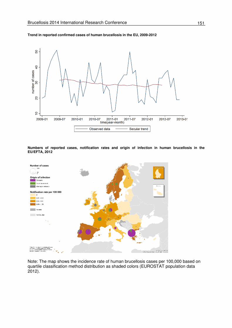
151 Brucellosis 2014 International Research Conference
Trend in reported confirmed cases of human brucellosis in the EU, 2009-2012
Numbers of reported cases, notification rates and origin of infection in human brucellosis in the EU/EFTA, 2012
Note: The map shows the incidence rate of human brucellosis cases per 100,000 based on quartile classification method distribution as shaded colors (EUROSTAT population data 2012).
152 Abstracts
Oral Presentations (O26-O29) O26: Human Brucella canis infection transmitted by contact with an asymptomatic infected dog Jorge C. Wallach1,2, Bettina Deodato2, Guillermo Benchetrit3, María E. Cortina1, Juan E. Ugalde1, Andrés E. Ciocchini1 and Diego J. Comerci1 1Instituto de Investigaciones Biotecnológicas ‘Dr. Rodolfo A. Ugalde’, Universidad Nacional de San Martín (IIB-UNSAM-CONICET), San Martín, Buenos Aires, Argentina; 2Unidad de Enfermedades Infecciosas, Hospital F.J. Muñiz, Buenos Aires, Argentina; 3 Servicio de Infectología, Centro Gallego, Buenos Aires, Argentina
Brucella (B.) canis, the main etiologic agent of canine brucellosis, can be transmitted to hu-mans by either laboratory accidents or close contact with infected dogs, principally after an abortion. Human B. canis infection is frequently mild or asymptomatic. In symptomatic cases, clinical signs are variable and typically the disease presents as an acute febrile illness with flu-like symptoms. Complications are infrequent and include arthritis, spondylitis and epidid-ymo-orchitis. Neurologic signs (including meningitis, uveitis and optic neuritis), anemia, inter-nal abscesses, nephritis, endocarditis and dermatitis can also occur and other organs and tissues can be affected, resulting in a wide variety of syndromes. Even though, antibiotic treatment is usually effective, relapses can occur. In untreated individuals, mortality rates vary from less than 2 % to 5 % and endocarditis and meningitis are the most frequent causes of death. Case report: A 37-year-old previously healthy male sought medical care in a general hospital due to fever, myalgia and headache that had begun 3 days earlier. No pathological signs were found by physical examination, and laboratory studies showed moderate elevation of transaminases and alkaline phosphatase. At that moment, two samples for blood culture were taken and symptomatic treatment was administered. The patient attended to control again 48 hours later with symptoms worsening, hepatomegaly was demonstrated by ultraso-nography, and increased levels of transaminases were found. Since the blood culture was positive for Gram-negative bacilli, hospitalization was decided and antibiotic treatment (ciprofloxacin 400 mg IV every 12 hours and ampicillin/sulbactam 1.5 g IV every 6 hours) was given. Few days later, the isolated bacteria were identified as Brucella spp. and the treatment was switched to streptomycin 1 g/d IM and doxycycline 100 mg b.i.d. orally. After treatment, the patient experienced clinical remission with normal transaminase values and negative results in subsequent blood cultures. After discharge, the patient was referred to the Hospital of Infectious Diseases FJ Muñiz for follow-up. Laboratory typing tests identified the isolated bacterium as B. canis. In addition, serum samples obtained during hospitalization were tested for antibodies against smooth Brucella strains (RBT, STA, STA-2ME, CFT and cELISA) and rough strains (RSAT and iELISA for B. canis). Positive results were obtained with RSAT and iELISA for B. canis, whereas the remaining tests were negative. The clinical examination and a questionnaire indicated that the patient was asymptomatic except for a mild intermittent diarrhea. Additionally, he reported to live with a healthy male dog which had presented positive blood cultures for B. canis. For this reason, we decided to carry out sero-logical tests on the patient’s family including his wife and their three children. His wife was asymptomatic and exhibited positive results by RSAT and iELISA, whereas the children were negative. This case demonstrates the transmission of canine brucellosis to pet owners from an appar-ently healthy dog which had a B. canis-positive blood culture. This type of transmission is not commonly seen in immunocompetent individuals, but it highlights the need for intensified controls of pet dogs to exclude a latent canine Brucella infection. E-mail of presenting author: [email protected]
153 Brucellosis 2014 International Research Conference
O27: Linked human and livestock study on seroprevalence and risk factors for brucel-losis in Kenya, 2012 Austine Bitek1, Eric M. Osoro1, Eric Ogola3, Ian Njeru4, Salome Wanyoike2 and Murithi R. Mbabu2
1Zoonotic Disease Unit, Nairobi, Kenya; 2Ministry of Agriculture, Livestock and Fisheries, Nairobi, Kenya, 3Kenya Medical Re-search Institute, Nairobi, Kenya; 4Ministry of Health, Nairobi, Kenya
Brucellosis is an endemic zoonotic disease in Kenya with dual burden i.e. significant eco-nomic losses among livestock and illness and disability in humans. Brucellosis studies in Kenya have previously focused on either animals or humans. We conducted a cross-sectional survey that investigated humans and livestock (cattle, sheep, goats and camels) in the same households. We assessed the seroprevalence and risk factors for human brucello-sis, animal brucellosis and their association. The study was conducted in three counties with different livestock production systems; households were selected randomly using a two-stage cluster sampling method by sub-location and then by household. Three persons were enrolled and livestock species proportional to the herd size were sampled per household. Questionnaires were answered by household heads and the persons sampled. Human sera were tested for Brucella IgG antibodies using competitive ELISA and animal sera were tested using an indirect ELISA. Risk factors were assessed using multivariate logistic regression. A total of 1,099 households, 2,811 persons and 11,039 production animals were enrolled. Overall, 14 % (95%CI 12-16) of households had at least one Brucella seropositive person and 15 % (95%CI 12-18) of the herds had at least one seropositive animal. Among humans sampled, 6.7 % (95%CI 5.6-7.8) were seropositive. Risk factors for human seropositivity in-cluded consuming unboiled milk (aOR=3.9, 95%CI 2.0-7.6), exposure to goats [herding, milk-ing, feeding] (aOR=2.5, 95%CI 1.6-4.0) and handling hides (aOR=3.9, 95%CI 2.5-6.2). Ani-mal seropositivity was linked to intermingling with wildlife (aOR=4.3, 95%CI 2.3-8.1) and keeping goats (aOR=2.7, 95%CI 1.4-5.2). The odds of human seropositivity given a sero-positive animal in the same household was 5.3 (95%CI 3.2-8.8).This linked survey shows that human and animal brucellosis seropositivity is associated with factors that increase ex-posure to seropositive animals. Our survey contributes to a better understanding of the bur-den of brucellosis in Kenya and helps to target health education programs. E-mail of presenting author: [email protected]
154 Abstracts
O28: Risk factors for human brucellosis in the Kween District, Eastern Uganda, 2011 – a case-control study Atuheire Emily1, Rutebemberwa Elizeus2, Kiguli Juliet2 and Nyakarahuka Luke3 1Ministry of Health, Kampala, Uganda; 2 Makerere University School of Public Health, Kampala, Uganda; 3Makerere University College of Veterinary Medicine, Animal Resources and Biosecurity, Kampala, Uganda
The global burden of brucellosis remains enormous, accounting for over 500,000 new infec-tions per year. The disease is endemic in most countries of Africa, Asia, South and Central America with incidence rates of up to 70 cases per 100,000 people. In Uganda, brucellosis is reported as an increasing public health hazard with a prevalence of 18-24 % in endemic are-as. This study was conducted to determine the risk factors for transmission of brucellosis to humans in the Kween District, Eastern Uganda and to recommend prevention and control methods. A facility based case-control study was carried out in a rural community in March 2011. A total of 56 cases and 141 controls were included. Cases and controls were residents of the Kween District at any age who were willing to participate in the study. Participants were iden-tified and enrolled when they attended outpatient services at selected facilities. A brucellosis case was defined to be a patient with a positive competitive ELISA result and presenting with at least two clinical symptoms that are suggestive of brucellosis. A control was an individual from the same village as the case having a negative serological test result. Blood samples were drawn and aliquoted before shipping to the Central Public Health Laboratory for labora-tory examination. Those who tested positive on rapid screening tests were further subjected to ELISA which was considered as the gold standard. Risk factor data were collected using individual structured questionnaire interviews. Data were entered into SPSS and exported to Stata and Epinfo statistical software programs for analysis. Cross tabulations, logistic regres-sion models and Fishers exact tests were used to establish associations at univariate and multivariate levels. P-values and odds ratios (OR) were used to determine statistical signifi-cance of independent variables. After adjusting for gender and age being in the age group of 21-50 years (OR 11, P=0.03), handling cow dung (OR 0.1, P=0.01), and being a male herding animals (OR 2.3, P=0.04) were found to be risk factors for brucellosis. In summary, there is a need to strengthen and prioritize the veterinary public health services at the community level in order to control brucellosis in humans. Communication programs aiming at behavior change are essential for empowering livestock keeping communities to prevent brucellosis transmission to humans. E-mail of presenting author: [email protected]
155 Brucellosis 2014 International Research Conference
O29: Epidemiological status of brucellosis in the Russian Federation Yury K. Kulakov¹, Marina D. Novikova¹ and Oleg D. Sklyarov² ¹Gamaleya Research Institute for Epidemiology and Microbiology of the Russian Ministry of Healthcare, Moscow, Russia; ²Federal State Institution «Russian State Center of Quality and Standardization of Pharmaceutical Preparations for Animals and Food» (VGNKI), Moscow, Russia
Brucellosis has caused about 300-500 reported new human cases per annum in the Russian Federation during the last decade. The official number of human brucellosis cases de-creased from 465 to 341 cases in the period 2012 to 2013. The morbidity rate was 0.33-0.24 per 100,000 people and in children below 17 years of age 0.17-0.08. Most new cases of hu-man brucellosis were detected in three federal districts of Russia: 298 cases (3.16 per 100,000 people) to 211 (2.23) in the North Caucasian Federal District, 75 (0.54) to 64 (0.46) in the Southern Federal District, 69 (0.36) to 44 (0.23) cases in the Siberian Federal District. These three districts covered 93.55 % of all human cases reported in the country. In the North Caucasian Federal District brucellosis was reported in almost all of the administrative territories except for the Republic of Ingushetia. The highest percentage of human cases were reported to be 45.81 % and 41.64 % (7.32 and 4.86 per 100,000 people) in the Repub-lic of Dagestan, 16.34 % and 18.48 % cases (2.73 and 2.26) in the Stavropol region in 2012 and 2013, respectively. Human brucellosis was also reported in all of the administrative terri-tories of the Southern Federal District with the highest percentage of cases in the Republic of Kalmykia i.e. 10.97 % and 12.9 % (17.62 and 15.29) in 2012 to 2013, respectively. In the Siberian Federal District, morbidity was 4.52 % and 3.52 % (6.82 and 3.89) in the Republic of Tyva, and 4.09 % and 4.12 % (1.72 and 1.27) in Trans-Baikal Territory in the respective years. In 2013, there were about 20.3 million cattle and over 22 million sheep in the country. The most affected regions for animal brucellosis were again the three federal districts already mentioned. In 2013, bovine brucellosis was reported in 22 regions of Russia in which 209 holdings (state and private farms) were affected by bovine brucellosis with 13.800 cattle in-fected. The highest number of bovine brucellosis cases was reported from the North Cauca-sus Federal District with 9,022 infected cattle or 65.2 % of all cases in Russia, the Southern Federal District with 3,389 cattle (24.5 %), and the Siberian Federal District with 1,134 cattle (8.2 %). In 2013, caprine brucellosis was reported in 30 holdings with 2,000 goats and sheep involved. About 80 % of sheep and goats and more than 50 % of cattle were located in pri-vate farms. Livestock owners were not capable to carry out expensive anti-brucellosis measures due to restricted financial resources. Most animals were infected on farms where goats and cattle were kept together and B. melitensis biovar 3 was the most prominent spe-cies originating from the Republic of Dagestan and Kalmykia. Summing up it can be pointed out that reported cases of human brucellosis have been found in 29 regions (34.94 %) of Russia, in 8 regions (9.64 %) brucellosis was only found in cattle (maybe accompanied by under-reporting of human brucellosis), in 6 regions (7.2 %) ‘free’ of brucellosis (travel-associated cases may still occur), in 42 regions (50.6 %) brucellosis was not detected in hu-mans and animals in 2013. Thus, in Russia the epidemiological situation is complicated due to missing controls of private animal farms, not ideal veterinary service, limited use of laboratory methods for active brucellosis surveillance in animals and humans. This lack of public veterinary health contrib-utes to the spread of animal brucellosis among cattle and sheep, a delayed culling of sick animals, the refusal of private owners to obey veterinary control measures, under-registration of new livestock. The situation is also worsened by uncontrolled introduction of animals suf-fering from brucellosis (mainly sheep) into territories previously free from brucellosis, the un-controlled movement of animals inside Russia and uncontrolled importation of animals from other CIS states bordering the Russian Federation (Kazakhstan, Azerbaijan, Mongolia). E-mail of presenting author: [email protected]
156 Abstracts
Poster Presentations (P85-P99) P85: Clinical and epidemiological parallels in human brucellosis cases Y.K. Khudayberdyev, N.E. Kadyrova and A.S. Kosymov Research Institute of Epidemiology, Microbiology and Infectious Diseases, Tashkent, Uzbekistan
The aim of our study was to assess the significance of peripheral blood and urine parameters in brucellosis patients and their correlation to clinical and epidemiological data. We investigated patients suffering from brucellosis as well as healthy volunteers from en-demic regions (Oblasts). The phenotype of acetylation (PA) and the levels of antigen-binding antibodies directed to tissue-specific antigens (ABL to TA ratio) were measured in all patients and some volunteers to determine the extent of pathogenic effects on joints, heart, brain and liver. The fast acetylation phenotype was determined in urine samples using 10 mg/kg of sulfathiazole as a test drug. The capacity of acetylation was considered slow unless it reached 50 % of average, and fast when it exceeded 50 % of average. Interviews were con-ducted according to an established protocol to identify possible cases of brucellosis in vari-ous social segments of the population. In addition, bacteriological and serological tests were performed to diagnose a Brucella infection. Brucellosis patients in various stages of disease were examined. As a result of the question-naires over 80 % of the residents in endemic regions turned out to be cattle owners and 57 % owned sheep and goats. At the same time, only 3 % of them belonged to the occupa-tional risk group of livestock owners. A total of 17.4 % of the participants were involved in cattle slaughtering and processing of livestock products. 31 % of the healthy volunteers were involved in lambing campaigns. 9.4 % revealed positive serological test results for brucello-sis. Peripheral blood parameters examined in a limited number of healthy volunteers were similar to that of patients with brucellosis. Acute brucellosis was characterized by a more foudroyant clinical course and a fast acetyla-tion phenotype. Chronic brucellosis was characterized by a subclinical and dull course and slow acetylation phenotype, which could be documented in nearly 60 % of the examined in-terviewees. More significant effects on joints, brain and the myocardium were found in pa-tients with a slow acetylation phenotypeas compared to fast acetylation. According to com-parative analysis of clinical and epidemiological data 9.4 % of the patients had experienced clinically non-manifesting brucellosis. E-mail of presenting author: [email protected]
157 Brucellosis 2014 International Research Conference
P86: Brucellosis laboratory diagnostics at Armenia’s National Center for Disease Con-trol and Prevention, 2009-2013 Gayane Gevorgyan National Center for Disease Control and Prevention (NCDCP), Especially Dangerous Infections Laboratory, Ministry of Health, Armenia
Brucellosis is one of the most common global zoonotic infections with over 500,000 new hu-man cases annually reported. Brucellosis is endemic in Armenia, with an average of 300 human cases recorded each year. As part of a continuous improvement process, we evalu-ated the laboratory surveillance system for brucellosis at the national level. A retrospective survey of brucellosis laboratory data originating from the National Center for Disease Control and Prevention (NCDCP) was conducted. Case files of all samples suspect-ing brucellosis and submitted to NCDCP in the years 2009-2013 were analysed for diagnos-tic tools used and results obtained. From 2009 to 2013, a total of 1,365 samples of any suspect human brucellosis case on the basis of standard case definition were sent to NCDCP for confirmatory laboratory diagnosis. Brucellosis was confirmed by Serum Agglutination Test (SAT) in combination with the Wright-Huddleston test. The samples were also tested by PCR. All 1,365 samples were posi-tive in SAT and PCR. The confirmed cases were recorded at the national level and reported to the World Health Organization (WHO). The highest incidence was recorded in 2009, with a rate of 10.7 % per 100,000 population, and the lowest incidence was recorded in 2013, with a rate of 7.2 % per 100,000 population. In all suspect samples which reached NCDCP brucellosis was laboratory confirmed. These results indicate that the standard case definition for brucellosis is accurate and is used ap-propriately by the health care providers in Armenia. The results also indicate that PCR is an alternative diagnostic tool for brucellosis; however, it is not recommended to be used as standalone test because SAT titres provide critical information for the diagnosis of brucello-sis. In order to improve the surveillance system in Armenia, genotyping of Brucella isolates should also be performed. Email of presenting author: [email protected]
158 Abstracts
P87: Role of laboratory methods in active surveillance of human brucellosis outbreaks on a part-time farm of the Moscow Zoo Marina D. Novikova and Yury K. Kulakov Gamaleya Research Institute of Epidemiology and Microbiology, Ministry of Healthcare of the Russian Federation, Moscow, Russia
Brucellosis is a dangerous zoonotic infection with chronic courses and disability of patients and of serious public health concern in the Russian Federation. Human cases are caused by the transmission of brucellae from their natural hosts (goats, sheep, pigs, and cattle). The aim of our study was to assess the role of laboratory methods in active surveillance of human brucellosis outbreaks on a part-time farm of the Moscow Zoo located about 100 km from Moscow. Epidemiological surveys were performed from 2003 to 2013 (twice a year in spring and autumn) involving more than 200 part-time farm employees who were working on the farm during outbreaks of sheep or cattle brucellosis. Blood sera were analysed using tube agglutination (Wright test), agglutination on glass slides (Huddelson), Coombs test, iE-LISA (indirect enzyme-linked immunosorbent assay) with the immunodominant s-LPS, real-time PCR targeting bcsp31. After the elimination of epizootic caprine brucellosis in 2003, the percentage of positively tested employees decreased from 42.7 % to 17.2/15.9 % in 2005/2006. Real-time PCR de-tected Brucella DNA in infected humans more efficiently than iELISA detected anti-Brucella antibodies. In 2007 and 2009, 5 and 11 workers were found infected, respectively. They had negative results from 2003 to 2006. The infection of these workers might be associated with outbreaks in 2007 and 2009 which were traced back to an infected 2-year-old yak. In 2012 and 2013, no acute brucellosis case was found, and a decrease of iELISA antibody titres in previously infected employees was noted. During the study period of 11 years, 42.2 % of the employees were diagnosed with brucellosis. These employees had contact to Brucella posi-tive animals, but only 14 % of these presented clinical signs and symptoms of disease. Active brucellosis surveillance by various laboratory techniques has been conducted during brucellosis outbreaks on a part-time farm of the Moscow Zoo. The laboratory proven human cases could be traced back to brucellosis outbreaks in sheep and in a yak in 2003, and 2007 and 2009, respectively. Real-time PCR was an effective tool to identify Brucella infections. The iELISA proved to be more sensitive in chronic patients than real-time PCR. Traditional serological methods were less sensitive and informative, but less time consuming compared to real-time PCR and iELISA. E-mail of presenting author: [email protected]
159 Brucellosis 2014 International Research Conference
P88: Combined diagnostics in a case study on human brucellosis Hudz Nataliia1, Nychyk Serhiy1, Svita Viktor2, Novokhatniy Uriy2, Postoienko Volodymyr1 and Sytiuk Mykola1 1Institute of Veterinary Medicine of the National Academy of Agrarian Sciences of Ukraine, Kyiv, Ukraine; 2State Institution «Ukrainian Center for Disease Control and Monitoring of Ministry of Health of Ukraine», Kyiv, Ukraine
Brucellosis is an infectious disease which affects small ruminants, bovine animals and pigs, and is dangerous for people. Currently, brucellosis is not officially reported in Ukraine. How-ever, because of the threat of import of the pathogen from abroad, timely and correct diagno-sis are extremely important. The aim of our study was to identify efficient approaches to diagnose human brucellosis. Epidemiological, clinical, and laboratory methods were used. Laboratory tests comprised tube agglutination (Huddelson), the Rait agglutination, and an indirect hemagglutination test. An Ukrainian citizen who worked at a construction side of stable at a livestock enterprise in Russia since October 2011 asked for medical help at the city hospital on 15th May 2013. He suffered from fever up to 40 °C, weakness, fatigue, drowsiness, joint pain in the upper limb, and a productive cough. An acute respiratory disease was initially diagnosed. However, the patient reported that two human brucellosis cases have been diagnosed on the cattle farm he was working at. Subsequently, the patient was tested positive for brucellosis and negative for tularemia. The Huddelson test was positive, the Rait test and the indirect hemagglutina-tion revealed anti-Brucella antibody titres, 1:200 and 1:640, respectively. Hence, the final diagnosis was ‘acute brucellosis, moderately severe’. A combination of laboratory tests and epidemiological investigation led to the timely detection of brucellosis in our patient. Whether the disease was acquired abroad cannot be clarified. In Ukraine, the implementation of effective countermeasures to prevent the introduction of bru-cellosis from endemic areas is of high relevance to control the spreading of the disease. E-mail of presenting author: [email protected]
160 Abstracts
P89: Human brucellosis in Georgia: Clinical and laboratory manifestations N. Kokaia, M. Kajaia, M. Murusidze and N. Iashvili S. Virsaladze Research Institute of Medical Parasitology and Tropical Medicine, Tbilisi, Georgia Human brucellosis is one of the most prevalent zoonosis in Georgia. According to national statistics, approximately 150 confirmed cases of brucellosis were recorded in the country annually. The disease in humans is mainly associated with animal contact and/or consuming animal products. We analyzed medical records of 543 patients at the Virsaladze Research Institute of Medical Parasitology and Tropical Medicine from 2009-2013 of whom brucellosis was diagnosed and treated. We assessed age, gender, clinical signs, laboratory investigations including com-plete blood count (CBC) and serological tests of the patients. During 2009-2013, a total of 2,423 patients were tested for brucellosis at the Institute. Among them, 543 patients (22.4 %) were positive tested for the disease. Confirmation of diagnosis was performed by Wright Agglutination test at a titer ≥1/200 and/or by enzyme-linked im-munosorbent assay (ELISA) to detect Brucella-specific IgM antibodies. Among patients, males predominated significantly (88.7 %). Brucellosis-confirmed patients were allocated to three different age groups. 67 % of the patients (363/543) belong to the largest group with ages ranging from 14-60 years. The second group consists of 24.4 % (133/543) of the pa-tients within the age group of >60 years, and only 8.6 % (47/543) of patients were in the third age group ranging between 0-14 years. The youngest patient of the study was 18 months. Most of the children with brucellosis reported a family history of the disease. 158 (29 %) out of 543 patients were hospitalized and treated at the clinic. The average dura-tion from symptoms onset to the diagnosis was 18 days. Delayed diagnosis for ≥1 month was observed in about 20 % of cases. Fever was recorded in 94.3 % (510/543), sweating in 75.8 % (412/543), chills in 77.7 % (422/543), fatigue in 77.3 % (420/543), osteoarticular and neuromuscular pain in 88 % (482/543), hepatomegaly in 34 %, (189/543) and splenomegaly in 16.3 % (89/543) of the investigated patients. Polylymphadenopathy was rare and observed only in 8.8 % (48/543) of cases. Among 543 brucellosis patients hematologic tests were per-formed for 454 patients. Anaemia was detected in 45.8 % (208/454) of cases. Leukopenia (WBC <4,300/mm3) was present in 7.54 % (34/454), thrombocytopenia (platelet count <150,000/mm3) in 26 % (118/454) and pancytopenia in 5.2 % (24/454) of the patients. Addi-tionally, a slight increase of liver enzyme levels was observed in 35.4 % (56/158) of cases. The most frequent complication of brucellosis was sacroileitis, which was observed in 13 % of the patients while epididymo-orchitis was detected only in 9 % of the cases. Both of these complications were associated with delayed diagnosis for more than 1 month. All adult pa-tients were treated with an antibiotic combination of streptomycin or gentamicin and doxycy-cline. Vast majority of patients 88.9 % (483/543) were cured after the full course of the treat-ment, 6.6 % (36/543) of patients interrupted the therapy and 4.5 % (24/543) of patients expe-rienced relapse of the disease. Brucellosis in humans remains one of the main health problems in Georgia. Nonspecific clin-ical signs cause delayed diagnosis of the disease, which can be associated with serious complications. Brucellosis should be considered for all patients who show signs of systemic inflammation and arthritis in endemic areas. E-mail of presenting author: [email protected]
161 Brucellosis 2014 International Research Conference
P90: Using molecular tools to identify the geographical origin of a case of human bru-cellosis Krishna Gopaul1, Jakub Muchowski1, Cyril Perrier1,2, Mark Koylass1, Amanda Dainty1, Antony Gatt3, Paul Caruana4 and Adrian Whatmore1
1Animal Health and Veterinary Laboratories Agency, Addlestone, United Kingdom; 2Université de Nimes, Nîmes, France; 3Health Promotion and Disease Prevention Directorate, Msida, Malta, 4Mater Dei Hospital, Msida, Malta
Brucellosis is a zoonosis of great socio-economic importance that causes reproductive prob-lems in ruminants and swine as well as a number of ailments in humans including general malaise. Although this disease has a global presence, a number of countries have invested a great deal of time and money on brucellosis eradication schemes and attain officially brucel-losis-free status. One of these countries is the Mediterranean nation of Malta. A strain of Brucella (B.) melitensis that had been isolated from a human in Malta was submit-ted in late 2013 to the WHO Collaborating Centre for Reference and Research on Brucellosis at AHVLA Weybridge. Maltese colleagues wanted to establish whether the causative agent had been imported or was an example of a local strain that had been present on the island prior to eradication. To this end, molecular typing tools were used to try and provide an an-swer to this question. Multi Locus Sequence Analysis (MLSA) and Variable Number Tandem Repeat (VNTR) test-ing are well-established tools widely used for typing and molecular epidemiology. As very little molecular data existed on Brucella strains from Malta, 15 B. melitensis isolates taken from both humans and livestock in Malta between 1950 and 1960 and present in the AHVLA strain archive were processed by these two methods alongside this current isolation. Using MLSA, we found that whilst the historical isolations all shared a common sequence type (ST7) which is found in other isolations in the Mediterranean/Southern Europe, the re-cently submitted case belonged to a ST associated with countries in the Horn of Africa. This observation was backed up by subsequently provided field epidemiological data which identi-fied the case as a recent arrival to Malta from the Horn of Africa. As expected, VNTR data showed more diversity within the 16 isolates tested but confirmed that the recent human iso-late was unrelated to historical local strains. In addition, whilst the MLVA-16 profiles of the historical local isolates were similar to each other, the profile generated from the recent hu-man isolation was very different to these. The example in this study illustrates the usefulness of molecular techniques in the unbiased geographical placement of a strain to add value to field epidemiological observations. Given advances in whole genome sequencing technology and its potential applications in phyloge-ography and microbial forensics, the strains are currently undergoing whole genome se-quencing to assess the added value of this approach. E-mail of presenting author: [email protected]
162 Abstracts
P91: Human brucellosis in Belgium: serological trends, isolates and molecular typing, 1998-2014 Marcella Mori1, Luisa Escobar-Calle1, Marjan Van Esbroeck2, Jacques Godfroid3 and David Fretin1 1Bacterial Zoonoses of Livestock, Operational Directorate Bacterial Diseases, Veterinary and Agro-chemical Research Centre (VAR), Brussels, Belgium; 2Tropical Medicine Institute, ITM, Antwerp, Belgium; 3Department of Food Safety and Infection Biolo-gy, Norwegian School of Veterinary Science, Tromsø, Norway
Belgium is a country free of brucellosis in livestock, with the latest domestic animal case oc-curring in 2000. In wildlife Brucella spp. can still be isolated from wild boars (B. suis bv 2 in Sus scrofa) and marine mammals. In late 2010 and spring 2012, the country was affected by two cluster cases of bovine brucellosis which, properly controlled, did not hamper the official-ly brucellosis-free (OBF) status of Belgium. To increase knowledge of human brucellosis in Belgium, we analyzed historical data of serological samples and clinical isolates collected in a reference setting during a 16-year period. Clinical samples were those submitted to the National Reference Centre outside of the labor medical testing. Confirmed laboratory case definition was that recommended by the World Health Organization (isolation of the bacteria or sero-convertion assessed with the IgG ELISA-(iELISA)). A total of 201 samples were sero-logically analyzed by Rose Bengal test (RBT) and iELISA. Thirty-three samples (15.7 %) were positive to RBT of which nine (27.3 %) were confirmed by the Wright agglutination test and fourteen (42.4 %) by the iELISA (with one associated with the direct isolation of Brucella spp.). Although not quantifiable due to inconsistent information on clinical data of the pa-tients, Rose Bengal test yielded false-positive results in various cases. A total of 32 clinical cases were identified by bacterial isolation. Biotyping of the isolated strains identified 26 as B. melitensis bv 3, three as B. melitensis bv 2, two as B. melitensis bv 1 and one as B. abortus bv 1. Multiple Loci VNTR Analysis confirmed and highlighted the diversity of those strains. Because B. melitensis is not present in small ruminants in Belgium, and because only B. abortus bv 3 has been isolated in cattle since the early 1990s, it is high-ly likely that all human cases cited here were imported or associated to a contamination hav-ing occurred abroad. Human brucellosis is yet a rare disease in Belgium, mainly allochtho-nous (or associated to professional work). Early detection of imported brucellosis in domestic animals is a prerequisite to prevent any human contamination associated to animals to occur in Belgium. E-mail of presenting author: [email protected]
163 Brucellosis 2014 International Research Conference
P92: Investigation of the risk factors to human brucellosis in Mubende district, Uganda Asiimwe Karimu Grace1, Nakavuma Jesca1, James Acai1 and Luke Nyakarahuka2 College of Veterinary Medicine, Animal Resources and Biosecurity, Makerere University, Kampala, Uganda; 2Uganda Virus Research Institute, Kampala Uganda
Brucellosis is a globally distributed zoonotic disease associated with more than 500,000 cas-es annually. Prevalence of 16 and 18-24 % has been reported in Africa and in Uganda, re-spectively. Human brucellosis infections are mainly associated with the consumption of un-pasteurized milk and its products or the direct contact with infected animals. In 2011, an in-creasing number of brucellosis cases from Mubende district were reported to the Ministry of Health Uganda. These cases prompted us to establish the prevalence and the main risk fac-tors for both human and animal brucellosis for this district. An unmatched one to one case control study involved extraction of brucellosis cases biodata from laboratory records. Active search for patients was done in communities. 52 cases of the target number of 100 cases were enrolled. Fifty-two controls accessed from immediate neighborhood considering age and gender were interviewed using structured questionnaires. Children's questionnaires were answered by their parents. Cattle older than two years and goats older than one year origi-nating from cases’ and controls’ homesteads were randomly selected and bled. Seropreva-lence in animals was determined using cELISA, risk factors were analysed using Chi square test that generated odds ratios. For humans, 52 cases and 52 controls were recruited for the study. The brucellosis sero-prevalence determined from hospital records was 31 %. The seroprevalence of brucellosis in cattle was 11 % at animal level and 38 % at herd level. In goats, prevalence was 36 % and 58 % at animal and herd level, respectively. Significant risk factors associated with contract-ing brucellosis in humans were: consumption of undercooked meat (the risk was eight times higher); not using protective wear while handling animals’ aborted fetuses and faeces (four times higher). In animals main risk factors were: history of animal abortions on farm and use of rented breeding bulls (eight times and five times higher, respectively). The increase in number of cases in Mubende district was found to be associated with the consumption of undercooked meat by farmers and failure to use protective wear while han-dling animals. History of abortions on farms and use of rented breeding bulls were associat-ed with high prevalence of brucellosis in animals. The government should strengthen pro-jects that will rise the awareness of rural farmers for the risk factors and the control measures for brucellosis. Regular testing of animals for brucellosis is advised. E-mail of presenting author: [email protected]
164 Abstracts
P93: Changing incidence of human brucellosis, Azerbaijan 1983-2009 Rita Ismayilova1, Ian Kracalik2,3, Rakif Abdullayev1, Mehriban Baghirova4, Narmin Ustun1, Mazahir Shikhiyev4, Ayden Talibzade1, Kliment Asadov4 and Jason K. Blackburn2,3
1Republican Anti-plague Station, Baku, Azerbaijan; 2Spatial Epidemiology & Ecology Research Laboratory, Department of Ge-ography, University of Florida, Gainesville, FL, USA; 3Emerging Pathogens Institute, University of Florida, Gainesville, FL, USA; 4State Veterinary Service, Baku, Azerbaijan
We assessed spatial and temporal trends in the epidemiology of human brucellosis in Azer-baijan from 1983 to 2009. Up to the mid-1990's there was a substantial increase in brucello-sis incidence (25 % annually) followed by an average decline in reporting of 5 % annually after the initiation of a livestock test-slaughter campaign. Our findings confirmed that brucel-losis was widespread in both humans and livestock. However, recent changes in the case distribution indicated an annual increase in human incidence among districts clustered in southeast compared to a decrease elsewhere. Males were disproportionately represented among cases (71 % male vs. 29 % female). The age specific average incidence was highest in the 15-19 years age group with 18.1 cases per 100,000 population, suggesting a primarily occupation route of exposure. Livestock control campaigns have likely improved the current brucellosis situation in Azerbaijan, although the disease appears to be re-emerging in the southeast. Our findings highlight the need for dynamic surveillance that takes into account the changing patterns of disease while focusing on high risk populations. E-mail of presenting author: [email protected]
165 Brucellosis 2014 International Research Conference
P94: Time series analysis of human brucellosis in Azerbaijan 2002-2010 Narmin Ustun1, Ian Kracalik2,3, Kliment Asadov4, Rita Ismayilova1, Mehriban Baghirova4, Mazahir Shikhiyev4, Ayden Talibzade1, Rakif Abdullayev1 and Jason K. Blackburn2,3
1Republican Anti-plague Station, Baku, Azerbaijan; 2Spatial Epidemiology & Ecology Research Laboratory, Department of Ge-ography, University of Florida, Gainesville, FL, USA; 3Emerging Pathogens Institute, University of Florida, Gainesville, FL, USA; 4State Veterinary Service, Baku, Azerbaijan
In Azerbaijan, the zoontic disease brucellosis is considered an endemic public and veterinary health threat. To assess the temporal dynamics of human brucellosis and target areas for control, we analysed monthly reporting from 2002 to 2010 using a time series analysis. Due to strong seasonality in the data, seasonal and non-seasonal Autoregressive Integrated Mov-ing Average (ARIMA) models were used to forecast future brucellosis case reporting 24 months ahead of time by fitting a series of models to the data and selecting the model with the lowest Akaike Information Criterion (AIC) score. Spatial differences in the seasonality of cases were assessed using geographic information systems (GIS). A total of 3,898 cases of human brucellosis were reported during 2002-2010 with significant seasonality in the sum-mer months. Model diagnostics and the AIC score indicated the ARIMA model with a (2,1,3) was the best fit. The trend in the data suggested a gradual decrease in the reporting of hu-man cases; however, we identified district local level heterogeneity in reporting trends and spatial differences in the seasonality of case reporting. Control of brucellosis requires coop-eration among multiple health agencies in order to properly formulate management strate-gies. Recent national reductions in the occurrence of human brucellosis in Azerbaijan high-light the impact that veterinary control measurements such as test-slaughter campaigns can have for human health. Further reduction of the number of human cases can only be reached if areas of high human and animal transmission are targeted. E-mail of presenting author: [email protected]
166 Abstracts
P95: Brucellosis epidemic situation dynamics in the Armenian population, 2011-2013 Liana Torosyan, Lilit Avetisyan and Artavazd Vanyan
National Center for Disease Control and Prevention, Ministry of Health, Armenia
Brucellosis is a dangerous disease for both humans and animals. It is endemic in several countries of the Caucasus region, including Armenia, and thus considered as a priority dis-ease for the region. In 2013, 219 new cases of brucellosis were recorded in Armenia (inci-dence rate of 7.2 per 100,000); in 2012, 227 cases (incidence rate of 6.9 per 100,000); in 2011, 260 cases (incidence rate of 8.0 per 100,000); in 2010, 295 cases (incidence rate of 9.2 per 100,000); in 2009, 346 cases (incidence rate of 10.7 per 100,000); and in 2008, 289 cases (incidence rate of 8.95 per 100,000). In this study, we used data from the Armenian epidemiological surveillance system to study the demographic distribution of human brucel-losis cases and identify risk factors of infection. Data were deduced from medical records of confirmed brucellosis patients registered in the country from 2011-2013. In total, 706 records were analysed with regard to demographic data (age, gender, residence [urban/rural]) and history of possible exposure. The epidemiology of human brucellosis in Armenia can be characterised as follows: 12 % of patients lived in urban and 88 % in rural communities; 92 % were adults (aged >18 years) and 8 % children (aged <18 years); and 70 % were women and 30 % men. Following risk factors were identified: exposure to animals was noted in 51 % (cows, sheep, horses and pigs), the purchase of milk and dairy products purchased from local farmer’s markets (i.e. non-state regulated markets) was recorded for 28 %, and no risk factor was identified in 21 % cases. Most brucellosis patients (88 %) were from rural communities. The prevalence of brucellosis was much higher in women (70 %). This is likely due to the fact that in Armenia women are responsible for milking animals and preparing food from animal products that may contain Brucella species. Disease cases occurred primarily in adults (92 %). A large majority of pa-tients had contact with animals before symptoms developed (79 %). This study provides some initial information on brucellosis cases concerning demographic structures and risk factors in Armenia. Our results are consistent with results of similar studies from endemic countries around the world. Email of presenting author: [email protected]
167 Brucellosis 2014 International Research Conference
P96: Brucella melitensis in humans in Venezuela: Mistaken identity of Brucella infection Andrés Sánchez-Cómbita1, Armindo Perozo-Mena1,2, Yustín Gómez-Paz2, Datty Rosales-Zambrano3,5, Araceli Contreras-Rodríguez3, Aurea Morales-Estrada3, Natasha Merchán-Maduro1, Sergio Rivera4 and Alfredo Sánchez-Villalobos4 1Faculty of Medicine, University of Zulia, Venezuela; 2CRB Autonomous Service of the University Hospital of Maracaibo, Zulia; 3 Department of Microbiology, National Polytechnic Institute, Mexico; 4Faculty of Veterinary Sciences, University of Zulia, Vene-zuela; 5Sixto David Rojo Laboratory, BIOMI, University of Andes, Venezuela
Brucellosis is a zoonotic disease transmitted by direct or indirect contact with infected ani-mals or their products. Nowadays, Brucella (B.) melitensis is the species most frequently reported as the cause of human brucellosis in the world. Human brucellosis is a multisystem-ic disease with a wide range of clinical signs and symptoms, and despite being a centenary problem, still presents many challenges, particularly in diagnosis. In Venezuela, brucellosis is a forgotten disease among physicians. Laboratory diagnosis is based on serological tests, because applying direct methods is only possible in a few laboratories. Serological results can be questionable and advanced molecular techniques are now always available. Consid-ering these facts it is not surprising that B. melitensis was frequently misdiagnosed in the past. This report describes a case of a 9-year-old girl with a prolonged febrile illness whose serol-ogy screening tests for brucellosis were positive (Rose Bengal test, Buffered Plate Antigen test, Fluorescent Polarization Assay). The medical interview revealed that the patient had often consumed fresh goat and cow milk cheese. For bacteriological diagnosis, two blood cultures were processed by Bact/Alert SA, being positive after 60 hours. Bacterial colonies were identified by the Vitek-2 system with a 99 % match for B. melitensis. The result was automatically reported by the software. Molecular diagnosis was performed at the National Polytechnic Institute of Mexico using the Bru-Multiplex PCR. The isolate was unmistakably diagnosed as B. abortus. The database of the Vitek-2 system considers all Brucella species as biovars of B. melitensis and can consequently not discriminate among species of the genus. Misidentification of Bru-cella species by commercial bacterial identification systems including the Vitek-2 has been previously reported. Diagnostic microbiology laboratories should be aware of potential misi-dentifications of brucellae using automated identification systems. This report confirms the presence of human brucellosis in Venezuela. However, the pres-ence of B. melitensis could not be confirmed as the causative agent. B. melitensis is widely spread in sheep and goats, where it is endemic, wheareas B. abortus is usually found in cat-tle. E-mail of presenting author: [email protected]
168 Abstracts
P97: Brucella seroprevalence study among stray and pet dogs in Georgia L. Malania1, M. Ramishvili1, G. Babuadze1, K. Goginashvili2, M. Donduashvili2, T. Tigilauri2, M. Kosoy4, M.P. Nikolich5, N. Trapaidze1,3, S. Goetz6, R.J. Arner6 and Paata Imnadze1 1National Center for Disease Control and Public Health, Tbilisi, Georgia; 2Laboratory of Ministry of Agriculture of Georgia; 3USAMRU-G Clinical Research Unit, Tbilisi, Georgia; 4Division of Vector-Borne Diseases, Centers for Disease Control and Prevention, Fort Collins, CO, USA; 5Walter Reed Army Institute of Research, Silver Spring, MD, USA; 6The Pennsylvania State University, University Park, PA, USA
Brucellosis is one of the most important zoonotic diseases worldwide. Brucellosis continues to cause significant medical, veterinary and socioeconomic problems and is endemic in Georgia. Although exposure to Brucella (B.) has been studied in cattle and small ruminants, no information is available of that in pets or wild animals in the country of Georgia. This study is the first attempt to record the detection of antibodies against Brucella spp. in serum sam-ples from stray and pet dogs collected in Tbilisi, Georgia in 2011. The Rose Bengal test (RBT) was performed to analyze serum samples collected from dogs for the presence of anti-Brucella spp. antibodies. RBT-positive samples were further investi-gated by fluorescence polarization assay (FPA) and ELISA to attempt confirmation. In addi-tion, positive samples were also tested by the slide agglutination test against five different pathogens: Yersinia (Y.) enterocolitica, Salmonella (S.) enteritidis, S. typhi, S. typhimurium, and E. coli O:157 to exclude false-positive results due to antigen cross-reactivity. Sera of 141 stray and 145 pet dogs were screened by RBT. The overall seroprevalence var-ied slightly between stray and pet dogs; a lower prevalence was detected in stray than that in pet dogs, 43.26 % and 51.7 %, respectively. Confirmation by FPA was successful in 5 of 11 RBT-positive samples that were negative for other infections. ELISA results were positive for all RBT-positive samples, regardless of infection status with other species. Out of the RBT-positive samples the samples were also positive for the following: Y. enterocolitica (69.73 %), S. enteritidis (25.65 %), S. typhi (37.5 %). S. typhimurium (21.71 %), E. coli (26.97 %).While the RBT assay does not allow the discrimination of antibodies against B. abortus and B. melitensis, the demonstrated high seroprevalence rate in both stray and pet dogs in Georgia suggests frequent exposure to one of the smooth lipopolysaccharide (LPS) Brucella spp. among these animals. Confirmation of RBT results was inconclusive as commercial FPA and ELISA assays are not validated for canine testing. Although the extent is unclear to which dogs transmit Brucella to human, the results obtained indicate a necessity to consider dogs as potential sources for human infection. To the best of our knowledge these are the first data reported for seroprevalence of Brucella spp. in dogs in Georgia. Future work will also include investigation in areas where the RBT test cross-reacts with non-Brucella microorgan-isms. This project will provide unique results to be incorporated into the public health ap-proach in Georgia. E-mail of presenting author: [email protected]
169 Brucellosis 2014 International Research Conference
P98: Evaluation of human brucellosis surveillance system in Georgia, 2010-2012 M. Ramishvili1,2, P. Imnadze1, L. Malania1, A. Navdarashvili1,2, M. Broladze1, T. Rush2, M. Geleishvili2 and E. Maes2 1National Center for Disease Control and Public Health, Tbilisi, Georgia; 2South Caucasus Field Epidemiology and Laboratory Training Program (SC/FELTP) Tbilisi, Georgia
Brucellosis is a highly contagious zoonosis affecting livestock and humans worldwide. It is recognized as a significant public health challenge with major economic and financial bur-dens. According to the WHO, about 500,000 cases of this disease are registered worldwide every year. The disease is transmitted to humans by contact with infected animals or the consumption of infected food products. The economic impact of the disease occurs through livestock disease and loss. According to national statistics, approximately 150 human brucel-losis cases were reported per year in Georgia in 2010-2012, although the actual numbers may be higher. The objectives of our evaluation were to identify strengths and weaknesses of the human brucellosis surveillance system in Georgia and to recommend improvements to the current surveillance system. For this purpose, current existing Georgian Guidelines for Brucellosis, policies and data from NCDC and EIDSS reports were reviewed. Data were also obtained from The Parasitology Center, Infectious Pathology Center, and Sepsis Center La-boratories. Brucellosis case definition was based on NCDC/DTRA definition, updated CDC 2001 evaluation guidelines, and other references. According to quantitative and qualitative assessments of the system attributes, the system is not simple and not flexible. Data quality, acceptability, representativeness of the system and timeliness are low. On average, 52.7 % of the monthly notification forms were filled completely in 2010-2012. A total of 190 samples were tested by ELISA and 34 (17.89 %) were positive. 2,279 Wright-Huddleston tests were performed, 440 (19.3 %) were positive. During the same period, 287 samples were cultured and 29 (10.1 %) showed Brucella growth. In 2010-2012, complete surveillance was conduct-ed for 472 probable cases and 19 Brucella outbreaks (two or more patients) were revealed. We could not calculate system sensitivity; however, the positive predictive value (PPV) for all tests in the surveillance system was approximately 18 %. In 2010-2012, the brucellosis inci-dence rate in Georgia decreased from 4.49 to 2.96 per 100,000 residents. This finding may be attributed to undiagnosed cases or a decreasing number of ruminants (for the last two years large numbers of sheep have been exported) or to a lack of patients seeking medical care. Culture is still considered the ‘gold standard’. However, it may not be sensitive. Not all cases are confirmed by culture and the system does not provide follow-up for all patients. Data quality and acceptability is low due to missing fields in EIDSS and slow reporting (inter-val between onset of symptoms and date of diagnosis is on average from 8 days until 5 months and up to one month from diagnosis until case registration). Cases entered into the system, however, have high validity. Representativeness is low, as this is a passive surveil-lance system and national screening has not been conducted for 30 years. Georgia does not have a national algorithm for brucellosis used by all labs. Brucellosis surveillance should be ongoing for better control of the disease in the country. The surveillance system should be evaluated periodically and recommendations should include quality, efficiency and useful-ness variables. Finally, a national testing algorithm should be standardized. E-mail of presenting author: [email protected]
170 Abstracts
P99: First outbreak of Brucella canis in a Finnish breeding kennel
Teresa Skrzypczak and Eija Seuna
Finnish Food Safety Authority Evira, Veterinary Bacteriology Research Unit, Helsinki, Finland
Canine brucellosis is caused by Brucella (B.) canis. In female dogs, the most common symp-tom is abortion, but an infected bitch can give birth to healthy, weak or dead puppies as well. In male dogs, the infection can cause infertility. Canine brucellosis is a zoonotic disease that is globally distributed. In Finland, B. canis was detected from a biopsy sample from lumbosa-cral vertebra of a stray dog imported from Russia for the first time in 2008. According to the Finnish legislation, B. canis infections belong to the category of immediately notifiable dis-eases. In 2011, a four-year-old Cane Corso bitch gave birth to 16 puppies. Six of them were dead and the rest required human assistance after birth. One and a half years after the de-livery, the bitch had vaginitis which did not respond to treatment and the bitch was spayed. At the age of two and a half years, one of the female puppies was brought to a clinic for neuter-ing as she did not show signs of estrus. Because the dog had foreign background, the clinic advised to test her for B. canis infection. The dog was tested positive for B. canis antibodies by rapid slid agglutination test (RSAT) without and with 2-mercaptoethanol. Consequently, other offspring and the parents were tested. Five out of nine puppies (four females and one male) and the bitch were positive for B. canis antibodies, and the parent male had a suspi-cious result. In addition, 21 dogs connected to the outbreak were tested, but all with negative results. Samples from both euthanized parents and one female puppy were sent for bacterio-logical investigation. The first seropositive dog was euthanized, but the owner refused to send the dog for autopsy. Bacteria out of the samples were investigated by Stamp staining and culturing on Farrel agar. Cultures were incubated at 37 °C under normal and 5 % CO2 atmosphere. All samples were negative for Brucella by culture and staining. This is the first report on brucellosis in a breeding kennel in Finland. Brucellosis was not suspected when the infected litter was born. It is known that infected dogs can be asymptomatic, or the symptoms can be mild or unspecific. The infection can persist in pet dogs for a long time without know-ing of the owner. Maybe the chronic stage of the disease or antibiotic treatment was the rea-son for the failure to recover Brucella from the seropositive dogs. Usually dogs suffering from Brucella infection do not respond to normal antibiotic treatment, but antibiotics may still ham-per the isolation of B. canis. In our opinion, the risk of canine brucellosis is underestimated in Finland. The risk of contracting canine brucellosis in the country is certainly low, but imported dogs and the use breeding services abroad are clear risk factors. The veterinarians and owners should be aware that Finnish dogs can carry B. canis. Thus each case of abortion, abnormal delivery or weak offspring should be investigated for B. canis. E-mail of presenting author: [email protected]
171 Brucellosis 2014 International Research Conference
Session 8: Livestock Brucellosis and Food Hygiene
Keynote Lecture: Jakob Zinsstag-Klopfenstein Swiss Tropical and Public Health Institute (TPH), Basel, Switzerland Towards a science of brucellosis elimination Brucellosis control by livestock mass vaccination and subsequent test-and-slaughter is effec-tive and in this way brucellosis has been eliminated in several countries. Yet, its control is stagnating or even re-emerging in many parts of the world, especially in Africa and Asia. De-pending on the context, the cross-sector nature of brucellosis involving wildlife, livestock and humans require a systemic integrated approach when aiming towards elimination. However, as this conference also shows, the public health and animal health sectors work too much in separation. While research on better vaccines is urgent and on-going, control and elimination of brucellosis is not merely an operational problem. Control and elimination of brucellosis is a science challenge by the complex involved processes and by its contribution to fundamental properties of the biology of brucellosis. From a matrix mathematical point of view, a brucello-sis intervention aiming at elimination can be considered as a major perturbation towards a disease free equilibrium. Here we outline the different components of a science of brucellosis elimination involving all actors in public and animal health. Prior to embark on brucellosis control and elimination activities the main reservoirs and their population dynamic must be known. The population dynamics of reservoir hosts directly de-termines the effective reproductive number (Re) of brucellosis transmission and the persis-tence of its transmission. Because of its long incubation period, the role of density depend-ence and contact networks in brucellosis transmission is not well understood and needs more research. Effective reproductive numbers were estimated at 1.2 in small ruminants and 1.7 in cattle in Mongolia and a vaccination coverage of 80% was recommended for the forth-coming mass vaccination campaign from 2000-2010. A recent study in Mongolia showed that since the end of the socialist period in 1990 the livestock population of goat, cattle and sheep changed in different ways. Especially the goat population rose sharply because of the high price of cashmere. However, all livestock species suffered heavy losses from recurrent snow storm catastrophes in 2001 and 2009. The near doubling of the Mongolian livestock popula-tion very likely contributed to the largely ineffective mass vaccination campaign (2000-2010) because the number of vaccinations was not sufficiently adjusted to the growing number of animals. The high human brucellosis seroprevalence in 2012 indicates that brucellosis transmission in Mongolia is ongoing and a new mass vaccination started, whereby the achieved vaccination coverage is now systematically assessed. Effective interventions against brucellosis rely on important enabling public conditions. Not only public or private veterinary services need be sufficiently staffed and being able to cover the area of intervention. Sufficient human and veterinary laboratory capacity is needed with the ability to characterized bacterial strains. Implementing test-and-slaughter control schemes require sufficient public funds to compensate farmers for culled stock and a rela-tively corruption-free environment. Above all, a societal consensus is needed by including all actors in so called ‘transdisciplinary’ stakeholder processes. Thereby, all involved stakehold-ers contribute to the identification of priority actions and may create trust among themselves. Understanding the disease ecology allows to identify the interventions with the highest effect. For this purpose, joint animal-human cross-sectional studies provide a snapshot of disease frequencies in the most important reservoir hosts and may indicate the main source of hu-man infection. In a representative study in Kyrgyzstan human seroprevalence was signifi-cantly related to sheep seroprevalence but not to goat and cattle. Such studies should be
172 Abstracts
complemented by molecular epidemiological studies ascertaining transmission pathways and reservoir hosts. In the Kyrgyz, example Brucella (B.) melitensis strains were isolated mostly from sheep and a few from cattle but none from goats. Unfortunately, the Kyrgyz health au-thorities refused to share human strains which would have allowed to ascertain the sources of human strains. In addition, diagnostic tests are most often not validated properly. Recent Bayesian analytical work allowed estimating true seroprevalences for several diagnostic tests without a Gold Standard. In addition to the validation of diagnostic test, the shear lack of di-agnostic tests in district public health centres leads to a huge underreporting of the disease. The Mongolian authorities have recently adopted a modified Rose Bengal test for human diagnosis in district health centres. Mathematical modelling of the transmission dynamics of brucellosis assists follow up the ef-fectiveness of interventions. Such models are the backbone to intervention economic analy-sis and can be used to assess the animal-human interface. In this way it can be shown that B. melitensis seems to be more readily transmitted to humans than B. abortus. In Mongolia, the small ruminant to human transmission constant was 13 times lower than that between small ruminants, i.e. one infected small ruminant infected 13 other small ruminants before one person was also infected. Assuming the cattle were mostly infected with B. abortus, the cattle to human transmission constant was 165 times lower than that between cattle trans-mission. Such findings are still very rare and need to be further assessed. A mathematical analysis of an elimination intervention provides the tool to validate fundamental properties of transmission dynamics like the threshold population below which brucellosis transmission is interrupted. For the pastoral system in Mongolia we estimated that the threshold density to interrupt transmission for cattle was 1.2 (min. 0.6; max. 8) cows/km2 and for small ruminants at about 6.8 (min. 4.5; max. 21) small ruminants/km2. Mathematical models of brucellosis should be combined with molecular strain characterization using full sequencing to address the risk of re-introduction into a previously brucellae free zone. The effectiveness of any intervention is the product of the vaccine efficacy times the achieved coverage. The coverage, the proportion of animals effectively reached by mass vaccination is determined by the vaccine availability, accessibility, affordability, acceptability and adequacy. Further it depends on the service provider compliance and the adherence to the mass vaccination by the animal holder. Understanding the determinants of the effective-ness of an intervention requires a close interdisciplinary collaboration among others between vaccine biology, health systems research, health economics, social and cultural sciences and animal health. Even if all interventions factors have a relatively high performance, they are all related to each other in a multiplicative way. This may lead to very low community effectiveness below the threshold to interrupt the transmission of brucellosis. Yet, the de-tailed understanding of the intervention factors is critical for achieving a successful interven-tion. Understanding the effectiveness of an intervention is a core element of the science of brucellosis elimination and is the focus of this talk. Novel quantitative models of intervention effectiveness should allow identifying the most sensitive effectiveness factors, which need to be harnessed to make an intervention successful. From our research on dog rabies elimination in African cities we have learned that above all, the monitoring and evaluation of vaccination coverage is a critical element for the follow up of an effective intervention. Often, the basic principles of random sampling proportional to size are not understood and it does not help if vaccination coverage assessments are only done in vaccinated herds. Vaccination coverage data, i.e. the proportion of effectively vaccinated animals, combined with field epidemiological seroprevalence and sequence data of isolated strains can be used to validate the effectiveness of the intervention as predicted by models like the ones cited above. Intervention methods depend on the prevalence of the disease and the available funds. It is well established that mass vaccination of livestock is recommended as long as brucellosis seroprevalence is above 1 %. Below this, a test-and-slaughter method is recommended,
173 Brucellosis 2014 International Research Conference
whereby seropositive animals would be culled after systematic sampling. However, we should bear in mind that most of the developing countries would not be able to compensate farmers for culled stock and the level of corruption would probably not allow a successful implementation of such schemes. Intervention methods should thus be carefully analysed within a given political and socio-economic context. Cross-sector economic analyses provide a societal perspective which could lead to the sharing of intervention cost between the live-stock and public health sectors (Roth et al. 2003). To achieve elimination, novel financing instruments could be examined like development impact bonds (DIB) which are currently examined for the elimination of sleeping sickness in Uganda. Thereby risks are shared be-tween national governments, institutional donors and private investors. Another precedent is the successful elimination of Rinderpest which could be re-invigorated for the elimination of livestock brucellosis at least in areas with negligible wildlife reservoirs. Brucellosis elimination is achievable also in developing countries. Above all, it requires a societal consensus addressing issues like compensation or intervention types which should not be decided over the head of livestock owners. Successful examples of disease elimina-tion shows that all actors need to be involved from the start, as all of them play an important role. Specifically the public health and animal health sectors should work as closely together as possible. Unfortunately, the structure of this conference reflects the poor communication between human and animal health and I hope that future brucellosis conferences will be in-tegrated, aiming at a joint problem solving of doctors and veterinarians. Regional approach-es, for example involving Mongolia, China and Russia will be needed to address issues of cross border transmission. In addition, in this way brucellosis control would probably makes a significant practical contribution to the creation of trust and peace building. The proposed science of brucellosis elimination is not alone, it can learn from other initiatives, like the sci-ence of Malaria eradication or the science of rabies elimination. Further orientations could aim at combining for example brucellosis and echinococcosis mass vaccination. We could also think about a locally adapted extended farm package including dog rabies, echinococ-cosis, brucellosis, anthrax, and FMD. Brucellosis can be eliminated but we need to work all together in an evidence based systemic way.
174 Abstracts
Oral Presentations (O30-O35) O30: Seroprevalence survey of animal brucellosis in Afghanistan Abul Hussain1, Zaiy Ghulam2, Abdul Baqi Rasuli3, Willy Schauwers4, Tania Dennison4 and Maken Ali4
1Department of Animal Health, Central Epidemiology Department, Kabul, Afghanistan; 2Central Veterinary Diagnostic and Re-search Laboratory, Kabul, Afghanistan, 3Sanitary Mandate Department, Kabul, Afghanistan; 4Animal Health Development Pro-gram, EU-project, Kabul, Afghanistan
The approach to control, prevention or eradication of brucellosis in a country or region will depend on many factors, such as the level of infection in the herds or flocks, type of hus-bandry, economic resources, public health impacts, and potential international trade implica-tions. Decision making by those charged with policy making is likely to be intuitive unless accurate and current epidemiological information is available. Several Middle Eastern and Central Asian countries have recently reported an increase in the incidence of human brucel-losis and the appearance of new foci. In recent years, the disease has re-emerged as being of major importance in Central Asia and has been found to be common in several localities of Afghanistan. Evidence based surveys carried out by officers from MoPH, MAIL (national and provincial level), AHDP and Massey University EpiCentre for New Zealand Agriculture Sup-port Program (ASP) have demonstrated that the disease is especially common in certain areas of Bamyan, Kabul, Kapisa, Kunduz, Baghlan and Badakhshan, where human clinical cases have been detected and subsequently, traced back to infection in cattle, sheep and goats in the same villages. During the winter and spring of 2010-2011, the US State Depart-ment funded the FAO to conduct a passive-surveillance survey of ‘Causes of Abortion’ on behalf of the Directorate of Animal Health. This included cases of abortions in sheep, goats and cattle, with the collection and testing of samples from aborted fetuses and placentas. In the study, Q fever and brucellosis were found to be two common causes of abortion. Before implementation of a brucellosis control program, accurate epidemiological information was required for decision makers and funding agencies. To provide this, a National Brucello-sis Sero-Survey (NBSS) was designed and conducted to determine the prevalence of brucel-losis in Afghanistan. Collection of data involved carrying out a national two-stage cross-sectional brucellosis serological survey of ruminants in all 34 provinces in Afghanistan. From November 2012 to April 2013, sera from 14,489 sheep, goats and cattle from 24 Kuchi flocks and 166 sedentary herds in 190 epidemiological units were collected (through the Sanitary Mandate: public-private partnership) and screened for antibodies to Brucella using the Rose Bengal test as the primary test and cELISA as the confirmatory test. The baseline individual brucellosis serological prevalence was 3.5 % (95%CI=3.1-3.9) for sheep, 2.3 % (95%CI=1.9-2.8) for goats and 2.6 % (95%CI=1.3-3.0 %) for cattle. The baseline herd & Kuchi sero-prevalence was 24 % (95%CI=18.7-30.8), which indicated that there are about 593,100 se-ropositive animals throughout the country. According to the analyzed data and test results, one or more sero-positive animals were found in 46 of 190 (166 Villages, 24 Kuchi flocks) randomly selected villages/Kuchi flocks, and about 7,524 villages/Kuchi flocks in Afghanistan have seropositive animals. The total number of villages in Afghanistan is 31,353 and about 10,000 Kuchi flocks. The herds with highest prevalence were found to be located in the western part of Afghanistan, while the medium and low herd prevalence areas were situated in the middle and the eastern part of Afghanistan (high herd prevalence >40 %, 40 %> medi-um >20 %, low <20 %). The information derived from this survey will serve as a baseline from which to measure the success (or otherwise) of the National Brucellosis Control Program (NBCP), which is the sub-ject of a separate program running from 2013-2018.
E-mail of presenting author: [email protected]
175 Brucellosis 2014 International Research Conference
O31: Descriptive epidemiology of bovine brucellosis in Gauteng Province, South Afri-ca, 2009-2013
K. Govindasamy1, P. Geertsma1, A. Potts2 and D.A. Abernethy3
1Gauteng Department of Agriculture and Rural Development, Pretoria, South Africa; 2Ondestepoort Veterinary Institute, Pretoria, South Africa; 3Faculty of Veterinary Science, University of Pretoria, South Africa
Brucellosis is a worldwide, zoonotic disease of economic importance. The prevalence of bo-vine brucellosis has increased in South Africa in recent years despite a national control pro-gramme utilising serological surveillance, test-and-slaughter and preventative vaccination. There is a paucity of published data on risk factors for both cattle and human health, and programme optimisation. This study is the first in a series to evaluate and mitigate the eco-nomic and zoonotic impact of brucellosis in Gauteng province, the smallest but most popu-lous province in the country. We analysed data from all serological samples (n=150,000; 2,746 herds) tested by the refer-ence laboratory (Onderstepoort Veterinary Institute) between 2009 and 2013. A complement fixation test (CFT) >60I U/ml was used to define a positive animal or herd to exclude S19 vaccine reactors. Data were captured using Access (Microsoft 2010) and analysed in Excel (Microsoft 2010) and RStudio (version 0.98.597). 2013 accounted for 36 % of all cattle test-ed. The median tested herd size was 24 (range 1–1,205). Herd prevalence increased from 17 % (2009) to 21 % (2013) in the province while animal prevalence decreased from 2.1 % in 2009 to 1.0 % in 2013. Herd size was a significant risk factor (Chi-square for trend =113.0, p<0.05; OR for herd size >150=3.85 that of smaller herds), which differs from anecdotal re-ports of higher prevalence in smallholder herds compared to larger, commercial units. This study emphasised the need to determine the zoonotic impact of brucellosis in Gauteng and to evaluate the current control strategy. We discuss the implications of this study and the further analyses that are underway to evaluate the representativeness of the herds tested, the effect of test thresholds and the relative sensitivity of the tests employed. E-mail of presenting author: [email protected]
176 Abstracts
O32: A nationwide cross-sectional study of ruminant brucellosis in Jordan I.I. Musallam1, M.N. Abo Shehada2, M. Omar3 and F.J. Guitian1
1The Royal Veterinary College, University of London, London, UK; 2London School of Hygiene and Tropical Medicine, University of London, London, UK;3Ministry of Agriculture, Zarqa, Jordan
Brucellosis is a neglected zoonosis that remains endemic in many of the Middle Eastern countries including Jordan. Control of brucellosis in ruminants may the keystone for the pre-vention of human infection. Reliable frequency estimates of ruminant brucellosis are needed to inform control but they are notably lacking in Jordan. Our objectives in this study are i) to estimate the true seroprevalence of brucellosis in cattle and small ruminants ii) to identify risk factors associated with Brucella spp. seropositivity at herd/flock level and iii) to describe the spatial distribution of ruminant brucellosis in Jordan. We conducted a cross-sectional study from May to October 2013 including all the twelve Jordanian governorates. A total of 204 cattle herds and 393 small ruminant flocks (289 sheep flocks, 52 goat flocks and 52 mixed flocks containing both sheep and goats) including 1,810 cows, 2,123 sheep and 541 goats were tested for Brucella spp. antibodies using ELISA. The true seroprevalence of herds/flocks with at least one seropositive animal was estimated at 22 % (95%CI: 16, 29), 45 % (95%CI: 30, 62), 70 % (95%CI: 55, 80), 34 % (95%CI: 28, 40) and 19 % (95%CI: 11, 25) for sheep, goat, mixed, all small ruminant flocks and cattle herds, respectively. The seropositive herds and flocks were heterogeneously distributed all over the country with the Northern governorates having the highest seroprevalence compared with Southern and middle governorates. Out of twelve studied variables at cattle herd level; adding new animals during the last year (OR=11.7; 95%CI: 2.8, 49.4) enhanced herd seropositivity, while disinfecting herds (OR=0.04; 95%CI: 0.01, 0.15), separate newly added animals (OR=0.09; 95%CI: 0.03, 0.29) and having calving pens (OR=0.14; 95%CI: 0.05, 0.43) reduced the risk of herd seropositivi-ty. In small ruminant flocks; lending/borrowing ram (OR=8.9; 95%CI: 3, 26.1), feeding abort-ed material to dog (OR=8; 95%CI: 3.5, 18.1), flocks with goats (OR=6.9; 95%CI: 3.1, 15.4), adding new animals to the flock in the last year (OR=5.8; 95%CI: 2.5, 13.6) and large flock size (OR=2.2; 95%CI: 1, 4.6) enhanced flock seropositivity, while separating newly added animals (OR=0.16; 95%CI: 0.05, 0.47), isolating aborted animals (OR= 0.19; 95%CI: 0.08, 0.46) and pen disinfection (OR=0.37; 95%CI: 0.16, 0.83) reduced the risk of flock seropositiv-ity . To our knowledge, this is the first nationwide study with probabilistic sampling aiming to es-timate the prevalence of ruminant brucellosis in Jordan. The results of this study confirm that ruminant brucellosis is widely spread in Jordan with high seroprevalence. The decision on how to better allocate resources to brucellosis control within a certain country or region should be based, among other considerations, on the existing frequency of infection. Fur-thermore, an understanding of spatial and demographic heterogeneities could help targeting control efforts towards specific subpopulations. Accordingly, the current study provides base-line information on the frequency and distribution of ruminant brucellosis in Jordan to inform the national control program. E-mail of presenting author: [email protected]
177 Brucellosis 2014 International Research Conference
O33: The identification of Brucella strains isolated during mass vaccination campaign with B. melitensis Rev1 and B. abortus S19 vaccines in Turkey Sevil Erdenlig, E. Ayhan Baklan, A. Murat Saytekin, Gulnur Saglam and M. Sencer Karagul
Pendik Veterinary Control Institute, National Reference Laboratory for Brucellosis, Istanbul, Turkey
Brucellosis still continues to be one of the major zoonotic agents in many parts of the world. The only logical way to control the disease in endemic regions or countries is vaccination of all animals in a flock or herd. In Turkey, a mass vaccination campaign was launched in the beginning of year 2012. According to this control program, all abortion cases were to be transmitted to relevant authorities. A total of 3,542 Brucella isolates received by the National Brucellosis Reference Laboratory were identified by conventional and molecular typing pro-cedures from 2012 to the end of 2013. 1,671 were isolated from cattle, 681 from sheep and 1,190 from goats. Of the cattle isolates, 1,273 were identifed as Brucella (B.) abortus bio-types, 394 B. melitensis biotypes and four B. abortus S19 vaccine strain. From the goat iso-lates, 1,076 were B. melitensis Rev1 vaccine strain, 104 B. melitensis biotypes and ten B. abortus biotypes. Isolates from sheep showed similar identification profile to those obtained from goats and 290 of the isolates were identified as B. melitensis Rev1 vaccine strain. The results showed high numbers of vaccines induced abortions in especially goats followed by sheep, while B. abortus S19 induced abortions were very few and considered as negligible. Such dramatic abortions due to vaccination could be happen if pregnancy status of the ani-mals is ignored, especially when mass vaccinations are practised. Cattle brucellosis caused by B. melitensis strains were found as dramatically high whilst B. abortus induced abortions were relatively much lower in sheep and goats. Regarding to atypical characteristics, penicil-line and thionine sensitivities were encountered mostly among others. According to the re-sults, it was recommended that animals should only be vaccinated when they are not preg-nant. Husbandry practices that include animals of different species being herded together increase the likelihood of animals being exposed to the disease. This factor should be taken into consideration in the planning and execution of control and eradication programs. E-mail of presenting author: [email protected]
178 Abstracts
O34: Temporal analysis and costs of ruminant brucellosis eradication program in Egypt between 1999 and 2011 M. Eltholth1,2, Y. Hegazy2, W. El-Tras2, M. Bruce1 and J. Rushton1 1Royal Veterinary College, London, UK, 2 Faculty of Veterinary Medicine, Kafrelsheikh University, Kafrelsheikh, Egypt
Brucellosis is a highly contagious disease affecting almost all domestic species, leading to severe economic losses due to abortion, infertility and reduced milk production. Data for the prevalence of brucellosis in ruminants in Egypt are scarce. Recent studies suggest the dis-ease is endemic in all ruminant species, with a high prevalence. Since 1981, the national control program involves testing all female ruminants older than six months and slaughtering serological positives, with voluntary vaccination of calves using Brucella abortus S19 vaccine and lambs and kids using Rev1 vaccine. The aim of this study was to assess the financial costs and the impact of the current control program on the temporal pattern of brucellosis among cattle, buffalo, sheep and goats using data from the active surveillance program be-tween 1999 and 2011. Statistical analyses were conducted to allow comparison between different years and ruminant species using IBM SPSS (IBM Corp.). To compare between seropositive proportions for different years for each species, a univariate binary logistic re-gression model, with seropositive as the response variable and year 1999 as a reference was used. The results showed that, the frequency of testing cattle was higher than other species with a maximum of 4.34 % in 2006. The proportion of tested buffalo and sheep was below 2 % except in 2001 (2.3 % in buffalo). The proportion of tested goat was <1 % apart from 2008 and 2009 in which 1.04 % and 1.06 % were tested, respectively. The highest pro-portion of seropositive was in sheep, followed by goat, cattle and buffalo with an average of 1.68 %, 1.18 %, 0.73 % and 0.48 %, respectively. The highest proportions of seropositive cattle (1.27 %), buffalo (1.08 %), sheep (3.65 %) and goat (3.22 %) were observed in 2002, 2008, 2009 and 2008, respectively. The proportion of seropositive cattle was significantly increased from 2000 to 2004 then significantly decreased from 2005 to 2011. The proportion of seropositive buffalo fluctuated year to year, however there was a significant increase in 2008 (OR 3.13, 95%CI 2.69-3.66, p<0.001). There was a decrease in the proportion of tested sheep that were seropositive during the study period except in 2001 and 2009 in which there was a significant increase. The proportion of seropositive goats increased in 2000 and 2001, and then decreased from 2002 to 2007. In 2008 there was a significant increase in the sero-positive proportion of goats (OR 2.53, 95%CI 2.21-2.90, p<0.001). The average annual cost for sampling and testing all ruminant species was US $1,658,048. The total costs for sam-pling and testing ruminants in the study period was US $21,554,630; the cost for cattle ac-counted for more than 50 %. The average annual compensation cost was US $1,427,546. The total compensation in the study period was more than US$18.5 million; about 65 % was spent on cattle. The average annual total cost for the eradication program including testing and compensation was more than US $3 million. The highest costs were for cattle particular-ly in year 2002 and 2003. The total cost for the eradication program including testing and compensation in the study period was more than US $40 million, from which more than 56 % for cattle. According to our assumption for voluntary vaccination, the annual cost was US $703,436. Despite significant government expenditure on brucellosis eradication, these re-sults indicate that brucellosis is still endemic in ruminants and a serious threat to humans in Egypt. Further studies are required for the effectiveness of the current control strategies and alternative strategies should be considered. The socio-economic impact of brucellosis and its control measures should be investigated. E-mail of presenting author: [email protected]
179 Brucellosis 2014 International Research Conference
O35: Investigating strategies to reduce the risk of brucellosis: Opinions of Albanian sheep farmers Mieghan Bruce1,2, Ylli Pema3, Xhelil Koleci4, Javier Guitian1,2, Nigel Poole5,2 and Jonathan Rushton1,2 1Royal Veterinary College, London, UK; 2Leverhulme Centre for Integrative Research on Agriculture and Health, London, UK; 3PAZA Project, Tirana, Albania; 4Agricultural University of Tirana, Tirana, Albania; 5School of Oriental and African Studies, London, UK
Brucellosis is considered an important zoonotic disease in Albania, with over 300 human cases reported annually. A national brucellosis control programme was initiated in 2012, in-volving the annual vaccination of all small ruminants for two consecutive years, followed by vaccination of replacement animals in subsequent years. The aim of the programme is to reduce brucellosis prevalence in small ruminants, thereby reducing transmission of the dis-ease to humans and cattle. The programme also aims to support private farm-level measures to reduce the risk of brucellosis entering into or spreading within a flock. The suc-cess of the programme is dependent on economic, social and political factors that influence its implementation and sustainability. Therefore an understanding of farmers’ knowledge of brucellosis and its control will provide core information to enhance success. However, very little is known about the level of knowledge regarding brucellosis transmission and the perception of risk amongst livestock owners in Albania. An understanding of farmers’ knowledge of the ways how brucellosis is introduced and maintained in flocks, and identifying the levers and barriers of risk mitigation strategies are needed. Important information in-cludes what measures they take to mitigate risk to themselves and to their livestock, and why they choose certain biosecurity measures and not others. The aim of the study was to inves-tigate the determinants of brucellosis prevention and control practices used by farmers in Albania to aid the design of feasible and sustainable disease control and public health poli-cies. Focus group discussions with sheep farmers were conducted to assess the importance of brucellosis and to explore the range of social, political and economic factors that influence the transmission of brucellosis or its control. The focus groups were conducted in Albanian, transcribed and translated into English, then analysed using the Framework Method tech-nique for qualitative data. Mapping transhumance, purchases and sales of live ruminants demonstrated that all regions of Albania were epidemiologically linked and transboundary movement with neighbouring countries occurs, including Kosovo, Macedonia and Greece. Brucellosis was considered a constraint to livestock production, although other causes of abortion were also mentioned. Participants frequently raised concerns that a definitive diagnosis was rarely reached. Knowledge on the foodborne risk of brucellosis was universal, and all participants reported heating milk before consumption or processing at home, although few farmers treated milk before selling it. In general, farmers were aware that direct contact with animals was a risk; however, many felt that ‘it wouldn’t happen to them’ thus rarely protected themselves when handling ruminants during parturition. The study identified a range of factors affecting the decision-making process by Albanian farmers with respect to brucellosis risk mitigation. It highlighted the need to shift from a na-tional brucellosis control programme to a more comprehensive strategy for zoonotic diseases in the Balkans. It also suggested that advising farmers on the broader benefits of biosecurity may be more successful than a disease focussed education policy. E-mail of presenting author: [email protected]
180 Abstracts
Poster Presentations (P100-P124) P100: Brucella suis biovar 2 in cattle in Europe: Results of an experimental infection D. Fretin1, M. Mori1, P. Michel1, J. Godfroid2 and C. Saegerman3
1Operational Directorate Bacterial Diseases, Veterinary and Agro-chemical Research Centre (VAR), Brussels, Belgium; 2Faculty of Biosciences, Fisheries and Economics, University of Tromsø, the Arctic University of Norway; 3Research Unit of Epidemiolo-gy and Risk Analysis applied to veterinary sciences (UREAR-ULg), Department of parasitic and infectious diseases, Faculty of Veterinary Medicine, University of Liège, Belgium
Last year, three member states of the EU (Belgium, Poland and France), that are ‘officially free of bovine brucellosis (OBF)’, reported the isolation of Brucella (B.) suis biovar 2 (BS2) in cattle showing brucellosis positive serological reactions. In the EU, infection associated with BS2 in cattle is the result of a spill over from a wildlife reservoir: wild boars (Sus scrofa) and hares (Lepus europaeus). In order to study the virulence of BS2 in cattle and to define accu-rate testing strategies, the following experimental infection was set up: three groups of heif-ers were infected; three animals with BS2; three with B. abortus biovar 3 (BA3) and two with Yersinia enterocolitica O:9 (YO:9). Brucella spp. were inoculated subcutaneously (1.5 107 BA3 and 1.08 107 BS2). YO:9 (1.5 1012) were administrated orally (per os). Animals were followed for 1 month post infection. Blood samples were taken every three days for brucellosis serology (indirect ELISA, SAW-EDTA, RBPT and CFT). For animals in-fected with BS2, positive reactions were observed with SAW-EDTA, RBPT and CFT. Sero-logical reactions appeared later (5 to 10 day after positive serology of B. abortus bv 3) and were of lesser amplitude compared to those observed for animals infected with BS3. In addi-tion, mild and transient serological reactions were observed in animals infected with YO:9: results in indirect ELISA test were rapidly positive (when and for how long) but negative at the end of the experiment for the two cattle. Combined SAT-EDTA, CFT and RBPT positive results were observed only for one animal. Lastly, a skin test with Brucellergen (Synbiotics®, Lyon, France) was performed at day 21 post infection. Animals infected with YO:9 tested negative. Conversely animals infected by Brucella spp. tested positive with slight reactions in animals infected with BS2 (average 1.36 mm for BS2 infected animal and 4.8 mm for the B. abortus infected group). After slaughter, bacteriological tests were performed in all the animals on 11 pairs of organs. For animals infected with BS2, two were positive (BS2 was isolated from three organs for one animal and only from one organ for the other animal). For the animal infected by BA3, bacteria were isolated in all animals (from five, seven and two organs respectively). Our results suggest that BS2 is less virulent than BS3 in cattle and that BS2 is unlikely to persist in cattle herds (R01<1). The results of the experimental infection with B. suis bv 2 reproduce field observations in OBF member states, and thus BS2 is likely to be the cause of positive serological reactions in cattle. In conclusion, the only diagnosis of certainty is the isolation of Brucella spp. YO:9 does not induce positivity in the brucellosis skin test, whereas BS2 does. In OBF-member states, tran-sient serology of limited amplitude, associated with positive skin test in a small number of animals in cattle herds, is suggestive of a BS2 spill over from the wildlife reservoir. E-mail of presenting author: [email protected]
181 Brucellosis 2014 International Research Conference
P101: Brucella abortus isolated for the first time from the cattle livestock in central region of Algeria Nedjma Lounes1, Abdallah Bouyoucef2, Malika Lazri3, Nabila Benamrouche3, Kheira Rahal3, Gilles Le Carrou4 and Bruno Garin-Bastuji4
1High National Veterinary School (ENSV), Algiers, Algeria; 2Department of Veterinary Sciences, Blida 2 University, Blida, Alge-ria; 3Medical Bacteriology Laboratory, Pasteur Institute of Algeria, Algiers, Algeria; 4EU/OIE/FAO Brucellosis Reference Labora-tory, French Agency for Food, Environmental and Occupational Health Safety (Anses), Maisons-Alfort, France
In Algeria, brucellosis was described for the first time at the end of 19th century. It has proved to be an endemic and widespread disease in the bovine population and several cases were detected in human. This disease causes significant economic losses. The available previous data were based only on serological surveys. Up today, no bacteriological investigations were carried out. Hence, the prevailing species and biotypes of Brucella in Algerian livestock are not yet known. In order to elucidate the epidemiology of this disease, isolation of Brucella is required. That is why our study aims to isolate and to identify the different biotypes of Bru-cella prevailing in the central region of Algeria from infected cattle in order to establish their distribution in this region. Indeed, the central region of Algeria includes 25 % of Algerian cat-tle population. A survey conducted between 2004 and 2005 showed that brucellosis was an important prevailing disease in this area. We found that the estimated herd and individual seroprevalence rates of the bovine brucellosis were about 3 % [CI 95 %: 2.31-3.69 %] and 0.8 % [CI 95 %: 0.68-0.94 %], respectively. In the same time, numerous human cases were detected (1,973 which corresponds to 19 per 100,000 habitants). Between October 2011 and May 2014, 160 samples, originating from infected animals (de-tected during screening and slaughtered under the control program) including 81 milks and 79 lymph tissues (41 retropharyngeal, 36 supramammary and two inguinal lymph nodes) were collected from 91 cows coming from seven departments of the central region. Brucella strain identifications were performed according to the technique described by the French standard AFNOR NF U47-105. The sensitivity of the isolated strains to several anti-biotics (streptomycin, rifampicin, gentamicin, tetracycline, doxycycline and trimethoprim-sulfamethoxazole) was tested by E-test method. A total of 50 strains of Brucella (B.) were isolated, 24 from milks and 26 from lymph nodes (seven from retropharyngeal, 17 from supramammary and two from inguinal lymph nodes). Forty-six strains belong to B. abortus bv 3, one to B. abortus bv 2, two to B. melitensis bv 3 and one to B. melitensis bv 2. It is the first time that B. abortus bv 2 and B. melitensis bv 2 were isolated from a region of the Maghreb. It is noteworthy that two of the isolated Brucella strains were found to be resistant to streptomycin, the main used antibiotic for the treatment of human brucellosis in Algeria. Far as we know, the study reported herein represents the first investigation in Algeria on the characterisation of Brucella strains from cattle. The obtained results may contribute (i) to es-tablish the epidemiological map of the distribution of different Brucella strains prevailing in the central region, (ii) to provide information which may be helpful in planning control pro-gram (iii) and to implement a better fight against this zoonosis. E-mail of presenting author: [email protected]
182 Abstracts
P102: Isolation and characterisation of Brucella abortus biovar 3 from cattle in a dairy herd in the southern highlands of Tanzania C. Mathew1, M. Stokstad2, T.B. Johansen3, S. Klevar4, R. Mdegela5, G. Mwamengele6, P. Michel7, D. Fretin8 and J. Godfroid9
1,2Norwegian University of Life Science, Oslo, Norway; 3,4National Veterinary Institute, Oslo, Norway; 5,6Sokoine University of Agriculture, Morogoro Tanzania; 7,8Veterinary and agrochemical research center, Brussels, Belgium; 9University of Tromsø, Tromsø, Norway
Brucellosis is a disease of veterinary public health and economic importance worldwide. Al-most all studies on livestock brucellosis carried out in Tanzania have reported brucellosis based on serology results. The aim of the present study was to isolate, identify and charac-terize Brucella spp. and to estimate it’s within-herd prevalence in dairy cattle and to investi-gate spillover effect to small ruminants and dogs from a dairy herd in Tanzania. This herd had about 350 cattle that are crossbred of Friesian/Ayshire with zebu or boran mingling to-gether with 130 goats, 90 sheep and six dogs. Animals are taken into unfenced pasture area during the day and kept indoors during the night with minimum contact with pastoral and oth-er dairy cattle around during grazing. Anti-brucellosis vaccination has never been performed in this farm. Serum from cattle, goats, sheep and dogs; milk and aborted materials from cows were used. Rose Bengal plate test, indirect ELISA and MRT were used to detect Brucella antibodies. Bacteriology was done using selective media whereas identification and charac-terization was done using PCR, gene sequencing and classical biotyping procedures. A high within-herdprevalence of 48 % (95%CI 0.4-0.5) for cattle was observed in this study by test-ing with a commercial indirect ELISA. In sheep and goats 2/35 and 1/50 positive individuals respectively were observed while all dogs were negative. Brucella spp. was isolated from aborted fetal organs and membranes from one cow. Brucella spp. specific PCR targeting IS711 indicated that the isolate belongs to the genus Brucella. Multiplex PCR (Bruce ladder) identified the isolate as B. abortus wild type, and classical biotyping classified as B. abortus bv 3. This is the first report of isolation and characterization of B. abortus bv 3 from dairy cat-tle in Tanzania. Importantly, dairy replacement heifers are sourced from this herd by small-holder communal farmers in the area posing risk of infection transmission. Serology has indi-cated spillover of Brucella infection from cattle to small ruminants but not to dogs. Since B. abortus is a potential zoonotic agent, further studies on human infection are necessary. E-mail of presenting author: [email protected]
183 Brucellosis 2014 International Research Conference
P103: Isolation of a Brucella suis strain from raw milk from a Bos indicus x Bos taurus cow, in Barinas State, Venezuela Datty Rosales-Zambrano1, Angela A. Lugo1, Aurea Y. Morales2, Félix D. Andueza1, Ahide Lopez-Merino2 and Aracely Contreras-Rodriguez2 1’Ahide López-Merino’ Zoonoses Research Laboratory, School of Pharmacy and Bioanalysis, ULA. Merida, Venezuela; 2National School of Biological Sciences, National Polytechnic Institute, DF, ‘Plan de Ayala’, Mexico
Brucellosis is a zoonosis present in the countries of the American continent. It has been known in Venezuela since 1930. The most important biotype in our country is Brucella (B.) abortus causing disease in cattle, followed by B. suis in pigs and wild fauna. Brucellosis is considered endemic in Barinas which is a livestock zone, from where confirmed cases in personnel at risk in slaughter and milking rooms have been reported. In general, isolation and classification are not done, diagnosis relies on serology. Fifteen individual samples from cow milk, collected from dual purpose dairy farms, with crossbreed cattle Bos taurus x Bos indicus, located in Sucre Municipality, Parish of Ticoporo, were studied in the months of May and June. The samples were aseptically taken, refrigerat-ed and sent within the first 24 hours to the processing laboratory. The milk samples were examined for antibodies to Brucella using the milk ring test (MRT) as screening test. Both suspected and positive samples were inoculated on Brucella agar and classic identification for morphology, biochemistry and special tests were carried out. The strains suspected to be Brucella were identified using API 20NE and those strains identified as Brucella spp. were subjected to DNA extraction and multiplex PCR for Brucella spp. to type them. The MRT detected four suspicious (26.66 %) and nine positive (60 %) cows. Five suspicious colonies were gained of which one was identified as Brucella spp. Finally, it was transferred to the National Polytechnic Institute of the NSBS (Mexico) and typied as B. suis. The last isolation of B. suis in Venezuela was done in 1991 from a Capybara (Hydrochoerus hydro-chaeris) in the Venezuelan llanos. Project funded: ULA-CDCHT Number FA-448-09-03C E-mail of presenting author: [email protected]
184 Abstracts
P104: Study of Brucella spp. circulating in milk-producing animals in Goygol region of Azerbaijan Aydin Huseynov1, Azer Suleymanov2, Aykhan Yusifov3,4 and Saida Aliyeva3,5 1Goygol Zonal Veterinary Laboratory, Goygol, Azerbaijan; 2 Goygol Rayonal Veterinary Office; 3Bechtel National Inc., Baku, Azerbaijan; 4Technology Management Corporation, Baku, Azerbaijan; 5Civilian Research and Development Foundation, Baku, Azerbaijan
Brucellosis was first diagnosed in the Republic of Azerbaijan (AJ) in 1922 and spreads quickly to more than two thirds of the country’s districts (or rayons) in <30 years. Changes in governmental system brought on by the collapse of the Soviet Union in 1991 have likely contributed to the persistence of the disease due to decreased funding for surveillance and eradication programs. AJ currently ranks 13th among countries with the highest incidence of disease, with an estimated annual incidence in the year 2,000 of >50 cases per million people. Six species of Brucella have been identified in AJ by previous research: Brucella (B.) suis, B. abortus, B. melitensis, B. neotome, B. ovis, and B. canis. Based on the information mentioned above, a project will be created that will characterize human and animal brucellosis in the Goygol region of AJ. The long term aim of this project is to enhance the quality of the surveillance system of AJ using appropriate sample collection and analytical methods. Thus, appropriate response measures can be taken, to reduce the overall incidence of brucellosis in man and animal. The Goygol region is located in a high risk area category according to a study of historic data that examined records from 1995 to 2009 and analyzed the spatial and temporal distribution of human brucellosis in AJ. It is also im-portant to emphasize that Goygol region is located in a seasonal movement area of farm animals (especially sheep). The project will be led by the Goygol Zonal Veterinary Laboratory and Rayonal Veterinary Office and sets out to accomplish the following objectives: 1) improve sample collection and transportation methods for Brucella surveillance; 2) determine the species and biovars of Brucella circulating in milk-producing animals in the selected region; 3) compare results of diagnostic assays testing blood and milk by bacteriology and serology (Rose Bengal/Milk Ring test, complement fixation test); 4) determine any Brucella isolate identified in milk as a potential source of infection for humans in the area; and 5) enhance skills of laboratory per-sonnel on Brucella diagnostic techniques. The hypothesis of this study is that monitoring of brucellosis in lactating livestock by using bacteriological detection methods on milk samples is more effective than on blood samples. In addition, expected outcomes of the project are the education of animal owners on brucel-losis; enhancement of laboratory and field sampling skills of veterinarians; decrease of eco-nomic losses and risk of transmission to humans; and implementation of anti-epidemic activi-ties at the proper level in Azerbaijan. Overall, this project will serve as a baseline for further research on the characterization of Brucella in other areas of Azerbaijan and will enhance overall surveillance and epidemiological analytical capabilities for brucellosis in the country. E-mail of presenting author: [email protected]
185 Brucellosis 2014 International Research Conference
P105: Update about typing of Brucella field strains isolated from livestock and wildlife populations in Italy, 2007-2012 Fabrizio De Massis, Katiuscia Zilli, Massimo Ancora, Guido Di Donato, Massimo Scacchia and Elisa Di Giannatale
1Istituto Zooprofilattico Sperimentale dell’Abruzzo e del Molise ‘‘G. Caporale’’, National and OIE Reference Laboratory for Bru-cellosis, Teramo, Italy
Brucellosis is a disease of public health concern in many countries around the world, where the disease is endemic and surveillance plans as well as animal vaccination programs are in place. Also territories free from the disease may be at risk given the fact that the geograph-ical distribution of brucellosis is constantly changing and new foci may emerge or re-emerge. Ten species are currently recognized within the genus Brucella (B.), and each species has distinctive host preferences, pathogenicity and epidemiology. The knowledge of the prevail-ing species and biovars of Brucella field strains isolated in outbreaks is an important epide-miological tool to better understand the epidemiology of the disease and to give support to the tracing-back of its origin, giving important information for the formulation of policies and strategies for the control of brucellosis in animal populations. The present study considers Brucella strains submitted for typing by the ltalian Istituti Zooprofilattici Sperimentali to the National Reference Laboratory for Brucellosis from January 2007 to December 2012. Brucel-la field strains were identified using both classical and molecular techniques. Species and biovar identification were performed by evaluating agglutination with anti-A, anti-M and anti-R monospecific sera, the production of H2S, CO2-dependence and the growth in the presence of basic fuchsin and thionin at final concentrations of 20 µg/ml. Results were confirmed with AMOS-PCR (Abortus Melitensis Ovis Suis-PCR) and PCR-fragment length polymorphism (PCR-RFLP) techniques. For biovar identification, amplification of omp2a, omp2b and omp31 genes was performed, followed by restriction analysis of the amplicons using endonucleases (Pstl, Hinfl, Taql, AvaIl, NeoI). The total number of strains typed during the period considered has been 4,390: 2,458 strains of B. abortus (56.0 %), 1,786 strains of B. melitensis (40.7 %), 143 strains of B. suis (3.3 %) and three strains of B. ceti (0.1 %). These figures include also the isolation of vaccine strains (58 strains of B. melitensis Rev1 and four strains of B. abortus RB51). 2,337 strains (53.2 %) isolated from cattle were typed as B. abortus bv 1, 3, 6, B. abortus strain RB51 and B. melitensis bv 3, 275 strains (6.3 %) isolated from water buffaloes were typed as B. abortus bv 1 and 3, B. abortus strain RB51 and B. melitensis bv 3. 1,618 strains (36.9 %) isolated from sheep and goats were typed as B. melitensis bv 1, 2 and 3; B. abortus bv 1, 3, 6, and B. melitensis strain Rev1. 136 strains (3.1 %) isolated from wild boar were typed as B. suis bv 2 and B. melitensis bv 3. Nine strains (0.21 %) isolated from pigs were typed as B. suis bv 2. Ten strains (0.23 %) isolated from man were typed as B. melitensis biovar 3 and B. abortus bv 3. Two strains (0.05 %) isolated from horses were typed as B. abortus bv 1 and three strains (0.07 %) isolated from dolphins were typed as B. ceti. The animal species affected, the biovars typed, the geographical origin and the spa-tial distribution of isolates are analysed and discussed. E-mail of presenting author: [email protected]
186 Abstracts
P106: Brucella species circulating in animals in Georgia, 2013-2014 (Interim report) M. Nikolaishvili, M. Zakareishvili, I. Beradze, M. Kokhreidze, L. Gelashvili, N. Vepkhvadze, T. Tigilauri, K. Goginashvili, E. Mamisashvili and M. Donduashvili Laboratory of the Ministry of Agriculture, Tbilisi, Georgia
Brucellosis is an endemic zoonotic disease in the country of Georgia. Prevalence among humans is 3.7 per 100,000 population (Georgia, NCDC). The prevalence of brucellosis among the cattle and sheep-goat population has not been precisely estimated yet. In 2012, a seroprevalence study found that 17 % of Georgian villages had a brucellosis prevalence of at least 10 % in cattle; and 10 % of villages had at least 10 % brucellosis prevalence in small ruminants. This investigation sought to characterize the prevalent Brucella species. Clinical samples (blood, serum, lymph node, milk and vaginal swab) from both cattle and small ruminants were collected from the animals (including the placenta and aborted fetal tissue (stomach, spleen lung). We cultured samples on Farrell’s medium. Serum was tested by Rose Bengal test and confirmed by fluorescent polarization assay (FPA). Isolated cultures were retested by the Brucella abortus/melitensis/ovis/suis (AMOS) PCR assay to differentiate specific Brucella species. Further identification of Brucella species was based on morpholo-gy, staining, and the metabolic profile (catalase, oxidase, and urease) and was compared to the AMOS test results. 140 samples from 35 cows and their aborted fetuses were submitted to the Georgia Labora-tory of the Ministry of Agriculture in 2013 (including four blood, 28 serum, 31 milk, 27 vaginal swabs, one lymph node, nine placentas, and 40 samples of aborted fetuses). The number of samples per animal varied from 2 to 7; twelve animals (34 %) had four samples. In total, 16 out of 35 cattle tested positive: 15 by both FPA and culture and one by culture only. Out of the 35 animals tested in this study, 15 tested positive using the Rose Bengal test and the FPA. A total of 14 positive cultures were isolated. All positive cultures were also tested by AMOS assay and classical microbiology identification methods; all 14 cultures were identified as B. abortus, however, several of these strains could not be typed using the AMOS PCR assay. This study shows that B. abortus is the common species in cattle; however, several of these strains exist as a B. abortus variant because they could not be properly identified with the Brucella AMOS assay. E-mail of presenting author: [email protected]
187 Brucellosis 2014 International Research Conference
P107: Brucella seropositivity among cattle in and around Kolkata, India Urmita Chakraborty, Devarati Dutta, Debasmita Chatterjee and Satadal Das Brucella Research Unit, Peerless Hospital & B K Roy Research Center, Kolkata, India.
Brucellosis is one of the important emerging zoonosis in developing countries and surprising-ly a neglected disease in India. The disease has manifold pathological consequences without specific symptoms among animals and human beings. Sporadic occurrence of brucellosis is mainly reported from the Southern and Northern part of India. No generalized data are avail-able regarding the disease in and around Kolkata, a metropolitan city of Eastern India. The present study was aimed to evaluate the burden of bovine brucellosis in this part of the coun-try. A total of 988 blood samples from apparently healthy cattle were collected at a centrally located government registered slaughterhouse of Kolkata catering all varieties of cattle in this area mainly from four adjoining districts of Kolkata. Brucellosis seropositivity was tested by Rose Bengal plate test (RBPT) and ELISA. The result of the study revealed the Brucella se-roreactivity of 4.85 % and 5.46 % by RBT and ELISA respectively. A possible cross reactivity was checked among the positive serum against salmonellosis (14.5 %) which is highly preva-lent in this area. Other cross reactive agents like Yersinia enterocolitica O:9, E. coli O:157 H:7, Pseudomonas maltophilia 555, Vibrio cholerae were not tested as they are not prevalent in this area. This study reflects the significant burden of bovine brucellosis in this area. Inter-estingly, all of the seropositive samples were from apparently healthy animals. This cross sectional study may contribute in developing strategies in diagnosis and control of this ig-nored disease in this part of the country. E-mail of presenting author: [email protected]
188 Abstracts
P108: Assessment of brucellosis epidemiology in the Republic of Georgia and pro-posal of sustainable prevention and control M. Duran-Ferrer1, L. Avaliani2, K. Chumburidze1, A. Pacios1, A. ElIdrissi3, A. Rozstalnyy4 and M. Herranz4 1FAO Regional Office for Europe and Central Asia; Consultant, Budapest, Hungary; 2LEPL National Food Agency, Tbilisi, Geor-gia; 3Animal Production and Health Division, FAO, Headquarters, Rome, Italy, 4FAO Regional Office for Europe and Central Asia, Budapest, Hungary
Animal brucellosis is present in cattle, sheep and goats in Georgia. Regarding disease occur-rence, the most recent available information has been provided by a National-Survey con-ducted by the National Food Agency (NFA) of the Ministry of Agriculture of Georgia, with the assistance of international stakeholders in 2011. The purpose of the survey was to estimate prevalence and geographical distribution of brucellosis at village level by regions. The survey was based on a random probability-based sampling scheme aiming to detect 10 % or greater prevalence with a 95 % confidence. At national level, the proportion of positive village-level herds/flocks was superior in cattle (16.3 %; 95%CI 4.2, 28.4) than in sheep and goats (8.1 %; 95%CI 0.1, 16.1), but with pronounced differences by regions. Territories where brucellosis was detected at a higher level (East and South Georgia) are epidemiologically linked by in-tense seasonal movements of flocks and herds for the use of natural pastures. Available data of human cases point to a non-homogeneous distribution along the country as well, and disease cases (166 in 2011) are higher again in the East and the South. The greatest risk factors for human beings are animal-related work, dairy production, and sheep ownership. In relation to species of Brucella (B.) involved, both B. abortus and B. melitensis have been isolated. Under the framework defined by the Georgia National Animal Health Program 2013-18, and the Animal Health Action Plans 2013-14, the NFA sought the assistance of the Food and Agriculture Organization of the United Nations (FAO), and other international stakeholders, to support the national veterinary authorities in their efforts to contain the disease in animals and to assist them in developing scientific-sound strategies for a sustainable and cost-effective national brucellosis control program. Under the approach of One Health Concept and the horizon of OIE and EU standards, FAO is currently assisting the NFA* in building veterinary capacities for an effective brucellosis risk assessment, risk management and risk communication in the country, through the development of working teams receiving intensive on-the-job training and permanent advice. The basis of the control will be the vaccination of animals at risk and the development of an active surveillance system. The vaccination of cattle and sheep and goats will be first implemented in high prevalence regions and extend-ed after to other parts of country, if necessary. The surveillance system, based on serology and bacteriology, will be designed to monitor the effectiveness of the vaccination campaign and also to give answers to the main existing information gaps regarding disease distribution and most prevalent Brucella species. Public awareness and collaboration between the public health and veterinary sectors will be part of the proposed program. *Support to brucellosis prevention, control and surveillance in Georgia (UTF/GEO/002/GEO), project funded by the EU and implemented by FAO. E-mail of presenting author: [email protected]
189 Brucellosis 2014 International Research Conference
P109: Prevalence of antibodies against Brucella in cattle production systems around Lake Mburo National Park Mwebembezi William¹, Grace Kagoro-Rugunda² and Claire Card³ ¹Mbarara Regional Veterinary Laboratory, Mbarara, Uganda; ²Mbarara, University of Science and Technology, Mbarara, Ugan-da; ³Western College of Clinical Veterinary Medicine, University of Saskatchewan, Saskatoon, Canada
The problem of brucellosis among animal owners and handlers is escalating day after day given its economic importance and the fact that it is transmissible to humans especially in developing countries where exposure risks and transmission avenues are not fully under-stood. The threat is worse among farmers along the wildlife-livestock interface where animals mingle during critical periods of marked shortage of pasture and water. A cross sectional study was conducted around Lake Mburo National Park in South Western Uganda, aiming at finding out and comparing the prevalence of antibodies against Brucella in the varying cattle production systems. The study was based on laboratory examination of bovine sera taken from varying production systems and breeds and animals aged older than one year. Brucella agglutination plate test was used as a screening test and confirmed by indirect ELISA. Fur-thermore, information was obtained through administration of a structured questionnaire tar-geting various stake holders in the communities. Results revealed the lowest individual ani-mal prevalence in the Traditional Dual Purpose Production system, followed by Commercial Dairy Production system, and the highest prevalence was recorded in the Transitional Dairy Production system. The Chi Square test showed significant variation in prevalence among production systems. The study revealed that introduction of new stock is an important way of transmission of brucellosis between herds. Re-stocking is also an important risk factor for transmission in the Transitional Dairy production system. Congested and restricted confine-ments in the commercial dairy production also provide better opportunities for intra herd transmission of brucellae. In conclusion, cattle that are less confined under Traditional Dual Purpose Production even when mixed with wild animals at the interface are at a lower risk of contracting the disease compared to those animals confined in the other two production sys-tems. The production system affects the risk of exposure. The study findings imply that con-trol measures directed to more vulnerable groups of animals and screening animals before they are allowed to move should be implemented to control inter herd transmission. E-mail of presenting author: [email protected]
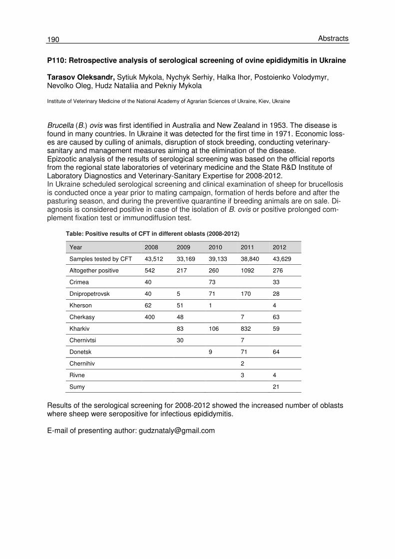
190 Abstracts
P110: Retrospective analysis of serological screening of ovine epididymitis in Ukraine Tarasov Oleksandr, Sytiuk Mykola, Nychyk Serhiy, Halka Ihor, Postoienko Volodymyr, Nevolko Oleg, Hudz Nataliia and Pekniy Mykola Institute of Veterinary Medicine of the National Academy of Agrarian Sciences of Ukraine, Kiev, Ukraine
Brucella (B.) ovis was first identified in Australia and New Zealand in 1953. The disease is found in many countries. In Ukraine it was detected for the first time in 1971. Economic loss-es are caused by culling of animals, disruption of stock breeding, conducting veterinary-sanitary and management measures aiming at the elimination of the disease. Epizootic analysis of the results of serological screening was based on the official reports from the regional state laboratories of veterinary medicine and the State R&D Institute of Laboratory Diagnostics and Veterinary-Sanitary Expertise for 2008-2012. In Ukraine scheduled serological screening and clinical examination of sheep for brucellosis is conducted once a year prior to mating campaign, formation of herds before and after the pasturing season, and during the preventive quarantine if breeding animals are on sale. Di-agnosis is considered positive in case of the isolation of B. ovis or positive prolonged com-plement fixation test or immunodiffusion test. Table: Positive results of CFT in different oblasts (2008-2012)
Year 2008 2009 2010 2011 2012
Samples tested by CFT 43,512 33,169 39,133 38,840 43,629
Altogether positive 542 217 260 1092 276
Crimea 40 73 33
Dnipropetrovsk 40 5 71 170 28
Kherson 62 51 1 4
Cherkasy 400 48 7 63
Kharkiv 83 106 832 59
Chernivtsi 30 7
Donetsk 9 71 64
Chernihiv 2
Rivne 3 4
Sumy 21
Results of the serological screening for 2008-2012 showed the increased number of oblasts where sheep were seropositive for infectious epididymitis. E-mail of presenting author: [email protected]
191 Brucellosis 2014 International Research Conference
P111: Estimation of the prevalence of brucellosis and identification of Brucella spe-cies among cattle and small ruminants in Georgia Lasha Avaliani and Ketevan Tsiklauri National Food Agency of Ministry of Agriculture of Georgia, Tbilisi, Georgia
Brucellosis is endemic in Georgia with several of thousands of positive animal cases and hundreds of human cases every year. Currently, Georgia uses a passive surveillance system and the endemic disease is still considered to be severely underestimated. This study was developed to identify which regions face the greatest risk so that cost-effective control pro-grams can be used in these areas. The study was planned in two phases. Phase one includ-ed the serology of animals in all regions of the country to identify ‘hot spots’ for Brucella in-fected animals. Phase two included isolation, identification, and typing of Brucella isolates that are circulating among cattle and small ruminants. Phase one: in total, 17,372 samples were randomly collected from cattle and small ruminants and tested by both the Rose Bengal test and the fluorescence polarization assay (FPA) at the Laboratory of the Ministry of Agri-culture. The results of this work demonstrated that brucellosis is not equally distributed be-tween different regions of Georgia. Some villages are hyper-endemic with a prevalence greater than 10 % in susceptible animals. Phase two: a total of 110 tissue samples from 35 cows and their aborted fetuses (lymph nodes, vaginal swabs, aborted fetus, placenta, and milk) were collected throughout the country. Samples were cultured on selective media to isolate brucellae, and then suspected isolates were analyzed by AMOS-PCR. In total, 14 samples from seven different animals were positive by culture. None of them was confirmed by AMOS PCR, demonstrating the limitation of the test in determining suspected isolates e.g. of B. abortus which may circulate in Georgia. The study results are being used to design new cost-effective brucellosis control programs to be managed at the regional level and the ef-forts will focus on the regions with the highest levels of infection. E-mail of presenting author: [email protected]
192 Abstracts
P112: Seroprevalence of brucellosis and risk factors assessment in caprines of Ramechhap District Yugal Raj Bindari and Sulochana Shrestha
Institute of Agriculture and Animal Science (IAAS), Kirtipur, Nepal
A study was conducted to assess the seroprevalence of brucellosis and its associated risk factors in goats of Ramechhap District. Two-stage sampling procedure was adopted; in the first step, VDCs were selected from the district and in the second step individual animal was selected randomly from the respected VDCs. In all, three VDCs (Bhaluwajor, Okhreni and Chuchure) were selected and then 502 animals were sampled. Serum was screened for Bru-cella melitensis employing Rose Bengal plate test (RBPT) and indirect ELISA (i-ELISA). The overall prevalence of brucellosis was found to be 5.8 % by i-ELISA and 6.3 % by RBPT. The prevalence varied from 3.4 % to 7.1 % in various VDCs. Correlation and regression analysis was made to check the relationship between the prevalence of disease in various VDCs in relation to the goat population. The results indicated strong and positive correlation (r=0.99) and regression statistics indicated significant association (F-statistic: 928.6; p=0.02). Sero-positivity significantly increases when herd size increases (p<0.05). Higher seroprevalence was found in the animals at lower altitude than at higher altitude (p<0.05). The disease prevalence was found to be significantly increasing with age (p<0.05). No statistically differ-ence was observed (p>0.05) in prevalence between males and females. However, disease was significantly associated with abortion (p<0.01). The highest prevalence was also found in goats kept in close association with other species in comparison to those kept separately. E-mail of presenting author: [email protected]
193 Brucellosis 2014 International Research Conference
P113: The status of bovine brucellosis in some parts of northern Nigeria H.M. Mai Animal Production Programme, School of Agriculture and Agricultural Technology, Abubakar Tafawa Balewa University, Bauchi, Bauchi State, Nigeria
The prevalence of bovine brucellosis in northern Nigeria appears to be on the increase. This study considered recent reports of the status of brucellosis in northern Nigeria. Multistage cluster sampling strategy, random sampling techniques and multiple logistic regression mod-els were used. The various serological tests carried out included Rose Bengal plate Test, milk ring test and ELISA. The states studied were Kaduna, Kano, Adamawa, Taraba, Jigawa and Bauchi. The individual animal-level prevalence ranged from 4.0 % to 26.3 %, while a herd-level prevalence of up to 77.5 % was reported. Several factors have been attributed to the increase of brucellosis such as management system, attitude of the pastoral Fulanis to-wards brucellosis, nomadism, crossing national borders, mixed herds/flocks, poor surveil-lance and control programs etc. Several other risk factors were significantly associated with brucellosis such as age, sex, pregnancy status, pastoral systems, herd size, handling facility, and presence of small ruminants in the same farm, introducing new animals from different sources and borrowing or sharing of bulls. The seemingly increase in brucellosis in cattle is of immense concern especially the fact that cattle and their products, particularly contami-nated milk, transmit the disease to humans. Consuming unpasteurized milk by Fulani herds-men in northern Nigeria is a very common practice, making them susceptible to brucellosis. This study highlights the epidemiological picture of brucellosis in parts of northern Nigeria. The control of brucellosis can be achieved by enlightening the farmers on brucellosis and its public health significance and instituting an annual vaccination program of cattle at national level. E-mail of presenting author: [email protected]
194 Abstracts
P114: Serological survey on brucellosis of domestic animals in Srem and South Backa (Autonomous Province of Vojvodina) Vidić Branka, Grgić Živoslav, Savić Sara, Marčić Doroteja, Vidić Vaso and Prica Nadežda* Scientific Veterinary Institute ‘Novi Sad’, Novi Sad, Serbia
Brucellosis is a very important zoonosis in Mediterranean countries, in terms of public health and economy. Brucellosis in Serbia has been monitored and controlled during the past few years within the Ministry of Agriculture, which is financing this control program. Brucellosis in pigs occurs sporadically as individual case. However, considering the presence of brucellosis in sheep and goats in the southern part of Serbia and the events that followed, brucellosis in sheep and goats has spread to the north of the country. The first case of brucellosis in sheep and goats in Vojvodina region was detected in 1999, and since then the disease has been spreading. Contribution to the spreading of the disease is made by uncontrolled animal trade, a nomadic way of keeping animals. Because of that, the decision was made that brucellosis should be monitored in sheep and goats and cattle. The categories of animals which are screened every year are the following: sheep and goats older than 6 months and cattle older than 12 months. The examination is done once a year for each animal. Serologically positive animals are safely disposed and the expenses for the diagnostic procedure and disposal are taken from the governmental budget. Within the control program of brucellosis in the region of Vojvodina during the period from 2001 to 2013, in total, 1,371,478 samples from domestic animals were analysed. Positive reactions were found in 1,344 sera samples, which is 0.097 %. The examined samples were from different animal species: 787,494 sera samples were from sheep and goats, 486,495 sera samples were from cattle, 95,807 from pigs, 1,151 from horses and 163 samples were from dogs. Following diagnostic methods were used: RBT, CFT and iELISA, and cELISA as conformation test. Positive findings were detected in 1,316 (0.17 %) sheep and goats, during the period from 2003 to 2007. During the same period, brucellosis was detected in eight hu-man patients as well. After this period of time, no new positive animal cases were found. Positive findings were detected in twelve (0.0024 %) sera from cattle which were from the same farms where brucellosis in sheep was found. Fifteen (0.015 %) serum samples from pigs and only one from horses were diagnosed as positive for the same period. E-mail of presenting author: [email protected]
195 Brucellosis 2014 International Research Conference
P115: Risk factors for bovine brucellosis in emerging or indigent farms Nkangala Dis-trict, Mpumalanga, South Africa – a case control study. Tendai J. Chiwome Clinical State Veterinarian, Mpumalanga Veterinary Services, Nkangala District, South Africa
Brucellosis is a controlled animal disease in South Africa in accordance with the Animal dis-eases Act 35 since 1984. It is a disease of zoonotic importance, because it causes significant reproductive losses in animals, annually. A case control study is being conducted with at least 40 farms known to be infected and 40 control farms. A case is defined as a bovine herd with at least one culture positive animal or at least two animals positive to the CFT between 2014 and 2015. Controls are bovine herds with negative test results in the last 3 years and no recorded outbreak within the last 5 years and no untested abortions in the last 6 months. A questionnaire is given to emerging or indi-gent farmers with a range of questions encompassing farm size and farming practices, loca-tion relative to game reserves specifically those with buffalos, milking practices, incidences of abortions in past 3 years and how they were managed, new animals introduced into herds and their source and history, details of animals culled or removed from herd, vaccination program against brucellosis in the herd, whether farmers share equipment/facilities/grazing with other herds (and the status of these herds), artificial insemination program (methods used and source of semen) and incidences of flu like symptoms in farm personnel. There is little known documented information about this group of farmers in Mpumalanga and their practices and attitudes. The study will help provide data on risk factors for brucellosis in these farms. The study is currently still underway. Farming practices, grazing practices and vaccination programs employed are expected to be major risk factors for brucellosis for emerging farmers. Vaccination of pregnant animals should be encouraged despite the risk of abortions because they are the most likely source of infection in a herd and thereby assist in eradication of the disease in communal farms. E-mail of presenting author: [email protected]
196 Abstracts
P116: Brucellosis outbreak in a rural endemic region of Mexico María Rosario Morales-García1,2, Ahidé López-Merino1 and Araceli Contreras-Rodríguez1 1Departamento de Microbiología, Escuela Nacional de Ciencias Biológicas, Instituto Politécnico Nacional, México, D.F., México; 2Departamento de Investigación, CICATA-Querétaro, Instituto Politécnico Nacional, Querétaro, Qro., Mexico
Brucellosis is a worldwide zoonotic disease. Humans can be infected mainly by either the consumption of raw milk and unpasteurised fresh cheeses or by contact with infected ani-mals, mainly in endemic regions. Here, we investigated a brucellosis outbreak in an endemic region of Mexico. Microbiological culture of human blood, raw milk from cows and goats, and fresh cheeses was performed to isolate Brucella. Identification of the bacteria was done by bacteriological procedures and by multiplex Bruce-ladder polymerase chain reaction. Brucella melitensis was isolated from patients, infected goats, and fresh goat cheeses, while B. abortus was isolated from cows. All patients had eaten fresh cheese but no occupational exposure to animals was reported. Results of molecular typing did not show any Brucella vaccine strains. The isolation, identification, and molecular characterisation of Brucella spp. in human brucellosis cases and infected animals are very important to identify the source of infection and to take control measures in endemic regions. E-mail of presenting author: [email protected]
197 Brucellosis 2014 International Research Conference
P117: Role of agricultural animals in brucellosis transmission in southern Kazakhstan T. Karibayev 1, I. Sytnik1, S. Tyulegenov1, R. Seidakhmetova1, A. Jailbekova1, B. Kozhako-va1, A. Bizhanova1, A. Tursunkulov1, M.P. Nikolich2, P.H. Elzer3 and J. Blackburn4 1RSE ‘National Reference Veterinary Center’, MoA, Kazakhstan; 2Walter Reed Army Institute of Research, Division of Bacterial & Rickettsial Diseases, Silver Spring, MD, USA; 3Animal Sciences and Natural Resources LSU Agricultural Center, USA; 4Spatial Epidemiology & Ecology Research Laboratory, Department of Geography, and the Emerging Pathogens Institute, University of Florida, Gainesville, FL, USA
Here we report on field surveillance made jointly by veterinary specialists and medical specialists at settlements in the Almaty and Zhambyl oblasts of southern Kazakhstan to examine backyard animals belonging to people who had confirmed brucellosis. 406 whole blood samples were collected from cattle, sheep and goats kept by different owners. 86 whole blood samples were from cattle and sheep owned by people of Alakol rayon of Almaty oblast; Rose Bengal test, CFT and FPA detected 17 seropositive blood samples. Brucella (B.) strains were isolated from the blood of two cows (B. abortus) and one sheep (B. melitensis) by culturing on solid nutrient media. Sick animals belonged to three persons from different settlements - from Aktubek village, Zhaupak village and Terekty village. It is worth noting that all these owners were seropositive for brucellosis as determined by the rayon Sanitary and Epidemiology Service (SES). In settlements of the Yenbekshikazakh ray-on, in the same oblast, 24 cattle were tested for brucellosis-specific antibodies using Brucella antigen. Six cattle were seropositive and only two cows were bacteremic. Further testing identified these Brucella isolates as B. abortus. Both cows belonged to a private owner living in Karaturuk village, who was already registered to have brucellosis. In Sarkan rayon of Al-maty oblast, there was another examination of animals from those private farms where own-ers were sick. However, no brucellosis-seropositive animals were detected among 36 cattle and 25 sheep. A bacteriological survey of all whole blood samples also showed negative results. This may be explained by the removal and slaughter of all Brucella-positive animals in these settlements earlier that year (in spring 2013) under the national program. A similar situation was observed in two rayons of Zhambyl oblast where 235 animal units were tested for brucellosis. Testing detected five seropositive cows and 22 seropositive small ruminants (sheep and goats). Brucella cultures were isolated from whole blood samples collected from three sheep kept in farms in Kilisbai village, Moinkum rayon, and four sheep from Shu rayon. It should be noted that sheep were kept in different farms and backyards, and there were also cases of brucellosis among members of the owners’ families. Analysis of the data above indicated that people in these southern Kazakhstan areas are infected through contact with diseased animals they own. The isolation rate of B. abortus is rare when compared to previ-ous studies of our group in 2007-08 and 2012-13, when B. melitensis was commonly isolat-ed. In order to shed light on animal-to-human transmission dynamics in this region we will conduct further studies to identify the genotypic characteristics of these isolates. E-mail of presenting author: [email protected]
198 Abstracts
P118: Epidemiological study of brucellosis in camels (Camelus dromedarius) in Cen-tral Sudan Abdelnassir Taha1, Enaam El Sanousi Ahmed1, Mohamed; Osman Elhaj Mona1 and L.L Perrett2 1Veterinary Research Institute (VRI), Khartoum, Sudan; 2Animal Health Veterinary Laboratories Agency (AHVLA), Surrey, UK
Camels in Sudan are farmed in forested grasslands together with sheep, goats and occa-sionally cattle; therefore, they are very susceptible to brucellosis caused by Brucella (B.) melitensis and B. abortus. Under these conditions, brucellosis has spread between these species and has appeared as a major constraint to camel breeding. Diagnosis of camel brucellosis is complicated by many difficulties, mainly because this dis-ease barely causes any apparent clinical signs in comparison to its clinical course in sheep and cattle. Until now, none of the commonly used serological tests can be considered as a recommend-ed test for brucellosis diagnosis in camel and most serological tests used have been driven from cattle without enough validation. This work has been formulated to contribute to the enhancement of diagnostic capabilities by studying bacteriological and different serological diagnostic instrumental features for detec-tion of brucellosis in camels in a distinct chosen district, Tamboul area, ElGazira State, Su-dan. A cross-sectional study was carried out during the year 2012 to estimate the seroprevalence of camel brucellosis. The sample size of the studied animals was estimated using the formu-la given for simple random sampling method, following the relevant formula for 95 % confi-dence and 5 % precision as by Thrusfield, 2005. A total of 500 serum samples were collected randomly from different herds, and 30 tissue samples were also collected from brucellosis seropositive camels in Tampoul slaughter house. Serum samples were subjected to serological investigation using Rose Bengal plate test (RBPT) as screening test, the results were then confirmed by competitive ELISA. The prevalence of the disease was found to be 11.6 % and 7.7 % by RBPT and cELISA, respec-tively. Further serological tests are still under investigation including the modified RBT and the complement fixation test (CFT). All 30 tissue samples (lymph nodes, placentas and spleens) were bacteriologically cultured, purified, identified and then characterized; four Brucella isolates were then recovered from the tissues. The isolated Brucella spp. were characterized as B. abortus bv 1; this finding document the isolation of biovar 1 from camels for the first time in Sudan. The study recommended continuous investigation of the disease in camels to reveal the var-ious impacts of the disease: epidemiologically, economically and its public health threat. All these collectively will help in planning for applied operational control approaches in different parts of Sudan. E-mail of presenting author: [email protected]
199 Brucellosis 2014 International Research Conference
P119: Case study in brucellosis re-emergence: the current status of brucellosis in humans, domestic livestock and the Saiga antelope in Kazakhstan W. Beauvais1, G. Nurtazina2, M. Orynbayev3, R. Kock1 and J. Guitian1 1Royal Veterinary College, London, UK; 2Ministry of Agriculture, Astana, Kazakhstan; 3Research Institute for Biological Safety Problems, Gvardeiskiy, Kazakhstan
The collapse of the Soviet Union in 1991 was followed by an increase in the reported inci-dence of brucellosis (and other diseases) in several Central Asian countries, including Ka-zakhstan. In Kazakhstan there was also a major shift in the way that livestock were kept, i.e. from large state-owned farms towards household-based subsistence farms. This has caused a major challenge for the veterinary services which were previously organized around the state-farm system. This also meant that more people are exposed to direct contact with live-stock now, and are therefore at risk to contract livestock zoonoses. In addition, the changing livestock systems may mean that livestock has increased contact with wildlife, such as the Saiga, a critically endangered migratory antelope that is probably susceptible to brucellosis. In 2007, a nationwide test-and-slaughter program was introduced in Kazakhstan, and there have been more than 100 million blood samples been tested. However, the official incidence of brucellosis in humans remains as high as 60 cases per 100,000 in some areas of the country, annually. There is a lack of published data on the burden of brucellosis in Kazakhstan in the peer-reviewed literature. A study in 1997-1998 found a seroprevalence of 2.4 % amongst domes-tic livestock; however, there was no random sampling done. The aim of this project was to analyze official data from the brucellosis test-and-slaughter program, official data on brucellosis in humans and available information on brucellosis in Saiga and to discuss the implications for brucellosis control in Kazakhstan. Official human incidence data were obtained from the Kazakh Ministry of Agriculture for the years 2007-2012, analysed for every oblast (province), age-group and rural versus urban area if available. Official livestock seroprevalence data were also obtained from the Kazakh Ministry of Agriculture for the years 2008-2012, and analysed for every oblast (province). Serosurvey results from Saiga conducted in 2012 and 2013 were obtained from the Re-search Institute for Biological Safety Problems. Descriptive statistics and mapping were done. The official statistics suggest that the incidence in humans may be decreasing, although cau-tion must be exercised in interpreting official data, particularly as there are ongoing changes within the Kazakh health system. There is a strong spatial pattern in human incidence, with a much higher incidence in the south of the country. Potential reasons for this include the high-er density of livestock in the south. The official seroprevalence in livestock is consistently very low, given the high incidence in people. This could be explained by some flexibility in interpretation of the tests. Although the numbers of Saiga tested were limited, there were no positive test results to Brucella antibodies. Current low numbers of Saiga and a likely high-prevalence in livestock suggests that the role of Saiga is currently not critical to brucellosis control; however, this fact could change in the future. Further work is needed to obtain more reliable seroprevalence data for domestic livestock and to evaluate the current control pro-gram. E-mail of presenting author: [email protected]
200 Abstracts
P120: System of the prevention of brucellosis in horses in Ukraine Peknyi Mykola, Nychyk Serhiy, Sytiuk Mykola, Halka Ihor and Ukhovskiy Vitaliy Institute of Veterinary Medicine of the National Academy of Agrarian Sciences of Ukraine, Kyiv, Ukraine Brucella abortus is a causative agent of brucellosis in horses, bovine animals, yaks, buffalos, camels and possibly dogs. Most often horses become infected during joint pasturing and via contacts with infected animals, which had aborted. Horses may be a source of infection for people depending on the spread of the disease. Transmission of the Brucella species is of epizootic importance. Brucellosis in horses has no clinical manifestation. Documented are infiltrations in withers and nape, rarely in other parts of the body, which then turn into abscesses, lameness because of arthritis of the forelegs and only sometimes of the hindlegs, and tendovaginitis. Results of the serological survey of brucellosis in horses in various regions were analyzed. Measures for the prevention of import and spread of the infections were developed for Ukraine. Over the last years, brucellosis in horses was registered in China, Kyrgyzstan, Rus-sia, and in some regions of Ukraine. The disease is connected to horse breeding. Brucellosis in horses has not been registered in Ukraine for over 40 years. Brucellosis in sheep and in bovine cattle was not registered since 1967 and 1980, respectively. According to veterinary instructions, horses are tested with Rose Bengal test and CFT when symptoms of brucellosis are noted or if there was contact to other animals of an outbreak or when horses are import-ed from abroad. In addition, seed bulls, cows, heifers, buffalos, rams, ewes without offspring, breeding boars and main breeding pigs are tested twice a year in an area where brucellosis was possibly imported. Horses positive to brucellosis were not registered during 5 years of serological survey. Due to well-organized work and adherence to the veterinary instructions Ukraine remains free from brucellosis in horses despite close relations with countries endemic for brucellosis. E-mail of presenting author: [email protected]
201 Brucellosis 2014 International Research Conference
P121: The brucellin skin test: A step towards more effective control of bovine brucel-losis in South Africa? N. Nyanhongo1,2, M. Hansen4, J Godfroid1,3 and A.L Michel1 1Department of Veterinary Tropical diseases, Faculty of Veterinary Science, University of Pretoria, Private Bag X04, South Africa; 2KwaZulu-Natal Department of Agriculture and Environmental Affairs, Vryheid Regional Veterinary Laboratory, Vryheid, South Africa; 3Department of Food Safety and Infection Biology, Norwegian School of Veterinary Science, Department of Food Safety and Infection Biology, Tromsø, Norway; 4Mpumalanga Department of Agriculture, Forestry and Fisheries, Standerton Veterinary Office, Standerton, South Africa
Bovine brucellosis is a bacterial disease of public health and veterinary significance. It is characterised by variable and sometimes latent incubation periods which make it difficult to control. No single diagnostic test can identify all stages of the disease with high sensitivity. While only a handful of countries have successfully eradicated brucellosis, South Africa is currently not only far from eradication but has seen an increase in outbreaks in recent years. The brucellin skin test (BST), which has been proved to identify some acute and chronic la-tent stages of brucellosis, was studied for the first time in South Africa by conducting a com-parative evaluation with serological tests. The objective of this study was to evaluate the BST in brucellosis free, as well as confirmed infected herds, particularly prior to routine brucellosis vaccination, under South African farming conditions. The results indicated that the BST, with a limited sensitivity of 42.86 % and a high specificity of 99.18 %, was unsuitable for use as a sole diagnostic test. However, the BST detected more brucellosis cases among unvaccinat-ed four to nine months old heifers in infected herds compared to the routinely used Rose Bengal test and complement fixation test. It was concluded that the BST, used in parallel with routine serology, particularly iELISA, could play a complementary role to enhance diagnostic sensitivity in the identification of infected heifer calves prior to vaccination and breeding; and thus expedite the elimination of brucellosis in chronically infected herds where eradication has proven problematic. E-mail of presenting author: [email protected]
202 Abstracts
P122: A retrospective analysis of animal brucellosis research in Egypt in the last 25 years Gamal Wareth1,2,3, Mandy C. Elschner1, Heinrich Neubauer1, Uwe Rösler2 and Falk Melzer1
1Friedrich-Loeffler-Institut, Federal Institute for Animal Health, Institute of Bacterial Infections and Zoonoses, Jena, Germany; 2Institute of Animal Health and Environmental Hygiene Centre for Infectious Medicine, Freie Universitaet-Berlin, Germany; 3Department of Pathology, Faculty of Veterinary Medicine, Benha University, Qalyobia, Egypt
Brucellosis is a highly contagious zoonotic disease of significance for public health and eco-nomic performance in endemic and non-endemic countries. In developing countries, brucel-losis is often a very common disease. A comprehensive, evidence-based assessment of lit-erature and officially available data on animal brucellosis is missing for Egypt. National and international scientific publications on serological investigations (n=53), isolation and biotyp-ing (n=34) of brucellae at the last 25 years were reviewed to verify the current status of bru-cellosis in animals in Egypt. Serological assays for brucellosis are well-established in Egypt and the results give indirect proof for the presence of brucellosis in cattle, buffaloes, sheep, goats and camels. Brucella (B.) melitensis bv 3, B. abortus bv 1 and B. suis bv 1 are the predominant isolates in Egypt and have been isolated from farm animals and Nile catfish. There seems to be a dis-crepancy between official seroprevalence data and data of scientific publications. Thus, it can only be assumed that Egypt is endemic with brucellosis and the disease is prevalent in all farm animal species, in the environment and in carrier hosts e.g. rats. The epidemiological situation of brucellosis in Egypt is complicated and needs clarification by a nationwide survey including genotyping of circulating brucellae. E-mail of presenting author: [email protected] and [email protected]
203 Brucellosis 2014 International Research Conference
P123: Development of a forecasting model for brucellosis spreading using the Italian cattle trade network Lara Savini, Fabrizio De Massis, Luca Candeloro, Paolo Calistri and Armando Giovannini
Istituto Zooprofilattico Sperimentale dell’Abruzzo e del Molise ‘‘G. Caporale’’, National and OIE Reference Laboratory for Brucel-losis, Teramo, Italy
Brucellosis caused by Brucella abortus is an important zoonosis that constitutes a serious hazard to public health. Prevention of human brucellosis depends on the control of the dis-ease in animals. Livestock movement data represent a valuable source of information to un-derstand the pattern of contacts between premises, which may determine the spread of the disease. The present study shows the development of a probabilistic brucellosis spreading simulation model, based on the Italian cattle trade network. The model takes into account both time and sequence of the events used for the simulation (cattle movements), and con-siders two main components: the dynamics of the disease at herd level (intra-herd spread-ing) and the dynamics of the disease following cattle movements (inter-herd spreading). The parameters for brucellosis spread used in the simulation model (rate of infection, rate of re-covery, etc.) were calculated on the basis of data derived from international peer-reviewed literature. Results of the simulation model have been compared to those generated through a deterministic approach. The model has been applied to the network of cattle movements recorded in 2009 and validated using real data on cases of cattle brucellosis recorded in the same year in Sicily, Italy. In particular, the following data have been extracted from the Ani-mal Health Information System (SANAN): brucellosis infected herds during the year and number of heads present. The reference network was generated from a brucellosis infected herd from Sicily by extracting from the National Database for Animal Identification and Regis-tration (BDN) all movements starting from that herd since the last brucellosis negative control until the date in which the herd was found as infected (i.e. a one-year period). In particular, the data considered from BDN about the network of movements were the following: node of origin, type of node, node of destination, number of animals moved, date(s) of movement(s), number of animals present in the herd on the date of movement, number of females aged equal or more than 18 months and present in the herd on the date of movement. The nodes of the network were those commercially connected directly or indirectly with the infected herd, and for which the number of animals entering and the timing of introduction were consistent to allow the transmission and the development of the infection. The numbers of nodes involved by the simulation, the numbers of movements concerning the network as well as the numbers of outbreaks identified by the model are discussed. The simulation model involves complex computational analysis; future developments may include the identi-fication of additional tools for analysis (to optimize the design of the model) as well as of a fine tuning of the proposed algorithms (to maximize its performance). E-mail of presenting author: [email protected]
204 Abstracts
P124: OIE Laboratory Twinning Sudan-UK: An empowering initiative for local laboratories which had a remarkable outcome for the control of brucellosis in Sudan
Enaam M. El Sanousi1, Judy Stack2, Taha Abdelnassir1 and Maha Khogaly1 1Veterinary Research Institute, Khatoum, Sudan; 2Animal Health Veterinary Laboratories Agency, Surrey, UK
OIE Laboratory Twinning was aimed to strengthen the candidate Brucella laboratory in Sudan by identifying its needs for laboratory capacity building, overcoming barriers and obstacles, networking and joint scientific activities. The candidate laboratory’s visibility is raised at national, regional, international conferences and meetings. The candidate laboratory at VRI sucessefully implemented the project activities.These activities included the production and standardization of biologicals (diagnostics and vaccines). Standard Operating Procedures (SOPs) for each product were implemented. The quality control of these products is done according to the OIE Manual and SOPs received from AHVLA. Brucella (B.) antigens (RBT, MRT, CFT and SAT), Brucella vaccines (S19 and Rev1), mono specific antisera (A and M) and The National Standard Serum are readily produced with high quality. The biological products also serve strategic aims: The amount of produced antigens is sufficient to test to 2 to 3 million animals of livestock to be exported (sheep, goats and camels) and the produced antigens can also be used for large scale diagnosis of thousands of samples annually. Several research activities were done this year: cultural, serological and molecular diagnosis of camel brucellosis was investigated; B. melitensis Rev1 vaccine and Rose Bengal antigen of B. melitensis were produced for the control of small ruminants’ brucellosis in Sudan; a study of acquired immunity against brucellosis using B. abortus S19 and B. melitensis strain Rev1 vaccines was done in Sudanese camels (Camelus dromedaries); an epizotological study of brucellosis in animals in Darfur State, Sudan, was conducted. Several activities towards the ‘One Health’ idea wereundertaken: the isolation of Brucella from humans with suspected brucellosis was initiated by a collaboration of the Ministry of Health, the Faculty of Medicine, University of Khartoum and the Nidiag (European Research Network). An external quality testing procedure (EQT) was set into force: The parent laboratory in UK sent various samples to be tested with the different technologies implemented in the Sudanese laboratory. These tests were successfully accomplished. In Khartoum State an epidemiological survey was done and the spatial distribution of brucellosis was sucsessfully investigated. Additionally, training courses in serological diagnosis of brucellosis using ovine, caprine, bovine and camel sera, and different serological tests (RPT, SAT, MRT and cELISA) were done at thedepartment of Brucella. The trainees were veterinarians, technical staff, veterinary paraprofessionals and vet students. Various national, regional and international activities were done. The parent laboratory experts will continue to give guidance to the Sudanese colleaguese. E-mail of presenting author: [email protected]
205 Brucellosis 2014 International Research Conference
Session 9: Wildlife Brucellosis
Keynote Lecture: Jaques Godfroid University of Tromsø, Department of Arctic and Marine Biology, Tromsø, Norway
Wildlife brucellosis: Spillover or sustainable infection? Interaction of humans and livestock with wildlife exposes them to the risk of pathogen spillo-ver. Livestock and wildlife may become intermediate or amplifier hosts from which pathogens can spill over into humans or humans can be infected via vectors. It has been estimated that 58 % of human pathogens are zoonotic, and 13 % are emerging, of which 73 % are zoonotic. Additionally, 26 % of human pathogens infect both domestic and wild animals. From a zoonotic perspective, brucellosis in humans is strongly linked to the management of infected animals and ingestion of unpasteurized dairy products. In this regard, there is a clear connection of brucellosis with the domestication of even-toed ungulates, milking practices, and fabrication of cheese and other dairy products. Domestication of sheep, cattle and goat, which occurred some 11,000-8,000 BCE, and clustering of wildlife reservoirs with narrower genetic backgrounds have provided a steady supply of emergent pathogens for humans. Domestication, anthropogenic modification of the environment and selection of animals by humans are not neutral phenomena. Consequently, it is expected that the prevalent extant Brucella strains have been selected through ‘narrow funnels’ connected to these processes. Nonetheless, reservoirs of Brucella spp. existed in indigenous African artiodactyla species (which include a significant number of potential Brucella hosts) long before the introduction of domesticated animals. A paleopathological study has suggested the presence of Brucella infections in australopithecines, already 2.5–2.3-million years ago. The first Brucella genome sequences were released in the early 2000s, and their analysis confirmed the previously held ideas concerning just how closely related they were. One of the key aspects of the biology of Brucella spp. that genomic sequencing has not yet been able to fully explain is the genetic basis of host specificity of the different Brucella spp. It is however clear that the preference of different Brucella spp. for a host is not based on a specific gene or pathogenicity island, but may rather be related to small genomic changes that affect expression or function of shared genomic content. Likewise, minor differences in the physiology of the different host species of Brucella spp. will also play a role in establishing the host specificity and preference of the different species. Most wildlife brucellosis studies have consisted of serological surveys, and pathogenesis studies are lacking. Differences in the disease course exist and are likely to be attributable to immunological and behavioral differences among species. Additionally, anthropogenic driv-ers may result in breaking down the species barrier by multiplying exposure, allowing ‘species jump’ for Brucella spp. which may favor the persistence of a particular Brucella (B.) spp. clone in a different ‘preferred’ host. Natural infection of B. abortus has been confirmed by culture in many wild artiodactyls, in-cluding bison (Bison bison), elk (Cervus elaphus), and African buffalo (Syncerus caffer). These species are known maintenance hosts of B. abortus under certain circumstances and may serve as reservoirs of the infection with the potential for transmission mainly to cattle and humans. Moose (Alces alces) appear unable to mount a protective immune response against the organism, usually resulting in mortality from B. abortus infection and the species is considered likely to be a dead-end host. Retained placentas, commonly seen following abortion or parturition in B. abortus infected cattle and bison are not observed in elk. Brucella abortus infection in female elk often results in abortion of the first calf. Mammary gland in-volvement is less frequent in elk than in cattle. Hygromata, carpal bursitis, synovitis and ten-
206 Abstracts
donitis are commonly seen. Based on natural observations and experimental work, of B. abortus infected wild canids are considered spillover hosts with little role, if any, in maintenance of the infection in other populations. Nowadays, in countries where bovine brucellosis eradication programmes are close to their end, there are no known sustainable reservoirs of B. abortus in wild species, other than bison and elk in and adjacent to the American National Parks of the Greater Yellowstone Area (GYA) and bison in the Canadian Wood Buffalo National Park. However, recent studies in feral pigs on the Atlantic coast of South Carolina, USA, may challenge these facts. Indeed, B. abortus wild-type and the B. abortus S19 and RB51 vaccine strains have been isolated from feral pigs that had been spatially separated from domestic livestock for decades. This is the first report of a wildlife reservoir of B. abortus outside the GYA in the USA. The epidemiological consequences of these findings are of the utmost importance. Indeed, if experimentally confirmed, this would mean that B. abortus was able to perform a ‘host species jump’ and feral pigs may have become a true reservoir of B. abortus. In Europe, natural infection of B. abortus in wild boar has, so far, not been reported. A recent survey in the Iberian Peninsula highlighted that wild ruminants were not a brucellosis reservoir for livestock. Brucella abortus was only isolated from a single red deer (Cervus elaphus), likely as a spillover from infected cattle. Thus, these results suggest that, in Europe, wild ruminants are occasional victims of brucellosis transmitted from infected livestock, rather than true reservoirs of the infection for livestock, to the contrary of what occurs in the GYA. Some years ago, spillover of B. melitensis from infected small ruminants has been documented in a few wildlife species, such as chamois (Rupicapra rupicapra) and ibex (Capra ibex) in the French and Italian Alps, and the Iberian wild goat (Capra pyrenaica) in Spain. These reports highlight the fact that B. melitensis infection in European wildlife was always linked to the small ruminant reservoir suggesting that wildlife species are unable to sustain the infection and cannot act as reservoirs for domestic animals and human beings. Given the progress in national eradication brucellosis programs in small ruminants in Europe, it was speculated that only anecdotal B. melitensis spillovers in wildlife would be reported. Surprisingly, the wildlife 2012 surveillance campaign in France, revealed a very high prevalence of brucellosis within the Alpine ibex population (>45 %) of the Bargy Massif, despite the absence of any new outbreak in domestic animals and while wild ungulates had been considered, up to now, as an epidemiological dead-end host in Europe. Brucella suis biovar 2 infection remains restricted to wild boar (Sus scrofa) and European brown hares (Lepus europaeus) in Europe, where B. suis has been eradicated in the pig industry for decades. Several outbreaks of B. suis biovar 2 have occurred in outdoor rearing systems of domestic pigs, with the source of infection being traced back to contacts with wild boar. Transmission from wild boar to pigs is believed to be through the venereal route, as crossed (striped) piglets have been reported, at least in France and Portugal; however, other routes might also be possible. The role of wild boar hunting (migration pressure, offal remaining in the field, hunters working on the premises, etc.) has not been fully investigated. Brucella suis biovar 2 strains isolated from hares and wild boars from Hungary showed substantial genetic divergence, suggesting separate lineages in each host and no instances of cross species infections, while across Europe, isolates from domestic pigs were closely related to isolates originating from both hares and wild boars. Feral pigs are rapidly expanding their ranges across the United States, with 44 of 50 states reporting populations. The rapid expansion into new areas has primarily been caused by unregulated human transportation of feral pigs to establish new populations for hunting. In at least 14 states, brucellosis has been documented to be present in feral pigs, with some populations showing high seroprevalence. Feral pigs have been implicated as the source of B. suis biovar 1 infections in humans in a number of cases in the United States. As behaviour and topography make elimination of established populations of feral pigs unachievable, effective separation from domestic livestock, and feral pig population control by hunting or
207 Brucellosis 2014 International Research Conference
targeted depopulation efforts has been implemented in order to avoid transmission of B. suis biovar 1 to livestock.Nevertheless, B. suis biovar 1 is regularly isolated from cattle in the United States and is becoming an emerging problem in cattle in South America. Brucella suis biovars 1, 2 and 3 are not considered to be pathogens in cattle. Indeed, cattle naturally infected with B. suis biovar 1 from feral pigs, and closely monitored for two years, did not experience abortion, did not transmit the agent to co-mingled negative cattle, and did not shed the organism except in the milk, which remained positive for two years. In Belgium, Brucella suis biovar 2 has recently been isolated from cattle without any induced pathology. After B. suis biovar 2 experimental infection in cattle, no gross pathology was seen and B. suis biovar 2 could not be isolated from carcasses 8 weeks post infection. Importantly, B. suis biovars 1 and 3 are true pathogens for humans, whereas B. suis biovar 2 is not. Recently, two independent studies suggest that hooded seals are not the definitive host of B. pinnipedialis. Indeed, gross pathology associated with B. pinnipedialis infection has not been described in seals and vertical transmission is not likely to occur, since B. pinnipedialis has never been isolated from females of reproductive age. These studies point toward the existence of a reservoir of Brucella spp. in the aquatic environment, likely in the food web. Fish constitute a significant part of the diet of seals and further studies are needed to determine if fish, or other aquatic organisms that are part of the diet of seals, can sustain a B. pinnipedialis infection in seal. There have been three reported cases of natural human infection associated with Brucella spp. from marine mammals. They were all associated with Brucella sequence type (ST) 27, although no contact with marine mammals was reported. Brucella ST27 has also been isolated in a dolphin and in a fur seal along the Californian coast. These findings suggest the hypothesis of a reservoir of Brucella spp. in the aquatic environment, possibly in fish or aquatic organisms, and that these marine Brucella spp. may spill over to marine mammals and humans. The basic reproductive number (R0) of an infection is the number of cases one case generates on average over the course of its infectious period, in an otherwise uninfected population. That is, for brucellosis to spread and for an effective animal to animal transmission of Brucella spp., it is required that R0 of Brucella spp. > 1. In contrast, if R0 < 1, then Brucella spp. will die out. Higher the R0 value, higher would be the chances for Brucella spp. to achieve a steady and successful adaptation in the host. The prevalence of brucellosis in some wildlife species is very low and thus, besides the classical factors considered to be important in transmission (host susceptibility, shedding, survival in the environment, etc.), the behaviour of individuals and interactions between wildlife and livestock may actually be the most important drivers for transmission. For instance, the reclusive calving behaviour of elk, which keeps the elk cow and calf away from the herd for several days after calving, minimises the risks of B. abortus transmission to other animals. On the other hand, in situations with important anthropic effects (i.e. intentional winter feeding, or co-mingling of wildlife on livestock feed grounds), the risk of infection increases dramatically, due to possible exposure to an infectious abortion near the feeding grounds. In the GYA, winter feeding changed the behaviour and density of free-ranging elk, the exposure rate to B. abortus, probably converting what would otherwise be a dead-end host in nature to a maintenance host of particular clones of B. abortus. A study of the B. melitensis infected Alpine ibex population of the Bargy Massif concluded on the persistence and circulation of B. melitensis local clone in the area, over a two decade-period, in this protected population. The high brucellosis prevalence reported in wildlife, and especially in ungulates, reflects the direct effects of artificial management on wildlife and brucellosis transmission. The important anthropogenic effects (e.g. habitat modifications, increased wildlife density, sedentary behavior, contacts with domestic animals, artificial feeding facilitating contacts, etc…) could account for the high brucellosis prevalence found in ibex in the Bargy Massif. Moreover, the genetic integrity of Alpine ibex in the Alpine massifs,
208 Abstracts
strongly modified by anthropic effects, might have also played a role. Indeed, the founding of new Ibex population in the Bargy Massif in the Alps came from a pool of few (15) animals, narrowing their genetic diversity. All these factors may have contributed to make the Bargy Massif ibex population a semi-domestic population able to self-sustain the B. melitensis infection, with high prevalence, which has never been reported before in wild ungulates. Brucella pinnipedialis has limited ability to survive within primary cells from hooded seals, thought to be the preferential host species, as well as human/murine cell lines. A lack of, or low, capacity to replicate and survive for prolonged periods within host cells, particularly macrophages, abolishes the ability to produce chronic infections. Seals may thus not be the primary host for B. pinnipedialis, but rather a ‘dead-end’ or spillover host susceptible to infection derived from other sources in the marine environment. Future research should aim at identifying the primary reservoir of B. pinnipedialis along with investigating potential virulent traits in marine mammal Brucella spp., important in the establishment of disease, either acute or chronic, in marine mammals and humans. The fact that B. abortus, B. melitensis and B. suis are isolated from non-preferential hosts more and more frequently indicates shortcomings in livestock control programmes and/or changes in husbandry practices or wildlife management. Anthropogenic drivers are thus some of the most important determinants for transmission of Brucella spp. to non-preferred species. Intracellular survival and replication is the hallmark of Brucella spp infection. Chronicity is linked to multiplication in macrophages, a feature facilitated by overriding the intracellular response following bacterial infection. A stealthy entry into host cells is essential to avoid detection by the immune system. Unfortunately, there are only very few studies of the pathobiology and the mechanisms of bacterial intracellular invasion and multiplication involving Brucella spp. in wildlife and this warrants further investigations. Lastly, the determinants of host specificity of Brucella spp. at the molecular level have not yet been identified, which remains one of the most important research issues.
209 Brucellosis 2014 International Research Conference
Oral Presentations (O36-O40) O36: Brucella melitensis at the wildlife-livestock-human interface in the Emirate of Abu Dhabi A.L. Chaber1,4, L. Lignereux2, D. Fretin3 and C. Saegerman1
1Research Unit of Epidemiology and Risk Analysis applied to veterinary sciences, Faculty of Veterinary Medicine, University of Liege, Liège, Belgium; 2Environment Agency Abu Dhabi, Abu Dhabi, United Arab Emirates; 3CODA-CERVA, Brussels, Belgium; 4 Wildlife Consultant L.L.C, Al Ain, United Arab Emirates
Brucellosis has been reported in almost all livestock species in the Middle East, especially in goats and camels. In the emirate of Abu Dhabi (UAE), 55.1 % of the 247 domestic farms sampled in 2010 were Brucella seropositive. B. melitensis has been isolated once in a Nubi-an Ibex (Capra ibex nubiana) in Dubai. Nevertheless there are no recent data accessible on the species and biovars circulating in the UAE. Both B. melitensis biovar 1 and 3 were re-ported on human cases from Tawam Hospital, Al Ain (Abu Dhabi), hence were co-circulating in the UAE in 1996. From 2000 to 2003, 6.5 % of the 998 patients admitted in the same hos-pital were Brucella-seropositive. The livestock management system in the UAE plays a role in Brucella spreading with the coexistence of several livestock species and because of mis-diagnosis and/or under reporting of animal and human cases. B. melitensis is rarely reported in wildlife but direct contact between free roaming livestock and wild ungulates is likely in the region. In this study we investigated the brucellosis seroprevalence within a large captive herd of scimitar horned oryx (SHO / Oryx dammah) to trace the source of infection. SHO are extinct in the wild since 2000 and conservation programmes rely heavily on captive stocks for possible future re-introduction. Among the 480 juvenile/subadult SHOs and the 400 adult SHOs tested, 75 % and 95 % were Brucella-seropositive, respectively, based on a Single Rose Bengal test. 15 sand gazelles housed within the same collection but 2 km away from the oryx herd were also tested seropositive. Clinically most of the male oryx suffer from orchi-tis while sand gazelles present mainly hygroma at the tibio-tarsal and metacarpo/tarso-phalangeal joints. B. melitensis biovar 1 was isolated from both oryx foetal stomach content and metacarpal fluid from a gazelle and genotyped using multiple-locus variable-number tandem-repeat (MLVA) at CODA-CERVA (Belgium). Both strains have a similar genetic pro-file to the BfR25 strain previously isolated from a goat in the UAE. Supposedly, oryx and/or gazelles initially acquired brucellosis infection from livestock, and later it started circulating among wildlife. Moreover, Brucella spp. that has not been genotyped has clinically infected three ungulate keepers handling contaminated materials in the same collection. All were cured following the Word Health Organisation treatment recommendations. A holistic ecosystem based approach should be used to tackle Brucella transmission and maintenance in heavily contaminated environment. Efforts in identifying species and subtyp-ing of Brucella isolates are paramount for any preventive (awareness campaign) and epide-miologic surveillance-control programme in endemic areas. Active surveillance of susceptible animals and occupational health screening of the workers will give a more accurate picture of Brucella distribution in the country and will help defining an appropriate control strategy. In heavily affected countries, mass vaccination programmes seem inevitable but require profi-cient veterinary services. Implementation of an eradication strategy to fight brucellosis in scimitar horned oryx will require preliminary studies on diagnosis tests, host/pathogen/environment interactions, vaccine assessment and appropriate veterinary protocols. E-mail of presenting author: [email protected]
210 Abstracts
O37: Brucella infection cases among stranded marine mammals recovered from the North Sea coasts of Belgium, the Netherlands and France (1999-2013) D. Fretin1, E. Alonso-Velasco1, T. Jauniaux1, M. Mori1 and J. Godfroid3 1Operational Directorate Bacterial Diseases, Veterinary and Agro-chemical Research Centre (VAR), Brussels, Belgium; 2 De-partment of Morphology and Pathology, Faculty of Veterinary Medicine, University of Liège, Liège, Belgium; 3Research Unit of Epidemiology and Risk Analysis applied to veterinary sciences (UREAR-ULg), Department of Parasitic and Infectious Diseases, Faculty of Veterinary Medicine, University of Liège, Liège, Belgium
A total of 203 stranded animals have been recovered from the North Sea coast during a fif-teen years surveillance program. The aim of this study was to determine the prevalence of Brucella infections among the stranded sea mammals population. A total of 15 Brucella strains were isolated from porpoises (n=6), grey seals (n=5), harbor seals (n=3) and a short-beaked common dolphin. In most cases, the infection was localized in the respiratory sys-tem. Additionally, two porpoises carried lung parasites harboring Brucella. Isolation of bacte-ria was confirmed by real-time PCR and strains were typed by Multi-Locus Variable Number Tandem Repeats Analysis (MLVA). Different genetic MLVA profiles were identified, demon-strating high strain variability among Brucella spp. circulating in marine mammals. As a po-tential zoonotic disease, the presence of Brucella spp. in marine mammals constitutes a bio-hazard for human health. E-mail of presenting author: [email protected]
211 Brucellosis 2014 International Research Conference
O38: First isolations of Brucella ceti from long-finned pilot whales (Globicephela me-las) and a sowerby‘s beaked whale (Mesoploden bidens) Geoffrey Foster1, Johanna L. Baily2, Adrian M. Whatmore3, Andrew C. Brownlow1
, Mark P. Dagleish2, Mark S. Koylass3, Rob Deaville4, Lorraine L. Perrett3, Emma J. Stubberfield3, Robert J. Reid1 and Nicholas J. Davison1
1SAC Consulting Veterinary Services, Inverness, UK; 2Moredun Research Institute, Edinburgh, UK; 3AHVLA Weybridge, UK; 4Institute of Zoology, London
The isolation of Brucella from cetaceans was first reported in 1994 from two harbour por-poises (Phocoena phocoena) and a short-beaked common dolphin (Delphinus delphis) in Scotland and from the aborted foetus of a captive bottlenose dolphin (Tursiops truncatus) in the USA. In the years since, B. ceti has been reported from several further species of ceta-cean including Atlantic white-sided dolphin (Lagenorhyncus acutus), striped dolphin (Stenella coeruleoalba), white-beaked dolphin (Lagenorhyncus albirostris), and minke whale (Balae-noptera acutorostrata). While further evidence of Brucella infection in further cetacean hosts have been provided by serological tests and detection of Brucella DNA in tissues from Bryde’s whales (Balaenoptera edenii), isolation of B. ceti following bacterial culture remains the gold standard for detection of Brucella in cetaceans. We report the first isolations of B. ceti from long-finned pilot whale (Globicephala melas) (LFPW) and Sowerby’s beaked whale (Mesoploden bidens) (SBW). Animals were reported through the Scottish Marine Animal Strandings Scheme (SMASS) and were subject to post mortem examinations which included selection of tissues for bacte-riology and histopathology.
Case 1: Brucella ceti was isolated from spleen, kidney and colorectal lymph node of a SBW. Although there was no correlation between isolation of B. ceti and histopathological lesions in individual tissues, a non-suppurative, necrotising encephalitis was detected, which was remi-niscent of that described in other cetacean species infected with B. ceti, some of which have only small numbers of colonies recovered from brain. It remains a possibility therefore that culture of brain in this case was not sufficiently sensitive to detect Brucella.
Case 2: B. ceti was isolated from testis and pus collected from the shoulder of the scapulo-humeral joint of a LFPW. The animal was one of ten LFPW for which bacteriology was car-ried out following a mass stranding event (MSE) of 70 LFPW, in which 19 animals died
Case 3: B. ceti was isolated from the CSF and brain of a single LFPW. Histopathology sup-ported a diagnosis of non-suppurative meningo-encephalitis as reported previously for Atlan-tic white-sided, striped and common dolphins. Multi-locus sequence typing identified the three isolates as belonging to ST26, a strain which is associated with pelagic delphinids in the north-east Atlantic. The recovery of B. ceti from a LFPW involved in a mass stranding event and from the brain of another LFPW, provide some evidence towards a potential role for B. ceti involvement in LFPW mass stranding events under the sick lead animal hypothesis. The recovery of B. ceti from the testis of a LFPW is of interest with regard to the impact of reproductive disease on cetaceans, which remains un-known and will likely require input from researchers in other fields. E-mail of presenting author: [email protected]
212 Abstracts
O39: Atlantic cod (Gadus morhua): A potential transmission host for Brucella pinnipe-dialis hooded seal (Cystophora cristata) strain?
A.K. Larsen1,2, I.H. Nymo1,2, S. Al Dahouk3, M. Seppola4,2, K.R. Bakkemo4,2 and J. God-froid1,2 1Research Group of Arctic Infection Biology, University of Tromsø – The Arctic University of Norway, Tromsø, Norway; 2Member of the Fram Centre – High North Research Centre for Climate and the Environment, Tromsø, Norway; 3Federal Institute for Risk Assessment, Berlin, Germany; 4 Norwegian College of Fishery Science, University of Tromsø – The Arctic University of Norway, Tromsø, Norway
Investigations of hooded seals (Cystophora cristata) have revealed high prevalences of Bru-cella-positive seals in the dramatically reduced Northeast Atlantic stock. However, pathologi-cal changes due to infection with B. pinnipedialis have not been identified in the hooded seal (HS). B. pinnipedialis HS strain is not able to multiply or establish a chronic infection in HS macrophages or epithelial cells in vitro. Age-dependent serological and bacteriological pat-terns for B. pinnipedialis have been identified in HS with a low probability of being positive for pups, a high probability for yearlings, followed by a decreasing probability with age, suggest-ing an environmental exposure post weaning during the first year of life followed by clear-ance of infection. This raises the question of a reservoir of B. pinnipedialis in the HS food web. For adult HS, the diet consists of Atlantic (Gadus morhua) and polar cod (Boreogadus saida) along with other species like e.g. deep-sea squid (Gonatus fabricii), redfish (Sebastes sp.), and Greenland halibut (Reinhardtius hippoglossoides). Primary leukocytes were isolated from head kidney of Atlantic cod and challenged with B. pinnipedialis sp. nov. (NCTC 12890) and a HS isolate (B17). Both bacterial strains entered the leukocytes and survived intracellularly without any major reduction in retrievable numbers for at least 48 hours. This is in contrast to the rapid elimination of intracellular bacteria from HS alveolar macrophages. Atlantic cod was experimentally infected with B. pinnipedialis HS strain. The fish was chal-lenged with either 105 (low dose) or 108 (high dose) B. pinnipedialis intraperitonally. At day 24 post infection (pi) no mortality in any of the infected groups has been recorded. Samples of blood, liver, spleen, muscle, heart, head kidney, female gonads and feces have been col-lected from 5 fish in each group at day 1, 7, 14 and 28 pi to determine the bacterial load. Our results show that B. pinnipedialis induce an extended bacteremia in Atlantic cod receiving the high dose. Bacteria were found in blood samples from all fish at day 1, 7, and 14. So far, preliminary results indicate that B. pinnipedialis HS strain may have a prolonged course of infection in the Atlantic cod, at least according to in vitro results generated from the use of primary cell cultures. A sustained bacteremia, compared to what has been observed in mammalian hosts, points in the same direction. This could imply that Atlantic cod may act as a transmission host for B. pinnipedialis HS strain in the marine environment. E-mail of presenting author: [email protected]
213 Brucellosis 2014 International Research Conference
O40: Characterization of Brucella species from stranded cetaceans in the United States since 2010 R. Tiller1, K. Terio2, C. Quance3, Q. Wu4, D. Fauquier5, K. Colegrove2, S. Venn-Watson6, M. Barbieri5,7, D. Rotstein8, S. Robbe-Austerman3, L. Schwacke4, T. Rowles5 and A. Hoffmaster1 United States Marine Mammal Stranding Network
1Division of High-Consequence Pathogens and Pathology, National Center for Emerging and Zoonotic Infectious Diseases, Centers for Disease Control and Prevention, Atlanta, GA, USA; 2Zoological Pathology Program, College of Veterinary Medicine, University of Illinois at Urbana-Champaign, Maywood, IL, USA; 3National Veterinary Services Laboratories, Animal and Plant Health Inspection Service, United States Department of Agriculture, Ames, IA, USA; 4Hollings Marine Laboratory, National Ocean Service, National Oceanic Atmospheric Administration, Charleston, SC, USA; 5Marine Mammal Health and Stranding Response Program, National Marine Fisheries Service, Silver Spring, MD, USA; 6National Marine Mammal Foundation, San Diego, CA, USA; 7The Marine Mammal Center, Sausalito, CA, USA; 8Marine Mammal Pathology Services, Olney, MD, USA
The first reported isolation of a Brucella species from a marine mammal was in 1994 from an aborted fetus of a captive bottlenose dolphin in the United States (US). Since then, dozens of studies have reported culture and serologically-positive animals in captive and wild (stranded or hunted) cetacean populations. This includes both animals exhibiting obvious clinical signs of disease and animals appearing disease free. There have also been reports of human bru-cellosis cases, although few, caused by certain marine Brucella strain types emphasizing the zoonotic potential of these species. In 2007, the Brucella marine species, B. ceti and B. pinnipedialis, were formally described and taxonomically named after the marine mam-mals they preferentially infect. B. ceti is most commonly associated with dolphin and por-poise populations and depending on the strain types, there have been varying degrees of virulence reported within this species. Genetic analysis of B. ceti isolates using a multi-locus sequence analysis (MLSA) scheme of nine housekeeping genes has shown three sequence types (STs) identified as STs 23, 26 and 27 associated with this species. Although brucello-sis in cetaceans is globally distributed, much of the strain typing data have been gathered from isolates predominantly originating from Europe, South and Central America. In essence, there is little known about the diversity of Brucella strains circulating in the cetacean popula-tions in US coastal waters. Due to an increase in cetacean ‘strandings’ and Unusual Mortality Events in the United States, a multi-agency investigation began in 2010 to better understand the disease burden of brucellosis in cetaceans and describe the strain diversity of Brucella species in cetaceans. Since 2010 tissue samples for molecular testing, culture, and histo-pathology have been obtained from 50 cetaceans stranding from 17 states throughout the US. Preliminary molecular analysis indicates the prevalence of previously described STs 23, 26 and 27. The identification of ST 27 in the US B. ceti isolates is notable and is the first re-port of this sequence type in the wild cetacean population. ST 27 has only been reported previously from a single cetacean and three human cases. A unique sequence type, ST 28, has also been discovered in this group of North America B. ceti isolates. Genetic analysis of marine mammal Brucella species in the US is ongoing. Future work will continue to reveal the true diversity of Brucella among marine mammals and their global distribution. E-mail of presenting author: [email protected]
214 Abstracts
Poster Presentations (P125-P135) P125: Genetic relatedness of Brucella suis biovar 2 isolates from wild boars, hares and pigs in Hungary and Europe Zsuzsa Kreizinger1, Jeffrey T. Foster2, Zsuzsanna Rónai3, Kinga M. Sulyok1, Enikő Wehmann1, Szilárd Jánosi3 and Miklós Gyuranecz1
1Institute for Veterinary Medical Research, Centre for Agricultural Research, Hungarian Academy of Sciences, Budapest, Hun-gary; 2Center for Microbial Genetics and Genomics, Northern Arizona University, Flagstaff, AZ, USA; 3Veterinary Diagnostic Directorate, National Food Chain Safety Office, Budapest, Hungary
Porcine brucellosis generally manifests as disorders in reproductive organs potentially lead-ing to serious losses in the swine industry. Brucella suis biovar 2 is endemic in European wild boar and hare populations, thus these species may play a significant role in the spread of the disease and serve as potential sources of infection for domestic pigs. The aim of this study was an epidemiologic analysis of porcine brucellosis in Hungary and a comparative analysis of B. suis bv 2 strains from Europe using multiple-locus variable-number tandem repeat analysis (MLVA). MLVA-16 and its MLVA-11 subset were used to determine the genotypes of 68 B. suis bv 2 isolates from Hungary. The results werefurther compared to the MLVA genotypes of European isolates. The analyses indicated a high genetic diversity of B. suis bv 2 in Hungary. Strains isolated from hares and wild boars showed substantial genetic diver-gence, suggesting separate lineages in each host and no instances of cross species infec-tions. The assessment of the European MLVA genotypes of wild boar isolates generally showed clustering based on geographic origin. In contrast, the hare isolates were more closely related to one another and did not cluster based on geographic origin. The limited relationship between geographic origin and genotype of the isolates from hares might be the result of cross-border live animal exchange. The results could also suggest that certain B. suis strains are more adapted to hares. Across Europe, isolates from domestic pigs were often closely related to isolates originating from both hares and wild boars, supporting the idea that wild animals are a source of brucellosis in domestic pigs. E-mail of presenting author: [email protected]
215 Brucellosis 2014 International Research Conference
P126: Anti-Brucella antibodies in wild boars in Poland Krzysztof Szulowski and Wojciech Iwaniak Department of Microbiology, National Veterinary Research Institute in Pulawy, Poland
Porcine brucellosis is an infectious disease caused by Brucella suis biovar 1, 2 or 3. The main clinical feature of B. suis infection is reproductive failure characterized by abortion, still-birth and infertility in sows, testicular lesions, asymmetry of testicles and infertility in boars. In Europe the most common agent of brucellosis in pigs is B. suis biovar 2, and wildlife (wild boars and hares) constitutes a source of infection for domestic pigs. Several outbreaks of B. suis biovar 2 have been confirmed in porcine outdoor rearing systems, even in fenced premises. The source of infection has been traced to contacts with wild boars. Transmission from wild boars to pigs is thought to be through the venereal route, but other routes are also possible. Systematic brucellosis monitoring in wildlife does not exist. In contrast veterinary surveillance of the animal health status is strictly regulated for domestic animals. In this study we determined the prevalence of anti-Brucella antibodies in wild boars from Poland by indi-rect ELISA. The ELISA kit developed at the National Veterinary Research Institute in Pulawy and utilized for examination of swine sera for anti-Brucella antibodies, was used for testing. The lipopolysaccharide (LPS) obtained from the strain B. abortus S19 was used as antigen, anti-swine immunoglobulins, conjugated with horseradish peroxidase, were used as the con-jugate, and ABTS with H2O2 as substrate. Assay controls consisted of strong positive (S++) and weak positive (S+) sera prepared on the basis of samples obtained from naturally Bru-cella infected pigs. A negative control (N-) was prepared from sera of healthy animals. Same criteria for the assessment of sera from wild boars and porcine sera were used. A total of 4,407 serum samples of wild boars (Sus scrofa) shot by hunters in Poland in 2012 were test-ed. Samples originated from 11 out of 15 voivodeships and 1,077 (24.44 %) seroreagents were detected. The highest prevalence of anti-Brucella antibodies in wild boars was found in Opolskie (39.9 %) and Wielkopolskie (37.29 %) voivodeships. On the other hand, the lowest percentage of positive sera was observed in Kujawsko-Pomorskie (13.74 %) and łódzkie (15.47 %). Results revealed vital differences in the level of prevalence of anti-Brucella anti-bodies in different districts, varying between 0-100 %. Results of the surveys show that wild boars constitute a huge reservoir of B. suis biovar 2 in Poland. E-mail of presenting author: [email protected]
216 Abstracts
P127: Brucella suis in wild boar (Sus scrofa) in the Netherlands Hendrik-Jan Roest1, Peter van Tulden1, Joke van der Giessen2, Yvette Bisselink1, Miriam Koene1, Frido de Zwart1 and Peter Willemsen1 1Central Veterinary Institute (CVI), Wageningen University and Research Centre, Lelystad, the Netherlands; 2National Institute of Public Health and the Environment (RIVM), Bilthoven, the Netherlands
So far, Brucella suis has been only detected in wild boars (Sus scrofa) originating from in countries bordering the Netherlands, but not in populations within the Netherlands. A monitoring program in wild boars in the Netherlands is currently in place to test for Food and Mouth Disease and Classical Swine Fever at the Central Veterinary Institute (CVI). Addi-tionally, these wild boars were tested for the presence of anti-Brucella antibodies to investi-gate the status of B. suis among wild boars in the Netherlands. A few wild boars were found serologically positive. Therefore, tonsils were collected and studied for the presence of Bru-cella spp. using classical culture techniques and PCR. A total of 54 animals were tested, mostly originating from the southern part of the Nether-lands. All tonsils were handled using methods described for the isolation of Brucella species by the OIE, subsequently tested by PCR targeting the IS711 element and if needed typed by MLVA. Two animals that originated from the southern part of the Netherlands close to the German and Belgium border showed viable brucellae in their tonsils. These samples were also posi-tive for the genetic marker IS711. Four tonsils of different animals were only positive tested by PCR. Brucella-typing of the isolates from enrichment cultures indicates that both strains belong to the species B. suis biovar 2. In conclusion, this is the first study that shows that B. suis is present in wild boars at least in the southern part of the Netherlands. E-mail of presenting author: [email protected]
217 Brucellosis 2014 International Research Conference
P128: Brucella suis biovar 2 infection in atypical hosts in France Maryne Jaÿ, Moulay-Ali Cherfa, Gilles Le Carrou, Antoine Drapeau, Yannick Corde, Virginie Mick and Bruno Garin-Bastuji Paris-Est University/ANSES, Animal Health Laboratory, Bacterial Zoonoses Unit, National Reference Centre for Human Brucel-losis, National & EU/OIE/FAO Reference Lab. for Animal Brucellosis, Maisons-Alfort, France
Brucella suis, mainly responsible for porcine brucellosis, is a zoonotic disease of public health and economic concern worldwide. B. suis biovar 2 has only been described in Europe and infects preferentially suids and hares, causing reproductive troubles. In France, B. suis bv 2 has been eradicated in domestic pigs with farming intensification and industrialisation (indoor commercial pig holdings using controlled artificial insemination), but an important wildlife reservoir remains (wild-boar population with a prevalence exceeding 30 % in certain areas, hares). At national level, no dedicated surveillance plan is implemented in production animals as well as in wildlife. Since 1993, 0-10 outdoor pig holdings have got infected with B. suis bv 2 due to contact with wild boars (generally through mating). Surprisingly, a B. suis bv 2 contamination was discovered in a cow in 2012 in Belgium. Retrospectively, similar cases in cattle in Poland were reported. in 2014, a B. suis bv 2 infection was confirmed in a cow in France. Two other cases were previously identified in France: a cow (2000) and a ewe (2009). All cases were discovered by accident during serological testing and no clinical signs were observed in infected animals. Infected animals remained isolated cases within the herd (negative serology on the entire herd) suggesting that B. suis bv 2 was not likely to be transmitted between ruminants. However, the isolation of B. suis bv 2 from different organs raises the question of B. suis bv 2 a sustainable infection and excretion. Epidemiological in-vestigations evidenced a close exposure of these ruminants to the wild reservoir (wild-boar field dressing in the cowshed, sheep/wild-boar cohabitation in pastures). To date, the conse-quences of ruminant B. suis bv 2 infections are unknown. Nevertheless, based on the few cases described, the infection seems asymptomatic and little/not contagious, suggesting a less pathogenicity in these species. The low pathogenicity of B. suis bv 2 in ruminants is also observed in humans. Unlike B. suis bv 1 and 3, B. suis bv 2 is considered as an opportunistic pathogen for humans. Indeed, human infection with B. suis bv 2 is rare, even in endemic areas and in exposed population (e.g. hunters). However, three B. suis bv 2 human cases, with clinical signs classically observed in human brucellosis, have been confirmed in France since 2004 in patients suffering from co-morbidity. Epidemiological investigations on these B. suis bv 2 atypical infections in humans and ruminants evidenced contamination risk fac-tors. Indeed, in almost all cases described, these atypical hosts were exposed to the B. suis bv 2 wild reservoir and were suffering from co-morbidity. Together with Brucella biochemical typing, recommended molecular methods allowed a rapid and specific identification of B. suis bv 2 (Bruce-Ladder suis) which is necessary to implement the adapted control strategy in ruminants (stamping out not necessary). Molecular typing approaches are under study, in order to investigate possible epidemiological relationship of these B. suis bv 2 ‘atypical’ strains with strains isolated in wildlife. B. suis bv 2 contamination of atypical hosts may be underestimated, especially in ruminants due to the absence of clinical signs. For now, B. suis bv 2 appears as a limited public health issue, but, as wild boar density is steadily increasing, B. suis bv 2 contamination could become an increasing threat to exposed immunocompro-mised people. To reduce this risk, preventive measures against B. suis bv 2 should be im-plemented (biosecurity awareness, responsible hunting practices). E-mail of presenting author: [email protected]
218 Abstracts
P129: Unexpected Brucella suis biovar 2 infection in a dairy cow in Belgium David Fretin1, Marcella Mori1, Guy Czaplicki2, Christian Quinet2, Benoît Maquet3, Jacques Godfroid4 and Claude Saegerman5 1Operational Directorate Bacterial Diseases, Veterinary and Agro-chemical Research Centre (VAR), Brussels, Belgium; 2Department of Animal Health, Association Régionale de Santé et d’Identification Animales (ARSIA), Loncin, Belgium; 3Veterinary practice, Corenne, Belgium; 4Faculty of Biosciences, Fisheries and Economics, University of Tromsø, the Arctic University of Norway, Norway; 5Research Unit of Epidemiology and Risk Analysis applied to veterinary sciences (UREAR-ULg), Fundamental and Applied Research for Animals & Health (FARAH), Department of Parasitic and Infectious Diseases, Faculty of Veterinary Medicine, University of Liège, Belgium
A spill over infection with Brucella suis biovar 2 in a dairy cow was detected during the Bel-gian national brucellosis screening program in spring 2012 (positive bulk tank milk indirect ELISA). All animals of this ‘reactor’ farm were further serologically tested. One animal classi-fied positive by SAT-EDTA and two independent indirect ELISAs, was slaughtered on April 23, 2012. This was a non-pregnant dairy cow (last calving in March 2011) older than four years, born on a farm that showed no obvious brucellosis cases. Bacteriological examination was conducted on the following samples collected at the slaughterhouse: spleen, uterus, lymph nodes (supramammary, retropharyngeal, iliac and submandibular) and the udder. Brucella spp. were only cultured from the spleen and the uterus and were further identified as B. suis bv 2. This case was epidemiologically linked to discarded offal in a pasture during the 2011 wikd boar hunting season. In livestock, the different Brucella species have preferred (but not exclusive) animal hosts. In bovines, in addition to B. bortus, infections caused by B. melitensis and B. suis have been previously reported. These cases are always epidemiologi-cally linked to the presence of a reservoir of Brucella spp. in their preferential hosts. Infection associated with B. suis in cattle is considered to be a ‘spill over’ from a wildlife reservoir. In Europe, wild boars (Sus scrofa) and hares (Lepus europaeus) constitutes the reservoir of B. suis bv 2. Only two cases of infection in cattle have been described previously in the litera-ture in Denmark and France, presumably related to B. suis bv 2 infected hares and wild boars, respectively. Since then, two other cases were reported in Europe, one in Poland (re-sult of bacteriological investigations in cattle slaughtered due to positive serological reactions for brucellosis) and one in France (under investigation). The results of the presented study indicate that B. suis bv 2 can infect cattle, and plays a role in the epidemiology and need to be considered in the control of bovine brucellosis. Lessons to be learned: Firstly, preventive measures must be implemented by hunters (awareness campaign, education on biosecurity and responsibility) and farmers (double fences for ‘at risk’ pastures). Secondly, typing of Bru-cella spp. is necessary to trace back the infection. Thirdly, a sound epidemiological inquiry must always be done in case of positive laboratory results in order to identify or exclude B. abortus infections as well as to investigate possible B. suis bv 2 infections. Fourthly, our bacteriological results (i.e., the absence of the bacterium from the samples collected at the abattoir from 111 additional bovines) suggest that stamping out is not necessary because B. suis bv 2 is not likely to be transmitted between cattle given that cattle are spill over hosts, not preferential hosts for B. suis bv 2 (i.e. the basic reproduction number, R0<1). Lastly, from a veterinary public health perspective, according to the literature B. suis bv 2 has a very low residual pathogenicity in humans. E-mail of presenting author: [email protected]
219 Brucellosis 2014 International Research Conference
P130: Impact of small mammal populations as a potential reservoir or vector for Bru-cella spp. dissemination Jens A. Hammerl1, Rainer G. Ulrich2, Christian Imholt3, Jens Jacob3, Nastasja Kratzmann2, Karsten Nöckler1, Bernd Appel1 and Sascha Al Dahouk1
1Federal Institute for Risk Assessment, Department of Biological Safety, Berlin, Germany; 2Friedrich-Loeffler-Institut, Federal Research Institute for Animal Health, OIE Collaborating Centre for Zoonoses in Europe, Institute for Novel and Emerging Infec-tious Diseases, Greifswald-Insel Riems, Germany; 3 Julius Kühn-Institut, Federal Research Institute for Cultivated Plants, Insti-tute for Plant Protection in Horticulture and Forests, Vertebrate Research Group, Münster, Germany
Brucellosis is a widespread zoonotic disease transmitted from an animal reservoir to hu-mans. In Germany, bovine and ovine/caprine brucellosis were eradicated more than a dec-ade ago and nowadays livestock is well-controlled. In contrast, the surveillance of wildlife is still challenging, and reliable data on the prevalence of brucellae in small mammal popula-tions do not exist. In order to assess the role of small mammals as a potential reservoir or vector for Brucella spp. dissemination a molecular survey was carried out. In this study, we used large scale rodent monitoring data to identify (a) the occurrence of Brucella in small mammal populations and their potential host species, (b) the spatial distribution of mammal populations with high prevalence as well as (c) intrinsic factors associated with the transmis-sion of brucellae. A total of 537 small mammals (rodents and shrews) which were trapped in four federal states (Mecklenburg-Western Pomerania, North Rhine-Westphalia, Thuringia and Baden-Wuerttemberg) located throughout Germany were investigated. Our survey provides evi-dence that Brucella is more commonly disseminated in Germany than previously expected. Using a two-step molecular real-time assay based on the detection of the Brucella-specific bcsp31 and IS711 sequences, 14.2 % (n=76) of the investigated animals were positive and originated mainly from Western and Southwestern Germany, where preliminary analyses suggest density dependent Brucella prevalence in bank voles (Myodes glareolus) and Apodemus spp. The molecular detection of brucellae in various rodent taxa and for the first time in shrew species shows that these animals may be naturally infected or at least have a history of exposure to the pathogen. Although the isolation of the bacteria from liver samples failed, results from molecular studies suggest that rodents and other small mammals might have an impact on the transmission of Brucella in the sylvatic and domestic cycle. E-mail of presenting author: [email protected]
220 Abstracts
P131: Unexpected Brucella melitensis outbreak in the Alpine Ibex (Capra ibex) population in the French Alps Jean Hars1, Amélie Vaniscotte 1, Maryne Jaÿ2, Jean-Philippe Herbaux1, Carole Toigo1, Philippe Gibert1, Séverine Rautureau3, Jean-Marie Le Horgne4, Jean-Jacques Pasquier5, Yvette Game6, Éric Maucci7, Dominique Gauthier8, Virginie Mick2 and Bruno Garin-Bastuji2
1French Hunting & Wildlife Agency (ONCFS), Wildlife Health Unit, Studies & Research Department, Gières, France; 2Paris-Est University/Anses, Animal Health Laboratory, EU/OIE/FAO & National Reference Laboratory for Brucellosis, Maisons-Alfort, France; 3French Ministry of Agriculture, Agro-Food Industry and Forestry, General Directorate for Food (DGAl), Paris, France; 4Departmental Veterinary Services of Haute-Savoie, Seynod, France; 5Departmental Hunters Association of Haute-Savoie, Villy Le Pelloux, Haute-Savoie, France; 6Departmental Veterinary Laboratory of Savoie, Chambery, France; 7Departmental Veteri-nary Laboratory of Haute-Savoie, Seynod, France; 8Departmental Veterinary Laboratory of Haute-Alpes, Gap, France
France has been officially free of bovine brucellosis since 2005, with no case in domes-tic/wild ruminants since 2003. In 2012, a human case and a related bovine outbreak due to B. melitensis biovar 3 occurred in the Bargy massif (Haute-Savoie, France). Epidemiological investigations did not identify any other case in domestic ruminants. An extensive survey was therefore implemented in wildlife: hunted chamois (Rupicapra rupicapra) (n=114), red deer (Cervus elaphus) (n=56) and roe deer (Capreolus capreolus) (n=65), as well as ran-domly captured Alpine ibex (Capra ibex) (n=77 – a protected species in France) were sero-logically tested in 2012-2013. Positive animals as well as Alpine ibex with clinical signs (ar-thritis and orchitis) were further submitted to bacteriological analyses. Only two chamois were found infected, while, surprisingly, the apparent seroprevalence reached 38 % in inves-tigated Alpine ibex animals (72 % in >5 year-old females). B. melitensis biovar 3 was isolat-ed from lesions/organs and from urogenital secretions of 16 out of 37 seropositive Alpine ibex. However, complementary surveys did not evidence the presence of infection in Alpine ibex in the two neighbouring massifs (Aravis, n=60; Sous-Dine, n=30). The isolates originating from the 2012 domestic/human outbreak harboured an identical genotype, confirming a recent and direct contamination from cattle to human. Interestingly, they clustered not only with isolates from wildlife in 2012, but also with local historical do-mestic isolates, in particular with the 1999 last bovine case in the same massif. Altogether, our results suggest that the recent bovine outbreak could have originated from the Alpine ibex population. Twenty captured Alpine ibex were equipped with GPS collars and 53 with VHF collars in order to monitor their movements, spatial behaviour and habitat selection.In order to mini-mize the risk of transmission to domestic ruminants, in October 2013 it was decided to cull all Alpine ibex aged over 5 years. The follow-up of the younger Alpine ibex population could provide the necessary epidemiological information to adapt the control measures. This is the first report of a B. melitensis spill-over from wildlife to domestic ruminants and of the sustainability of the infection in Alpine ibex.
E-mail of presenting author: [email protected]
221 Brucellosis 2014 International Research Conference
P132: A rapid test for identifying B. melitensis infection in an Alpine ibex (Capra ibex) reservoir in the French Alps Yannick Corde1, Antoine Drapeau1, Yvette Game2, Éric Maucci3, Jean Hars 4, Maryne Jaÿ1 and Bruno Garin-Bastuji1
1Paris-Est University/Anses, Animal Health Laboratory, EU/OIE/FAO & National Reference Laboratory for Brucellosis, Maisons-Alfort, France; 2Departmental Veterinary Laboratory of Savoie (LDAV 73), Chambery, France; 3Departmental Veterinary Labora-tory of Haute-Savoie (LIDAL 74), Seynod, France; 4 French Hunting & Wildlife Agency (ONCFS), Wildlife Health Unit, Studies & Research Department, Gières, France
France has been officially free of bovine brucellosis since 2005 and though a regular exhaus-tive surveillance in the whole French territory. Since 2003 no infection has been reported in all domestic ruminants. In the French Alps, a bovine outbreak due to B. melitensis biovar 3 was confirmed in April 2012 and identified as the source of two human cases. Epidemiologi-cal investigations implemented in the concerned Bargy massif did not identify any other case in domestic ruminants, but a huge prevalence was observed in wild Alpine ibex (Capra ibex) which isa protected species in France. Thus, unlike the hunted species, animals may be killed only for sanitary reasons (i.e. seropositive or diseased animals). In order to control the disease in the Alpine ibex local population, a test-and-slaughter programme has been im-plemented since fall 2012. Animals are first captured (chemical immobilization) for blood sampling (laboratory tests required) and identified (Ear-tags and GPS/VHF collar). Animals with positive Rose Bengal test (RBT)/ complement fixiation test (CFT) results are then tracked (collars), and killed. Laminar flow immuno-chromatographic assays (LFIA) are rapid tests applicable on many samples, including whole-blood, and efficient for rapid diagnosis, in human brucellosis particularly. In order to reduce time and cost of the eradication strategy (expensive GPS/VHF equipment, two field interventions), our study aimed at assessing, in Alpine ibex, a commercial LFIA designed for sheep and goat brucellosis diagnosis, in com-parison with the OIE prescribed tests for small ruminant brucellosis, i.e. RBT and CFT. This study included 172 ibex sera sampled in 2012-2013 in the Bargy massif, the Interna-tional Standard anti-Brucella melitensis Serum (ISaBmS) and the EU Goat Brucella Standard Serum (EUGBSS). Standard sera were tested at the lower limit of detection defined for the indirect ELISA (ISaBmS 1/64; EUGBSS 1/8). The standards were diluted in whole sheep blood in order to evaluate the sensitivity of LFIA in field versus laboratory conditions (dilution in negative serum). LFIA (Bionote, South Korea) was then compared on field serum samples to RBT and CFT (Idexx, France), and to two ELISA assays: a sheep and goat specific indi-rect ELISA (iELISA; Idexx, France) and a blocking ELISA (Ingenasa, Spain). Our results showed a very good correlation between the LFIA and the other tests. On the 172 sera, the consistency of LFIA with other tests exceeded 97.6 % no matter which test was applied. Moreover, the ISaBmS and EUGBSS standards, directly diluted in whole blood, were positive in LFIA. The reliability of LFIA in Alpine ibex seems very good in terms of sensitivity and specificity in comparison with OIE approved laboratory tests for ovine/caprine brucellosis. Furthermore, this test can be performed by unskilled people, in the field, with fast results (<20 min) and easy interpretation. A first trial in field conditions on Alpine ibex whole-blood confirmed the reliability obtained in laboratory conditions. LFIA could therefore be used for the immediate serological diagnosis of brucellosis the Alpine ibex in order to make a rapid sanitary decision. E-mail of presenting author: [email protected]
222 Abstracts
P133: First report of the isolation of Brucella pinnipedialis from a bearded seal (Erig-nathus barbatus) Geoffrey Foster1, Johanna L. Baily2, Andrew Brownlow1
, Mark P. Dagleish2, Mark S. Koylass3, Lorraine L. Perrett3, Ingebjørg H. Nymo4, Claire E. Dawson3, Barry McGovern1 and Adrian M. Whatmore3
1SAC Consulting Veterinary Services, Inverness, UK; 2Moredun Research Institute, Edinburgh, UK; 3AHVLA Weybridge, UK; 4University of Tromsø – the Arctic University of Tromsø, Norway
The isolation of Brucella from seals was first reported in 1994 from common seals (Phoca vitulina) in Scotland. In the two decades since, Brucella has been recovered from several other species of seal, including Pacific harbour seal (Phoca vitulina richardsii), grey seal (Ha-lichoerus grypus), hooded seal (Cystophara cristata), ringed seal (Phoca hispida) and harp seal (Phoca groenlandica) and Brucella pinnipedialis has been described for isolates with seals as their preferred host. Serological studies have provided further presumptive evidence that infections are widespread amongst other pinniped hosts and populations, however, iso-lation following bacterial culture remains the gold standard for detection of B. pinnipedialis in seals. Bearded seals (Erignathus barbatus) are members of the phocid family of ‘true seals’ and represent the only species within the genus Erignathus. They have a patchy circumpolar dis-tribution throughout the Arctic between 45 and 85 N and are rarely observed in Scottish wa-ters. In February 2012, a stranded bearded seal was reported through the Scottish Marine Animal Strandings Scheme (SMASS). The animal had stranded on the Aberdeenshire coast of the north-eastern Scottish mainland and represented the first report of a stranded bearded seal in Scotland since records began in 1992. The carcase was transported to SAC Consulting Veterinary Services, Inverness, for a post mortem examination and tissues selected for bacterial culture and histopathology. Samples of lung, liver, spleen, kidney, brain, mesenteric lymph node, bladder and small intestine were cultured on Columbia sheep blood agar and Farrell’s medium and incubated at 37° C in air with 5 % added CO2. Colonies typical of Brucella were recovered from the mesenteric lymph node after four days and further characterisation identified the organism as B. pinnipedialis. Multi locus sequence analysis demonstrated that the isolate belonged to ST24, which is as-sociated typically with seals. Histopathological examination of a range of tissues suggested that the likely cause of death of this animal was verminous granulomatous encephalitis. Mesenteric lymph node was not amongst the tissues examined for histopathology. In contrast to cetaceans, for which a broad range of pathologies associated with Brucella infection has been reported, there has been a paucity of such reports from pinnipeds, with isolation of B. pinnipedialis made in several cases from animals that were apparently healthy, including seals that had been shot by fishermen. Brucella infection was suggested as a pos-sible cause of abortions in California sea lions (Zalophus californianus) and the possibility that Brucella may be causing hitherto unrecognised reproductive problems in different spe-cies and populations of free-ranging pinnipeds, including bearded seals, is a concern and represents a major challenge for researchers studying these animals. E-mail of presenting author: [email protected]
223 Brucellosis 2014 International Research Conference
P134: No evidence of Brucella spp. infection in free living Amazon River Dolphins (Inia geoffrensis) in Amazon region, Brazil L.B. Keid1, R.M. Soares2, M.P. Rocca2, L.M. Paulin3, F.C.W. Rosas4 and V.M.F. Da Silva4
1Faculdade de Zootecnia e Engenharia de Alimentos da Universidade de São Paulo, Pirassununga, SP, Brazil; 2Faculdade de Medicina Veterinária e Zootecnia da Universidade de São Paulo, São Paulo, SP, Brazil; 3Instituto Biológico, São Paulo, SP, Brasil; 4Instituto Nacional de Pesquisas da Amazônia, Manaus, AM, Brazil
The objective of this study was to investigate the occurrence of Brucella spp. infection in free living Amazon River Dolphin (Inia geoffrensis), from the Mamirauá Sustainable Development Reserve, in Tefé municipality, Amazonas, Brazil. Samples were collected from 161 free living animals of both sexes and different ages during two expeditions, in 2010 and 2011. Samples of serum (n=67), milk (n= 29) and genital, anal, nasal, oral and cutaneous lesions swabs (n=340) were collected. Detection of antibodies against smooth Brucella was performed us-ing the Rose Bengal test (RBT) and the Fluorescent Polarization Assay (FPA). Milk and swab samples were cultured on Tryptose Agar with 5 % of fetal bovine serum and the follow-ing antibiotic mixture: vancomycin (3 mg/L), colistin methanesulphonate (7.5 mg/L), nystatin (12.500 UI/L), amphotericin B (2.5 mg/L) and nitrofurantoin (10 mg/L). DNA from Gram-negative bacteria was extracted by boiling preparation and were tested by PCR using a pair of primers directed to the 16S-23S interspace region (ITS-PCR). Those with positive results were characterized by sequencing the 16S ribosomal DNA and the recA gene. All 67 serum samples were negative in RBT and FPA. No Brucella-like bacteria were isolated from the 340 samples tested by microbiological culture; however, an isolate showed a positive result in ITS-PCR and was characterized as Ochrobactrum intermedium according to the sequence of the 16S ribosomal DNA and recA gene. The genus Ochrobactrum has been considered an expanding genus, with several species being identified in the last decades and O. intermedium is an emerging human opportunistic pathogen. It was not possible to evalu-ate the sanitary relevance of this detection since the isolate was obtained from a free living animal that appeared clinically normal during sampling. E-mail of the presenting author: [email protected]
224 Abstracts
P135: Brucella pinnipedialis in harbor seals (Phoca vitulina) in the Netherlands Peter Willemsen1, Michiel Kroese1, Lisa Beckers1, Yvette Bisselink1, Jooske IJzer3, Peter van Tulden1, Miriam Koene1, Joke van der Giessen1,2 and Hendrik-Jan Roest1 1Central Veterinary Institute (CVI), part of Wageningen University and Research Centre, Lelystad, the Netherlands; 2National Institute of Public Health and the Environment (RIVM), Bilthoven, the Netherlands; 3Utrecht University, Faculty of Veterinary Medicine, Department of Pathobiology, the Netherlands
Brucella spp. were detected in marine mammals in the ‘90s of the last century. Brucella pin-nipedialis was identified in pinnipeds in countries around the North Sea, but not along the Dutch coast. Stranded harbor seals (Phoca vitulina) were investigated at the Faculty of Veterinary Medi-cine (Utrecht University). Selected organs and nematodes from lungs were sent to the Cen-tral Veterinary Institute (CVI) to test for the presence of Brucella spp. In total, 132 samples from 39 animals were analysed using classical bacteriology and PCR. All samples were cultured using the standard protocol for the isolation of Brucella as previ-ously described by the OIE. Suspected culture samples were screened by PCR, targeting the IS711 element. One third of the harbor seals (12 of the 39: 31 %) were found to be positive for Brucella spp. Brucellae were detected in lungs, lungworms, pulmonary lymphnodes, spleen and liver. All Brucella isolates were typed as B. pinnipedialis. In conclusion, the B. pinnipedialis prevalence in harbor seals stranded at the Dutch coast was estimated to be around one third within the population. This is in accordance with previ-ous reports on the prevalence along the coasts of Scotland, Northern Ireland and Germany and thereby extends the geographical distribution of B. pinnipedialis to the Dutch coastal areas. E-mail of presenting author: [email protected]

225 Brucellosis 2014 International Research Conference