Embed Size (px)
Citation preview

[ 429 ]
BRITISH HYPOGEOUS FUNGI
BY LILIAN E. HAWKER
Department of Botany, University of Bristol
(Communicated by W. Brown, F.R.S.-Received 16 May 1953- Revised 30 July 1953)
CONTENTS PAGE
INTRODUCTION 430
Methods of collection 430 Examination and preservation of material 431 Habitat 431
(a) Nutrition 432 (b) Soil conditions (H-ion concentration, texture and aeration) 432 (c) Weather 433
KEY TO THE GENERA OF BRITISH HYPOGEOUS FUNGI 435
PHYCOMYCETES 436
Mucorales. Endogonaceae 436 Systematic position of Endogone 444
ASCOMYCETES 444
Plectascales. Elaphomycetaceae 444 Systematic position of Elaphomyces 453
Tuberales 453 (I) Pseudotuberaceae 454 (II) Geneaceae 459 (III) Eutuberaceae 467 (IV) Terfeziaceae 503
Theories of relationship within the Tuberales 506
BASIDIOMYCETES 507
(I) Hysterangiaceae 507 (II) Hydnangiaceae 512 (III) Hymenogastraceae 523 (IV) Rhizopogonaceae 536
The relationship between the hypogeous Gasteromycetes and other fungi 540
REFERENCES 542
The study of hypogeous fungi has been neglected in Britain from the time of Berkeley & Broome until that of the present investigation. During the years 1948-53 some 700* collections have been made, mainly in the Bristol area, but also from other parts of England, Scotland, North Wales and Northern Ireland. These include members of the Phycomycetes (Endogone spp.), Ascomycetes (Elaphomycetaceae, Tuberales) and Basidiomycetes (Gasteromycetes). Some species were found sufficiently often to permit tentative conclusions to be drawn relating to the effect of weather and soil conditions on the production of fruit-bodies.
Most of the species previously recorded in Britain have been collected and some new records made. Descriptions are given of all recorded British species, and most of these are illustrated by line drawings made from fresh material. Details of development are given for representative species and the probable relationships within the group and with other fungi are discussed.
Increased to 1000 by end of 1953, later collections not listed in this paper unless of special interest.
VOL. 237. B. 650. (Price 27s.) [Published 17 February 1954 52
LILIAN E. HAWKER ON
INTRODUCTION
Many fungi pass the greater part of their life cycle within the soil, but the majority of those producing large fruit-bodies either produce them above the soil surface or if they are at first subterranean they emerge before maturing. The true hypogeous fungi, however, produce relatively large fruit-bodies which complete their development beneath the surface of the soil or are at least embedded in the covering of humus or litter. None of them has any dehiscence mechanism by which the spores are shed. The latter are set free by the decay of the surrounding hyphae or are probably dispersed by soil-inhabiting invertebrates or small mammals which eat the fruit-bodies. It is not known whether the spores are capable of normal germination. The hypogeous habit is found amongst the Zygomycetes, Ascomy- cetes and Basidiomycetes, but the similarity of habitat has induced a superficial morpho- logical resemblance between the mature fruit-bodies despite the great differences in their modes of development.
The early studies of European hypogeous fungi by Micheli (1729), Fries (1822), Vittadini
(I83I, I842), Tulasne (I843, i844, i851), Zobel (I854), Chatin (1892) and Hesse (i89I, 1894) have been continued by Fischer (I896-I927), Bucholtz (1897-1912), Jaczewski (I909), Hollos (i9II), Soehner (1913-5I), Bataille (192I, I923), Lohwag (I924-39), Knapp (I924-52),
Malen~on (I938) and others. The North American species have been studied by Harkness
(1899), Lloyd (I922), Gilkey (1916, 1939), Dodge & Zeller (I918-36) and Coker & Couch
(1928), and the hypogeous Gasteromycetes of South Africa (Bottomley 1948) and Australasia (Cunningham 1944) have been investigated. The hypogeous fungi of Britain, however, have been neglected since the days of Berkeley & Broome (I846-75). Only occasional references occur in the Foray Lists published by the British Mycological Society, and these are usually to two common species of Elaphomyces. The present writer became interested in hypogeous fungi as the result of the collection of specimens of Tuber puberulum by Dr P. H. Gregory at the Belfast Foray of the British Mycological Society in September 1948, and, with the aid of colleagues and students of the Department of Botany, University of Bristol, and of a few mycological colleagues elsewhere, has since made some 700 separate collec- tions. These include most of the species previously recorded for Britain, together with one new species, a new variety and several known species not hitherto recorded as British. Much of this material was in sufficient abundance to permit developmental studies to be made. Most of the descriptions given below were compiled as a result of the examination of fresh material which was compared with herbarium specimens and checked against earlier descriptions. A few, which are indicated in the text, were based solely on a study of preserved material at the Herbarium, Royal Botanic Gardens, Kew, and at the British Museum (Natural History).
Methods of collection The edible truffles are still collected in France, Italy and elsewhere on the continent
of Europe with the aid of trained pigs or dogs. No such trained animals are available in this country, and search has been made by scraping away the leaf litter and loose surface soil under suitable trees with a small hand-rake. While many fruit-bodies may be missed by this method it has the advantage that the collection is not limited to mature specimens of those species whose scent is attractive to animals, but is likely to contain a representative
430

BRITISH HYPOGEOUS FUNGI
collection of these and of immature specimens or specimens of odourless varieties. Experience soon showed what type of site would be most likely to yield fruit-bodies, while it was often advantageous to investigate the neighbourhood of small holes dug by animals.
Complete records were kept of the places where specimens were collected, and these included particulars of the nature and H-ion concentration of the soil, surface vegetation, if any, and the species of tree beneath which the fungus was found. Records of weather were also made. These have given some indication of the conditions favouring development of the commoner species.
Examination and preservation of material A full record was made of size, colour, general external and internal appearance, spore
type and size, odour, etc., of each collection in a fresh condition. The specimens were then either dried or preserved in formalin alcohol. Frequently, half of a particular collection (or fruit-body) was dried and half was pickled. For the study of development, portions of fruit-bodies were fixed and embedded in wax by a method employing butyl alcohol and avoiding the use of xylol (which causes shrinkage and distortion) based on that used by Professor Nannfeldt of the University of Uppsala, Sweden. Serial sections were cut and stained with carbol fuchsin (or safranin) and light green for the study of gross morphology or with iron alum haemotoxylon counter-stained with light green for cytological studies. Staining with gentian violet did not in general give good cytological preparations of these fungi. For the investigation of mature fruit-bodies, hand sections or occasionally freezing microtome sections were preserved in glycerine. All drawings, other than those of whole fruit-bodies, were made with the aid of the camera lucida.
Habitat The large majority of the collections were made under trees, usually in woodlands but
occasionally under isolated trees in parks or hedgerows. A few specimens were obtained some distance from the nearest tree, but in these examples the soil had been undisturbed for long periods and thus it is possible that the unbroken mycelium extended through it for a considerable distance. Some species were found under a number of different kinds of tree, but many were associated with one only. Beech, evergreen oak, lime and conifers (both indigenous and planted) have been the most usual tree associates, while ash and deciduous oakwoods have been relatively poor in hypogeous fungi. There is little doubt that many of these fungi are intimately associated with tree roots in the formation of ectotrophic mycorrhizal mantles. Claims have been made for a mycorrhizal association between various trees and Elaphomyces granulatus (Reess i88o), E. muricatus (Lewton-Brain 1901), Tuber borchii (Mattirolo 1934), T. magnatum (Sappa 1946), Rhizopogon luteolus (Young 1937) and R. roseolus (Modess I939). Fruit-bodies of various species collected by the writer have been found to be in very close association with mycorrhizae, and others have been equally closely associated with pseudomycorrhizae (Melin 1917), e.g. Endogone microcarpa. The hyphae of the true mycorrhizal mantles often show a close resemblance to those of parts of the fruit-body and its surrounding mycelium. The latter may sometimes be traced through the soil from fruit-body to root. An unusual orange mycelium which, in culture, felts together to form a sheet of hyphae with a greenish black metallic sheen, was isolated from the mantles of beech mycorrhizae and from immature fruit-bodies of Tuber excavatum.
52-2
431

LILIAN E. HAWKER ON
This mycelium has not been induced to produce truffles, but both isolates have caused the development of typical mycorrhizal roots on beech seedlings grown under aseptic con- ditions. Further work of this type is planned.
The conditions which favour the development of fruit-bodies are not fully understood and almost certainly vary to some extent with the species. Nevertheless, it is possible to draw some general conclusions.
(a) Nutrition Melin and his co-workers (Melin & Lindeberg 1939; Melin & Norkrans 1942, 1948),
have shown that growth in artificial culture of certain Basidiomycetes known to be mycorrhiza-formers, including the hypogeous Rhizopogon roseolus, is stimulated by the addition to the medium of mixtures of amino-acids and of growth substances, of which vitamin B1 was the most effective. The isolate referred to above and believed to be Tuber excavatum and a culture of Hymenogaster tener behaved similarly to Melin's mycorrhizal fungi, that is, they grew better on peptone or asparagine than with nitrates or ammonium salts as sole source of nitrogen, and they were stimulated by the presence of an external supply of vitamin B1. In their natural habitats it is likely that these fungi obtain both organic nitrogen and growth substances from tree roots.
Observations in the field with a number of species confirm those of De Ferry de la Bellone (I888), who found that the edible truffles, Tuber melanosporum and Tuber aestivum were most plentiful in association with mature trees in their prime and were seldom found under either either very young trees or old decaying ones.* The present writer has frequently found numerous fruit-bodies round the stumps of trees felled the previous year. It is thus probable that, as with many other fungi, the hypogeous species produce their fruit-bodies only on initially well-nourished mycelia but that the onset of starvation leads to increased fructification.
Some species produce their fruit-bodies only in close association with tree roots. The abundant mycelium of Hysterangium nephriticum has been observed in several localities spreading beneath the surface of the soil under beech trees. The fruit-bodies, which are produced in large numbers, occurred only within a few inches of a large root. Fruit-bodies of Tuber excavatum have been collected in quantity by following the course of a large root, while the fruit-bodies of Elaphomyces granulatus frequently grow around the roots of pine which become embedded in the 'cortex' and 'crust' of the fungus. Others produce their fruit-bodies at some distance from any large roots, and it is not always possible to trace a mycelial connexion between fructification and root.
(b) Soil conditions (Hydrogen-ion concentration; texture and aeration) De Ferry de la Bellone (i888) points out that the edible truffles of France occur only on
light calcareous soils and are never found in clays or in acid soils. This is also true of the majority of British species, which occur in greatest abundance in slightly alkaline soils. There are, however, a number of exceptions such as Elaphomyces granulatus, Genea hispidula, G. klotzschii and Melanogaster variegatus var. broomeianus which, while they do occur in alkaline
* Elaphomyces spp. are an exception and E. muricatus was the only species found by the writer in September 1952 in the famous 'truffle walk' at Savernake Forest, an avenue of old beeches, which, according to the estate records, formerly yielded Tuber aestivum in quantity.
432

BRITISH HYPOGEOUS FUNGI
soils, are also found in acid ones. Elaphomyces granulatus is actually commoner in acid than in alkaline soils, which is not surprising, since it is most frequently associated with the Scots pine. In this connexion it is of interest that very few species and individuals have been obtained from the Chiltern beechwoods, where the surface soil is usually the acid clay with flints which overlies the chalk, compared with the large numbers from the Cotswold beechwoods where the soil is alkaline. A relatively large number of specimens have resulted from a few hurried investigations of beechwoods on chalk soil in Kent and Sussex. The texture of the soil is also important, and no specimens have been found in heavy soils although some have been found in the leaf litter overlying such soils. Species of Elaphomyces and most of the hypogeous Gasteromycetes are commonly found at the junction of the shallow layer of loose top soil with the hard pan formed by the harder subsoil. A somewhat similar habitat is provided by the hard edge of a woodland path. Fruit-bodies are frequently partially, but never completely, embedded in the hard layer. The difficulty of the expansion of the growing fruit-body in a hard soil is probably one factor in this distribution, and fruit- bodies which during development encounter a hard surface such as a root or a stone are frequently much distorted and lobed. Aeration is almost certainly a more important factor, and all species are extremely susceptible to conditions of poor aeration. Even those forms, such as Tuber puberulum, which normally grow within the layer of partially decayed leaves, are not found where the leaves drift to form a thick layer. Other species, such as T. excavatum, are found only in places such as the edges of woods, artificial mounds or banks or near the top of slopes, where leaves do not accumulate. Slight disturbance of the soil may stimulate some species, presumably through improved aeration. Thus large numbers of young fruit- bodies of T. puberulum were found on returning to a patch which when searched, and therefore disturbed, some months earlier had yielded only a few. Tuber aestivum, was found in quantity in the grounds of the University of Bristol under holly trees one year after the ground had been dug, after having been undisturbed for many years. No fruit-bodies were discovered at the first digging. In general, however, the soil must be relatively stable, so that steep slopes or rabbit warrens arens are seldom suitable. Some species are frequent under a thin covering of moss. Moss or herbaceous plants growing thickly over the floor of a woodland prevent the development of most species, although there are a few exceptions such as Sclerogaster compactus, Balsamia spp. and Arcangeliella stephensii which have been found among crowded roots of dog's mercury or ivy. The effect of dense undergrowth is most probably to reduce the aeration of the soil and the presence of such plants may possibly explain the absence of hypogeous fungi from deciduous oakwoods and ashwoods in the west of England. The soils of the French oak- woods, which do yield truffles, are lighter and consequently probably better aerated.
(c) Weather
Collections have been made over the years 1948-53. Certain woods in the Bristol area, notably three areas on the Cotswolds near Wotton-under-Edge, Gloucestershire; the Blaise Castle and Kingsweston estate in north Bristol; Abbot's Pool, Somerset and the area known as Goblin Coombe and the surrounding limestone cliffs at Cleeve, Somerset, have been visited at frequent intervals. The relative frequency of a number of species in these areas has varied considerably in different years. While the probable exhaustion of the
433

LILIAN E. HAWKER ON
mycelium following the production of an unusually large crop of fruit-bodies is a factor to be considered, yet mycorrhizal forms are likely to be able to restore nutrients to the mycelium fairly rapidly. Moreover, the incidence of the fruit-bodies of particular species can be closely correlated with the prevailing conditions of temperature, rainfall and soil water content.
The relation between fungus and environment depends partly on the length of the fruiting cycle of a particular species. Thus the majority of the Tuberales, together with the Phycomycetous Endogone lactiflua, produce only one batch of fruit-bodies annually, and these usually take several months or even the greater part of a year to mature. The actual initiation of fruiting is dependent upon environmental conditions and may be delayed by periods of abnormal cold or drought in the early spring. Maturation may be delayed by either excessive rainfall or drought later in the year. Such slowly developing fruit-bodies are of a relatively leathery or hard texture, and thus, while development may be delayed it is rarely entirely inhibited. The final size of mature fruit-bodies may be greatly reduced by drought during the growing period. Thus with these groups, environment influences the number of young fruit-bodies produced, the dates of initiation and maturation and the size of the mature fructifications.
Mature fruit-bodies of species of Elaphomyces are usually to be found at all times of the year and independently of weather conditions. Young fruit-bodies, however, are scarce in prolonged periods of cold or drought and are abundant soon after such conditions are replaced by more favourable ones, irrespective of the time of year. Thus, in contrast to the Tuberales, the mycelium of these fungi is at all seasons potentially capable of the initiation of fruiting and only severely unfavourable conditions stop the process.
The Basidiomycetous hypogeous fungi are also to be found at all times of the year if conditions of temperature and soil moisture favour the particular species. Since their development is rapid and their soft texture is such that the mature fruit-bodies are short- lived, there is usually no trace of these to be found in very cold or very dry weather. Moreover, the young fruit-bodies abort if unfavourable conditions occur during develop- ment. Drought has a more serious effect than low temperature on this group. Two large patches of mycelium of Hysterangium nephriticum were kept under observation from May 1951 to October 1952. Fruit-bodies were numerous only when warm, moist conditions continued for several weeks, i.e. early summer of 1951, while a rapid alternation of wet and dry periods in the spring of 1952 caused the abortion of successive crops of young fruit-bodies. The mycelium, however, survived both cold and drought. No Basidiomycetous hypogeous fungi were found in the Bristol area during the dry summer of 1949, but these were abundant soon after the return of wet conditions in early autumn and have been found sporadically throughout the summer in subsequent wetter years.
The genus Amylocarpus which is usually included in the Tuberales is excluded from this account, since the single collection of A. encephaloides made by Currey in 1858 occurred on pieces of wood washed up on the sands near Swansea, south Wales, and was not truly hypogeous. Pseudobalsamia microspora, the so-called 'truffle' of cultivated mushroom beds, is excluded, since it is not truly hypogeous and its systematic position is doubtful. Ceno- coccum geophilum is also excluded, since it produces hypogeous sclerotia in common with many other unrelated fungi, none of which is considered here.
434
BRITISH HYPOGEOUS FUNGI
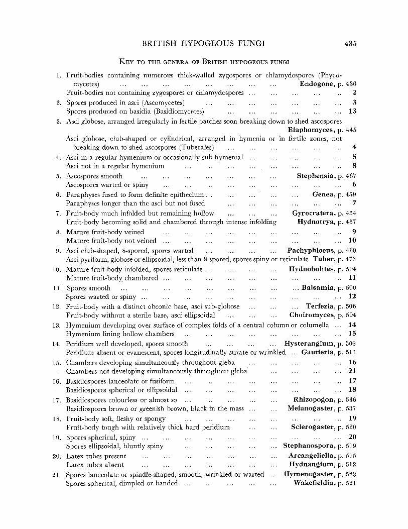
KEY TO THE GENERA OF BRITISH HYPOGEOUS FUNGI
1. Fruit-bodies containing numerous thick-walled zygospores or chlamydospores (Phyco- mycetes) ... ... ... ... ... ... ... ... Endogone, p. 436
Fruit-bodies not containing zygospores or chlamydospores ... ... ... .. ... 2 2. Spores produced in asci (Ascomycetes) ... .. .... .. ... ... ... 3
Spores produced on basidia (Basidiomycetes) ... ... ... ... ... ... 13 3. Asci globose, arranged irregularly in fertile patches soon breaking down to shed ascospores
Elaphomyces, p. 445 Asci globose, club-shaped or cylindrical, arranged in hymenia or
breaking down to shed ascospores (Tuberales) ......
4. Asci in a regular hymenium or occasionally sub-hymenial ... Asci not in a regular hymenium ... ... ... ... .
5. Ascospores smooth ... ... ... ......
Ascospores warted or spiny ... ... .........
6. Paraphyses fused to form definite epithecium ......
Paraphyses longer than the asci but not fused ... ...
7. Fruit-body much infolded but remaining hollow ... ... ...
Fruit-body becoming solid and chambered through intense infolding 8. Mature fruit-body veined ... ... ... ... ...
Mature fruit-body not veined ... ... .. ... ...
9. Asci club-shaped, 8-spored, spores warted ... ... ...
in fertile zones, not ... ... .. 4
... 5 .. ... 8
Stephensia, p. 467 ... . ... 6
... Genea, p. 459 . 7
Gyrocratera, p. 454 Hydnotrya, p. 457
. 9
... ... ... 10
Pachyphloeus, p. 469 Asci pyriform, globose or ellipsoidal, less than 8-spored, spores spiny or reticulate Tuber, p. 473
10. Mature fruit-body infolded, spores reticulate ... ... ... ... Hydnobolites, p. 504 Mature fruit-body chambered ... ... ... ...... ... ... ... ... 11
11. Spores smooth ... ... ... ... ... ... ... ... ... Balsamia, p. 500
Spores warted or spiny . .. ... ... . ... ... ... ... ... 12
12. Fruit-body with a distinct obconic base, asci sub-globose ... ... ... Terfezia, p. 506
Fruit-body without a sterile base, asci ellipsoidal ... ... ... Choiromyces, p. 504
13. Hymenium developing over surface of complex folds of a central column or columella ... 14
Hymenium lining hollow chambers ... ... ... ... ... ... ... ... 15
14. Peridium well developed, spores smooth ... .. ... ... Hysterangium, p. 509 Peridium absent or evanescent, spores longitudinally striate or wrinkled ... Gautieria, p. 511
15. Chambers developing simultaneously throughout gleba ... Chambers not developing simultaneously throughout gleba
16. Basidiospores lanceolate or fusiform ... ... ...
Basidiospores spherical or ellipsoidal ... ... ...
17. Basidiospores colourless or almost so ... ... ...
Basidiospores brown or greenish brown, black in the mass ...
18. Fruit-body soft, fleshy or spongy ... ... ...
Fruit-body tough with relatively thick hard peridium ...
19. Spores spherical, spiny ...... ... ...
Spores ellipsoidal, bluntly spiny ... ... ...
20. Latex tubes present ... ......... ... Latex tubes absent ... ... ... ...
21. Spores lanceolate or spindle-shaped, smooth, wrinkled or warted
Spores spherical, dimpled or banded .........
... ... . ... 16 ... ... ... ... 21
... ... ... ... 17 ... ... ... ... 18 . Rhizopogon, p. 536
... Melanogaster, p. 537
... ... ... ... 19 . Sclerogaster, p. 520
... ... ... ... 20 ... Stephanospora, p. 519
.. Arcangeliella, p. 515
. Hydnangium, p. 512
.. Hymenogaster, p. 523 Wakefieldia, p. 521
435
LILIAN E. HAWKER ON
The author has had no opportunity of examining type material of the majority of the species described. Material included in Broome's Herbarium at the British Museum (Natural History), in the Berkeley Herbarium and in some other British and European collections in the Herbarium of the Royal Botanic Gardens at Kew has been examined.
Original descriptions and figures of type material have also been studied.
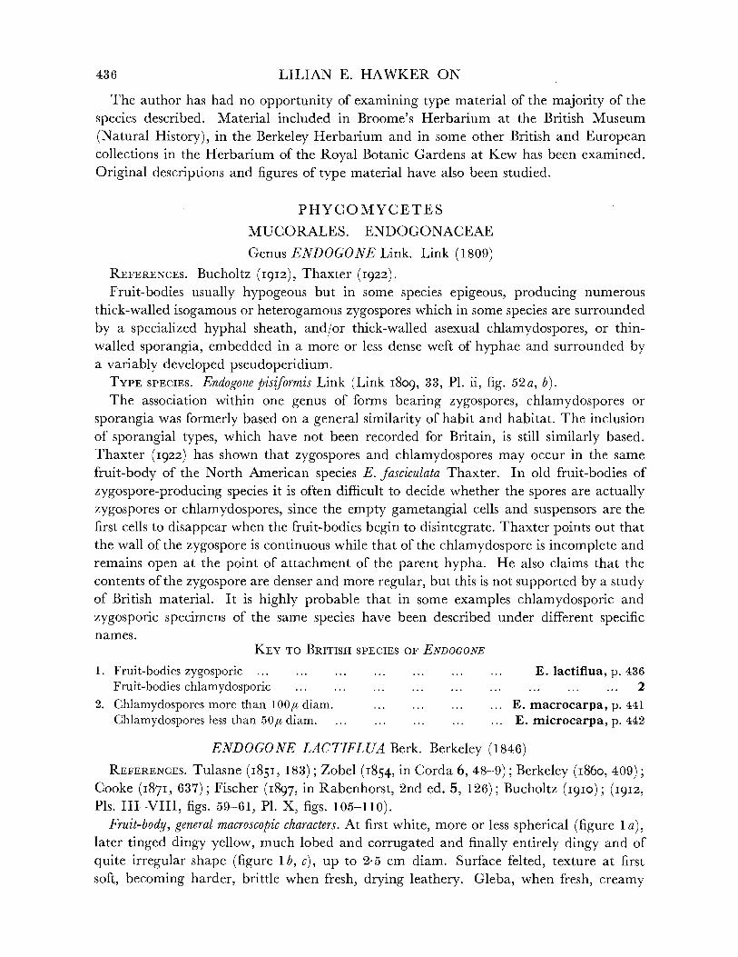
PHYCOMYCETES
MUCORALES. ENDOGONACEAE
Genus ENDOGONE Link. Link (1809) REFERENCES. Bucholtz (1912), Thaxter (1922). Fruit-bodies usually hypogeous but in some species epigeous, producing numerous
thick-walled isogamous or heterogamous zygospores which in some species are surrounded by a specialized hyphal sheath, and/or thick-walled asexual chlamydospores, or thin- walled sporangia, embedded in a more or less dense weft of hyphae and surrounded by a variably developed pseudoperidium.
TYPE SPECIES. Endogone pisiformis Link (Link I809, 33, P1. ii, fig. 52a, b). The association within one genus of forms bearing zygospores, chlamydospores or
sporangia was formerly based on a general similarity of habit and habitat. The inclusion of sporangial types, which have not been recorded for Britain, is still similarly based. Thaxter (1922) has shown that zygospores and chlamydospores may occur in the same
fruit-body of the North American species E. fasciculata Thaxter. In old fruit-bodies of
zygospore-producing species it is often difficult to decide whether the spores are actually zygospores or chlamydospores, since the empty gametangial cells and suspensors are the first cells to disappear when the fruit-bodies begin to disintegrate. Thaxter points out that the wall of the zygospore is continuous while that of the chlamydospore is incomplete and remains open at the point of attachment of the parent hypha. He also claims that the contents of the zygospore are denser and more regular, but this is not supported by a study of British material. It is highly probable that in some examples chlamydosporic and
zygosporic specimens of the same species have been described under different specific names.
KEY TO BRITISH SPECIES OF ENDOGONE
1. Fruit-bodies zygosporic ... ... ... ... ... ... ... E. lactiflua, p. 436 Fruit-bodies chlamydosporic ............ ... .. ... ... 2
2. Chlamydospores more than 100/6 diam. ... ... ... ... E. macrocarpa, p. 441 Chlamydospores less than 50/, diam. ... ... . .... ... E. microcarpa, p. 442
ENDOGONE LACTIFLUA Berk. Berkeley (1846) REFERENCES. Tulasne (1851, 183); Zobel (I854, in Corda 6, 48-9); Berkeley (I860, 409);
Cooke (1871, 637); Fischer (1897, in Rabenhorst, 2nd ed. 5, 126); Bucholtz (1910); (1912, Pls. III-VIII, figs. 59-61, P1. X, figs. 105-110).
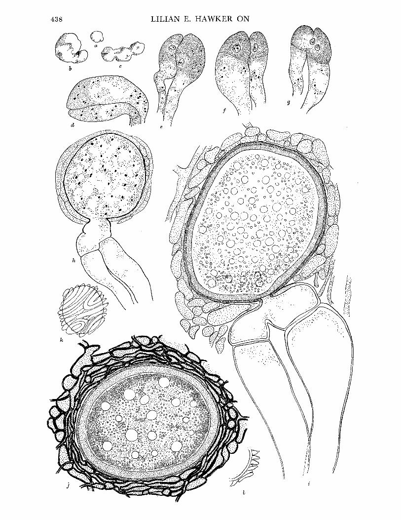
Fruit-body, general macroscopic characters. At first white, more or less spherical (figure 1 a), later tinged dingy yellow, much lobed and corrugated and finally entirely dingy and of
quite irregular shape (figure 1 b, c), up to 2-5 cm diam. Surface felted, texture at first soft, becoming harder, brittle when fresh, drying leathery. Gleba, when fresh, creamy
436

BRITISH HYPOGEOUS FUNGI
white in young specimens, becoming flushed apricot yellow, then entirely pinkish orange or finally dingy cinnamon, with increasing maturity. Zygospores just detectable with naked eye as orange granules in older specimens which also yield cream to orange latex when cut. Fruit-bodies may grow around small sticks or dead leaves of conifers which thus become embedded in them. Often much eaten, sometimes by slugs, but no odour detectable.
Pseudoperidium. No true peridium, but outer zone of fruit-body consists of rather more closely interwoven hyphae than those of central part. This differentiation becomes more obvious as fruit-body matures. Zygospores usually not formed in this peripheral zone but occasionally they may be quite near surface.
Gleba. At first consisting of loosely interwoven hyphae without latex tubes, later more closely woven and latex tubes conspicuous. Hyphae coenocytic, diam. 5 to 7#, with numerous small nuclei and occasional septa. Latex tubes diam. 10 Q with dense contents.
Development of zygospores. A fully illustrated account has been given by Bucholtz (1912). His figures show some evidence of shrinkage of the material, doubtless due to his use of xylol during processing. Examination of British material confirms Bucholtz's account with some minor differences.
Paired hyphae scattered through the young fruit-body develop into progametangia. These are swollen and somewhat curved, with dense contents and numerous small nuclei (figure 1 d). They lie parallel to one another for some distance from their tips, and one is usually slightly larger than the other. Oily contents concentrate at the tips and a single nucleus in each progametangium enlarges and comes to lie in a clear area (figure 1 e), while the rest of the nuclei in the tips disintegrate. Cross-walls then form at or near the widest part of the progametangia cutting off the uninucleate gametangia from the multi- nucleate suspensor cells (figure if). The wall between the gametangia then breaks down, and the single nucleus from the smaller one passes into the larger one together with most or all of the oily contents (figure 1 g). The fertilized gametangium then grows out into a bud-like projection which rapidly swells to form a spherical or ellipsoidal sac into which the contents of the gametangium pass. No conclusive evidence of nuclear fusion in the gametangium was seen, although the two nuclei were seen in close association, so that it is likely that the two nuclei both pass into the sac as claimed by Bucholtz. According to his figures they remain undivided, but the British material shows numerous nuclei in the sac long before it reaches its maximum size (figure 1 h). When most of the contents of the gametangium have passed into the sac a thin wall is laid down lining it, thus delimiting the young zygospore and cutting off the nearly empty gametangium. The young zygospore continues to increase in size and the wall increases in thickness to 9 to 10 u and becomes at first pale yellow and finally deep reddish orange in colour, remaining smooth but showing two distinct layers in section (figure 1 i,j). Where the zygospores are crowded the wall may be flattened by the pressure of adjacent spores giving an angular appearance. Where such pressure is absent the spore is ellipsoidal or less often almost spherical. The contents
rapidly become very dense with numerous colourless oil globules. A sheath of sterile hyphae begins to develop round the zygosporic sac at an early stage and increases as the spore matures. This sheath varies in different specimens, but may finally be up to 30,u thick and then consists of one to several layers of yellow, thick-walled hyphae encircling the zygo- spore in a more or less spiral manner (figure 1 i j, k). In stained sections these sheath
437
VOL. 237, B, 53
\~.-,; % :'.' ''':-:: : .-:''-i:::::::::::::
q
NO ":aNAMVH '3 NVIrIr

BRITISH HYPOGEOUS FUNGI
hyphae often have the appearance termed 'flammenkronen' by Bucholtz (figure 1 1), due to the projection of the hyphae out into the matrix of the fruit-body and to the affinity of their thickened walls for various stains, in contrast to the zygospore wall which remains unstained.
The fruit-bodies develop slowly. Conjugating stages have not been found later than October. The gametangia continue to increase in size as the zygospore grows, but when the spore is mature they rapidly disappear. In late autumn the vegetative hyphae begin to disintegrate and thus to set free the zygospores. No record of germination of the zygo- spores exists and attempts to culture the fungus have consistently failed.
Normally all the zygospores in any one fruit-body are at approximately the same stage, although the gametangia tend to develop first in the outer part of the fruit-body. Occasion- ally, however, the majority of the spores cease to develop and abort, while a few continue to develop to maturity.
Bucholtz points out that the size of the mature zygospores varies considerably in different collections. This variation in size has been noted in numerous collections made recently in various parts of Britain. The size of mature zygospores varied in different fruit- bodies from 80-92-100 x 60-71-80 to 140-189-200 x 120-145-150#.* No correlation could be made between size of zygospores and date or place of collection or identity of tree beneath which the fruit-bodies were found, but the differences were directly correlated with the density of distribution of the spores within the individual fruit-body. The size decreased with an increase in the number of spores per unit volume of fruit-body. Such a difference could be seen between different individuals of the same collection or even within the same specimen. There is no justification therefore for the separation of strains on the basis of spore size. All other macroscopic and microscopic characters were essentially similar in all collections made.
Habitat and periodicity. The material in Broome's Herbarium on which Berkeley's original description was based was collected at Chudleigh (Devonshire) and a further collection was made in Shropshire. The Foray records of the British Mycological Society report the fungus from Thirlmere, Cumberland. During the present study collections were made in
* The middle figure is the mean.
FIGURE 1. Endogone lactiflua. (a) Young fruit-body, almost spherical, x 2. (b), (c) Older fruit-bodies, showing irregular shape, x 2. (d) Young multinucleate progametangia. (e) Older progame- tangia, showing enlargement of single nucleus in each and concentration of oily contents in tips. (f) Uninucleate gametangia cut off from suspensor cells by transverse walls. (g) Nucleus of smaller gametangium migrating into larger one. (h) Young multinucleate zygospore with sheath hyphae beginning to develop. (d)-(h) Drawn from material stained iron alum haema- toxylon and light green. (i) Nearly mature zygospore, drawn from fresh unstained material. Note oily contents of spore, 2-layered wall, enlargement of empty gametangia and suspensors and development of sheath hyphae. (j) Mature zygospore, showing fully developed hyphal sheath. Composite drawing, contents of zygospore drawn from fresh material, sheath from stained preparation. (d)-(j) x 1500. (k) Surface view of nearly mature zygospore, showing spiral arrangement of sheath hyphae. (1) Part of section of mature zygospore cut in plane parallel to that of k and showing 'flammenkrone' effect (Bucholtz I912) due to projecting folds of sheath hyphae; cf. j, which is of a specimen cut at right angles to the plane of k, I and so more or less in the plane of the enveloping hyphae. (k, 1) x 460.
53-2
439

LILIAN E. HAWKER ON
d
9
0I
b
e
/It I
n
a
c
440
BRITISH HYPOGEOUS FUNGI
Devonshire (Dartington Hall, 107*), Gloucestershire (Cotswold Hills, near Wotton-under- Edge, 281, 446, 455, 548, 647, 648, 649), Somerset (Cleeve, 313), Herefordshire (Haugh Wood, 470, 471), Perthshire (near Loch Tay, 407, 414, 416, 422) and Caernarvonshire
(Nant-y-Garth, Bangor, 242; Bettws-y-coed, 258; Nant Heilyn, 260, 261). Mature fruit-bodies are found only in summer and early autumn. Locally abundant. The present collections were made in coniferous woods or plantations (including pine,
spruce, silver fir and larch). In no case could any connexion be traced between fruit- bodies of Endogone lactiflua and typical ectotrophic mycorrhizal mantles, but in most cases the tree roots in the neighbourhood of the fruit-bodies showed the typical pseudomycor- rhizae described by Melin (1917). It is likely, therefore, that this fungus is a true parasite on the roots of coniferous trees.
ENDOGONE MACROCARPA (Tul.), Tulasne. Tulasne (I85I, p. 182, P1. XX, fig. 1) REFERENCES. Fischer (1897, 125); Bucholtz (I912, P1. VIII, figs. 62-70; P1. IX, figs.
71-4); Thaxter (I922). Syn. Glomus macrocarpus Tulasne (I845, 63). Fruit-body, general macroscopic characters. Irregularly globose or lobed, up to size of a
hazelnut, usually smaller, at first pallid, later yellow, finally dingy brown, at first soft, becoming firm and usually less compact, cut surface of gleba white, then yellow, finally reddish brown, granular when mature owing to presence of large chlamydospores just visible to naked eye (figure 2a), latex absent, odourless.
Pseudoperidium. No true peridium, outer zone of hyphae without chlamydospores, often enclosing soil particles, but otherwise similar to hyphal network of gleba.
Gleba. Loosely interwoven, thin-walled, aseptate hyphae (10 to 30,c diam.) with charac- teristic wide-angled more or less dichotomous branching (figure 2 b), contents yellow, oily. Chlamydospores (140-190-230 x 120-145-180u) terminal on hyphal branches, notcrowded, at first thin-walled, hyaline, with dense, oily contents. Wall becoming thickened (5 to O1,U) and bright yellow, oil drops finally conspicuous (figure 2 c to g). Chlamydospores at different stages of development may be present in the same fruit-body, but the majority develop more or less simultaneously. No hyphal sheath develops round the spores.
Habitat and periodicity. Single mature specimens were collected on two occasions (May 1952, May 1953) under beech in a copse of mixed beech and larch near Wotton-under-
* Figures throughout refer to the writer's collections.
FIGURE 2. Endogone macrocarpa (a) Longitudinal section of mature fruit-body, showing granular effect due to large chlamydospores, x 2. (b) Characteristic hyphae with wide-angled dichotomous branching. (c)-(g) Stages in development of chlamydospores. (f)-(g) x 250. E. microcarpa. (h) Chlamydospores drawn to same scale for comparison with those of E. macrocarpa. (i) Fruit- body attached to stick, x 4. (j) Slender loosely woven hyphae of pseudoperidium, showing hairs which project beyond surface. (k) Portion of gleba of mature fruit-body, showing charac- teristic thick-walled hyphae with wide-angled dichotomous branching (cf. E. macrocarpus) and mature chlamydospores with thick walls and oily contents. Note that interior of spore is still in communication with parent hyphae. (l)-(q) Stages in development of chlamydospores. (r) Chlamydospore with unusual swelling at base. (j)-(r) x 740.
441
LILIAN E. HAWKER ON
Edge, Gloucestershire (606, 722) and once from Failand, Somerset (949). A larger collection was made under yew at Kingsweston, Bristol (947). Specimens in Herb. Broome, Brit. Museum, labelled Endogone pisiformis from Somerset (Brockley Coombe, Leigh Woods) and Northamptonshire (Maidwell), are all E. macrocarpa.
It is almost certain that Berkeley & Broome's (1846) description of E. pisiformis Link (see also Berkeley (i860) and Cooke (1871)) was based on these specimens of E. macrocarpa. Cooke (1871) reports thatTulasne examined this material and considered it to be E. macro-
carpa. The specimen in Herb. Broome from Whitby, Yorks, labelled E. macrocarpa is, how- ever, actually E. microcarpa. E. macrocarpa is probably relatively uncommon in Britain. The large size of the chlamydospores distinguishes it clearly from E. microcarpa.
Thaxter (1922) suggests that this species may be the chlamydospore stage of E. lactiflua. This is unlikely, since the characteristic hyphae of E. macrocarpa are not seen in E. lactiflua while latex is present in the latter and absent in the former.
ENDOGONE MICROCARPA (Tul.), Tulasne. Tulasne (I85I, p. 182, P1. XX, fig. 2) REFERENCES. Zobel (I854, in Corda, 6, 48); Bucholtz (I912); Thaxter (I922); Knapp
(1952). Syn. Glomus microcarpus Tulasne (I844, 63). Fruit-body, general macroscopic characters. Small, seldom more than 5 mm diam., at first
more or less spherical, then flattened or lobed, often closely adpressed to surface of dead leaf or stick (figure 2i). Surface felted but not visibly hairy. At first chalky white, then becoming dull straw-coloured and finally dingy yellow-brown. Cut surface of gleba homo- geneous, firm, at first dingy white then ochre yellow and finally yellow-brown, always somewhat darker than the exterior, no latex present. No odour detectable.
Pseudoperidium. No true peridium but outer hyphae of fruit-body slender (1 to 2# diam.), more loosely interwoven than central ones and a few of them projecting into the soil or terminating as short septate hairs (figure 2j). Small soil particles may be entangled in these loose peripheral hyphae.
Gleba. Consisting of loosely interwoven hyphae (diam. 2 to 8yu), much branched, occasionally septate, bearing terminal, almost spherical chlamydospores (diam. 40-42-48 x 35-38-42#) with walls 2 to 5,u thick. The hyphae show wide-angled branching similar to that of E. macrocarpa and become largely used up as the chlamydospores mature (figure 2k).
Development of chlamydospores (figure 21 to q). Chlamydospores develop from swollen ends of hyphal branches. These are often in pairs (figure 2m, n), but no sign of conjugation is seen and the apparent pairing may be merely the result of the crowding of the young spores. It is possible, however, that the chlamydospores are actually parthenogenetic zygospores, and that the ability to conjugate was lost early in the evolution of the species. Figure 2r shows a hypha which has cut off a chlamydospore and is cutting off other swollen cells below it. This condition is unusual, but there is no evidence that the basal cell is of gametangial nature. The wall of the normal chlamydospore thickens and becomes yellow, but a narrow channel connecting the mature spore with the parent hypha remains open (in contrast to the zygospores of E. lactiflua). From an early stage the contents consist of large oil globules which are sufficiently uniform in size to give the appearance of spores and may account for the confusion that exists between this species and E. pisiformis. These oil drops are much
442
BRITISH HYPOGEOUS FUNGI
more crowded than those of E. macrocarpa. The majority of the chlamydospores in a particular fruit-body are usually at approximately the same stage of development, but old and young fruit-bodies may be found close together, presumably borne on the same mycelium.
Habitat and periodicity. Specimens in Herb. Broome, British Museum from Credenhill Camp and Brockley Coombe (labelled Glomus macrocarpus), Whitby (labelled E. macrocarpa), Kings Cliffe (unlabelled) and Saltford (unlabelled). Numerous collections made during the present study as follows: Bristol (Stoke Bishop 484, 485; Blaise Castle, 56, 65, 66, 67, 81, 280, 431, 540, 652, 655, 691; Leigh Woods, 509, 514, 516, 521, 679), Somerset (Cleeve, 37, 43, 73, 76, 192, 194, 200, 312, 333, 335, 513, 557, 583, 695, 713; Orchardleigh, Frome, 49, 223; Portbury, 49, 207, 215, 217, 343; Wraxall, 100, 101, 322; Brockley Coombe, 268, 271, 274, 396, 491; Brockley Road, 380, 623; Abbot's Pool, Failand, 286, 528), Gloucester- shire (Staunton, Forest of Dean, 462; Newark Park, 503), Herefordshire (Downton Gorge, 464), Wiltshire (Savernake Forest, 364), Caernarvonshire (Vaynol Park, Bangor, 229; Bettws-y-coed, 246). The fungus has been found most frequently under yew or pine but has also occurred under various deciduous trees. It is frequently associated with pseudo- mycorrhizae of which it is probably the cause. Locally abundant.
Fructifications of an unidentified species of Endogone in association with mycorrhizal strawberry roots have recently been described by Mosse (I953). Material has been examined by the present writer. While this fungus is undoubtedly a chlamydosporic species of Endogone, it is not attributable to any of the three species described above, nor does it closely resemble any of the species described by Thaxter (I922). The fruit-bodies are about 1 mm in diameter and contain 2 to 32 (usually 2 to 6) thick-walled yellow chlamydospores (92 to 197u diam.) embedded in a loose mass of hyphae with branching similar to that figured for E. macrocarpa and E. microcarpa (figure 2). Further description and identification must await the results of Miss Mosse's studies now in progress.
Doubtful record ENDOGONE PISIFORMIS Link
This was recorded by Bucknall (1878) from Hanham, Bristol, but I have been unable to trace the specimen. In view of the frequency of Endogone microcarpa in this district and the confusion between these two species it is probable that this specimen was actually E. microcarpa. The specimens in Herb. Broome British Museum, labelled E. pisiformis are all E. macrocarpa, so that Bucknall's specimen may have been this species. The record of E. pisiformis in the account of the Bangor Foray of the British Mycological Society held in September 1950 was an error and the specimen was E. microcarpa. E. pisiformis was first described by Link (1809) as a form showing normal zygospore production. Fischer (1897), writing in Rabenhorst's Kryptogamenflora, considered that E. pisiformis and E. microcarpa were synonyms but figured the latter as sporangial. Bucholtz (I912) erroneously referred to E. pisiformis as sporangial and to E. microcarpa as zygosporic. Thaxter (1922) cleared the matter up and includes only zygosporic material in E. pisiformis and reserves the name E. microcarpa for fruit-bodies forming the typical small chlamydospores. Unfortunately, Knapp (I952) has reverted to the use of the name E. pisiformis for sporangial forms. Sporangial forms have not been recorded in Britain.
443

LILIAN E. HAWKER ON
Systematic position of Endogone There is little doubt that the species showing zygospore formation are advanced types
of Zygomycetes. The usually aseptate nature of the ground hyphae of the fruit-body is definitely a phycomycetous character. The formation of the gametangia is not unlike the early stages of conjugation in Phycomyces, while the passage of the contents of the fertilized gametangium into a bud or vesicle in which the zygospore develops is paralleled in the Piptocephalaceae. Grouping of the paired gametangia is .seen in some members of the Entomophthorales, while the single zygospore of Mortierella is embedded in a sterile hyphal sheath resembling that of Endogone lactifua. In spite of the ascomycetous affinities shown by the relatively large fruit-body, the extra-gametangial development of the zygospores and the septation of the sheath hyphae, it is likely that Endogone represents the end of an evolutionary side branch. Nevertheless, its structure supports the hypothesis that the simple filamentous Endomycetales and such forms as Gymnoascus with simple fruit-bodies may have evolved from an ancestral Zygomycete along lines parallel to the evolution of Endogone.
If the sporangial types which have been described do actually belong to the same genus, then Endogone is obviously closely related to the simple Zygomycetes. It is possible that some of the 'sporangial' stages described were actually chlamydospores containing oil drops of regular size. Cultural studies by Kanouse (1936) indicate that the sporangial forms are not related to the zygosporic ones.
The chlamydospore stage remains a problem. The close resemblance of these spores to mature zygospores suggests that they may be azygospores formed parthogenetically. The production of chlamydospores is, however, a characteristic of the Zygomycetes. The usually aseptate nature of the ground hyphae of the chlamydosporic fruit-body suggests a phycomycetous derivation.
The British species are only three in number and therefore not representative, but the differences in habitat and general structure are such that it is practically certain that these species are distinct. As already pointed out, the mature zygospores of E. lactiflua often show no trace of gametangia and could easily be mistaken for chlamydospores. The other two species are definitely chlamydosporic from the earliest stage. They may be distinguished from one another by the great difference in chlamydospore size.
The solution of the problem of the three types of fruit-body attributed to Endogone can only be solved by careful cultural studies of a larger number of species than is available in Britain.
ASCOMYCETES
PLECTASCALES
Asci globose, evanescent, irregularly arranged in sporocarp.
ELAPHOMYCETACEAE
Fruit-bodies subterranean, spore mass powdery at maturity. Dodge (1929) divides this family into two tribes: Elaphomyceteae, in which the central core is cottony in texture, and Mesophelliae, with a corky or woody core. All the British species belong to the genus Elaphomyces of the first of these tribes.
444
BRITISH HYPOGEOUS FUNGI
Genus ELAPHOMYCES Nees ex Fr. Fries (1829, p. 57) Fruit-bodies consisting of a central core or gleba surrounded by a peridium which
consists of two layers, an inner true peridium and an outer layer which in many species splits into pyramidal warts. This outer layer has been termed the 'cortex'. This is an unfortunate choice, but its long usage makes it necessary to retain this term. The whole fruit-body is, in some species, surrounded by the 'crust' which consists of a layer of soil particles bound together by hyphae. No sexual organs are known, but groups of asci arise from ascogenous hyphae which develop in small knots near the periphery of the core. The asci break down at an early stage and the spores complete their development after liberation. The spore mass is finally powdery.
Dodge (1929) divides the genus into two sections, subgenus Malocoderma Vitt. with a more or less fleshy cortex becoming wrinkled, but not spiny, and with spores less than
151I diam., and subgenus Scleroderma Vitt. with a hard cortex and with larger spores (diam. 15 to 50/u). In view of the general acceptance of the Gasteromycete genus Sclero- derma this is not a permissible use of the name. Nevertheless, the distinction between the two groups is a useful one. All the British species fall in the so-called 'scleroderma' section except Elaphomyces citrinus which is a doubtful record.
TYPE SPECIES. E. granulatus Fr.
KEY TO BRITISH SPECIES OF ELAPHOMrCES
1. Cortex soft, ascospores less than 15/t diam. ... ... ... ... E. citrinus, p. 453 Cortex hard, ascospores more than 15/t diam. ... ... . .. ... ... ...... 2
2. Cortex verrucose or echinulate ... ... ... ... ... ... 3 Cortex smooth or nearly so ... ... ... ... ... ... ... ... ... 5
3. Peridium more or less homogeneous in section ... ... ... ... ... ... 4 Peridium marbled in section ... ... ... ... ... E. muricatus, p. 449
4. Peridium ochraceous, verrucose ... ... ... ... ... ... E. granulatus, p. 445 Peridium greyish, echinulate ... . ... ... ... ... E. aculeatus, p. 451
5. Peridium thin, grey-brown to russet ... ... ....... ... E. leucosporus, p. 451 Peridium thick, brownish black ... ... ... ... ... E. anthracinus, p. 452
ELAPHOMYCES GRANULATUS Fr., Fries (I829, p. 58) REFERENCES. Berkeley & Broome (I84I, 430, P1. XI, fig. 10); Tulasne (I841, 22, P1. I,
fig. 3; P1. II, fig. 7; PI. IV, fig. 3); (I851, 109-10, PI. XIX, fig. 4); Vittadini (I842, 78, P1. III, fig. 7); Berkeley (i86o, 378); Cooke (i871, 750); Hesse (i894, 70-2, PI. XIII, figs. 1-7, P1. XXI, fig. 55); Massee (I909, 249).
-Syn. ? Lycoperdon solidum Linnaeus (1737, 369). ? L. cervinum Linnaeus (1753, 1183). Hypogeum cervinum Persoon (I797, 7). Hypogaeum cervinum Gray (I821, 582, P1. I). Scleroderma cervinum Persoon (I801, 156, P1. IV, fig. 2). Tuber cervinum Nees v. Esenbeck (I816, 161, P1. XV, fig. 147). Lycoperdastrum cervinum Kuntze (I891, P1. I). Elaphomyces officinalis Nees v. Esenbeck (1821-3, P1. I). Phymatium flvum Chevallier (1826, 361, P1. X, fig. 6). Elaphomyces vulgaris var. granulatus Corda (I84I, 25-6). E. cervinus Schlectendal (1824, 166); Fischer (I897, 94-5, figs. 1-4 on p. 82);
Hennings (I905, 91, fig. 2); Dodge (1929); Knapp (1952). VOL. 237, B. 54
445

LILIAN E. HAWKER ON
t..N;~~~~~~~~~~~~~~~~~~~~~~~~~~~~,-
A 11~~~~~~~~~~~~~~~
a ?~~~~~~~~~~~~~~~~~~~~~~~~4
- C- -
C C C') e~~~~~~~9
k s~~~~~~~~~~~~~~~~~~~~~~~~~~~~~~~~~~
3 4W
WI W2~~~~~W
446
BRITISH HYPOGEOUS FUNGI
This fungus has been known from the sixteenth century and was used as an aphrodisiac. The specific name was deliberately changed from cervinus to granulatus by Fries in part 1 of vol. 3 of the Systema Mycologica published in 1829. Dodge (1929) considered that the correct name was Elaphomyces cervinus (L. ex S. F. Gray) Schlect., and Knapp (I952) cites it as E. cervinus (Pers.) Schroter syn. E. granulatus Fr. By the latest definition of the Rules at the Botanical Congress at Stockholm 1950 any citation in any volume of the Systema or the Elenchus is the starting point in naming Ascomycetes. Hence, since Gray's paper is earlier than Fries's citation in vol. 3, Elaphomyces granulatus Fr. is the valid name. This name has always been used in British papers.
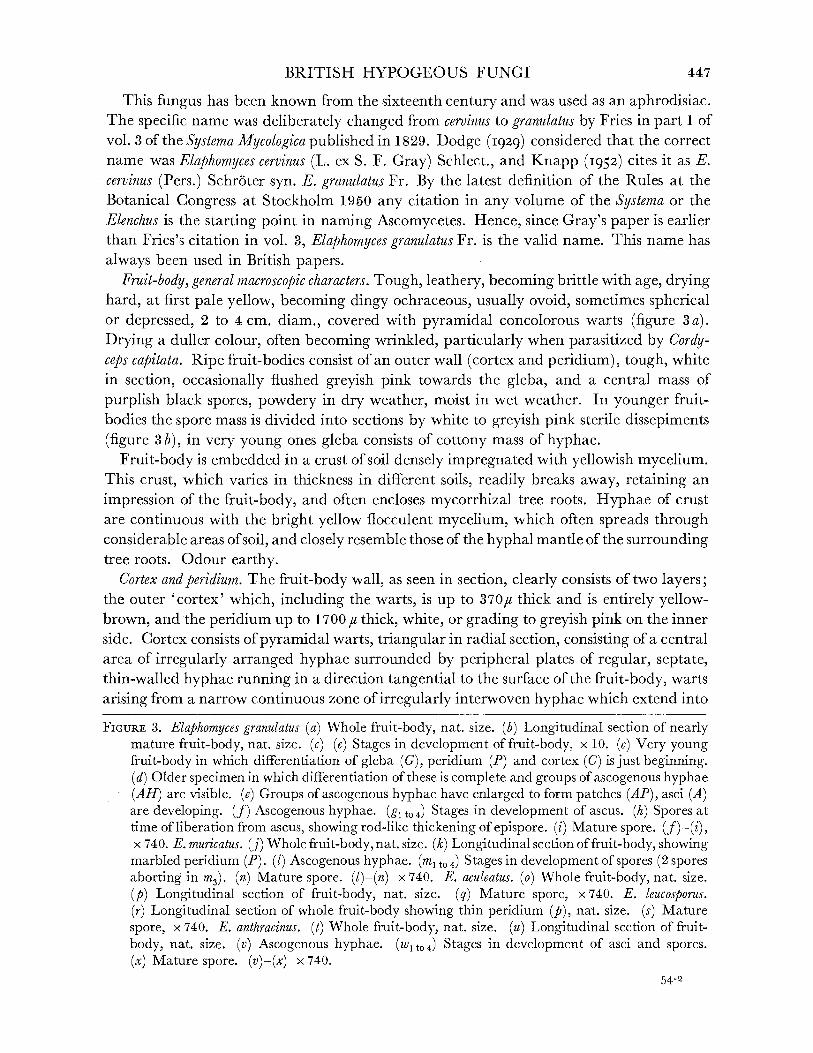
Fruit-body, general macroscopic characters. Tough, leathery, becoming brittle with age, drying hard, at first pale yellow, becoming dingy ochraceous, usually ovoid, sometimes spherical or depressed, 2 to 4 cm. diam., covered with pyramidal concolorous warts (figure 3 a). Drying a duller colour, often becoming wrinkled, particularly when parasitized by Cordy- ceps capitata. Ripe fruit-bodies consist of an outer wall (cortex and peridium), tough, white in section, occasionally flushed greyish pink towards the gleba, and a central mass of purplish black spores, powdery in dry weather, moist in wet weather. In younger fruit- bodies the spore mass is divided into sections by white to greyish pink sterile dissepiments
(figure 3b), in very young ones gleba consists of cottony mass of hyphae. Fruit-body is embedded in a crust of soil densely impregnated with yellowish mycelium.
This crust, which varies in thickness in different soils, readily breaks away, retaining an
impression of the fruit-body, and often encloses mycorrhizal tree roots. Hyphae of crust are continuous with the bright yellow flocculent mycelium, which often spreads through considerable areas of soil, and closely resemble those of the hyphal mantle of the surrounding tree roots. Odour earthy.
Cortex and peridium. The fruit-body wall, as seen in section, clearly consists of two layers; the outer 'cortex' which, including the warts, is up to 370,u thick and is entirely yellow- brown, and the peridium up to 1700 It thick, white, or grading to greyish pink on the inner side. Cortex consists of pyramidal warts, triangular in radial section, consisting of a central area of irregularly arranged hyphae surrounded by peripheral plates of regular, septate, thin-walled hyphae running in a direction tangential to the surface of the fruit-body, warts
arising from a narrow continuous zone of irregularly interwoven hyphae which extend into
FIGURE 3. Elaphomyces granulatus (a) Whole fruit-body, nat. size. (b) Longitudinal section of nearly mature fruit-body, nat. size. (c)-(e) Stages in development of fruit-body, x 10. (c) Very young fruit-body in which differentiation of gleba (G), peridium (P) and cortex (C) is just beginning. (d) Older specimen in which differentiation of these is complete and groups of ascogenous hyphae (AH) are visible. (e) Groups of ascogenous hyphae have enlarged to form patches (AP), asci (A) are developing. (f) Ascogenous hyphae. (gl to 4) Stages in development of ascus. (h) Spores at time of liberation from ascus, showing rod-like thickening of epispore. (i) Mature spore. (f)-(i), x 740. E. muricatus. (j) Whole fruit-body, nat. size. (k) Longitudinal section of fruit-body, showing marbled peridium (P). (1) Ascogenous hyphae. (m t0 4) Stages in development of spores (2 spores aborting in m3). (n) Mature spore. (I)-(n) x 740. E. aculeatus. (o) Whole fruit-body, nat. size. (p) Longitudinal section of fruit-body, nat. size. (q) Mature spore, x 740. E. leucosporus. (r) Longitudinal section of whole fruit-body showing thin peridium (p), nat. size. (s) Mature spore, x 740. E. anthracinus. (t) Whole fruit-body, nat. size. (u) Longitudinal section of fruit- body, nat. size. (v) Ascogenous hyphae. (wI to4) Stages in development of asci and spores. (x) Mature spore. (v)-(x) x 740.
54-2
447
LILIAN E. HAWKER ON
the peridium. A study of young fruit-bodies suggests that the warts arise by splitting of the plates of tangential hyphae which fail to keep pace with the peripheral expansion of the peridium. The peridium consists of a firm pseudoparenchyma which is softer but less brittle than the cortex. The hyphae of the outer part, next the cortex, are closely inter- woven, diameter 3,u, mostly arranged periclinally, becoming looser and tending to a radial arrangement towards the inside where the hyphae are stouter (diam. 6 I). Throughout the peridium irregularly arranged bands of hyphae occur, but these are not visible to the naked eye (cf. Elaphomyces muricatus).
Asci. Globose to pyriform, 35 to 45u diam., evanescent, usually 6-spored (figure 3g4 t 4).
Ascospores. Brownish black at maturity, spherical, surface divided into shallow blocks by irregular cracks, 24 to 32,c diam., wall up to 10 i thick (figure 3 i).
Development offruit-body, asci and spores. At an early stage (fruit-bodies up to 5 mm diam.) only the cortex is clearly differentiated, and the densely woven peridial hyphae are not sharply divided from the loosely woven hyphae of the central core, which is often pinkish in colour (figure 3 c, d). Small knots of fine hyphae, which readily stain with aniline blue, arise near the outer edge of this core (figure 3 d) and develop rapidly to give groups of stout, coiled, much-branched, septate ascogenous hyphae with dense contents (figure 3f). These are said to be binucleate (Reess & Fisch I887; Dodge 1929). Asci soon arise from the terminal cells of the short branches of the ascogenous hyphae (figure 3 e, g1, 2). The con- tents of these young asci are granular with numerous oil drops. Staining with aceto- carmine shows very small nuclei. Details of nuclear division could not be followed, but the young ascus contains two nuclei which probably fuse to give a uninucleate stage, and later divide by three successive divisions. At some stage in these divisions some nuclei abort so that six finally remain. The spores develop around these nuclei and soon become thick-walled, showing a peripheral arrangement of granules in rows giving a number of radial rods (figure 394, h) which, after release of the spores from the ascus, rapidly turn brown or black. Cracks developing between these rods give the spore its characteristic appearance (figure 3 i). The asci often remain in communication with the stalk cells until spore differentiation is far advanced and finally break down when the spores are about half their final size (Clemencet 1932). At this stage separate ascogenous areas can still be seen separated by bands or dissepiments of the original core hyphae (figure 3 b). As the spores mature the latter are used up and the glebal cavity finally contains only spores interspersed with a few capillitium threads.
Habitat and periodicity. The records suggest that this fungus is common throughout Great Britain. It is recorded several times in British Mycological Society Foray lists and some 20 to 30 separate collections, mainly in western England, were made during the present investigations as follows: Bristol (Leigh Woods, 203, 344), Gloucestershire (Forest of Dean, 460; Wotton-under-Edge, 7, 10), Somerset (Cleeve, 102; Emborough, 31, 574; Brockley Coombe, 387; Abbot's Pool, Failand, 6, 18, 55, 68, 284, 321, 522, 569, 574a, 611, 617, 715; Portbury, 128, 129, 130), Devonshire (Woodbury Ring, 115; Stoke Wood, Exeter, 116), Essex (Epping Forest, 21b), Caernarvonshire (Nant-y-garth, 240, 241; Bettws-y-coed, 244, 249), Perthshire (Glen Lochay, 417) and was previously found at Mortimer Common, Hampshire. Some other recent collections from different parts of the country are preserved in the Herbarium of the Royal Botanic Gardens, Kew.
448

BRITISH HYPOGEOUS FUNGI
It is most frequently found on light acid soil or peat under Scots pine, but is also found under deciduous oak, sweet chestnut and occasionally under beech, including very old trees of each species.
In a study of several collections made in different localities under different species of tree Dicker (unpublished work in this department) observed slight variations in details of structure and in spore size. One collection from a beechwood near Wotton-under-Edge, Gloucestershire, differed from the type in the more pronounced banded arrangement of the peridial hyphae and the faintly marbled appearance of the inner peridium. This may have been a hybrid between E. granulatus and the common beechwood species E. muricatus
(see below). A suggestion that it might be a strain of E. asperulus was shown to be untenable when it was compared with material of that species collected by the writer in Norway. Genuine specimens of E. asperulus have not been found in Britain. Material assigned to E. granulatus, however, has proved to be very variable and more work on this species is desirable. In the present stage of our knowledge it is best to regard this as a species group covering a range of closely related forms.
Fruit-bodies, in favourable localities, are usually present in large numbers at all times of the year, but initiation of young ones is inhibited by extreme cold or extreme drought. They are most frequently partially embedded in the hard pan, which underlies the surface layers of leaf litter or humus, or are pressed against a large root or stone. They are seldom present at a depth of more than 3 in. from the surface and usually occur only in well- drained situations. They are frequently eaten by soil-inhabiting invertebrates, or by rodents. The spores are probably dispersed in this way, but no attempt to germinate them has succeeded and they were even recovered apparently undamaged and still incapable of germination from the faeces of captive rabbits to which they had been fed.
ELAPHOMYCES MURICATUS Fr., Fries (I829, p. 59) REFERENCES. Berkeley (I841, 430); Rabenhorst (I844, 291); Zobel (I854, 51, PI. X,
fig. 97); Quelet (I873, 379); Dodge (1929). Syn. ? Lycoperdon scabrum Willdenow (1787).
? Scleroderma cervinum /f scabrum Persoon (I801-8, 157). Elaphomyces variegatus Vittadini (1831, 68-9, P1. IV, fig. 4); (1843, 220); Tulasne
(1841, 23, P1. I, fig. 4; P1. II, figs. 4, 11; P1. IV, fig. 1); (i85I, 108-9, P1. III, fig. 8); Berkeley (i860o, 378); Cooke (1871, 749); Reess & Fisch (1887); Hesse (1894, 72-3, P1. XIII, figs. 8-16); Fischer (1897, 91); Massee (1909, 378); Dodge (1929);
Ramsbottom & Balfour-Browne (1951); Knapp (1952). ? Ceraunium scabrum and muricatum Wallroth (I833, 406-7). Elaphomyces vulgaris oc muricatus and y variegatus Corda (1871, 21, 27, Pls. VII
and IX). E. hirtus Tulasne (I841, 23). E. scaber Schr6ter (i893, 223).
This species is readily distinguished from Elaphomyces granulatus by the marbled peridium, as seen in section, smaller spores and the regular form, smaller size and darker tawny colour of the fruit-body. Dodge (I929) distinguishes E. variegatus Vitt. from E. muricatus Fries by differences in the coloration of the marbled peridium and its slightly smaller
449
LILIAN E. HAWKER ON
spores, but these differences are within the range of variation in material collected during the present investigation.
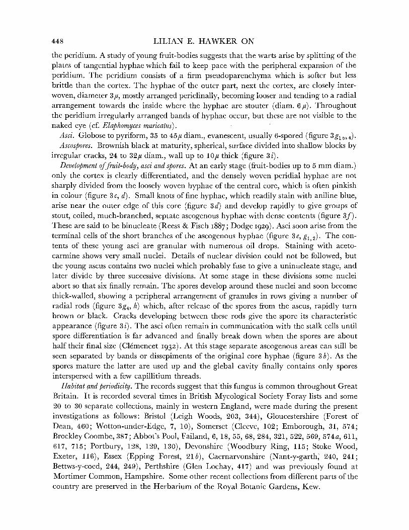
Fruit-body, general macroscopic characters. Tough, leathery, becoming brittle with age, drying hard, at first yellow then bright tawny orange, finally dingy yellow brown, usually spherical, up to 2 cm diam., covered with small pyramidal warts (figure 3j), often
parasitized by Cordyceps ophioglossoides. General organization and development of fruit-
body similar to that of Elaphomyces granulatus, from which it is distinguished by the peridium, which in section is mottled or marbled with yellowish white veins surrounding pink to chestnut brown areas, and by the smaller spores. Crust poorly developed and the
surrounding flocculent yellow mycelium less conspicuous than with E. granulatus. Odour, weak, earthy.
Cortex and peridium. Both thinner than in E. granulatus (ca. 200 and 500 to 2000#u respectively). Peridium marbled in section with yellowish white anastomosing veins
surrounding small pink to chestnut brown areas, colour darkening generally towards centre of fruit-body, marbled effect due to large air spaces and fissures (figure 3k).
Gleba. Similar to that of E. granulatus but hyphal strands which separate ascogenous areas in immature fruit-bodies are often pink or purplish fawn in colour. Spore masses bluish black or occasionally brownish black. Capillitium variable, sometimes profuse, sometimes sparse.
Asci. Globose, 30 to 40/x diam., evanescent, usually 4-spored, occasionally 2-spored
(figure 31, m1 to 4)
Ascospores. Purplish black at maturity, spherical, 18 to 24,u diam., surface cracked into blocks to a depth of 2,u, rods making up wall in developing spores more coarsely granular than in E. granulatus, wall up to 8,u thick (figure 3n).
Development. Similar to that of E. granulatus.. Habitat and periodicity. Common in beechwoods in south and west England, occasionally
found under other species of trees but never under conifers. It is probably equally common in other parts of Great Britain but has not been looked for so intensively. British Myco- logical Society Foray lists record it from Scotland (Forres), north Wales (Bangor) and Northern Ireland (Belfast), while its parasite Cordyceps ophioglossoides is recorded from
Keswick, Aviemore, Norwich and north Wales and occurs in Windsor Park. Numerous collections were made during the present investigation as follows: Gloucestershire (Cots- wold Hills, near Wotton-under-Edge, and Dursley, 3, 8, 11, 12, 13, 45, 86, 93, 104, 170, 293, 298, 307, 353, 373, 397, 398, 576, 589, 590, 597, 598, 590, 597, 598, 614, 646, 685, 717, 720, 728; Michael Wood, 103; Forest of Dean, 403, 459), Somerset (Cleeve, 571; Emborough, 573; Abbot's Pool, Failand, 320, 523, 534, 618, 716; Portbury, 131, 132), Herefordshire (Downton Gorge, 468), Devonshire (Berry Pomeroy, 108), Wiltshire (Wylie Valley, 90; Savernake Forest, 360-3, 367, 680), Oxfordshire (Kingwood Common, 26), Buckinghamshire (Beaconsfield, 653), Surrey (Mickleham, 60, 92), Caernarvonshire (Vay- nol Park, Bangor, 238; Bettws-y-coed, 245, 248). Slightly alkaline soil is the most usual habitat in south-west England. The fruit-bodies are usually in the humus layer or leaf litter and in contrast to E. granulatus, are seldom found partially embedded in the under-
lying hard pan. Mature fruit-bodies occur all the year round as with E. granulatus. Fruit-bodies often
fail to develop spores even when not attacked by Cordyceps.
450

BRITISH HYPOGEOUS FUNGI
ELAPHOMYCES ACULEA TUS Vitt., Vittadini (I83I, 70, P1. III, fig. 12) REFERENCES. Vittadini (i842, 79); Tulasne (I841, 24-5, P1. I, fig. 5; P1. II, fig. 6;
P1. III, fig. 3); (I851, 111); Fischer (I897, 98). Syn. Lycoperdastrum aculeatum Kuntze (189I).
Elaphomyces rubescens Hesse (1894, 75-7, P1. XIV, figs. 1-7; P1. XXII, figs. 1-5, 7, 9, 15, 18-24, 29); Fischer (I897, 97).
Fruit-body, general macroscopic characters. Tough and leathery, becoming brittle with age, drying hard, spherical, 1 to 2 cm diam., cortex black or greyish black with 3 to 4 angled, pointed, dark spines embedded in thin dark crust of soil and hyphae, peridium whitish to sooty grey in section, gleba at first with numerous dissepiments which disappear at maturity to leave mass of powdery sooty-black spores (figure 3 o, p). Mycelium not visible in surrounding soil.
Cortex and peridium. Cortex black in section, conical warts or spines consisting of alternating bands of fused black hyphae and plates of tangentially arranged light brown hyphae, inner layer black, pseudosclerenchymatous. Peridium whitish on outside becoming grey towards gleba, consisting of closely interwoven and fused hyphae.
Asci. Pyriform 40 to 50 x 35 to 45/u, evanescent, usually 8-spored. Ascospores. Sooty-black at maturity, spherical 14 to 17# diam., surface cracked into
blocks as in E. granulatus and E. muricatus (figure 3 q). Capillitium consisting of copious long grey threads.
Habitat and periodicity. Under beech in calcareous woods in summer (Knapp I952). Only two collections of this fungus have been made in Britain (Synwell Hill, Wotton-
under-Edge, Gloucestershire, 15, 5 September 1949, Hawker (1952) and near Dursley, Gloucestershire (760)). The description has therefore been compiled from an examination of this material and from descriptions of continental specimens. As the Gloucestershire specimens were all mature no studies of development could be made. The structure of the mature specimen is, however, essentially similar to that of other species, so that it is likely that development is also similar. Shortly after the first collection was made the wood was cut down and no specimens were found in the following summer.
ELAPHOMYCES LEUCOSPORUS Vitt., Vittadini (I842, 71, P1. III, fig. 1) REFERENCES. Vittadini (I842, 215, P1. III, fig. 1); Tulasne (I85I, 104); Fischer (I897, 86);
Massee (I909, 249-50). The specific name leucosporus was originally given to immature specimens in which, as
in other species, the spores were still colourless or light brown. According to Massee the spores of the type material became dark in storage, and those of the single collection hitherto made in Britain, by Broome at Chudleigh, Devon, were also dark. Three mature fruit-bodies collected on separate occasions during the present investigation also had dark
spores. Fruit-bodies, general macroscopic characters. Brittle, thin wall readily breaking, reddish
brown in British specimens but of variable colour in continental ones, spherical 0-5 to 1 cm diam., minutely papillate. Spores powdery, brownish to purplish black in mass, interspersed with fine capillitium threads (figure 3r). Crust poorly developed or absent. Odour slight.
451

LILIAN E. HAWKER ON
Cortex andperidium. Together less than 1 mm thick, usually thinner, cortex dark, peridium often bright russet-red or even salmon-red in section.
Gleba. Powdery mass of blue-black spores and capillitium threads at maturity. Asci. Not seen by writer, said by Fischer (I897) to be 4- to 8-spored. Disappearing before
spores are mature.
Ascospores. Brownish black when mature, spherical, diam. 20 to 22,u, av. 20 5u (this size is from recent collections and is slightly larger than that given for Vittadini's type material which was obviously not mature since the spores were light-coloured) (figure 3s).
Habitat, distribution, periodicity. In humus under trees, originally described as under oak, one specimen in present collection under evergreen oak (Quercus Ilex) and two under beech. Broome's original collection was made at Chudleigh, Devonshire. During the present investigation a single mature fruit-body was collected from each of the following: Gloucestershire (Westridge, Wotton-under-Edge, 306), Somerset (Cleeve, 137; Wraxall, 346). This is obviously an uncommon species, or at least it rarely produces fruit-bodies. The three specimens were collected in different months, suggesting that, like the commoner species, it is able to form fruit-bodies at any time of the year.
ELAPHOMYCES ANTHRACINUS Vitt., Vittadini (I831, 66, P1. III, fig. 8) REFERENCES. Vittadini (i842, 72-3); (I843, 216-17); Berkeley & Broome (I846, 81);
Tulasne (1851, 106, P1. XIX, fig. 5); Cooke (I87I, 749); Fischer (1897, 89-90); Massee
(1909). Syn. Lycoperdastrum anthracinum, L. pyriforme Kuntze (I891, 858, P1. I).
Elaphomyces pyriformis Vittadini (1842, 72); Tulasne (1851, 107, P1. III, fig. 4); Fischer (I897, 90-1).
E. uliginosus Hesse (I894, 67-9, P1. XXII, figs. 8, 28, 30); Fischer (I897, 88). E.plumbeus Hesse (1894, 69-70, P1. XIV, figs. 15-18; P1. XXI, figs. 53-4; P1. XXII,
fig. 6); Fischer (I897, 88).
Fruit-bodies, general macroscopic characters. Hard, with carbonaceous exterior, when mature, always sooty or brownish black, globose to ovoid or depressed, often grooved, seldom more than 1 cm diam., minutely verrucose, appearing smooth and dull to naked eye (figure 3 t). Crust poorly developed or absent, but floccose hyphae present in surrounding soil. These are usually dark brown and inconspicuous or occasionally bluish or greenish grey. (Dodge (1929) separates those with blue or green mycelium as Elaphomyces uliginosus and states that this form has larger spores. No such difference in spore size is shown by British material.) In section peridium is dingy white to grey, gleba dingy white, cottony in young specimens, filled powdery mass of sooty spores in mature specimens or spores may not fill gleba, leaving central hollow (figure 3 u). Spores often fail to develop.
Cortex and peridium. Cortex around 220c thick, general structure of warts similar to those of E. granulatus, but valleys between them partially filled with large hyphae, so that only the tips of the warts project above the general level, inner layer of pseudoparenchyma beneath warts, all cortical hyphae dark and thick-walled (at least 1,c). Peridium about OOO,/ thick, composed of firm pseudoparenchyma of thin-walled hyphae showing banded
arrangement as in E. granulatus, but homogeneous in appearance to naked eye. Outer
452

BRITISH HYPOGEOUS FUNGI
hyphae of peridium closely interwoven, narrow (2,u diam.), grading to more loosely inter- woven wider hyphae (up to 13,# diam.) on inner edge.
Asci. Spherical to pyriform, 50 to 55,# diam., thin-walled, evanescent, usually 8-spored (figure 3 w to 4).
Ascospores. Sooty-black at maturity, spherical, 16 to 20, diam. (av. 18.2,u), slightly cracked at periphery (figure 3x). Younger spores hyaline and showing walls up to 5,# thick composed of rods as in other species described above.
Development. Similar to that of E. granulatus and E. muricatus. Groups of ascogenous hyphae (figure 3v) arise near periphery of core and produce groups of asci. Dissepiments disappearing during maturation of spores and remaining as a few capillitium threads.
Habitat and periodicity. This species was said to be rare by Massee (1909). A specimen in Herb. Broome, British Museum, was collected in Leigh Woods, Bristol, February 1845. In the present investigation this fungus has been found to be locally abundant in certain calcareous beechwoods of the Cotswold district of Gloucestershire (near Wotton-under- Edge and Dursley, 1, 7, 8, 10, 11, 69, 94, 105, 302, 323, 350-2, 368, 375, 575, 587, 721, 727). The fruit-bodies are formed within the humus layer and have been collected at all times of the year. Disturbance of the soil does not prevent the development of fruit-bodies, since numerous collections have been made from October 1948 to 1952 under the same beech tree. It has not been found in any other area during this investigation.
Doubtful record ELAPHOMYCES CITRINUS Vitt., Vittadini (1831, 65, P1. IV, fig. 16) Massee (I909) states that this species has been recorded as occurring in England but
gives no reference and was unable to trace any British specimens. No British specimen exists in the herbaria at Kew or the British Museum. It can be distinguished from all confirmed British species by the persistent covering of lemon-coloured mycelium, soft thick flesh, white, tinged with green, and by the small spores (8 to 10,i diam.).
Systematic position of Elaphomyces
Dodge (1929) considers that two main lines of evolution may have arisen from the coremial forms of Penicillium. One, by a further development of the stipe, may have led to Onygena and the Trichocomaceae, and the other, by a 'progressive differentiation of
peridium and degeneration of the stipe and sterile tissue in the centre of the fructification', may have led to the Elaphomycetaceae. A study of the structure, development and
arrangement of the ascogenous hyphae and asci of Elaphomyces supports the view that this
genus is a highly advanced member of the Plectascales.
TUBERALES
Fleshy to leathery; fruit-bodies simple with hymenium lining single cavity, opening to surface by a pore; or complex with hymenium corrugated or folded, lining irregular cavities which open to surface by one or more pores, or lining labyrinthine chambers or
originally lining such cavities or chambers and later becoming separated by sterile
partitions into apparently unorganized ascogenous areas. Asci cylindrical, clavate, pyri- form or globose, 8- to 1-spored. Spores smooth or variously sculptured.
The mature forms of these fungi are very varied and would appear to be related only by their hypogeous habit, but, as the number of species in which the development is known
453
VOL. 237. B. 55

LILIAN E. HAWKER ON
increases, it becomes clear that this group is a natural one of forms resembling each other in morphology and development. Early workers subdivided it in an arbitrary manner, but developmental studies have made possible several recent attempts at a more logical arrangement. The various theories of relationship within the group will be discussed after a systematic consideration of the British species, but the most recent system of subdivisions due to Knapp (I950, revised I952) will be followed provisionally. Knapp recognizes four families: I, Pseudotuberaceae (p. 454); II, Geneaceae (p. 459); III, Eutuberaceae (p. 467); and IV, Terfeziaceae (p. 503).
FAMILY I. PSEUDOTUBERACEAE
Gleba non-lacunose, asci parallel in hymenium, or with a few lying in subhymenium, cylindrical or clavate, 8-spored. Two genera (Gyrocratera and Hydnotrya) are represented in Britain. Knapp also includes the non-British genera Hydnocystis, Geopora and Geoporella. Originally he also included the British genus Balsamia, but has since recommended its transfer to the Eutuberaceae as a result of further developmental studies.
Genus GYROCRATERA P. Henn., Hennings (i899, 41, 8) Fruit-body simple, with single cavity opening by a pore or folded into complex chambers,
asci cylindrical, 8-spored, shorter than the paraphyses, spores warted. TYPE SPECIES. Gyrocratera ploettneriana P. Henn.
GYROCRATERA PLOETTNERIANA P. Henn., Hennings (I899, 41, 9) REFERENCES. Fischer (I927); Saccardo & Sydow (I902, 315); Knapp (1950). First collected in Britain by Corner in Bedfordshire 16 May and 4 December 1926, later
at Bagshot Heath, Surrey, 22 September 1927. A fourth collection made by Nicholls (during present investigation) in south Devon (Dartington Hall, 12 May 1950, 106). Slight differences between material from different localities may be of varietal but not of specific order.
Fruit-bodies, general macroscopic characters. Subterranean, up to 3 cm in diain., hollow, irregular in shape, increasingly folded and lobed with age, opening by one or more pores or may open out to resemble Sepultaria with scarcely any infolding of hymenium, peridium tending to split when mature, waxy firm but brittle in texture, smooth, colour variable, pale fawn, flesh-coloured, dull ochre or with distinct olive tinge, darkening when bruised (figure 4a to g). Hymenium seen in section as deep flesh-coloured to blood red streak. Odour faint, unpleasant.
Peridium. About 1 000, thick, minutely hairy at first, becoming smooth, pseudoparenchy- matous, consisting of an outer layer (100,u thick) of somewhat inflated coloured cells representing the fused tips of radiating hyphae and sometimes projecting as fine hairs (7 to 15pc); and an inner layer (ca. 900,u) cells of which are more or less in rows radiating from hymenium towards margin (3 to 10u thick), grading into closely interwoven sub- hymenial layer.
Asci. Cylindrical, indehiscent, with rounded apex sometimes slightly swollen just behind apex but tapering slightly to actual tip and tapering gradually to a short stalk at the base (figure 4h), width ca. 35,u, irregular when mature, wall showing tendency to bulge round
454

BRITISH HYPOGEOUS FUNGI
spores, length up to 360y, 4- to 8-spored, mean number of spores varying in different
collections, nuclear divisions often not simultaneous. Paraphyses. Colourless, straight, free from one another, slightly swollen at tip which is
cut off by a septum (figure 4i), otherwise cylindrical and sparsely septate, projecting to a distance of 150#u above tips of asci.
Ascospores. Uniseriate, ellipsoidal to subglobose, red-brown, covered with irregular flat, angular warts or flanges, 1-5 to 5.0,u high, which may anastomose to give a reticulum
(figure 4j). Size varies in different collections (see table 1), 20-26-34x 16-21-27/U, excluding warts.
Development. The fruit-body is first recognizable as a minute disk 1 mm across and 0 5 mm thick. It expands and the margin curls up at the edge, resembling first a saucer and then becoming cup-like, as in a half-opened Peziza, with a round opening (figure 4 a, b). At this stage the fruit-body varies from 3 to 8 mm, and occasionally may persist in this form until maturity. Usually, however, the inside of the cup becomes ridged and folded (figure 4e), and, in extreme examples, the folds may reach across the original simple cavity and divide it into several smaller ones, completely or incompletely separated from each other, the walls of which also show smaller wrinkles or folds (figure 4g). Simultaneously with this elaboration of the interior, tending to the obliteration of the original cavity, lobing of the growing fruit-body also occurs, often to such an extent that the mature ascophore is
split nearly to the centre (figure 4f). Meanwhile the original opening may become of
irregular shape (figure 4c) or may be divided into two or three separate openings, owing to the growth of the folds across it, or may become distorted and concealed in one of the
grooves developing in the fruit-body. Finally, the largest fruit-bodies may show star-
shaped splits developing from any point on the surface and resulting in the tearing apart of sections of the fructification to reveal the hymenium.
TABLE 1
character Bedfordshire Devonshire Surrey shape simple or convoluted simple or convoluted simple to extremely
and/or split and/or split complex colour of fruit-body pale fawn to flesh pale fawn to ochre, black fawn but with olive tinge
when bruised colour of hymenium blood-red flesh flesh asci 4- to 8-spored, mostly 5- to 8-spored, never mostly 8-spored, sub-
7-spored, occasionally subhymenial hymenial asci not seen subhymenial
ascospores ellipsoidal, warts some- ellipsoidal, warts seldom subglobose, warts fusing times fusing to give fusing to give bands or flanges reticulate structure
size of ascospores, 24-34 x 19-24,a 20-29 x 16-23/, 30-35 x 25-30/, excluding warts
Not all specimens reach such a degree of complexity and the fungus may mature at any stage. Mature spore-bearing specimens may be less than 1 cm in diameter and retain the
simple single chamber or they may show any degree of complexity between this and a much folded, several-chambered and split structure.
Variation within the species. The differences between collections made from the three British localities are shown in table 1.
55-2
455

LILIAN E. HAWKER ON
s
I
456
b
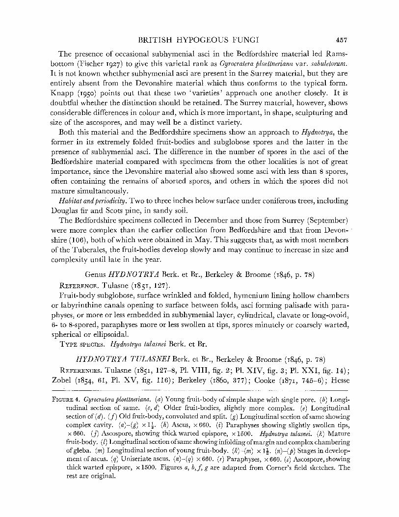
BRITISH HYPOGEOUS FUNGI
The presence of occasional subhymenial asci in the Bedfordshire material led Rams- bottom (Fischer 1927) to give this varietal rank as Gyrocratera ploettneriana var. sabuletorum. It is not known whether subhymenial asci are present in the Surrey material, but they are entirely absent from the Devonshire material which thus conforms to the typical form. Knapp (1950) points out that these two 'varieties' approach one another closely. It is doubtful whether the distinction should be retained. The Surrey material, however, shows considerable differences in colour and, which is more important, in shape, sculpturing and size of the ascospores, and may well be a distinct variety.
Both this material and the Bedfordshire specimens show an approach to Hydnotrya, the former in its extremely folded fruit-bodies and subglobose spores and the latter in the presence of subhymenial asci. The difference in the number of spores in the asci of the Bedfordshire material compared with specimens from the other localities is not of great importance, since the Devonshire material also showed some asci with less than 8 spores, often containing the remains of aborted spores, and others in which the spores did not mature simultaneously.
Habitat and periodicity. Two to three inches below surface under coniferous trees, including Douglas fir and Scots pine, in sandy soil.
The Bedfordshire specimens collected in December and those from Surrey (September) were more complex than the earlier collection from Bedfordshire and that from Devon- shire (106), both of which were obtained in May. This suggests that, as with most members of the Tuberales, the fruit-bodies develop slowly and may continue to increase in size and complexity until late in the year.
Genus HYDNOTRYA Berk. et Br., Berkeley & Broome (i846, p. 78) REFERENCE. Tulasne (I851, 127). Fruit-body subglobose, surface wrinkled and folded, hymenium lining hollow chambers
or labyrinthine canals opening to surface between folds, asci forming palisade with para- physes, or more or less embedded in subhymenial layer, cylindrical, clavate or long-ovoid, 6- to 8-spored, paraphyses more or less swollen at tips, spores minutely or coarsely warted, spherical or ellipsoidal.
TYPE SPECIES. Hydnotrya tulasnei Berk. et Br.
HYDNOTRYA TULASNEI Berk. et Br., Berkeley & Broome (I846, p. 78) REFERENCES. Tulasne (1851, 127-8, P1. VIII, fig. 2; P1. XIV, fig. 3; P1. XXI, fig. 14);
Zobel (I854, 61, P1. XV, fig. 116); Berkeley (i860, 377); Cooke (1871, 745-6); Hesse
FIGURE 4. Gyrocratera ploettneriana. (a) Young fruit-body of simple shape with single pore. (b) Longi- tudinal section of same. (c, d) Older fruit-bodies, slightly more complex. (e) Longitudinal section of (d). (f) Old fruit-body, convoluted and split. (g) Longitudinal section of same showing complex cavity. (a)-(g) x 1. (h) Ascus, x 660. (i) Paraphyses showing slightly swollen tips, x 660. (j) Ascospore, showing thick warted epispore, x 1500. Hydnotrya tulasnei. (k) Mature
fruit-body. (1) Longitudinal section of same showing infolding of margin and complex chambering ofgleba. (m) Longitudinal section of young fruit-body. (k)-(m) x 1. (n)-(p) Stages in develop- ment of ascus. (q) Uniseriate ascus. (n)-(q) x 660. (r) Paraphyses, x 660. (s) Ascospore, showing thick warted epispore, x 1500. Figures a, b,f, g are adapted from Corner's field sketches. The rest are original.
457

LILIAN E. HAWKER ON
(1894, 52-3, P1. XII, fig. 4); Fischer (1897, 26, figs. 1-3 on p. 15); (1927, 113); Massee
(1909, 254, P1. XVII, figs. 3, 6, 14); Knapp (I950, 101-18). Syn. Hydnobolites Tulasnei Berkeley & Broome (I844, 351).
Rhizopogon Tulasnei Zobel (I854, 61, P1. XV, fig. 116). Fruit-body, general macroscopic characters. Irregularly globose, often lobed, surface knobbly
or wrinkled, deeply infolded and fused (figure 4k), minutely downy to scurfy when young, later smooth, waxy, soft but firm, up to 4 cm diam., red-brown to foxy red or dark flesh-coloured, discolouring to blackish red when bruised. Readily discolouring and decomposing when attacked by eelworms. Gleba lighter, cream, pink or russet, according to age, convoluted and chambered (figure 41, m). Odour musty or of mushrooms but becoming unpleasant when decomposed.
Peridium. In young specimens outer peridium consisting of parallel, radially arranged elements, the projecting ends of which give downy effect, soon becoming irregularly dis- torted with thick, coloured walls but still showing tendency to radial arrangement (cf. Gyrocratera), 2 or 3 cells thick, merging with broad inner zone of regular fused elements, closely interwoven and not following any particular direction. The outer layer is con- tinuous with the palisade layer lining the folds of the young gleba and resembles it in
young fruit-bodies. Development of gleba. Becoming increasingly convoluted and chambered with age (figure
41). Young specimens relatively simple and obviously infolded (figure 4m). Paraphyses at first septate, with equal or only slightly swollen tips (figure 4r) not fused, later some extend across canal or fold and become knotted and interwoven with those of opposite side, thus producing closed chambers. Ascogenous hyphae arise in small dense knots at base of palisade. Asci develop from these and push their way into palisade of paraphyses, but are usually rather shorter than the paraphyses, or some develop in reverse direction, extending into subhymenial layer.
Asci. Usually club-shaped with spores arranged irregularly (figure 4 n top) but occasion- ally in old fruit-bodies, cylindrical and uniseriate (figure 4q), probably as a result of pressure due to the expansion of the hymenium, sessile when young, sometimes shortly stalked later, tip blunt, no apical pore, 150-230 x 35-70u, 8-spored.
Ascospores. Spherical 20-28-35# diam. (excluding warts), epispore thickened and irregularly warted, warts 5 to 7,u deep, red-brown (figure 4s).
Distribution, habitat and periodicity. Not uncommon, usually in rather light soil, or sandy soil mixed with humus up to 3 in. below surface. Recorded from number of places in Britain. During present investigation found in Hertfordshire (Panshanger Wood, 156, under oak), Caernarvonshire (Nant Heilyn, 259, under spruce) and Perthshire (Glen Lochay, 419, under beech). This fungus is obviously not associated with any particular species of tree, since European collections are also recorded from under various trees (Fischer I897; Pirk I949). It probably develops a single generation of fruit-bodies each year. These mature in late summer.
Gyrocratera and Hydnotrya are obviously fairly closely related, since the general structure of the fruit-body, the form of the paraphyses and the sculpturing of the spores are very similar. H. tulasnei happens to be a particularly complex and probably advanced species, but certain non-British species such as H. carnea and the North American species H. ellipso-
458
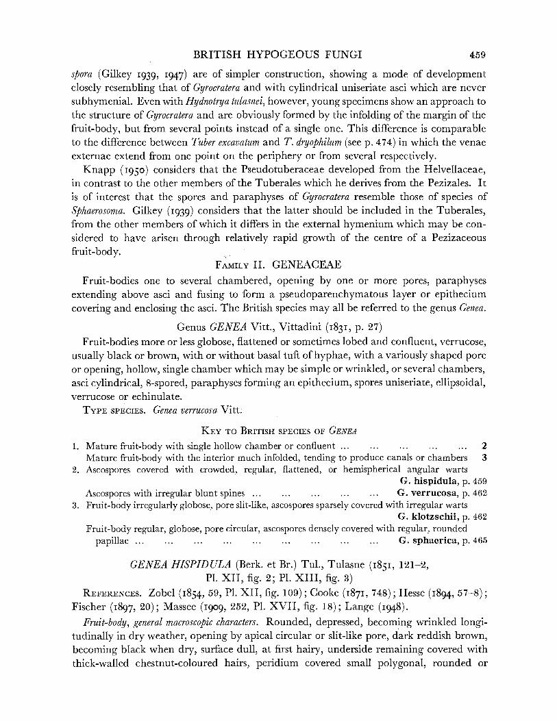
BRITISH HYPOGEOUS FUNGI
spora (Gilkey I939, I947) are of simpler construction, showing a mode of development closely resembling that of Gyrocratera and with cylindrical uniseriate asci which are never subhymenial. Even with Hydnotrya tulasnei, however, young specimens show an approach to the structure of Gyrocratera and are obviously formed by the infolding of the margin of the fruit-body, but from several points instead of a single one. This difference is comparable to the difference between Tuber excavatum and T. dryophilum (see p. 474) in which the venae externae extend from one point on the periphery or from several respectively.
Knapp (I950) considers that the Pseudotuberaceae developed from the Helvellaceae, in contrast to the other members of the Tuberales which he derives from the Pezizales. It is of interest that the spores and paraphyses of Gyrocratera resemble those of species of Sphaerosoma. Gilkey (I939) considers that the latter should be included in the Tuberales, from the other members of which it differs in the external hymenium which may be con- sidered to have arisen through relatively rapid growth of the centre of a Pezizaceous fruit-body.
FAMILY II. GENEACEAE
Fruit-bodies one to several chambered, opening by one or more pores, paraphyses extending above asci and fusing to form a pseudoparenchymatous layer or epithecium covering and enclosing the asci. The British species may all be referred to the genus Genea.
Genus GENEA Vitt., Vittadini (I831, p. 27) Fruit-bodies more or less globose, flattened or sometimes lobed and confluent, verrucose,
usually black or brown, with or without basal tuft of hyphae, with a variously shaped pore or opening, hollow, single chamber which may be simple or wrinkled, or several chambers, asci cylindrical, 8-spored, paraphyses forming an epithecium, spores uniseriate, ellipsoidal, verrucose or echinulate.
TYPE SPECIES. Genea verrucosa Vitt.
KEY TO BRITISH SPECIES OF GENEA
1. Mature fruit-body with single hollow chamber or confluent ... ... ... ... ... 2 Mature fruit-body with the interior much infolded, tending to produce canals or chambers 3
2. Ascospores covered with crowded, regular, flattened, or hemispherical angular warts G. hispidula, p. 459
Ascospores with irregular blunt spines ... ... ...... ... G. verrucosa, p. 462 3. Fruit-body irregularly globose, pore slit-like, ascospores sparsely covered with irregular warts
G. klotzschii, p. 462
Fruit-body regular, globose, pore circular, ascospores densely covered with regular, rounded papillae ... ... ... ... ... ... ... ... ... G. sphaerica, p. 465
GENEA HISPIDULA (Berk. et Br.) Tul., Tulasne (I851, 121-2, P1. XII, fig. 2; P1. XIII, fig. 3)
REFERENCES. Zobel (I854, 59, P1. XII, fig. 109); Cooke (I871, 748); Hesse (I894, 57-8); Fischer (I897, 20); Massee (i909, 252, P1. XVII, fig. 18); Lange (I948).
Fruit-body, general macroscopic characters. Rounded, depressed, becoming wrinkled longi- tudinally in dry weather, opening by apical circular or slit-like pore, dark reddish brown, becoming black when dry, surface dull, at first hairy, underside remaining covered with thick-walled chestnut-coloured hairs, peridium covered small polygonal, rounded or
459

LILIAN E. HAWKER ON
a b
c
g
I
460
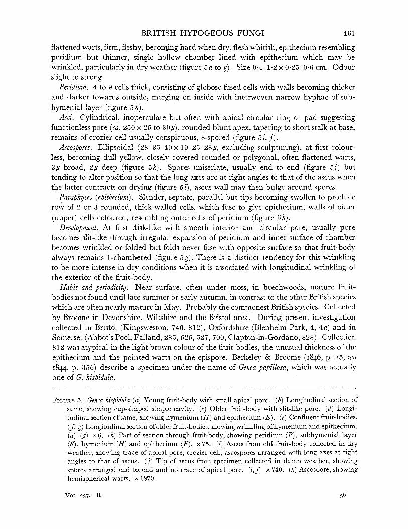
BRITISH HYPOGEOUS FUNGI
flattened warts, firm, fleshy, beconmng hard when dry, flesh whitish, epithecium resembling peridium but thinner, single hollow chamber lined with epithecium which may be wrinkled, particularly in dry weather (figure 5 a to g). Size 0-4-1.2 x 0-25-0-6 cm. Odour slight to strong.
Peridium. 4 to 9 cells thick, consisting of globose fused cells with walls becoming thicker and darker towards outside, merging on inside with interwoven narrow hyphae of sub- hymenial layer (figure 5h).
Asci. Cylindrical, inoperculate but often with apical circular ring or pad suggesting functionless pore (ca. 250 x 25 to 30/,), rounded blunt apex, tapering to short stalk at base, remains of crozier cell usually conspicuous, 8-spored (figure 5 i, j).
Ascospores. Ellipsoidal (28-35-40 x 19-25-28u, excluding sculpturing), at first colour- less, becoming dull yellow, closely covered rounded or polygonal, often flattened warts, 3,u broad, 2,# deep (figure 5k). Spores uniseriate, usually end to end (figure 5j) but tending to alter position so that the long axes are at right angles to that of the ascus when the latter contracts on drying (figure 5i), ascus wall may then bulge around spores.
Paraphyses (epithecium). Slender, septate, parallel but tips becoming swollen to produce row of 2 or 3 rounded, thick-walled cells, which fuse to give epithecium, walls of outer (upper) cells coloured, resembling outer cells of peridium (figure 5h).
Development. At first disk-like with smooth interior and circular pore, usually pore becomes slit-like through irregular expansion of peridium and inner surface of chamber becomes wrinkled or folded but folds never fuse with opposite surface so that fruit-body always remains 1-chambered (figure 5g). There is a distinct tendency for this wrinkling to be more intense in dry conditions when it is associated with longitudinal wrinkling of the exterior of the fruit-body.
Habit and periodicity. Near surface, often under moss, in beechwoods, mature fruit- bodies not found until late summer or early autumn, in contrast to the other British species which are often nearly mature in May. Probably the commonest British species. Collected by Broome in Devonshire, Wiltshire and the Bristol area. During present investigation collected in Bristol (Kingsweston, 746, 812), Oxfordshire (Blenheim Park, 4, 4 a) and in Somerset (Abbot's Pool, Failand, 285, 525, 527, 700, Clapton-in-Gordano, 828). Collection 812 was atypical in the light brown colour of the fruit-bodies, the unusual thickness of the epithecium and the pointed warts on the epispore. Berkeley & Broome (1846, p. 75, not
i844, p. 356) describe a specimen under the name of Genea papillosa, which was actually one of G. hispidula.
FIGURE 5. Genea hispidula (a) Young fruit-body with small apical pore. (b) Longitudinal section of same, showing cup-shaped simple cavity. (c) Older fruit-body with slit-like pore. (d) Longi- tudinal section of same, showing hymenium (H) and epithecium (E). (e) Confluent fruit-bodies. (f, g) Longitudinal section of older fruit-bodies, showing wrinkling of hymenium and epithecium. (a)-(g) x 6. (h) Part of section through fruit-body, showing peridium (P), subhymenial layer (S), hymenium (H) and epithecium (E). x 75. (i) Ascus from old fruit-body collected in dry weather, showing trace of apical pore, crozier cell, ascospores arranged with long axes at right angles to that of ascus. (j) Tip of ascus from specimen collected in damp weather, showing spores arranged end to end and no trace of apical pore. (i, j) x 740. (k) Ascospore, showing hemispherical warts, x 1870.
461
VOL. 237. B. 56
LILIAN E. HAWKER ON
GENEA VERRUCOSA Vitt., Vittadini (i851i 28, P1. II, fig. 7 M-P) REFERENCES. Tulasne (1843, 378); (1851, 119, P1. IV, fig. 1; P1. XII, fig. 3; P1. XIII,
fig. 5); Berkeley (i860, 378); Cooke (1871, 748); Hesse (1894, 55-6); Fischer (1897, 22); Massee (I909, 251); non Klotzsch (I84I, 874).
Syn. G. perlata Zobel (I854, 57, P1. XII, fig. 104). G. kunzeana Zobel (I854, 56, P1. XI, fig. 102).
Fruit-body, general macroscopic characters. Globose, depressed, with single chamber, opening by circular pore, or irregular and confluent and thus with more than one chamber, interior smooth or slightly wrinkled, up to 2 cm diam., attached by basal cushion or stalk to mass of blackish brown hyphae, peridium black, shining, covered small polygonal slab-like warts, flesh greyish white, drying yellowish, epithecium well developed, resembling peridium, outer layers hard, brittle, flesh soft, drying hard (figure 6a). Odour slight to strong and unpleasant.
Peridium. Outer layer (3 to 4 cells deep) of thick-walled cells rather more radially elongated than in Genea hispidula, merging into layer of thin-walled usually tangentially arranged cells, which in turn merges with small-celled subhymenial layer (figure 6b).
Asci. Cylindrical (180-220x25-28,), often slightly constricted between the spores, rounded apex, sometimes with slight indication of functionless pore, tapering only slightly to base, remains of crozier cell not usually visible, 8-spored (figure 6c, d).
Ascospores. Ellipsoidal, varying in size in different collections (21-28-30 x 18-20-23u), hyaline, echinulate, spines blunt, rather regularly arranged, not crowded (figure 6e).
Paraphyses (epithecium). Paraphyses not showing parallel, filiform structure as in G. hispi- dula but fusing at level of apices of asci to form pseudoparenchymatous epithecium, outer layer of cells with thickened dark walls resembling peridium (figure 6b).
Habitat and distribution. Near surface of soil or under moss in mixed woodland, mature fruit-bodies found from May until early autumn at least. Probably not uncommon. Collected by Broome in Wiltshire, Shropshire and the Bristol area. During present investigation found in Caernarvonshire (Vaynol Park, Bangor, 237) and in Somerset (Cleeve, 385). A specimen described by Berkeley & Broome (1844, p. 356, not 1846, p. 75) as G. papillosa was actually G. verrucosa.
GENEA KLOTZSCHII Berk. et Br., Berkeley & Broome (1846, p. 78) REFERENCES. Tulasne (185I, 120, P1. XIII, fig. 4); Zobel, (1854, 57, P1. XI, fig. 101);
Berkeley (I860, 378); Cooke (187I, 748); Hesse (I894, 56-7); Fischer (I897, 23); Massee (1909, 251-2, P1. XVII, fig. 9).
This species was first described by Klotzsch (I84I, p. 874) as Genea verrucosa. Fruit-body, general macroscopic characters. Irregularly globose, or becoming more or less
flattened and angular, irregularly lobed or ribbed (figure 7 a to f), 1- 0 to 1*5 cm diam., seated on short, readily separable fleshy stalk from which brown hairs grow out into soil, black, shining, covered numerous small, more or less pyramidal warts. Apical opening at first more or less circular may remain so but more often becomes sunken in a groove or at meeting of grooves and is then slit-like, triangular or stellate according to degree of ribbing of mature fruit-body, may elongate to as much as one-third of the width of the fruit-body. Interior cavity simple in young specimens, becoming much folded in older specimens
462

BRITISH HYPOGEOUS FUNGI
forming almost, or completely, closed chambers (figure 7g to 1). Flesh white to slate-grey, much thicker and fruit-body harder than in previous two species. Odour said to be unpleasant when ripe, none in young specimens.
0
d~
00
(P), subhymenial layer (S), hymenium (H) and epithecium (E), x 100. (c) Ascus. (d) Tip of ascus, both showing traces of pore formation. (c, d) x 740. (e) Ascospore, showing sparsely echinulate epispore, x 1870.
56-2
463

b
LILIAN E. HAWKER ON
a
cd
f e
9 . I 1
h
m
'6
00
0 OQ
0
iv
0
j
I
k
FIGURE 7. Genea klotzschii. (a)-(f) Fruit-bodies of various degrees of complexity. (g)-(l)Longi- tudinal section of these, showing increasing folding of hymenium and epithecium and decreasing size of cavities. (a)-(l) x 2?. (m)-(p) Stages in development of ascus, showing persistent crozier cell, irregular development of spores and polar development of warts on the spores, x 750. (q) Mature ascospore, showing irregular angular warts, x 1870.
464

BRITISH HYPOGEOUS FUNGI
Peridium. Thicker than in G. hispidula or G. verrucosa but similarly constructed. Asci. At first club-shaped, then cylindrical (270-320 x 35-42,c), apex rounded, no trace
of a pore, walls constricted between spores, tapering sharply to base with remains of square-ended hook cell clearly visible, very occasionally subhymenial, usually 8-spored but occasionally 6- or 7-spored from abortion of developing spores, usually uniseriate but occasionally one or two spores may be out of line (figure 7 m to p). Spores sometimes not formed simultaneously (figure 7 o).
Ascospores. At first spherical, thin-walled, smooth, hyaline and aguttulate, becoming ellipsoidal, 25-34-45 x 20-27-32u (excluding warts), irregularly and sparsely warted with block-like or pyramidal warts, up to 4#u deep, 2-guttulate (guttules 8 to 9,u diam.) or less often 1-, 3- or more than 3-guttulate, colourless. Warts form first at poles of spore later developing over rest of surface (figure 7 q).
Paraphyses (epithecium). Forming well-marked epithecial layer, thicker than in G. hispidula or G. verrucosa.
Habitat andperiodicity. Up to 4 in. deep in soil or even, as at Savernake, in clay, position indicated by presence of flocculent white mycelium on surface of soil which is revealed when leaf litter and loose humus are scraped away. This feature was noted by Berkeley & Broome (I846) in their original collection at Stapleton, Bristol, and seen in collections made at Savernake Forest, Wiltshire (359, 365), and near Cleeve, Somerset (383), during the present investigation. Under beech, maturing as early as May. Probably not uncommon.
A single specimen collected in June 1952 at Mickleham, Surrey (613), differed from the type in having a bright orange peridium, which rapidly became black. Immature speci- mens from Savernake were black when collected.
GENEA SPHAERICA Tul., Tulasne (1843, p. 378) REFERENCES. Tulasne (1851, 120-1, P1. IV, fig. 2; P1. XII, fig. 1; P1. XIII, fig. 6);
Hesse (1894, 54-5, P1. XII, fig. 9; P1. XVI, fig. 32); Fischer (1897, 24); Ramsbottom (1923, 197).
Fruit-body, general macroscopic characters. Spherical, usually not lobed or ribbed, diam. ca. 1 cm, with short stalk arising from prominent tuft of hairs (figure 8 a); peridium black, covered with small, polygonal, flattened warts, duller in appearance than Genea klotzschii, apical pore small and circular. Interior of fruit-body much folded, cavity nearly filled (figure 8b, c). Flesh pallid, greyish white. Odour, none.
Peridium. Thick, construction similar to that of other species. Asci. Clavate, becoming cylindrical (190-220x28-35u), with 8 uniseriate spores, or
remaining clavate, when spores tend to become biseriate, apex bluntly rounded, no trace of pore, tapering to base in lower third of the ascus, remains of hook cell prominent, square-ended.
Ascospores. Broadly ellipsoidal, colourless, size varying with collection and age, 25-30-35 x 19-24-28U (excluding warts), covered with shining, hemispherical, bead-like warts of radius 2-3,c (figure 8 h, i).
Paraphyses (epithecium). Septate, cylindrical, 1-5 to 2-0u thick, fusing above asci to give pseudoparenchyma as in other species.
465

LILIAN E. HAWKER ON
b c
2-1
h.
466

BRITISH HYPOGEOUS FUNGI
Habitat and periodicity. Near surface of soil, under beech in early summer. Collected by Broome in Leigh Woods, Bristol, and found during present investigation in several beech- woods on the Cotswold Hills, near Wotton-under-Edge (389, 393, 726) and Dursley (498), Gloucestershire. Probably less common than the other British species.
These four species show a progressive increase in complexity. Thus in G. hispidula and G. verrucosa the chamber of the fruit-body is comparatively simple and the asci are cylindrical, contain 8 uniseriate spores and show an indication of a vestigial apical pore. In G. verrucosa the epithecium is more fully developed than in G. hispidula but less so than in the other two species. G. klotzschii and G. sphaerica have more complex, fleshy fruit- bodies with a more pronounced thickening and warting of the epithecium and a tendency to irregular development of spores, to abortion of one or more spores in the ascus, to a biseriate arrangement and occasionally to the production of subhymenial asci, while the asci have no trace of an apical pore. Some specimens of G. sphaerica approach Pachyphloeus in the solidity of the fruit-body.
FAMILY III. EUTUBERACEAE Fruit-bodies veined, veins (venae externae) branching from one or more points on the
peridium, in mature specimens veins usually filled by secondary growth of paraphyses. Asci cylindrical, clavate or subglobose, 8-spored or less.
The British species may be referred to the genera Stephensia, Pachyphloeus, Tuber and Balsamia.
Genus STEPHENSIA Tul., Tulasne (1845, p. 1433) Fruit-body subglobose or flattened, peridium felted, single glebal cavity opening by
basal pore, hymenium closely folded, paraphyses not fused, asci narrow, cylindrical, 8-spored, spores spherical, smooth.
TYPE SPECIES. Stephensia bombycina (Vitt.) Tul.
STEPHENSIA BOMBYCINA (Vitt.) Tul., Tulasne (i845, P. 1433) REFERENCES. Tulasne (I85I, 130, P1. XII, fig. 4); Berkeley (I860, 377); Cooke (I871,
745); Fischer (I897, 29-30, figs. 1-4 on p. 16); Massee (I909, 254-5, P1. XVII, fig. 12). Syn. Genea bombycina Vittadini (1831, 29, P1. III, fig. 13; P1. IV, fig. 8); Berkeley &
Broome (i844, 357). Fruit-body, general macroscopic characters. More or less globose, often flattened, lobed or
longitudinally wrinkled, with basal pore or opening, size of fruit-body up to 2 cm, light brown or dingy yellow brown, felted with short hairs, gleba cream with deeper cream or straw-coloured veins (figure 9 a, b, c). Fleshy but firm. Odour strong and unpleasant, of drains or overripe Camembert cheese.
Peridium. Consists of narrow outer brown-coloured layer (ca. 100# thick) and wider cream layer (ca. 700,u thick), hyphal elements arranged more or less parallel to one another radiating towards outside (2 to 6, diam.), in outer coloured zone cells inflated, walls
FIGURE 8. Genea spherica. (a) Fruit-body, showing regular shape, apical pore and basal mycelial tuft. (b)-(c) Longitudinal section of fruit-bodies, showing labyrinthine cavity. (a)-(c) x 21. (d)-(f) Stages in development of asci, showing persistent crozier cell and in (e) tendency to arrangement of spores in more than one row. (g) Paraphyses. (d)-(g) x 660. (A) Immature ascospore, showing large oil globule. (i) Mature ascospore, showing hemispherical warts. (h)-(i) x 1500.
467
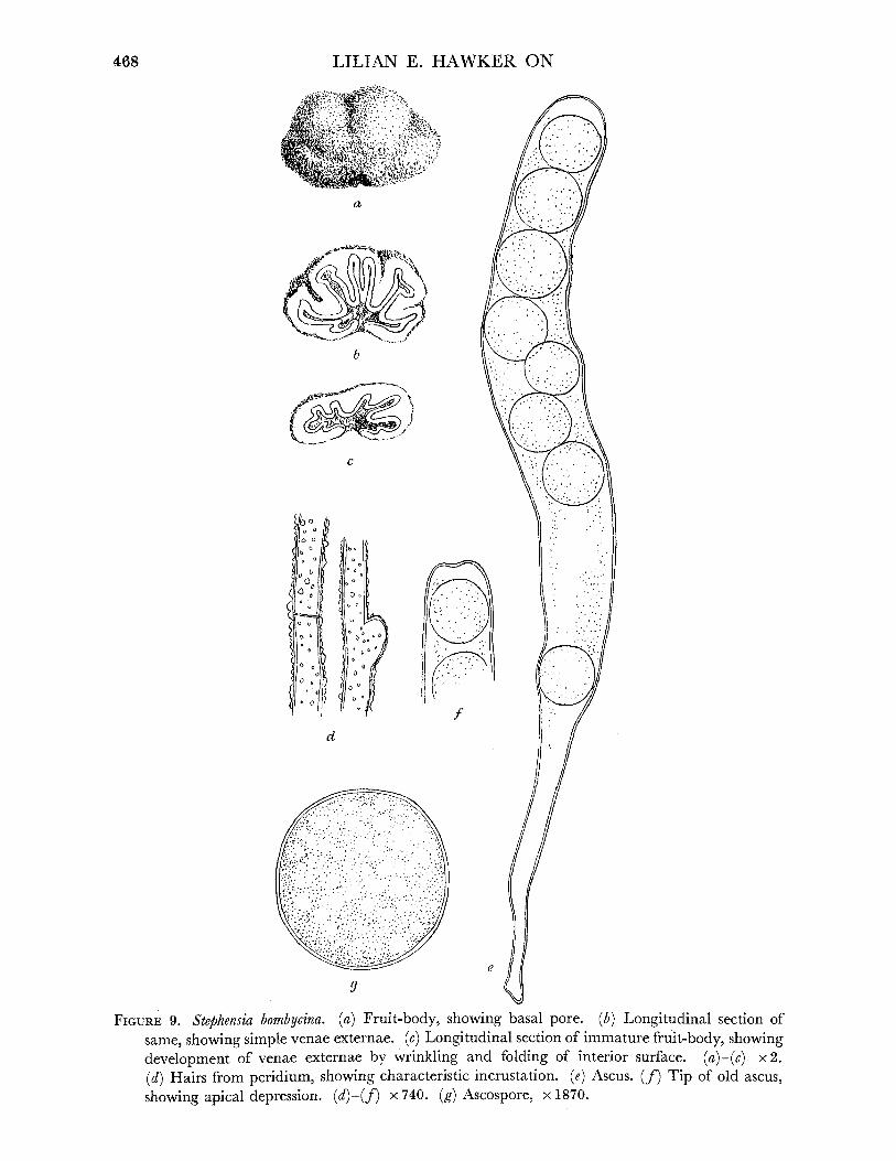
468 LILIAN E. HAWKER ON
a
b
C
' c I
c
0 0 '0
0 0
11
0 I )
6 / 0 0iI
/ 0
d
e
g
FIGURE 9. Stephensia bombycina. (a) Fruit-body, showing basal pore. (b) Longitudinal section of same, showing simple venae externae. (c) Longitudinal section of immature fruit-body, showing development of venae externae by wrinkling and folding of interior surface. (a)-(c) x 2.
(d) Hairs from peridium, showing characteristic incrustation. (e) Ascus. (f) Tip of old ascus, showing apical depression. (d)-(f) x 740. (g) Ascospore, x 1870.

BRITISH HYPOGEOUS FUNGI
thick and coloured, hyphae terminating in hairs which derive colour from curious in- crustation or sculpturing of wall (figure 9 d). In young specimens these hairs may be one or more millimetres long, in older specimens they tend to be broken or rubbed off.
Gleba. Trama consisting of loosely interwoven hyphae, becoming more densely inter- woven in subhymenial layer. Hymenium regular palisade of asci and paraphyses lining venae externae, paraphyses usually extending beyond asci but seldom entirely filling the cavities between venae externae.
Asci. Cylindrical, 180-270 x 20-30,u rounded apices, occasionally showing an apical depression, but no definite indication of a vestigial pore similar to that in Genea hispidula, tapering gradually in lower half to long narrow stalk, 8-spored, uniseriate (figure 9e,f).
Ascospores. Spherical, 19-21-26,u, colourless, thin-walled (figure 9g). Paraphyses. Narrow, 2-3# diam., septate, colourless. Habitat and Distribution. Under coniferous trees, in undisturbed soil, at or near surface,
often partially embedded in clay or at edge of path or under moss. August to October. Reported from Devonshire, Dorset, Wiltshire and the Bristol area. Recently found at Stoke
Bishop, Bristol (481, 482, 563), at or near the site of one of Broome's collections (Broome I874-6), in Somerset (Burrington 777, 789) and Gloucestershire Wotton-under-Edge 931.)
Genus PACHYPHLOEUS Tul., Tulasne (I844, p. 60)
Fruit-body subglobose, usually with short stalk and basal mycelial tuft, surface irregularly warted or bluntly spiny, interior compact, folded, venae externae arising from apical pore or from several points on periphery, hymenium irregular, asci clavate or almost cylindrical with a bulbous base (except in one North American species attributed to this genus, Pachyphloeus virescens Gilkey, in which the asci are said to be globose), 8 spores, irregularly arranged, spores spherical, bluntly or acutely spiny.
TYPE SPECIES. P. melanoxanthus Tul.
KEY TO BRITISH SPECIES OF PACHrPHLOEUS
1. Spores with pointed spines ... ... ... ... ... P. melanoxanthus, p. 469. Spores with blunt spines or warts ... ... ... ... ... ... ... ... 2
2. Fruit-body at first yellow then brown to black, spores with short blunt spines P. citrinus, p. 471.
Fruit-body purple black, spores with short blunt warts ... P. conglomeratus, p. 471.
PACHYPHLOEUS MELANOXANTHUS Tul., Tulasne (1844, p. 69) REFERENCES. Berkeley & Broome (I846, 79); Tulasne (I85I, 1P31-2, P1. IV, fig. 6;
P1. XIV, fig. 4); Zobel (1854, 63); Berkeley (i86o, 377); Cooke (i871, 743); Hesse (I894, 39-41, P1. XII, fig. 8; P1. XV, fig. 10; P1. XVI, figs. 20, 25); Fischer (1897, 31, figs. 1-4 on p. 17); Massee (i909, 255, P1. XVII, fig. 11).
Syn. Choiromyces melanoxanthum, C. viridis, Tuber melanoxanthus Berkeley & Broome (I844, 359). Rhizopogon melanoxanthus Zobel (I854, P1. IV, fig. 113).
Fruit-body, general macroscopic characters. More or less globose, sometimes shortly stalked, with an apical or lateral opening, size of a hazel nut, at first yellowish green, becoming black, covered polygonal warts, gleba olive-yellow or dusky, becoming black when dry, with few veins. Odour slight, not unpleasant,
469
VOL 237. B. 57

LILIAN E. HAWKER ON
Peridium. Pseudoparenchymatous with thin reddish or violet-brown cell walls. Asci. Clavate 80-150 x 25-45u, 8-spored, spores irregularly arranged. Ascospores. Spherical, 13 to 17u diam., hyaline to light yellow-green, rather densely
covered with spines, about 3,# long (figure 10 g). No fresh material of this species seen during this investigation. Broome's material was
obtained at Chudleigh (Devonshire), Bowood Park (Wiltshire) and in the Bristol area (in districts now built over or otherwise altered).
b
f
C
(C 00 0% ~ CC C)ooO%1
(_"C' ~~~-/ i
e
9 d
FIGURE 10. Pachyphloeus conglomeratus. (a) Fruit-body, showing grooves and basal stalk. (b) Longi- tudinal section of same, showing sparsely branched venae externae. (a)-(b) x 2. (c)-(e) Stages in development of ascus, showing bulbous base and irregularly arranged ascospores, x 740. (f) Ascospore, showing small blunt regularly arranged warts. P. melanoxanthus. (g) Ascospore, showing numerous slender spines. P. citrinus. (h) Ascospore, showing small, blunt, irregularly arranged warts. (f)-(h) x 1870.
470
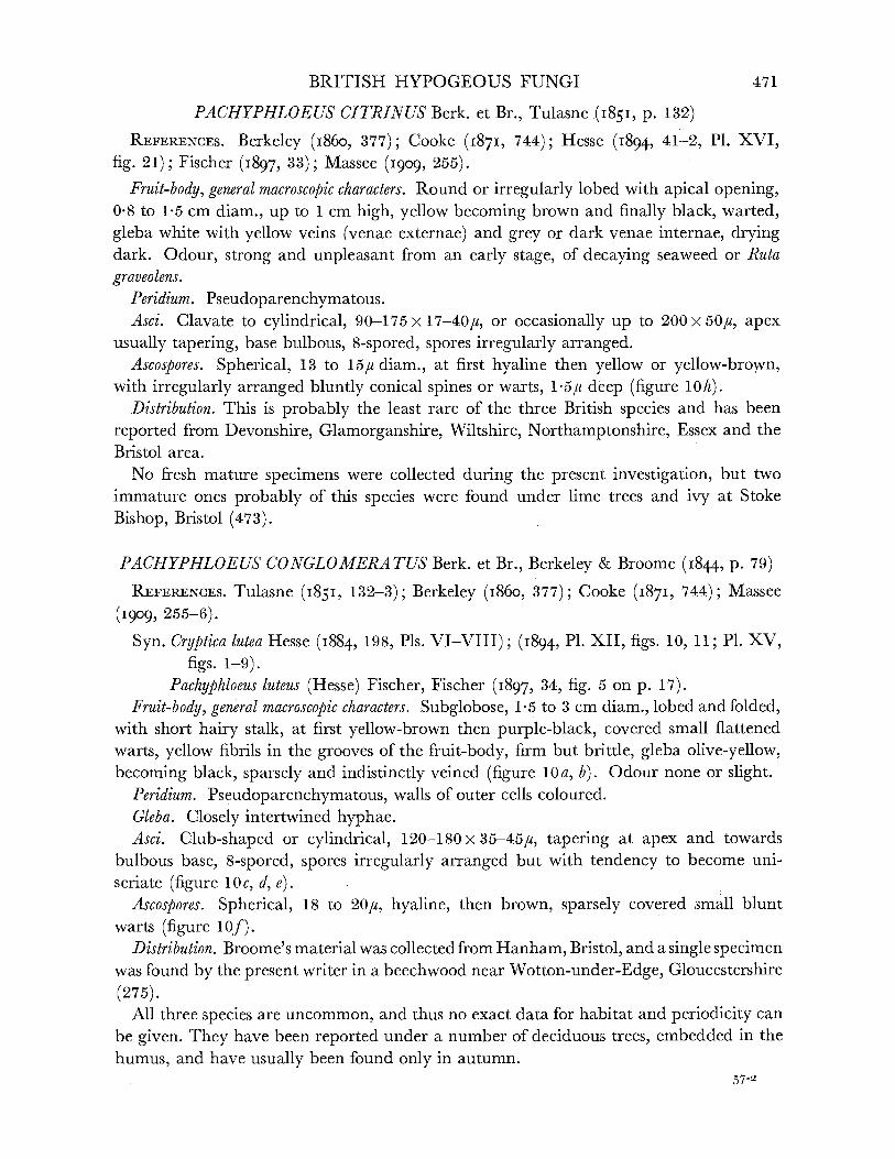
BRITISH HYPOGEOUS FUNGI
PACHYPHLOEUS CITRINUS Berk. et Br., Tulasne (1851, p. 132)
REFERENCES. Berkeley (i86o, 377); Cooke (i871, 744); Hesse (I894, 41-2, P1. XVI,
fig. 21); Fischer (I897, 33); Massee (1909, 255).
Fruit-body, general macroscopic characters. Round or irregularly lobed with apical opening, 0*8 to 1-5 cm diam., up to 1 cm high, yellow becoming brown and finally black, warted, gleba white with yellow veins (venae externae) and grey or dark venae internae, drying dark. Odour, strong and unpleasant from an early stage, of decaying seaweed or Ruta graveolens.
Peridium. Pseudoparenchymatous. Asci. Clavate to cylindrical, 90-175x17-40/,, or occasionally up to 200x50u, apex
usually tapering, base bulbous, 8-spored, spores irregularly arranged. Ascospores. Spherical, 13 to 15# diam., at first hyaline then yellow or yellow-brown,
with irregularly arranged bluntly conical spines or warts, 15#u deep (figure 10h). Distribution. This is probably the least rare of the three British species and has been
reported from Devonshire, Glamorganshire, Wiltshire, Northamptonshire, Essex and the Bristol area.
No fresh mature specimens were collected during the present investigation, but two immature ones probably of this species were found under lime trees and ivy at Stoke Bishop, Bristol (473).
PACHYPHLOEUS CONGLOMERATUS Berk. et Br., Berkeley & Broome (1844, p. 79)
REFERENCES. Tulasne (1851, 132-3); Berkeley (1860, 377); Cooke (1871, 744); Massee
(I909, 255-6).
Syn. Cryptica lutea Hesse (I884, 198, Pls. VI-VIII); (I894, P1. XII, figs. 10, 11; P1. XV, figs. 1-9).
Pachyphloeus luteus (Hesse) Fischer, Fischer (i897, 34, fig. 5 on p. 17). Fruit-body, general macroscopic characters. Subglobose, 1-5 to 3 cm diam., lobed and folded,
with short hairy stalk, at first yellow-brown then purple-black, covered small flattened warts, yellow fibrils in the grooves of the fruit-body, firm but brittle, gleba olive-yellow, becoming black, sparsely and indistinctly veined (figure 10a, b). Odour none or slight.
Peridium. Pseudoparenchymatous, walls of outer cells coloured. Gleba. Closely intertwined hyphae. Asci. Club-shaped or cylindrical, 120-180x35-45u, tapering at apex and towards
bulbous base, 8-spored, spores irregularly arranged but with tendency to become uni- seriate (figure 10c, d, e).
Ascospores. Spherical, 18 to 20u, hyaline, then brown, sparsely covered small blunt warts (figure 10f).
Distribution. Broome's material was collected from Hanham, Bristol, and a single specimen was found by the present writer in a beechwood near Wotton-under-Edge, Gloucestershire
(275). All three species are uncommon, and thus no exact data for habitat and periodicity can
be given. They have been reported under a number of deciduous trees, embedded in the humus, and have usually been found only in autumn.
57-2
471
e
f
h 9
I i L 9
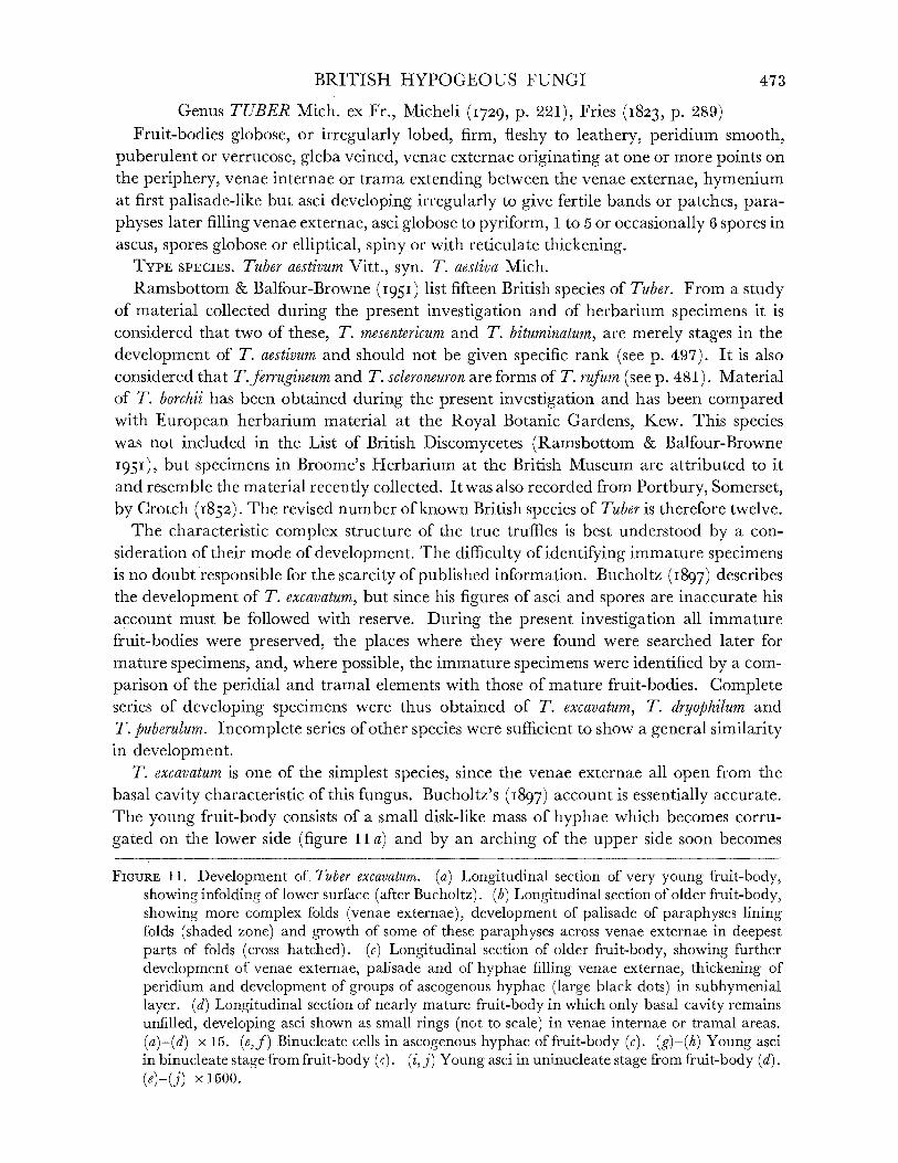
BRITISH HYPOGEOUS FUNGI
Genus TUBER Mich. ex Fr., Micheli (1729, p. 221), Fries (I823, p. 289) Fruit-bodies globose, or irregularly lobed, firm, fleshy to leathery, peridium smooth,
puberulent or verrucose, gleba veined, venae externae originating at one or more points on the periphery, venae internae or trama extending between the venae externae, hymenium at first palisade-like but asci developing irregularly to give fertile bands or patches, para- physes later filling venae externae, asci globose to pyriform, 1 to 5 or occasionally 6 spores in ascus, spores globose or elliptical, spiny or with reticulate thickening.
TYPE SPECIES. Tuber aestivum Vitt., syn. T. aestiva Mich.
Ramsbottom & Balfour-Browne (I95I) list fifteen British species of Tuber. From a study of material collected during the present investigation and of herbarium specimens it is considered that two of these, T. mesentericum and T. bituminatum, are merely stages in the development of T. aestivum and should not be given specific rank (see p. 497). It is also considered that T.ferrugineum and T. scleroneuron are forms of T. rufum (see p. 481). Material of T. borchii has been obtained during the present investigation and has been compared with European herbarium material at the Royal Botanic Gardens, Kew. This species was not included in the List of British Discomycetes (Ramsbottom & Balfour-Browne
1951), but specimens in Broome's Herbarium at the British Museum are attributed to it and resemble the material recently collected. It was also recorded from Portbury, Somerset, by Crotch (i852). The revised number of known British species of Tuber is therefore twelve.
The characteristic complex structure of the true truffles is best understood by a con- sideration of their mode of development. The difficulty of identifying immature specimens is no doubt responsible for the scarcity of published information. Bucholtz (1897) describes the development of T. excavatum, but since his figures of asci and spores are inaccurate his account must be followed with reserve. During the present investigation all immature fruit-bodies were preserved, the places where they were found were searched later for mature specimens, and, where possible, the immature specimens were identified by a com- parison of the peridial and tramal elements with those of mature fruit-bodies. Complete series of developing specimens were thus obtained of T. excavatum, T. dryophilum and T. puberulum. Incomplete series of other species were sufficient to show a general similarity in development.
T. excavatum is one of the simplest species, since the venae externae all open from the basal cavity characteristic of this fungus. Bucholtz's (1897) account is essentially accurate. The young fruit-body consists of a small disk-like mass of hyphae which becomes corru-
gated on the lower side (figure 11 a) and by an arching of the upper side soon becomes
FIGURE 11. Development of Tuber excavatum. (a) Longitudinal section of very young fruit-body, showing infolding of lower surface (after Bucholtz). (b) Longitudinal section of older fruit-body, showing more complex folds (venae externae), development of palisade of paraphyses lining folds (shaded zone) and growth of some of these paraphyses across venae externae in deepest parts of folds (cross hatched). (c) Longitudinal section of older fruit-body, showing further development of venae externae, palisade and of hyphae filling venae externae, thickening of peridium and development of groups of ascogenous hyphae (large black dots) in subhymenial layer. (d) Longitudinal section of nearly mature fruit-body in which only basal cavity remains unfilled, developing asci shown as small rings (not to scale) in venae internae or tramal areas. (a)-(d) x 15. (e,f) Binucleate cells in ascogenous hyphae of fruit-body (c). (g)-(h) Young asci in binucleate stage from fruit-body (<). (i, j) Young asci in uninucleate stage from fruit-body (d). (e)-(j) x 1500.
473
LILIAN E. HAWKER ON
more or less spherical, with a large basal opening giving access to a hollow chamber, into which the corrugations of the inner surface extend (figure 11 b). At this stage the fruit-body is essentially similar in structure to that of a young specimen of Stephensia bombycina. The inner surface soon becomes more complex so that the cavity is reduced in relative size and becomes a mass of branched channels, the so-called 'venae externae'. A regular palisade- like layer of narrow septate paraphyses develops over the surface of the much wrinkled trama or 'venae internae' (figure 11 c). Beneath this palisade binucleate cells arise at a number of points (figure 1l1 e,f). There is some evidence that these are produced by the fusion of two tramal cells, but this could not be determined with certainty. From these binucleate cells small knots of deeply staining ascogenous hyphae develop and rapidly extend through the trama and subhymenium, finally producing masses of short-stalked, globose or ellipsoidal asci, which are quite irregularly arranged. In mature fruit-bodies the trama is almost completely replaced by the ascus masses. Meanwhile the paraphyses grow out to form a loose weft of hyphae filling the venae externae and extending into the basal cavity (figure 11 d). This spongy filling which contains numerous air spaces is responsible for the contrast in appearance between the venae externae and the more compact venae internae or tramal plates.
Tuber dryophilum and T. puberulum are of more complex structure than T. excavatum, since the venae externae develop from more than one point on the periphery. In some specimens of T. puberulum the majority of the venae externae converge towards the base of the fruit-
body, but in T. dryophilum there is no such distinction between those developing from the base and those from other parts of those from peripothery. Immature specimens of these two species are more or less spherical masses of hyphae. The surface develops irregularly so that cavities are produced by the infolding of the margin at various points (figure 12 a). The lining of these cavities becomes wrinkled in a complex manner, and the wrinkles rapidly become so crowded that the opposite surfaces touch one another and the cavities become almost completely filled. Paraphyses grow out as in T. excavatum and interlock to complete the filling up of the original cavities and to bridge the openings where the venae externae reach the surface (figure 12 b) The asci develop from surface (figure 12b). The asci develop from subhymenial knots of ascogenous hyphae and are irregularly arranged as in T. excavatum.
The nuclei of the ascogenous hyphae and of the young asci are small and nuclear details have not been observed with certainty. Binucleate cells have been seen in young ascogenous hyphae of T. excavatum (figure 11 e, f), and young asci have also been seen in the binucleate condition (figure 11 g, h). The asci soon become uninucleate (figure ll i,j) and remain in this condition until the fruit-bodies reach almost full size. The fusion nucleus becomes much inflated and, in the resting condition, is surrounded by a distinct membrane. The chromatin is seen as small irregular pieces embedded in the layer immediately within the membrane. The central part of the nucleus is clear and contains a single large, deeply staining nucleolus (figure 15f).
Greis (1939) in a study of ascus development in T. aestivum and T. brumale claims that the division of the nucleus follows a division of chromatin material and the production of a spindle within the nuclear membrane, and that the nucleolus is extruded before this
segregation of chromatin takes place. No confirmation of either of these suggestions has been obtained in the present investigation. In stained microtome sections of fixed material
474

BRITISH HYPOGEOUS FUNGI
the nucleolus was occasionally seen outside the nucleus, the membrane of which appeared to be damaged. This was not seen in sections of fresh material stained with aceto-carmine. It is likely that the membrane of the inflated nucleus is readily damaged during micro- toming. No definite division stages have been observed even in material fixed in the field immediately after collection. It is concluded that the actual nuclear divisions take place very rapidly and probably occur at a definite time of the day and are thus easily missed.
b
FIGURE 12. (a) Longitudinal section of young undifferentiated fruit-body of Tuber dryophilum with margin infolding at two places in plane of section. (b) Longitudinal section of older fruit-body of T. dryophilum, showing developing venae externae, lined with palisade of paraphyses (shaded zone) and entirely filled with loose hyphae (cross-hatched areas) which fan out at surface of fruit-body and fuse with peridium thus completely closing the venae externae. Contrast with young fruit-body of T. excavatum (figure 11 b) at approximately same stage of development (i.e. before the development of ascogenous hyphae) in which the venae externae are not filled. (a, b) x 20. (c)-(g) Types of venation of fruit-bodies of Tuber spp. (after Malen9on 1938). (c) = Malen.on's type 1 in which veins converge to a snall basal cushion or cavity. (d) = Type 2, in which the main venation is as in type 1, but a few subsidiary veins open to other points on the surface. (e) = Type 3, in which veins converge to larger basal area. (f) Type 4, in which main venation is as in type 3, but a few subsidiary veins open to other points on the surface. (g) = Type 5, in which there is no convergence of veins to the base, but numerous systems opening to irregularly distributed points on the surface.
475
LILIAN E. HAWKER ON
Asci containing two or four nuclei are frequently present, and it is of interest that these nuclei are often grouped together in the centre of the ascus (figure 15h). In view of the fact that germination of the ascospore and the subsequent development of a mycelium has never been observed with certainty,* it is not impossible that the nuclear divisions are abnormal. The unusual features described by Greis or an irregular fission of the nuclear material might then occur. The nuclei are seen to abort at any stage, giving the irregular number of spores characteristic of the genus. It is possible that some of the spores are genetically abnormal and non-viable as a result of the loss of function due to the hypogeous habit.
The spores develop by the aggregation of cytoplasm around the nuclei and the secretion of a wall. The remaining cytoplasm of the ascus is used up in the formation of the intricately sculptured spore walls. As might be expected, the size of the spores is inversely correlated with the number in the ascus.
All species of Tuber show considerable changes in shape, colour and other characters during development. Variation within the species, due partly to differences in environ- mental factors, is often considerable. It is thus difficult to formulate descriptions that will permit the ready identification of specimens at all stages of development. In fact no one character is sufficient to separate species, and a number of such characters as presence or absence of warts on the peridium, colour, texture, venation of gleba,t shape of asci, number of spores in the ascus and size, shape and sculpturing of the spores must be con- sidered. For this reason the key given below is supplemented by a table of characters
(table 2). It is certain that the confusion which still exists in the classification of this genus is partly a result of different specific names having been given to the same species according to its age or to its response to environmental variations.
Several attempts to divide the genus into subgenera have been made. Thus Vittadini
(1831) divides the genus into Tubera spuria and T. genuina (Eu-Tuber), and Wallroth (I833) separates off the subgenus Aschion (corresponding to Tubera spuria) to include all those, such as Tuber excavatum, which are of hard or leathery texture. The most satisfactory method is the recent one of Knapp (I950), who divides the genus into a number of groups. Most of these groups are fairly natural ones and this arrangement will be followed here, the groups being placed in approximate order of complexity. Knapp's groups are as follows, although he does not place them in this order: (1) Rufum group, p. 478, including the British species Tuber rufum, T. nitidum and the doubtful species T. ferrugineum and T. scleroneuron, characterized by a more regular arrangement of the asci, the stalked, clavate asci and the spiny ellipsoidal spores. (2) Excavatum group, p. 482, including a single British species, Tuber excavatum, and characterized by tough texture, venae externae con- verging to a definite basal cavity and the coarsely reticulate spores. (3) Puberulum group A, p. 485, including the British species Tuber puberulum, T. rapaeodorum, T. borchii and T. macu- latum, characterized by the soft texture and globose to shortly ellipsoidal spores with deep,
* Boulanger (1903) claimed to have observed the splitting of the exospore in T. melanosporum, but no mycelium developed.
t Malenqon (1938) distinguishes five types of venation (figure 12c to g), according to whether the veins converge to a small area at the base or to a larger basal area, with or without subsidiary convergences to other points on the periphery or whether there is complete absence of basal convergence. These arrange- ments are of some use in distinguishing species, but it is not easy to distinguish types 1, 2, 3 and 4 from each other and more than one type may occur in a single collection of the same species.
476
TABLE 2
species T. rufum
T. nitidum
T. excavatum
T. puberulum
T. rapaeodorum
T. maculatum
T. borchii
T. dryophilum
T. foetidum
size of mature
fruit-body (mm) 10-15
shape, colour, surface, texture
irregularly globose, foxy-red, minutely warted, firm to hard
ca. 10 globose, golden brown, shining, firm to hard
colour of gleba
white-+ lilac-> red-brown
white - reddish grey
10-30 irregular with basal white -> cavity, dull ochre- ochre -+
brown, rough, leathery purple-brow: 5-17 irregularly globose,
white -- dingy -* mole-grey, persistently puberulent, soft
10-15 globose, dingy-> straw -+ mottled, smooth, firm
5-20 irregular, white, or more translucent --mottled,
variable, waxy, brittle
5-20 globose or lobed, dull or more yellow-brown to
chocolate, smooth, firm
5-20 irregular, dull ochraceous to choco- late, smooth, firm
ca. 10 globose, foxy-red, minutely warted, firm
T. aestivum , 30-90 irregularly warted, black, covered large warts, hard
20-50 globose or lobed, brown to black, small warts, fleshy
ca. 20 globose, purple-black, covered large warts, firm to hard
white -+lilac- grey -+
purple- black
white -+ grey -+
purple- black
white - clay- coloured blackish-red
white pink -> chocolate
white - pink- purple-browi
white -, pink-+ purple- brown
white -> yellow- grey
white -> purple- black
white -+
bluish grey
veins
numerous, type 3 or 4, dingy white or reddish
rather few, type 1, chalk white
few, type 1, cream
n numerous, type 2
or 3, white, distinct
numerous, type 4, chalk white or cream, distinct
few, type 5, chalk white
few, type 1 or 2, white, later reddish
numerous, type 5, dingy white
n
fairly numerous, type 4 or 5, white, later brown
numerous, type 5, white to grey
numerous, type 5, white, later brown, distinct
fairly numerous, type 1 or 2, white, indistinct
I
mature asci ovoid, stalked
ovoid, stalked
globose, sessile
large globose, sessile
ovoid to pyriform, sessile or shortly stalked
ovoid sessile
ovoid to clavate, short-stalked with bulbous base
globose, sessile
globose sessile
globose, sessile
ellipsoidal, often shortly stalked
ellipsoidal, sessile
most usual average no. of size of spores spore
in ascus (it) 4 20x17*
to 39x 24
4 24x17 to
40 x 24 2-4 31x23
to 51 x 31
1 33x31 to
43 x 40
2-3 31 x23 to
43 x 36
1-3 31 x 27 to
36x31
2 30 x 20 to
43 x 29
4 33 x 26 to
51 x 32
1-3 29 x 19 to
40 x 26
4 24x18 to
35 x 24
2 Large, up to 80 x 45 av. 60 x 35
4 29 x 20
colour and sculpturing
of mature spore pale brown, numerous
short spines
pale brown, numerous slender spines
dark red-brown, irregularly reticulate, deep with wide mesh
dark red-brown, regularly reticulate, deep with small mesh
dark red-brown, variable, reticulate, usually regular and small-meshed, fragile
yellow-brown, regularly reticulate, deep, rather small- meshed
red-brown, regularly or irregularly reticu- late, shallow, mesh variable in size
pale golden brown, deeply reticulate, mesh wide and irregular
dark brown, irregularly reticulate, shallow, mesh small to medium
yellow-brown, irregu- larly reticulate, depth and mesh variable, fragile
dark brown, reticu- late, shallow, mesh small but irregular
purple-brown, numerous pointed spines up to 4/, long
* According to number in ascus.
0 r
H 1-1 C/O)
o
0
0
V2
T. macro- o sporurn
T. brumale

LILIAN E. HAWKER ON
usually small-meshed, regularly reticulate sculpturing. (4) Puberulum group B, p. 493, including the British species Tuber dryophilum and T. foetidum and differing from Puberulum group A in the long-ellipsoidal, irregularly wide-meshed reticulate spores, and (5) Aestivum group, p. 495, including the British species T. aestivum (including T. mesentericum and T. bituminatum), T. macrosporum and T. brumale and distinguished by the coarsely verrucose
peridium. The members of this group show considerable differences in spore characters. It is not considered to be a natural group but is retained for convenience in identification.
KEY TO BRITISH SPECIES OF TUBER
1. Peridium smooth, scurfy or puberulent, light coloured, yellow, brown or reddish brown, never entirely black ... ... ... ... ... ... ... ... ... ... 2
Peridium coarsely verrucose, black, purplish black or brown-black ... ... ... 10 2. Spores spiny ... ... ... ... ... ... . .. ... .. ... 3
Spores reticulate ... ... ... ... ... ... ... ... ... ... ... 4 3. Fruit-body smooth, shining, yellow-brown, gleba cream ... ... T. nitidum, p. 479
Fruit-body dull, smooth or scurfy, red-brown ... ... ... ... T. rufum, p. 478 4. Fruit-body with conspicuous basal cavity ... ... ... ... T. excavatum, p. 482
Fruit-body without conspicuous basal cavity ... ... ... ... ... ... ... 5 5. Fruit-body puberulent throughout development ... ... T. puberulum, p. 485
Fruit-body smooth or scurfy or puberulent only when young ... ... ... ... 6 6. Fruit-body waxy, lobed, at first white, becoming irregularly stained yellow T. maculatum, p. 487
Fruit-body firm or soft, not waxy, at first white, soon becoming ochre yellow, reddish or potato-coloured ... ... ... ... ... ... ... ... ... ... 7
7. Fruit-body dingy white to ochre, often becoming cracked and blackened, gleba at first cream, finally almost black, veins always yellow-white, numerous, conspicuous, peridium sharply marked off from gleba ... ... ... ... ...T. rapaeodorum, p. 489
Fruit-body reddish or potato-coloured at maturity ... ... ... ....... ... 8 8. Fruit-body reddish ... ... ... ... ... ... ... T. foetidum, p. 494
Fruit-body potato-coloured or golden brown, not red ... ... ... ... ... 9 9. Fruit-body potato-coloured or golden brown, veins sparsely branched and from base
T. borchii, p. 491 Fruit-body potato to milk chocolate-coloured, veins densely branched and originating from
more than one point on periphery ... ... ... ... ... T. dryophilum, p. 493 10. Spores spiny ... ... ... ... ... ... ...... T. brumale, p. 500
Spores reticulate ...... ... ... ... ... ... ... ... ... ... ... ... 11 11. Spores large, up to 80,C, peridial warts irregular ... ... T. macrosporum, p. 499
Spores never more than 50/t, peridial warts large, regular ... T. aestivum, p. 495
(i) Rufum group
TUBER RUFUM Pico ex Fr., Pico (I778, p. 80); Fries (I823, p. 292) REFERENCES. Vittadini (1831, 48, P1. I, fig. 1); Tulasne (1843, 380); (1851, 141-2, P1. VI,
fig. 2; P1. XVIII, fig. 2); Berkeley & Broome (1844, 359); Berkeley (i860o, 376); Cooke
(1871, 741); Hesse (1894, 11-12, P1. XI, figs. 10, 11; P1. XVI, fig. 4); Massee (1909, 259).
Syn. Tuber suillum Bornholz (1827, 25). T. cinereum Tulasne (I844, 62). T. rufum f. typicum Fischer (I897, 57, figs. 6 and 7 on p. 19). Oogaster rufus Zobel (1854, 71, P1. XVI, fig. 123). ?0. Lespiaultii Zobel (1854, 72, P1. XVI, fig. 119).
478
BRITISH HYPOGEOUS FUNGI
Fruit-body, general macroscopic characters. Globose when young, usually becoming irregularly globose or distinctly bumpy or lobed when mature, base hollow or flattened, small, seldom more than 2-0 cm diameter, peridium foxy red, becoming darker with age, surface rarely smooth, usually minutely warted or granular or minutely cracked, not unlike Balsamia (compared by De Ferry de la Bellone (i888) to a dog's nose) (figure 13a), texture firm, drying very hard, gleba at first white with cream veins, becoming flesh or with lilac or yellow tinge, or finally red-brown, veins becoming dingy white or even dark, venation usually MalenSon's type 4 or occasionally 3, arising from basal pad (figure 13 b). Odour slight or may become strong, unpleasant and phenolic in old specimens.
Peridium. Usually rather thin, less than 0-5 mm, distinct from gleba, consisting of outer layer of radially arranged hyphae with large cells with thick, coloured walls, apparently fused together, and inner layer of tangentially orientated, narrow, interwoven hyphae resembling those of the trama.
Gleba. Veins usually much branched, hymenium at first regular, asci arising at first within the hymenium but becoming crowded and irregularly arranged in older specimens, not developing simultaneously, hence asci at various stages are present in same fruit-body.
Asci. Pyriform or clavate with definite stalk, long when young, becoming relatively shorter with age, but never entirely disappearing, bulbous base indicating origin from a crozier, 60-80 x 45-70/Q, excluding stalk, stalk 20 to 60,u long (figure 13c-f).
no. of spores in ascus 1 2 3 4 5+ no. of asci in counts from four collections 32 36 63 188 14
Ascospores. Ellipsoidal to spindle-shaped, at first colourless, later becoming pale brown, covered with spines (figure 13g), size of spore varying with number in asci as follows:
1 spore in ascus: 27-39-46 x 19-24-27u. 2 spores in ascus: 18-28-39 x 17-20-26/u. 3 spores in ascus: 17-23-30 x 15-18-20u. 4 or more spores in ascus: 17-20-28 x 14-17-20,.
Habitat and periodicity. In light soil under beech, evergreen oak or conifers, mature spores not usually present until July or August but young fruit-bodies seen as early as February. Fairly common, previously recorded from Bristol, Wiltshire, Somerset, Devonshire and Essex. Collected during present study from Bristol (Blaise Castle 27, 78, 79, 124, 345, 356, 654), Somerset (Burrington Coombe, forming part of collections 424-6), Gloucester- shire (Cotswold Hills near Wotton-under-Edge and Dursley, 11, 33, 82, 84, 338, 390, 391, 437, 453, 604, 637; Leighterton, 175), Sussex (Friston Forest, 392, 714), Kent (Otford,
705), Caernarvonshire (Vaynol Park, Bangor, 230; Bettws-y-coed, 250).
TUBER NITIDUM Vitt., Vittadini (i83I, 48, P1. II, fig. 10; P1. V, fig. 3)
REFERENCES. Berkeley & Broome (I844, 359); Tulasne (185I, 142); Berkeley (I86o, 376); Cooke (i87I, 741); Hesse (1894, 12-13, P1. XVI, fig. 4); Massee (i909, 260 (mis- printed as T. intidum) P1. XVII, fig. 7).
Syn. Rhizopogon nitidus Rabenhorst (I884, 247). Oogaster nitidus Zobel (I854, 71, P1. XV, fig. 117). T. rufum f. nitidum Fischer (I897, 59).
58-2
479
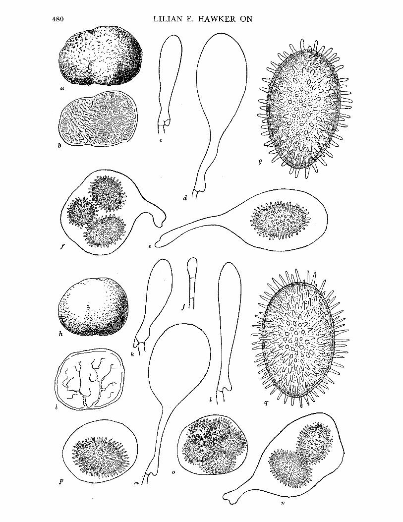
480 LILIAN E. HAWKER ON
aj;:,?, :,; ?~ r~ ._
'K?"H "~' JJ ~~, .~ \,,//'~f,
d
f
p
BRITISH HYPOGEOUS FUNGI
Fruit-body, general macroscopic characters. Spherical or slightly flattened at base, small, seldom more than 1 cm diam. (figure 13h), peridium golden brown, smooth or slightly papillate but shining, not puberulent, texture firm to hard, gleba white, later becoming grey to reddish grey, veins always chalk white, few, arising from base (Malen5on's Type 1 or 3), figure 13 i. Odour none.
Peridium. 0-5 to 0*8 mm thick, similar in structure to Tuber rufum. Gleba. Veins fewer and less branched than in T. rufum, hymenium at first regular, asci
later becoming irregularly arranged. Asci. Stalked as in T. rufum, 50-80 x 45-60,u (figure 13j to p).
no. of spores in ascus 1 2 3 4 5+ no. of asci in four counts from a single collection 5 8 8 19 0
Ascospores. As in T. rufum, but spines more slender and more densely crowded (figure 13 q). Size: 1 spore in ascus: ca. 40 x 24,u.
2 spores in ascus: 27-31-37 x 18-20-24u. 3 spores in ascus: 22-25-30 x 16-19-218 . 4 or more spores in ascus: 20-24-27 x 15-17-19#.
Habitat and periodicity. In the present investigation recorded only twice, under beech, from Bristol and Wotton-under-Edge, Gloucestershire, (54, 486), European records from under beech, oak and ash. No mature spores seen before September, season given as August to November by Fischer (I897). Almost certainly less common than T. rufum. Recorded from Wiltshire, Gloucestershire and Somerset. The two species are obviously closely related but are distinct, differing in colour, shape, venation and in the nature of the spines on the spores. The characteristic shape and development of the asci of these species are closely similar.
Ramsbottom & Balfour-Browne give also T. ferrugineum and T. scleroneuron as British records, but it is doubtful whether these are distinct species or are merely names given to stages or varieties of T. rufum. Both are said to be rare and very little material of either is available. T. ferrugineum was first described by Vittadini (1831) as a form with reticulate spores. Hesse (I894) and Fischer (1897) also record it as having reticulate spores. Massee (1909) and later MalenSon (1938) include it in the section with spiny spores. It is thus highly probable that two different organisms have been described under this name, and from the descriptions, the form with echinulate spores differs from T. rufum only in its softer texture and in having fewer veins and smaller spores. This corresponds to T. Bonetti Roumg. described by Malengon (1938). A single collection of two specimens made near
Wotton-under-Edge, Gloucestershire, during the present investigation was identified by
FIGURE 13. Tuber rufum. (a) Mature fruit-body, showing slightly irregular, granular surface. (b) Longi- tudinal section of same, showing venation of type 2 or 4. (a, b) x 3. (c)-(e) Stages in development of asci, showing characteristic long stalks. (f) Ascus from fruit-body with small spores possibly attributable to T. bonetti. (c)-(f) x 660. (g) Mature spore, showing blunt spines, x 1500. T. nitidum. (h) Mature fruit-body, showing smooth, shining surface. (i) Longitudinal
section of same, showing venation of type 1. (A, i) x 3. (j)-(p) Stages in development of ascus, showing characteristic stalk persisting until maturity. (j)-(p) x 660. (q) Mature spore, showing numerous pointed spines, x 1500.
481

LILIAN E. HAWKER ON
Miss Wakefield, of the Royal Botanic Gardens, Kew, as T. Bonetti. The differences from T. rufum are, however, so slight that it seems likely that T. Bonetti and T. ferrugineum (in the sense of Massee and Malen?on) both refer to a rather unusual variant of T. rufum. T. scleroneuron, on the other hand, is said to differ from a typical specimen of T. rufum in its firmer cartilaginous texture, its deeper red-brown colour and in the less conspicuously stalked asci. All these characters are those which might be the result of age or dry con- ditions, and it is unlikely that T. scleroneuron is a distinct species.
T. rufum and T. nitidum are of a relatively simple organization. The convergence of all or most of the venae externae from a basal pad or hollow shows a similarity to the simple infolded fruit-body of Stephensia, while the retention of a hymenial arrangement of the asci until the development of the fruit-body is well advanced, together with the stalked, clavate asci which remain attached until mature, and perhaps also the prevalence of regularly four-spored asci, are primitive characters relating these species to the Pezizales. In one specimen of T. rufum a few cylindrical asci containing four uniseriate spores were observed. Such asci have not been seen in any other species of Tuber.
(ii) Excavatum group
TUBER EXCAVATUM Vitt., Vittadini (183I, p. 49, P1. 1, fig. 7)
REFERENCES. Tulasne (1843, 381); (i851, 144, P1. VI, fig. 1; P1. XVII, fig. 5); Smith
(1891, 11-12); Cooke (I865, 137-40); (I87I, 740); Broome (I874, 290-8). Hesse (I894, 18-20, P1. XI, fig. 5-9; P1. XVI, fig. 8; P1. XIX, fig. 1-7); Fischer (i897, 55); Massee
(1909, 257-8, P1. XVII, fig. 1). Syn. Aschionfuscum Wallroth (I833, 866).
Rhizopogon excavatus Rabenhorst (I844, 246). Tuberfuscum Corda (I837, 25, P1. VII, fig. 298); Zobel (1854, P1. XX, fig. 142). T. (Vittadinion) montagnei Zobel (I854, 75).
Fruit-body, general macroscopic characters. More or less globose, often flattened or lobed, always with a definite basal cavity (figure 14 a to d), up to 3 cm diameter, at first clear yellow, becoming dull ochraceous to potato-coloured, sometimes with a reddish tinge, surface smooth at first, then scurfy due to splitting of outer layer of peridium into minute warts, texture firm, drying very hard, gleba at first white then ochre yellow, sometimes tinged flesh pink, finally dull red to purple-black, veins few, cream to dingy yellow, radiating from basal cavity, venae internae at first conspicuous becoming indistinguishable so that young fruit-bodies appear to be more finely veined than old ones. Odour none or slight or sometimes strong and unpleasant.
Peridium. Outer layer (ca. 350#u thick) of large fused hyphae with thick yellow-brown walls (diam. up to 20/, walls 1 to 2,# thick), inner layer (ca. 250,u thick) continuous with
trama, of thin-walled, closely interwoven slender hyphae (1.5 to 3,u thick). Gleba. Firm, regular palisade of paraphyses at first lining venae externae, asci arising
irregularly in subhymenial layer and eventually almost obscuring venae internae. Asci. Thin-walled, hyaline, more or less globose (figure 14 e), shortly stalked when young,
stalk usually indistinguishable at maturity, 95-100 x 75-90,u most frequently 4-spored,
482

'OLS[ x 'oodsido jo IJOM:ou xejn..t Qsreoo ;sc o o 'sNsods OM1 2UImT.euoo snost aQinveju (a) *g x (p)-(v) 'Apoq-:lnij jo SuliIlds poetide pue AX.TA^O epseq ?uiMoqs 'ou-es
jo uoioDs ipuipniS2uo-J (p) *AiTA^o piseq Moqis o: Mopq Luoij pM9TxA Apoq-lmjj iJaunoA (a) *nudo felUTJ;9XQ WeUDA qvLI.M OIUT AITA3o peSeq 2IUMO14S pUa eSJo UOpi9S IjUIpn2Suo-' (q) >3pJio OXl ?uipul munipluad aejnumS S2uTMOqs 'Apoq-:mnj omsEu AlsesN (v) 'uwntvavxa atqnj 'tl aun)IAo
IV
giJ fUd SflOlJDOJAH HSIRIfI G8Fi

LILIAN E. HAWKER ON
but average number of spores per ascus decreases with age (see table below) and varies with individual fruit-bodies and between different collections:
total no. of spores in ascus 1 2 3 4 5 6 counted
collected September 8 18 11 46 12 1 96 collected October from same locality 36 42 46 54 8 1 187
Ascospores. At first hyaline and globose, later golden brown, finally dark reddish brown and ellipsoidal. Epispore deeply reticulate (3 to 7#u deep), mesh coarse, very irregular (figure 14e), walls of reticulation 1 to 3,c thick and darker than rest of spore, inner wall of spore ca. 2,u thick. Size varying with number of spores in ascus, all measurements excluding sculpturing:
1 spore in ascus: 38-51-55 x 28-31-32#. 2 spores in ascus: 32-42-45 x 22-28-30#. 3 spores in ascus: 25-36-43 x 24-27-30#. 4 spores in ascus: 26-34-42 x 18-23-29#. 5 or more spores in ascus: 22-31-40 x 16-23-29#.
Habitat and periodicity. 1 to 2 in. deep in light calcareous soil in beechwoods, although one specimen was found under yew at some distance from nearest beech. European specimens collected also under oak. During present investigation usually found in light or well-drained soils such as the top of a slope or the slopes of badger earths or artificial dykes and not found in wet years. In dry warm springs young fruit-bodies have been collected as early as January, but asci are not usually formed until May or June and no spores are formed earlier than August. The fruit-bodies are not fully mature until October. Not uncommon in southern England, previously recorded from Gloucestershire, Somerset, Wiltshire and Kent and found during present investigation in beechwoods in Gloucester- shire (Cotswold Hills, near Wotton-under-Edge, 4, 5, 7, 10, 11, 12, 32, 83, 98, 99, 308, 325, 369, 493, 639), Somerset (Frome, 347; Leigh Woods, 918), Wiltshire (Wylie Valley, 89, 119), Oxfordshire (Blenheim Park, 21), Kent (Otford, 619, 709, 711) and Surrey (Mickleham, 682).
This species is a very variable one and, moreover, all the diagnostic characters alter as the fruit-body matures. Attempts to subdivide it may sometimes have been the result of the examination of specimens of different ages. According to E. Fischer (1923) the species T. fulgens Quel. is distinct from T. excavatum owing to its brighter colour and more regular reticulations of the spore. In the present investigation specimens of brighter colour than usual have been found and some of these had more regularly reticulated spores, but these were nearly always immature specimens and the differences were not sufficient to assign them to T. fulgens. Fischer subdivides T. excavatum into two subspecies typicum Fisch. and lapideum (Matt.) Fisch. on the basis of the presence or absence of peridial and tramal hyphae which stain blue with iodine, and he further subdivides each of these into var. longisporum and var. brevisporum according to the shape of the spores. No blueing of hyphae in response to iodine occurred in material collected during the present investigation and spore shape depended upon age, so that all the mature specimens would fall into Fischer's T. excavatum Vitt. s.sp. lapideum (Matt.) Fisch. var. longisporum.
T'he presence of a distinct basal cavity in this species is a primitive character linking Tuber with such a form as Stephensia. The shape of the asci and loss of the ascus stalk at
484

BRITISH HYPOGEOUS FUNGI
maturity, however, may be considered to be more advanced than the Tuber rufum type of stalked ascus, while the entirely irregular arrangement of the asci is also an advanced character.
(iii) Puberulum group A
TUBER PUBERULUM Berk. et Br., Berkeley & Broome (I846, p. 81) REFERENCES. Tulasne (I851, 148, P1. XIX, fig. 11); Berkeley (i86o, 736); Cooke (I871,
741-2); Hesse (1894, 29-30, P1. XII, figs. 15-18; P1. XV, fig. 12; P1. XVI, fig. 17); Fischer
(1897, 45); Massee (I909, 258-9). Fruit-body, general macroscopic characters. At first globose, later may become lobed or
irregular, up to 1*7 cm diam. but seldom more than 1 -0 cm, at first pure white, then cream or dingy yellowish grey or pinkish, finally mole grey, tinged purple or tan with lighter streaks in the grooves where venae externae reach surface, puberulent, with short hairs giving a velvety appearance, becoming smooth on surface of bumps or lobes but always remaining puberulent in grooves, texture fragile at first and waxy, later fleshy but firmer, gleba white at first, then flushed flesh-pink or lilac-grey, later purplish black, venae externae always white, numerous, much branched, arising from base and also from other points on periphery (figure 15a, b). Odour none unless parasitized and then unpleasant.
Peridium. Soft, definite but very thin, composed of closely interwoven, uniform, narrow hyphae, continuous with trama, covered with groups of short pointed hairs, septate, bulbous at base (figure 16k).
Gleba. At first with definite palisade-like hymenium and well-developed tramal dis- sepiments (venae internae), but trama largely used up in development of asci so that the whole of the gleba between the venae externae is a mass of asci embedded in a few sterile hyphae.
Ascus. Thin-walled, ovoid to globose, 70 to 90,u, at first shortly stalked but stalks soon become indistinguishable and asci appear to be free within the gleba (figure 15c to i). Asci most frequently 1-spored, less often 2- or 3-spored, seldom or never more than 4-spored.
no. of spores in asci 1 2 3 4 5+ no. of asci 43 33 30 11 0
Ascospores. Usually almost spherical but occasionally shortly ellipsoidal, at first hyaline, then pale golden brown, finally deep red-brown, maturation of spores is responsible for changes in colour of gleba with age. Epispore sculptured with deep, small-meshed, thin-
walled, regular reticulations (figure 15j). Some variation in size of the meshes may be seen even within the same ascus, but this variation is less than with most other species. Size (excluding sculpturing) varies with number of spores in ascus:
1 spore in ascus: 34-43-50 x 33-40-49/u. 2 spores in ascus: 32-36-45 x 28-34-42u. 3-4 spores in ascus: 31-33-36 x 28-31-35,.
Habitat and periodicity. This is the commonest British species, widely distributed in southern England, gregarious. Over 100 separate collections have been made during the last four years, most of which have been made under beech, but some under other deciduous trees or under conifers, as follows: Bristol (Blaise Castle, 38, 278; Leigh Woods, 508), Gloucestershire (Cotswold Hills near Wotton-under-Edge and Dursley, 2, 150, 165-9, 177-80, 276, 277, 282, 283, 287, 288, 290-2, 297, 300, 303, 309-11, 316, 324, 327, 328,
485
VOL. 237. B. 59
LILIAN E. HAWKER ON
at
C J d. A e)
. .
< ; ? X , . . * . . , . . s . . ;.\
/..; :..-; '-.:. .,...' .... X
/.: . ., :, ., .- : - ......................... ;.' : ..
A _ *2 *}*** -*'2-'
<_e".'-' / f '_w*; 4 siswv
. 5 _ v
486

BRITISH HYPOGEOUS FUNGI
330, 436, 439, 443, 452, 456, 472, 497, 541, 542, 547, 553, 570, 577, 591, 642, 643, 656, 683, 684; Leighterton, 174; Newark Park, 501, 502, 504; Culkerton, 317), Somerset (Cleeve, 30, 42, 191, 196-8, 201, 314, 381, 512, 533, 534, 554, 621, 712; Portbury, 113, 114, 206, 208-10, 212, 265, 400, 423; Brockley Coombe, 269, 273, 490; Wraxall, 487, 488, 641; Abbot's Pool, Failand, 526), Oxfordshire (Bagley Wood, 17), Surrey (Boxhill, 319), Caernarvonshire (Vaynol Park, Bangor, 226, 232, 235; Bettws-y-coed, 252). Usually found in considerable numbers 1 to 3 in. below surface, lying free in humus or occasionally under deeper layer of leaves, never actually embedded in clay or hard soil, occasionally in sandy or stony soil but then few and small. Favoured by damp conditions and most numerous in the wet year of 1950. Not found in acid soils. In a mild winter fruit-bodies may be found as early as February, but usually are not found until later, asci occasionally formed as early as March but spores not developed before July and in most specimens not until August, by which time peridium and gleba begin to colour. Young colourless fruit- bodies with no spores may be found as late as early November, but the majority are mature by late October. Occasionally a few fruit-bodies survive until the following spring. The soft ripe fruit-body is readily attacked by eelworms and then rapidly undergoes bacterial decomposition with the development of a strong, unpleasant odour, which may account for the otherwise inexplicable confusion with T. rapaeodorum. Fruit-bodies of all ages may be attacked by the Pyrenomycetous fungus, Battarina inclusa, and then become distorted or enlarged and may be of abnormally light colour.
TUBER MACULATUM Vitt., Vittadini (I83I, p. 45, P1. III, fig. 16) REFERENCES. Tulasne (1843, 380); (1851, 148-9, P1. XIX, fig. 9); Zobel (1854, 78-9,
P1. XVIII, fig. 133); Hesse (1894, 26-7, P1. XVI, fig. 14; P1. XXI, figs. 1-8); Fischer
(1897, 47); Ramsbottom (I923, 201).
Fruit-body, general macroscopic characters. From the first very irregular, knobbly, wrinkled or bumpy (figure 16 a, b),b), size said to be up to that of a hen's egg (Fischer 1897; Malen.on I938), but British specimens all much smaller and seldom more than 2 cm in diameter, at first pure white, translucent, later becoming discoloured yellow in patches or variably coloured, reddish or spotted blue grey, surface smooth, waxy, soft, brittle, gleba at first white with a water-soaked appearance, traversed by a few chalk-white veins, later clay- coloured to blackish russet but veins remaining white, venae externae few at first and sparsely branched, later more numerous, arising in the grooves at several points of the exterior (figure 16c). Odour faint, characteristic, not unpleasant, taste bitter.
FIGURE 15. Tuber puberulum. (a) Nearly mature fruit-body.- (b) Longitudinal section of same, showing indistinct venation, veins numerous and arranged according to types 4 or 5. (a, b) x 4. (c)-(i) Stages in development of ascus (c, d, e and h drawn from fresh material;f, g and i from material stained iron alum haematoxylon). (c) Young binucleate ascus. (d, e) Older elongated uni- nucleate asci. (f) Ascus containing single enlarged nucleus with prominent nucleolus and
peripheral network of chromatin. (g) Nucleus in which chromatin network is disintegrating (small nucleus on left is a vegetative one for comparison of size). (h) Ascus with four nuclei surrounded by dense cytoplasm. (i) Ascus with two nuclei (fresh material), showing oily nature of cytoplasm surrounding nuclei. (c)-(i) x 700. (j) Mature spore, showing regular, small- meshed reticulate thickening of epispore and nearly spherical shape of spore, x 1600. (k) Charac- teristic pointed, septate hairs arising from outer cells of peridium, x 700.
59-2
487

488 LILIAN E. HAWKER ON
b
f 9
BRITISH HYPOGEOUS FUNGI
Peridium. Indefinite at first, outer hyphae ending blindly to give felted appearance, soon
becoming smooth, except in grooves where a few hairs may persist, peridium more clearly defined in mature specimens, 300 to 400/ thick, composed of closely interwoven thick
hyphae (up to 25,u diam.), walls of outer 2 or 3 layers finally becoming thickened and
slightly coloured. Gleba. Trama continuous with inner layer of peridium, asci at first rather regularly
arranged, less crowded than in Tuber puberulum. Asci. Ovoid (70-110 x 50-70#) shortly stalked or sessile, 1- to 3-spored. Ascospores. Globose or broadly ellipsoidal or becoming longer ellipsoidal with age, at
first hyaline, becoming yellow-brown, thick-walled, deeply reticulate, meshes regular and rather small, walls of reticulations at first delicate (figure 16h), later becoming rather thick but regular. Size variable:
1 spore in ascus: 31-36-43 x 27-31-32u. 2 or 3 spores in ascus: 28-31-39 x 20-27-32,.
Habitat and periodicity. In Europe reported from under a number of deciduous trees, British specimens from under both deciduous and coniferous trees, 1 to 2 in. deep in humus, usually not mature until late autumn. Collected during present investigation in Gloucestershire (Wotton-under-Edge, 164, 638, 663, 690), Herefordshire (Haugh Wood,
469), Somerset (Cleeve, 956), Devonshire (Stoke Wood, Exeter, 117), Sussex (Friston Forest, 568, 681), Suffolk (Barton Mills, 539) and Perthshire (Loch Tay, 413; Glen Lochay, 418). These specimens closely resembled material collected by Corner in Spain and identi- fied by Malen?on, but of the specimens in Broome's Herbarium, British Museum, only that from Herefordshire is undoubtedly this species.
Specimens of T. puberulum are often much lobed and then resemble T. maculatum, but the latter is never entirely puberulent, and when young has a translucent, waxy appearance. The spores are at first nearly spherical, becoming broadly ellipsoidal, and differ from those of T. puberulum in the thicker wall and the wider mesh of the reticulations.
TUBER RAPAEODORUM Tul., Tulasne (I843, p. 380, P1. XVIII, fig. 1) REFERENCES. Tulasne (1851, 147, P1. V, fig. 4); Hesse (1894, 28, P1. XVI, fig. 18);
Massee (I909, 258). Fruit-body, general macroscopic characters. Ovoid to irregularly lobed, up to 1.5 cm diam.
(figure 16d), dingy white to light straw-coloured, sometimes mottled reddish brown,
FIGURE 16. Tuber maculatum. (a, b) Immature, lobed fruit-bodies. (c) Longitudinal section of (a) showing venation of type 2, peridium not clearly defined. T. rapaeodorum. (d) Mature fruit- body, showing irregular development of pigment and tendency to crack. (e) Longitudinal section of same, showing distinct peridium, dark gleba and venation of type 2. T. borchii. (f) Nearly mature fruit-body. (g) Longitudinal section of same, showing sparse venation of type 1. (a)-(g) x 4. (h) Mature spore of T. maculatum, showing nearly spherical shape, thick wall with delicate, regular reticulations of wider mesh than in T. puberulum (see figure 15j). (i,j) Mature spores of T. rapaeodorum, showing ellipsoidal shape, dark colour, shallow, small- meshed, somewhat irregular reticulation. (k) Mature ascus of T. borchii, showing bulbous base and wide difference between size of mesh of reticulations on spores within same ascus. A third spore is aborting. (1) Mature spore of T. borchii, showing regular, small-meshed reticulations typical of the majority of spores of this species. (h)-(l) x 500.
489

LILIAN E. HAWKER ON
finally becoming dingy brown with darker patches, at first puberulent, then smooth, dry, finally tending to develop numerous fine cracks giving a somewhat warted appearance in old specimens, firmer than Tuber puberulum but not hard, gleba white at first then becoming cream, greyish yellow or pinkish grey and finally purple-black. Peridium and venae externae conspicuous, sharply defined (in contrast to T. puberulum) and always chalky white or cream. Venae externae from base and other points on surface, fairly numerous and much branched (figure 16e). Odour none in young specimens, usually strong and unpleasant (cruciferous) in old specimens.
Peridium. 600 to 900/u thick, thickness varying even within same fruit-body, distinct, composed of rounded irregular, fused elements 10 to 50,u wide. Walls of outer layers become thickened and yellow-brown.
Gleba. Trama at first covered with regular palisade of paraphyses, asci developing irregularly in trama.
Asci. Ovoid to pyriform (70-100 x 50-70,u) at first with conspicuous remains of crozier, then shortly stalked becoming sessile, mostly 2- or 3-spored.
no. of spores in ascus 1 2 3 4 5+ no. of asci 37 52 57 35 2
Ascospores. Very variable, usually ellipsoidal, particularly in old specimens, reticulate network usually small and fairly regular (figure 16i), but often large or with large and small meshes on the same spore (figure 16j), deep at first but fragile and becoming shallow or collapsing when ripe, spores at first hyaline, then yellow, finally rather dark red-brown, giving dark colour to gleba. Size variable according to number of spores in ascus:
1 spore in ascus: 29-43-54 x 24-36-43u. 2 or more spores in ascus: 22-31-48 x 15-23-30u.
Habitat andperiodicity. Collections made in present investigation in Bristol (Stoke Bishop, 476), Gloucestershire (Cotswold Hills near Wotton-under-Edge, 25, 445, 447, 451, 454, 458, 549-51, 650, 686-8) and Somerset (Failand, 529-31), all under larch, except one under a lime tree, 1 to 3 in. deep in needle layer or loose soil, not in acid soils, not found in wet situations or in wet seasons (cf. puberulum). Annual periodicity as in T. puberulum. Probably less common than that species.
Some confusion exists in the literature between this species and T. puberulum, and the various published descriptions suggest that some of these have been made from insufficient and immature material. Fischer (1897) considers rapaeodorum to be a variety of puberulum, but Malen?on (1938) considers it to be a distinct species, which is supported by the present study. T. rapaeodorum differs from T. puberulum in its firmer texture, the evanescent nature of the hairs, in its more clearly defined peridium and in the elliptical spores with greater irregularity of spore size and sculpture. The odour of radishes claimed as characteristic
by many observers occurs only in mature fruit-bodies, almost certainly as a result of bacterial attack, and is valueless as a diagnostic character.
490

BRITISH HYPOGEOUS FUNGI
TUBER BORCHII Vitt., Vittadini (I831, pp. 44-5, P1. I, fig. 3) REFERENCES. Tulasne (1851, 145-6, P1. V, fig. 1; P1. XXI, fig. 13); Hesse (I894, 24-5,
P1. XVI, fig. 1); Fischer (1897, 46). Syn. Tuber albidum Pico (1788, 79).
T. album Bulliard (1791-8, 80). T. mixtum Risso (I844, 567). Rhizopogon Borchii Rabenhorst (I844, 246). T. elegans Zobel (1854, P1. XXI, fig. 139; figured as T. borchianum Zobel, 77-8,
P1. XIX, fig. 137). Fruit-body, general macroscopic characters. Globose or somewhat lobed, often with basal
depression but no cavity (figure 16f), European specimens said to be up to size of hen's egg, British specimens never more than 2 cm diam., at first whitish then clay-coloured, golden-brown or potato-coloured to chocolate, white or light yellow in grooves, at first downy later smooth, shining when wet, hairs persisting in the folds, firm but not leathery or hard, gleba white, tinged flesh pink, becoming milk chocolate coloured and mottled, veins few, white, later discoloured reddish, venation type 1 or 2 (figure 16g). Odour earthy, or may be unpleasant when ripe.
Peridium. 500 to 800,u thick, grading from large coloured spherical elements, up to 22/z diam. with walls 1 *5p thick, on the outer edge to slender, colourless, thin-walled but still spherical elements on inner side, more loosely constructed than in Tuber excavatum or T. dryophilum. Hairs on young fruit-bodies and in folds of older ones 30 to 40,u long, 1-5 to 10-o thick.
Gleba. Venae internae not conspicuous when ripe, paraphyses thin and septate, asci irregularly arranged.
Asci. Irregularly globose, pyriform or clavate, shortly stalked with bulbous base (figure 16k), usually 2-spored, less often 1- or 3-spored, occasionally 4 or more.
no. of spores in ascus 1 2 3 4 5 + no. of asci 83 92 62 27 2
There is a tendency for the spores to abort at a late stage in development, so that the pro- portion of asci with three or more spores decreases with increase in age of the specimen.
Ascospores. Very variable, ellipsoidal to narrow-ellipsoidal, at first hyaline, finally red- brown, wall reticulately thickened, mesh small, shallow and regular or wide, deep and
irregular, often contrasted in different spores in same ascus (figure 16k, 1), walls of network rather fine and prolonged at corners into spines, size varying to some extent with number in ascus but also independently.
1 spore in ascus: 35-45-52 x 26-29-33,. 2 spores in ascus: 22-34-48 x 18-22-31u. 3 or more spores in ascus: 19-30-37 x 16-20-27#u.
Habitat and periodicity. In humus under beech in calcareous soil, spores maturing in late summer or autumn. Collections were made in Bristol (Stoke Bishop, 475), Gloucestershire (Cotswold Hills near Wotton-under-Edge and Dursley, 157, 495, 552, 636, 645; Newark Park, 505) and Perthshire (Loch Tay, 412, 415).
This species has been previously recorded in Britain only once (Crotch I852), but
specimens from Chudleigh, Devonshire and Abbot's Leigh, Somerset, in the Herb.
491

492 LILIAN E. HAWKER ON
a

BRITISH HYPOGEOUS FUNGI
Broome, British Museum, are attributed to it. As it bears a close superficial resemblance to T. dryophilum specimens may have been attributed to the latter. Its few, sparsely branched venae externae, the presence of a large proportion of small-meshed regularly reticulate spores and the preponderance of 1- and 2-spored asci distinguish it clearly from T. dryophilum. The venation of the gleba and the colour of the mature fruit-bodies distinguish it from T. rapaeodorum, while the ellipsoidal spores differ markedly from the spherical ones of T. puberulum and young specimens of T. maculatum.
This group is a fairly natural one. Its members are similar in the structure of the peridium, the tendency to puberulence at least in early stages, the development and shape of the asci and the predominance of regularly reticulated spores.
(iv) Dryophilum group (-Knapp's puberulum group B) TUBER DRYOPHILUM Tul., Tulasne (I844, p. 62)
REFERENCES. Tulasne (1851, 147, P1. V, fig. 3; P1. XIX, fig. 8); Berkeley & Broome
(1846, 80); Berkeley (i86o, 376); Cooke (I87I, 742); Hesse (I894, 25-6, P1. XVI, fig. 15); Fischer (I897, 51); Massee (i909, 258).
Fruit-body, general macroscopic characters. Roundish or flattened-oblong, entire, lobed, wrinkled or knobbly, often with a basal depression but no cavity (figure 17 a), up to 2 cm diam., at first light clay to potato-coloured, finally purplish chocolate colour, like Tuber borchii but duller, at first shortly puberulent, soon quite smooth but not polished, firm but soft, gleba at first cream with white venae externae, then pink to lilac grey, finally milk chocolate to purplish brown with dingy white veins, veins numerous, much branched, arising at a number of points on periphery (figure 17 b), tendency for gleba to break into oblong pieces along the veins, suggesting imperfect joining of opposite faces. Odour earthy or none.
Peridium. Similar in construction to that of T. excavatum but not splitting to give granular or scurfy surface as in that species, 250 to 400# thick, outer layer of large thick-walled hyphae, walls fused, pale brown, inner layer of smaller, closely interwoven hyphae con- tinuous with and similar to those of trama.
Gleba. Very numerous and much-branched venae externae, imperfectly 'sealed' by growth of paraphyses and therefore gleba tending to split into sections, palisade of para- physes regular at first, asci forming irregularly and filling trama.
Asci. Thin-walled, hyaline, globose or ovoid, usually sessile, 50-60 x 40-50,u, usually 3- or 4-spored, less often 1 or 2, seldom more than 4 (figure 17f).
no. of spores in ascus 1 2 3 4 5 + no. of asci 32 51 72 151 3
FIGURE 17. Tuber dryophilum. (a) Mature fruit-body. (b) Longitudinal section of same, showing venation of type 2. T. foetidum. (c, d) Nearly mature fruit-bodies, showing irregular shape and granular surface. (e) Longitudinal section of (c), showing venation of type 2 with fewer branches than in T. dryophilum. (a)-(e) x 2. (f) Nearly mature ascus of T. dryophilum. (g) Mature spore of T. dryophilum; note irregular large-meshed reticulations which are more delicate than those of T. excavatum (see figure 14 e). (h, i) Mature spores of T.foetidum, showing reticulate thickening with medium-sized, rather irregular meshes. (f)-(i) x 1870.
493
VOL. 237. B. 6o

LILIAN E. HAWKER ON
Ascospores. Broadly to narrowly ellipsoidal, at first hyaline, later pale golden brown, epispore deeply reticulate, meshes large and irregular with undulating walls as in T. excava- tum, but walls of the meshes much more delicate and less coloured, subsidiary ridges may form within meshes of main reticulation, ridges of reticulation tending to break and collapse in old or parasitized specimens, size and shape varying with number in ascus (figure 17f, g).
1 spore in ascus: 32-51-59 x 26-32-36,u. 2 spores in ascus: 31-37-47 x 27-29-32u. 3 spores in ascus: 28-36-46 x 24-28-36u. 4 spores in ascus: 26-33-41 x 22-26-30u.
Habitat and periodicity. Under beech in fairly well-drained localities, asci formed as early as May, but mature spores not present until late summer or autumn. Fairly common. Collected during present investigation in Bristol (Hanham, 565; Blaise Castle, 432, 615, 651), Gloucestershire (Cotswold Hills near Wotton-under-Edge and Dursley, 34, 370, 371, 377, 644), Oxfordshire (Blenheim Park, 20) and Wiltshire (Savernake Forest, 366). The material in Broome's Herbarium was collected in the Bristol area with the exception of one doubtful specimen.
This species bears a superficial resemblance to T. excavatum and T. borchii but differs from the former in the absence of a basal cavity, the smooth peridium, the type of venation and the more delicate reticulations of the spore wall and from the latter in its duller surface, more numerous venae externae and the irregularly large-meshed reticulations of the spore wall.
TUBER FOETIDUM Vitt., Vittadini (1831, p. 41, P1. I, fig. 8; P1. III, fig. 11)
REFERENCES. Tulasne (1851, 140-1, P1. XVII, fig. 7); Zobel (1854, 80, P1. XIX, fig. 135); Fischer (1897, 49); not Massee (i909, 257).
Fruit-body, general macroscopic characters. Closely resembling Tuber rufum in external and internal characters (but differing in the reticulate spores). More or less globose or may be wrinkled or lobed or indented (figure 17c, d), about 1 cm diam., tawny red-brown, smooth or minutely granular or warted, firm but softer than T. rufum, gleba at first white then grey or flesh-coloured to reddish or purplish brown, venae externae thick, branched and anastomosing, arising at a number of points on the periphery (figure 17e), whitish, becoming dingy brown and less conspicuous. Odour said to be strong of asafoetida or rancid oil, no odour in young specimens but strong in mature ones collected during present investigation.
Peridium. Rather sharply marked off from gleba, outer layer of radially arranged fused hyphae with thick yellow walls, inner layer of interwoven hyphae arranged tangentially.
Gleba. Large patches of irregularly arranged asci, leaving only thin tramal strands in mature specimens.
Asci. Ellipsoidal, 70-90 x 65-75#, most often 3-spored:
no. of spores in asci 1 2 3 4 5 + no. of asci 13 23 61 25 3
494

BRITISH HYPOGEOUS FUNGI
Ascospores. Ellipsoidal, at first hyaline then dark brown, epispore with rather shallow, reticulate thickening with medium-sized meshes, the walls of which are undulating as in T. dryophilum (figure 17 h, i). Size varying with number of spores in ascus:
1 spore in ascus: 34-40-44 x 20-26-28#. 2 spores in ascus: 22-31-42 x 18-22-25/u. 3 or more spores in ascus: 19-29-32 x 15-19-21,.
Habitat and periodicity. Collections made in this investigation, from calcareous larch- woods at Burrington Coombe, Somerset (424-6, 790, 793) and Wotton-under-Edge, Gloucestershire (902, 927). Spores not quite mature in late August. Probably not common or may have been frequently taken for T. rufum from which it differs in sculpturing of spores, shape of asci and fewer venae externae. The earlier collection made on the Mendips was at first thought to be of T. rufum but was later found to contain specimens of both species. All collections agree with the descriptions of this species given by Fischer (1897) and MalenSon (1938), but Massee's (I909) description differs in important particulars and is more applicable to T. borchii, suggesting that his specimens were of the latter species.
(v) Aestivum group TUBER AESTIVUM Vitt., Vittadini (1831, pp. 38-9, P1. II, fig. 4; P1. V, fig. 11)
REFERENCES. Tulasne (1843, 380); (1851, 137-8, P1. VII, fig. 3); Berkeley (i860, 376, P1. XXIII, fig. 3); Cooke (1871, 738-9); Hesse (1894, 14, P1. XI, fig. 1-4; P1. XVI, fig. 6; P1. XX); Fischer (I897, 37); Massee (I909, 256).
Syn. Tubera aestiva Micheli (I729, 221). Tuber cibarium Sowerby (I797-1815, P1. 309). T. albidum Fries (I823, 291). T. mesentericum Vittadini (I83I, 40, P1. III, fig. 19); Tulasne (I843, 380). T. bituminatum Berk. et Broome (1851, 183). T. culinare var. aestiva Zobel (I854, 82, P1. XVIII, fig. 129).
Fruit-body, general macroscopic characters. Irregularly globose often with hollow base, up to 9 cm diam., black or brownish or purplish black, covered slightly flattened 5- or 6-sided pyramidal warts which are often split or striate along their faces or the sides are trans- versely wrinkled (figure 18 a), warts 3 to 5 mm wide, 2 or 3 mm deep, peridium glossy when wet, drying duller, hard, drying very hard, gleba at first dingy white then cream, finally dingy yellow-grey or putty-coloured or occasionally tinged red or purple, very numerous much-branched veins arising from many points on surface (figure 18 b). Odour and taste pleasant except in old specimens where odour becomes phenolic. Young specimens more or less odourless, odour of mature ones nutty or of mushroom.
Peridium. Consists of a thin black outer layer of more or less fused spherical cells with thick dark walls and a wide inner layer of nearly colourless, narrow, rather thin-walled hyphae, continuous with trama which is composed of similar hyphal elements. Total thickness of peridium varies considerably according to age and the position of the warts.
Gleba. Whitish, becoming yellow, fawn or occasionally vinous, trama much reduced as asci develop, until in old or dry material it is seen as narrow dark lines parallel to the white venae externae. Asci irregularly arranged from the first. Flesh firm but readily breaks into small flakes along the lines of the venae externae.
60-2
495
496 LILIAN E. HAWKER ON
b
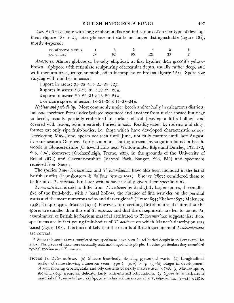
BRITISH HYPOGEOUS FUNGI
Asci. At first clavate with long or short stalks and indications of crozier type of develop- ment (figure 18c to h), later globose and stalks no longer distinguishable (figure 18i), mostly 4-spored:
no. of spores in ascus 1 2 3 4 5 6 no. of asci 28 62 85 121 35 2
Ascospores. Almost globose or broadly elliptical, at first hyaline then greenish yellow- brown. Epispore with reticulate sculpturing of irregular depth, usually rather deep, and with medium-sized, irregular mesh, often incomplete or broken (figure 18i). Spore size varying with number in ascus:
1 spore in ascus: 31-35-41 x 21-24-32#. 2 spores in ascus: 26-28-32 x 19-22-28#. 3 spores in ascus: 20-26-31 x 18-20-24#. 4 or more spores in ascus: 18-24-30 x 14-28-24#.
Habitat and periodicity. Most commonly under beech and/or holly in calcareous districts, but one specimen from under isolated sycamore and another from under spruce but near to beech, usually partially embedded in surface of soil (leaving a little hollow) and covered with leaves, seldom entirely buried in soil. Readily eaten by rodents and slugs, former eat only ripe fruit-bodies, i.e. those which have developed characteristic odour. Developing May-June, spores not seen until June, not fully mature until late August, in some seasons October. Fairly common. During present investigation found in beech- woods in Gloucestershire (Cotswold Hills near Wotton-under-Edge and Dursley, 172, 182, 295, 394), Somerset (Orchardleigh, Frome, 222), in the grounds of the University of Bristol (974) and Caernarvonshire (Vaynol Park, Bangor, 225, 239) and specimens received from Sussex.
The species Tuber mesentericum and T. bituminatum have also been included in the list of British truffles (Ramsbottom & Balfour Brown I95I). Fischer (I897) considered these to be forms of T. aestivum, but later writers have usually given them specific rank.
T. mesentericum is said to differ from T. aestivum by its slightly larger spores, the smaller size of the fruit-body, with a basal hollow, the absence of fine wrinkles on the peridial warts and the more numerous veins and darker gleba* (Hesse I894; Fischer i897; Malengon 1938; Knapp 1950). Massee (gog1909), however, in describing British material claims that the spores are smaller than those of T. aestivum and that the dissepiments are less tortuous. An examination of British herbarium material attributed to T. mesentericum suggests that these specimens are in fact young fruit-bodies of T. aestivum on which Massee's description was based (figure 18j). It is thus unlikely that the records of British specimens of T. mesentericum are correct.
* Since this account was completed two specimens have been found buried deeply in soil excavated by a fox. The glebas of these were unusually dark and tinged with purple. In other particulars they resembled typical specimens of T. aestivum.
FIGURE 18. Tuber aestivum. (a) Mature fruit-body, showing pyramidal warts. (b) Longitudinal section of same showing numerous veins, type 5. (a, b) x 1-. (c)-(h) Stages in development of asci, showing crozier, stalk and oily contents of nearly mature asci, x 740. (i) Mature spore, showing deep, irregular, delicate, fairly wide-meshed reticulations. (j) Spore from herbarium material of T. mesentericum. (k) Spore from herbarium material of T. bituminatum. (i)-(k) x 1870.
497
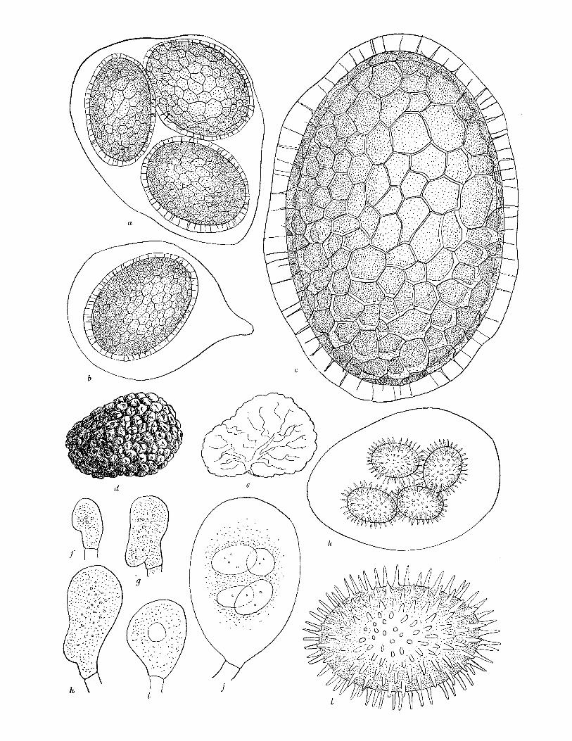
0.., ,I
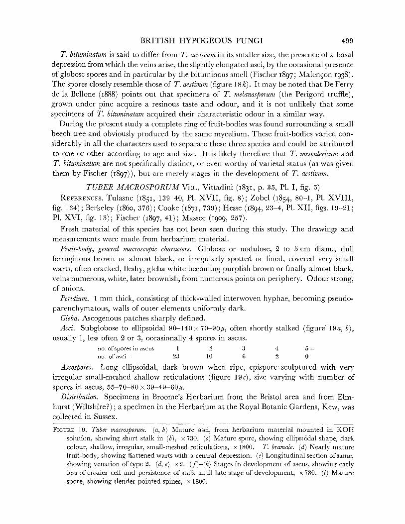
BRITISH HYPOGEOUS FUNGI
T. bituminatum is said to differ from T. aestivum in its smaller size, the presence of a basal depression from which the veins arise, the slightly elongated asci, by the occasional presence of globose spores and in particular by the bituminous smell (Fischer 1897; Malen?on I938). The spores closely resemble those of T. aestivum (figure 18k). It may be noted that De Ferry de la Bellone (i888) points out that specimens of T. melanosporum (the Perigord truffle), grown under pine acquire a resinous taste and odour, and it is not unlikely that some specimens of T. bituminatum acquired their characteristic odour in a similar way.
During the present study a complete ring of fruit-bodies was found surrounding a small beech tree and obviously produced by the same mycelium. These fruit-bodies varied con-
siderably in all the characters used to separate these three species and could be attributed to one or other according to age and size. It is likely therefore that T. mesentericum and T. bituminatum are not specifically distinct, or even worthy of varietal status (as was given them by Fischer (I897)), but are merely stages in the development of T. aestivum.
TUBER MACROSPORUM Vitt., Vittadini (I831, p. 35, P1. I, fig. 5) REFERENCES. Tulasne (I851, 139-40, P1. XVII, fig. 8); Zobel (1854, 80-1, P1. XVIII,
fig. 134); Berkeley (I860, 376); Cooke (I87I, 739); Hesse (I894, 23-4, P1. XII, figs. 19-21; P1. XVI, fig. 13); Fischer (1897, 41); Massee (I909, 257).
Fresh material of this species has not been seen during this study. The drawings and measurements were made from herbarium material.
Fruit-body, general macroscopic characters. Globose or nodulose, 2 to 5 cm diam., dull
ferruginous brown or almost black, or irregularly spotted or lined, covered very small warts, often cracked, fleshy, gleba white becoming purplish brown or finally almost black, veins numerous, white, later brownish, from numerous points on periphery. Odour strong, of onions.
Peridium. 1 mm thick, consisting of thick-walled interwoven hyphae, becoming pseudo- parenchymatous, walls of outer elements uniformly dark.
Gleba. Ascogenous patches sharply defined. Asci. Subglobose to ellipsoidal 90-140 x 70-90pu, often shortly stalked (figure 19a, b),
usually 1, less often 2 or 3, occasionally 4 spores in ascus. no. of spores in ascus 1 2 3 4 5+ no. of asci 23 10 6 2 0
Ascospores. Long ellipsoidal, dark brown when ripe, epispore sculptured with very irregular small-meshed shallow reticulations (figure 19c), size varying with number of
spores in ascus, 55-70-80 x 39-49-60,u. Distribution. Specimens in Broome's Herbarium from the Bristol area and from Elm-
hurst (Wiltshire?); a specimen in the Herbarium at the Royal Botanic Gardens, Kew, was collected in Sussex.
FIGURE 19. Tuber macrosporum. (a, b) Mature asci, from herbarium material mounted in KOH
solution, showing short stalk in (b), x 730. (c) Mature spore, showing ellipsoidal shape, dark
colour, shallow, irregular, small-meshed reticulations, x 1800. T. brumale. (d) Nearly mature
fruit-body, showing flattened warts with a central depression. (e) Longitudinal section of same, showing venation of type 2. (d, e) x 2. (f)-(k) Stages in development of ascus, showing early loss of crozier cell and persistence of stalk until late stage of development, x 730. (1) Mature
spore, showing slender pointed spines, x 1800.
499

LILIAN E. HAWKER ON
TUBER BRUMALE Vitt., Vittadini (I831, p. 37-8, P1. I, fig. 6) REFERENCES. Tulasne (I843, 380); (I85I, 135, P1. VII, fig. 2; P1. XVII, fig. 3); Berkeley
(1860, 376); Cooke (1871, 740-1); Hesse (1894, 7-9, P1. XI, figs. 14-16; P1. XVI, fig. 1); Massee (1909, 259, P1. XVII, fig. 23).
Syn. Tuber brumale f. typicum Fischer (1897, 42). Oogaster brumale Zobel, 73, P1. XVII, fig. 127.
This species is very close to Tuber melanosporum, the Perigord truffle, and was at first
thought by Berkeley (1844) to be the latter. Fischer (1897) considered T. melanosporum to be a variety of T. brumale, separated from it by its darker colour and the reddish venae externae. According to Knapp (1950) the fruit-body and the spores of the Perigord truffle are larger than those of T. brumale. Malengon (1938) points out that the spores of the former are opaque when mature and those of the latter remain translucent.
Fruit-body, general macroscopic characters. More or less spherical, sometimes angular or
ridged, usually with a basal depression, around 2 cm diam., dull purplish black, covered small roughly polygonal warts (1 to 2 mm diam. at base), with flattened or excavated
apices (figure 19d), firm to hard, gleba at first white, becoming violet-grey or blue-grey, with a translucent, water-soaked appearance, veins moderately numerous, mainly converging to base, occasionally from other points of periphery, white, rather indistinct (figure 19e). Odour pleasant, nutty, rather like that of T. aestivum or may become strong and unpleasant.
Peridium. Pseudoparenchymatous, walls of outer elements coloured. Gleba. Asci very irregularly arranged from the first. Asci. Broadly ellipsoidal (ca. 100 x 70u), at first shortly stalked, developing from short
thick croziers (figure 19f to k), mostly 3- to 5-spored. no. of spores in ascus 1 2 3 4 5 6 no. of asci 5 6 18 30 22 3
Ascospores. Elliptical, at first hyaline, then pale brown, finally dark purple-brown, but translucent, densely covered with long (up to 4pu) pointed rather stiff spines (figure 191), size 34-29-26 x 26-20-18u, varying only-slightly with number in ascus.
Habitat and periodicity. Not common, collections in 1951 and 1952 made under lime trees at Stoke Bishop, Bristol (564, 706), previously recorded from Wiltshire, Somerset and Dorset. Resembles Perigord truffle in maturing late in year, November to December.
Genus BALSAMIA Vitt., Vittadini (1831, p. 30) Fruit-body nodulose, fleshy, warted, reddish brown, gleba white, chambered when
mature, asci ellipsoidal, 8-spored, spores smooth, hyaline. This genus has usually been separated from the Eu-tuberaceae, and as late as 1950 was
included in the Pseudotuberaceae by Knapp. Further work on the development of the
fruit-body showed that it is first infolded and that the chambers develop by an irregular fusing of the opposite faces of intricate folds, and Knapp (I952) revised his classification and included Balsamia in the Eu-tuberaceae. Developmental studies during the present investigation confirm this.
TYPE SPECIES. B. vulgaris Vitt.
500

BRITISH HYPOGEOUS FUNGI
KEY TO BRITISH SPECIES OF BALSAMIA.
1. Spores long-ellipsoidal (av. 30 x 13.5,u), asci clavate ... ... ... B. vulgaris. p. 501 Spores short-ellipsoidal (ca. 20 x 13,u) ... ... ...... ...... ... ... ... 2
2. Chambers scattered, asci citriform ... ... ... .. ... B. platyspora, p. 501 Chambers crowded, asci stalked ... ... ... ... ... B. fragiformis, p. 503
BALSAMIA VULGARIS Vitt., Vittadini (1831, p. 30, P1. I, fig. 2; P1. V, fig. 6) REFERENCES. Tulasne (1843, 379); (I851, 123-4, PI. IV, fig. 4; PI. XV, fig. 1); Zobel
(1854, 59, P1. X, fig. 99); Smith (I891, 359); Hesse (I894, 35-6, P1. XVI, fig. 29); Fischer
(I897, 63, figs. 1, 2 on p. 62); Massee (I909, 252). Fruit-body, general macroscopic characters. Globose or more or less wrinkled, up to 4 cm
diam., foxy red, covered with minute darker granular warts (figure 20a), brittle, gleba creamy white, becoming yellowish, at first with venae externae opening at various points, then with numerous crowded angular to curved chambers (figure 20b, c). Odour none at first, becoming strong, unpleasant, phenolic, associated with bacterial breakdown of
fruit-body. Peridium. Outer layer of dark, thick-walled more or less globose or somewhat radially
elongated cells, grading into inner layer of colourless thinner-walled globose hyphae. Gleba. Consists of interwoven thin-walled colourless hyphae. Asci. Clavate, stalked (figure 20d, e) 50-80 x 20-40U, 8-spored. Ascospores. Cylindrical with curved ends, 25-30-42 x 10-13-5-18u, hyaline, smooth,
thin-walled with three large oil drops (figure 20f). Habitat and periodicity. In humus under beech, maturing in late summer, autumn or
winter. Collected during present investigation in two beechwoods, on the Cotswold Hills, Gloucestershire (158, 689). Specimens in Broome's Herbarium, British Museum, from Devonshire and the Bristol area.
BALSAMIA PLATYSPORA Berk. et Br., Berkeley & Broome (1844, p. 358) REFERENCES. Tulasne (i85I, 124-5, P1. XV, fig. 11); Berkeley (I86o, 378); Cooke
(I87I, 747); Hesse (I894, 36, P1. XVI, fig. 30); Fischer (1897, 65, figs. 3 and 4 on p. 62); Massee (I909, 253, P1. XVII, fig. 13).
Fruit-body, general macroscopic characters. Irregular shape, often elongated and knobbly, often cracked, usually more than 1 cm in longest diam., seldom more than 2 cm, general colour impression foxy red but due to dark chestnut warts on yellow-red ground, yellow colour more apparent in grooves of fruit-body, warts slightly larger and less densely crowded than in other two species (figure 20g), firm but brittle, gleba white with few sinuous compressed chambers, dissepiments correspondingly wide, venae externae origin- ally fewer than in other species, becoming sealed at intervals to give chambers (figure 20h), odour none at first, then strong, unpleasant, phenolic.
Peridium and gleba. Of similar elements to those described for Balsamia vulgaris. Asci. Prominent hook retained even in mature asci which are lemon-shaped, sessile,
8-spored (figure 20i, j), 60-80 x 30-40u.
Ascospores. Globose to ellipsoidal, size 19-22-28 x 12-13-16#, hyaline, smooth, thin-
walled, single large oil globule and many smaller ones (figure 20k).
VOL. 237. B.
501

LILIAN E. HAWKER ON ..
b 'n O
'` na:bq Db(\c Ob ;?
,, o
? --ab?. ,. ,3 b .O 0, r0, u -36iri ;ar--.? r D :'Zi/4co:b4 "o : 6'00o
-O o de
9
(3 o (il c?t
.i Dt)0:
C='
?;;;??i;?;IJ Ps:
c
nar I0t-'c, b:
d
r sk s FIGURE 20. Balsamia vulgaris. (a) Mature fruit-body, showing irregularly globose form and small
warts. (b) Longitudinal section of part of (a), showing small, hollow, angular chambers. (c) Longi- tudinal section of immature fruit-body, showing development of chambers by infolding of margin. (a)-(c) x 2 . (d) Young clavate ascus. (e) Mature ascus with eight spores. (d, e) x 750. (f) Mature spore, showing smooth wall and three large oil drops with scattered smaller ones. x 1870. B.platyspora. (g) Mature fruit-body, showing irregular shape and small warts. (h) Longi- tudinal section of same, showing scattered, elongated, hollow chambers. (g, h) x 21. (i) Young ascus, showing prominent and distinctive crozier. (j) Mature ascus, showing citriform shape,
502
BRITISH HYPOGEOUS FUNGI
Habitat and periodicity. Under beech or other deciduous trees, spores maturing late in
year. Collected during present investigation at Blaise Castle (39) and Leigh Woods (515), Bristol. Specimens in Broome's Herbarium, British Museum, from Herefordshire and Wiltshire.
BALSAMIA FRAGIFORMIS (Tul.) Tulasne, Tulasne (I85I, p. 125, P1. V, fig. 3) REFERENCES. Hesse (1894, 33-4, P1. XII, fig. 12-14; P1. XVI, fig. 28; P1. XVII;
P1. XVIII; P1. XXI, figs. 27-48); Fischer (I897, 64); Massee (I909, 253 (wrongly spelt as B.fagiformis)).
Syn. Balsamia polysperma Tulasne (1843, 379); non Vittadini (1831, 31). Fruit-body, general macroscopic characters. Globose or flattened or much lobed, small, usually
less than 1 cm diam., foxy red, densely covered minute darker warts (figure 201), firm but brittle, gleba white, numerous small hollow angular chambers (figure 20m, n). Odour none at first, strong and phenolic when mature.
Peridium and gleba. Of similar elements to those described for Balsamia vulgaris. Asci. Globose or ovoid, long stalked, 8-spored (figure 200 to q), 40-65 x 30-45#,
excluding stalk.
Ascospores. Ellipsoidal, size 13-19-22 x 12-13-14pu, increasing with ageing of fruit-body and therefore not a good diagnostic character (different authors give widely differing measurements for this species), single large central oil drop and numerous smaller ones, smooth, thin-walled, hyaline (figure 20r).
Habitat andperiodicity. In soil 1 to 3 in. deep under beech, lime, oak, etc., mature spores not earlier than September and fruit-bodies found as late as November or even December.
Specimens in Broome's Herbarium, British Museum, from Batheaston and Bathford. Collected during present investigation in Bristol (Stoke Bishop, 474, 592), Gloucestershire
(Cotswold Hills near Dursley, 496) and Caernarvonshire (Vaynol Park, Bangor, 224). These three species are closely similar and are most readily distinguished macroscopic-
ally by size and the arrangement of the chambers and microscopically by ascus shape. B. vulgaris is also distinguished by its longer cylindrical spores.
FAMILY (IV). TERFEZIACEAE
Distinguished by the chambered fruit-body, venae externae absent, or present only at
very early stages, palisade of paraphyses absent or poorly developed. The British species belong to the genera Hydnobolites and Choiromyces and there is a doubtful record of Terfezia leonis.
crozier and eight spores. (i,j) x 750. (k) Mature spore, showing smooth wall and large oil drop with numerous smaller ones, x 1870. B. fragiformis. (1) Mature fruit-body, showing lobing and small warts. (m) Longitudinal section of immature fruit-body, showing infolding of margin. (n) Longitudinal section of mature fruit-body, showing numerous small angular chambers. (l)-(n) x 22. (o, p) Young asci, showing crozier. (q) Mature ascus, showing stalk and eight spores, crozier has almost disappeared. (o)-(q) x 750. (r) Mature spore, showing smooth wall, large oil drop and scattered smaller ones x 1870. (s) Diagram to show infolding of margin of young fruit-body and development of palisade of paraphyses (shaded zone), x 30.
6I-2
503
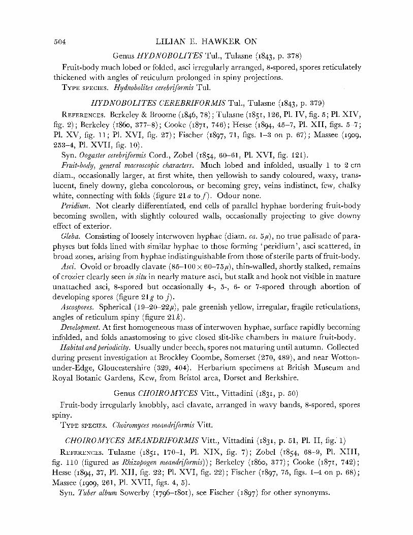
LILIAN E. HAWKER ON
Genus HYDNOBOLITES Tul., Tulasne (1843, p. 378) Fruit-body much lobed or folded, asci irregularly arranged, 8-spored, spores reticulately
thickened with angles of reticulum prolonged in spiny projections. TYPE SPECIES. Hydnobolites cerebriformis Tul.
HYDNOBOLITES CEREBRIFORMIS Tul., Tulasne (1843, p. 379) REFERENCES. Berkeley & Broome (I846, 78); Tulasne (I85I, 126, P1. IV, fig. 5; P1. XIV,
fig. 2); Berkeley (I860, 377-8); Cooke (I87I, 746); Hesse (1894, 45-7, P1. XII, figs. 5-7; P1. XV, fig. 11; P1. XVI, fig. 27); Fischer (I897, 71, figs. 1-3 on p. 67); Massee (I909, 253-4, P1. XVII, fig. 10).
Syn. Oogaster cerebriformis Cord., Zobel (I854, 60-61, P1. XVI, fig. 121). Fruit-body, general macroscopic characters. Much lobed and infolded, usually 1 to 2 cm
diam., occasionally larger, at first white, then yellowish to sandy coloured, waxy, trans-
lucent, finely downy, gleba concolorous, or becoming grey, veins indistinct, few, chalky white, connecting with folds (figure 21 a tof). Odour none.
Peridium. Not clearly differentiated, end cells of parallel hyphae bordering fruit-body becoming swollen, with slightly coloured walls, occasionally projecting to give downy effect of exterior.
Gleba. Consisting of loosely interwoven hyphae (diam. ca. 54u), no true palisade of para- physes but folds lined with similar hyphae to those forming 'peridium', asci scattered, in broad zones, arising from hyphae indistinguishable from those of sterile parts of fruit-body.
Asci. Ovoid or broadly clavate (85-100 x 60-75,u), thin-walled, shortly stalked, remains of crozier clearly seen in situ in nearly mature asci, but stalk and hook not visible in mature unattached asci, 8-spored but occasionally 4-, 5-, 6- or 7-spored through abortion of
developing spores (figure 21g to j). Ascospores. Spherical (19-20-22,u), pale greenish yellow, irregular, fragile reticulations,
angles of reticulum spiny (figure 21 k). Development. At first homogeneous mass of interwoven hyphae, surface rapidly becoming
infolded, and folds anastomosing to give closed slit-like chambers in mature fruit-body. Habitat andperiodicity. Usually under beech, spores not maturing until autumn. Collected
during present investigation at Brockley Coombe, Somerset (270, 489), and near Wotton-
under-Edge, Gloucestershire (329, 404). Herbarium specimens at British Museum and
Royal Botanic Gardens, Kew, from Bristol area, Dorset and Berkshire.
Genus CHOIROMYCES Vitt., Vittadini (1831, p. 50)
Fruit-body irregularly knobbly, asci clavate, arranged in wavy bands, 8-spored, spores spiny.
TYPE SPECIES. Choiromyces meandriformis Vitt.
CHOIROMYCES MEANDRIFORMIS Vitt., Vittadini (1831, p. 51, P1. II, fig. 1) REFERENCES. Tulasne (1851, 170-1, P1. XIX, fig. 7); Zobel (1854, 68-9, P1. XIII,
fig. 110 (figured as Rhizopogen meandriformis)); Berkeley (I860, 377); Cooke (I87I, 742); Hesse (1894, 37, P1. XII, fig. 22; P1. XVI, fig. 22); Fischer (1897, 75, figs. 1-4 on p. 68); Massee (I909, 261, P1. XVII, figs. 4, 5).
Syn. Tuber album Sowerby (I796-I801), see Fischer (1897) for other synonyms.
504
BRITISH HYPOGEOUS FUNGI 505
No fresh material of this species seen during present investigation, but herbarium and
pickled material examined. No young specimens were available and little is known of the mode of development. The youngest fruit-body examined and figured by Bucholtz (1908) already showed the presence of closed chambers.
Fruit-body, general macroscopic characters. Irregularly globose, grooved, cracked or knobbly, often 10 to 12 cm diam., drying very wrinkled, light golden brown to brown, drying
d1 b c 0 f
k
FIGURE 21. Hydnobolites cerebriformis. (a, b, c) Mature fruit-bodies, showing irregular, lobed and convoluted shape. (d, e,f) Longitudinal section of same, showing infolded margins and sparse venation. (a)-(f) x 2. (g)-(j) Stages in development of ascus showing persistent crozier, x750. (k) Mature spore, showing irregular reticulate thickening, x 1870. (1) Choiromyces
meandriformis. Mature spore, showing irregular thick, blunt spines, x 1870.
ab
I
c
,?- ,I
LILIAN E. HAWKER ON
pale brown, smooth, soft, fleshy, drying hard, gleba white, becoming yellowish, in section
showing numerous irregular wavy ochraceous lines. Odour strong, aromatic. Peridium. Not clearly differentiated. Gleba. Of interwoven thin-walled hyphae. Asci. In irregular lines, clavate (120-180 x 35-70u), 8-spored. Ascospores. Spherical (16 to 21u diam.), light yellow, irregularly covered with curved
blunt spines up to 4,u long (figure 211). Habitat. Variable. Mature British herbarium specimens dated September or later, a
specimen dated August was immature. Widely distributed in England.
Doubtful record TERFEZIA LEONIS Tul., Tulasne (1846, p. 432, P1. XXIV, figs. 22-30) REFERENCES. Tulasne (1851, 173-5, P1. VII, fig. 5; P1. XV, fig. 3); Fischer (1897, 77,
figs. 1-4 on p. 69); Massee (I909, 261-2). Syn. Tulasneina leonis Zobel (I854, p. 64-7, P1. XVI, fig. 122). Subglobose to pyriform, with short obconic base, 4 to 10 cm diam., whitish, becoming
discoloured, asci large, globose or broadly ellipsoidal, 8-spored, spores spherical, coarsely nodulose, hyaline or finally slightly coloured, 19 to 25 #u diam. Odour weak.
Theories of relationship within the Tuberales The hypogeous habit of the Tuberales is usually considered to be a secondary develop-
ment and is assumed to be accompanied by certain degenerate characters. These are most
apparent in those forms which differ the most from the cup-like Peziza type of fruit-body from which they are assumed to have developed. The chief characters in which the Tuberales show a divergence from the hypothetical Peziza-like ancestor are the wrinkling of the hymenium, leading ultimately to a compact fruit-body, the secondary growth of the
paraphyses to form an epithecium which encloses the asci, the tendency to the loss of the
parallel arrangement of the asci which in extreme examples are arranged quite irregularly, together with the reversion to globose asci and the increasing frequency of nuclear
abortion within the ascus, giving irregular numbers of spores, occasionally in some species and constantly in others. As might be expected a particular species does not show the same
degree of advancement or degeneration in all these tendencies, so that it is impossible to
arrange the group in order of general complexity. The most simple form in almost every respect is Gyrocratera, and this has been the starting
point for several schemes of probable development. Thus Bucholtz (1903) derived the more
complex genera from Gyrocratera by way of Stephensia but considered that Gyrocratera itself
might have developed from Genea from which a separate line led to Choiromyces by way of Genabea. Genea, however, is unlikely to be in the direct line of development, since the fusion of the paraphyses to form a warted epithecium is not paralleled in any other group and
permits of no further development. Fischer (1910) suggests that Genea is a sideline derived from Gyrocratera, and Gilkey (I939) also considers Genea and Genabea to be the end of a line of development. MalenCon (1938), however, considers that Pseudobalsamia and Pachyphloeus are further developments in this direction in spite of the absence of a fused inner cortex or
epithecium. In all attempts to derive Stephensia and Tuber from a simple Pezizaceous ancestor the
difficulty of orientation of the fruit-body is encountered, since the opening in Stephensia
506
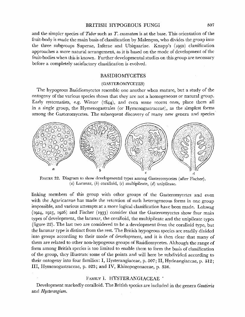
BRITISH HYPOGEOUS FUNGI
and the simpler species of Tuber such as T. excavatum is at the base. This orientation of the fruit-body is made the main basis of classification by Malengon, who divides the group into the three subgroups Superae, Inferae and Ubiquariae. Knapp's (1950) classification approaches a more natural arrangement, as it is based on the mode of development of the fruit-bodies when this is known. Further developmental studies on this group are necessary before a completely satisfactory classification is evolved.
BASIDIOMYCETES
(GASTEROMYCETES) The hypogeous Basidiomycetes resemble one another when mature, but a study of the
ontogeny of the various species shows that they are not a homogeneous or natural group. Early systematists, e.g. Winter (I844), and even some recent ones, place them all in a single group, the Hymenogastrales (or Hymenogastraceae), as the simplest forms
among the Gasteromycetes. The subsequent discovery of many new genera and species
QOiX~~~~~~~~~~~~~~~~~secies
~a \? ~~~b c d
FIGURE 22. Diagram to show developmental types among Gasteromycetes (after Fischer). (a) Lacunar, (b) coralloid, (c) multipileate, (d) unipileate.
linking members of this group with other groups of the Gasteromycetes and even with the Agaricaceae has made the retention of such heterogeneous forms in one group impossible, and various attempts at a more logical classification have been made. Lohwag (1924, I925, 1926) and Fischer (I933) consider that the Gasteromycetes show four main types of development, the lacunar, the coralloid, the multipileate and the unipileate types (figure 22). The last two are considered to be a development from the coralloid type, but the lacunar type is distinct from the rest. The British hypogeous species are readily divided into groups according to their mode of development, and it is then clear that many of them are related to other non-hypogeous groups of Basidiomycetes. Although the range of form among British species is too limited to enable them to form the basis of classification of the group, they illustrate some of the points and will here be subdivided according to their ontogeny into four families: I, Hysterangiaceae, p. 507; II, Hydnangiaceae, p. 512; III, Hymenogastraceae, p. 523; and IV, Rhizopogonaceae, p. 536.
FAMILY I. HYSTERANGIACEAE'
Development markedly coralloid. The British species are included in the genera Gautieria and Hysterangium.
507

-????- \ .?- ?
`- ?--? . =, '''. j t ?.,=`'
....?.:'..=.. : --?'- ?'-?. ?-... i,_.-=-., r'?
S L=-'=-
I.
"'
;? " :\-;2??l;b; '?"r r\ ?- -??-
t.r'_T.:=.? ?r:l==:?. ,... '-l-Li',J ;C??-i-?4i-i: ,i.
?-- '' j;s
?; ;r
:.,;?s.f;a'9a=:t:
.-;.- 4i--.C-,.-~ -Y i~~~~~c ~ ~ ..
; t I
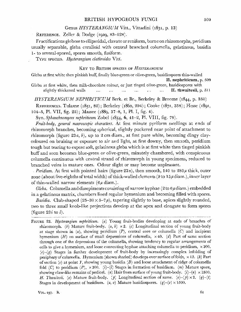
BRITISH HYPOGEOUS FUNGI
Genus HYSTERANGIUM Vitt., Vittadini (I831, p. 13) REFERENCE. Zeller & Dodge (1929, 83-128). Fructifications globose to ellipsoidal, clavate or reniform, borne on rhizomorphs, peridium
usually separable, gleba coralloid with central branched columella, gelatinous, basidia 1- to several-spored, spores smooth, fusiform.
TYPE SPECIES. Hysterangium clathroides Vitt.
KEY TO BRITISH SPECIES OF HrSTERANGIUM
Gleba at first white then pinkish buff, finally blue-green or olive-green, basidiospores thin-walled H. nephriticum, p. 509
Gleba at first white, then milk-chocolate colour, or just tinged olive-green, basidiospores with
slightly thickened walls ... ... .. ... ... .. H. thwaitesii, p. 511
HYSTERANGIUM NEPHRITICUM Berk. et Br., Berkeley & Broome (I844, p. 350) REFERENCES. Tulasne (1851, 82); Berkeley (I860, 294); Cooke (1871, 358); Hesse (1891,
104-5, P1. VII, fig. 25); Massee (1889, 37-8, 1, P1. I, fig. 4). Syn. Sphanchnomyces nephriticum Zobel (1854, 6, 41-2, P1. VIII, fig. 79). Fruit-body, general macroscopic characters. At first minute pyriform swellings at ends of
rhizomorph branches, becoming spherical, slightly puckered near point of attachment to
rhizomorph (figure 23a, b), up to 2 cm diam., at first pure white, becoming dingy clay- coloured on bruising or exposure to air and light, at first downy, then smooth, peridium tough but tearing to expose soft, gelatinous gleba which is at first white then tinged pinkish buff and soon becomes blue-green or olive-green, minutely chambered, with conspicuous columella continuous with central strand of rhizomorph in young specimens, reduced to
branched veins in mature ones. Odour slight or may become unpleasant. Peridium. At first with pointed hairs (figure 23 n), then smooth, 140 to 280# thick, outer
zone (about five-eighths of total width) of thick-walled elements (9 to 15, diam.), inner layer of thin-walled narrow elements (6/# diam.).
Gleba. Columella and dissepiments consisting of narrow hyphae (2 to 6/ diam.) embedded in a gelatinous matrix, chambers lined regular hymenium and becoming filled with spores.
Basidia. Club-shaped (25-30 x 5-7u), tapering slightly to base, apices slightly rounded, two to three small knob-like projections develop at the apex and elongate to form spores
(figure 23 i to 1).
FIGURE 23. Hysterangium nephriticum. (a) Young fruit-bodies developing at ends of branches of rhizomorph. (b) Mature fruit-body. (a, b) x 2. (c) Longitudinal section of young fruit-body at stage shown in (a), showing peridium (P), central core or columella (C) and incipient hymenium (H) on surface of small depressions of columella, x 40. (d) Part of same section through one of the depressions of the columella, showing tendency to regular arrangements of cells to give a hymenium, and loose connecting hyphae attaching columella to peridium, x 300. (e)-(g) Stages in further development of fruit-body by increasingly complex infolding of periphery of columella. Hymenium (shown shaded) develops over surface of folds, x 12. (h) Part of section (e) at point Y, showing young basidia (B) and loose attachment of ridge of columella fold (C) to peridium (P), x 300. (i)-(l) Stages in formation of basidium. (m) Mature spore, showing claw-like remains of pedicel. (n) Hair from surface of young fruit-body. (i)-(n) x 1500. H. Thwaitesii. (o) Mature fruit-body. (p) Longitudinal section of same. (o)-(p)x2. (q)-(t) Stages in development of basidium. (u, v) Mature basidiospores. (q)-(v) x 1500,
Vol, 237. B,
509
LILIAN E. HAWKER ON
Basidiospores. Almost sessile, thin-walled, with homogeneous contents, elongated and tapering at both ends, with a short claw-like outgrowth at the point of attachment and with rounded or sometimes pointed apices, 15-20 x 5-6u (figure 23m), pale brown.
Development. The smallest healthy fruit-body examined (figure 23a) was 2*0 mm long x 1 0 mm wide. The peridium was an extension of the outer layer of the parent rhizomorph but was slightly thicker. It consisted of loosely interwoven hyphae but was more compact than the periphery of the rhizomorph and was itself more compact towards the outside than next the central core or columella. The latter was continuous with the firm central strand of the rhizomorph and consisted of smaller elements more closely inter- woven than those of the peridium. These were arranged more irregularly than the elements of the central strand of the rhizomorph which were mostly parallel to one another. At a number of points the outer edge of the columella was slightly depressed (figure 23c), indicating the beginnings of the development of folds resembling those described for other species. These depressions were lined with a definite layer of radially arranged oblong cells constituting the beginnings of the hymenium (figure 23 d). Hand sections of slightly older specimens showed that the depressions had become deeper and that the hymenial layer was more clearly defined. Serial sections of a larger fruit-body (ellipsoidal in shape and of size 3*5 x 2-0 mm) showed a definite differentiation of the peridium into a thin closely interwoven outer zone and a less compact inner zone. The columella was of denser more homogeneous structure than in the younger specimen. Deep compound wrinkles had developed at the margin of the columella (figure 23e,f). These folds had obviously developed by growth of the apices, the hyphae of which had fanned out and become firmly attached to but distinct from the peridium (figure 23 h). The hymenium was well developed over the interior of the folds up to but not including the apical region in contact with the peridium. In yet older fruit-bodies, nearly spherical in shape and of size up to 10 mm in diameter (figure 23g), the peridium was rather thicker and had lost the characteristic pointed hairs seen in young specimens. The columella occupied relatively less volume than in younger fruit-bodies, and the wrinkling of the fertile peripheral parts had become so complex that it had the appearance in section of numerous small closed chambers. Serial sections showed that these were merely pockets in the complex folds.
Thus the development of the fruit-bodies of Hysterangium nephriticum is essentially similar to that of H. clathroides (Rehsteiner 1892) and H. stoloniferum var. americanum (Fitzpatrick I913) .
Habitat and periodicity. Near surface under leaves in calcareous woods usually near large roots, surrounded by mass of pure white flocculent mycelium, which aggregates to form rhizomorphs which bear the fruit-bodies, mycelium almost certainly directly connected with mantles of small mycorrhizal roots. At all times of year except in very cold or very dry periods, fruit-bodies readily aborting with onset of unfavourable conditions. Fairly common. Previously recorded from Bristol area. Collected during present study in Gloucestershire (Cotswold Hills near Wotton-under-Edge, 109, 326, 582, 625) and Somerset (Brockley Coombe, 378, 399, 592, 624; Cleeve, 698). Usually under beech. The specimens from Cleeve were found under pine and differed from those under beech in the reddening of the peridium on exposure.
510
BRITISH HYPOGEOUS FUNGI
HYSTERANGIUM THWAITESII Berk. et Br., Berkeley & Broome (1848, P. 267) REFERENCES. Tulasne (1851, 82-3); Berkeley (i860, 294); Cooke (1871, 358); Hesse
(1891, 105, P1. VII, figs. 20, 46); Massee (1889, 38, P1. IV, fig. 80). Syn. Splanchnomyces Thwaitesii Zobel (I854, 42). Fruit-body, general macroscopic characters, spherical or slightly irregular (figure 23o) 1 to
2 cm diam., silky white, becoming rufous when bruised or exposed, peridium thin, tough, gleba soft, at first white, then colour of milk chocolate or with olive-green tinge, numerous convoluted folds filled with spores (figure 23p), columella disappearing early, and in mature
specimens visible only as a few narrow branched strands, odour slight. Peridium. 160 to 1804u thick, splitting when dry, composed of interwoven colourless
hyphae, 3 to 5,u diam. Gleba. With a sterile outer zone and dissepiments formed of gelatinous hyphae.
Hymenium breaking down early. Basidia. Club-shaped, with blunt apex and tapering slightly to base (40-50 x 10-1 2,),
2- to 4-spored. In a large collection from Stoke Wood, Exeter, the basidia were almost all 2-spored; in a single fruit-body from Cleeve, Somerset they were mostly 4-spored (figure 23 q to t).
Basidiospores. On short sterigmata, ovoid, tapering to base with apex varying from blunt to pointed, 17-21 x 6-8,u, pale brown, with rather thicker walls than those of H. nephriticum (figure 23u, v).
Habitat. Under leaves of deciduous trees, sometimes in rather wet situations, usually surrounded by abundant white mycelium. At all times of year except in dry or cold periods. Probably less common than H. nephriticum. Recorded from Bristol area and found during present investigation in Somerset (Cleeve, 72) and Devonshire (Stoke Wood, Exeter, 118).
Genus GAUTIERIA Vitt., Vittadini (I83I, p. 25) REFERENCE. Dodge & Zeller (I934, 692); Zeller (1949). Fructifications spherical to irregular, borne on a simple or branched rhizomorph or
mycelial strand, columella present in young specimens, variable in mature ones, peridium evanescent, cavities of gleba formed by infolding of periphery as in Hysterangium, basidia clavate, usually 2-spored, spores ellipsoidal to fusiform, longitudinally striate.
TYPE SPECIES. Gautieria morchellaeformis Vitt.
Dodge & Zeller (I934) include in this genus some species with a persistent peridium. Recent studies show that some of these are obviously more appropriately placed in other
genera, and it has been suggested that the genus Gautieria should be restricted to those forms in which the peridium is absent or evanescent (Corner and Hawker I953).
GAUTIERIA MORCHELLAEFORMIS Vitt., Vittadini (1831, p. 26, P1. III, fig. 6) REFERENCES. Tulasne (1851, 62); Winter (1883, 873); Hesse (I891, 109-10); Zobel (1854,
34, P1. VII, fig. 62); Hawker (i952, 282, fig. 2, A-F).
Fruit-body, general macroscopic characters. At first spherical to ellipsoidal, then irregularly lobed and convoluted, up to 2 cm. diam., arising from mass of grey-white mycelium and borne on much-branched poorly defined mycelial strands, white, becoming dingy yellow and finally dull brown, soft, spongy, peridium disappearing early, leaving much-infolded
gleba exposed, columella prominent at first, soon almost disappearing by continued 62-2
511

LILIAN E. HAWKER ON
infolding of periphery to give labyrinthine chambers (figure 24a to d). Odour none at first, later strong and somewhat unpleasant.
Peridium. Present only in very early stages as loose mass of inflated hyphae (figure 24e). Gleba. At first with slightly infolded margin, later complex with branched and anasto-
mosing folds lined with hymenium which is continuous with outer sterile layer of exposed edges of folds (figure 24f).
Basidia. Clavate, tapering gradually to base and slightly to flat top, 40-50 x 8-12d, bearing 2 spores on short slender pedicels (figure 24g, h).
Basidiospores. Lemon-shaped, 12-18-24x 8-11-12-5,u, exospore thick, longitudinally wrinkled, dark brown (figure 24i,j).
Habitat and periodicity. Collections made in present investigation were all from well- drained slopes in different parts of a beechwood, near Wotton-under-Edge, Gloucester- shire (14, 110, 596) (Hawker 1952). Collections were made at various times of year after rain. Fruit-bodies shrivel and abort in dry periods. This is the only record of this fungus in Britain.
Rehsteiner (1892) suggested that the Clathraceae may have originated from a form like
Hysterangium. Later Fischer (I900) suggested that Protubera and Phallogaster might represent transitional forms, and Fitzpatrick (I913) proposed a series of increasing complexity thus: Gautieria -> Chamonixia -+ Hysterangium - Protubera -PIPhallogaster -- Clathraceae. The structure of the peridium, the gelatinous, greenish grey, often evil-smelling gleba, its mode of
development and the shape and arrangement of the spores in Hysterangium, all suggest relationship with Clathrus. The structure of the rhizomorph of Hysterangium nephriticum resembles those of Phallus impudicus and Mutinus caninus, and is unlike that of other Gastero-
mycetes such as Hymenogaster tener and Lycoperdon sp. (Townsend I952). The development of the hymenial folds from the periphery of the columella links Gautieria with Hysterangium. In Bottomley's (I948) classification of the Gasteromycetes the two genera are placed close
together. It is doubtful, however, whether Gautieria is more primitive, as suggested by Fitzpatrick. The spores of this genus are definitely more complex than those of Hyster- angium, and the presence of a peridium is so uniform in the Gasteromycetes and it is so well developed in Hysterangium that its loss in Gautieria may well be a secondary develop- ment. This genus would then be best considered as a somewhat degenerate side line from a Hysterangium-like ancestor. The two genera would then have diverged from the common ancestor, which may have had smooth spores and a poorly developed peridium.
FAMILY II. HYDNANGIACEAE
Spores spherical or ellipsoidal, echinulate or verrucose. The early development of most
species is unknown, since differentiation is more or less complete long before full size is attained. In those which have been studied the development is unipileate, but in some
species the columella may completely disappear before the fruit-body is mature.
Genus HYDNANGIUM Wallr., Wallroth (I839, p. 465) REFERENCE. Dodge & Zeller (I936). Peridium thin, membranaceous, no sterile base, chambers numerous, gyrose, spores
spherical, echinulate to verrucose borne on long sterigmata. TYPE SPECIES. Hydnangium carneum Wallr.
512
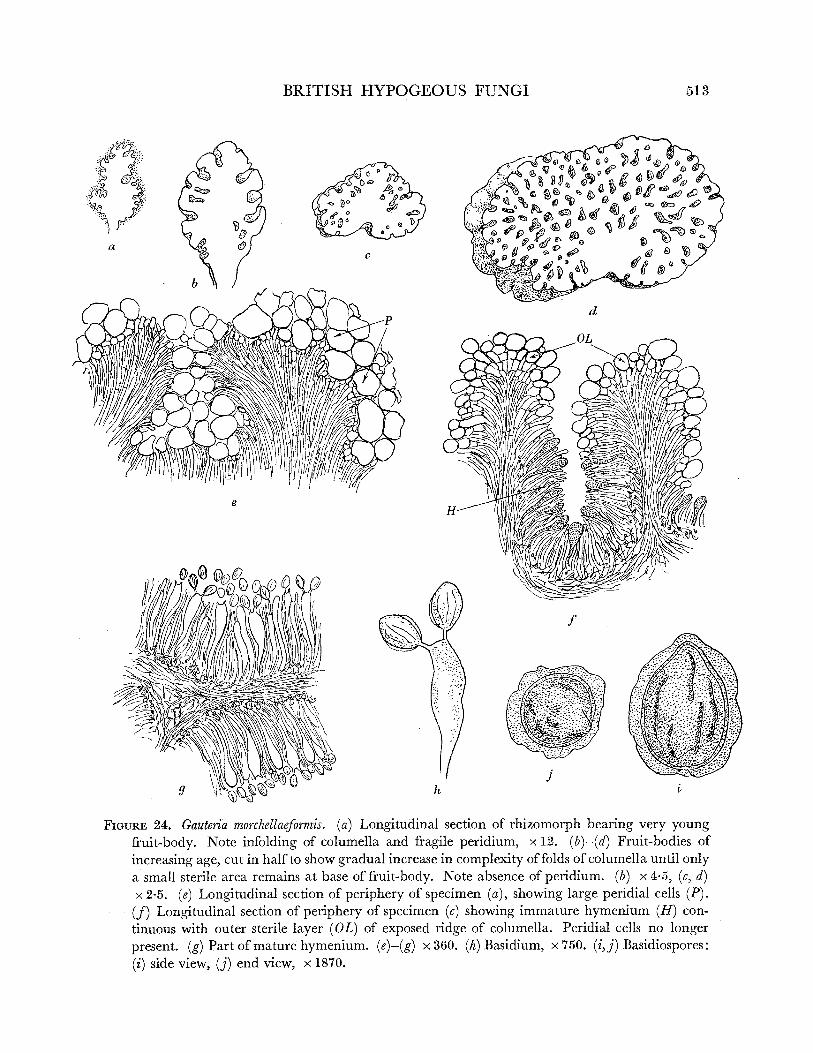
BRITISH HYPOGEOUS FUNGI
d
e
f
0 h 1~
FIGURE 24. Gauteria morchellaeformis. (a) Longitudinal section of rhizomorph bearing very young fruit-body. Note infolding of columella and fragile peridium, x 12. (b)-(d) Fruit-bodies of increasing age, cut in half to show gradual increase in complexity of folds of columella until only a small sterile area remains at base of fruit-body. Note absence of peridium. (b) x 45, (c, d) x2-5. (e) Longitudinal section of periphery of specimen (a), showing large peridial cells (P). (J) Longitudinal section of periphery of specimen (c) showing immature hymenium (H) con- tinuous with outer sterile layer (OL) of exposed ridge of columella. Peridial cells no longer present. (g) Part of mature hymenium. (e)-(g) x360. (h) Basidium, x750. (i,j) Basidiospores: (i) side view, (j) end view, x 1870.
513
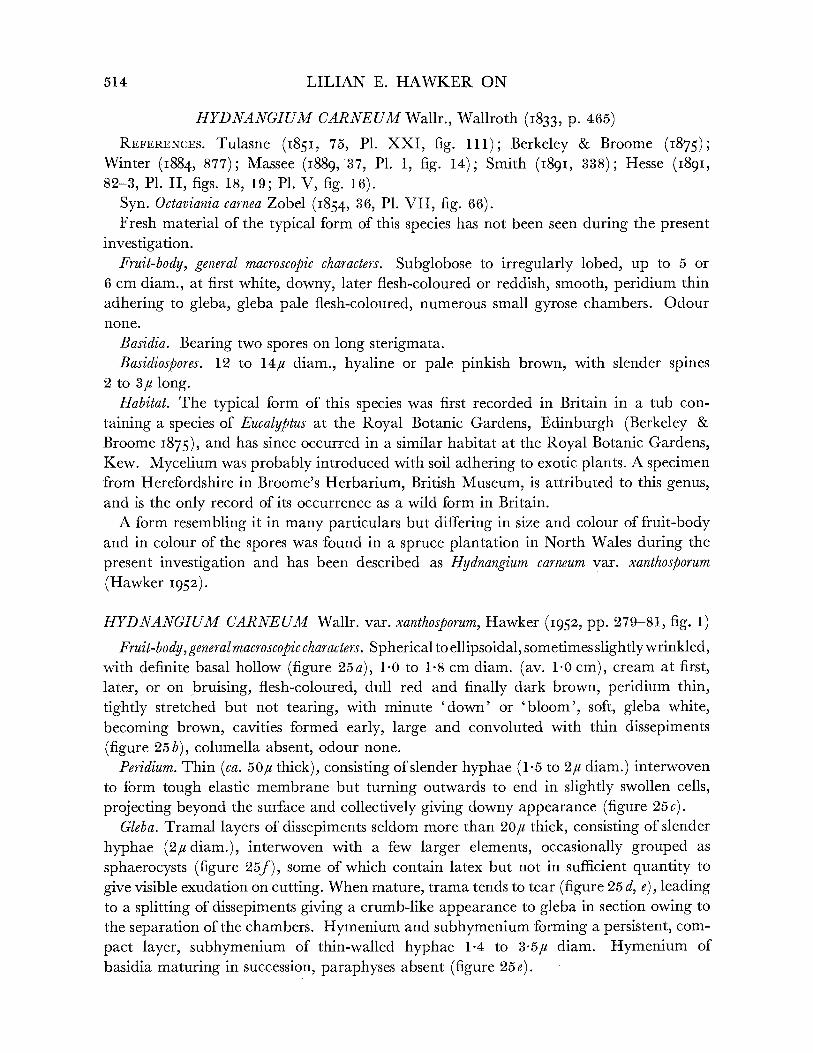
LILIAN E. HAWKER ON
HYDNANGIUM CARNEUM Wallr., Wallroth (1833, p. 465) REFERENCES. Tulasne (185I, 75, P1. XXI, fig. 111); Berkeley & Broome (1875);
Winter (1884, 877); Massee (I889, 37, P1I. I, fig. 14); Smith (I89I, 338); Hesse (I891, 82-3, P1. II, figs. 18, 19; P1. V, fig. 16).
Syn. Octaviania carnea Zobel (1854, 36, P1. VII, fig. 66). Fresh material of the typical form of this species has not been seen during the present
investigation. Fruit-body, general macroscopic characters. Subglobose to irregularly lobed, up to 5 or
6 cm diam., at first white, downy, later flesh-coloured or reddish, smooth, peridium thin
adhering to gleba, gleba pale flesh-coloured, numerous small gyrose chambers. Odour none.
Basidia. Bearing two spores on long sterigmata. Basidiospores. 12 to 14#u diam., hyaline or pale pinkish brown, with slender spines
2 to 3,u long. Habitat. The typical form of this species was first recorded in Britain in a tub con-
taining a species of Eucalyptus at the Royal Botanic Gardens, Edinburgh (Berkeley & Broome 1875), and has since occurred in a similar habitat at the Royal Botanic Gardens, Kew. Mycelium was probably introduced with soil adhering to exotic plants. A specimen from Herefordshire in Broome's Herbarium, British Museum, is attributed to this genus, and is the only record of its occurrence as a wild form in Britain.
A form resembling it in many particulars but differing in size and colour of fruit-body and in colour of the spores was found in a spruce plantation in North Wales during the
present investigation and has been described as Hydnangium carneum var. xanthosporum (Hawker 1952).
HYDNANGIUM CARNEUM Wallr. var. xanthosporum, Hawker (I952, pp. 279-81, fig. 1)
Fruit-body, general macroscopic characters. Spherical to ellipsoidal, sometimes slightly wrinkled, with definite basal hollow (figure 25a), 1.0 to 1-8 cm diam. (av. 1.0 cm), cream at first, later, or on bruising, flesh-coloured, dull red and finally dark brown, peridium thin, tightly stretched but not tearing, with minute 'down' or 'bloom', soft, gleba white, becoming brown, cavities formed early, large and convoluted with thin dissepiments (figure 25b), columella absent, odour none.
Peridium. Thin (ca. 50,u thick), consisting of slender hyphae (1-5 to 2,u diam.) interwoven to form tough elastic membrane but turning outwards to end in slightly swollen cells, projecting beyond the surface and collectively giving downy appearance (figure 25c).
Gleba. Tramal layers of dissepiments seldom more than 20,u thick, consisting of slender
hyphae (2u diam.), interwoven with a few larger elements, occasionally grouped as
sphaerocysts (figure 25f), some of which contain latex but not in sufficient quantity to
give visible exudation on cutting. When mature, trama tends to tear (figure 25 d, e), leading to a splitting of dissepiments giving a crumb-like appearance to gleba in section owing to the separation of the chambers. Hymenium and subhymenium forming a persistent, com-
pact layer, subhymenium of thin-walled hyphae 1*4 to 3.5# diam. Hymenium of basidia maturing in succession, paraphyses absent (figure 25e).
514

BRITISH HYPOGEOUS FUNGI
Basidia. Clavate, widest part about one-third of length from apex, 34 x 11 5,u av. size, borne on definite stalk cell, bearing two spores on long sterigmata (figure 25g, to 4)5 occasion-
ally 1- or 3-spored. Basidiospores. Spherical, 10 to 14-5u, av. 11-5 (excluding spines), at first hyaline and
smooth, later pale yellow-brown, with blunt spines (ca. 3,u long), amyloid, staining black with Melzer's solution (figure 25i to 1).
Habitat. Just below needle cover in dense spruce plantation, gregarious, no obvious
mycelium observed. Mature specimens collected in September in Caernarvonshire (Bettws-y-coed, 247, 251, 255, 257; Vaynol Park, Bangor, 228).
This variety differs from the typical form in the smaller size, more regular shape, in the colour of the fruit-body and gleba, which are flesh pink to red in the type material, and in the yellow-brown spores.
Genus ARCANGELIELLA Cav., Cavara (I900, pp. 117-128) REFERENCE. Zeller & Dodge (I936, 612). Syn. Octaviania Vitt. (I83I, 15-20). Peridium tough, gleba chambered, sterile base or columella usually present, exuding
latex when cut, spores spherical, echinulate or verrucose. The generic name Arcangeliella is preferred to Octaviania, since, according to Zeller &
Dodge (I936), the latter was based on type material which proved to be a specimen of
Melanogaster. The name Octaviania would thus become invalid. The present writer has had no opportunity of verifying this statement. The name Arcangeliella has been generally adopted by recent European workers, e.g. Soehner (I949).
KEY TO BRITISH SPECIES OF ARCANGELIELLA
Fruit-body becoming purple-black or greenish blue when bruised, latex hyphae few, latex not exuding freely from cut surfaces, basidiospores coarsely echinulate ... ...
A. asterosperma, p. 515 Fruit-body becoming rufous on exposure, never becoming green, latex exuding copiously from
cut surfaces, basidiospores with few short spines ... ... ... A. stephensii, p. 517
ARCANGELIELLA ASTEROSPERMA (Vitt.) Zeller & Dodge, Zeller & Dodge (I936, p. 631)
REFERENCES. Tulasne (1843, P1. XVII, fig. 21); (1851, 77-8, PI. XI, fig. 1); Berkeley & Broome (1847); Zobel (1854, 35-6, P1. VII, fig. 64); Berkeley (i860, 292); Cooke (1871, 355); Winter (1884, 878, figs. 1-3 on p. 871); Massee (1889, 31, P1. I, fig. 2); Hesse (1891, 72-4, PI. III, figs. 1-7; P1. V, fig. 15; P1. VI, fig. 4).
Syn. Octaviania asterosperma Vitt., Vittadini (1831, p. 17, P1. III, fig. 7; P1. V, fig. 9a). Fruit-body, general macroscopic characters. Globose or sometimes irregular or slightly
flattened, up to 3 cm diam., usually smaller, peridium smooth, tending to split, at first white, often slightly veined blue, becoming purple-black or greenish with age or when
bruised, gleba at first white, then honey-coloured with white dissepiments, then dark clay- coloured, soft, gelatinous, no obvious exudation of latex when cut, numerous chambers, sinuous but not hollow, columella or sterile base present, much reduced in old specimens (figure 25m, n). Odour pleasant, nutty, sweet.
515

LILIAN E. HAWKER ON
b '., X-J9
I.
U t y~~~~~~~~~~~~~~~~~
~LOKO It -: ." . -V . .
.
4, : -IPO
Q~~~~~~
'I
`?? C- K<1
&C p~Ic
S3
m K
n,
516
a

BRITISH HYPOGEOUS FUNGI
Peridium. About 100,u thick, of irregularly interwoven hyphae (1 5 to 2/u diam.), running parallel to the surface to give a firm membrane but not sharply distinguished from tramal
hyphae. Gleba. Tramal hyphae 1*5 to 2/i diam., in loose bundle in the plane of the dissepiment,
latex hyphae few, rather wider, not conspicuous, dissepiments 100 to 150fu thick. Basidia. In regular hymenium, at first pyriform then more or less cylindrical, 40 x 101u,
usually 2-spored (figure 250 to 3).
Basidiospores. Globose, coarsely echinulate, pale brown, 11 to 14#u, borne on long tapering sterigmata, shed into cavity when mature (figure 25p).
Habitat. Usually in humus under leaves in calcareous beechwoods, found only after wet
period, mycelium said to be abundant, but not seen in collections made in three Cotswold beechwoods near Wotton-under-Edge, Gloucestershire (171, 543-5, 729), during present investigation. Fairly common, widely distributed in southern England.
ARCANGELIELLA STEPHENSII (Berk. et Br.) Zeller & Dodge, Zeller & Dodge (1936, p. 413)
REFERENCES. Zobel (I854, 36, P1. VII, fig. 67); Cooke (1871, 355); Massee (I889, 31-2, P1. I, fig. 3).
Syn. Hydnangium stephensii Berk. et Br. (1846, 13, 352). Octaviana stephensii Tulasne (I85I, 78, P1. XXI, fig. 6).
Fruit-body, general macroscopic characters. Globose, puckered at base, up to 1 cm. diam., cream, tinged orange, later becoming rufous, smooth, soft, gleba white, exuding white
latex, said by some observers to become rufous on exposure but remaining white in single fresh specimen seen (figure 25 q, r). Odour none.
Peridium. Thin, ca. 100,u, of interwoven hyphae running parallel to surface, firm but not
sharply marked off from gleba. Gleba. Dissepiments 75 to 150,u thick, tramal hyphae 2 to 3,/ diam., brown, irregularly
arranged, loosely intertwined, latex hyphae large and conspicuous, of irregular diam.
(up to 10/). Basidia. In regular hymenium, clavate, brown, 40 x 7/, bearing 2 spores on long
sterigmata (figure 25 s t 3).
FIGURE 25. Hydnangium carneum var. xanthosporum. (a) Mature fruit-body, showing basal depression. (b) Longitudinal section of same, showing thin peridium and numerous hollow chambers. (a, b) x 1. (c) Interwoven hyphae of outer layer of peridium ending blindly at surface and giving downy appearance of fruit-body, x 750. (d) Longitudinal section of part of periphery of mature fruit-body, showing thin peridium (P), hollow chambers (C) and breakdown of trama (T), x 40. (e) Portion of (d) enlarged, showing hymenium (H), torn tramal hyphae (T) and latex vessels (L), x 200. (f) Sphaerocysts from trama just beneath peridium, seen in tangential section, x 750. (glto 4) Developing basidia. (h) Unusual 3-spored basidium. (g)-(h) x 750. (i)-(l) Basidiospores, showing irregular blunt spines. x 1870. Arcangeliella asterosperma. (m) Mature fruit-body. (n) Longitudinal section of same, x 1-3. (01 to 3) Stages in development of basidia, x750. (p) Basidiospore, showing blunt spines, x 1870. A. stephensii. (q) Mature fruit-body. (r) Longitudinal section of same, x 11. (s to 3) Stages in development of basidia, x 750. (y-z) Basidiospores, showing short spines, x 1870.
51.7
VOL. 237. B. 63
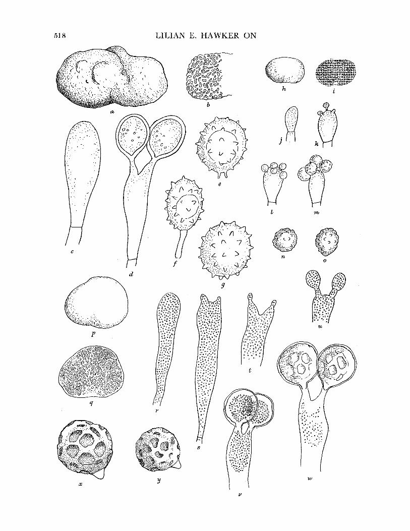
LILIAN E. HAWKER ON
hi
b
j
fm
. 3
.7
0t n/ 0
9
6.6 06 .6666
6.666b
6 6b6 0 66 6.6.c 66 "<.6
6o 666 .6
66.6.64 66)
r00
y
0 0 . ;?a *. 0 Ob
.6 6 6gt 66 6.6 66
.'6. 6 .666 66.6
66 \ . . .6
" .O .1 6 o,
d. o . 6 6 6
L66 6 6 666
.6.6.666.6
6U6. 66 6.6 cO
6d 6 ,
j.6.6o SaD O?
w
Xv
518.
c
P
q
x
?,4 ? -? ?:? ... ?,
:: I
I?

BRITISH HYPOGEOUS FUNGI
Basidiospores. Subglobose or broadly elliptical, pale brown, thick epispore with few short spines, 14-5-15-5 x 11-12u, shed into cavity when mature (figure 25t).
Habitat. 1 to 2 in. deep in soil, among roots of ivy, etc., often under lime trees. Found during present investigation in Leigh Woods, Bristol (510), and previously recorded from the same locality, from other places in the Bristol area and from Wraxall, Somerset.
Genus STEPHANOSPORA Pat., Patouillard (I914, p. 349) Peridium thin, flocculent or lacking, basidia cylindrical, spores ovoid or spindle-shaped,
echinulate with small basal collar or scar. TYPES SPECIES. Stephanospora carotaecolor.
STEPHANOSPORA CAROTAECOLOR (Berk. et Br.) Pat.,. Patouillard (1914, p. 349); Zeller & Dodge (1935, p. 365)
Syn. Hydnangium carotaecolor Berk. et Br. in Berkeley & Broome (1844, 13, 351). Tulasne
(I851, 75-6, P1. XXI, fig. 4); Berkeley (I860, 293-4); Cooke (I871, 358); Winter
(I884, 877-8, fig. on p. 871); Massee (1889, 36-7, P1. I, fig. 6). Octaviania carotaecolor Zobel (I854, 36, P1. VII, fig. 65).
Fruit-body, general macroscopic characters. Irregularly globose or more often oblong, much wrinkled, about 2-0 x 1-0 cm (figure 26a), orange (colour of ripe carrot), soft and fleshy, peridium at first downy, thin or disappearing, revealing sponge-like gleba, moist when cut, trama bright orange-red, hymenium paler or white, lining large convolute chambers, sterile base lacking (figure 26 b). No mycelial attachment visible. Odour of decaying puff balls.
Peridium. At first composed of loose hyphae, later disappearing. Gleba. Dissepiments 100 to 200,u thick, consisting of trama of inflated cells of irregular
size up to 20,u diam. interspersed with slender hyphae, some of which extend across cavities. Basidia. In regular hymenium, at first clavate, later elongated 20-40 x 7-10,u usually
2-spored with short sterigmata (figure 26c, d). Basidiospores. Pale yellow-brown, subglobose to ovoid, 11-16 x 9-1 2u (excluding spines),
showing projection or scar at point of attachment but not a definite collar, sparsely echinulate, spines short (1.0 to 1-5,), conical, pointed (figure 26e-g).
Habitat. On or near surface of leaf layer under trees, particularly conifers. Collected
during present investigation at Bristol (Leigh Woods, 941), in Somerset (Cleeve, 836) and Sussex (Friston, 536), previous records from the Bristol area and Swanage, Dorset.
FIGURE 26. Stephanospora carotaecolor. (a) Mature fruit-body. (b) Longitudinal section of same, showing absence of peridium and of sterile base, large convolute chambers. (a, b) x 3. (c, d) Stages in development of basidia, x 1870. (e)-(g) Basidiospores showing short conical spines, projection at point of attachment of spore to sterigma (e) or deciduous sterigma (f). (e)-(g) x 1870. Sclerogaster compactus. (h) Mature fruit-body. (i) Longitudinal section of same, showing minute chambers. (h, i) x 3. (j)-(m) Stages in development of basidia, showing irregularly arranged sessile spores of indefinite number, x 1870. (n, o) Basidiospores, x 1870. Wakefieldia macrospora. (p) Mature fruit-body. (q) Longitudinal section of same, showing numerous chambers and immature peripheral patches. (p, q) x 3. (r)-(w) Stages in development of basidia, x 1870. (x, ,y) Basidiospores, showing dimpled thickening and hyaline papillae at points of attachment to sterigmata, x 1870.
63-2
519

LILIAN E. HAWKER ON
Genus SCLEROGASTER Hesse, Hesse (189I, p. 84) Mycelium abundant, fruit-bodies small, peridium white, fluffy, not separable from
gleba, gleba yellow, columella not always present, chambers minute, basidia 4- to 8-spored, spores small, spherical, minutely verrucose or echinulate.
TYPE SPECIES. Sclerogaster compactus (Tul.) Sacc.
SCLEROGASTER COMPACTUS (Tul.) Sacc., Saccardo (1895, p. 170) REFERENCE. Dodge & Zeller (1936, 573). Syn. Octaviania compacta Tul. (1844, 55); (I851, 79, P1. XI, fig. 3); Massee (1889, 1, P1. I,
fig. 12); Smith (I891, 337). Hydnangium compactum Quelet (I870-5). Octaviania compacta Kuntze (1891).
Fruit-body, general macroscopic characters. Spherical, small, less than 1 cm diam., pure white, covered by loose flocculent mycelium, firm to hard, gleba greenish yellow, minutely chambered (figure 26h, i). Columella absent in mature specimens. Odour none.
Peridium. 100 to 300u thick, not separable, outer layer of hairs 2 to 3#u thick, inner layer of inflated thin-walled cells, variable size, up to 15l diam., becoming smaller towards inside and then resembling the tramal elements.
Gleba. Tramal elements similar to those of inner peridium. Basidia. At first ellipsoidal then cylindrical (10-15 x 5-8u) (figure 26j-m), evanescent,
3- to 6-spored, spores shed into and filling cavity at early stage. Basidiospores. Spherical, 4 to 6,u diam., thick-walled, sparsely and minutely warted, pale
yellow-brown in mass, borne on very short sterigmata (figure 26n, o). Habitat. Among roots of herbaceous plants in woods, fruit-bodies produced in large
numbers from mass of white flocculent mycelium. Found during present investigation at Blaise Castle, Bristol, (62, 279) and at Cleeve, Somerset, 983, previously reported from Leigh Woods, Bristol, and Kent (Shoreham and Otford).
Doubtful species SCLEROGASTER BROOMEIANUS Zeller & Dodge, Zeller & Dodge (1935, p. 370)
This species is said by Dodge & Zeller (1936) to differ from last in smaller flattened fruit- bodies (0.5 x 0.3 cm), separable, thicker (280#) peridium composed of prosenchymatous hyphae (3 to 4u thick), wider dissepiments consisting of slender loosely interwoven hyphae embedded in a gel, small clavate basidia (7-8 x 3-4,u), minutely and sparsely echinulate spores (5 to 7# diam.). Since Dodge & Zeller's description was compiled from dried material the appearance of the peridial and tramal hyphae and measurements of cells are probably unreliable as a guide, since the inflated cells of Sclerogaster compactus tend to collapse and become unrecognizable in dried material.
Habitat. Among grass roots. Zeller & Dodge (1935) base this species on Broome's material collected near Shoreham, Kent.
Doubtful species SCLEROGASTER LANATUS Hesse, Hesse (I89I, p. 85-6, P1. V, fig. 11); Dodge & Zeller (1936, p. 568)
Hesse considered this species to be identical with Tulasne's Octaviania compacta, but both Soehner (1949) and Dodge & Zeller (1936) consider it to be a distinct species.
520)
BRITISH HYPOGEOUS FUNGI
It is said to differ from Sclerogaster compactus in the thick (280,) 2-layered peridium of which the outer layer consists of loosely interwoven hyphae with abundant crystal deposits, readily tearing away, and the inner resembles the tramal elements, i.e. slender periclinal hyphae embedded in gel, cavities not filled with spores, wider dissepiments (35,u wide), cylindrical basidia (14 x 4,), 4- to 8-spored, and the smooth or nearly so spores 4 to 6#u diam.
Habitat. Under litter layer in coniferous woods. Dodge & Zeller (1936) assign Broome's material from Otford, Kent, to this species.
Excluded species SCLEROGASTER MACROSPORUS Hawker
This species is now described (p. 521) as Wakefieldia macrospora (Hawker) Hawker n.comb., as a result of further studies of its development and the discovery of a related form from Malaya.
The position of the genus Sclerogaster within the Hydnangiaceae is doubtful and its inclusion there rests solely on the spherical spores. Developmental studies are lacking and
reports vary as to the presence or absence of a columella. The irregular number of spores on the basidium suggests a possible affinity with Rhizopogon, and it may well be that the
development of the fruit-bodies is lacunose as in that genus. For the present it is perhaps best to leave Sclerogaster in the Hydnangiaceae, as suggested by Soehner (1949), to await
developmental studies. There is some doubt as to the subdivision of this genus into species. Among modern
authors Bataille (1923) considers that the genus contains the single species S. compactus, while Soehner (I949) and Dodge & Zeller (I936) recognize at least four species based on the thickness of the peridium and small differences in spore size.
An examination of Broome's herbarium specimens and of large collections made at Blaise Castle and at Cleeve, Somerset during this investigation suggests that the British material is all S. compactus at various stages of development. The thickness of the peridium and dissepiments decreases, the peridium loses its loose outer felt of mycelium, and the size of basidia and spores and the degree of echinulation of the spores increase with age. Young specimens from the Blaise Castle material fitted Dodge & Zeller's descriptions of S. lanatus and S. broomeianus, while older specimens were typical of S. compactus. Thus it seems most likely that Broome's specimens, too, should be placed in this species.
Genus WAKEFIELDIA Corner & Hawker (I953) Fruit-bodies spherical, attached to short rhizoidal fibrils or poorly developed rhizo-
morphs at base, white, columella or sterile base absent in mature specimen, basidia 2- to 4-spored, spores spherical, sculpturing plate-like or pitted, not echinulate.
TYPE SPECIES. Wakefieldia striaespora Corner & Hawker.
WAKEFIELDIA MACROSPORA (Hawker) Hawker n.comb.
Syn. Sclerogaster macrosporum Hawker, Hawker (I95I, pp. 216-19). This fungus was first collected by MissJ. Fraymouth at Wotton-under-Edge, Gloucester-
shire, on 31 December 1949. Further collections have been made from the same wood. It
521
LILIAN E. HAWKER ON
was provisionally placed in Sclerogaster (Hawker 1951), although it was pointed out that its characters did not entirely agree with those of any existing genus. Later an examination of hypogeous fungi collected by Corner in Malaya revealed a specimen which differed from S. macrosporus only in the sculpturing of the epispore. The Malayan form was described (Corner & Hawker I953) as the type species of the new genus Wakefieldia to which Sclerogaster macrosporus is transferred as Wakefieldia [macrospora (Hawker) Hawker n.comb.
The youngest available material of these two species shows the chambers already well
developed and no trace of a columella. In W. macrospora the chambers develop first in the centre of the fruit-body and the peripheral ones develop irregularly, so that peripheral patches of non-sporing chambers are present in young material. In the absence of early developmental stages this genus is best placed provisionally in the Hydnangiaceae on account of its spherical spores.
Fruit-body, general macroscopic characters. Globose, wrinkled, attached to a poorly developed rhizomorph from which they readily become detached, leaving small basal depression 1*0 to 2*5 cm diam., dingy white, smooth, peridium thin, often splitting but not separable, gleba at first pinkish purple, becoming brownish purple, cavities numerous, small, empty, developing first in centre of gleba leaving peripheral pockets of immature cavities (figure 26p, q). Odour none.
Peridium. Thin, entire. Gleba. Dissepiments very thin. Basidia. At first club-shaped, then cylindrical and projecting above level of hymenium,
50-70 x 5-9,c, 2-spored, hyaline (figure 26 r to w).
Basidiospores. At first colourless, smooth, pyriform, becoming golden brown, more or less
spherical with colourless papilla at point of attachment to basidium (cf. Stephanospora), thick-
walled, irregularly dimpled or pitted, 13 to 18fl diam. (figure 26x, y). Habitat. Several collections made at or near surface of soil in a calcareous beechwood
near Wotton-under-Edge, Gloucestershire (51, 71, 97, 457), at all times of year. The Hydnangiaceae (excluding Sclerogaster, the position of which has already been dis-
cussed) has been the subject of recent research, particularly in France. Malengon (I931) and Heim (1936, 1948) have developed an early suggestion made by Bucholtz (1903) and have shown certain striking resemblances between the genera Russula and Lactarius among agarics and the Hydnangiaceae. The spherical, spiny, often amyloid, spores and the loosely constructed fruit-body, often showing sphaerocysts and latex cells, of the typical species of
Hydnangiaceae are characteristic also of the Russula and Lactarius group and transition forms between the two groups exist in the tropical species of Elasmomyces, while the family Secotiaceae (not represented in Britain) also provides a link with the agarics. It has been
suggested that the hypogeous species of the Hydnangiaceae have been derived from
primitive forms of Russula, and that these genera should all be grouped in the Astero-
sporeae of which the hypogeous forms would constitute the family Asterogastraceae. This
grouping has much to recommend it as an attempt at a natural arrangement, and a study of the British species of Hydnangium, Arcangeliella and Stephanospora produces some evidence in its favour and no real objection to it. A final decision, however, must await further studies.
522
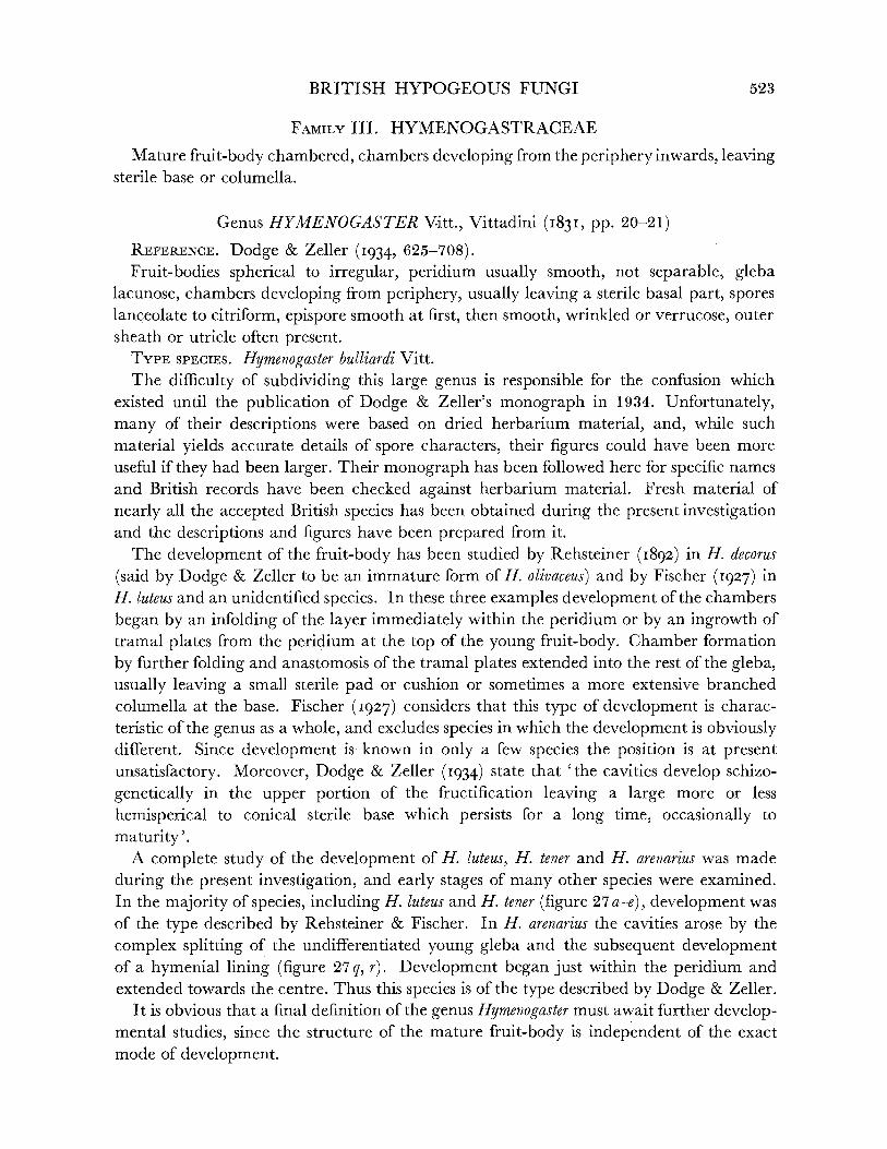
BRITISH HYPOGEOUS FUNGI
FAMILY III. HYMENOGASTRACEAE
Mature fruit-body chambered, chambers developing from the periphery inwards, leaving sterile base or columella.
Genus HYMENOGASTER Vitt., Vittadini (I831, pp. 20-21) REFERENCE. Dodge & Zeller (I934, 625-708). Fruit-bodies spherical to irregular, peridium usually smooth, not separable, gleba
lacunose, chambers developing from periphery, usually leaving a sterile basal part, spores lanceolate to citriform, epispore smooth at first, then smooth, wrinkled or verrucose, outer sheath or utricle often present.
TYPE SPECIES. Hymenogaster bulliardi Vitt. The difficulty of subdividing this large genus is responsible for the confusion which
existed until the publication of Dodge & Zeller's monograph in 1934. Unfortunately, many of their descriptions were based on dried herbarium material, and, while such material yields accurate details of spore characters, their figures could have been more useful if they had been larger. Their monograph has been followed here for specific names and British records have been checked against herbarium material. Fresh material of
nearly all the accepted British species has been obtained during the present investigation and the descriptions and figures have been prepared from it.
The development of the fruit-body has been studied by Rehsteiner (I892) in H. decorus
(said by Dodge & Zeller to be an immature form of H. olivaceus) and by Fischer (1927) in H. luteus and an unidentified species. In these three examples development of the chambers
began by an infolding of the layer immediately within the peridium or by an ingrowth of tramal plates from the peridium at the top of the young fruit-body. Chamber formation
by further folding and anastomosis of the tramal plates extended into the rest of the gleba, usually leaving a small sterile pad or cushion or sometimes a more extensive branched columella at the base. Fischer (I927) considers that this type of development is charac- teristic of the genus as a whole, and excludes species in which the development is obviously different. Since development is known in only a few species the position is at present unsatisfactory. Moreover, Dodge & Zeller (1934) state that 'the cavities develop schizo-
genetically in the upper portion of the fructification leaving a large more or less
hemisperical to conical sterile base which persists for a long time, occasionally to
maturity'. A complete study of the development of H. luteus, H. tener and H. arenarius was made
during the present investigation, and early stages of many other species were examined. In the majority of species, including H. luteus and H. tener (figure 27 a-e), development was of the type described by Rehsteiner & Fischer. In H. arenarius the cavities arose by the
complex splitting of the undifferentiated young gleba and the subsequent development of a hymenial lining (figure 27 q, r). Development began just within the peridium and extended towards the centre. Thus this species is of the type described by Dodge & Zeller.
It is obvious that a final definition of the genus Hymenogaster must await further develop- mental studies, since the structure of the mature fruit-body is independent of the exact mode of development.
523
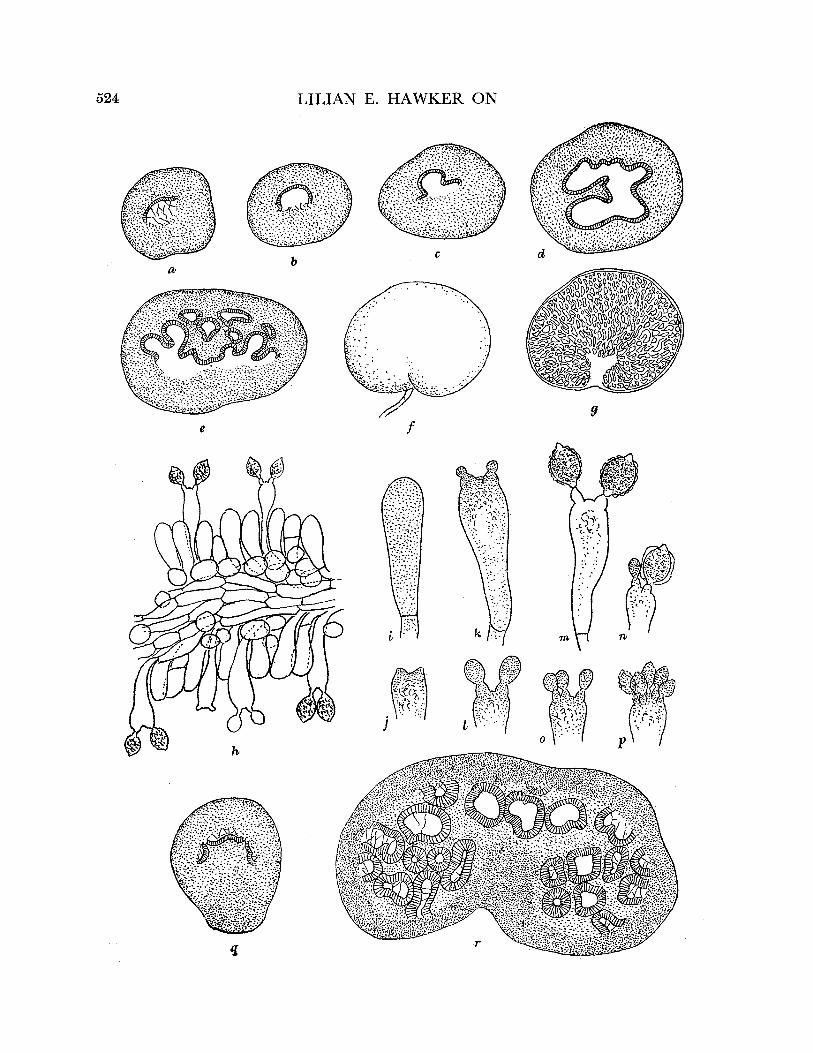
LILIAN E. HAWKER ON
y~~~~~~~~~~~~~~~~~~~~~~~~~- ..
(r ~ ~ ~ ~ ~ ~ ~ (
I ~ ~I L t ~ ~ ~ ~ ~ ~ ~ .. -
o I
I f
524
e
'
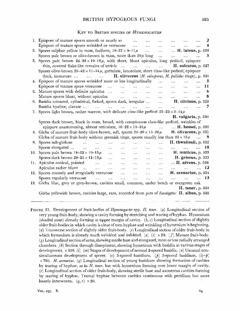
BRITISH HYPOGEOUS FUNGI 525
KEY TO BRITISH SPECIES OF HTMENOGASTER
1. Epispore of mature spores smooth or nearly so ... ... ... ... ... ... 2
Epispore of mature spores wrinkled or verrucose ... ... ... ... ... ... 4 2. Spores sulphur yellow in mass, fusiform, 18-22 x 9-11, ... ... ... H. luteus, p. 526
Spores pale brown or olive-brown in mass, more than 20/, long ... ... ... ... 3 3. Spores pale brown 24-36 x 10-16/t, with short, blunt apiculus, long pedicel, epispore
thin, covered flake-like remains of utricle ... ... ... ... H. sulcatus, p. 527
Spores olive-brown 29-42 x 11-14,a, guttulate, lanceolate, short claw-like pedicel, epispore thick, immature ... ... ... H. olivaceus (H. calosporus, H. pallidus stage), p. 531
4. Epispore of mature spores wrinkled more or less longitudinally ... ... ... ... 5
Epispore of mature spore verrucose ... ... ... ... ... ... ... ... 11 5. Mature spores with definite apiculus ... ... ... ... ... ... ... ... 6
Mature spores blunt, without apiculus ... ... ... ... ... ... ... 8 6. Basidia coloured, cylindrical, forked, spores dark, irregular ... ... H. citrinus, p. 529
Basidia hyaline, clavate ... ... ... ... ... .. .. ... ... ... 7 7. Spores light brown, rather narrow, with delicate claw-like pedicel 22-23 x 9-14,u
H. vulgaris, p. 530
Spores dark brown, black in mass, broad, with conspicuous claw-like pedicel, wrinkles of
epispore anastomosing, almost reticulate, 16-22 x 10-15,/ ... ... H. hessei, p. 530 8. Gleba of mature fruit-body olive-brown, soft, spores 20-30 x 15-20,u H. olivaceus, p. 531
Gleba of mature fruit-body without greenish tinge, spores usually less than 25 x 15,a ... 9 9. Spores sub-globose ... ... ... ...... ... ... H. thwaitesii, p. 532
Spores elongated ... ... ... ... .... .... .... .... .. ... ... 10 10. Spores pale brown 18-23 x 10-15/, ... ... ... ... ... H. muticus, p. 533
Spores dark brown 20-25 x 11-13# ... ... . ..... ... H. griseus, p. 533 11. Apiculus conical, pointed ... ... ... ... ... ... ... H. niveus, p. 536
Apiculus rather blunt ... ... ... ... ... ... ... ... ... ... 12 12. Spores coarsely and irregularly verrucose ... ....... ... H. arenarius, p. 534
Spores regularly verrucose ... ... ... ... ... ... ... ... ... 13 13. Gleba lilac, grey or grey-brown, cavities small, common, under beech or evergreen oak
H. tener, p. 534 Gleba yellowish brown, cavities large, rare, recorded from pots of Eucalyptus H. albus, p. 535
FIGURE 27. Development of fruit-bodies of Hymenogaster spp. H. tener. (a) Longitudinal section of
very young fruit-body, showing a cavity forming by stretching and tearing ofhyphae. Hymenium (shaded zone) already forming at upper margin of cavity. (b, c) Longitudinal section of slightly older fruit-bodies in which cavity is clear of torn hyphae and wrinkling of hymenium is beginning. (d) Transverse section of slightly older fruit-body. (e) Longitudinal section of older fruit-body in which hymenium is already much wrinkled and in folded. (a)-(e) x 20. (f) Mature fruit-body. (g) Longitudinal section of same, showing sterile base and elongated, more or less radially arranged chambers.. (A) Section through dissepiment, showing hymenium with basidia at various stages of
development, x 350. (i)-(m) Stages of development of normal 2-spored basidia. (n) Unusual non- simultaneous development of spores. (o) 3-spored basidium. (p) 5-spored basidium. (i)-p) x 750. H. arenarius. (q) Longitudinal section of young basidium showing formation of cavities
by tearing of hyphae, as in H. tener, but with hymenium forming over lower margin of cavity. (r) Longitudinal section of older fruit-body, showing sterile base and numerous cavities forming by tearing of hyphae. Tramal hyphae between cavities continuous with peridium but more
loosely interwoven. (q, r) x 20.
VOL. 237. B. 64

LILIAN E. HAWKER ON
The basidia in all species are well developed, clavate and hyaline (except in H. citrinus, where they are elongated and coloured), are persistent to a late stage and usually collapse only after the spores have been shed into the cavity. Most frequently they are 2-spored, but 4-spored basidia are common in some species and 5-spored, 3-spored or 1-spored ones occur occasionally. In all species, with the exception of H. citrinus, the spores are at first globose, thin-walled, colourless and smooth. In some species the spores remain smooth and light coloured until maturity, but the shape alters and they become more or less elongated longitudinally. In most species they become pale to dark brown and the epispore becomes wrinkled or verrucose, while the utricle, which at first may be an inflated transparent envelope, contracts and adheres closely to the epispore. In many species an apiculus develops as the spores grow, which may be relatively large and may be retained until maturity or may disappear or break off. Thus the spore characters alter considerably during development and, unless one is familiar with a species at all stages, it is not usually possible to identify immature specimens with certainty. This has led to a multiplicity of specific names, many of which have been based on immature material, as in H. olivaceus, the younger stages of which have been recorded as H. calosporus or H. pallidus, while the mature stage was known as H. decorus (Dodge & Zeller I934).
Eleven species have been recorded from Britain, together with two doubtful records and H. albus (H. Klotzschii), which is probably not native as it is known only from pots of exotic plants in botanic gardens.
Dodge & Zeller (I934) divide the genus primarily on the thickness of the peridium. This is a variable character and their key is consequently difficult to use. An attempt is made here to divide the British species mainly on the basis of the spore characters in mature specimens. This is not necessarily a natural grouping.
HYMENOGASTER LUTEUS Vitt., Vittadini (183I, p. 22, P1. III, fig. 9; P1. V, fig. 7)
REFERENCES. Berkeley & Broome (I844, 340); Tulasne (I85I, 65-6, P1. I, fig. 3); Berkeley (i860, 295); Cooke (1871, 360); Winter (1884, 875); Massee (1889, 43, P1. I, fig. 18); Hesse (1891, 130, P1. VII, fig. 41); Smith (1908, 493); Dodge & Zeller (1934, 677, P1. XVIII, fig. 18).
Syn. Splanchnomyces luteus Zobel (i854, P1. VIII, fig. 76). S. Berkeleyanus Zobel (I854, 43, P1. VIII, fig. 85).
Fruit-body, general macroscopic characters. More or less globose at first, often becoming irregularly lobed, small, usually 1 cm or less but up to 17 mm diam., often borne on white rhizomorph, peridium thin, at first pure white becoming dingy yellow to brown, smooth, silky, soft but firm, consistency of cheese, gleba at first white, soon becoming greenish sulphur yellow, cavities numerous, small, soon filled spores, sterile veins frequently present radiating from sterile base (figure 28a). Odour at first none, then strong, characteristic, sweetish, reminiscent of vanillin but also phenolic and unpleasant.
Peridium. 40-50u thick, of slender thick-walled hyphae. Gleba. Dissepiments thin (11 to 18,u thick) of slender hyphae similar to those of peridium,
hymenium regular, lining small cavities.
526

BRITISH HYPOGEOUS FUNGI
Basidia. Hyaline, clavate, tapering to base (20-25 x 5-6,u), usually bearing 2 almost sessile spores (figure 28b, c).
Basidiospores. Ovoid to fusiform, often asymmetrical or angular, usually with pointed apiculus but occasionally blunt, tapering to base, with narrow claw-like pedicel, thick- walled, smooth, 1-guttulate, greenish yellow, translucent, 18-22 x 7-11 (figure 28 d). Size given by Massee is too long. Triangular spores, as noted by Massee, not infrequent (figure 28 e) and, since they are borne singly on the basidium, probably due to fusion or fascicula- tion of the normal two spores.
Habitat and periodicity. Usually embedded 1 to 2 in. deep in needle layer under conifers but occasionally under deciduous trees. One large collection made in a garden path which had been undisturbed for several years and was at least 100 yards from nearest trees (Scots pines). The mycelium may have extended for this distance through the soil. Tendency to form fruit-bodies in contact with hard surface such as clay layer, stones or tree roots. Usually in calcareous districts. At all times of the year but disappearing in dry or very cold periods. Often eaten by soil invertebrates. Very common in southern England, not recorded from the north.
Abundant material during present investigation from Bristol (Blaise Castle, 61, 63, 430), Gloucestershire (Cotswold Hills, near Wotton-under-Edge, Dursley and Frocester, 152, 163, 185, 339, 402, 579, 581, 595, 605, 628, 629, 633, 661, 664, 718, 724), Somerset (Cleeve, 41, 74, 188, 195, 382; Frome, 48, 701), Wiltshire (Wylie Valley, 88), Oxfordshire (Beacon Hill, Watlington, 47), Surrey (Mickleham, 91), Kent (Otford, 702, 703) and Caernarvonshire (Vaynol Park, Bangor, 236).
HYMENOGASTER SULCATUS Hesse, Hesse (1891, p. 111-12, P1. VII, fig. 27)
REFERENCES. Soehner (I923, 157); Dodge & Zeller (I934, 671). Fruit-body, general macroscopic characters. More or less globose, wrinkled, grooved or
irregular, sometimes flattened, usually less than 1 cm diam., at first dingy white or yellowish, becoming dark brown, peridium somewhat flocculent when young, gleba soft and spongy but brittle, fawn to yellow-brown, chambers fairly large, sterile veins some- times present but sterile base indistinct. Odour slight.
Peridium. About 1 00,u thick, outer layer loosely interwoven, inner of compact interwoven hyphae.
Gleba. Dissepiments 25 to 37,u thick, of loosely woven hyphae. Basidia. Clavate, rather long and slender, 2- or rarely 3-spored. Basidiospores. Ovoid, tapering gradually to basal claw, short thimble-shaped apiculus,
pale golden yellow or straw coloured in mass, thick epispore, with flakes of torn utricle adhering giving rough effect (figure 28f), aguttulate, 24-36 x 10-1 6,.
Habitat. Said to be in humus layer under oak and beech, single collection during present investigation under lime trees at Stoke Bishop, Bristol (558). Specimen in British Museum collected by Broome in North Wales and attributed by him to Hymenogaster vulgaris was identified by Dodge & Zeller (I934) as H. sulcatus.
64-2
527

LILIAN E. HAWKER ON
Ti
q,2
'sf wy U2v.-
I528

BRITISH HYPOGEOUS FUNGI
HYMENOGASTER CITRINUS Vitt., Vittadini (183I, p. 21, P1. III, fig. 2; P1. V, fig. 9b)
REFERENCES. Tulasne (1843, 19, 374, P1. XVII, figs. 9-10); (1851, 69-70, P1. I, fig. 1; P1. X, fig. 3); Berkeley & Broome (1844, 346); Berkeley (i860, 296); Cooke (1871, 361); Winter (I884, 875); Massee (I889, 45, P1. I, fig. 8); Hesse (I891, 112-13, P1. VII, fig. 29); Soehner (I923, 157); Dodge & Zeller (I934, 647, P1. XVIII, fig. 9).
Syn. Splanchnomyces citrinus Zobel (I854, 43-4, P1. IX, 87). S. citrus (Zobel) (1854, 43, P1. IX, 86). Hymenogaster citrus Tulasne (I854, 43).
Fruit-body, general macroscopic characters. More or less globose, sometimes irregular or wrinkled, up to 4 cm but usually smaller av. ca. 1 cm, at first white, soon becoming bright golden yellow and later dingy, finally, or when bruised, rufous black, peridium silky, smooth, gleba firm but soft, brittle when dry, at first white, soon becoming greenish yellow, finally, or when bruised, reddish black, chambers variable, usually rather small, sterile base present, at first with sterile 'veins' radiating from it, sometimes attached to
poorly developed rhizomorph. Odour none at first, becoming cheese-like or phenolic. Peridium. 80 to 150f# thick, outer layer of coarse loosely interwoven hyphae, inner
prosenchymatous. Gleba. Dissepiments 60 to 80,# thick, of compact prosenchymatous hyphae, hymenium
coloured golden brown from early stage. Basidia. At first more or less cylindrical, sometimes irregularly knobbly or twisted,
golden brown, 2-spored or occasionally 1-spored, sterigmata short or sometimes very long (figure 28g1 t 4)
Basidiospores. At first pyriform with blunt apex, later elongated with prominent apiculus, coloured from the first, golden brown then dark reddish brown, at first smooth and translucent but soon becoming much wrinkled and opaque, claw-like pedicel at base, 14-24-46 x 17-13-19,u, but size varying in different collections, which may account for the differences in published accounts and figures of this fungus (figure 28h).
Habitat and periodicity. In humus layer under various deciduous and coniferous trees in calcareous woods, at all times of year when conditions are favourable. Found during present investigation in Gloucestershire (Cotswold Hills, near Wotton-under-Edge and Dursley, 299, 331, 340, 580, 594, 626), Somerset (Cleeve, 699) and Caernarvonshire
(Vaynol Park, Bangor, 231). Previously recorded from various localities in southern England.
FIGURE 28. Hymenogaster luteus. (a) Longitudinal section of mature fruit-body. (b) Young basidium. (c) Mature basidium. (d) Basidiospore. (e) Abnormal triangular spore. H. sulcatus. (f) Basidio- spore showing torn remains of utricle. H. citrinus. (gl to4) Stages in development of basidia. (h) Basidiospore. H. vulgaris. (i) Longitudinal section of fruit-body, showing sterile basal cushion. (j1 to 2) Young basidia. (k1 to 2) Basidiospores. H. hessei. (1) Basidiospore. H. olivaceus. (m) Mature fruit-body. (n) Longitudinal section of same. (o) Young basidiospore. (p) Mature basidiospore. H. muticus. (ql to 2) Basidiospores. H. griseus. (rl to 2) Basidiospores. H. arenarius. (s) Basidiospore seen from side. (t) Basidiospore seen from end. H. tener. (u) Basidiospore. Fruit-bodies, x 21. Basidia, x 700. Basidiospores, x 1600.
529

LILIAN E. HAWKER ON
HYMENOGASTER VULGARIS Tul., Berkeley & Broome (I846, p. 74) REFERENCES. Tulasne (1851, 67-8, P1. X, fig. 10); Berkeley (i860, 296); Cooke (I871,
361); Massee (I889, 44, PI. I, fig. 13 (inaccurate)); Soehner (I923, 158); Dodge & Zeller
(I934, 665, P1. XVIII, fig. 39); Hawker (1953, fig. 2 G,H,I); not Hesse (I891, 114-15). Syn. Rhizopogon albus Fries (I823, 2, 293-4).
Hymenogaster albus Fries (I849). Splanchnomyces Tulasneanus Zobel (I854, 43).
The specimen described by Tulasne (1843, 374) as Hymenogaster griseus is thought by Dodge & Zeller (I934) to have been H. vulgaris.
Fruit-body, general macroscopic characters. More or less globose, often longitudinally wrinkled and slightly flattened, sometimes lobed, basal depression often present, not attached to distinct rhizomorph, seldom more than 1-0 cm diam., usually smaller, at first white, some- times with reddish tinge at base, soon becoming biscuit cream, dingy yellow or grey, finally clay-coloured or dull brown, soft, waxy and spongy, readily deliquescing when parasitized, peridium often splitting and tearing, gleba at first white, then mottled lilac- grey or grey-brown, finally cinnamon coloured, drying cinnamon or reddish brown, chambers variable but usually rather large, gyrose, empty, sterile cushion at base arched over basal depression (figure 28 i), but radiating sterile veins absent. Odour slight but may become unpleasant in old or parasitized specimens.
Peridium. Thin, composed of compact hyphae, separable in old specimens. Gleba. Dissepiments 40 to 45,# thick, becoming gelatinous. Basidia. At first club-shaped, 30-45 x 7-10,u tapering to base, finally flat-topped, usually
2-spored, occasionally 3- or 4-spored (figure 28jto 2). Sterigmata conical, short. Basidiospores. Spindle-shaped, widest part usually nearer apex, definite apiculus and
small basal claw, at first smooth, hyaline and with conspicuous colourless, bladder-like utricle, later golden brown, with closely adhering wrinkled utricle, av. size 25 x ll,
(figure 28 k to 2). Habitat and periodicity. In humus, usually under beech, less frequently under evergreen
oak, sweet chestnut or spruce, at all times of year when conditions are suitable, common but varying greatly in frequency from year to year, usually less common following a severe winter or cold spring. Abundant material collected during present investigation in Bristol
(Blaise Castle, 434, 435, 612; Stoke Bishop, 561), Gloucestershire (Cotswold Hills, near
Wotton-under-Edge, 438, 444, 450, 588), Somerset (Portbury, 120, 122, 204, 213, 216, 218, 219, 342, 354; Cleeve, 193, 337, 384, 586; Brockley Coombe, 272, 379), Herefordshire
(Downton Gorge, 466), Caernarvonshire (Vaynol Park, Bangor, 234) and Perthshire
(Loch Tay, 408, 410, 411).
HYMENOGASTER HESSEI Soehner, Soehner (1923, p. 185)
REFERENCES. Dodge & Zeller (I934, 673, P1. XVIII, fig. 15); Hawker (I952, 283, fig. 2J, K, L).
Hesse's (i891, 114-15, PI. II, figs 14-15; P1. VII, fig. 30) description of Hymenogaster vulgaris differs widely from the type of this species, and Soehner was undoubtedly justified in giving specific rank to this fungus as H. hessei.
530

BRITISH HYPOGEOUS FUNGI
Fruit-body, general macroscopic characters. Externally indistinguishable from H. vulgaris, mature gleba becoming dark chocolate or reddish black, drying dull black, sterile base less conspicuous than in H. vulgaris except in young specimens.
Peridium and gleba. Structure as in H. vulgaris. Basidia. As in H. vulgaris. Basidiospores. Broader than in H. vulgaris, apiculus less conspicuous or even lacking,
basal claw wide and very prominent, mature spores very dark reddish black, deeply wrinkled, wrinkles anastomosing to give reticulate folds, av. 25 x 15/u (figure 281).
Habitat and periodicity. In humus under evergreen oak or occasionally under spruce, periodicity as in H. vulgaris, rather less common. Collected during present study in Bristol (Blaise Castle, 35; Leigh Woods, 517-20), Gloucestershire (Cotswold Hills near Wotton- under-Edge, 441, 449), Somerset (Cleeve, 189), Herefordshire (Downton Gorge, 467) and Caernarvonshire (Bettws-y-coed, 254). A specimen was also seen from Hertfordshire. Not previously recorded in Britain, but some of the specimens attributed to H. vulgaris may have been H. hessei.
Hawker (1952) recorded the presence of H. hessei in Britain but found a close series of forms intermediate between this species and H. vulgaris, particularly in mixed woodlands. Typical material of the two species is distinct, but they are probably sufficiently close to one another to permit the development of intermediate forms through anastomosis of hyphae of neighbouring mycelia and the consequent establishment of a heterokaryotic condition. So far it has not been possible to induce the spores to germinate and so proof is lacking.
HYMENOGASTER OLIVACEUS Vitt., Vittadini (1831, pp. 24-5, P1. V, fig. 9c)
REFERENCES. Tulasne (1843 (2), 19, 374; (1851, 70-1); Berkeley (i860, 296); Cooke
(I871, 362); Winter (I884, 876); Massee (i889, 45-6, P1. I, figs. 15-18); Hesse (i891, 126-7); Dodge & Zeller (i934, 661-3, P1. XVIII, fig. 25).
Syn. Splanchnomyces Cordaeanus, Zobel (i854, 42, PI. VIII, fig. 80). S. olivaceus Zobel (I854, 44-5, P1. XIII, fig. 107). S. Broomeanus Zobel (I854, P1. XIII, fig. 107 (nomen nudum). Hymenogaster olivaceus var. modestus Berkeley & Broome (1846, 74); Tulasne (1951, 71). H. decorus Tulasne (i843, 374); (i851, 67, P1. X, fig. 9); Cooke (I87I, 360-1);
Winter (I884, 876); Massee (I889, 43-44); Hesse (i891, 115-160, P1. VII, fig. 32); Soehner (I923, 158).
H. pallidus Berkeley & Broome (1846, 74); Tulasne (1851, 68-9); Cooke (I871, 361); Hesse (I891, 131).
H. calosporus Tulasne (i851, 70, P1. X, fig. 4); Hesse (1891, 129-30, P1. VII, fig. 34); Soehner (I923, 158).
Fruit-body, general macroscopic characters. Usually very irregular in shape, often with hyphal tuft with adhering soil at base, up to 4 cm in longest diameter, but usually not more than 2-5 cm, often only 1 cm or less, peridium thin, often cracked, at first whitish, rapidly dis- colouring on exposure, then dingy olive brown, surface smooth but dull, at first firm then soft and spongy, gleba at first white then mottled cinnamon buff, dissepiments whitish, finally entirely dingy olive brown, often with black water-soaked decayed areas, sterile
531
LILIAN E. HAWKER ON
base inconspicuous except in young specimens, old specimens sometimes hollow in centre, at all ages much tunnelled and eaten by soil invertebrates (figure 29 m, n). Odour slight or may become unpleasant when parasitized.
Peridium. Thin (75 to 100f thick), composed of large thin-walled hyphae. Gleba. Dissepiments 20 to 30,u thick, of slender, somewhat gelatinous, interwoven hyphae. Basidia. Slender, cylindrical 30-40 x 5-7,u, usually 2-spored, soon becoming thin and
collapsing, shedding spores into cavity. Basidiospores. Arise as globose, hyaline swellings at ends of short sterigmata which may
elongate later, then become fusiform or lanceolate (25-35 x 9-12,u), thick-walled, smooth with long conical apiculus and slender two-pronged claw-like pedicel, 1- or 2-guttulate, pale olive yellow, utricle closely applied, later (usually at or after shedding) becoming dark red-brown, utricle much wrinkled, apiculus less conspicuous, spores relatively broader (29-42 x 1 1-1 4) (figure 28 o, p).
The striking changes in spore form and the delay in the final maturing of the dark, wrinkled stage are responsible for the confusion that existed before an examination of all available herbarium material by Dodge & Zeller (I934) showed that Hymenogaster pallidus, H. decorus, H. calosporus and H. olivaceus var. modestus are all stages of the same fungus, H. olivaceus.
Habitat and periodicity. In humus layer, often as much as 3 to 4 in. deep, under deciduous or coniferous trees, at all times of year when conditions are suitable, often found in large numbers 2 to 3 weeks after the end of a dry period, disappearing rapidly after reaching maturity, very common in south-west England, collected during the present study in Bristol (Blaise Castle, 694; Hanham, 566; Stoke Bishop, 478-9, 483, 622, 708; Leigh Woods, 511), Gloucestershire (Cotswold Hills, near Wotton-under-Edge, and Dursley, 151, 154, 155, 159, 160, 176, 296, 374, 376, 401, 405, 406, 440, 448, 465, 494,
499, 500, 506, 598-603, 607, 608, 610, 630-2, 640, 658, 659, 723), Somerset (Cleeve, 315, 386, 556; Brockley Coombe, 267; Portbury, 266; Orchardleigh, Frome, 220, 221), Herefordshire (Downton Gorge, 465), Hertfordshire (Tring, 304-5), Caernarvonshire
(Bettws-y-coed, 253) and Perthshire (Loch Tay, 421).
HYMENOGASTER THWAITESII Berk. et Br., Berkeley & Broome (I846, p. 75)
REFERENCES. Tulasne (I851, 71, P1. X, fig. 11); Berkeley (i86o, 297); Cooke (I871,
362-3); Massee (I889, 47-8, P1. I, fig. 25); Hesse (I891, 125-6, P1. VII, fig. 46); Dodge & Zeller (I934, 669, P1. XVIII, fig. 36).
No fresh material of this rare species has been collected during this investigation. Description compiled from published accounts.
Fruit-body, general macroscopic characters. Globose, about the size of a pea, dingy white, stained brown, smooth, firm, peridium thin, gleba at first whitish then brown.
Peridium. 110 to 120u thick, of hyaline slender hyphae. Gleba. Dissepiments thin, cavities elongated. Basidia. Cylindrical, slender, 2-spored, mixed with paraphyses. Basidiospores. Usually globose or nearly so, 11 to 13u diam., occasionally elongated
(22 x 14,c), apiculus small, blunt or absent, claw-like pedicel sometimes present, wrinkled or irregularly verrucose, dark red-brown.
532

BRITISH HYPOGEOUS FUNGI
Habitat, etc. Rare, under beech or oak in autumn, reported from Portbury (near Bristol) and Tunbridge Wells.
HYMENOGASTER MUTICUS Berk. et Br., Berkeley & Broome (1846, p. 267) REFERENCES. Tulasne (1851, 65, P1. X, fig. 7); Berkeley (i860, 295); Cooke (1871, 360);
Massee (I889, 42-3, P1. I, fig. 20 (inaccurate)); Hesse (i891, 118-19); Dodge & Zeller
(I934, 650-1, PI. XVIII, fig. 22). No specimens of this species collected during this investigation; description compiled
from herbarium material and published accounts.
Fruit-body, general macroscopic characters. Globose, or occasionally irregular, wrinkled when dry, ca. 1 cm diam., white at first then tinged brown, finally dark brown, drying dingy ochre, peridium separable, tending to crack, gleba yellow-brown to reddish or dark brown, cavities large and irregular, particularly towards centre, giving shrunken appearance to dried specimens.
Peridium. 150 to 200# thick, homogeneous, prosenchymatous, distinct from gleba. Gleba. Dissepiments thin (20 to 40/u thick), fibrous. Basidia. Rather large, 2-spored. Basidiospores. At first hyaline, smooth, becoming pale golden to reddish brown, obtuse apex,
small claw-like pedicel, covered with undulating wrinkles 15-23 x 7-11 u (figure 28q0t 2).
Neither Massee's nor Dodge & Zeller's figures are typical. Habitat, etc. In humus under coniferous trees. Previously recorded from Bristol area
and a specimen seen from Tring, Hertfordshire.
HYMENOGASTER GRISEUS Vitt., Vittadini (I831, p. 23, P1. III, fig. 15)
REFERENCES. Tulasne (i851, 69); Winter (1883, 875); Massee (1889, 48); Hesse (I89i, 131-2, P1. VII, fig. 36); Dodge & Zeller (1934, 651, P1. XVIII, fig. 14); not Tulasne
(1843, 374). Fruit-body, general macroscopic characters. More or less spherical or flattened and slightly
lobed, 0-5 to 1-5 cm diam., at first whitish, downy, then smooth, buff-brown, firm, peridium in section light cream to buff, thick, distinct, dissepiments similar, hymenium dark, giving mottled effect, becoming entirely dark brown, chambers medium size. Odour
sweet, of Convallaria. Peridium. Unusually thick (up to 1 mm), distinct from gleba but not separable, composed
of densely interwoven hyphae. Gleba. Dissepiments 30 to 40# thick, of slender hyphae. Basidia. More or less cylindrical, usually 2-spored. Basidiospores. Spindle-shaped, apex slightly pointed or obtuse, basal end showing claw-
like process but no true pedicel, with undulating wrinkled utricle or irregularly tuberculate, dark umber brown, 20-25 x 11-13/t (figure 2 8 r to 2).
Habitat and periodicity. In humus under various trees, in calcareous woods, at any time of year but most frequent in late summer, not uncommon or locally common in western
England. Collected by Broome in the Bristol area and by the present writer in Bristol
(Stoke Bishop, 477) and in Gloucestershire (Cotswold Hills, near Wotton-under-Edge, 162, 657, 660, 665, 725).
533
VOL. 237. B, 65

LILIAN E. HAWKER ON
HYMENOGASTER ARENARIUS Tul., Tulasne (i844, p. 55)
REFERENCES. Tulasne (1851, 73, P1. X, fig. 2); Hesse (1891, 124-5); Soehner (1923,
192-202); Dodge & Zeller (I934, 674-5). Syn. H. pusillus Berkeley & Broome (1846, 75); Tulasne (i85I, 73); Berkeley (i860, 297);
Cooke (I87I, 383); Massee (I889, 48-9, PI. I, fig. 21). Fruit-body, general macroscopic characters. Subglobose, lobed or depressed, up to 1 cm
diam., greyish white, mat surface, gleba white, then tan, finally greyish black, sterile base inconspicuous, chambers roughly triangular, large, giving spongy texture. Odour slight.
Peridium. 200 to 320# thick, composed of thin-walled hyphae (7 to 8,u diam.), numerous crystals present.
Gleba. Dissepiments up to 100,u thick, trama of large thin-walled hyphae, subhymenium wide.
Basidia. Cylindrical (30-35 x 5-7/u), bearing two spores on long sterigmata. Basidiospores. Ellipsoidal to citriform, red-brown, coarsely and irregularly verrucose,
symmetrical with pallid apex and pedicel, 11-18 x 8-11 u (figure 28s, t). Habitat. In light soil under trees. Not common. Collections made during present
investigation in Bristol (Stoke Bishop, 560, 710; Hanham, 567; Blaise Castle, 616), and Gloucestershire (Synwell, 492). Previously recorded (as H. pusillus) from the Bristol area and Rushton, Northamptonshire.
HYMENOGASTER TENER Berk. et Br., Berkeley & Broome (I844, p. 349)
REFERENCES. Berkeley & Broome (I846, 75); Tulasne (I85I, 72-3, P1. I, fig. 4; P1. X, fig. 1); Berkeley (i860, 296); Cooke (187I, 362); Massee (1889, 46-7, P1. I, fig. 1; P1. IV, fig. 54); Hesse (189I, 122, P1. VII, fig. 47); Dodge & Zeller (I934, 642-3, P1. XVIII, fig. 35).
Syn. Hymenogaster argenteus Tulasne (I844, 55). H. lilacinus Berkeley (I86o, 305); not Tulasne (I843, 19, 374). Splanchnomyces tener Zobel (I854, 44, P1. XII, fig. 108). S. tulasneanus Zobel (I854, 43, P1. VIII, fig. 84).
Fruit-body, general macroscopic characters. Globose to lobed, often puckered at base, usually borne on well-developed white rhizomorph (figure 27f), 0 5 to 2-0 cm diam., usually about 1 cm diam., at first pure white, later dingy white or sometimes becoming reddish when bruised, at first silky, soft but brittle, peridium tending to split in old specimens, gleba at first pure white then lilac to mushroom pink, grey or greyish brown when mature, colour developing first at periphery, columella at first prominent, later reduced to sterile basal cushion (figure 27g). Odour characteristic not unpleasant.
Peridium. Variable thickness, outer layer of loosely woven hyphae, inner layer more compact.
Gleba. Dissepiments 30 to 40,u thick, trama of hyphae arranged in plane of septum, subhymenial layer pseudoparenchymatous (figure 27h), branched columella at first, later reduced to sterile basal pad, chambers small at first, later becoming elongated and radiating from base, spores forming first at periphery.
Basidia. Broadly clavate, ca. 11 x 1 9,u, 1- to 4-spored, most usually 2-spored, sterigmata short, conical (figure 27 i-p).
534

BRITISH HYPOGEOUS FUNGI
Basidiospores. Spindle-shaped to citriform, at first smooth, hyaline, with bladder-like utricle, later verrucose, yellow-brown, finally red-brown, with warts somewhat obscured at first by wrinkled utricle (figure 28u), apiculus at first clearly defined, hyaline, tending to become less conspicuous in mature spores, pedicel short, hyaline. Size very variable, but constant in a particular collection or fruit-body, typically 17-20 x 10-13/,, but often smaller 10-15 x 7-11 /. This small-spored form is similar to the typical form in most other particulars but with a tendency to red staining of peridium and to a reddish brown colour of the gleba. It agrees with Hymenogaster tener var. mutabilis (Soehner 1923 c) which has been given specific rank by Dodge & Zeller (1934) as H. mutabilis (Soehner) Zeller & Dodge. It is unfortunate that this variety has been given specific rank, since H. tener is a very variable species and this form is within the species range described by Knapp (1941). All the material (some fifty separate collections) collected during the present investigation, together with British herbarium material, falls within Knapp's (1941) concept of H. tener.
Habitat and periodicity. Fruit-bodies often gregarious, borne on well-developed branched
rhizomorphs, usually on surface of soil below loose layer of leaf litter and humus, not more than 2 in. deep, under a variety of deciduous trees, usually in calcareous districts, at all times of year when conditions are favourable. Previously recorded from numerous localities in south and west England. Collected during present study in Bristol (Blaise Castle, 40, 64, 125, 149, 355, 427-9, 692-3; Stoke Bishop, 480, 559, 709; Leigh Woods, 572), in Gloucestershire (Cotswold Hills, near Wotton-under-Edge, 127, 153, 609, 627, 635, 719), Somerset (Cleeve, 75, 190, 199, 334, 336, 535, 553, 584, 585, 696; Portbury, 112, 121, 211) and Perthshire (Loch Tay, 409).
This species may exist in a number of biologic races. The variation in spore size has
already been noted above. Most of the collections in the present investigation have been made under evergreen oak or beech. Those under evergreen oak were typical specimens in which the basidia were almost always 2-spored and the spores were of the larger size. Those from under beech often showed variation in the number of spores in the basidium from 1 to 5; the spores did not always mature simultaneously on a single basidium and
were, in general, of the smaller size.
HYMENOGASTER ALBUS (Klotzsch.) Berk. et Br., Berkeley & Broome (I844, p. 349)
REFERENCES. Dodge & Zeller (I934, 640-1, P1. XVIII, fig. 2). Syn. Rhizopogon albus Berkeley (1836, 229); not Fries (i821).
Hymenangium album Klotzsch (I84I, 466). Splanchnomyces albus Zobel (I854, 40, P1. VIII, fig. 82). Hymenogaster Klotzschii Tulasne (i85i, 64-5, P1. X, fig. 12); Winter (1883, 875);
Massee (I889,42, P1. I, fig. 24); Hesse (I89, 123-4, P1. II, figs. 10-13; P. VII, fig. 48). This species is almost certainly not indigenous either in Britain or in western Europe
generally, since it has been found only in pots of cultivated plants, often Eucalyptus sp. in
greenhouses. The British record is from J. D. Hooker's collection, October 1830 in the Botanic Garden, Glasgow.
Fruit-body, general macroscopic characters. Subglobose, fibrillose at base, size of hazel nut, white, becoming yellowish, drying maize yellow, gleba at first pallid becoming rufous
ochre, cavities large. 65-2
535

LILIAN E. HAWKER ON
Basidiospores. Broadly ellipsoid or citriform, 11-14-20 x 8-10-13u, ends obtuse, minutely verrucose.
Doubtful records. (1) HYMENOGASTER NIVEUS Vitt. does not feature in any lists of British species. Dodge & Zeller (1934) refer to a specimen from England sent to Lloyd Herbarium by Broome, but no similar specimen could be found in Broome's collection at the British Museum.
(2) H. LYCOPERDINEUS Vitt., listed by Smith (1908) without reference to source, not given as British by Dodge & Zeller (I934).
The following species recorded in early lists have since been renamed or identified with other species: H. pallidus, H. decorus and H. olivaceus var. modestus identified with H. olivaceus (p. 531); H. pusillus, renamed H. arenarius (p. 534); H. lilacinus, identified with H. tener (p. 534); H. Klotzschzi, renamed H. albus (p. 535); a specimen referred to (Berkeley i860, p. 304) as H. populetorum found to be incorrectly named and referred to H. olivaceus (Smith I908, p. 313).
FAMILY IV. RHIZOPOGONACEAE
Development of gleba lacunose. The British species are included in the genera Rhizopogon and Melanogaster.
Genus RHIZOPOGON Fr., Fries (1817-18; I823, p. 293) Fruit-bodies more or less globose, attached to definite rhizomorph or with fibrils or
strands adpressed to the peridium, peridium continuous, gleba lacunose, number of spores on basidium variable, spores long-ellipsoidal, smooth, hyaline or light coloured.
TYPE SPECIES. Rhizopogon luteolus Fr.
KEY TO BRITISH SPECIES OF RHIZOPOGON
Fruit-body dingy yellow to olive-brown, leathery, cracked ... ... R. luteolus, p. 536 Fruit-body at first white, becoming sulphur yellow, reddening when touched, finally becoming
yellow-brown R. rubescens, p. 537
RHIZOPOGON LUTEOLUS Fr., Fries (I817-I8) REFERENCES. Fries (1823, 294); Vittadini (1831, 53); Tulasne (1851, 87-8, P1. I, fig. 5;
P1. XI, fig. 5); Winter (1884, 880); Massee (1889, 40-1, P1. I, fig. 9); Hesse (1891, 87-9, P1. II, figs. 1-4; P1. V, figs. 5-7; P1. VII, fig. 26; P1. IX, fig. 26); Zobel (1854, 38, P1. VII, fig. 69).
Fresh material not seen.
Fruit-body, general macroscopic characters. Globose or irregular, approximately walnut- sized, at first whitish, soon becoming honey-coloured, dingy yellow or olive-brown, clothed with numerous slender strands, peridium thick, becoming corky or leathery, often cracked, gleba at first white then, as spores ripen, dingy olive-yellow, numerous rounded chambers, at first empty, later filled spores. Odour slight at first, then strong and unpleasant.
Basidia. 4- to 8-spored. Basidiospores. Narrowly ellipsoidal, smooth, 2-guttulate, at first hyaline, later pale olive,
5-8 x 3,u (figure 29 a).
536
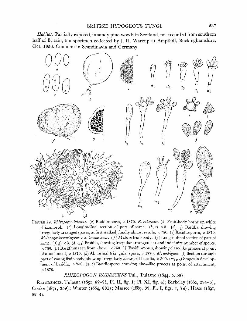
BRITISH HYPOGEOUS FUNGI
Habitat. Partially exposed, in sandy pine-woods in Scotland, not recorded from southern half of Britain, but specimen collected by J. H. Warcup at Ampthill, Buckinghamshire, Oct. 1950. Common in Scandinavia and Germany.
[? v // Ar c d, d
4l h.2 43
FIGURE 29. Rhizopogon luteolus. (a) Basidiospores, x 1870. R. rubescens. (b) Fruit-body borne on white rhizomorph. (c) Longitudinal section of part of same. (b, c) x3. (d1t05) Basidia showing irregularly arranged spores, at first stalked, firstnally almost sessile, nallyx 750. (e) Basidiospores, x 1870. Melanogaster variegatus var. broomeianus. (f) Mature fruit-body. (g) Longitudinal section of part of same. (f, g) x 3. (h to0 4) Basidia, showing irregular arrangement and indefinite number of spores, x 750. (i) Basidium seen from above, x 750. (j) Basidiospores, showing claw-like process at point
of attachment, x 1870. (k) Abnormal triangular spore, x 1870. M. ambiguus. (1) Section through part of young fruit-body, showing irregularly arranged basidia, x 300. (mI t 3) Stages in develop- ment of basidia, x 750. (n, o) Basidiospores showing claw-like process at point of attachment, x 1870.
RHIZOPOGON RUBESCENS Tul., Tulasne (I844, p. 58) REFERENCES. Tulasne (185I, 89-91, P1. II, fig. 1; P1. XI, fig. 4); Berkeley (i86o, 294-5);
Cooke (1871, 359); Winter (1884, 881); Massee (1889, 39, P1. 1, figs. 7, 7a); Hesse (1891, 92-4).
537

LILIAN E. HAWKER ON
Fruit-body, general macroscopic characters. Globose, usually less than 1 cm diam., at first pure white, borne on white rhizomorph (figure 29 b), resembling young fruit-body of Phallus impudicus but reddening when touched, later sulphur yellow or flushed red, finally becoming dingy yellow or brown, smooth, soft, peridium thin, gleba at first waxy, white, becoming gelatinous, greenish yellow or sometimes reddish, chambers numerous, minute (figure 29 c). Odour none at first, later strong and unpleasant of decaying meat or bad eggs.
Peridium. Very thin. Gleba. Dissepiments thin, chambers becoming filled with spores. Basidia. Clavate 20 x 4,u, bearing variable number (usually 5) of irregularly arranged
sessile spores (figure 29 d to 5), soon shedding spores and collapsing. Basidiospores. Long ellipsoidal, at first colourless then pale yellow-green, 2 or more
guttules, thin-walled, 6-9 x 3-4# (figure 29 e). Habitat andperiodicity. Under pine, 1 to 3 in. deep in needle layer, in calcareous or sandy
districts, found only in autumn, not common, gregarious. Reported by Massee (1889) from Chudleigh, Devon, and found during present investigation on limestone cliffs above Goblin Coombe, Cleeve, Somerset (36, 532, 697).
Recently a third species of Rhizopogon has been collected near Burrington, Somerset
(847, 993, 994) which does not fit any published description. The material is being investigated.
Genus MELANOGASTER Corda, in Sturm's Deutschlands Flora (iii), XI, 1 (1837) Fruit-bodies more or less globose, peridium tomentose, dissepiments thick, cavities large,
filled with gelatinous matrix in which basidia develop irregularly, no regular hymenium, spores usually dark coloured.
TYPE SPECIES. Melanogaster variegatus Vitt. Zeller & Dodge (1936) consider that the British material represents three distinct species,
two of which are rather closely related and one of which is based on a single collection.
KEY TO BRITISH SPECIES OF MELANOGASTER
1. Spores small, 6-8 x 3-4/t. ... ... ... M. variegatus var. broomeianus, p. 538 Spores relatively large, more than 10/ long. ... ... ... ... ... 2
2. Spores 12-16 x 6-8,/. ... ... ... ... ... M. ambiguus, p. 539 Spores 13-18 x 7-4-83. ... ... ... ... ... ... ... M. intermedius, p. 540
MELA NO GAS TER VARIEGA TUS Vitt. var. BR 0 OMEIANUS Berkeley, Berkeley (I86o, p. 293)
REFERENCES. Tulasne (1851, 93, P1. II, fig. 4); Winter (1884, 882-3); Massee (1889, 34, P1. I, fig. 11); Hesse (I891, 62, P1. IV, fig. 5).
Syn. Melanogaster broomeianus Berk. et Br. (i844, 350); Tulasne (I843, 19, 377, P1. XVII, fig. 23); Zobel (I854, 46, P1. IX, fig. 90); Zeller & Dodge (1936, 639-55).
Tuber moschatum Sowerby (I797-1815, P1. 426).
538

BRITISH HYPOGEOUS FUNGI
This fungus, known as the Red or Bath Truffle, was formerly sold in the market at Bath and was preferred to the true truffles.
Fruit-body, general macroscopic characters. Irregularly globose to lobed, sometimes con- fluent, often flattened at top and with slight basal projection, 2 to 4 cm diam., peridium felted, often with adpressed fibrils, at first pale greenish yellow, then mustard yellow, becoming reddish brown, blackening when bruised, often cracked, gleba purplish black with numerous gelatinous cavities exuding slimy mass of spores when cut, thick whitish dissepiments (figure 29f, g). Odour none or slight, pleasant. Fruit-body rapidly decaying with development of unpleasant odour.
Peridium. Variable in thickness 50 to 200pu, but usually 50 to 100,u, loose hyphae with large vesicular cells, coloured towards the exterior, continuous with trama.
Gleba. Trama of subgelatinous hyaline hyphae, cavities arising by gelatinization of numerous patches, basidia developing irregularly in cavity, no true hymenium.
Basidia. Clavate, sinuous, tapering to base (figure 29/h to 4, i), hyaline, 3- to 4-spored, or occasionally 5-spored, spores irregularly arranged on short sterigmata and not always developing simultaneously, basidia soon shedding spores and collapsing.
Basidiospores. At first ellipsoidal and hyaline, then ovoid (6-9 x 3-4,u), olive-brown with several guttules, obtuse with small claw-like process at point of attachment (figure 29j). Triangular (probably fused) spores occasionally present (cf. Hymenogaster luteus) (figure 29 k).
Habitat. At or near surface of soil, covered by leaves or humus, under beech or less frequently other deciduous trees, in both acid and alkaline soils, at all times of year when conditions are favourable, much eaten by rabbits. Collected during present study in Bristol (Leigh Woods, 507, 678), Gloucestershire (Cotswold Hills, near Wotton-under- Edge, and Dursley, 57, 181, 395, 546), Somerset (Cleeve, 202), Surrey (Mickleham, 58, 59) and Caernarvonshire (Vaynol Park, Bangor, 227, 233). Specimens received from south and east England.
Melanogaster variegatus var. broomeianus differs from the type in the colour of the dissepi- ments which are orange in the latter, and in the smaller, lighter-coloured spores. It is doubtful whether these differences are sufficient to warrant specific status (as proposed by Zeller & Dodge 1936a), particularly as one recent collection from Leigh Woods, Bristol, showed a tendency for the dissepiments to become orange with age. This was not observed in any other of the twelve separate collections made in various localities during this investigation, but the single example indicates that the colour of the dissepiments is not necessarily a constant character. The present author therefore prefers to retain this form as a variety of M. variegatus rather than as a distinct species.
MELANOGASTER AMBIGUUS (Vitt.) Tul., Tulasne (I843, p. 377)
REFERENCES. Tulasne (I85I, 94-5, P1. II, fig. 5; P1. XII, fig. 5); Berkeley (i86o, 293); Cooke (I87I, 356-7); Massee (i889, 34-5, P1. I, fig. 5-5b); Hesse (I891, 62-4, PI. IV, figs. 6-9; P1. V, fig. 4; PI. VI, fig. 2); Winter (I891, 882-3); Zeller & Dodge (I936, 23,
639-55). Syn. Octaviania ambigua Vittadini (1831, 18).
Melanogaster Klotzschii Zobel (I854, 45-6, P1. IX, fig. 88).
539
LILIAN E. HAWKER ON
Fruit-body, general macroscopic characters. Globose or ellipsoidal, regular, usually not wrinkled or lobed, 1 to 3 cm diam., at first dull reddish brown, becoming potato-coloured, or even olive-brown, blotched black particularly when bruised, peridium smooth but dull surface, brown in section, finally cracking in a network to reveal gleba white at first, then with blue-black chambers separated by pure white or later dingy white dissepiments, chambers rounded with gelatinous contents, black slimy mass of spores exuding when cut. Odour at first sweetish but unpleasant, rubbery or like decaying toadstools, later strong and foetid.
Peridium. Continuous with trama. Gleba. Basidia rather more regularly arranged than in M. variegatus var. broomeianus, but
no continuous hymenium (figure 291). Basidia. Clavate, tapering to base 2- to 4-spored, spores borne on slender sterigmata
(1 to 6,u long), irregularly arranged but usually developing simultaneously (figure 29 ml to 3).
Basidiospores. At first ovoid, hyaline, then spindle-shaped with pointed apex and claw- like process at base, thick-walled, olive-brown, with one or more guttulae, 12-16 x 6-8,c
(figure 29 n, o). Habitat. In humus under beech, and occasionally other trees, at all times of the year,
widespread (reported from numerous places in Britain as far north as Jedburgh) but probably less common in western England than M. variegatus var. broomeianus, since only four collections from two localities, Blaise Castle, Bristol (52) and near Wotton-under-Edge, Gloucestershire (111, 372, 578), have been made during this investigation. A specimen was also received from Harpenden, Hertfordshire (537).
MELANOGASTER INTERMEDIUS (Berkeley) Zeller & Dodge, Zeller & Dodge (1936, p. 639)
Syn. Melanogaster variegatus var. intermedius Berkeley & Broome (I844, 354). M. ambiguus var. intermedius Tulasne (1851, 95); Berkeley (i860o, 293); Cooke (1871,
357); Massee (i889, 35-6). This species is based on a single collection made by Broome in Spye Park, Wiltshire.
It differs from Melanogaster ambiguus in the larger size of the spores, 18-13 x 7A4-8,u, in the shape of the spores which are obtuse, in the yellowish dissepiments and in the bright rusty colour of the fruit-body. It is again doubtful whether Zeller & Dodge (1936) were justified in giving this specimen specific rank in the absence of further collections.
The relationship between the hypogeous Gasteromycetes and other fungi Most earlier writers assumed that the hypogeous Gasteromycetes were relatively primitive
forms. Recently Holm (1949) produced evidence for the theory that some at least may have been derived from an Ascomycete related to Tuber. The observations of clamp connexions in certain species of Tuber (Greis 1938) support this view, and Holm further suggests that the epigeous Gasteromycetes and the Hymenomycetes may have been derived from the hypogeous forms.
Several schemes have been elaborated deriving various groups of epigeous Gastero- mycetes from hypogeous forms. Thus Rehsteiner (1892) proposed the following relationships:
Hysterangium -> Clathraceae, Hymenogaster -> Phallaceae, Rhizopogon -> Lycoperdales.
540
BRITISH HYPOGEOUS FUNGI
The first of these was also postulated by Fischer (1908) and elaborated by Fitzpatrick (I913) and has already been discussed above (p. 512).
For the second of Rehsteiner's groups, that of a relationship between Hymenogaster and the Phallaceae, there seems to be little evidence. The third suggested relationship, that of Rhizopogon and the Lycoperdales, although supported by Fischer (1927), seems to be equally unlikely (the spores of the Lycoperdales are more like those of Sclerogaster than those of any other hypogeous Gasteromycete). In neither of these groupings are spore characters or other structural features similar. Fischer (I927) further suggested that Octaviania (Arcangeliella) might have led to the Nidulariaceae. This is supported by the frequency with which the tramal hyphae disintegrate in such related forms as Hydnangium carneum var.
xanthosporum as already pointed out. The spores of the Hydnangiaceae, however, are quite different from those of the Nidulariaceae while such scanty knowledge as we have of the development of the former group does not suggest affinity with the Nidulariaceae. A relation- ship between the latter and the Rhizopogonaceae is more likely, since the spores are not unlike and the development of both Rhizopogon and Melanogaster is definitely lacunose. The irregular development of both basidia and spores in these genera, however, suggests that they may be degenerate forms.
The opposite view was taken by De Bary (i884), who considered that the Gasteromycetes as a whole were derived from the Agaricales. This view is strongly supported by Heim (1948) and other French mycologists who consider that some at least of the hypogeous forms are the end-points of a degeneration series. Thus Bucholtz (1903), Malen9on (1923)
and Heim (1948) consider with some justification that the Hydnangiaceae have developed from the Russula-Lactarius group, as already discussed (p. 522). The same workers (Heim I948) have also produced evidence for a relationship between Rhizopogon and the Boletus- Paxillus group, which is further supported by the discovery of an intermediate form in North America (Malen^on 1931), while Romagnesi (I933) remarked upon the similarity between the spores of the non-British genus Richoniella and the agaric, Rhodophyllus. Heim, in several publications (summarized I948), has pointed out the relationship of Secotium both to the agarics and to certain hypogeous Gasteromycetes.
Thus with the probable exception of the Hysterangiales the hypogeous Gasteromycetes are best considered as forms which have degenerated from various epigeous groups. The position of the Tuberales among the Ascomycetes is thus paralleled, to some extent, but whereas the Tuberales is a group the members of which are obviously related, the hypo- geous Gasteromycetes are almost certainly a group of forms with diverse origins and showing few interrelationships.
Thanks are due to the University of Bristol for a grant towards the expenses of collecting material; to Miss J. Fraymouth, Dr S. M. Mount, Dr V. 0. Nicholls, Dr B. E. J. Wheeler and other past and present members of the Department of Botany, University of Bristol, and to Miss E. Reyner for help in collecting the bulk of the specimens; to Mr P. C. K. Austwick, Mr E. J. H. Corner, Professor P. H. Gregory, Professor C. T. Ingold, Dr M. Lange, Miss B. Mosse, Professor A. Nannfeldt, Dr J. Ramsbottom and Dr D. P. Rogers who sent specimens; to the Director, Royal Botanic Gardens, Kew, for the loan of specimens and for access to herbarium material and books; to Dr G. Taylor and Mrs Balfour-Browne for
VOL. 237. B.
541
66

LILIAN E. HAWKER ON
access to herbarium material and books at the British Museum (Natural History); to Miss J. Fraymouth, who made most of the permanent preparations used in developmental studies; to Dr S. M. Mount for assistance in checking herbarium material and literature; to Mrs Balfour-Browne, Dr G. R. Bisby, Dr R. W. G. Dennis and Miss E. M. Wakefield for advice on points of nomenclature; and in particular to Miss E. M. Wakefield, late of the Royal Botanic Gardens, Kew, for much help and encouragement, particularly in the early stages of this study, and for a critical reading of the manuscript. Thanks are also due to Mr E. J. H. Corner for permission to use diagrams and data from his field notes on Gyrocratera ploettneriana and to the Editor and publishers of the Transactions of the British
Mycological Society for permission to include illustrations (forming part of figures 25 and 26) from my papers in that journal.
Representative sets of material are to be deposited at the Herbaria of the Royal Botanic Gardens, Kew,,and the Department of Botany, British Museum (Natural History).
REFERENCES
Bambeke, C. van 1904 Sur l' evolution nucleaire et la sporulation chez Hydnangium carneum Wallr. Mem. Acad. R. Belg. 54, 1-44.
Bataille, F. 192I Flore analytique et descriptive des Tuberoidees de l'Europe et de l'Afrique du Nord. Bull. Soc. mycol. Fr. 37, 155-207.
Bataille, F. 1923 Flore analytique et descriptive des Hymenogastracees d'Europe. Bull. Soc. mycol. Fr. 39, 157-96.
Berkeley, M. J. i86o Outlines of Britishfungology. London. Berkeley, M. J. & Broome, C. E. I837-1885 Notices of British Fungi 1841, Ann. Mag. Nat. Hist.
(1), 6, 430-9; I844, ibid. (1), 13, 340-60; i848, ibid. (2), 2, 259-68; 1851, ibid. (2), 7, 176-89; 1859, ibid. (3), 3, 356-77; i86i, ibid. (3), 7, 373-82; 1865, ibid. (3), 15, 312-22, 444-52; i866, ibid. (3), 18, 121-9; I879, ibid. (5), 3, 202-12.
Berkeley, M. J. & Broome, C. E. I846 Notices of British hypogaeous fungi. Ann. Mag. Nat. Hist.
(1), 18, 73-82. Bornholz, A. v. 1827 Della coltivazione dei Tartufi. Milan. Boudier, E. 1876 Du parasitisme probable de quelques especes du genre Elaphomyces et de la recherche
de ces Tuberacees. Bull. Soc. bot. Fr. 23, 115-19. Boulanger, E. I903 Germination de l'ascospore de la truffe. Bull. Soc. mycol. Fr. 19, 26-46.
Bottomley, A. M. 1948 Gasteromycetes of South Africa. Bothalia, 4, pt. ii, 473-810.
Broome, C. E. i874-6 Notes on Bristol fungi. Proc. Bristol Nat. Soc. (N.S.), 1, 290-8. Bucholtz, F. i897 Zur Entwickelungsgeschichte der Tuberaceen. Ber. dtsch. bot. Ges. 15, 211-16. Bucholtz, F. I903 Zur Morphologie und Systematik der Fungi hypogaei. Ann. mycol., Berl., 1,
152-74. Bucholtz, F. 1908 Zur Entwicklung des Choiromyces Fruchtkorper. Ann. mycol., Berl., 6, 539-50. Bucholtz, F. 1910 Zur Entwicklungsgeschichte des Balsamiaceen-Fruchtkorpers, nebst Bemerkungen
zur Verwandtschaft der Tuberineen. Ann. mycol., Berl., 8, 121-41. Bucholtz, F. I9II Ueber die Befruchtung von Endogone lactiflua Berk. Ann. mycol., Berl., 9, 329-30. Bucholtz, F. 1912 Beitrage zur Kenntnis der Gattung Endogone Link. Beih. bot. zbl. 29 (2), 147-224,
pls. 3-10.
Bucknall, C. 1878 The fungi of the Bristol district. Proc. Bristol Nat. Soc. 2, 60, 348; 3, 68; 4, 58; 6, 145; Index, 6, 425-75.
Bulliard, P. 179I-8. Histoire des champignons de France. Paris. Cavara, F. 1900 Arcangeliella. Nuovo. G. bot. ital. 7, 117-128. Chatin, A. I892 La Truffe. Paris.
542
BRITISH HYPOGEOUS FUNGI
Chevallier, F. i826 Flore generale des environs de Paris, 1. Paris. Clemencet, M. 1932 Contribution a l etude du developpement et de l'anatomie des Ascomycetes
hypoges. Les Elaphomycetacees. Le Botaniste, 24, 3-106. Coker, W. C. & Couch, J. N. i928 The Gasteromycetes of the eastern United States and Canada. 201 pp.
North Carolina. Cooke, M. C. 187I Handbook of British fungi. London. Cooke, M. C. i865 Tuber excavatum Vitt. and other British truffles. J. Bot., Lond., 3, 137-40. Corda, A. C. J. I84I Iconesfungorum. Prague. Corda, A. C. J. 1854 See Zobel. Corda, A. C. J. I837 Pilze. in Sturm, Deutschlands Flora, 3. Nuremberg. Corner, E. J. H. & Hawker, L. E. 1953 Hypogeous fungi from Malaya. Trans. Brit. Mycol. Soc. 36,
125-37. Crotch, W. R. 1852. List of fungi found in the neighbourhood of Bristol, Bath and Taunton. Proc.
Somersetsh. Archaeol. Nat. Hist. Soc. for 1852. Cunningham, G. H. I944 The Gasteromycetes of Australia and New Zealand, 15, 236 pp. Dunedin. Currey, F. 1858 On the existence of amorphous starch in a new Tuberaceous fungus. Proc. Roy. Soc.
9, 119-23. De Bary, A. I844 Vergleichende Morphologie und Biologie der Pilze, Mycetozoen und Bacterien. Leipzig. De Bary, A. i863 Uber die Fruchtentwicklung der Ascomyceten. Leipzig. De Ferry de la Bellone I888 La Truffe, L8tude sur les truffes et les truffiexs. Paris. Dodge, C. W. I929 The higher Plectascales. Ann. mycol., Berl., 27, 145-84. Dodge, C. W. & Zeller, S. M. I934 Hymenogaster and related genera. Ann. Mo. Bot. Gdn, 21, 625-708. Dodge, C. W. & Zeller, S. M. 1936 Hydnangium and related genera. Ann. Mo. Bot. Gdn, 23, 565-98. Fischer, E. i896 Ueber den Parallelismus der Tuberaceen und Gastromyceten. Ber. dtsch. bot. Ges.
14, 301-11. Fischer, E. i897 Tuberaceen. In Rabenhorst, Kryptogamenflora, 2nd ed. 5. Leipzig. Fischer, E. I897a Tuberineae. In Engler and Prantl, Die Natiirlichen Pflanzenfamilien, Teil 1,
Abt. i, pp. 278-90. Leipzig. Fischer, E. 1900 Hymenogastrineae. In Engler and Prantl, Die Naturlichen Pflanzenfamilien, Teil 1,
Abt. i, pp. 296-313. Leipzig. Fischer, E. go1900o a Bemerkungen uber die Tuberaceengattungen Gyrocratera und Hydnotrya. Hedwigia,
39, 48-51. Fischer, E. I9I0 Neuese Untersuchungen uber die Verwandtschaftsverhaltnisse der Tuberineen und
Helvellineen. Z. Bot. 2, 718-22. Fischer, E. 1923 Zur Systematik der Schweizerischen Trtiffein aus den Gruppen von Tuber excavatum
und rufum. Verh. naturf. Ges. Basel, 35 (1), 34-50. Fischer, E. 1927 Mykologische Beitrage. No. 32. Zur Entwicklungeschichte der Fruchtkorper von
Hymenogaster. No. 33. Die Tuberineen gattungen Hydnotrya und Gyrocratera und ihre gegen- seitige Beziehungen. Mitt. naturf. Ges. Bern, for 1926, 99-114.
Fischer, E. 194I Hymenogaster tener Berk. Schweiz. Z. Pilzk. 19, 41-2. Fitzpatrick, H. M. I913 A comparative study of the development of the fruit-body in Phallogaster,
Hysterangium and Gautieria. Ann. mycol., Berl., 2, 119-49. Fries, E. M. I817-18 Symbolae Gasteromycetae. Lund. Fries, E. M. 1821-32 Systema mycologica (1821), 1, Lund; (1822-3), 2, Lund; (1829-32), 3, Greifswald. Fries, E. M. I846 Summa Vegetabilium Scandinaviae. Stockholm and Leipzig. Gilkey, H. M. I916 A revision of the Tuberales of California. Univ. Calif. Publ. Bot. 6. Gilkey, H. M. 1939 Tuberales of North America. Oregon State Monographs, 1. Gilkey, H. M. 1947 New or otherwise noteworthy species of Tuberales. Mycologia, 39, 441-52.
Gray, J. E. I82I Natural arrangement of British plants. London. Greis, H. 1938 Die Enstehung der Basidiomycetenschnallen aus den Ascomycetenhaken. J. wiss.
Bot. 86, 81-106. 66-2
543

LILIAN E. HAWKER ON
Greis, H. 1938 Die Sexualvorgange bei Tuber aestivum und T. brumale. Biol. Zbl. 58, 617-31.
Greis, H. I939 Ascusentwicklung von Tuber aestivum und T. brumale. Z. Bot. 34, 129-78.
Harkness, H. W. I899 Californian hypogaeous fungi. Proc. Calif. Acad. Sci. (3), 1, 241-92. Hawker, L. E. I95I Hypogaeous Fungi. I. A hypogaeous Gasteromycete, Sclerogaster macrosporus.
Trans. Brit. Mycol. Soc. 34, 216-19.
Hawker, L. E. 1952 Hypogaeous Fungi. II. A new variety of Hydnangium carneum Wallr. from North Wales. III. Three new British records: Gautieria morchellaeformis Vitt., Hymenogaster hessei Soehner and Elaphomyces aculeatus Vitt. Trans. Brit. Mycol. Soc. 35, 279-84.
Heim, R. 1936 Sur la parente entre Lactarius et certains Gasteromycetes. C.R. Acad. Sci., Paris, 202, 2101-3.
Heim, R. I948 Phylogeny and natural classification of macrofungi. Trans. Brit. Mycol. Soc. 30, 161-78.
Hennings, P. 1899 Gyrocratera, eine neue Tuberaceengattung, sowie einige neue und seltene Asco- myceten aus der Mark. Ver. Prov. Brandenberg, 41, 87-91.
Hennings, P. I905 Tuberaceae in Krypt. Fl. Mark. Brandenberg, 7, 150-62.
Hesse, R. I891 Hypogaeen Deutschlands. 1. Die Hymenogastreen. Marburg. Hesse, R. 1894 Hypogaeen Deutschlands. 2. Die Tuberaceen. Marburg. Hollos, L. I9I1 Fungi Hypogaei Hungariae. Budapest. Holm, L. 1949 Some aspects on the origin of the Gasteromycetes. Svensk bot. Tidskr. 43, 65-71.
Jaczewski, A. L. i909 Monographie des Tuberacees de la Suisse. Bull. Herb. Boissier, 4, 591-682.
Kanouse, B. B. I936 Studies of two species of Endogone in culture. Mycologia 27, 47-62. Klotzsch, J. F. I84I In Dietrich A. Flora Marchica, oder Bescheibung der in der Mark Brandenberg
wildwachsunden Pflanzen. Berlin.
Knapp, A. I924 Entwicklung von Balsamia platyspora Berk. Schweiz. Z. Pilzk. 17 (1), 5-8.
Knapp, A. I94i Die Hypogaeen um Basel. In Erinnerung an Prof. Dr. Ed. Fischer, Bern. 7 Fort- setzung. Schweiz. Z. Pilzk. 19, 40-3.
Knapp, A. 1950-52 Die europaischen Hypogaeen-Gattungen und ihre Gattungstypen. Schweiz. Z. Pilzk. (1950), 28 (3), 29-42; 28 (7), 101-16; 28 (10), 153-79; (1951), 29 (4), 65-92; 29 (7), 133-148; (1952), 30 (3), 33-43; 30 (6) 81-92.
Kuntze, C. E. 0. i89i Revisio generum Plantarum, 1. Leipzig. Lange, M. 1948 Genea hispidula Berk. En for Danmark ny hypogoeish Storsvamp. Dansk hot. Tidskr.
40, 214-16.
Lewton-Brain, L. I901 The mycorrhiza of Elaphomyces variegatus. Ann. Bot., Lond., 15, 521-30. Link, H. F. 1809 Observationes in Ordines Plantarum naturelles. Ges. naturf. Freunde. zu Berlin.
Magazin. f.d. neuesten Entdeckungen in den gesammten Naturkunde, 3, 33.
Linnaeus, C. I737 Flora Lapp. Amsterdam.
Linnaeus, C. 1753 Species Plantarum, 2. Stockholm.
Lloyd, C. G. I922 The Hymenogastraceae. The Octaviania group. The genus Arcangeliella. Mycologia, 7, 1138-43.
Lohwag, H. V. 1924 Zur Stellung und Systematik der Gastromyceten. Verh. zool. bot. Ges. Wien, 70, 38-55.
Lohwag, H. V. I925 Zur Entwicklungsgeschichte und Morphologie der Gastromyceten. Beih. Bot. Zbl. 42, 298-9.
Lohwag, H. V. 1926 Die Homologien im Fruchtk6rperbau der h6heren Pilze. Biol. gen. 2, 148-82, 575-608.
Lohwag, H. V. I939 Mykologische Studien. XVI. Tuberineenstudien. Ann. mycol., Berl., 37, 455-504.
Malen?on, G. 193I La Serie des Asterospores. Trav. crypt. dedies a Louis Mangin, pp. 377-96. Paris.
Malengon, G. 1938 Les truffes Europeenes. Rev. mycol., Paris, (N.S.), 3, 1-92. Massee, G. 1889 A monograph of British Gasteromycetes. Ann. Bot., Lond., 4, 1-103. Massee, G. 1909 The structure and affinities of British Tuberaceae. Ann. Bot., Lond., 23, 243-63.
544

BRITISH HYPOGEOUS FUNGI
Mattiroto, 0. I888 Illustrazione di tre nuove speci di Tuberacee Italiane. Mem. Acad. Sci. Torino,
(2), 37, 1-19. Mattirolo, 0. 1934 Rapporti simbiotici sviluppatisitra il tartufo 'bianchelto' (Tuber borchii Vittadini)
et i proppi americana detti canadesi. Ann. R. Accad. Agric. Torino, 70, 3-10. Melin, E. 1917 Studier over der norrlandska myrmarkernas vegetation. Norrldndskt Handbibliolek, 7.
Melin, E. & Lindeberg, G. I939 Uber den Einfluss von Aneurin und Biotin auf das Wachstum
einiger Mykorrhizenpilze. Bot. Notiser, pp. 241-5. Melin, E. & Norkrans, B. 1942 Uber den Einfluss der Pyrimidin- und der Thiazol-Komponente des
Aneurins auf das Wachstum von Wurzelpilzen. Svensk bot. Tidskr. 36, 271-86. Melin, E. & Norkrans, B. 1948 Amino acids and the growth of Lactarius deliciosus (L.) Fr. Physiol.
Plant. 1, 176-84. Micheli, P. A. 1729 Nova Plantarum Genera. Florence. Modess, 0. 1939 Experimctelle Untersuchungen fiber Hymenomyceten und Gasteromyceten als
Mykorrhizabilden bei Kiefer und Fichte. Svensk bot. Tidskr. 33, 91-3. Modess, 0. I94I Zur Kenntniss der Mykorrhizenbilden von Kiefer und Fichte. Symb. bot. Upsaliens,
5 (i), 147 pp. Mosse, Barbara I953 Fructifications associated with mycorrhizal strawberry roots. Nature, Lond.,
171, 974. Nees v. Esenbeck, C. G. I817 Das System der Pilze und Schwamme. Wurzberg. Nees v. Essenbeck, C. G. 1821-3 Plantae officinales. Dusseldorf. Parks, H. C. I92I Californian Hypogaeous Fungi. Tuberaceae. Mycologia, 13, 301-14. Patouillard, N. 1914 Contribution a la Flore Mycologique dujura. Bull. Soc. mycol. Fr. 30, 349-54.
Persoon, C. H. 1797 Disp. Meth. Fung. Leipzig. Persoon, C. H. i8oi Synopsis Methodica Fungorum. G6ttingen. Petri, L. I902 Lo sviluppo del corpo fruttifero dell' Hydnangium carneum Wallr. Rendiconte del
Congresso botanico di Palermo, Maggio, 148-51. Pico, V. 1788 Melethemata inauguralia de fungorum generatione et propagatione. Turin. Pirk, W. I949 Die Rote Truffel, Hydnotrya Tulasnei Berk. et Br. Schweiz. Z. Pilzk. 27, 21-9.
Quelet, L. i872-5 Les champignons du Jura et des Vosges. Paris. Rabenhorst, L. i844 Deutschlands Kryptogamenflora, 1st ed. Leipzig: (see Fischer (i897), Winter
(I884)). Ramsbottom, J. 1923 Handbook of the larger British fungi. London. Ramsbottom, J. & Balfour-Browne, F. L. 1951 List of Discomycetes recorded from the British Isles.
Trans. Brit. Mycol. Soc. 34, 38-137. Reess, M. i88o Ueber den Parasitismus von Elaphomyces granulatus. S.B. phys.-med. Soz. Erlangen,
12, 103-7. Reess, M. i88oa Uber den Parasitismus von Elaphomyces granulatus. Bot. Z. 38, 729-33. Reess, M. i885 Ueber Elaphomyces und sonstige Wurzelpilze. Ber. dtsch. bot. Ges. 3, 293-5. Reess, M. & Fisch, C. 1887 Untersuchungen uber Bau und Lebensgeschichte der Hirschtruffel
Elaphomyces. Bibl. bot., Stuttgart, 7, 1-24. Rehsteiner, H. 1892 Beitrage zur Entwicklungsgeschichte der Fruchtkorper einiger Gasteromyceten.
Bot. Z. 50, 761-71, 777-92. Risso, J. A. 1844 Fl. Nice. Nice. Romagnesi, H. 1933 Le genre Richoniella, chalnon angiocarpe de la serie des Rhodogoniospores.
Bull. Soc. mycol. Fr. 49, 433-4. Saccardo, P. A. & Sydow, H. I902 Syll. Fung. 16. Padua. Sappa, F. 1946 Ricerche biologiche sul Tuber magnatum Pico. La germinazione delle spore e
caratteri della micorrhiza. Nuovo G. bot. Ital. 47, 155-98. Schlectendahl, D. F. L. v. 1824 Flora Berol. 2. Cryptogamia. Berlin. Schroter, J. I893 In Cohn, Krypt. Fl. Schlesien, 3. Breslau. Smith, W. G. i891 Supplement to Berkeley's Outlines of British Fungology. London.
545

LILIAN E. HAWKER ON BRITISH HYPOGEOUS FUNGI
Smith, W. G. 1908 British Basidiomycetes. London. Soehner, F. 1913 Die Formenkreis von Hymenogaster tener Berk. et Br. Hedwigia. 64, 192-202.
Soehner, F. 1923 Hydnangium stephensii Berk. Z. Pilzk. 2 (vii), 153-6. Soehner, F. 1923 a Berichtegungen zu der in Dr Rickens vademecum aufgeftihrten Hymenogastreen.
Z. Pilzk. 2 (vii), 156-8. Soehner, F. 1923 b Ueber das Suchen von Truffein Physikalischbiologische Wachstumsbedingungen
der Hypogaeen (Schattenpflanzen). Z. Pilzk. 2 (viii), 164-70. Soehner, F. I942, 1944 Tuberaceen. Studien I and II. Notizbl. bot. Gart. Mus. Berlin-Dahlem, 15
(v, vii). Soehner, F. I949 Deutsche Hydnangiaceae. Z. Pilzk. (iii), 11-20. Soehner, F. I95i Tuberaceen-Studien. III. Z. Pilzk. (viii), 7-16. Sowerby, J. I797-1809 Coloured figures of English fungi or mushrooms. Plate 309. London. Stephens, H. 0. 1840 On the mycology of the neighbourhood of Bristol. Ann. Mag. Nat. Hist. 4, 246-53. Thaxter, R. 1922 A revision of the Endogoneae. Proc. Amer. Acad. Arts Sci. 57, 291-350. Townsend, B. B. 1952 Rhizomorphs. Part of Thesis for the Degree of Ph.D. in the University of
Bristol. Tulasne, L. R. & Tulasne, C. 184I Observations sur le genre Elaphomyces et description de quelques
especes nouvelles. Ann. Sci. nat. Bot. (2), 16, 5-29. Tulasne, L. R. & Tulasne, C. I843 Champignons hypoges de la famille des Lycoperdacees, observes
dans les environs de Paris et les Departements de la Vienne et d'Indre-et-Loire. Ann. Sci. nat. Bot. (2), 19, 373-81.
Tulasne, L. R. & Tulasne, C. 1844 Fungi nonnulli hypogaei novi v. minuscogniti auct. Nuovo G. bot. ital. 2 (1), 55-63.
Tulasne, L. R. & Tulasne, C. 1845 Memoire pour servir a l'histoire des champignons hypoges suivi de leur monographie et accompagne de plances. C.R. Acad. Sci., Paris, 21, 1432-6.
Tulasne, L. R. & Tulasne, C. I85i Fungi hypogaei. Histoire et monographie des champignons hypogds. Paris.
Vittadini, C. 183I Monographia Tuberacearum. Milan. Vittadini, C. 1842 Monographie Lycoperdineum. Milan. (Also published, I843, in Mem. R. Acad.
Sci. Torino, 2 (v), 216). Wallroth, K. F. W. 1833 In Dietrich, Flora Krypt. Germanica, 2. Nuremberg. Willdenow, C. L. I787 Flora Berolinensis prodromus. Berlin. Winter, G. I884 In Rabenhorst's Deutschlands Kryptogamenflora, 2nd ed. 1. Leipzig. Young, H. E. 1937 Rhizopogon luteolus, a mycorrhizal fungus of Pinus. Forestry, 2, 30-1. Zeller, S. M. I949 Keys to Gasteromycetes. Mycologia, 41, 36-58. Zeller, S. M. & Dodge, C. W. 1918 Rhizopogon in North America. Ann. Mo. Bot. Gdn, 5, 1-36. Zeller, S. M. & Dodge, C. W. 1918 a Gautieria in North America. Ann. Mo. Bot. Gdn, 5, 133-42. Zeller, S. M. & Dodge, C. W. 1929 Hysterangium in North America. Ann. Mo. Bot. Gdn, 16, 83-128. Zeller, S. M. & Dodge, C. W. 1935 New species of Hydnangiaceae. Ann. Mo. Bot. Gdn, 22, 365-73. Zeller, S. M. & Dodge, C. W. 1936 Elasmomyces, Arcangeliella and Macowanites. Ann. Mo. Bot. Gdn,
23, 599-638.
Zeller, S. M. & Dodge, C. W. 1936a Melanogaster. Ann. Mo. Bot. Gdn, 23, 639-55.
Zobel, J. B. 1854 In Corda, Icones Fungorum, 6. Prague.
546