Embed Size (px)
Citation preview
brief reports
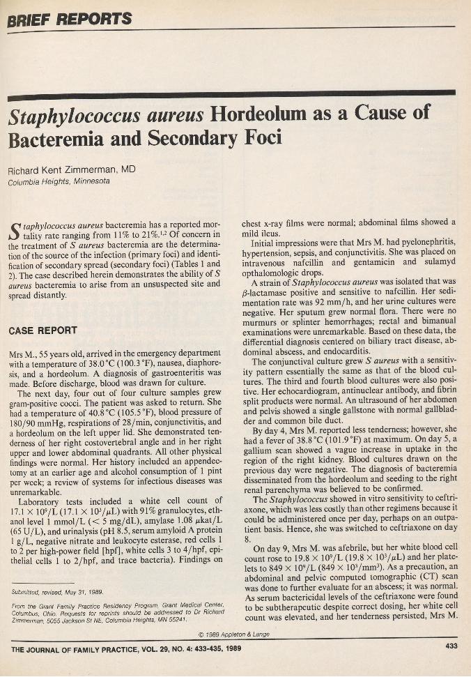
Staphylococcus aureus Hordeolum as a Cause of Bacteremia and Secondary FociRichard Kent Zimmerman, MDColumbia Heights, Minnesota
taphylococcus aureus bacteremia has a reported mortality rate ranging from 11% to 21%.U2 Of concern in
the treatment of S aureus bacteremia are the determination of the source of the infection (primary foci) and identification of secondary spread (secondary foci) (Tables 1 and 2). The case described herein demonstrates the ability of S aureus bacteremia to arise from an unsuspected site and spread distantly.
CASE REPORT
Mrs M., 55 years old, arrived in the emergency department with a temperature of 38.0 °C (100.3 °F), nausea, diaphoresis, and a hordeolum. A diagnosis of gastroenteritis was made. Before discharge, blood was drawn for culture.
The next day, four out of four culture samples grew gram-positive cocci. The patient was asked to return. She had a temperature of 40.8 °C (105.5 °F), blood pressure of 180/90 mmHg, respirations of 28/min, conjunctivitis, and a hordeolum on the left upper lid. She demonstrated tenderness of her right costovertebral angle and in her right upper and lower abdominal quadrants. All other physical findings were normal. Her history included an appendectomy at an earlier age and alcohol consumption of 1 pint per week; a review of systems for infectious diseases was unremarkable.
Laboratory tests included a white cell count of 17.1 X 10⁹/L (17.1 X KT/mL) with 91% granulocytes, ethanol level 1 mmol/L ( < 5 mg/dL), amylase 1.08 /xkat/L (65 U/L), and urinalysis (pH 8.5, serum amyloid A protein 1 g/L, negative nitrate and leukocyte esterase, red cells 1 to 2 per high-power field [hpf], white cells 3 to 4/hpf, epithelial cells 1 to 2/hpf, and trace bacteria). Findings on
Submitted, revised, May 31, 1989.
From the Grant Family Practice Residency Program, Grant Medical Center, Columbus, Ohio. Requests for reprints should be addressed to Dr Richard Zimmerman, 5055 Jackson St NE, Columbia Heights, MN 55241.
chest x-ray films were normal; abdominal films showed a mild ileus.
Initial impressions were that Mrs M. had pyelonephritis, hypertension, sepsis, and conjunctivitis. She was placed on intravenous nafcillin and gentamicin and sulamyd opthalomologic drops.
A strain of Staphylococcus aureus was isolated that was /3-lactamase positive and sensitive to nafcillin. Her sedimentation rate was 92 mm/h, and her urine cultures were negative. Her sputum grew normal flora. There were no murmurs or splinter hemorrhages; rectal and bimanual examinations were unremarkable. Based on these data, the differential diagnosis centered on biliary tract disease, abdominal abscess, and endocarditis.
The conjunctival culture grew S aureus with a sensitivity pattern essentially the same as that of the blood cultures. The third and fourth blood cultures were also positive. Her echocardiogram, antinuclear antibody, and fibrin split products were normal. An ultrasound of her abdomen and pelvis showed a single gallstone with normal gallbladder and common bile duct.
By day 4, Mrs M. reported less tenderness; however, she had a fever of 38.8 °C (101.9 °F) at maximum. On day 5, a gallium scan showed a vague increase in uptake in the region of the right kidney. Blood cultures drawn on the previous day were negative. The diagnosis of bacteremia disseminated from the hordeolum and seeding to the right renal parenchyma was believed to be confirmed.
The Staphylococcus showed in vitro sensitivity to ceftriaxone, which was less costly than other regimens because it could be administered once per day, perhaps on an outpatient basis. Hence, she was switched to ceftriaxone on day 8.
On day 9, Mrs M. was afebrile, but her white blood cell count rose to 19.8 X 10⁹/L (19.8 X 10³//u.L) and her platelets to 849 X 109/L (849 X 103/m m 3). As a precaution, an abdominal and pelvic computed tomographic (CT) scan was done to further evaluate for an abscess; it was normal. As serum bactericidal levels of the ceftriaxone were found to be subtherapeutic despite correct dosing, her white cell count was elevated, and her tenderness persisted, Mrs M.
© 7989 Appleton & Lange
THE JOURNAL OF FAMILY PRACTICE, VOL. 29, NO. 4: 433-435, 1989 433
STAPHYLOCOCCUS HORDEOLUM
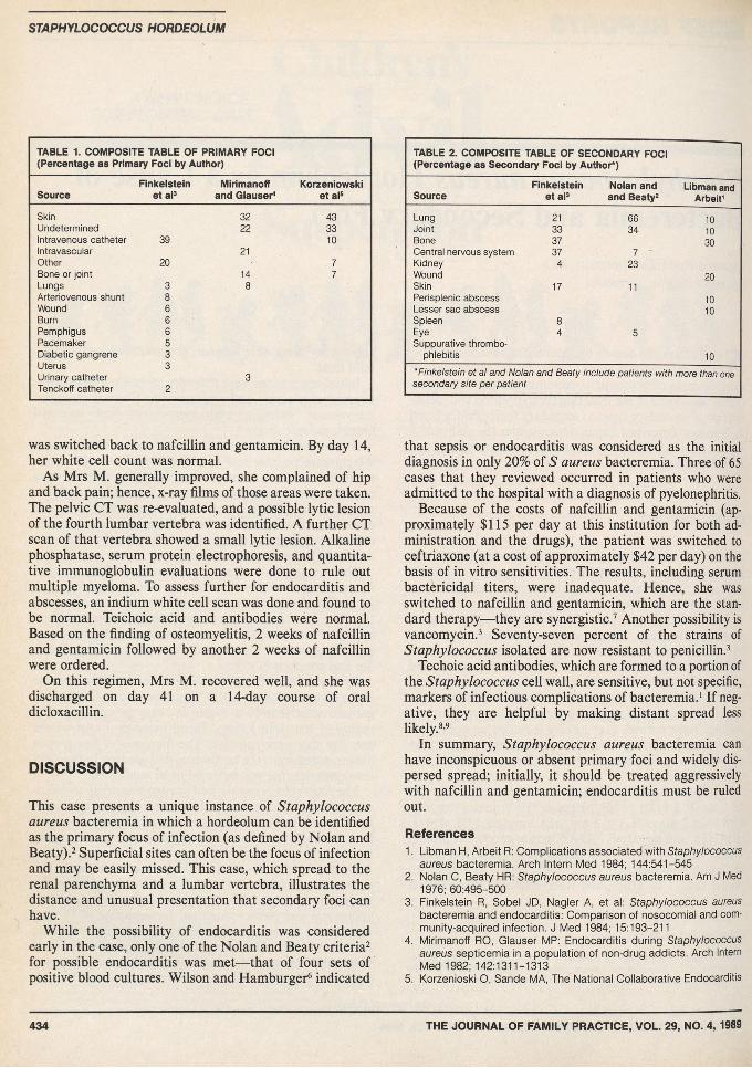
TABLE 1. COMPOSITE TABLE OF PRIMARY FOCI (Percentage as Primary Foci by Author)
SourceFinkelstein
et al3Mirimanoff
and Glauser4Korzeniowski
et al5
Skin 32 43Undetermined 22 33Intravenous catheter 39 10Intravascular 21Other 20 7Bone or joint 14 7Lungs 3 8Arteriovenous shunt 8Wound 6Bum 6Pemphigus 6Pacemaker 5Diabetic gangrene 3Uterus 3Urinary catheter 3Tenckoff catheter 2
was switched back to nafcillin and gentamicin. By day 14, her white cell count was normal.
As Mrs M. generally improved, she complained of hip and back pain; hence, x-ray films of those areas were taken. The pelvic CT was re-evaluated, and a possible lytic lesion of the fourth lumbar vertebra was identified. A further CT scan of that vertebra showed a small lytic lesion. Alkaline phosphatase, serum protein electrophoresis, and quantitative immunoglobulin evaluations were done to rule out multiple myeloma. To assess further for endocarditis and abscesses, an indium white cell scan was done and found to be normal. Teichoic acid and antibodies were normal. Based on the finding of osteomyelitis, 2 weeks of nafcillin and gentamicin followed by another 2 weeks of nafcillin were ordered.
On this regimen, Mrs M. recovered well, and she was discharged on day 41 on a 14-day course of oral dicloxacillin.
DISCUSSION
This case presents a unique instance of Staphylococcus aureus bacteremia in which a hordeolum can be identified as the primary focus of infection (as defined by Nolan and Beaty).2 Superficial sites can often be the focus of infection and may be easily missed. This case, which spread to the renal parenchyma and a lumbar vertebra, illustrates the distance and unusual presentation that secondary foci can have.
While the possibility of endocarditis was considered early in the case, only one of the Nolan and Beaty criteria2 for possible endocarditis was met— that of four sets of positive blood cultures. Wilson and Hamburger6 indicated
TABLE 2. COMPOSITE TABLE OF SECONDARY FOCI (Percentage as Secondary Foci by Author*)
SourceFinkelstein
et al3Nolan and and Beaty2
Libman and Arbeit1
Lung 21 66 10Joint 33 34 10Bone 37 30Central nervous system 37 7Kidney 4 23Wound 20Skin 17 11Perisplenic abscess 10Lesser sac abscess 10Spleen 8Eye 4 5Suppurative thrombo-
phlebitis 10"Finkelstein et at and Nolan and Beaty include patients with more than one secondary site per patient
that sepsis or endocarditis was considered as the initial diagnosis in only 20% of 5 aureus bacteremia. Three of 65 cases that they reviewed occurred in patients who were admitted to the hospital with a diagnosis of pyelonephritis.
Because of the costs of nafcillin and gentamicin (approximately $115 per day at this institution for both administration and the drugs), the patient was switched to ceftriaxone (at a cost of approximately $42 per day) on the basis of in vitro sensitivities. The results, including serum bactericidal titers, were inadequate. Hence, she was switched to nafcillin and gentamicin, which are the standard therapy— they are synergistic.7 Another possibility is vancomycin.3 Seventy-seven percent of the strains of Staphylococcus isolated are now resistant to penicillin.3
Techoic acid antibodies, which are formed to a portion of the Staphylococcus cell wall, are sensitive, but not specific, markers of infectious complications of bacteremia.1 If negative, they are helpful by making distant spread less likely.8’9
In summary, Staphylococcus aureus bacteremia can have inconspicuous or absent primary foci and widely dispersed spread; initially, it should be treated aggressively with nafcillin and gentamicin; endocarditis must be ruled out.
References1. Libman H, Arbeit R: Complications associated with Staphylococcus
aureus bacteremia. Arch Intern Med 1984; 144:541-5452. Nolan C, Beaty HR: Staphylococcus aureus bacteremia. Am J Med
1976; 60:495-5003. Finkelstein R, Sobel JD, Nagler A, et al: Staphylococcus aureus
bacteremia and endocarditis: Comparison of nosocomial and community-acquired infection. J Med 1984; 15:193-211
4. Mirimanoff RO, Glauser MP: Endocarditis during Staphylococcus aureus septicemia in a population of non-drug addicts. Arch Intern Med 1982; 142:1311-1313
5. Korzenioski O, Sande MA, The National Collaborative Endocarditis
434 THE JOURNAL OF FAMILY PRACTICE, VOL. 29, NO. 4, 1989
STAPHYLOCOCCUS h o r d e o l u m
Study Group: Combination antimicrobial therapy for Staphylococcus aureus endocarditis in patients addicted to parenteral drugs and in nonaddicts. Ann Intern Med 1982, 97:496-503
6. Wilson R, Hamburger M: Fifteen years' experience with Staphylococcus septicemia in a large city hospital. Am J Med 1957; 22:437-457
7. Licht MC, Hamilton J: Penicillinase-resistant penicillin/gentamicin synergism. Arch Intern Med 1979; 139:1094-1098
8. Tuazon CU, Sheagren JN, Choa MS, et al: Staphylococcus aureus bacteremia: Relationship between formation of antibodies to teichoic acid and development of metastatic abscesses. J Infect Dis 1978; 137:57-62
9. Verbrugh HA, Peters R, Goessens WHF, et al: Distinguishing complicated from uncomplicated bacteremia caused by Staphylococcus aureus'. The value of "new” and "old" serological tests. J Infect Dis 1986; 153:109-115
If you dorit already ifttim evoulo
have a ltm u s ll,ife time vou looked into i t
Since we introduced the Titmus II in 1985, this little wonder has proven itself time and again in thousands of doctors' offices all across America. The results are conclusive: The Titmus II is easy, fast and accurate.
With the Titmus II, screening takes only 5 minutes. And a wide range of visual functions can be assessed: far, near, intermediate and peripheral vision, color perception, muscle balance, depth perception and binocu- larity. It even screens for hyperopia—one more way the Titmus II Vision Tester
is far superior to a wall chart.The Titmus II is lightweight and
compact. Its micro-digital remote control is easy to use, and the photo electric sensor ensures correct head
positioning at all times. And command of all test operations is right at your fingertips. Your patients will appreciate your up-to-date screening methods, and you will appreciate the increased
convenience and profitability the Titmus II will bring to your practice.
Tb team more about why the Titmus II is well worth looking into, call the Titmus Instrument Group at (800) 446-1802; in Virginia (800) 552-1869, or write Titmus atPO. Box 191, Petersburg, Virginia 23804-0191.
T lT m U S *Focusing on the future
THE JOURNAL OF FAMILY PRACTICE, VOL. 29, NO. 4, 1989 435