Embed Size (px)
Citation preview

Bridging the Gap between Plant and MammalianPolyamine Catabolism: A Novel PeroxisomalPolyamine Oxidase Responsible for a FullBack-Conversion Pathway in Arabidopsis1[W][OA]
Panagiotis N. Moschou, Maite Sanmartin, Athina H. Andriopoulou, Enrique Rojo,Jose J. Sanchez-Serrano, and Kalliopi A. Roubelakis-Angelakis*
Department of Biology, University of Crete, 71409 Heraklion, Greece (P.N.M., A.H.A., K.A.R.-A.); andCentro Nacional de Biotecnologia-Consejo Superior de Investigaciones Cientıficas, UniversidadAutonoma de Madrid, 28049 Madrid, Spain (M.S., E.R., J.J.S.-S.)
In contrast to animals, where polyamine (PA) catabolism efficiently converts spermine (Spm) to putrescine (Put), plants havebeen considered to possess a PA catabolic pathway producing 1,3-diaminopropane, D1-pyrroline, the corresponding aldehyde,and hydrogen peroxide but unable to back-convert Spm to Put. Arabidopsis (Arabidopsis thaliana) genome contains at least fiveputative PA oxidase (PAO) members with yet-unknown localization and physiological role(s). AtPAO1 was recently identifiedas an enzyme similar to the mammalian Spm oxidase, which converts Spm to spermidine (Spd). In this work, we haveperformed in silico analysis of the five Arabidopsis genes and have identified PAO3 (AtPAO3) as a nontypical PAO, in terms ofhomology, compared to other known PAOs. We have expressed the gene AtPAO3 and have purified a protein corresponding toit using the inducible heterologous expression system of Escherichia coli. AtPAO3 catalyzed the sequential conversion/oxidation of Spm to Spd, and of Spd to Put, thus exhibiting functional homology to the mammalian PAOs. The best substratefor this pathway was Spd, whereas the N1-acetyl-derivatives of Spm and Spd were oxidized less efficiently. On the other hand,no activity was detected when diamines (agmatine, cadaverine, and Put) were used as substrates. Moreover, although AtPAO3does not exhibit significant similarity to the other known PAOs, it is efficiently inhibited by guazatine, a potent PAO inhibitor.AtPAO3 contains a peroxisomal targeting motif at the C terminus, and it targets green fluorescence protein to peroxisomeswhen fused at the N terminus but not at the C terminus. These results reveal that AtPAO3 is a peroxisomal protein and that theC terminus of the protein contains the sorting information. The overall data reinforce the view that plants and mammalspossess a similar PA oxidation system, concerning both the subcellular localization and the mode of its action.
Polyamines (PAs) are well-known, low-molecular-mass organic compounds, and the most abundantones, putrescine (Put), spermidine (Spd), and sperm-ine (Spm), exert a multifunctional role in plant devel-opment and defense (Thomas and Thomas, 2001;Mehta et al., 2002; Capell et al., 2004; Yamaguchiet al., 2006). Insights into the role of plant PA catab-olism have been constantly gained in the last fewyears, and it has been correlated with diverse pro-
cesses such as cell growth and development and pro-grammed cell death (Ha et al., 1997; Yoda et al., 2003;Amendola et al., 2005; Paschalidis and Roubelakis-Angelakis, 2005a, 2005b; Moschou et al., 2008).
Oxidation is the main catabolic pathway of PAs. PAoxidation in mammals is exerted by multiple enzy-matic activities with PA oxidase (PAO; EC 1.5.3.11),mainly localized in peroxisomes, and converting Spmto Put, via Spd producing hydrogen peroxide (H2O2;Schrader and Fahimi, 2004). The best substrates forthis back-conversion process are the acetyl-derivativesof Spd and Spm (Schrader and Fahimi, 2004). On thecontrary, in plants, PA oxidation is exerted mainlyby apoplastic PAOs, oxidizing Spm and Spd to 1,3-diaminopropane, H2O2, and the corresponding alde-hyde, thus being responsible for the terminal catabolismof PAs (Rea et al., 2004; Cona et al., 2006). Interestingly,none of the known plant PAOs has been reported tolocalize to peroxisomes (Angelini et al., 1995; Conaet al., 2005), while only the PAO from barley (Hordeumvulgare) has been reported to be localized in the vacuole(Cervelli et al., 2004). Also, Tavladoraki et al. (2006)reported that the Arabidopsis (Arabidopsis thaliana) geneAtPAO1 product could be localized in the cytoplasm,leaving an open window for novel PAO localization.
1 This work was supported by the National and European re-sources (EPEAKII-Pythagoras), Greek-Spain Bilateral Agreement,and COST858, COST FA065 Actions; by the Consejo Superior deInvestigaciones Cientıficas I3P Programme cofinanced by the Euro-pean Social Fund (to M.S.); and by the Onassis Foundation (schol-arship to A.H.A.).
* Corresponding author; e-mail [email protected] author responsible for distribution of materials integral to the
findings presented in this article in accordance with the policydescribed in the Instructions for Authors (www.plantphysiol.org) is:Kalliopi A. Roubelakis-Angelakis ([email protected]).
[W] The online version of this article contains Web-only data.[OA] Open Access articles can be viewed online without a sub-
scription.www.plantphysiol.org/cgi/doi/10.1104/pp.108.123802
Plant Physiology, August 2008, Vol. 147, pp. 1845–1857, www.plantphysiol.org � 2008 American Society of Plant Biologists 1845 www.plantphysiol.orgon July 8, 2018 - Published by Downloaded from
Copyright © 2008 American Society of Plant Biologists. All rights reserved.

Biochemical evidence for the existence of a back-conversion pathway in plants was presented by DelDuca et al. (1995) and Tassoni et al. (2000), who foundthat exogenously supplied Spd to Helianthus tuberosuschloroplasts and Arabidopsis plants, respectively, wasconverted to Put, but the enzymatic activity(ies) re-sponsible for this conversion could not be determinedat that time; it was hypothesized that an enzymesimilar to the animal PAO enzyme responsible for this
pathway should exist in plants. Recently, Tavladorakiet al. (2006) identified the Arabidopsis gene AtPAO1 asa PAO able to convert Spm to Spd, thus being func-tionally similar to the mammalian Spm oxidase (SMO;EC 1.5.3.1), giving new insights into the plant PA catab-olism and thus reinforcing the view that additionalPAOs similar to animal PAOs should exist in plants.
In contrast to their mammalian counterparts, acorrelation with PA oxidation has not been established
Figure 1. AtPAO3 sequence alignment with other known plant and mammalian PAOs. AtPAO3 (At3g59050; GenBankaccession no. AY143905) amino acid sequence similarity with AtPAO1 (At5g13700; GenBank accession no. NM_121373),AtPAO2 (At2g43020; GenBank accession no. AF364952), AtPAO4 (At1g65840; GenBank accession no. AF364953), AtPAO5(At4g29720; GenBank accession no. AK118203), maize (ZmPAO; GenBank accession no. NM_001111636), Homo PAO(H. sapiens PAO; GenBank accession no. NM_152911), Mus (GenBank accession no. NM_153783), Homo SMO (H. sapiensSMO; GenBank accession no. NM_175839), and tobacco (NtPAO; GenBank accession no. AB200262) PAOs. Conserved motifsspanning the protein sequences are also indicated (black bars). Alignment and (default) shading were accomplished usingClustalW version 1.8.
Moschou et al.
1846 Plant Physiol. Vol. 147, 2008 www.plantphysiol.orgon July 8, 2018 - Published by Downloaded from
Copyright © 2008 American Society of Plant Biologists. All rights reserved.

yet for plant peroxisomes (Hayashi and Nishimura,2006). On the other hand, plant peroxisomes possess awide array of functions, exhibiting significant plastic-ity in response to changes in their cellular environment(Nyathi and Baker, 2006). Peroxisomes contain numer-ous enzymes involved in oxidative metabolism of avariety of substrates and also detoxifying enzymes tocompensate for the high production rate of reactiveoxygen species (ROS), especially from flavin-containingoxidases. Moreover, plant peroxisomes contribute tothe defense against oxidative challenge, for whichb-oxidation is the most conserved process. In additionto this role in plant defense, molecules with hormonalaction, such as jasmonic acid (JA), auxin, and salicylicacid (SA), are all derived from peroxisomal b-oxidationin plants. Although mammalian peroxisomes possess aPA-derived ROS source via flavin-containing PAOs(Wu et al., 2003), the same process for plant peroxi-somes remains obscure. However, a thorough knowl-edge of the oxidative sources of plant peroxisomes isrequired to further understand the physiological role(s)of ROS.
In this work, an in silico analysis of the Arabidopsisgenome for putative PAO genes was performed, whichrevealed that AtPAO3 is a putative peroxisomal pro-tein. To further explore this finding, heterologousexpression of the gene encoding for AtPAO3 andpurification of the respective protein was performed.Transient expression of the AtPAO3 protein fused tothe C terminus of the GFP protein revealed thatAtPAO3 is localized in organelles. The peroxisomallocalization of the AtPAO3 was further documentedby colocalization experiments in Nicotiana benthamianaleaves. Finally, its novel back-converting enzymaticactivity able to convert Spm to Spd and Spd to Put wasestablished. This is the second enzyme characterizedso far that possesses back-converting activity, whilethe first was that reported by Tavladoraki et al. (2006),also in Arabidopsis. Our overall results presentedherein support that plant peroxisomes possess PAcatabolic activity and that PA catabolism in plants andmammals share critical features.
RESULTS
PAO3 Cloning and Sequence Comparison
In silico analysis of the Arabidopsis genomeshowed that it contains five putative PAOs, AtPAO1(At5g13700; GenBank accession no. NM_121373),AtPAO2 (At2g43020; GenBank accession no. AF364952),AtPAO3 (At3g59050; GenBank accession no. AY143905),AtPAO4 (At1g65840; GenBank accession no. AF364953),and AtPAO5 (At4g29720; GenBank accession no.AK118203), all of which encode proteins with a singleamine oxidase domain (Tavladoraki et al., 2006).Irrespective of their origin, all PAO proteins containfour conserved motifs (Fig. 1). More specifically, PAOsin their N terminus (amino acids 30–60) contain aflavin adenine dinucleotide (FAD)-binding motif,while the positions 300 to 330, 380 to 410, and 440 to460 approximately contain three specific PAO motifs(Fig. 1). All these motifs were present in AtPAO3 asindicated using the Simple Modular ArchitectureResearch Tool software (Fig. 1; Letunic et al., 2006).Sequence comparison and phylogenetic analysis ofthe five putative genes indicated that PAO genes inArabidopsis can be separated into three paraphyleticgroups (Fig. 2). More specifically, the gene AtPAO1 canbe classified with the well-characterized plant PAOgenes from maize (Zea mays; ZmPAO; GenBank acces-sion no. NM_001111636; Tavladoraki et al., 1998) andtobacco (Nicotiana tabacum; NtPAO; GenBank accessionno. AB200262; Yoda et al., 2006). On the other hand,AtPAO2, AtPAO3, and AtPAO4 can be clustered to-gether in one group, while AtPAO5 is the most distinctPAO gene (Fig. 2).
In mammals, PAOs able to back-convert PAs arelocalized in peroxisomes (Pledgie et al., 2005). Fromthe above-mentioned Arabidopsis genes, AtPAO2,AtPAO3, and AtPAO4 are predicted from the AraPeroxdatabase to be localized in plant peroxisomes(Reumann et al., 2004), containing a peroxisomal tar-geting signal 1-type signal responsible for the locali-zation of mammalian PAOs in peroxisomes. These
Figure 2. Phylogenetic tree representing the evolu-tionary relationship of the AtPAO3 gene with otherknown plant and mammalian PAOs. AtPAO3 (At3g59050;GenBank accession no. AY143905) sequence similar-ity with AtPAO1 (At5g13700; GenBank accession no.NM_121373), AtPAO2 (At2g43020; GenBank acces-sion no. AF364952), AtPAO4 (At1g65840; GenBankaccession no. AF364953), AtPAO5 (At4g29720; Gen-Bank accession no. AK118203), maize (ZmPAO;GenBank accession no. NM_001111636), HomoPAO (H. sapiens PAO; GenBank accession no.NM_152911), mouse (PAO; GenBank accession no.NM_153783), Homo SMO (H. sapiens SMO; Gen-Bank accession no. NM_175839), and tobacco(NtPAO; GenBank accession no. AB200262) PAOs.
Plant Peroxisomes Back-Convert Polyamines
Plant Physiol. Vol. 147, 2008 1847 www.plantphysiol.orgon July 8, 2018 - Published by Downloaded from
Copyright © 2008 American Society of Plant Biologists. All rights reserved.

triggered us to further investigate AtPAO3 as a startingpoint for the comparative analysis between plant andmammalian PA catabolism. AtPAO3 protein exhibits84% identity with AtPAO2, 77% identity with an un-known protein from Vitis vinifera (GenBank accessionno. CU459235.1), 72% identity with Os04g0623300gene product from Oryza sativa (GenBank accessionno. NM_001060458.1), and 61% identity with AtPAO4.The similarity to other PAOs, including those localizedin peroxisomes of mammals, was very low.
The coding region of the AtPAO3 cDNA was ob-tained by performing PCR on a pUNI 51 vectorcontaining the AtPAO3 cDNA (Arabidopsis BiologicalResource Center [ABRC] Stock Center, clone no.U21196). The size of the AtPAO3 coding region is1,467 bp. In addition, we checked the expression ofthe gene in both leaves and roots using reversetranscription (RT)-PCR and detected that the geneis mostly expressed in the leaves (SupplementalFig. S1).
AtPAO3 Heterologous Expression and
Protein Purification
AtPAO3 encodes for a 488-amino acid protein, witha calculated molecular mass of 54.1 kD and pI 5.81. Tooverexpress the AtPAO3 protein in a heterologoussystem, the AtPAO3 cDNA containing the completeopen reading frame was introduced into the pDEST-TH1 vector, to fuse AtPAO3 to the maltose-bindingprotein (MBP; MBP:PAO3) under the inducible tacpromoter (fusion of the trp and lac promoters; Fig. 3A).This construct was introduced into Escherichia coliBL21 cells and induced with isopropyl b-D-thiogalacto-pyranoside (IPTG) for protein expression (Fig. 3B).AtPAO3 protein was detected at high levels in total celllysates as soon as 1 h after induction (Fig. 3B). At 37�C,the accumulation of the MBP:PAO3 protein was foundmostly in the pellet fraction of cell lysates, as indicatedusing an anti-MBP-specific polyclonal antibody (Fig.3C). After decreasing the culture temperature from37�C to 18�C and the inductive IPTG concentration
Figure 3. AtPAO3 expression in E. coli and AtPAO3 protein digestion and chromatographic purification. A, The construct usedto produce recombinant AtPAO3 in E. coli BL21 cells. B, Time course of AtPAO3 protein accumulation in the total cellularextracts from induced E. coli BL21 cells. C, Detection of the MBP:PAO3 protein fusion in the soluble fraction, in noninduced(2IPTG) and induced cells (1IPTG), using an anti-MBP polyclonal antibody at different temperatures. At higher temperatures(e.g. 37�C), the MBP:PAO3 protein accumulated mostly in the pellet fraction. D, First step protein expression and purification. 1,Cell lysate from noninduced culture; 2, supernatant of the cell lysate from induced culture; 3, pellet of the cell lysate frominduced culture; 4, initial flow through the amylose resin; 5, part of the amylose resin before elution of the protein; 6, second flowthrough the amylose resin; 7, final flow through the amylose resin (10th); 8, elution fractions 4 and 5 from the amylose resin(recombinant MBP:PAO3 eluted between fractions 3–7). E, Digestion of the eluted MBP:PAO3 protein with the specific proteaseXa factor for 24 h. As a control, MBP:PAO3 protein was used without addition of Xa factor (left). F, Gel filtration of the MBP:PAO3protein purified with the amylose resin producing a single MBP:PAO3 band and AtPAO3 protein after digestion and purificationwith Xa factor. The data presented are from a single representative experiment that was repeated twice with similar results.
Moschou et al.
1848 Plant Physiol. Vol. 147, 2008 www.plantphysiol.orgon July 8, 2018 - Published by Downloaded from
Copyright © 2008 American Society of Plant Biologists. All rights reserved.
from 1 mM to 0.05 mM, higher MBP:PAO3 proteinaccumulation was detected in the soluble fraction (Fig.3C). Lower temperatures or IPTG concentrations didnot have any significant effect on the amount ofMBP:PAO3 protein accumulation (data not shown).
To purify the MBP:PAO3 protein, supernatants fromcell lysates from induced cultures were passed throughan amylose resin to bind the MBP tag, and AtPAO3protein was purified to .80% (Fig. 3D). Furthermore,digestion of the MBP:PAO3 fusion with the Xa factor, aprotease that specifically cleaves the junction betweenthe MBP and PAO3 in the Ile-Leu-Glu-Gly-Arg se-quence, resulted in two separate proteins, MBP with amolecular mass of 42 kD and AtPAO3 with a molec-ular mass of 54 kD (Fig. 3E). The MBP:PAO3 proteinwas further purified using gel filtration (Fig. 3F). Morespecifically, the MBP:PAO3 protein sample was ap-plied again to an amylose resin and subsequently to aSephadex gel filtration column equilibrated with MBPbuffer, pH 7.0. Elution was performed with the samebuffer, collecting fractions of 3.0 mL. The procedure wasrepeated twice, and the MBP:PAO3 protein was dialyzedin MBP buffer. More than 3 mg of recombinant MBP:PAO3L21 culture could be obtained after the gel filtration step at18�C and inductive IPTG 0.05 mM (Fig. 3F).
The AtPAO3 protein was further purified from theMBP tag after digestion with the Xa factor by applyingit to an amylose resin to rebind the MBP. The Xa factorwas removed through a hydroxyapatite column, re-sulting in a single band corresponding to the AtPAO3protein (Fig. 3F). The final purified protein was sub-sequently used for the following experiments.
AtPAO3 Is a FAD-Binding Protein
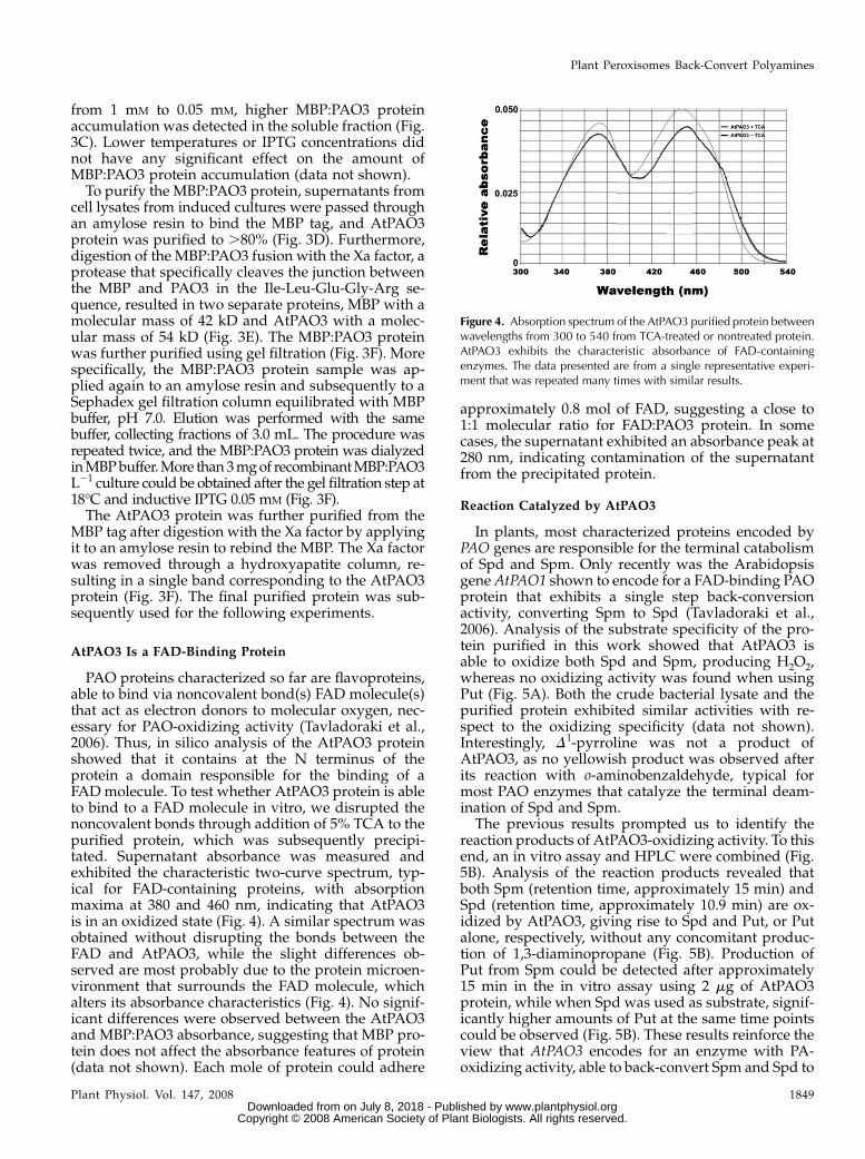
PAO proteins characterized so far are flavoproteins,able to bind via noncovalent bond(s) FAD molecule(s)that act as electron donors to molecular oxygen, nec-essary for PAO-oxidizing activity (Tavladoraki et al.,2006). Thus, in silico analysis of the AtPAO3 proteinshowed that it contains at the N terminus of theprotein a domain responsible for the binding of aFAD molecule. To test whether AtPAO3 protein is ableto bind to a FAD molecule in vitro, we disrupted thenoncovalent bonds through addition of 5% TCA to thepurified protein, which was subsequently precipi-tated. Supernatant absorbance was measured andexhibited the characteristic two-curve spectrum, typ-ical for FAD-containing proteins, with absorptionmaxima at 380 and 460 nm, indicating that AtPAO3is in an oxidized state (Fig. 4). A similar spectrum wasobtained without disrupting the bonds between theFAD and AtPAO3, while the slight differences ob-served are most probably due to the protein microen-vironment that surrounds the FAD molecule, whichalters its absorbance characteristics (Fig. 4). No signif-icant differences were observed between the AtPAO3and MBP:PAO3 absorbance, suggesting that MBP pro-tein does not affect the absorbance features of protein(data not shown). Each mole of protein could adhere
approximately 0.8 mol of FAD, suggesting a close to1:1 molecular ratio for FAD:PAO3 protein. In somecases, the supernatant exhibited an absorbance peak at280 nm, indicating contamination of the supernatantfrom the precipitated protein.
Reaction Catalyzed by AtPAO3
In plants, most characterized proteins encoded byPAO genes are responsible for the terminal catabolismof Spd and Spm. Only recently was the Arabidopsisgene AtPAO1 shown to encode for a FAD-binding PAOprotein that exhibits a single step back-conversionactivity, converting Spm to Spd (Tavladoraki et al.,2006). Analysis of the substrate specificity of the pro-tein purified in this work showed that AtPAO3 isable to oxidize both Spd and Spm, producing H2O2,whereas no oxidizing activity was found when usingPut (Fig. 5A). Both the crude bacterial lysate and thepurified protein exhibited similar activities with re-spect to the oxidizing specificity (data not shown).Interestingly, D1-pyrroline was not a product ofAtPAO3, as no yellowish product was observed afterits reaction with o-aminobenzaldehyde, typical formost PAO enzymes that catalyze the terminal deam-ination of Spd and Spm.
The previous results prompted us to identify thereaction products of AtPAO3-oxidizing activity. To thisend, an in vitro assay and HPLC were combined (Fig.5B). Analysis of the reaction products revealed thatboth Spm (retention time, approximately 15 min) andSpd (retention time, approximately 10.9 min) are ox-idized by AtPAO3, giving rise to Spd and Put, or Putalone, respectively, without any concomitant produc-tion of 1,3-diaminopropane (Fig. 5B). Production ofPut from Spm could be detected after approximately15 min in the in vitro assay using 2 mg of AtPAO3protein, while when Spd was used as substrate, signif-icantly higher amounts of Put at the same time pointscould be observed (Fig. 5B). These results reinforce theview that AtPAO3 encodes for an enzyme with PA-oxidizing activity, able to back-convert Spm and Spd to
Figure 4. Absorption spectrum of the AtPAO3 purified protein betweenwavelengths from 300 to 540 from TCA-treated or nontreated protein.AtPAO3 exhibits the characteristic absorbance of FAD-containingenzymes. The data presented are from a single representative experi-ment that was repeated many times with similar results.
Plant Peroxisomes Back-Convert Polyamines
Plant Physiol. Vol. 147, 2008 1849 www.plantphysiol.orgon July 8, 2018 - Published by Downloaded from
Copyright © 2008 American Society of Plant Biologists. All rights reserved.

the final product Put in a two-step reaction producingH2O2 as end-product.
The pH optimum for the oxidation of the substrateswas found to be 7.5 (Fig. 5C), similar to AtPAO1 (pH 5 8;Tavladoraki et al., 2006) but different from that ofZmPAO (pH 6.5). Interestingly, the enzyme wasquickly inactivated at lower pH values, and at pH .9.5 or , 6.5, no significant activity was found (Fig. 5C).The pH dependence of the enzyme did not followthe bell-shaped dependency of AtPAO1, suggestingdifferent ionization groups within the active site(Tavladoraki et al., 2006; results herein). Interestingly,the mouse (Mus musculus) peroxisomal PAO exhibitssimilar pH dependence (Wu et al., 2003).
The optimum temperature was found to be 37�C,similar to the Homo sapiens peroxisomal PAO (Wanget al., 2003), whereas at higher temperatures, theenzyme was quickly inactivated (Fig. 5C). Moreover,equimolar amounts of Spd and Put to Spm were pro-
duced via AtPAO3 oxidation activity (Fig. 5D). Theenzyme exhibited a progressive inactivation with time,possibly due to end-product inhibition, either throughH2O2 or Put (Fig. 5D).
To further confirm the back-converting activity ofAtPAO3, a radiometric method was employed usingradiolabeled PAs as substrates, and the conversionefficiency to each product was estimated as a fractionof time (Fig. 6). Thus, 80% of the initial Spd was con-verted to Put within 1 h, while 65% of initial Spm wasconverted to Spd and Put at the same time point (Fig. 6;Spd and Spm, respectively). On the contrary, radiola-beled Put was not recognized as substrate by AtPAO3.
Kinetic Parameters of AtPAO3
Because the known mammalian PAOs oxidizeN1-acetyl derivatives with higher affinity than thenonacetylated ones (Wang et al., 2003), the in vitro
Figure 5. Biochemical properties of AtPAO3. A, Relative H2O2 production (CPM, counts per minute normalized to controls)using Spd and Spm as substrates (10 mM each). B, HPLC analysis of the AtPAO3-dependent Spm conversion to Spd (15 min) andPut (15 and 60 min; AtPAO31Spm), and Spd conversion to Put (15 and 60 min; AtPAO31 Spd). A total 1 mM substrate was usedand 2 mg of AtPAO3 enzyme. The peaks with retention time varying between approximately 7 to 8 min correspond to traces ofbenzoyl chloride used as derivatization reagent. C, AtPAO3 dependence from pH and temperature. D, Conversion of Spm to Spdand Put by AtPAO3 as a fraction of time, using 2 mg of AtPAO3 enzyme and Spm as substrate. Data are the means from threeindependent experiments 6 SE.
Moschou et al.
1850 Plant Physiol. Vol. 147, 2008 www.plantphysiol.orgon July 8, 2018 - Published by Downloaded from
Copyright © 2008 American Society of Plant Biologists. All rights reserved.

activity of AtPAO3 was determined not only usingSpd and Spm as substrates, but also using N1-AcSpd,N8-AcSpd, and N1-AcSpm (Table I). AtPAO3 wasfound to oxidize not only Spd (Km 5 0.204 mM)and Spm (Km 5 0.588 mM) but also N1-AcSpd(Km 5 1 mM) and N1-AcSpm (Km 5 2 mM), convertingthem to AcSpd and AcPut, respectively (Table I; datanot shown). The substrate specificity was Spd .Spm . N1-AcSpd . N1-AcSpm (Table I), suggestingthat AtPAO3 differs with respect to the substratespecificity from the functionally homologous mam-malian enzymes.
In addition, the inhibition constants of two well-known PAO inhibitors, guazatine and aminoguani-dine, were determined using Spd as substrate (Bacchiet al., 2004; Tavladoraki et al., 2006). Guazatine, themost potent inhibitor characterized so far for PAOenzymes, including AtPAO1 (Tavladoraki et al., 2006),significantly inhibited AtPAO3 activity with a Ki of0.028 mM (Table I). Aminoguanidine also inhibitedPAO3 but with a higher Ki of 1.08 mM (Table I). Inaddition, Put, the end-product of PAO3 activity andN8-AcSpd, also inhibited the enzyme (Table I; Ki 5 61.5mM and Ki 5 40.8 mM, respectively). Put similarlyinhibited AtPAO3 when using Spm, N1-AcSpd, andN1-AcSpm as substrates (Ki 5 60.1, 50.2, and 40.9 mM,respectively).
PAO3 Is Localized in Plant Peroxisomes
In silico protein localization prediction softwarefailed to identify AtPAO3 as a putative peroxisomalprotein. However, AtPAO3 contains a peroxisomaltargeting signal 1, consisting of a Ser, an Arg, and aMet residue (SRM sequence; amino acids 486–488) atthe C-terminal end of the protein, which has beenshown to target proteins to peroxisomes in plants(Reumann et al., 2004). To investigate the in vivoAtPAO3 localization, we fused AtPAO3 cDNA to ei-ther the C-terminal (35S:GFP:PAO3) or N-terminal(35S:PAO3:GFP) part of the GFP. Agrobacterium-mediated transient expression of the 35S:GFP:PAO3in N. benthamiana leaves showed a green fluorescentpattern in small spots distributed throughout thecytoplasm of the epidermal cells (Fig. 7A). In contrast,when 35S:PAO3:GFP construct was used, a diffusefluorescence throughout the cytoplasm was found(Fig. 7B), suggesting that the fusion remained as asoluble protein in the cytosol. These results indicatethat AtPAO3 has targeting information to small cyto-plasmic organelles and that the sorting motifs aremasked when GFP is fused to the C terminus.
To determine where AtPAO3 is localized, we per-formed a colocalization experiment using yellowfluorescent protein (YFP) and Cherry markers of com-partments that resembled the spots observed in35S:GFP:PAO3 (d35S:YFP:SKL, d35S:mCherry:SKL forperoxisomal and d35S:mYFP for mitochondrial local-ization; SKL represents the motif Ser-Lys-Leu respon-sible for peroxisomal localization; Nelson et al., 2007;Fig. 6C). Agrobacterium-mediated transient expressionof the 35S:GFP:PAO3 in N. benthamiana leaves withsimultaneous cotransformation of the d35S:mCherry:SKL(Fig. 7C, top), or the d35S:YFP:SKL (Fig. 7C, bottom)
Figure 6. Radiometric assay for the detection of AtPAO3 reactionproducts and conversion efficiency using Spm and Spd as substrates.Data are the means from three independent experiments 6 SE.
Table I. Kinetics in the presence/absence of specific inhibitorsfor the PAO3 enzyme
Data are the means from a single representative experiment.
Substrate Km Kcat Kcat
mMa s21b Km
21
Agmatine 0 0 –Cadaverine 0 0 –Put 0 0 –Spd 0.204 1.250 6.12Spm 0.588 0.188 0.31N1-AcSpd 1 0.042 0.042N 8-AcSpd .5000 0 –N1-AcSpm 2 0.020 0.01Inhibitor Ki (mM)c – –Guazatine 0.028 – –Aminoguanidine 1.08 – –Put 61.5N8-AcSpd 40.8 – –
aSubstrate concentrations were ,1.25 mM. Data are the means fromthree independent experiments 6SE. b10 mM substrate was used forKcat determination. cInhibitors concentrations were ,1.25 mM.Data are the means from three independent experiments 6SE.
Plant Peroxisomes Back-Convert Polyamines
Plant Physiol. Vol. 147, 2008 1851 www.plantphysiol.orgon July 8, 2018 - Published by Downloaded from
Copyright © 2008 American Society of Plant Biologists. All rights reserved.
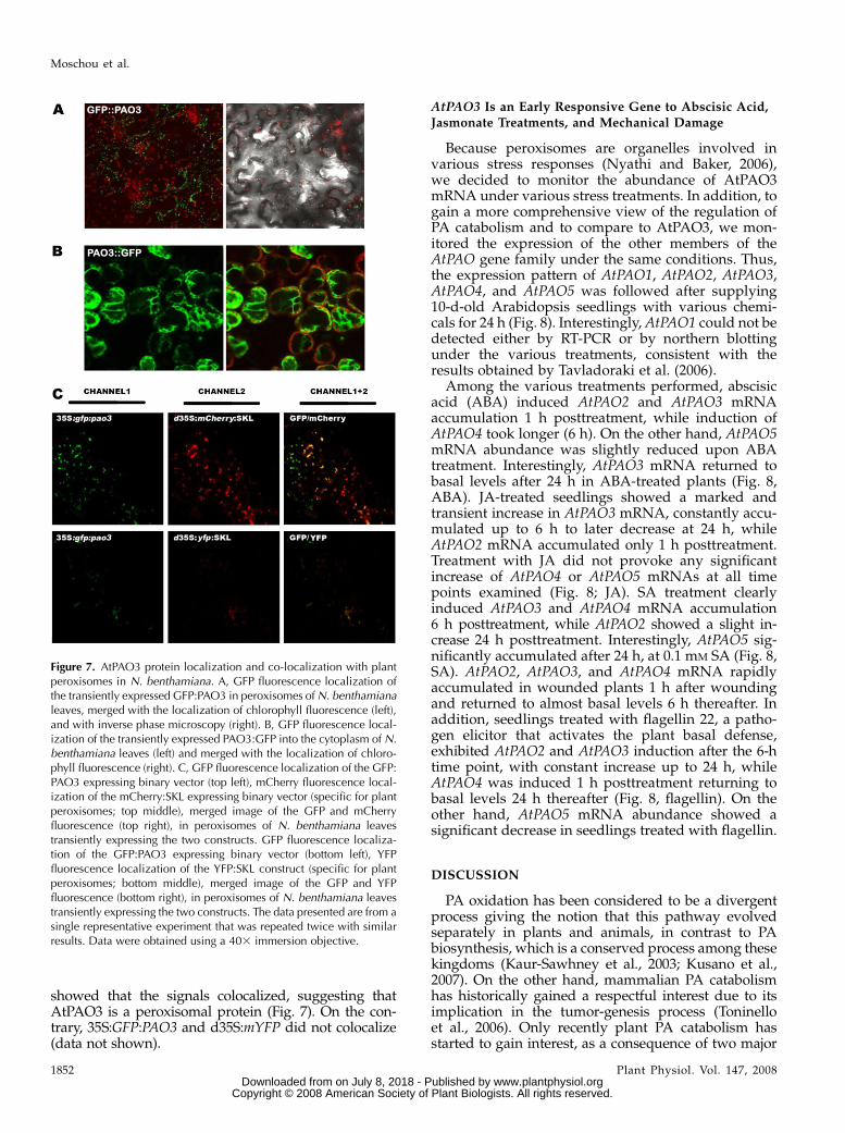
showed that the signals colocalized, suggesting thatAtPAO3 is a peroxisomal protein (Fig. 7). On the con-trary, 35S:GFP:PAO3 and d35S:mYFP did not colocalize(data not shown).
AtPAO3 Is an Early Responsive Gene to Abscisic Acid,
Jasmonate Treatments, and Mechanical Damage
Because peroxisomes are organelles involved invarious stress responses (Nyathi and Baker, 2006),we decided to monitor the abundance of AtPAO3mRNA under various stress treatments. In addition, togain a more comprehensive view of the regulation ofPA catabolism and to compare to AtPAO3, we mon-itored the expression of the other members of theAtPAO gene family under the same conditions. Thus,the expression pattern of AtPAO1, AtPAO2, AtPAO3,AtPAO4, and AtPAO5 was followed after supplying10-d-old Arabidopsis seedlings with various chemi-cals for 24 h (Fig. 8). Interestingly, AtPAO1 could not bedetected either by RT-PCR or by northern blottingunder the various treatments, consistent with theresults obtained by Tavladoraki et al. (2006).
Among the various treatments performed, abscisicacid (ABA) induced AtPAO2 and AtPAO3 mRNAaccumulation 1 h posttreatment, while induction ofAtPAO4 took longer (6 h). On the other hand, AtPAO5mRNA abundance was slightly reduced upon ABAtreatment. Interestingly, AtPAO3 mRNA returned tobasal levels after 24 h in ABA-treated plants (Fig. 8,ABA). JA-treated seedlings showed a marked andtransient increase in AtPAO3 mRNA, constantly accu-mulated up to 6 h to later decrease at 24 h, whileAtPAO2 mRNA accumulated only 1 h posttreatment.Treatment with JA did not provoke any significantincrease of AtPAO4 or AtPAO5 mRNAs at all timepoints examined (Fig. 8; JA). SA treatment clearlyinduced AtPAO3 and AtPAO4 mRNA accumulation6 h posttreatment, while AtPAO2 showed a slight in-crease 24 h posttreatment. Interestingly, AtPAO5 sig-nificantly accumulated after 24 h, at 0.1 mM SA (Fig. 8,SA). AtPAO2, AtPAO3, and AtPAO4 mRNA rapidlyaccumulated in wounded plants 1 h after woundingand returned to almost basal levels 6 h thereafter. Inaddition, seedlings treated with flagellin 22, a patho-gen elicitor that activates the plant basal defense,exhibited AtPAO2 and AtPAO3 induction after the 6-htime point, with constant increase up to 24 h, whileAtPAO4 was induced 1 h posttreatment returning tobasal levels 24 h thereafter (Fig. 8, flagellin). On theother hand, AtPAO5 mRNA abundance showed asignificant decrease in seedlings treated with flagellin.
DISCUSSION
PA oxidation has been considered to be a divergentprocess giving the notion that this pathway evolvedseparately in plants and animals, in contrast to PAbiosynthesis, which is a conserved process among thesekingdoms (Kaur-Sawhney et al., 2003; Kusano et al.,2007). On the other hand, mammalian PA catabolismhas historically gained a respectful interest due to itsimplication in the tumor-genesis process (Toninelloet al., 2006). Only recently plant PA catabolism hasstarted to gain interest, as a consequence of two major
Figure 7. AtPAO3 protein localization and co-localization with plantperoxisomes in N. benthamiana. A, GFP fluorescence localization ofthe transiently expressed GFP:PAO3 in peroxisomes of N. benthamianaleaves, merged with the localization of chlorophyll fluorescence (left),and with inverse phase microscopy (right). B, GFP fluorescence local-ization of the transiently expressed PAO3:GFP into the cytoplasm of N.benthamiana leaves (left) and merged with the localization of chloro-phyll fluorescence (right). C, GFP fluorescence localization of the GFP:PAO3 expressing binary vector (top left), mCherry fluorescence local-ization of the mCherry:SKL expressing binary vector (specific for plantperoxisomes; top middle), merged image of the GFP and mCherryfluorescence (top right), in peroxisomes of N. benthamiana leavestransiently expressing the two constructs. GFP fluorescence localiza-tion of the GFP:PAO3 expressing binary vector (bottom left), YFPfluorescence localization of the YFP:SKL construct (specific for plantperoxisomes; bottom middle), merged image of the GFP and YFPfluorescence (bottom right), in peroxisomes of N. benthamiana leavestransiently expressing the two constructs. The data presented are from asingle representative experiment that was repeated twice with similarresults. Data were obtained using a 403 immersion objective.
Moschou et al.
1852 Plant Physiol. Vol. 147, 2008 www.plantphysiol.orgon July 8, 2018 - Published by Downloaded from
Copyright © 2008 American Society of Plant Biologists. All rights reserved.

facts, the complete analysis of the plant PA biosyntheticpathway, and the correlation of PA catabolism withprogrammed cell death processes, both at physiologicaland under stress conditions (Tiburcio et al., 1994; Yodaet al., 2003, 2006; Rea et al., 2004; Paschalidis andRoubelakis-Angelakis., 2005b; Moschou et al., 2008).
In an attempt to bridge the gap in our knowledge ofPA oxidation between plants and animals, we strivedto search the Arabidopsis genome with the scope toidentify putative genes that could be functionallysimilar to the mammalian PAOs in terms of specificactivity and localization. Using data obtained fromcombinatorial in silico analysis and the expressionpatterns of pao genes, and taking into account thepossible localization predicted by AraPerox database(Reumann et al., 2004), we concluded that AtPAO3gene could serve as a starting point for our research. Inthis work, we have described the purification of re-combinant AtPAO3 using the heterologous expressionsystem of E. coli (Fig. 3). In contrast to previous studiesthat either failed to express the protein in the solublefraction (Tavladoraki et al., 2006) or introduced signif-icant changes (deletion of the N terminus of theprotein) in the structure of the PAO protein in orderto obtain it in the soluble fraction (Yoda et al., 2006), wehave managed to produce the protein in a soluble formusing a fusion of the protein to MBP protein. Theresults have revealed that AtPAO3 exhibits the typicalmammalian PAO back-conversion activity being ableto produce Put as a final oxidation product (Figs. 5 and6). A striking difference between the two counterpartsis the substrate specificity.
More specifically, in mammals, Spd/Spm N1-acetyl-transferase (SSAT) is the responsible enzyme for theacetylation of Spd and Spm, and the correspondingacetylated derivatives are orientated toward the cata-
bolic pathway in peroxisomes, where di-acetyl-Spm and N1-acetyl-Spd serve as the optimum sub-strate for PAOs (SSAT-dependent PA catabolism). Incontrast, AtPAO3 exhibits strong preference for non-acetylated Spd and Spm, indicating that AtPAO3probably does not depend upon a yet-unrecognizedSSAT activity (SSAT-independent catabolism; Table I).Moreover, AtPAO1 also showed preference for Spmand not its acetylated derivatives, suggesting that it isalso SSAT independent (Tavladoraki et al., 2006). Al-though Spm can be oxidized by the mammalian per-oxisomal PAO, it is a poor substrate when compared toN1-acetyl-Spm and N1-acetyl-Spd. In mammals, thenecessity of Spm and Spd acetylation relates to the factthat both PAs are acetylated in order to be transportedfrom the cells and eventually being excreted (Seiler,1995; Wu et al., 2003). High PAO levels in mammalscould prevent transport of N1-acetyl-Spm and N1-acetyl-Spd from the cell and increase the Spd andPut levels of the PA pool (Wu et al., 2003). On the otherhand, these PA analogues in plants are of unknownphysiological function and they can only be found intrace amounts in Arabidopsis, while in H. tuberosumrelatively high titers of N1-acetyl-Spd were found in chlo-roplasts upon exogenous Put addition (Del Duca et al.,1995; Tassoni et al., 2000). Moreover, Del Duca et al.(1995) hypothesized that exogenous Put addition causesthe inhibition of a PAO activity responsible for theconversion of Spd to Put. This is now reinforced by ourresults in which Put, the end-product of the full back-conversion pathway, exerted an inhibitory effect (TableI). The biochemical evidence for the existence of thePAO-dependent back-conversion pathway in the pe-rennial dicot plant H. tuberosus indicates that the PAback-conversion is a common pathway in plants, ratherthan species-specific.
Figure 8. AtPAO2, AtPAO3,AtPAO4, and AtPAO5 mRNA abun-dance under various chemical stimuli1, 6, and 24 h after the correspond-ing treatment (each well contains 10mg of total RNA and exposure timewas 48 h). The data presented arefrom a single representative experi-ment that was repeated twice withsimilar results.
Plant Peroxisomes Back-Convert Polyamines
Plant Physiol. Vol. 147, 2008 1853 www.plantphysiol.orgon July 8, 2018 - Published by Downloaded from
Copyright © 2008 American Society of Plant Biologists. All rights reserved.

Interestingly, AtPAO1 and AtPAO3 exhibit a signif-icant difference in their respective mRNA abundancein plant cells. More specifically, AtPAO3 is a ratherconstitutively expressed mRNA at relatively highlevels in plant tissues (Supplemental Fig. S1), whereasAtPAO1 could only be detected using nested RT-PCR(Tavladoraki et al., 2006; results herein). In mammals,the peroxisomal PAO is also constitutively expressedat high levels, whereas the SMO is essentially aninducible gene (Pledgie et al., 2005). This striking sim-ilarity could imply a type of regulation of PA back-conversion with a yet-unknown role for both plantsand animals. With respect to the SSAT dependence,AtPAO3 mimics AtPAO1 that is also involved in apathway in which the SSAT probably does not partic-ipate. On the other hand, although both AtPAO1 andAtPAO3 follow a SSAT-independent pathway, theirpreference for substrates is radically different. AtPAO1exhibits strong resemblance to the mammalian SMO,converting Spm to Spd but unable to convert Spd toPut (Tavladoraki et al., 2006), whereas AtPAO3 isable to oxidize both Spm and Spd, exhibiting prefer-ence to Spd (Table I; Fig. 6). In mammals, when PAOoxidizes N1-acetyl-Spm or N1-acetyl-Spd, FAD is re-duced and subsequently oxidized by O2 to generateH2O2, and in our study, AtPAO3 also produced H2O2(Fig. 5). In contrast to Fsm1, the PAO homolog fromyeast (Saccharomyces cerevisiae), AtPAO3 is unable tooxidize N8-acetyl-Spd (Landry and Sternglanz, 2003).Thus, Spd produced by AtPAO1 from Spm might be ofphysiological significance, because this can be furtherused by AtPAO3 to efficiently produce Put.
Most known animal PAOs are localized in peroxi-somes; AtPAO3 is so far the second organellar PAOrecognized in plants in addition to PAO from barley,which is sorted to vacuoles (Cervelli et al., 2004). Thelocalization of AtPAO3 in peroxisomes gives directevidence for the implication of this organelle in PAoxidation and back-conversion in plant tissues (Fig. 7).This compartmentalization could be of physiologicalimportance considering the oxidative nature of PAO,because the produced H2O2 could be efficiently scav-enged through the abundant peroxisomal catalase.Moreover, the apparent nonorganellar localization ofAtPAO1, which functionally resembles the mamma-lian SMO and the AtPAO3 sorting to peroxisomes,could suggest a similar system to the mammalianoxidation system but lacking the SSAT.
The strong induction of the AtPAO3 by JA (early),wounding (early), SA (middle to late), and flagellin(constant; Fig. 8) suggests the relevance of the enzymeto stress defense responses (Alvarez et al., 1998; Asaiet al., 2000). The H2O2 produced by the AtPAO-oxidizing activity could act as a second messengerthat signals downstream events under biotic stress, asdescribed previously (Orozco-Cardenas et al., 2001;Takahashi et al., 2004; Vacca et al., 2004; Sasaki-Sekimoto et al., 2005; Skopelitis et al., 2006; Moschouet al., 2008). Moreover, PAs have been implicated in thewound response in Arabidopsis (Perez-Amador et al.,
2002). In mammals, another product of the oxidationof the N1-acetylated PAs, 3-acetamidopropanal, maybe enzymatically deacetylated to yield cytotoxic3-aminopropanal that is thought to contribute, eitheralone or in concert with H2O2, to tissue damage fol-lowing traumatic injury (Wu et al., 2003). What isworth noticing is the pattern of flagellin-inducedAtPAO3 increase, which in contrast to the other treat-ments produces a constantly increasing pattern ratherthan a transient increase (Fig. 8). The previous impliesthat the differential signal perception between bioticand abiotic stress produces alternative patterns ofAtPAO3 activation. In the case of abiotic stress, PAoxidation from AtPAO3 could have a signaling role,whereas in the case of biotic stress, AtPAO3 could pro-duce H2O2 for a prolonged period of time to participatein this way in the plant’s basal defense against patho-genic bacteria.
Our overall results support that AtPAO3 is a perox-isomal PAO, able to back-convert Spm to Spd and sub-sequently Spd to Put, and provides plant peroxisomeswith a new role in PA oxidation/back-conversion.Moreover, the correlation of AtPAO3 with stress re-sponses involving oxidative burst gives evidence fora PA-peroxisomal interplay under (a)biotic stress con-ditions.
MATERIALS AND METHODS
Chemicals
All chemicals were purchased from Sigma unless stated otherwise, while
radiolabeled PAs were purchased from Amersham.
In Silico Analysis and cDNA Acquisition
For the multiple protein alignment and the phylogenetic tree construction,
the Expasy Database tools were used (http://au.expasy.org/tools/). For motif
and domain scan, the Simple Modular Architecture Research Tool database
was used (http://smart.embl.de). The cDNA encoding for the AtPAO3 (clone
no. U21196) was provided by the ABRC stock center (http://www.arabidopsis.
org).
Plant Material and Treatments
Seeds of Arabidopsis (Arabidopsis thaliana) ecotype Columbia-0 were sur-
face sterilized and sown in 24-well (10 seeds per well) tissue culture clusters,
containing 1 mL/well of sterile Murashige and Skoog medium (Murashige
and Skoog salts; Duchefa) supplemented with 0.5% Suc and grown with
shaking (150 rpm) in a culture room under 16-h-d (26�C)/8-h night (22�C)
diurnal cycles. Fresh medium, 500 mL/well, was added 9 d after sowing, and
experiments were conducted 1 d later. Prior to treatment initiation, the
medium remaining in the wells was removed and 1 mL of fresh medium
with the different compounds tested was added.
Gene Constructs
For the AtPAO3 cDNA cloning into vector pDONR207, the primer pairs
GGGGACAAGTTTGTACAAAAAAGCAGGCTTGATGGAGTCCGGAGGCA-
AC (PAO3-FOR) and GGGGACCACTTTGTACAAGAAAGCTGGGTATTA-
CATACGGGAGATCAGAAG (PAO3-REV) were used, and inserts were
cloned into the pDEST-TH1 (Hammrastrom et al., 2002) and pGWB6 (GFP:
PAO3) vectors using the GATEWAY recombination system (Invitrogen). To
eliminate the stop codon of PAO3 cDNA, the PAO3-REV-no stop primer
Moschou et al.
1854 Plant Physiol. Vol. 147, 2008 www.plantphysiol.orgon July 8, 2018 - Published by Downloaded from
Copyright © 2008 American Society of Plant Biologists. All rights reserved.

GGGGACCACTTTGTACAAGAAAGCTGGGTATTACATACGGGAGATCA-
GCG was used in combination with PAO3-FOR, and AtPAO3 was cloned into
a second pDONR207 vector and further transferred into the pGWB5
(PAO3:GFP) vector using the GATEWAY recombination system. The vector
pDEST-TH1 was used for protein expression, whereas vectors pGWB6 and
pGWB5 correspond to the N-terminal and C-terminal GFP binary fusion
vectors, respectively. All vectors prepared were direct sequenced.
PCR amplifications were performed using a thermal PTC-200 PCR cycler
and the Pfu (Promega) or Taq (Minotech) polymerase following standard PCR
protocols.
Protein Expression and Purification
Escherichia coli strain BL21 cells were transformed with standard transfor-
mation protocols and were induced for protein expression by adding 0.05 to
1 mM IPTG in the Luria Nutrient Broth culture media supplemented with 100
mg mL21 ampicillin and 2 g L21 Glc after reaching OD600 5 0.5. The cells were
harvested at 2,500g for 20 min at RT and frozen overnight at 280�C. Cells were
dissolved in 10 mL of MBP buffer (20 mM Tris, pH 7.4, 400 mM NaCl, and 1 mM
EDTA, 0.5 mM PMSF) and lysed at 4�C in a sonicator using four cycles of 15 to
20 s. Cell debris (pellet fraction) was removed after a centrifugation step at
10,000g at 4�C, and supernatant was filtrated using Miracloth and passed
through an amylose resin (Biolabs) at 4�C. Protein was eluted using MBP
buffer supplemented with 20 mM maltose, and collected fractions were
electrophoretically resolved.
Protein fusion was digested using the protease Xa factor as described by
the manufacturer (Biolabs). The Xa factor was removed using a hydroxy-
apatite column as described by the manufacturer (Biolabs).
In Vitro Activity Assays
In vitro activity was determined using four different assays. To determine
directly the H2O2 production, a modified method of luminol measurement
was used. Thus, 2 mg of purified protein were incubated in a 690-mL H2O2
lumination buffer (10 mM Tris, pH 7.0, 1 mM CaCl2, 0.1 mM KCl) supplemented
with 100 mL 2.5 mg mL21 luminol, 10 mL of 0.1 unit mL21 peroxidase, and 1 to
10 mM final concentration of Put 2HCl, Spd 3HCl, or Spm 4HCl. Counts were
measured in a scintillation counter (Beckman) for 10 min, and values
were taken in the linear part of the assay. For determination of the kinetic
parameters, the method of the oxidation of 4-aminopterine was used
(Tavladoraki et al., 2006). Analysis of AtPAO3 oxidation products was
performed by incubating saturating amounts of substrate with 4 mg of
purified protein, in a final volume of 200 mL, buffered with 100 mM Tris, pH
7.5. Reaction was stopped by the addition of equal volume of 5% (v/v)
perchloric acid. The samples were centrifuged for 20 min at 12,000g at 4�C, the
supernatant was derivatized, and PAs were determined as previously de-
scribed, using an HP 1100 High Performance Liquid Chromatographer
(Hewlett-Packard; Kotzabasis et al., 1993). Additionally, reaction products
were estimated using radiolabeled Spm by a modification of the radiometric
method of Paschalidis and Roubelakis-Angelakis (2005a), using [1,4-14C]Put,
[1,4-14C]Spd, [1,4-14C]Spm (Amersham; specific activities, 4.37 GBq mmol21)
as labeled substrates and 4 mg of AtPAO3 enzyme. The assay mixture
contained 0.2 mL of Tris, pH 7.5, 1 mM unlabeled Put, Spd, and Spm and 3.7
KBq of [1,4-14C]Put, [1,4-14C]Spd, [1,4-14C]Spm. After incubation on a shaker at
37�C for 60 min, the reaction was terminated by adding 150 mL of saturated
sodium carbonate. Products were separated on a thin layer chromatography
plate using as a mobile phase Chlorophorm/Triethylamine 4:1 (v/v). The plate
was dried out for 10 min at 60�C, the radioactivity was estimated after scraping
off the spots from the plate, and the powder from each spot was measured
using a scintillation counter (Beckman). To identify the retention factor of each
spot, 1 mM standards of PAs were also used and were visualized under UV light
following dansylation as described by Flores and Galston (1982). Additionally,
radiolabeled products were dansylated to maintain the Rr accuracy.
Standard calibration curves for N1-AcSpm, N1-AcSpd, and N8-AcSpd were
additionally prepared. The D1-aminopyrroline production was assayed by
using the o-aminobenzaldehyde method and the formation of a yellowish
product, as described in Moschou et al. (2008).
Protein Extraction, Western Blotting, RNA Extraction,and Northern Blotting
Proteins were extracted and treated as described in Papadakis and
Roubelakis-Angelakis (2005). Total protein extracts were electrophoretically
resolved using SDS-PAGE, transferred to membranes, and hybridized against
an anti-MBP polyclonal antibody. Total RNA was extracted with the optimized
hot-phenol method according to Rojo et al. (1998), transferred to nylon mem-
branes, and hybridized against 32P-labeled probes. All hybridizations were
performed at 67�C and the appropriate controls were used, including RNA
ladders, for the correct size of each transcript to be estimated.
Templates for probes were obtained using the following primers
and standard PCR conditions: for PAO1, GGTAGTGGTGAAGACAGAGG
(PAO1-FOR) and AGCTTGTGCCTGAGATAAA (PAO1-REV); PAO2, ATG-
GAGTCCAGGAAAACTC (PAO2-FOR) and GAGACGAGATATAAGAAG
(PAO2-REV); PAO3, GGGGACAAGTTTGTACAAAAAAGCAGGCTTGATG-
GAGTCCGGAGGCAAC (PAO3-FOR) and GGGGACCACTTTGTACAA-
GAAAGCTGGGTATTACATACGGGAGATCAGAAG (PAO3-REV); PAO4,
GGTCACCAAACCCTTCATC (PAO4-FOR) and TTGAGCAAACGATGGA-
GAAG (PAO4-REV); PAO5 ATGGCGAAGAAAGCAAGA (PAO5-FOR) and
AAAATTACATTTGTAAT (PAO5-REV).
RT-PCR Analysis
For semiquantitative RT-PCR reactions, total mRNA from leaves and roots
was extracted and subjected to DNase I RNase-free treatment (Invitrogen) for
45 min at 37�C. After checking the RNA in agarose gel, the samples were
subjected to RT-PCR reactions using polyTas primer and the Super RT enzyme
according to the manufacturer’s instructions (HT Biotechnology). The samples
were normalized according to actin and the primers TTGAGCACATTCT-
GGAAGAGGTG (forward PAO3) and ACACTTTGCCGAGATGGTTTCAG
(reverse PAO3) were used for AtPAO3-specific 30 cycle (exponential phase)
quantitative RT-PCR reactions. Reactions with samples treated with DNase I
but not subjected to RT were also used as negative controls.
Transient Expression and Colocalization
Agrobacterium tumefaciens strain C58C1 cells were transformed with the
appropriate binary vectors using the freeze-thaw method and PCR confirmed.
A. tumefaciens positive clones were grown in Luria-Bertani until OD600 5 0.6
and were pelleted after a centrifugation step at 3,000g for 20 min. Cells were
resuspended in MMA (Murashige and Skoog salts, 10 mM MES, pH 5.7, 0.2
mM acetosyringone, 3% Suc) until OD600 5 0.5, incubated at RT for 1 h, and
infiltrated in Nicotiana benthamiana leaves, using a 1-mL syringe. Leaves were
analyzed after 48 h using a Leica TCS-NT confocal microscope, using the 403
objective. The excitation/emission wavelength was 480/508 nm for GFP, 513/
527 nm for YFP, and 587/610 nm for mCherry. The YFP-PRX (PRX: peroxisomal)
and mCherry-PRX constructs were purchased from the ABRC Stock Center
(http://www.arabidopsis.org).
Colocalization was performed by mixing the MMA-resuspended A.
tumefaciens GFP:PAO3 with A. tumefaciens YFP-PRX or mCherry-PRX in a
ratio 1.2:1 (v/v). Merge of the images was performed using the Leica Confocal
software version 2.61.
Sequence data from this article can be found in the GenBank/EMBL data
libraries under accession numbers NM_121373 (AtPAO1), AF364952 (AtPAO2),
NM_115767 (AtPAO3), AF364953 (AtPAO4), and AK118203 (AtPAO5).
Supplemental Data
The following materials are available in the online version of this article.
Supplemental Figure S1. Expression of AtPAO3 in leaves and roots.
cDNAs were equalized according to actin.
ACKNOWLEDGMENT
The authors are grateful to Dr. Helena Berglund (Karolinska Institute,
Sweden) for the generous gift of the pDEST-TH1 vector.
Received May 31, 2008; accepted June 18, 2008; published June 26, 2008.
LITERATURE CITED
Alvarez ME, Pennell RI, Meijer PJ, Ishikawa A, Dixon RA, Lamb C (1998)
Reactive oxygen intermediates mediate a systemic signal network in the
establishment of plant immunity. Cell 92: 773–784
Plant Peroxisomes Back-Convert Polyamines
Plant Physiol. Vol. 147, 2008 1855 www.plantphysiol.orgon July 8, 2018 - Published by Downloaded from
Copyright © 2008 American Society of Plant Biologists. All rights reserved.

Amendola R, Bellini A, Cervelli M, Degan P, Marcocci L, Martini F,
Mariottini P(2005) Direct oxidative DNA damage, apoptosis and radio
sensitivity by spermine oxidase activities in mouse neuroblastoma cells.
Biochim Biophys Acta 1755: 15–24
Angelini R, Federico R, Bonfante P (1995) Maize polyamine oxidase: anti-
body production and ultrastructural localization. J Plant Physiol 145:
686–692
Asai T, Stone JM, Heard JE, Kovtun Y, Yorgey P, Sheen J, Ausubel FM
(2000) Fumonisin B1-induced cell death in Arabidopsis protoplasts re-
quires jasmonate-, ethylene-, and salicylate-dependent signaling path-
ways. Plant Cell 12: 1823–1835
Bacchi CJ, Rattendi D, Faciane E, Yarlett N, Weiss LM, Frydman B, Woster
P, Wei B, Marton LJ, Wittner M (2004) Polyamine metabolism in a
member of the phylum Microspora (Encephalitozoon cuniculi): effects of
polyamine analogues. Microbiology 150: 1215–1224
Capell T, Bassie L, Christou P (2004) Modulation of the polyamine
biosynthetic pathway in transgenic rice confers tolerance to drought
stress. Proc Natl Acad Sci USA 101: 9909–9914
Cervelli M, Caro O, Penta A, Angelini R, Federico R, Vitale A, Mariottini
P (2004) A novel C-terminal sequence from barley polyamine oxidase is
a vacuolar sorting signal. Plant J 40: 410–418
Cona A, Moreno S, Cenci F, Federico R, Angelini R (2005) Cellular re-
distribution of flavin-containing polyamine oxidase in differentiating
root and mesocotyl of Zea mays L. seedlings. Planta 221: 265–276
Cona A, Rea G, Angelini R, Federico R, Tavladoraki P (2006) Functions of
amine oxidases in plant development and defence. Trends Plant Sci 11:
80–88
Del Duca S, Beninati S, Serafini-Fracassini D (1995) Polyamines in
chloroplasts: identification of their glutamyl and acetyl derivatives.
Biochem J 305: 233–237
Flores HE, Galston AW (1982) Analysis of polyamines in higher plants by
high performance liquid chromatography. Plant Physiol 69: 701–706
Ha HC, Woster PM, Yager JD, Casero RA (1997) The role of polyamine
catabolism in polyamine analogue-induced programmed cell death.
Proc Natl Acad Sci USA 94: 11557–11562
Hammarstrom M, Hellgren N, Van der Berg S, Berglund H, Hard T (2002)
Rapid screening for improved solubility of small human proteins
produced as fusion proteins in Escherichia coli. Protein Sci 11: 313–321
Hayashi M, Nishimura M (2006) Arabidopsis thaliana: a model organism to
study plant peroxisomes. Biochim Biophys Acta 1763: 1382–1391
Kaur-Sawhney R, Tiburcio AF, Atabella T, Galston AW (2003) Polyamines
in plants: an overview. J Cell Mol Biol 2: 1–12
Kotzabasis K, Christakis-Hampsas MD, Roubelakis-Angelakis KA
(1993) A narrow bore HPLC method for the identification and quanti-
tation of free, conjugated and bound polyamines. Anal Biochem 214:
484–487
Kusano T, Yamaguchi K, Berberich T, Takahashi Y (2007) Advances in
polyamine research in 2007. J Plant Res 120: 345–350
Landry J, Sternglanz R (2003) Yeast Fms1 is a FAD-utilizing polyamine
oxidase. Biochem Biophys Res Commun 303: 771–776
Letunic I, Copley RR, Pils B, Pinkert S, Schultz J, Bork P (2006) SMART 5:
domains in the context of genomes and networks. Nucleic Acids Res 34:
D257–260
Mehta RA, Cassol T, Li N, Ali N, Handa AK, Mattoo AK (2002)
Engineered polyamine accumulation in tomato enhances phytonutrient
content, juice quality, and vine life. Nat Biotechnol 20: 613–618
Moschou P, Dellis I, Paschalidis K, Roubelakis-Angelakis KA (2008)
Transgenic tobacco plants over-expressing polyamine oxidase are not
able to cope with oxidative burst generated by abiotic factors. Physiol
Plant 133: 140–156
Nelson BK, Cai X, Nebenfuhr A (2007) A multi-color set of in vivo
organelle markers for colocalization studies in Arabidopsis and other
plants. Plant J 51: 1126–1136
Nyathi Y, Baker A (2006) Plant peroxisomes as a source of signaling
molecules. Biochim Biophys Acta 1763: 1478–1495
Orozco-Cardenas ML, Narvaez-Vasquez J, Ryan CA (2001) Hydrogen
peroxide acts as a second messenger for the induction of defence genes
in tomato plants in response to wounding, systemin and methyl
jasmonate. Plant Cell 13: 179–191
Papadakis AK, Roubelakis-Angelakis KA (2005) Polyamines inhibit
NADPH oxidase-mediated superoxide generation and putrescine pre-
vents programmed cell death induced by polyamine oxidase-generated
hydrogen peroxide. Planta 220: 826–837
Paschalidis KA, Roubelakis-Angelakis KA (2005a) Spatial and temporal
distribution of polyamine levels and polyamine anabolism in different
organs/tissues of the tobacco plant. Correlations with age, cell divi-
sion/expansion, and differentiation. Plant Physiol 138: 142–152
Paschalidis KA, Roubelakis-Angelakis KA (2005b) Sites and regulation of
polyamine catabolism in the tobacco plant. Correlations with cell
division/expansion, cell cycle progression, and vascular development.
Plant Physiol 138: 2174–2184
Perez-Amador MA, Leon J, Green PJ, Carbonell J (2002) Induction of the
arginine decarboxylase ADC2 gene provides evidence for the involve-
ment of polyamines in the wound response in Arabidopsis. Plant
Physiol 130: 1454–1463
Pledgie A, Huang Y, Hacker A, Zhang Z, Woster PM, Davidson NE, Casero
RA (2005) Spermine oxidase SMO (PAOh1), not N1-acetylpolyamine
oxidase PAO, is the primary source of cytotoxic H2O2 in polyamine
analogue-treated human breast cancer cell lines. J Biol Chem 280: 39843–
39851
Rea G, de Pinto MC, Tavazza R, Biondi S, Gobbi V, Ferrante P, De Gara L,
Federico R, Angelini R, Tavladoraki P (2004) Ectopic expression of
maize polyamine oxidase and pea copper amine oxidase in the cell wall
of tobacco plants. Plant Physiol 134: 1414–1426
Reumann S, Ma C, Lemke S, Babujee L (2004) AraPerox. A database of
putative Arabidopsis proteins from plant peroxisomes. Plant Physiol
136: 2587–2608
Rojo E, Titarenko E, Leon J, Berger S, Vancanneyt G, Sanchez-Serrano JJ
(1998) Reversible protein phosphorylation regulates jasmonic acid-
dependent and -independent wound signal transduction pathways in
Arabidopsis thaliana. Plant J 13: 153–165
Sasaki-Sekimoto Y, Taki N, Obayashi T, Aono M, Matsumoto F, Sakurai
N, Suzuki H, Hirai MY, Noji M, Saito K, et al (2005) Coordinated
activation of metabolic pathways for antioxidants and defence com-
pounds by jasmonates and their roles in stress tolerance in Arabidopsis.
Plant J 44: 653–668
Schrader M, Fahimi DH (2004) Mammalian peroxisomes and reactive
oxygen species. Histochem Cell Biol 122: 383–393
Seiler N (1995) Polyamine oxidase, properties and functions. Prog Brain
Res 106: 333–344
Skopelitis DS, Paranychianakis NV, Paschalidis KA, Pliakonis ED, Delis
ID, Yakoumakis DI, Kouvarakis A, Papadakis AK, Stephanou EG,
Roubelakis-Angelakis KA (2006) Abiotic stress generates ROS that
signal expression of anionic glutamate dehydrogenases to form gluta-
mate for proline synthesis in tobacco and grapevine. Plant Cell 18:
2767–2781
Takahashi Y, Uehara Y, Berberich T, Ito A, Saitoh H, Miyazaki A, Terauchi
R, Kusano T (2004) A subset of hypersensitive response marker genes,
including HSR203J, is the downstream target of a spermine signal
transduction pathway in tobacco. Plant J 40: 586–595
Tassoni A, Van Buuren M, Franceschetti M, Fornale IS, Bagni N (2000)
Polyamine content and metabolism in Arabidopsis thaliana and effect
of spermidine on plant development. Plant Physiol Biochem 38:
383–393
Tavladoraki P, Rossi MN, Saccuti G, Perez-Amador MA, Polticelli F,
Angelini R, Federico R (2006) Heterologous expression and biochem-
ical characterization of a polyamine oxidase from Arabidopsis involved
in polyamine back conversion. Plant Physiol 149: 1519–1532
Tavladoraki P, Schinina ME, Cecconi F, Di Agostino S, Manera F, Rea G,
Mariottini P, Federico R, Angelini R (1998) Maize polyamine oxidase:
primary structure from protein and cDNA sequencing. FEBS Lett 426:
62–66
Thomas T, Thomas TJ (2001) Polyamines in cell growth and cell death:
molecular mechanism and therapeutic applications. Cell Mol Life Sci 58:
244–258
Tiburcio AF, Besford RT, Borrell A (1994) Posttranslational regulation of
arginine decarboxylase synthesis by spermine in osmotically-stressed
oat leaves. Biochem Soc Trans 22: 455S
Toninello A, Pietrangeli P, De Marchi U, Salvi M, MondovI B
(2006) Amine oxidases in apoptosis and cancer. Biochim Biophys Acta
1765: 1–13
Vacca RA, de Pinto MC, Valenti D, Passarella S, Marra E, Gara DL (2004)
Production of reactive oxygen species, alteration of cytosolic ascorbate
peroxidase, and impairment of mitochondrial metabolism are early
events in heat shochinduced programmed cell death in tobacco bright-
yellow 2 cells. Plant Physiol 134: 1100–1112
Moschou et al.
1856 Plant Physiol. Vol. 147, 2008 www.plantphysiol.orgon July 8, 2018 - Published by Downloaded from
Copyright © 2008 American Society of Plant Biologists. All rights reserved.

Wang Y, Murray-Stewart T, Devereux W, Hacker A, Frydman B, Woster
PM, Casero RA Jr (2003) Properties of purified human poly-
amine oxidase, PAOh1/SMO. Biochem Biophys Res Commun 304:
605–611
Wu T, Yankovskaya V, McIntire WS (2003) Cloning, sequencing, and
heterologous expression of the murine peroxisomal flavoprotein
N1-acetylated polyamine oxidase. J Biol Chem 278: 20514–20525
Yamaguchi K, Takahashi Y, Berberich T, Imai A, Miyazaki A, Takahashi
T, Michael A, Kusano T (2006) The polyamine spermine protects
against high salt stress in Arabidopsis thaliana. FEBS Lett 22: 6783–8
Yoda H, Hiroi Y, Sano H (2006) Polyamine oxidase is one of the key
elements for oxidative burst to induce programmed cell death in tobacco
cultured cells. Plant Physiol 142: 193–206
Yoda H, Yamaguchi Y, Sano H (2003) Induction of hypersensitive cell death
by hydrogen peroxide produced through polyamine degradation in
tobacco plants. Plant Physiol 132: 1973–1981
Plant Peroxisomes Back-Convert Polyamines
Plant Physiol. Vol. 147, 2008 1857 www.plantphysiol.orgon July 8, 2018 - Published by Downloaded from
Copyright © 2008 American Society of Plant Biologists. All rights reserved.