Embed Size (px)
Citation preview

Brian Stith, DOPGY-3
Via Christi Family Medicine Residency

Definition Screening Initial Evaluation Labs to follow Medications Diet Overview When to refer

20 million Americans have CKD (1 out of 9 persons)
Higher morbidity and mortality on dialysis Diabetic life expectancy is 2 years (25% death
rate/year) Non-diabetic is 5 years
Renal Failure – 9th leading cause of death Medicare can't support the cost – and it
is getting worse

Total of 512,502 CKD patients in 2006 Total of 355,000 ESRD and l51,502
transplant patients CKD – 6.6% of Medicare population,
but19.4% of the cost Total cost of the ESRD program in the US
was approximately $39.46 billion in 2008

ESRD 1.2% of Medicare population, but 8.2% of
the cost $ 71,889/year for those on Hemodialysis $53,327/year for those on Peritoneald
Dialysis Transplant
$24,952/year $75,000-l50,000 for actual transplant and 3
months of follow-up

Evidence of structural or functional renal abnormalities that persists for at least 3 months With or without a decrease in GFR Most common manifestation of CKD is albuminuria
Or GFR persistently below 60 mL/minute/1.73 m2,
which is below the level of kidney function expected to occur with aging No clear relationship between eGFR and CKD
clinical manifestations, but they tend to occur at lower eGFR levels

CKD refers to the many clinical abnormalities that progressively worsen as kidney function declines
Results from a large number of systemic diseases damaging the kidney or from disorders that are intrinsic to the kidney

GFR – # of functioning nephrons Assessment of GFR
1. Serum Creatinine Concentration2. Creatinine Clearance (24 hr urine sample)3. Estimation Equations based on serum
creatinine MDRD, Cockroft-Gault
www.mdrd.com Accounts for some variables – age, gender, race,
body size Best overall measurement of renal function

Normal GFR 100-125 ml/min Stage l - GFR > 90 m/min with
proteinuria Stage 2 - GFR: 60-89 m/min Stage 3 - GFR: 30-59 m/min Stage 4 - GFR: 15-29 m/min Stage 5 - GFR < 15 m/min or dialysis

Normal GFR is 100-125 mL/minute until age 40 After age 40, normal GFR loss is 0.5-0.75
mL/min/year Example: For an 80 year old patient: (80 - 40 years) X 0.5-0.75 mL/min/year = 20-30
mL/min Normal GFR for this patient should be
100-l25 mL/min – 20-30 mL/min = 70-95 mL/min This is CKD level 2 even with normal aging
Diabetic Nephropathy – May lose 2-20 mL/min/year GFR below 60 represents loss of ½ or more of the
adult normal renal function

Diabetes mellitus (45%) Most common cause of ESRD in all
racial/ethnic groups Hypertension (27%) Polycystic kidney disease Glomerulonephritis Vesico-ureteral reflux Nephrolithiasis

Renovascular disease (very common) Glomenrulonephritis
Wegeners, Goodpastures, Lupus Membranous nephropathy
Hepatitis B & C, Cancer Renal papillary necrosis (rare) Autonomic neuropathy of the bladder Urinary tact infection Pyelonephritis Contrast Nephropathy

Annually Hypertension Diabetes mellitus Cardiovascular disease Family history of renal disease
Consider annual testing Persistent hematuria (after exclusion of other
causes) Recurrent UTI’s Systemic illnesses that can affect the kidney (i.e.
SLE, Hyperuricemia, Multiple myeloma)

History and Physical History of comorbid conditions and length
of disease HTN, Diabetes mellitus, CV disease, Lower urinary
tract symptoms, Hepatitis B and C, HIV, Nephrolithiasis
Chronic pain syndrome? – concern for long term NSAID use
No symptoms are specific or diagnostic for CKD
Assess for Family History of renal diseases

Review Meds – causing/contributing to CKD *NSAIDs Diuretics Lithium Cyclosporine Tacromilus Antivirals Chemotherapeutic medications Dietary or Herbal supplements

Physical Exam Vitals Volume Status
Serial weights, JVD, Edema BMI Assess for abdominal or femoral bruit
May indicate renal artery stenosis Cardiac rub – present in advanced CKD
(uremia)

Renal sonogram – structural examination Normal size indicates amenable to medical
treatment Large kidneys (>13cm)
Seen with DM, amyloid, infiltrative disease, HIV nephropathy
Small kidneys – suggests irreversible disease Asymmetry
Suggests renovascular disease or ureteral obstruction May be a congenital abnormality
Labs CMP (K+, Na+, Ca2+, HCO3
-, BUN, Cr, Glucose) Phosphorous UA with microurinalysis CBC UA
Add protein-to-creatinine ration of 1+ or more Random urine albumin-to-creatinine ratio
(those with DM) Lipid panel

Anemia – Erythropoietin deficiency Hypocalcemia – Secondary to low Vitamin D Acidosis – Bone will act as buffer & dump
calcium Osteomalacia/osteopenia/osteoporosis Secondary hyperparathyroidism Malnutrition
Albumin <3.8 increases mortality in ESRD due to low immunoglobulin production
Fluid control

When CKD stage 3, 4, or 5 CMP CBC Fasting Lipid panel PTH intact UA with micro 25-OH Vitamin D levels Uric acid Urine Protein-to-Creatinine Ratio
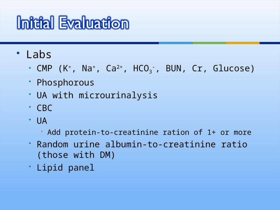
Phosphorous Goal – 2.7-4.6 mg/dL For CKD stage 5, goal is 3.5-5.5 mg/dL
Total calcium Goal – use lab reference range For CKD stage 5, goal is 8.4-9.5 mg/dL

Causes of anemia in CKD Reduced erythropoietin production Shortened RBC survival Iron Deficiency
Treatment Replace Iron if deficient Recombinant human erythropoietin
Initially at 80-100 units/kg/week SQ and titrate Treatment goal of Hgb 11-13

Causes of secondary hyperparathyroidism in CKD Phosphate retention Decreased free calcium Decreased Vitamin D1,25
Kidney function is required to convert Vitamin D25 to Vitamin D1,25 Reaction stimulated to PTH
Patients with CKD have low circulating Vitamin D1,25, low Vitamin D25, and increased PTH, even before demonstratable hyperphosphatemia and hypocalcemia
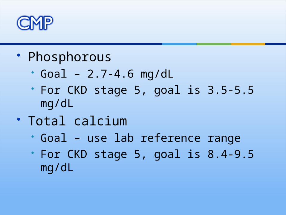
Goal PTH intact level – to control secondary hyperparthyroidism 35-70 pg/mL with eGFR 30-59 (state III) 70-110 pg/mL with eGFR 15-29 (stage
IV) 150-300 pg/mL for dialysis pts or eGFR
<15

The kidney is the location of 1-hydroxylation to make the active form of vitamin D (calcitriol)
Deficiency is associated with secondary hyperparathyroidism
Treatment helps regulate PTH levels via vitamin D receptors on the parathyroid
Deficiency associated with increased albuminuria
Goal level is for Vitamin D25 is > 30 ng/mL

CKD patients have a decreased ability to excrete uric acid
Theorized that hyperuricemia may contribute to CKD progression Thus, treatment with allopurinol may slow
disease
General goal is a uric acid <5.0 Levels >5.2 have been correlated
greatly with CKD disease progression

Marker of renal damage Two classes of proteins – Albumin, Globulins Potent independent risk factor for
progression of renal disease and an independent cardiovascular risk factor
Initially assess with Urine Dip First morning sample preferred, random is
acceptable +1 reached at excretion of 300-500 mg/day
(upper limit of normal for proteinuria is 150mg/day)

Spot Urine Protein/Creatinine ratio
24 hr urine protein
Lab Method Advantages
• Strong correlation with 24 hr urine protein•Patient convenience•Rapid Results
More accurate quantification of proteinuria
Disadvantages
Less accurate for proteinura >4g/day and <500mg/day
•Poor patient compliance•Time consuming•Delay in obtaining results

Urine Ratios Albumin to Creatinine Ratio
More precise at lower concentrations More expensive Use for screening in patients at increased risk of CKD
Diabetes mellitus, Hypertension
Protein to Creatinine Ratio Many of the studies on treatment of CKD stratified
patients based on this value Recommended due to cost benefit Closely correlates to 24 hour urine protein sample Used to trend proteinuria, if albumin/creatinine ratio
is high

Monitoring proteinuria in CKD patients should be done with quantitative measurements
Urine Protein-to-Creatinine Ratio Normal
< 150 mg/24 hour sample < 0.2 g/g (> 200 mg protein/mg creatinine)
Nephrotic Range > 3g /24 hours > 3.5 mg /mg

Low Phosphorous Studies have shown significant decrease in
PTH and improvements in bone histology in mild CKD
Low Protein Insufficient evidence to use for disease progression May delay onset of uremic symptoms in those
close to needing dialysis Patient needs at least 0.6-0.8 g/kg/day
Low Potassium – prevents hyperkalemia

No NSAIDs Use Tylenol or narcotic pain medications
When GFR <30 Save non-dominant arm from IV, PICC lines,
needles Saves veins for future grafting of AV fistula
Discuss options of fistula/graft/peritoneal dialysis
Transplant evaluation – able to get if GFR <20 Dialysis at GFR <15

ACE-I/ARBs Slows progression independent of BP
effect Monitor Cr and K+ 1-2 weeks after
initiation Should be continued in most patients
unless: Acute decline in GFR by >30% within 2
weeks of starting the medicine K+ > 6, despite appropriate treatment

ACE-I/ARBs Insufficient evidence to recommend
combo of ACE-I and ARB to slow disease progression
Only benefit seen in non-diabetic CKD patients with concomitant IgA nephropathy (Berger’s disease)

Lowers systemic BP Lowers glomerular pressure Increase renal blood flow

Reduces proteinuria Even if BP is controlled and patient is
normotensive, doses of ACE-I/ARBs should be raised even greater than recommended
To reduce protein excretion to levels <500mg (level most optimal to protect the kidney)
Natriuretic (salt excretion) Decrease in Aldosterone production Inhibits Angiotensin II, cytokines, growth
factors, and macrophages

Diuretics Use in patients with volume overload May be useful to also control potassium
levels HCTZ not useful in patients with GFR is
<30 Not able to get to the distal tubule at that
low of GFR Use Loops + Metalazone

Allopurinol Titrate to uric acid <5.0 There is no risk of causing further renal
failure Max dose is 900 mg/day
Uloric (Febuxostat) Another option for hyperuricemia Up to 80 mg/day More expensive

Statins Lipid lowering is important in CKD, especially
in nephrotic range CKD Studies suggest that high lipid levels
contribute to CKD disease progression Hyperlipidemia experimentally activates
mesangial cells and increases production of macrophage chemotactic factors, fibronectin, type IV collagen, plasminogen activator-1, reactive O2 species
If diabetic, remember goal LDL is <70

Phosphate Binders Indicated with patients with elevated
PTH and increased phosphate, despite phosphate restriction for 2-4 weeks
Calcium containing – preferred Calcium carbonate
Do not use H2 blockers/PPI Needs acid to become active
Calcium acetate

Phosphate Binders Non-calcium containing
Sevelamer Lanthanum
Aluminum hydroxide – former med of choice, but out of favor due to aluminum toxicity

Vitamin D Cholecalciferol (Vitamin D3) – preferred Ergocalciferol (Vitamin D2)
Vitamin D analogues - not routinely used Use – when PTH is still high, despite
correcting Phosphorus and Calcium to <9.5 mg/dL
Calcidiol (25-hydroxyvitamin) Calcitriol (1,25-dihydroxyvitamin D)

Sensipar (Cinacalcet) Treatment of secondary hyperparathyroidism
in patients not on dialysis Not currently approved for this use Increases sensitivity of calcium sensing
receptor on parathyroid gland Lowers PTH Lowers Calcium Lowers Phosphorus
Prevents bone disease related to CKD

Be alert that other diseases can develop in addition to diabetic nephropathy
Hypertensive nephrosclerosis is common Renal artery stenosis can occur anytime
Suggested if creatinine rises > 0.2-0.3 mg/dl and high potassium after starting an ACE-I
Suggested is >1cm size difference in renal ultrasound
Diabetics are at higher risk of contrast nephropathy Arteriograms, heart catherization, CT scan, IVP's

Goal Blood Pressure <130/80 mm Hg Reduce proteinuria with ACE-I/ARBs
Goal <1g/day Control phosphate – diet, binders Maintain Vitamin D25 >30 ng/mL Prevent hyperparathyroidism
Correct Vitamin D, Phosphorus, Calcium

Correct Anemia – goal Hgb 11-13 mg/dL Give diuretics for volume overload Control K+ - diet restriction, diuretics Protein intake at least 0.6-0.8 g/kg/day Control metabolic acidosis with oral
sodium citrate Tight diabetes mellitus control
HgA1c goal <7.0 (6.5 even better)

Underlying cause is unclear after basic work-up Renal biopsy is indicated eGFR < 30 mL/min/1.73m2
Facilitate education, planning of dialysis/transplant Rapid progression of CKD
GFR decline 50% in less than 6 months with no obvious cause
Superimposed acute renal failure Metabolic complications
Anemia, Secondary hyperparathyroidism Management is beyond your comfort level

Epidemiology and risk factors for chronic kidney disease. McClellan WM – Med Clin North Am – 01-MAY-2005; 89(3): 419-45
Cecil, R. L., Goldman, L., & Schafer, A. I. (2012).Goldman's Cecil medicine. Philadelphia: Elsevier/Saunders.
National Kidney Foundation – Am J Kidney Dis – 01-FEB-2002; 39(2 Suppl 1): S1-266
Chronic Kidney Disease Working Group. (2008). VA/DoD clinical practice guideline for the management of chronic kidney disease in primary care. Version 2.0. Washington, DC: Veterans Health Administration and Department of Defense.
Outpatient management of chronic kidney disease: proteinuria, anemia and bone disease as therapeutic targets. Lam A – Dis Mon – 01-APR-2010; 56(4): 215-32
Bope, E. T., Rakel, R. E., Kellerman, R. D., & Conn, H. F. (2011). Conn's current therapy 2011. Philadelphia, Pa: Saunders/Elsevier.
Rose, Burton D MD. Evaluation of isolated proteinuria in adults. In: UpToDate, Basow, DS (Ed),UpToDate, Waltham, MA, 2011.
Post, Theodore W MD. Overview of the management of chronic kidney disease in adults. In: UpToDate, Basow, DS (Ed),UpToDate, Waltham, MA, 2011.