Embed Size (px)
Citation preview
Breaking the one antibody–one target axiom
C. Sinha Fang Guo, Sanjib Das, Barbara M. Mueller, Carlos F. Barbas, III, Richard A. Lerner, and Subhash
doi:10.1073/pnas.0603822103 2006;103;11009-11014; originally published online Jul 5, 2006; PNAS
This information is current as of October 2006.
& ServicesOnline Information
www.pnas.org/cgi/content/full/103/29/11009etc., can be found at: High-resolution figures, a citation map, links to PubMed and Google Scholar,
Supplementary Material www.pnas.org/cgi/content/full/0603822103/DC1
Supplementary material can be found at:
References www.pnas.org/cgi/content/full/103/29/11009#BIBL
This article cites 23 articles, 8 of which you can access for free at:
www.pnas.org/cgi/content/full/103/29/11009#otherarticlesThis article has been cited by other articles:
E-mail Alerts. click hereat the top right corner of the article or
Receive free email alerts when new articles cite this article - sign up in the box
Rights & Permissions www.pnas.org/misc/rightperm.shtml
To reproduce this article in part (figures, tables) or in entirety, see:
Reprints www.pnas.org/misc/reprints.shtml
To order reprints, see:
Notes:
Breaking the one antibody–one target axiomFang Guo*†, Sanjib Das*†, Barbara M. Mueller‡, Carlos F. Barbas III*, Richard A. Lerner*§, and Subhash C. Sinha*§
*The Skaggs Institute for Chemical Biology and Department of Molecular Biology, The Scripps Research Institute, 10550 North Torrey Pines Road,La Jolla, CA 92037; and ‡Cancer Biology Division, La Jolla Institute for Molecular Medicine, San Diego, CA 92121
Contributed by Richard A. Lerner, May 12, 2006
Studies at the interface of chemistry and biology have allowed usto develop an immunotherapeutic approach called chemically pro-grammed antibodies (cpAbs), which combines the merits of tradi-tional small-molecule drug design with immunotherapy. In thisapproach, a catalytic antibody catalyzes the covalent conjugationof a small molecule or peptide to the active site of the antibody,effectively recruiting the binding specificity of the conjugatedmolecule to the antibody. In essence, this technology provides thetools for breaking the ‘‘one antibody–one target axiom’’ of im-munochemistry. Our studies in this area have focused on using thechemistry of the well studied aldolase catalytic antibodies of whichmAb 38C2 is a member. Previously, we explored reversible assem-bly of cpAbs available through diketone chemistry. In this article,we explore a unique proadapter assembly strategy wherein anantibody 38C2-catalyzed transformation unveils a reactive tag thatthen reacts to form a stable covalent bond with the antibody. Anintegrin �v�3 antagonist was synthesized with the designed pro-adapter and studied using human breast cancer cell lines MDA-MB-231 and MDA-MB-435. We demonstrate that this approachallows for (i) the effective assembly of cpAbs in vitro and in vivo,(ii) selective retargeting of 38C2 to integrin �v�3 expressing breastcancer cell lines, (iii) intracellular delivery of cpAbs into cells, (iv)dramatically increased circulatory half-life, and (v) substantialenhancement of the therapeutic effect over the peptidomimeticitself in animal models of breast cancer metastasis. We believe thatthis technology possesses potential for the treatment and diag-nosis of disease.
catalytic antibody � chemical programming � combinatorial antibodylibraries
Monoclonal antibodies are a rapidly growing class of thera-peutics for a wide variety of diseases (1, 2). Some of the
advantages of antibodies include their relative lack of nonspecifictoxicity, long half-life, and ease of access from patient-derived orsynthetic combinatorial antibody libraries. For certain diseases,such as cancer, that antibodies can carry their own effector func-tions is of prime importance because the antibody specificity directsthe killing function endemic to the effector domain, the Fc. It hasalways been axiomatic in immunochemistry that even though onemay desire one or more of the advantageous properties common toall antibodies, due to their clonal nature, each task requires adifferent antibody. A solution to this problem, namely chemicallyprogrammed antibodies (cpAbs), has emerged at the interface ofchemistry and biology: One can use different low-molecular-weighttargeting agents (programming agents or adapters) to selectivelytarget the same antibody to different sites for different uses (3). Thisstrategy has the advantage that only a single antibody is required fora multiplicity of tasks, and it taps into the unlimited chemicaldiversity and the specificity that can be engendered by organicsynthesis (4). The antibody provides the organic compound ahalf-life, biodistribution, valency, and effector function that it maynot otherwise have.
The cpAb approach that we have reported is unique in thatsmall synthetic molecules or peptides and catalytic mAbs reactin a self-assembly process and become linked through a covalentbond. This covalent modification results in the reprogrammingof the specificity of the antibody with the binding specificity ofthe small molecule. The resulting conjugate of small molecule
and antibody is a cpAb. Significantly, we have demonstrated thatchemical programming of a catalytic antibody can occur both invitro and in vivo to have a therapeutic effect in disease models (3,5). Key to this approach is the development of catalytic anti-bodies that operate using covalent reaction mechanisms (6, 7).mAb 38C2 is an antibody of this type, an aldolase antibodygenerated by reactive immunization that contains a highlyreactive lysine residue that is key to its activity. Small moleculesor targeting agents are adapted to work in this approach byaddition of a reactive tag that the antibody, through its catalyticfunction, selectively processes to form a covalent link betweenitself and the programming agent.
Thus, to selectively target the antibody to particular cells, anantibody-reactive tag is linked to a targeting agent that is a ligandfor the desired cellular receptor(s). In this study, we directcatalytic aldolase antibodies to the integrin �v�3. The integrins�v�3 and �v�5 are intriguing targets for cancer therapeuticsbecause these receptors are expressed both on a variety ofcancers and on the activated endothelial cells of the angiogenicvasculature they induce (8, 9, 10). The results presented herediffer from previous studies (3, 4, 5, 11), in that the reactive tagsstudied here can be considered proadapters as the antibody usestwo catalytic steps to generate a stable covalent complex. Ourearlier studies in this area focused on the use of reactive tags thatprovided for reversible enaminone-attachment chemistry. In thisnew approach, the reactive tag is first catalytically activated bya retro-aldol reaction that unveils a reactive vinyl ketone that issubsequently covalently attached to the antibody through aMichael addition reaction. In this article, we explore the chem-
Conflict of interest statement: Patents related to this work have been licensed to CovX, Inc.in which C.F.B., R.A.L., and S.C.S. maintain an equity position.
Freely available online through the PNAS open access option.
Abbreviations: cpAbs, chemically programmed antibodies; ESI, electrospray ionization.
†F.G. and S.D. contributed equally to this work.
§To whom correspondence may be addressed. E-mail: [email protected] or [email protected].
© 2006 by The National Academy of Sciences of the USA
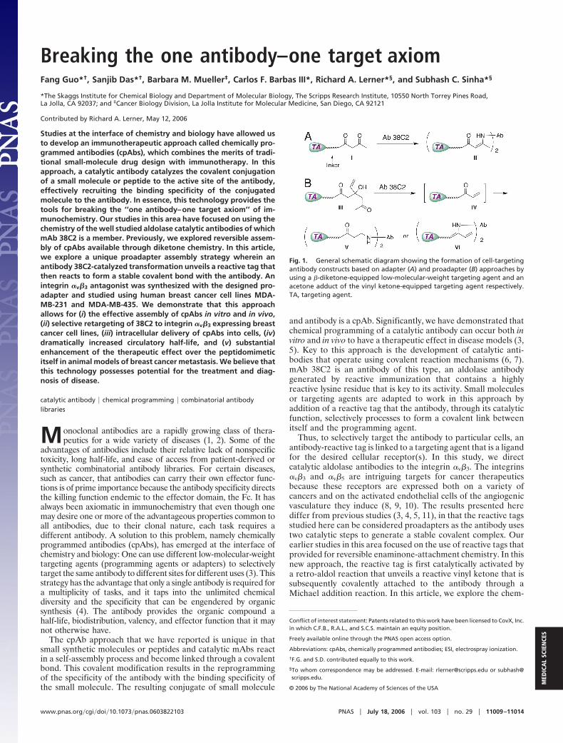
Fig. 1. General schematic diagram showing the formation of cell-targetingantibody constructs based on adapter (A) and proadapter (B) approaches byusing a �-diketone-equipped low-molecular-weight targeting agent and anacetone adduct of the vinyl ketone-equipped targeting agent respectively.TA, targeting agent.
www.pnas.org�cgi�doi�10.1073�pnas.0603822103 PNAS � July 18, 2006 � vol. 103 � no. 29 � 11009–11014
MED
ICA
LSC
IEN
CES
istry, biology, and therapeutic potential of this proadapterstrategy and a peptidomimetic targeting agent in cancer.
Results and DiscussionIn our previous reports, we reacted the small-molecule antago-nists of �v�3 and �v�5 integrins equipped with a diketone linker,such as I, with the reactive lysine residues in the aldolaseantibody 38C2-binding sites to form the corresponding enami-none derivative, II (Fig. 1A) (3, 4, 5, 11). In the proadapterapproach, we anticipated that a targeting agent equipped with atertiary aldol linker, such as III, would undergo a 38C2-catalyzedretro-aldol reaction (12) to produce an adapter possessing areactive linker, such as the vinyl ketone IV. The ketone IV wouldthen react as a Michael acceptor with the key nucleophilic aminein the antibody active site to produce conjugate V, a cpAb.Arguably, the intermediate IV could also react with 38C2forming the corresponding dibenamine complex VI, but in theend that would also be converted to the thermodynamicallystable Michael adduct V. In preliminary studies, we found thatmethylvinyl ketone rapidly inactivated the antibody, indicatingthat electrophiles of this type would be suitable as reactive tagsif their inherent reactivity could be controlled (S.C.S. and S.Abraham, unpublished results). It should be noted that inthe structurally and functionally related constructs II and V, theprimary differences are the formation and breakdown of theconjugates. Thus, II is reversible, whereas conjugate V is sub-stantially more stable (Fig. 1B).
We observed that both I and III react specifically and quan-titatively with the antibody and two equivalents of either com-pound is sufficient to completely inhibit the catalytic activity of38C2, indicating that the key lysine residue in each of the twoactive sites of the antibody are labeled (Fig. 1B). The role of thealdol functionality of III is to mask the reactive vinyl ketonelinker that would be expected to react readily with a variety ofprotein nucleophiles. Because this reactive functionality is onlyrevealed in the active site of the antibody after the retro-aldolreaction, it was anticipated that the vinyl ketone functionalitywould react with the catalytic lysine as soon as it was unveiledand before dissociating from the reactive site. Loss of catalytic
reactivity of 38C2 after incubation of III with antibody supportsthis hypothesis. Prolinker III, given its inherent inertness beforeactivation, should have potential synthetic advantages because itshould be inert to reaction with nucleophilic groups that mightbe present on targeting agents.
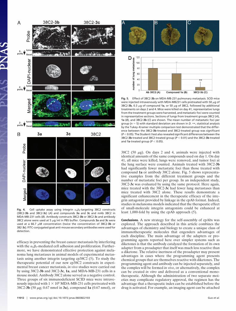
�v�3 Integrin-Targeting Agents for the Antibody Construct Formation.To target 38C2 to �v�3 integrin using the proadapter approach,two homologous programming agents (2a and 3a) were prepared(see latter). Both agents possessed an analog of 1, compound 1a,which binds the �v�3 integrin with high affinity (Fig. 2), as thetargeting agent (13). Compound 1a was functionalized with anacetone adduct of the vinyl ketone linkers. This adduct was likelyto be a substrate for the retro-aldol reaction catalyzed by theantibody 38C2 to afford 2b or 3b, which should undergo Michaeladdition with 38C2 to give V (Fig. 1B). As controls for theevaluation of V, the analogous diketone-containing program-ming agents 2c and 3c were also prepared. The latter compoundsshould react with 38C2 to give II as shown in Fig. 1 A. Synthesisof compounds 2a–3a and 2c–3c is described in the supportinginformation, which is published on the PNAS web site.
Antibody Construct Formation. To assess the potential of 2a, 3a, 2c,and 3c as programming agents, these compounds were separatelymixed with antibody 38C2 at a ratio of 2:1, and the mixtures wereincubated at 37°C for 2 h. A fluorescence assay, based on usingmethodol as the substrate, was used to assess time-dependentinactivation of the catalytic activity of the antibody (14). Inac-tivation of aldolase activity should be indicative of modificationof the catalytic lysine residue and, thus, chemical programmingof the antibody. In the absence of the programming agents, 38C2rapidly catalyzed the retro-aldol reaction of methodol to producethe fluorescent product 6-methoxy-2-naphthaldehyde. In con-trast, the antibody-programming agent constructs¶ were com-
¶The cpAb 38C2 using targeting agents 2a or 3a were named as 38C2-2b and 38C2-3b,respectively, based on the fact that compounds 2b and 3b were the expected ligands thatconjugated with 38C2. Similarly, the analogous chemically programmed 38C2 Fab (orcp38C2Fab) construct obtained from 3a was named as 382Fab-3b.
Fig. 2. Structures of the �v�3 integrin-targeting antagonists equipped with an acetone adduct of a vinyl ketone, a vinyl ketone, or a diketone for chemicalprogramming of the aldolase antibody.
11010 � www.pnas.org�cgi�doi�10.1073�pnas.0603822103 Guo et al.

pletely inactive, indicating that after conjugation the active siteof the catalytic antibody was occupied. These observationsclearly supported the assumption that vinyl ketones, 2b and 3b,produced in situ from their acetone adducts, reacted with theactive site of the antibody and also reinforced the previouslydescribed construct formation from the analogous diketonecompounds 2c and 3c.
The chemical programming of antibody 38C2 using 2b or 3bwas also analyzed by MALDI-TOF mass spectrometry for whichwe used both antibody 38C2 and its Fab fragment. The chemi-cally programmed 38C2 Fab (or cp38C2Fab) was prepared byusing a 1:1 mixture of the Fab and compounds 3a or 3c, and theirformation was initially analyzed by using the fluorescence assay,as described above. In the mass spectra, chemically programmed38C2 (i.e., 38C2-3b¶ and 38C2-3c) showed addition of �2molecules of the programming agents to the average mass of38C2. Similarly, the analogous cp38C2Fab constructs preparedfrom 3b or 3c (i.e., 38C2Fab-3b¶ or 38C2F-3c) showed theaddition of approximately one molecule of the programmingagent to the average mass of the Fab. The average mass peaks38C2 Fab, 38C2Fab-3b, and 38C2Fab-3c were recorded at48,410, 49,354 and 49,378 mass units, respectively (see support-ing information for a comparative MALDI-TOF mass spectra of38C2 Fab, 38C2Fab-3b, and 38C2Fab-3c). These observationsindicated that the reactive site lysine residues in 38C2 andcp38C2Fabs were labeled specifically compared with any of themany other lysine residues found in the covalent structure of theantibody or Fab.
Binding of Antibody Constructs to �v�3 Integrin-Expressing Cells.Next, we evaluated binding of the cp38C2 derivatives to �v�3integrin-expressing cells by cell f low cytometry. The two celllines used, MDA-MB-435 and MDA-MB-231, are immortalizedhuman breast cancer cell lines, and both cell lines express highlevels of �v�3 integrin (15, 16). All cpAb constructs and controlanti-integrin antibody LM609 bound efficiently to these cells(Fig. 3A). As expected, antibody 38C2 alone did not bind to thesecells. Flow cytometric staining with the cp38C2s, i.e., 38C2-3band 38C2-3c at 25 and 5 �g�ml, produced profiles with nearlyidentical f luorescence intensities, but fluorescence decreasedconsiderably at 1 �g�ml and lower concentrations (Fig. 3B). This
data implied that 38C2-3b and 38C2-3c provides maximal stain-ing after incubation at a concentration of �5 �g�ml (33 nM).
In Vivo Assembly of cpAbs. In previous studies (3, 5), we noted thata diketone linker-equipped integrin antagonist SCS-873 was ableto conjugate in vivo with antibody 38C2 and that the resultingcp38C2 had a serum half-life �200 times longer than that of theantagonist itself. The half-life of the SCS-873-based cp38C2 was�3 days (3), whereas the half-life determined for 38C2 itself was�4 days (17). Effective in vivo assembly allowed both theantagonist and 38C2 to be administered separately to inhibit thetumor growth in animal models. Here we studied in vivoassembly using the proadapter approach. Key to this assembly isantibody-catalyzed retro-aldolization to provide the vinyl ketoneproduct that could then self-attach to 38C2. Our previous studiesconcerning 38C2-catalyzed prodrug activation in vivo providesprecedence for this approach (18). To evaluate this, we carriedout experiments using compound 2a and the conventionaldiketone 2c as a control. Antibody 38C2 (1 mg in 100 �l of PBSbuffer) was administered i.v. to three mice followed by i.p.administration of compounds 2a (1 mg in 100 �l of buffer), 2c(1 mg in 100 �l of buffer), or PBS (100 �l). Sera were obtainedat regular intervals (24, 48, 72, 96, and 168 h) was examined forthe presence of cp38C2 by using flow cytometry and MDA-MB-231 cells. This study confirmed that the complex, cp38C2, wasformed in vivo with both 2a and 2c. Sera from the mouse lackingthe targeting agent did not show any binding to the cells. Thus,compound 2a was effectively processed by 38C2 in vivo to formvinyl ketone 2b, which then self-assembled to form cp38C2. Theresulting cp38C2-V species (i.e., 38C2-2b) was stable in serum,with a half-life of �60 h as analyzed by comparing the meanfluorescence intensity; similar results were observed for thecp38C2-II species (�60 h for 38C2-2c, as well).
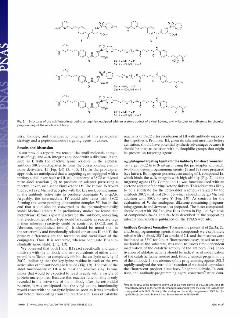
Cellular-Uptake of the Antibody Adapter Constructs. It is well estab-lished that �v�3��v�5 integrin–ligand complexes are rapidlyinternalized via an integrin-dependent endocytosis pathway.Examples of ligands that are effectively internalized includeviruses (19) and �v-integrin-blocking mAb. In contrast, inter-nalization of the cyclic synthetic arginine–glycine–aspartatemotif (cRGD) peptides targeted for �v�3 takes place by anintegrin-independent fluid-phase endocytosis pathway (20). Toassess the feasibility of using the integrin-targeting cp38C2constructs for drug delivery, we evaluated the internalization ofthe cp38C2 variants. Internalization of 38C2-3b and 38C2-3c intoMDA-MB-231 cells was studied. Briefly, 150,000 cells wereincubated with 38C2-3b or 38C2-3c (5 �g�ml in cell culturemedium; 1 ml) for 15 min on a coverslip. The cell–38C2-3b andcell–38C2-3c ternary complexes were then fixed by using 2%paraformaldehyde in 0.01 M PBS and incubated with FITC-conjugated goat anti-mouse secondary antibody (10 �g/ml) for60 min. Similar experiments were carried out by using 38C2alone as the negative control. The cells were analyzed by usingconfocal laser scanning microscopy (21). Cells treated with38C2-3b and 38C2-3c showed intense intracellular fluorescence,whereas cells treated with 3a, 3c, and 38C2 alone did not (Fig.4B). Therefore, 38C2-3b and 38C2-3c were rapidly internalizedprobably through an integrin-mediated endocytosis mechanism.If a nonintegrin-mediated or Fc-based internalization mecha-nism was operative, f luorescence should have been observedwith antibody 38C2 alone.
Prevention of Breast Cancer Metastasis. Breast cancer is treatableif diagnosed early. Nevertheless, the prognosis is considerablyworse if patients have secondary tumors in distant organs.Prevention of breast cancer metastasis is clearly a significantgoal. Both a small-molecule �v�3 integrin antagonists (22) andan antibody specific for �v�3 (23) have shown remarkable
Fig. 3. Flow cytometry histograms showing the binding of 38C2-3b, 38C2-3c,and 38C2 alone (A) and binding of serial dilutions of 38C2-3b to MDA-MB-231cells (B). In A, 38C2-3b and 38C2-3c prepared from 38C2 (1 eq) and 3a or 3c (2eq) were diluted to 25 �g�ml. In B, the 38C2-3b (25 �g�ml) construct used inA was further diluted 5�, 25�, and 125�. In all experiments, 38C2 alone wasused at 25 �g�ml, LM609 was used at 10 �g/ml dilution, and FITC-conjugatedgoat anti-mouse secondary antibodies were used for detection. The y axisgives the number of events in linear scale, the x axis gives the fluorescenceintensity in logarithmic scale.
Guo et al. PNAS � July 18, 2006 � vol. 103 � no. 29 � 11011
MED
ICA
LSC
IEN
CES
efficacy in preventing the breast cancer metastasis by interferingwith the �v�3-mediated cell adhesion and proliferation. Further-more, we have demonstrated effective protection against mela-noma lung metastases in animal models of experimental metas-tasis using another integrin targeting cp38C2 (5). To study thetherapeutic potential of our new cp38C2 constructs in experi-mental breast cancer metastasis, in vivo studies were carried outby using 38C2-3b and 38C2-3c, 1a, and MDA-MB-231 cells in amouse model. Antibody 38C2 alone served as a negative control.Three groups of six immunodeficient SCID mice were intrave-nously injected with 1 � 106 MDA-MB-231 cells pretreated with38C2-3b (50 �g; 0.67 nmol in 3a), compound 1a (0.67 nmol), or
38C2 (50 �g). On days 2 and 4, animals were injected withidentical amounts of the same compounds used on day 1. On day41, all mice were killed, lungs were removed, and tumor foci atthe lung surface were counted. Animals treated with 38C2-3bhad significantly fewer metastatic foci than those treated withcompound 1a or antibody 38C2 alone. Fig. 5 shows representa-tive examples from the different treatment groups and thenumber of metastatic foci per group. In an independent study,38C2-3c was evaluated by using the same protocol. Here again,mice treated with the 38C2-3c had fewer lung metastases thanmice treated with 38C2 alone. These results demonstrate asignificant enhancement in the therapeutic efficacy of the inte-grin antagonist provided by linkage in the cpAb format. Indeed,studies in melanoma models indicated that the therapeutic effectof small-molecule integrin antagonists could be enhanced atleast 1,000-fold by using the cpAb approach (5).
Conclusions. A new strategy for the self-assembly of cpAbs wasexplored. The approach described in this article combines theadvantages of chemistry and biology to create a unique class ofimmunotherapeutic molecules that engenders advantages ofeach discipline. The main advantage of the adapters or pro-gramming agents reported here over simpler systems such asdiketones is that the antibody catalyzed the formation of its ownadapter from a proadapter that itself was much less reactive thana diketone. The relative inertness of the proadapter may presentadvantages in cases where the programming agent presentschemical groups that are themselves reactive with diketones. Theprogramming agent and antibody can be injected separately, andthe complex will be formed in vivo, or alternatively, the complexcan be created in vitro and delivered as a conventional mono-therapeutic. Although the administration of two separate moi-eties may complicate regulatory approval, the regimen has theadvantage that a therapeutic index can be established before thedrug is activated. For example, an imaging agent can be attached
Fig. 4. Cell uptake assay using integrin �v�3-targeting 38C2 constructs(38C2-3b and 38C2-3c) (A) and compounds 3a and 3c and mAb 38C2 inMDA-MB-231 cells (B). Antibody constructs 38C2-3b or 38C2-3c and antibody38C2 alone were used at 5 �g�ml in PBS buffer. Compounds 3a and 3c wereused at a 66.7 �M concentration (twice the concentration of 38C2-3b or382-3c). FITC-conjugated goat anti-mouse secondary antibodies were used fordetection.
Fig. 5. Effect of 38C2-3b on MDA-MB-231 pulmonary metastasis. SCID micewere injected intravenously with MDA-MB231 cells pretreated with 50 �g of38C2-3b, 0.3 �g of compound 1a, or 50 �g of 38C2, followed by additionaltreatments on days 2 and 4. Mice were killed on day 41, representative lungsfrom the treatment groups were harvested, and metastatic foci were countedin representative sections. Sections of lungs from treatment groups 38C2 (A),1a (B), and 38C2-3b (C) are shown. The mean number of metastatic foci pergroup (n � 5) with standard deviation are shown in D. **, statistical analysisby the Tukey–Kramer multiple comparison test demonstrated that the differ-ence between the 38C2-3b-treated and 38C2-treated group was significant(P � 0.05). The Student t test also revealed significant differences between the38C2-3b-treated and 38C2-treated group (P � 0.01) and the 38C2-3b-treatedand 1a-treated group (P � 0.05).
11012 � www.pnas.org�cgi�doi�10.1073�pnas.0603822103 Guo et al.
to the proadapter, allowing the physician to monitor localizationof a drug before arming the agent with the effector functions ofthe antibody molecule. Of course, the complex can also beformed in vitro if such preselectivity is not deemed necessary.Such complexes will circulate for �60 h, giving the adaptergreatly extended half-life relative to the small molecule andtunable pharmacokinetics. Half-lives of cpAbs in humans areanticipated to be significantly greater than those observed inmice, as is the case for conventional mAbs. Therapeutic studiesin experimental breast cancer metastasis models demonstratethe increase in efficacy that can be provided to a small moleculethrough coupling with an antibody effector.
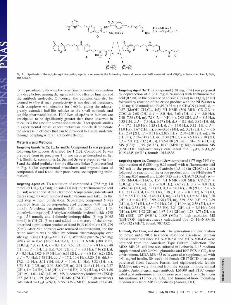
Materials and MethodsTargeting Agents 1a, 2a, 2c, 3a, and 3c. Compound 1a was preparedfollowing the process described for 1 (13). Compound 2c wasprepared from its precursor 4 in two steps as described earlier(4). Similarly, compounds 2a, 3a, and 3c were prepared via 4 or5 and the aldol prolinker 6 or the diketone linker 7, as describedin Fig. 6 (for experimental procedures and physical data ofcompounds 5 and 6 and their precursors, see supporting infor-mation).
Targeting Agent 2a. To a solution of compound 4 (785 mg; 1.0mmol) in CH2Cl2 (3 ml), anisole (1.0 ml) and trif luoroacetic acid(1.0 ml) were added. After 2 h at room temperature, solvents andexcess reagents were removed under vacuum and taken to thenext step without purification. Separately, compound 6 wasprepared from the corresponding acid precursor (450 mg; 1.3mmol), N-hydroxy succinimide (180 mg; 1.56 mmol), 1-(3-dimethylaminopropyl)-3-ethylcarbodiimide hydrochloride (298mg; 1.56 mmol), and 4-dimethylaminopyridine (8 mg; 0.065mmol) in CH2Cl2 (5 ml) and added to a mixture of the above-described deprotection product of 4 and Et3N (0.5 ml) in CH3CN(5 ml). After 24 h, solvents were removed under vacuum, and thecrude mixture was purified by column chromatography oversilica gel using CH2Cl2–MeOH (9:1) affording pure 2a (745 mg;78%). Rf � 0.45 (MeOH–CH2Cl2, 1:5); 1H NMR (500 MHz,CDCl3): 7.70 (2H, d, J � 8.1 Hz), 7.57 (2H, d, J � 7.8 Hz), 7.48(1H, dd, J � 7.4, 1.2 Hz), 7.37 (2H, d, J � 8.5 Hz), 7.16 (2H, d,J � 8.1 Hz), 7.02–6.99 (4H, m), 6.35 (2H, d, J � 8.8 Hz), 6.28 (2H,d, J � 7.4 Hz), 5.78 (1H, dd, J � 17.2, 10.6 Hz), 5.20 (1H, dd, J �17.2, 1.1 Hz), 5.11 (1H, dd, J � 10.6, 1.1 Hz), 3.82 (1H, m),3.73–3.31 (12H, m), 3.06–3.00 (2H, m), 2.91–2.46 (11H, m), 2.27(2H, t, J � 7.4 Hz), 2.18 (2H, t, J � 6.6 Hz), 2.09 (3H, s), 1.92–1.88(2H, m), 1.81–1.63 (4H, m); MS [electrospray ionization (ESI)]:957 (MH�), 979 (MNa�); HRMS (ESI-TOF high-accuracy)calculated for C50H65N6O11S: 957.4353 (MH�), found: 957.4348.
Targeting Agent 3a. This compound (181 mg; 75%) was preparedby deprotection of 5 (200 mg; 0.24 mmol) with trif luoroaceticacid (0.5 ml) in the presence of anisole (0.5 ml) in CH2Cl2 (2 ml)followed by reaction of the crude product with the NHS-ester 6(160 mg; 0.36 mmol) and Et3N (0.25 ml) in CH3CN (3.0 ml). Rf �0.37 (MeOH–CH2Cl2, 1:5); 1H NMR (500 MHz, CD3OD �CDCl3): 7.68 (2H, d, J � 8.0 Hz), 7.63 (2H, d, J � 8.0 Hz),7.40–7.36 (3H, m), 7.18–7.14 (4H, m), 7.03 (2H, d, J � 8.5 Hz),6.33 (1H, d, J � 7.5 Hz), 6.27 (1H, d, J � 8.2 Hz), 5.82 (1H, dd,J � 17.5, 11.0 Hz), 5.23 (1H, d, J � 17.0 Hz), 5.12 (1H, d, J �11.0 Hz), 3.67 (1H, m), 3.56–3.36 (14H, m), 3.21 (2H, t, J � 6.5Hz), 2.94 (2H, t, J � 8.5 Hz), 2.82 (3H, s), 2.84–2.81 (2H, m), 2.76(1H, m), 2.63–2.47 (5H, m), 2.30 (2H, t, J � 7.5 Hz), 2.18 (2H,t, J � 7.0 Hz), 2.12 (3H, s), 1.92–1.88 (2H, m), 1.81–1.68 (6H, m);MS (ESI): 1,015 (MH�), 1037 (MNa�); high-resolution MS(ESI-TOF high-accuracy) calculated for C53H71N6O12S:1015.4845 (MH�), found: 1015.4838.
Targeting Agent 3c. Compound 3c was prepared (175 mg; 74%) bydeprotection of 5 (200 mg; 0.24 mmol) with trif luoroacetic acid(0.5 ml) in the presence of anisole (0.5 ml) in CH2Cl2 (2 ml)followed by reaction of the crude product with the NHS-ester 7(160 mg, 0.36 mmol) and Et3N (0.25 ml) in CH3CN (3.0 ml). Rf �0.35 (MeOH–CH2Cl2, 1:5); 1H NMR (500 MHz, CD3OD �CDCl3): 7.74 (2H, d, J � 8.0 Hz), 7.67 (2H, d, J � 8.0 Hz),7.49–7.46 (3H, m), 7.23 (2H, d, J � 8.0 Hz), 7.18 (2H, d, J � 7.5Hz), 7.11 (2H, d, J � 8.0 Hz), 6.38 (1H, d, J � 8.0 Hz), 6.35 (1H,d, J � 8.5 Hz), 3.63–3.48 (14H, m), 3.43 (2H, t, J � 6.0 Hz), 3.28(2H, t, J � 6.2 Hz), 2.99–2.96 (2H, m), 2.91–2.86 (4H, m), 2.89(3H, s), 2.67 (2H, t, J � 7.0 Hz), 2.61 (1H, br, s), 2.56 (2H, t, J �8.0 Hz), 2.35 (2H, t, J � 7.5 Hz), 2.24 (2H, t, J � 7.5 Hz), 2.04(3H, s), 1.98–1.92 (2H, m), 1.87–1.81 (2H, m), 1.78–1.73 (2H, m);MS (ESI): 987 (MH�), 1,009 (MNa�); high-resolution MS(ESI-TOF high-accuracy) calculated for C51H67N6O12S:987.4532 (MH�), found: 987.4525.
Antibody, Cell Lines, and Animals. The generation and purificationof mouse mAb 38C2 has been described elsewhere. Humanbreast cancer cell lines MDA-MB-231 and MDA-MB-435 wereobtained from the American Type Culture Collection. TheMDA-MB-231 cell line was cultured in Leibovitz L-15 mediumsupplemented with 2 mM L-glutamine and 10% FCS in CO2-freeenvironment. MDA-MB-435 cells were also supplemented with0.01 mg�ml insulin. Six-week-old female CB17-SCID mice werepurchased from Taconic Farms, and eight-week-old femaleBALB�c mice were obtained from the Scripps in-house animalfacility. Anti-integrin �v�3 antibody LM609 and FITC conju-gated goat anti-mouse antibody were purchased from ChemiconInternational (Temecula, CA), and Immuno-Fluore mountingmedium was from MP Biomedicals (Aurora, OH).
Fig. 6. Synthesis of the �v�3 integrin-targeting agents. a represents the following chemical procedure: trifluoroacetic acid, CH2Cl2, anisole, then 6 or 7, Et3N,and CH3CN.
Guo et al. PNAS � July 18, 2006 � vol. 103 � no. 29 � 11013
MED
ICA
LSC
IEN
CES
Formation of Antibody Construct and Evaluation of Binding to �v�3
Integrin-Expressing Cells. The generation and purification ofmouse mAb 38C2 has been described elsewhere. The antibodyconstructs (38C2-2b, -2c, -3b, and -3c) were prepared by mixinga solution of compound 2a, 2c, 3a, or 3c (100 �M; 3.3 �l) withantibody 38C2 (50 �M; 3.3 �l) in PBS buffer (total volume, 50�l), and the mixtures were incubated at 37°C for 2 h. Cells weredetached by brief trypsinization with 0.25% (wt�vol) trypsin, 1mM EDTA, washed with PBS, and resuspended at a concen-tration of 1 � 106 cells per milliliter in flow cytometry buffer [1%(wt�vol) BSA�25 mM Hepes in PBS, pH 7.4]. Aliquots of 100 �lcontaining 1 � 105 cells were distributed into wells of a V-bottom96-well plate (Corning) for indirect immunofluorescence stain-ing in the presence of serial dilutions (1:20, 1:100, 1:500, and1:2500) of cpAbs 38C2-2b, 38C2-2c, 38C2-3b, or 38C2-3c in flowcytometry buffer. After the constructs were incubated with cellsfor 1 h, FITC-conjugated goat anti-mouse polyclonal antibodies(10 �g�ml, in flow cytometry buffer) were added to the mixtureand further incubated for 45 min at room temperature. Finally,samples were analyzed by flow cytometry using a FACScaninstrument (Becton Dickinson).
For the in vivo antibody construct formation, three 8-week-oldBALB�c mice were injected i.v. (tail vein) with 100 �l of 10mg�ml antibody 38C2 in PBS. Compounds 2a and 2c wereinjected i.p. as 100 �l of 10 mg�ml in 50% PBS�25% DMSO�25% ethanol. Sera were prepared by centrifuging eye bleedstaken 24, 48, 72, 96, and 168 h after the injections. By using a1:100 dilution in flow cytometry buffer, the prepared sera wereanalyzed by flow cytometry as described above.
Cell-Uptake of the Antibody Constructs. Cover slides in 24-wellplates were kept under UV for 2 h. MDA-MB-231 cells weredetached by brief trypsinization with 0.25% (wt�vol) trypsin�1mM EDTA, washed with PBS, and resuspended at a concen-tration of 1.5 � 105 cells per milliliter. The suspended cells weredistributed into wells. After 24 h, medium was removed, and38C2-3b or 38C2-3c (prepared as before using 1 eq of 38C2 and2 eq of 3a or 3c, respectively), compounds 3a or 3c, or antibody38C2 alone was added into wells. They were incubated at 37°Cfor 15 min and then fixed using 2% paraformaldehyde in 0.01 MPBS for 10 min followed by 0.2% Triton X-100 in PBS at room
temperature for 2 min. After the cells were rinsed with PBScontaining 0.001% Triton X-100, they were incubated with 10%normal goat serum at room temperature for 60 min and againrinsed with PBS containing 0.001% Triton X-100. Cells werenext incubated with FITC-conjugated goat anti-mouse (10 �g/ml) at room temperature for 1 h, rinsed using PBS containing0.001% Triton X-100, incubated with DAPI (10 �g�ml) (Mo-lecular Probes) at room temperature for 60 min, and mountedonto slides using the Immuno-Fluore mounting medium. Fixedand stained samples were then viewed by using a RainbowRadiance 2100 laser scanning confocal system attached to aNikon TE200-U inverted microscope (Bio-Rad). Images wereacquired using LASER SHARP 2000 (Bio-Rad) software and pro-cessed in LSM EXAMINER (Zeiss) software.
Prevention of Breast Cancer Metastasis. MDA-MB-231cells (1 �106) were suspended in 100 �l of serum-free medium andinjected into the tail vein in 6-week-old female CB17-SCID mice,including 38C2-3b (50 �g; 0.67 nmol in 3a), compound 1a (0.67nmol), 38C2 (50 �g), and buffer alone. Animals were furtherinjected on days 2 and 4, with the identical amounts of theconstruct, compound, or antibody. On day 41, all mice werekilled, lungs were removed, and tumor foci at the lung surfacewere counted by anatomy microscope. Statistical analysis by theTukey–Kramer multiple comparison test demonstrated a signif-icant difference between the 38C2-3b-treated and 38C2-treatedgroup (P � 0.05). Student’s t test also revealed significantdifferences between the 38C2-3b-treated and 38C2-treatedgroup (P � 0.01) and the 38C2-3b-treated and 1a-treated group(P � 0.05). All of the animal experiments were approved by theInstitutional Animal Care and Use Committee of the ScrippsResearch Institute before the experiments were started.
We thank Dr. William B. Kiosses (The Core Microscopy Facility, TheScripps Research Institute) for helping with the cell-uptake assay;Roberta Fuller for the ELISA; and Drs. Christoph Rader [NationalCancer Institute, National Institutes of Health (NIH), Bethesda],Mikhail Popkov, and Fujie Tanaka for helpful discussions. Financialsupport was provided by the Skaggs Institute for Chemical Biology,Department of Defense Grant W81XWH-04-1-0717 (to S.C.S.), andNIH Grant 5R01CA104045 (to C.F.B.).
1. O’Mahony, D. & Bishop, M. R. (2006) Front. Biosci. 11, 1620–1635.2. Chester, K., Pedley, B., Tolner, B., Violet, J., Mayer, A., Sharma, S., Boxer, G.,
Green, A., Nagl, S. & Begent, R. (2004) Tumor Biol. 25, 91–98.3. Rader, C., Sinha, S. C., Popkov, M., Lerner, R. A. & Barbas, C. F., III (2003)
Proc. Natl. Acad. Sci. USA 100, 5396–5400.4. Li, L. S., Rader, C., Matsushita, M., Das, S., Barbas, C. F., III, Lerner, R. A.
& Sinha, S. C. (2004) J. Med. Chem. 47, 5630–5640.5. Popkov, M., Rader, C., Gonzalez, B., Sinha, S. C. & Barbas, C. F., III (2006)
Int. J. Cancer 119, 1194–1207.6. Wagner, J., Lerner, R. A. & Barbas, C. F., III (1995) Science 270, 1797–
1800.7. Barbas, C. F., III, Heine, A., Zhong, G., Hoffmann, T., Gramatikova, S.,
Bjornestedt, R., List, B., Anderson, J., Stura, E. A., Wilson, E. A. & Lerner,R. A. (1997) Science 278, 2085–2092.
8. Mousa, S. A. (2000) Emerg. Ther. Targets 4, 143–153.9. Liapis, H., Flath, A. & Kitazawa, S. (1996) Diagn. Mol. Pathol. 5, 127–135.
10. Stupack, D. G. & Cheresh, D. A. (2004) Curr. Top. Dev. Biol. 64,207–238.
11. Rader, C., Turner, J. M, Heine, A., Shabat, D., Sinha, C. C, Wilson, I. A,Lerner, R. A & Barbas, C. F., III (2003) J. Mol. Biol. 332, 889–899.
12. List, B., Shabat, D., Zhong, G., Turner, J. M., Li, A., Bui, T., Anderson, J.,Lerner, R. A. & Barbas, C. F., III (1999) J. Am. Chem. Soc. 121, 7283–7291.
13. Duggan, M. E., Duong, L. T., Fisher, J. E., Hamill, T. G., Hoffman, W. F., Huff,J. R., Ihle, N. C., Leu, C.-T., Nagy, R. M., Perkins, J. J., et al. (2000) J. Med.Chem. 43, 3736–3745.
14. List, B., Barbas, C. F., III & Lerner, R. A. (1998) Proc. Natl. Acad. Sci. USA95, 15351–15355.
15. Meyer, T., Marshall, J. F. & Hart, I. R. (1998) Br. J. Cancer 77, 530–536.16. Felding-Habermann, B., O’Toole, T. E., Smith, J. W., Fransvea, E., Ruggeri,
Z. M., Ginsberg, M. H., Hughes, P. E., Pampori, N., Shattil, S. J., Saveni, A.& Mueller, B. M. (2001) Proc. Natl. Acad. Sci. USA 98, 1853–1858.
17. Shabat, D., Rader, C., List, B., Lerner, R. A. & Barbas, C. F., III (1999) Proc.Natl. Acad. Sci. USA 96, 6925–6930.
18. Shabat, D., Lode, H. N., Pertl, U., Reisfeld, R. A., Rader, C., Lerner, R. A. &Barbas, C. F., III (2001) Proc. Natl. Acad. Sci. USA 98, 7528–7533.
19. Wickham, T. J., Mathias, P., Cheresh, D. A. & Nemerow, G. R. (1993) Cell 73,309–319.
20. Castel, S., Pagan, R., Mitjans, F., Piulats, J., Goodman, S., Jonczyk, A., Huber,F., Vilaro, S. & Reina, M. (2001) Lab. Invest. 81, 1615–1626.
21. Cullander, C (1999) Methods Mol. Biol. 122, 59–73.22. Shannon, K. E., Keene, J. L., Settle, S. L., Westlin, T. D., Schroeter, S.,
Ruminski, P. G. & Griggs, D. W. (2004) Clin. Exp. Metastasis 21, 129–138.23. Felding-Habermann, B., Lerner, R. A., Lillo, A., Zhuang, S., Weber, M. R.,
Arrues, S., Gao, C., Mao, S., Saven, A. & Janda, K. D. (2004) Proc. Natl. Acad.Sci. USA 101, 17210–17215.
11014 � www.pnas.org�cgi�doi�10.1073�pnas.0603822103 Guo et al.