Embed Size (px)
Citation preview
Egyptian Informatics Journal (2015) xxx, xxx–xxx
Cairo University
Egyptian Informatics Journal
www.elsevier.com/locate/eijwww.sciencedirect.com
ORIGINAL ARTICLE
Brain tumor segmentation based on a hybrid
clustering technique
* Corresponding author at: 44th Hamza Ibn Abdulmotaleb ST.,
Ahmed Maher ST., Mansoura City, Dakahlia Gov., A.R.E., Egypt.
Tel.: +20 10 281 42 420.
E-mail addresses: [email protected], [email protected]
(E. Abdel-Maksoud), [email protected] (M. Elmogy), actt_egypt@
yahoo.com (R. Al-Awadi).
Peer review under responsibility of Faculty of Computers and
Information, Cairo University.
Production and hosting by Elsevier
http://dx.doi.org/10.1016/j.eij.2015.01.0031110-8665 � 2015 Production and hosting by Elsevier B.V. on behalf of Faculty of Computers and Information, Cairo University.
Please cite this article in press as: Abdel-Maksoud E et al., Brain tumor segmentation based on a hybrid clustering technique, Egyptian Informatics J (2015dx.doi.org/10.1016/j.eij.2015.01.003
Eman Abdel-Maksoud a,*, Mohammed Elmogy b, Rashid Al-Awadi c
a Information Systems Dept., Faculty of Computers and Information, Mansoura University, Egyptb Information Technology Dept., Faculty of Computers and Information, Mansoura University, Egyptc Communication Dept., Faculty of Engineering, Mansoura University, Egypt
Received 30 April 2014; revised 15 January 2015; accepted 18 January 2015
KEYWORDS
Medical image segmentation;
Brain tumor segmentation;
K-means clustering;
Fuzzy C-means;
Expectation Maximization
Abstract Image segmentation refers to the process of partitioning an image into mutually exclu-
sive regions. It can be considered as the most essential and crucial process for facilitating the delin-
eation, characterization, and visualization of regions of interest in any medical image. Despite
intensive research, segmentation remains a challenging problem due to the diverse image content,
cluttered objects, occlusion, image noise, non-uniform object texture, and other factors. There
are many algorithms and techniques available for image segmentation but still there needs to
develop an efficient, fast technique of medical image segmentation.
This paper presents an efficient image segmentation approach using K-means clustering tech-
nique integrated with Fuzzy C-means algorithm. It is followed by thresholding and level set segmen-
tation stages to provide an accurate brain tumor detection. The proposed technique can get benefits
of the K-means clustering for image segmentation in the aspects of minimal computation time. In
addition, it can get advantages of the Fuzzy C-means in the aspects of accuracy. The performance
), http://
2 E. Abdel-Maksoud et al.
Please cite this article in press as: Abdel-Makdx.doi.org/10.1016/j.eij.2015.01.003
of the proposed image segmentation approach was evaluated by comparing it with some state of the
art segmentation algorithms in case of accuracy, processing time, and performance. The accuracy
was evaluated by comparing the results with the ground truth of each processed image. The exper-
imental results clarify the effectiveness of our proposed approach to deal with a higher number of
segmentation problems via improving the segmentation quality and accuracy in minimal execution
time.
� 2015 Production and hosting by Elsevier B.V. on behalf of Faculty of Computers and Information,
Cairo University.
1. Introduction
Image segmentation refers to the process of partitioning a dig-ital image into multiple regions. The goal of segmentation is to
change the representation of an image to be more meaningfuland easier to analyze. It is used in order to locate objects andboundaries in images. The result of image segmentation occurs
as a set of regions that collectively covers the entire image [1].Therefore, medical image segmentation plays a significant rolein clinical diagnosis. It can be considered as a difficult problem
because medical images commonly have poor contrasts, differ-ent types of noise, and missing or diffusive boundaries [2]. Theanatomy of the brain can be scanned by Magnetic Resonance
Imaging (MRI) scan or computed tomography (CT) scan. TheMRI scan is more comfortable than CT scan for diagnosis. Itis not affect the human body because it does not use any radi-ation. It is based on the magnetic field and radio waves [3]. On
the other hand, brain tumor is one of the leading causes ofdeath among people. It is evidence that the chance of survivalcan be increased if the tumor is detected correctly at its early
stage. In most cases, the physician gives the treatment forthe strokes rather than the treatment for the tumor. Therefore,detection of the tumor is essential for the treatment. The life-
time of the person who affected by the brain tumor willincrease if it is detected early [4]. Thus, there is a need for anefficient medical image segmentation method with some pre-ferred properties such as minimum user interaction, fast com-
putation, accurate, and robust segmentation results [5].On the other hand, image segmentation algorithms are
based on one of the two fundamental properties of image
intensity values: discontinuity and similarity [6]. In the formalcategory, the segmentation approach is based on partitioningthe processed image based on changes in intensity, such as
edges and corners. The second one is based on partitioningan image into regions that are similar due to a set of predefinedcriteria. Therefore, there are many segmentation techniques
which can be broadly used, such as histogram based methods,edge-based methods, artificial neural network based segmenta-tion methods, physical model based approaches, region-basedmethods (region splitting, growing, and merging), and cluster-
ing methods (Fuzzy C-means clustering, K-means clustering,Mean Shift, and Expectation Maximization) [7–9].
There are many challenging issues to image segmentation
like development of a unified approach that can be appliedto all types of images and applications. Even, the selection ofan appropriate technique for a particular kind of image is a
difficult problem. Thus, there is no universal accepted methodfor image segmentation. So, it remains a challenging problemin image processing and computer vision fields [10].
soud E et al., Brain tumor segmentatio
One view of image segmentation is a clustering problemthat concerns how to determine which pixels in an imagebelong together most appropriately. There is an extensive liter-
ature on the methods that perform image segmentation basedon clustering techniques. These methods usually show cluster-ing in one of the two different ways, either by partitioning or
by grouping pixels. In partitioning, the whole image is dividedinto regions that are ‘‘good’’ according to some criteria.Whereas in the grouping, the pixels are collected togetherbased on some assumptions that determine how to group pref-
erably [11]. There are many clustering algorithms that can beused in image segmentation process, such as hard clusteringor K-means clusters, and Fuzzy clustering. Therefore, cluster-
ing is a challenging field. It can be used as a stand-alone tool togain insight into the distribution of data in different clustersfor further analysis. Cluster analysis serves as a pre-processing
step for other algorithms, such as classification that wouldthen operate on detected clusters [12].
We used image segmentation techniques based on cluster-
ing to detect the brain tumor and calculating the tumor area.We developed a novel image segmentation approach, calledK-means integrated with Fuzzy C-means (KIFCM), forabnormal MRI images. We integrated K-means clustering
algorithm with the Fuzzy C-means algorithm to overcomethe limitations and get benefits of them. After clustering stage,the extraction of the tumor is done automatically without user
interaction by using thresholding and level set methods to con-tour the tumor area. The last stage of our proposed techniqueis calculating the tumor area in the processed image. K-means
algorithm can detect a brain tumor faster than Fuzzy C-means.However, Fuzzy C-means predicted tumor cells that are notpredicted by K-means algorithm. The proposed techniquegives an accurate result as compared to the K-means algo-
rithm. Even though, original Fuzzy C-means algorithm yieldsgood results for segmenting noise free images, it fails to seg-ment noisy images. Therefore, we get benefits from integrating
these two algorithms to reduce the number of iterations, whichaffects execution time and gives an accurate result in tumordetection.
This paper is organized as follows. In Section 2, the currentscientific research in medical image segmentation is intro-duced. Section 3 presents the materials and methods used in
this work. It describes the image datasets used in this work.It also shows the proposed medical image segmentation systembased on clustering. Section 4 depicts the experimental resultsobtained from the evaluation of the proposed methods using
three types of data sets and discusses the central questionsderived from them. Finally, conclusion and future work aredrawn in Section 5.
n based on a hybrid clustering technique, Egyptian Informatics J (2015), http://
Brain tumor segmentation based on a hybrid clustering technique 3
2. Related work
Medical image segmentation is considered as a hot researchtopic. Several researchers have suggested various methodolo-
gies and algorithms for image segmentation. For example,Bandhyopadhyay and Paul [13] proposed a brain tumor seg-mentation method based on K-means clustering technique.
The method consists of three steps: K-means algorithm basedsegmentation, local standard deviation guided grid basedcoarse grain localization, and local standard deviation guidedgrid based fine grain localization. The extraction of the brain
tumor region from the processed image requires the segmenta-tion of the brain MRI images to two segments. One segmentcontains the normal brain cells consisting of Grey Matter
(GM), White Matter (WM), and the Cerebral Spinal Fluid(CSF). The second segment contains the tumor cells of thebrain. The segmentation technique is constraint by the fact
that the images need to be of adjacent imaging layer. Theimage fusion method gave a good result in fusing multipleimages. In particular cases, it resulted in the loss of intensity.
Moreover, it also ignored the finer anatomic details, such astwists and turns in the boundary of the tumor or overlappingregion of gray and white matters in the brain.
Meena and Raja [14] proposed an approach of Spatial
Fuzzy C-means (PET-SFCM) clustering algorithm on PositronEmission Tomography (PET) scan image datasets. The algo-rithm is joining the spatial neighborhood information with
classical FCM and updating the objective function of eachcluster. Spatial relationship of neighboring pixel is an aid ofimage segmentation. These neighboring pixels are highly reno-
vated the same feature data. In spatial domain, the member-ships of the neighbor centered are specified to obtain thecluster distribution statistics. They calculated the weighting
function based on these statistics and applied into the member-ship function. Their algorithm is tested on data collection ofpatients with Alzheimer’s disease. They did not calculateobjective based quality assessment that could analyze images
and did not report their quality without human involvement.Glavan and Holban [15] proposed system that using a con-
volution neural network (CNN) as pixel classifier for the seg-
mentation process of some X-ray images. The systemanalyzes each pixel from the image and tries to classify theminto two classes: bone and non-bone. They attempted to sepa-
rate the bone tissue area from the rest of the image. TheirCNN obtained the best results in contrast to other configura-tions. For ensuring a minimum training time of the network,they used only the interest areas from an image. Their method
recognized the significant bone areas, but the problemsappeared when the bone area presented irregularities and takemore execution time in training.
Tatiraju and Mehta [16] introduced image segmentationusing K-means clustering, Expectation Maximization (EM),and Normalized Cuts (NC). They analyzed the two former
unsupervised learning algorithms and compared them with agraph-based algorithm, the Normalized Cut algorithm. Theyapplied the partitioning algorithm to gray-scaled images with
varying value of k (number of clusters). For smaller values ofk, the K-means and EM algorithms give good results. For lar-ger values of k, the segmentation is very coarse; many clustersappear in the images at discrete places. The NCuts algorithm
gave good results for larger value of k, but it takes a long time.
Please cite this article in press as: Abdel-Maksoud E et al., Brain tumor segmentatiodx.doi.org/10.1016/j.eij.2015.01.003
Yerpude and Dubey [17] proposed color image segmenta-tion using K-Medoids Clustering. The idea of the algorithmis to find clusters of objects by finding the Medoids for each
cluster. Each remaining object is clustered with the Medoidor representative objects to which it is the most similar. K-Medoids method uses representative objects as reference
points rather than taking the mean value of the objects in eachcluster. The algorithm takes the input parameter k and thenumber of clusters to be partitioned among a set of n objects.
The segmented images are highly dependent on the number ofsegments or centers. They did not consider finding optimalnumber of segments to provide more accurate results.
Islam and Ahmed [18] proposed image segmentation tech-
nique based on K-means, K-Mediods, and Hierarchical clus-tering technologies. They made a comparison between thesethree clustering techniques on natural images to find the
advantages and disadvantages of each algorithm. After apply-ing these algorithms, they mentioned that the K-means Clus-tering method has better performance and easy to implement
than other clustering methods.On the other hand, other several researchers have suggested
various hybrid algorithms for image segmentation. For exam-
ple, Christe et al. [19] made the integration between K-meansand Fuzzy C-means. They chose the number of clusters, fuzzi-ness, distance, and stopping the criterion. Then, they initializedthe memberships randomly or getting from K-means and in
iterations, recalculating centers and memberships until theobjective function reached. The advantage of their method isthat it can deal with overlapping grayscale intensities. The dis-
advantage of their proposed method is that it cannot clearlydefined borders between tissues successfully. Although, it min-imizes the within-class sum square errors, but its performance
degrade when applied to noise corrupted images. They solvedthis problem by the preprocessing step before applying the inte-gration. They compared their result with KM, FCM, and the
integration FKM in case of under-segmentation and over-seg-mentation. They proved that FKM gives minimum under orover-segmentation, but they did not demonstrate what abouttime of each algorithm or in the integration method.
Funmilola et al. [20] made the Fuzzy K-C-means method,which carries more of Fuzzy C-means properties than that ofK-means. The algorithm reads the image, determines the iter-
ations, reduces the iterations by distance checker, gets the sizeof the image, concatenates the dimension, generates large dataitems with distance calculation, and reduces repetition when
possible distance has been attained. The iteration begins byidentifying significant component of data then it stops whenpossible identification elapses. Fuzzy K-C-means works ongrayscale images like Fuzzy C-means. It generates the same
number of iterations as in Fuzzy C-means. The authorsreduced the iterations by checking the distances only. The dis-advantage is that the result of their proposed method is similar
to the outcome of the Fuzzy C-means algorithm except insome images. The time of Fuzzy C-means is greater than bymaximum 2 s than their proposed method.
Wilson and Dhas [21] used K-means and Fuzzy C-meansrespectively to detect the iron in brain SWI. The extraction ofthe iron region in the brain is made by K-means and Fuzzy C-
means clustering method. The SWI is compared for brain ironusing K-means and FCM methods. The tests done on FuzzyC-means indicates that the iron regions are easily visible thanthe output of K-means image. The main disadvantage of their
n based on a hybrid clustering technique, Egyptian Informatics J (2015), http://
4 E. Abdel-Maksoud et al.
method is that they did not make an integration that gets meritsof the two methods and overcome the disadvantages of them.
In this paper, we tested the performance of the most four
famous clustering techniques: K-means, Fuzzy C-means, MeanShift, and Maximization Expectation. We made a comparisonbetween our proposed technique and these algorithms in
aspects of processing time and accuracy. The tested algorithmswere applied on three different data sets consist of 255 MRIimages of the brain contain tumor cells. In our integration,
we eliminated the user interaction, saved time, retained imageinformation, removed the inference of points that, of course,avoided over-segmentation and under-segmentation andachieved the accuracy.
3. The proposed medical image segmentation system
There are some medical image segmentation systems which useK-means algorithm for detecting mass tumor in brain [22]. TheK-means algorithm is fast and simple to run on large datasets,but it suffers from incomplete detection of tumor, mainly if it is
a malignant tumor. On the other hand, other systems use FuzzyC-means algorithm because it retains the more information of
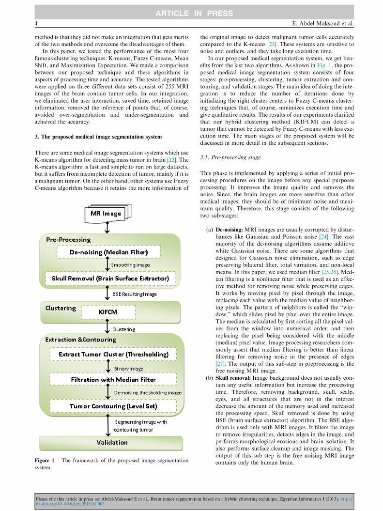
Figure 1 The framework of the proposed image segmentation
system.
Please cite this article in press as: Abdel-Maksoud E et al., Brain tumor segmentatiodx.doi.org/10.1016/j.eij.2015.01.003
the original image to detect malignant tumor cells accuratelycompared to the K-means [23]. These systems are sensitive tonoise and outliers, and they take long execution time.
In our proposed medical segmentation system, we get ben-efits from the last two algorithms. As shown in Fig. 1, the pro-posed medical image segmentation system consists of four
stages: pre-processing, clustering, tumor extraction and con-touring, and validation stages. The main idea of doing the inte-gration is to reduce the number of iterations done by
initializing the right cluster centers to Fuzzy C-means cluster-ing techniques that, of course, minimizes execution time andgive qualitative results. The results of our experiments clarifiedthat our hybrid clustering method (KIFCM) can detect a
tumor that cannot be detected by Fuzzy C-means with less exe-cution time. The main stages of the proposed system will bediscussed in more detail in the subsequent sections.
3.1. Pre-processing stage
This phase is implemented by applying a series of initial pro-
cessing procedures on the image before any special purposesprocessing. It improves the image quality and removes thenoise. Since, the brain images are more sensitive than other
medical images; they should be of minimum noise and maxi-mum quality. Therefore, this stage consists of the followingtwo sub-stages:
(a) De-noising: MRI images are usually corrupted by distur-bances like Gaussian and Poisson noise [24]. The vastmajority of the de-noising algorithms assume additive
white Gaussian noise. There are some algorithms thatdesigned for Gaussian noise elimination, such as edgepreserving bilateral filter, total variation, and non-local
means. In this paper, we used median filter [25,26]. Med-ian filtering is a nonlinear filter that is used as an effec-tive method for removing noise while preserving edges.
It works by moving pixel by pixel through the image,replacing each value with the median value of neighbor-ing pixels. The pattern of neighbors is called the ‘‘win-dow,’’ which slides pixel by pixel over the entire image.
The median is calculated by first sorting all the pixel val-ues from the window into numerical order, and thenreplacing the pixel being considered with the middle
(median) pixel value. Image processing researchers com-monly assert that median filtering is better than linearfiltering for removing noise in the presence of edges
[27]. The output of this sub-step in preprocessing is thefree noising MRI image.
(b) Skull removal: Image background does not usually con-tain any useful information but increase the processing
time. Therefore, removing background, skull, scalp,eyes, and all structures that are not in the interestdecrease the amount of the memory used and increased
the processing speed. Skull removed is done by usingBSE (brain surface extractor) algorithm. The BSE algo-rithm is used only with MRI images. It filters the image
to remove irregularities, detects edges in the image, andperforms morphological erosions and brain isolation. Italso performs surface cleanup and image masking. The
output of this sub step is the free noising MRI imagecontains only the human brain.
n based on a hybrid clustering technique, Egyptian Informatics J (2015), http://
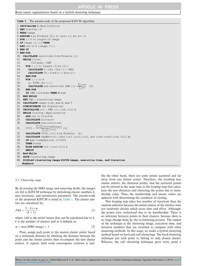
Table 1 The pseudo-code of the proposed KIFCM algorithm.
1. INITIALIZE K, Maxiteration
2. SET Iterfcm = 0
3. READ image
4. ASSIGN m as Formula (2); h = zero (1, m); hc = h
5. FOR i = 0 to length of image
6. IF image (i) > 0 THEN
7. Add one to h (image (i))
8. END IF
9. END FOR
10. CALCULATE centroids from Formula (1)
11. WHILE (true)
12. Old mean =MU13. FOR i = 1 to length (find (h))
14. CALCULATE C = abs (Ind (i)-MU)15. CALCULATE CC = find(c == min(c))
16. END FOR
17. FOR i = 1 to k
18. a = FIND (hc == i)
19. CALCULATE new centroidsMU (i) =
Pa�hðaÞPhðaÞ (3)
20. END FOR
21. IFMU =old mean THEN Break
22. END WHILE
23. SET IMA = clustering image
24. CALCULATE image size, max X, max Y
25. CONCATINATE the dimensions
26. INITIALIZE cc1 =MU (1), cc2, till k
27. WHILE Iterfcm < Maxiteration
28. ADD one to Iterfcm
29. CALCULATE distance
30. CALCULATE new centroids
31. CCC1 =
P PU1�U1�doubleðimageÞð ÞP P
U1�U1ð Þ (4)
32. CALCULATE CCC2, till k as Formula (4)
33. CALCULATE tmpMatrix = [abs (cc1-ccc1)/cc1, abs (cc2-ccc2)/cc2, till k]
34. IF max (tempmatrix) <0.0001
35. THEN break
36. ELSE ASSIGN cc1 = ccc1 till k
37. ENDIF
38. End While
39. SAVE Clustering image
40. DISPLAY clustering image KIFCM image, execution time, and iteration
Numbers
Brain tumor segmentation based on a hybrid clustering technique 5
3.2. Clustering stage
By de-noising the MRI image and removing skulls, the imagesare fed to KIFCM technique by initializing cluster numbers k,max iterations, and termination parameter. The pseudo-code
of the proposed KIFCM is listed in Table 1. The cluster cen-ters are calculated by:
MU ¼ ð1 : kÞ � m
ðkþ 1Þ ð1Þ
whereMU is the initial means that can be calculated due to k.k is the number of clusters and m is defined as:
m ¼ maxðMRI imageÞ þ 1 ð2Þ
Then, assign each point to the nearest cluster center basedon a minimum distance by checking the distance between thepoint and the cluster centers then re-compute the new cluster
centers. It repeats until some convergence criterion is met.
Please cite this article in press as: Abdel-Maksoud E et al., Brain tumor segmentatiodx.doi.org/10.1016/j.eij.2015.01.003
On the other hand, there are some points scattered and faraway from any cluster center. Therefore, the resulting new
cluster centers, the clustered points, and the scattered pointscan be entered in the same time to the looping step that calcu-lates the new distances and clustering the points due to mem-
bership value. Then, the membership and means values areupdated with determining the condition of closing.
This looping step takes less number of iterations than the
random selection because the initial centers of the clusters werenot randomly chosen which saves time and effort. Although,the points were reclustered due to its membership. There isno inference between points in their clusters, because there is
no huge change done by the re-clustering process. The outputof the technique is the clustering image, execution time, anditeration numbers that are recorded to compare with other
clustering methods. In this stage, we make a hybrid clusteringmethod based on hard and soft clusterings. The hard clusteringtechnique put each point to belong to only closest cluster.
Whereas, the soft clustering technique gives every point a
n based on a hybrid clustering technique, Egyptian Informatics J (2015), http://
6 E. Abdel-Maksoud et al.
degree of membership, rather than belonging wholly to justone cluster.
3.3. Extraction and contouring stage
In this stage, we used two segmentation methods: thresholdingand active contour level set methods:
(a) Thresholding segmentation: It is intensity-based segmen-tation. Thresholding or image binarization is one of the
important techniques in image processing and computervision. It is used to extract the object from the back-ground. The segmented image, which is obtained by
thresholding, has the advantages of smaller storagespace, fast processing speed, and ease of manipulation,compared with gray level image which usually containsa large number of gray levels (maximum 256 levels)
[28]. The output of this step is the segmenting image withdark background and lighting tumor area.
(b) Active contour by level set: Active contours have been
used for image segmentation and boundary trackingsince the first introduction of snakes by Kass et al.[29]. The basic idea is to start with initial boundary
shapes represented in a form of closed curves, i.e. con-tours, and iteratively modify them by applying shrink/expansion operations according to the constraints. Theused active contour method show robust segmentation
capabilities in medical images where traditional segmen-tation methods show poor performance. An advantageof the active contours as an image segmentation method
is that they partition an image into sub-regions with con-tinuous boundaries. While the edge detectors based onthe threshold or local filtering, it often results in
discontinuous boundaries. The use of level set theoryhas provided more flexibility and convenience in theimplementation of active contours. Depending on the
implementation scheme, active contours can use variousproperties used for other segmentation methods such asedges, statistics, and texture. Level set algorithm isdemonstrated in details by Lee [30].
The clustering image is entered to the binarization processusing inverse thresholding method with iteration number
equals 3. The noise of the image is removed by using the med-ian filter that eliminates the small regions that are far awayfrom the tumor cluster. We can consider this step as a post-
processing step in our system. Of course, these two steps canbe converted to one step if the classical FCM is used which
Table 2 The pseudocode of the extraction and contouring stages.
1. BINARIZE image
2. APPLY median filter
3. SAVE thresholding image
4. CALL level set function
5. SAVE resulting image
6. DISPLAY the segmenting image with contoured tumor regi
7. CALCULATE total pixels =numel (BW)
8. CALCULATE white pixels nwhite=P
BWð:Þ (5)
9. CALCULATE black pixels nblack=total pixels – nwhite
10. CALCULATE ratio= nwhitenblack
Please cite this article in press as: Abdel-Maksoud E et al., Brain tumor segmentatiodx.doi.org/10.1016/j.eij.2015.01.003
user can enter the cluster to be a threshold or appeared onlyin image. In our proposed technique, we get rid of user inter-action that may be true or false. After that, the thresholding
image with the lighting tumor cluster is fed to the level set.Level set contours the tumor area of the thresholding imageon the original image. The output of this step is the threshold-
ing image and original free noising image with contouringtumor area. The tumor area can be calculated by computingthe white pixels of total pixels of the image. The pseudo code
of the extraction and contouring is illustrated in Table 2.
3.4. Validation stage
In validation stage, the segmented images by KIFCM werecompared to the ground truth in cases of the third data setas illustrated in experimental results. It compared to the typicalimages as in the second data set, but the first one does not have
any ground truth. The results were evaluated by performancematrix that contains the precision and recall. Precision is thecorrect segmentation that refers to the percentage of true posi-
tive. In other words, it is the number of pixels that belong to acluster and is segmented into that cluster. Recall, or sensitivityis defined as the number of the true positives divided by the
total number of elements that belong to the positive clus-ter [31–33]. The performance matrix will be illustrated indetails in Section 4.2. The results of each technique wererecorded in the following tables according to accuracy, execu-
tion time, number of iterations and performance metrics thatmentioned before and represented.
4. Experimental results
4.1. Data sets
In order to check the performance of our image segmentationapproach, we used three benchmark data sets. The first one is
the Digital Imaging and Communications in Medicine(DICOM) data set [34]. DICOM consists of 22 images that con-tain brain tumors. All DICOM image files are encoded in
JPEG2000 transfer syntax with ‘‘.DCM’’ extension. It has noground truth images for the contained images. The second dataset is Brain Web data set [35]. It contains simulated brain MRIdata based on two anatomical models: normal and multiple
sclerosis (MS). Full 3-dimensional data volumes have been sim-ulated using three sequences (T1-, T2-, and proton density-(PD-) weighted) and a variety of slice thicknesses, noise levels,
and levels of intensity non-uniformity. The files contained inthis data set have extension of‘‘.MNC’’. Its T1 modality is
ons
n based on a hybrid clustering technique, Egyptian Informatics J (2015), http://
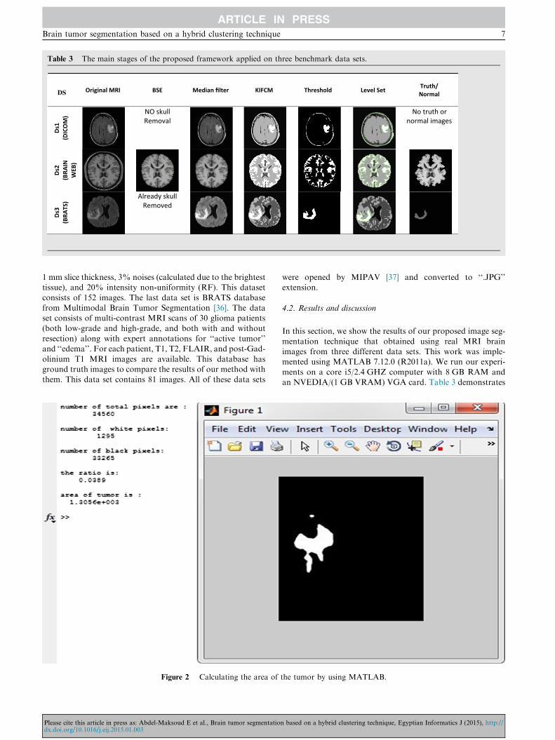
Table 3 The main stages of the proposed framework applied on three benchmark data sets.
SD Original MRI BSE Median filter KIFCM Threshold Level Set Truth/Normal
Ds1
(DIC
OM
) NO skullRemoval
No truth or normal images
Ds2
(BRA
IN
WEB
)
Ds3
(BRA
TS)
Already skullRemoved
Brain tumor segmentation based on a hybrid clustering technique 7
1 mm slice thickness, 3% noises (calculated due to the brightesttissue), and 20% intensity non-uniformity (RF). This dataset
consists of 152 images. The last data set is BRATS databasefrom Multimodal Brain Tumor Segmentation [36]. The dataset consists of multi-contrast MRI scans of 30 glioma patients
(both low-grade and high-grade, and both with and withoutresection) along with expert annotations for ‘‘active tumor’’and ‘‘edema’’. For each patient, T1, T2, FLAIR, and post-Gad-
olinium T1 MRI images are available. This database hasground truth images to compare the results of our method withthem. This data set contains 81 images. All of these data sets
Figure 2 Calculating the area of
Please cite this article in press as: Abdel-Maksoud E et al., Brain tumor segmentatiodx.doi.org/10.1016/j.eij.2015.01.003
were opened by MIPAV [37] and converted to ‘‘.JPG’’extension.
4.2. Results and discussion
In this section, we show the results of our proposed image seg-
mentation technique that obtained using real MRI brainimages from three different data sets. This work was imple-mented using MATLAB 7.12.0 (R2011a). We run our experi-
ments on a core i5/2.4 GHZ computer with 8 GB RAM andan NVEDIA/(1 GB VRAM) VGA card. Table 3 demonstrates
the tumor by using MATLAB.
n based on a hybrid clustering technique, Egyptian Informatics J (2015), http://
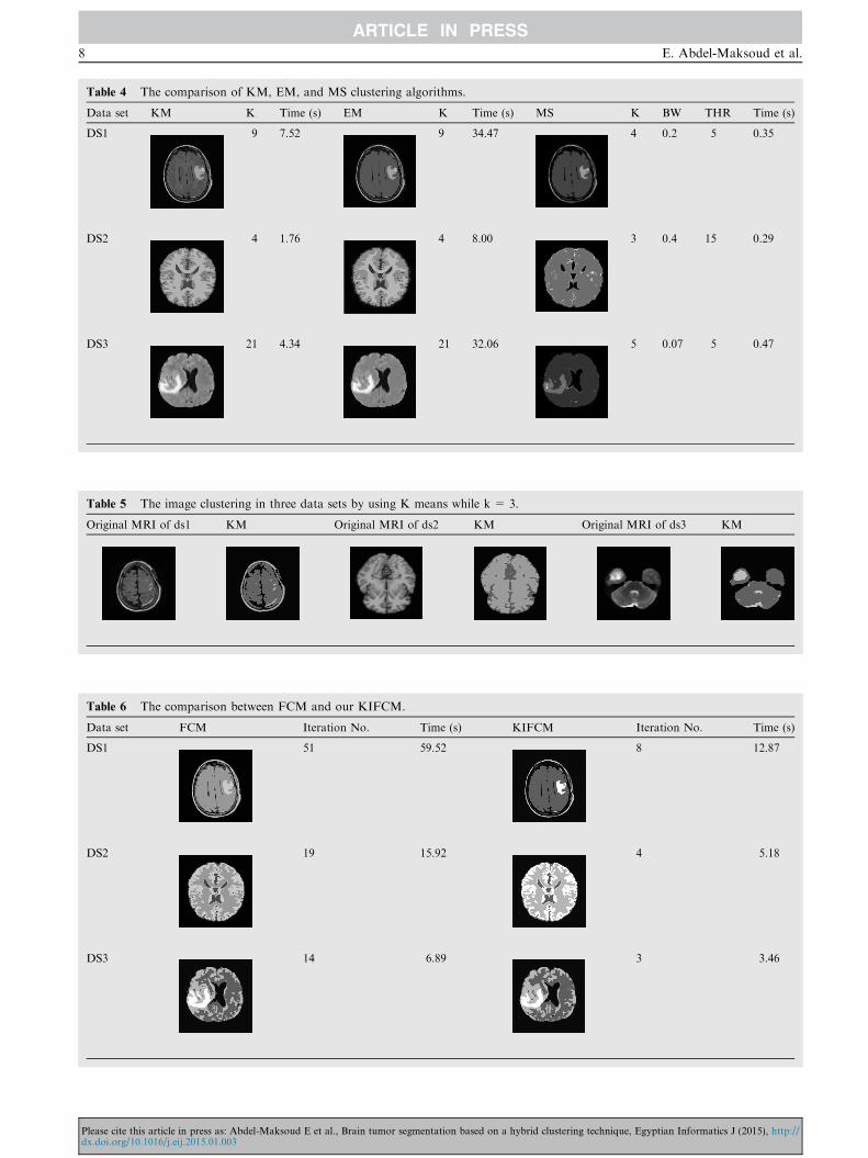
Table 4 The comparison of KM, EM, and MS clustering algorithms.
Data set KM K Time (s) EM K Time (s) MS K BW THR Time (s)
DS1 9 7.52 9 34.47 4 0.2 5 0.35
DS2 4 1.76 4 8.00 3 0.4 15 0.29
DS3 21 4.34 21 32.06 5 0.07 5 0.47
Table 5 The image clustering in three data sets by using K means while k = 3.
Original MRI of ds1 KM Original MRI of ds2 KM Original MRI of ds3 KM
Table 6 The comparison between FCM and our KIFCM.
Data set FCM Iteration No. Time (s) KIFCM Iteration No. Time (s)
DS1 51 59.52 8 12.87
DS2 19 15.92 4 5.18
DS3 14 6.89 3 3.46
8 E. Abdel-Maksoud et al.
Please cite this article in press as: Abdel-Maksoud E et al., Brain tumor segmentation based on a hybrid clustering technique, Egyptian Informatics J (2015), http://dx.doi.org/10.1016/j.eij.2015.01.003
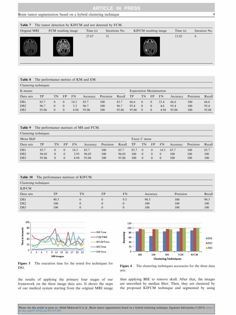
Table 7 The tumor detection by KIFCM and not detected by FCM.
Original MRI FCM resulting image Time (s) Iterations No. KIFCM resulting image Time (s) Iteration No.
27.87 51 13.92 8
Table 8 The performance metrics of KM and EM.
Clustering techniques
K-means Expectation Maximization
Data sets TP TN FP FN Accuracy Precision Recall TP TN FP FN Accuracy Precision Recall
DS1 85.7 0 0 14.3 85.7 100 85.7 66.6 0 0 33.4 66.6 100 66.6
DS2 96.7 0 0 3.3 96.7 100 96.7 95.4 0 0 4.6 95.4 100 95.4
DS3 95.06 0 0 4.94 95.06 100 95.06 95.06 0 0 4.94 95.06 100 95.06
Table 9 The performance matrices of MS and FCM.
Clustering techniques
Mean Shift Fuzzy C mean
Data sets TP TN FP FN Accuracy Precision Recall TP TN FP FN Accuracy Precision Recall
DS1 85.7 0 0 14.3 85.7 100 85.7 85.7 0 0 14.3 85.7 100 85.7
DS2 96.05 0 0 3.95 96.05 100 96.05 100 0 0 0 100 100 100
DS3 95.06 0 0 4.94 95.06 100 95.06 100 0 0 0 100 100 100
Table 10 The performance matrices of KIFCM.
Clustering techniques
KIFCM
Data sets TP TN FP FN Accuracy Precision Recall
DS1 90.5 0 0 9.5 90.5 100 90.5
DS2 100 0 0 0 100 100 100
DS3 100 0 0 0 100 100 100
Figure 3 The execution time for the tested five techniques for
DS1.Figure 4 The clustering techniques accuracies for the three data
sets.
Brain tumor segmentation based on a hybrid clustering technique 9
the results of applying the primary four stages of ourframework on the three image data sets. It shows the stepsof our medical system starting from the original MRI image
Please cite this article in press as: Abdel-Maksoud E et al., Brain tumor segmentatiodx.doi.org/10.1016/j.eij.2015.01.003
then applying BSE to remove skull. After that, the imagesare smoothed by median filter. Then, they are clustered bythe proposed KIFCM technique and segmented by using
n based on a hybrid clustering technique, Egyptian Informatics J (2015), http://
10 E. Abdel-Maksoud et al.
thresholding and contouring the tumor region by level set.Fig. 2 shows a snapshot of calculating the area of the tumor.
As shown in Table 4, we used the same number of clusters
for Expectation Maximization (EM) and K-means (KM) toevaluate them under the same conditions due to efficiency ofsegmentation and processing time. We can observe that EM
like KM in accuracy but it takes longer time (T in seconds)than KM. On the other hand, the Mean Shift (MS) clusteringtechnique need to supply the parameters of bandwidth and
threshold. It calculates a number of clusters K and consumedtime in clustering. By doing the experiments on all images ofthe three data sets using the MS, we found that the best resultsin image clusters can be obtained if bandwidth = 0.2 and
threshold = 5. By decreasing the bandwidth for the samethreshold, it gives best results in less time. Whereas, MS isnot accurate at all the time, it takes less processing time if
the cluster number K = 3, but it does not give accurate results.On the contrary, if K equals to 3 in KM, it gives accurateresults in most of the cases as shown in Table 5. We also found
that without skull removal, it increases the processing time onall techniques. On the contrary, when removing the skull as inDS2 or using images with removed skull like in DS3, the pro-
cessing time is reduced as shown in Table 4.As shown in Table 6, we observed that KIFCM seems like
FCM (Fuzzy C Means) in accuracy but KIFCM take less pro-cessing time than FCM with less iteration. In the first data set
(DS1), the iteration number of FCM clustering technique is 51when max iteration is greater than 50, and the processing timeis 59.52 s. On the other hand, the iteration number to cluster
the same image in our technique is 8 when the max iterationis greater than 15. The clustering time is 12.87 s with initialcluster k = 6 and 4 cluster centers and the result is apparent
for the user to discover the tumor with his eyes before doingthresholding and level set stages.
In some images, we found that the KIFCM method is more
accurate than FCM, which is demonstrated in Table 7. We canobserve that when clustering the image with FCM it takes 51iterations in 27.87 s, and the resulting image is not accurateand has overlapped area. However, when we clustered the
same image with our technique KIFCM, it uses 8 iterationsin 13.92 s. The tumor in the second image of the table thatwas clustered with our technique KIFCM is clearer to the user
than in the first image which is clustered by the FCMtechnique.
The comparison was done between the five tested
techniques according to the following performance measures:
True Positive ðTPÞ¼No of resulted images having brain tumor
total No of images
ð6Þ
True Negative ðTNÞ ¼ No of images that haven0t tumor
total No of images
ð7Þ
False PositiveðFPÞ¼No of images that haven0t tumor and detected positive
total No of images
ð8Þ
False Negative ðFNÞ¼No of images have tumor and not detected
total No of imagesð9Þ
Please cite this article in press as: Abdel-Maksoud E et al., Brain tumor segmentatiodx.doi.org/10.1016/j.eij.2015.01.003
Precision ¼ TP
ðTPþ FPÞ
� �ð10Þ
Recall ¼ TP
ðTPþ FNÞ
� �ð11Þ
Accuracy ¼ ðTPþ TNÞðTPþ TNþ FPþ FNÞ
� �ð12Þ
Table 8 shows the performance comparisons between KMand EM. The results prove that the accuracy of the EM isapproximately equal to the accuracy of the KM in the last
two data sets (DS2 and DS3). In first data set (DS1), the accu-racy of the KM is 85.7%, whereas the accuracy of the EM is66.6%. As shown in Tables 8 and 9, we can observe that the
performance of the MS technique seems to be the same asKM except in DS2. Tables 9 and 10 ensure that KIFCM ismore accurate than FCM.
Fig. 3 represents the execution time of the clustering stage for
the five tested clustering techniques for DS1 as a sample. Itshows that FCM takes the longest execution time in the cluster-ing process and is followed by EM. On the other hand, our tech-
nique (KIFCM) takes the third level in the execution time. TheKM is in the fourth level, and the MS is in the fifth level. There-fore, theMS is the least execution time. Fig. 4 shows the ranking
of the five clustering techniques according to the accuracy.From the previous figures and tables, it is very clear that
our proposed technique is the most accurate one with minimalexecution time. Although, our proposed technique takes
longer time than KM and MS, but KIFCM takes minimal exe-cution time compared to FCM and EM. Although FCM ismore accurate than KM, MS and EM but also KIFCM is
more accurate than FCM.
5. Conclusion
Image segmentation plays a significant role in medical image.In the field of medical diagnosis, an extensive diversity ofimaging techniques is available presently, such as CT and
MRI. MRI is the most effectively image model used for diag-nostic image examination for brain tumor. The MRI scan ismore comfortable than CT scan for diagnosis. On the other
hand, K-mean algorithm can detect a brain tumor faster thanFuzzy C-means, but Fuzzy C-means can predict tumor cellsaccurately. Original Fuzzy C-means algorithm fails to segmentimage corrupted by noise, outliers, and other imaging artifacts.
Therefore, we developed a new approach that integrates the K-means clustering algorithm with the Fuzzy C-means algorithmto detect brain tumor accurately and in minimal execution
time. Our framework consists of four stages: pre-processing(de-noising and skull removal), clustering (integration of K-means and Fuzzy C-means), extraction and contouring (thres-
holding and level set), and validation stages. From the exper-imental results, we proved the effectiveness of our approachin brain tumor segmentation by comparing it with four state-
of-the-art algorithms: K-means, Expectation Maximization,Mean Shift, and Fuzzy C-means. Our proposed system deter-mines the initial cluster k value to minimize the execution time.The performance of the proposed technique, its minimization
time strategy, and its quality has been demonstrated in severalexperiments. In future work, the 3D evaluation of the braintumor detection using 3D slicer will be carried out. As well
n based on a hybrid clustering technique, Egyptian Informatics J (2015), http://
Brain tumor segmentation based on a hybrid clustering technique 11
as to increase the efficiency of the segmentation process, anintensity adjustment process will provide more challengingand may allow us to refine our segmentation techniques to
the MRI brain tumor segmentation.
References
[1] Janani V, Meena P. Image segmentation for tumor detection using
fuzzy inference system. Int J Comput Sci Mobile Comput
(IJCSMC) 2013;2(5):244–8.
[2] Dong B, Chien A, SHEN Z. Frame based segmentation for
medical images. Commun Math Sci 2010;32(4):1724–39.
[3] Patel J, Doshi K. A study of segmentation methods for detection
of tumor in brain MRI. Adv Electron Electr Eng
2014;4(3):279–84.
[4] Rohit M, Kabade S, Gaikwad MS. Segmentation of brain tumour
and its area calculation in brain MRI images using K-mean
clustering and Fuzzy C-mean algorithm. Int J Comput Sci Eng
Technol (IJCSET) 2013;4(5):524–31.
[5] Aslam HA, Ramashri T, Ahsan MIA. A new approach to image
segmentation for brain tumor detection using pillar K-means
algorithm. Int J Adv Res Comput Commun Eng 2013;2:1429–36.
[6] Acharya J, Gadhiya S, Raviya. Segmentation techniques for
image analysis: a review. Int J Comput Sci Manage Res
2013;2(4):1218–21.
[7] Naik D, Shah P. A review on image segmentation clustering
algorithms. Int J Comput Sci Inform Technol 2014;5(3):3289–93.
[8] Christe SA, Malathy K, Kandaswamy A. Improved hybrid
segmentation of brain MRI tissue and tumor using statistical
features. ICTACT J Image Video Process 2010;1(1):34–49.
[9] Seerha GK, Kaur R. Review on recent image segmentation
techniques. Int J Comput Sci Eng (IJCSE) 2013;5(2):109–12.
[10] Dass R, Priyanka, Devi S. Image segmentation techniques. Int J
Electron Commun Technol 2012;3(1):66–70.
[11] Kaur J, Agrawal S, Vig R. Integration of clustering, optimization
and partial differential equation method for improved image
segmentation. Int J Image Graph Signal Process 2012;4(11):26–33.
[12] Panda M, Patra MR. some clustering algorithms to enhance the
performance of the network intrusion detection system. J Theor
Appl Inform Technol 2008;4(8):795–801.
[13] Bandhyopadhyay SK, Paul TU. Automatic segmentation of brain
tumour from multiple images of brain MRI. Int J Appl Innovat
Eng Manage (IJAIEM) 2013;2(1):240–8.
[14] Meena A, Raja K. Spatial Fuzzy C-means PET image segmen-
tation of neurodegenerative disorder spatial Fuzzy C-means PET
image segmentation of neurodegenerative disorder. Indian J
Comput Sci Eng (IJCSE) 2013;4(1):50–5.
[15] Glavan CC, Holban S. Segmentation of bone structure in X-ray
images using convolutional neural network. Adv Electr Comput
Eng 2013;13(1):1–8.
[16] Tatiraju S, Mehta A. Image Segmentation using k means
clustering, EM and normalized Cuts, University Of California
Irvine, technical report.
[17] Yerpude A, Dubey S. Colour image segmentation using
K-medoids clustering. Int J Comput Technol Appl 2012;3(1):
152–4.
Please cite this article in press as: Abdel-Maksoud E et al., Brain tumor segmentatiodx.doi.org/10.1016/j.eij.2015.01.003
[18] Islam S, Ahmed M. Implementation of image segmentation for
natural images using clustering methods. Int J Emerg Technol
Adv Eng 2013;3(3):175–80.
[19] Christe SA, Malathy K, Kandaswamy A. Improved hybrid
segmentation of brain MRI tissue and tumor using statistical
features. J Image Video Process 2010;1(1):43–9.
[20] Funmilola A, Oke OA, Adedeji TO, Alade OM, Adewusi EA.
Fuzzy K-C-means clustering algorithm for medical image seg-
mentation. J Informat Eng Appl 2012;2(6):21–32.
[21] Wilson B, Dhas JPM. An experimental analysis of Fuzzy C-means
and K-means segmentation algorithm for iron detection in brain
SWI using Matlab. Int J Comput Appl 2014;104(15):36–8.
[22] Mohan P, AL V, Devi BRS, Kavitha BC. Intelligent based brain
tumor detection using ACO. Int J Innovat Res Comput Commun
Eng 2013;1(9):2143–50.
[23] Anandgaonkar G, Sable G. Brain tumor detection and identifi-
cation from T1 post contrast MR images using cluster based
segmentation. Int J Sci Res 2014;3(4):814–7.
[24] Rodrigues I, Sanches J, Dias J. Denoising of medical images
corrupted by poisson noise. In: Image processing, ICIP 2008. 15th
IEEE international conference on; 2008. p. 1756–9.
[25] Auckland University. <https://www.cs.auckland.ac.nz/courses/
compsci373s1c/PatricesLectures/Image%20 Filtering_2up.pdf>;
[accessed 15.03.14].
[26] Web Services for School of Industrial Engineering and Manage-
ment. <http://www2.it.lut.fi/kurssit/07-08/ct20A6100/seminars/
Median_Filtering.pdf>; [accessed 15.03.14].
[27] Castro A, Donoho DL. Does median filter truly preserve edges
better than linear filtering? Ann Stat 2009;37(3):1172–206.
[28] Saini R, Dutta M. Image segmentation for uneven lighting images
using adaptive thresholding and dynamic window based on
incremental window growing approach. Int J Comput Appl
2012;56(13):31–6.
[29] Kass M, Witkin A, Terzopoulos D. Snakes, active contour model.
Int J Comput Vision 1988;1(4):321–31.
[30] Lee CP. Robust image segmentation using active contours: level
set approaches, PhD thesis, Department of Electrical and Com-
puter Engineering North Carolina State University; 2005. p. 18–
30. [Chapter 3].
[31] Kdnuggets. <http://www.kdnuggets.com/faq/precision-recall.
html>; [accessed 23.04.14].
[32] Weng CG, Poon J. A new evaluation measure for imbalanced
datasets. In: Seventh Australasian data mining conference (Aus
DM 2008), South Australia: ACS, vol. 87; 2008. p. 27–32.
[33] Dakua SP. Use of chaos concept in medical image segmentation.
Comput Methods Biomech Biomed Eng: Imag Visual
2013;1(1):28–36.
[34] Brain Web: Simulated Brain Database. <http://brainweb.bic.
mni.mcgill.ca/brainweb/>; [accessed 30.12.13].
[35] DICOM Samples Image Sets. <http://www.osirix-viewer.com/
datasets/>; [accessed 29.12.13].
[36] MICCA Nice 2012. <http://www2.imm.dtu.dk/projects/
BRATS2012/data.html>; [accessed 12.01.14].
[37] NIH Center for Information Technology. <http://mipav.cit.nih.
gov/>; [accessed 07.04.14].
n based on a hybrid clustering technique, Egyptian Informatics J (2015), http://