Embed Size (px)
Citation preview

HomeostasisResponse to Outside and Inside Environment

I. General comparison of the body’s 2 major control systemsA. General comparisonProperties Nervous SystemEndocrine SystemAnatomy System of neuronalpathways highlyorganized into CNS andPNS. Each nervous cellterminates directly on itstarget cell.
Includes a number ofstructurally unrelatedorgans, which are widelydispersed throughoutthe body. Notanatomically linked totarget cells.General functions Coordinates the rapid,precise responses. Primarily controlsmetabolism and theactivities that requireduration, not speed.Specificity of actionDepends on closephysical associationbetween the nervous andtarget cell.Determined by presenceof specific receptors on ttarget cells. Hormonesbind to receptors in alock-and key fashion.Route of chemicalmessenger Neurotransmitter isreleased into synapticcleft and diffuses a veryshort distance to targetcell.Hormones are releasedinto the blood andtherefore can circulatethroughout the body.Speed of response Rapid (milliseconds). Slow (minute to hours):Complex mechanism ofaction.Duration of action Brief (milliseconds):Neurotransmitter is takenback up to nervousterminal or inactivate byenzymes within synapticcleft.Long (minutes to daysor even longer):hormone may remainbound to receptor.

Nervous System
• Central Nervous System (CNS) – Brain
– Spinal Cord
– Olfactory
– Optic nerves
• Almost no regeneration?????
• Peripheral Nervous system (PNS) – Autonomic Nervous System
SympatheticParasympathetic
– Somatic
– Cranial Nerves (3-12)
• Some regeneration

Efferent Division
Afferent Division

What are the components of CNS ?
• Neuron
• Glia

Neurons
Cells that specialized for transmitted chemical and electrical signals from one part of the body to another.
CELL BODY
AXONMyelin sheath
Schwann cellNode of Ranvier
Synaptic terminals
Dendrites Nucleus Synapses

Impulse
Presynaptic neuron
Vesicle
Transmitters
Synaptic cleft
Receptors
Postsynapticneuron
Postsynaptic activity

Classifying Neurons
• Number of axons and dendrites
• Type of connections
• Type of neurotransmitter
• unipolar, bipolar, multipolar
• sensory, motor, interneurons
• Acetylcholin, Dopamine

Glia Cells
• Astrocytes
• Oligodendrocytes
• Microglia
• Ependymal cells

Possible Roles of Glia Cells
• supporting element
• producing myelin
• scavengers - removing debris
• buffer
• guide migration in course of development
• help to form special lining in the capillaries - Blood Brain Barrier (BBB)

Anyone touched human brain?

bumps = gyrigrooves =sulci (fissures)

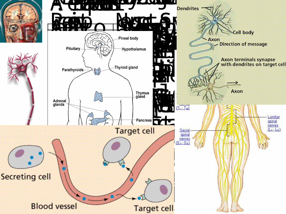
Is the brain hard or soft?
The brain is soft.
How it is protected?

QuickTime™ and a decompressor
are needed to see this picture.


QuickTime™ and aCinepak decompressor
are needed to see this picture.
CSF

QuickTime™ and aVideo decompressor
are needed to see this picture.
QuickTime™ and aVideo decompressor
are needed to see this picture.
Ventricles

Blood-Brain barrier

Cortical layer
I
II
III
IV
V
VI
Blood vessel
Neuron

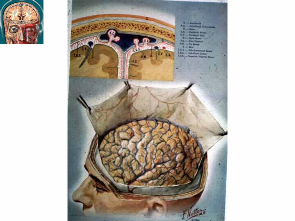
Mapping the brain function
How much % of the brain are we using?
10%
50%
100%


Frontallobe
Temporallobe
Parietal lobe
Occipitallobe
Cerebellum
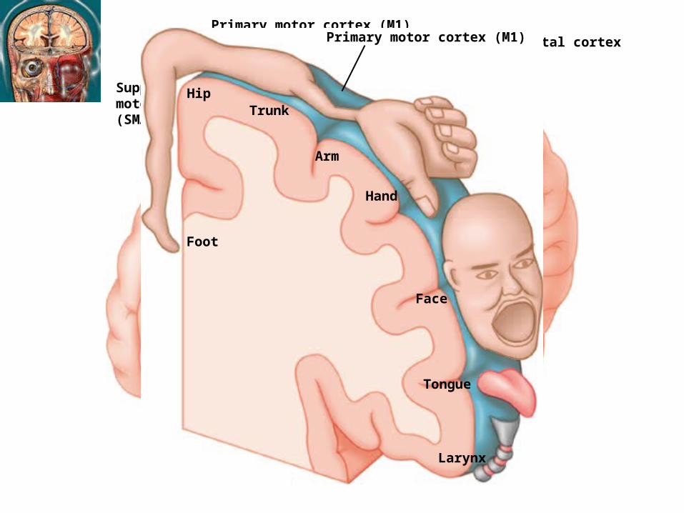
Primary motor cortex (M1)Posterior parietal cortex
Premotor cortex(PMA)
Supplementarymotor cortex(SMA)
Primary motor cortex (M1)
Foot
HipTrunk
Arm
Hand
Face
Tongue
Larynx

Broca’sarea
Parsopercularis
Motor cortex Somatosensory cortex
Sensory associativecortex
PrimaryAuditory cortex
Wernicke’sarea
Visual associativecortex
Visualcortex

Evidence for localization
• Broca (1861)
Expressive Aphsia
can understand but cannot speak
• Wernicke (1876)
Receptive Aphsia
can speak but cannot understand

Sensory Stimuli
Sensation
Perception
Phenomena in Environment
Excitation in Sensory Nerve
Integration in Sensory CNS
Speech

QuickTime™ and a decompressor
are needed to see this picture.


LeftAuditorycortex
RightAuditorycortex
Cochlea Medial geniculate nucleus
Inferior colliculus
SuperiorOlivarynucleus
IpsilateralCochlearnucleus
Auditorynerve fiber
QuickTime™ and a decompressor
are needed to see this picture.

Optic nerve
Optic tract
Lateral geniculate nucleus
Optic radiation
Optic chiasm
Primary visual cortex

Line
Retina
Lateralgeniculatenucleus
PrimaryVisualCortex (V1)
What you see, what you get

QuickTime™ and aGIF decompressor
are needed to see this picture.
QuickTime™ and aGIF decompressor
are needed to see this picture.
QuickTime™ and aGIF decompressor
are needed to see this picture.

QuickTime™ and aGIF decompressor
are needed to see this picture.

QuickTime™ and aGIF decompressor
are needed to see this picture.

Processing of sound


QuickTime™ and aVideo decompressor
are needed to see this picture.


QuickTime™ and aVideo decompressor
are needed to see this picture.


Amygdala
Hippocampus

QuickTime™ and aAnimation decompressor
are needed to see this picture.

Coordination and control of voluntary movement.
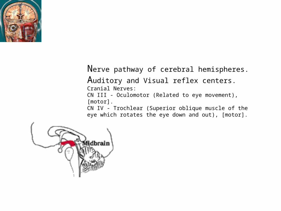
Nerve pathway of cerebral hemispheres.
Auditory and Visual reflex centers.Cranial Nerves:CN III - Oculomotor (Related to eye movement), [motor]. CN IV - Trochlear (Superior oblique muscle of the eye which rotates the eye down and out), [motor].

Respiratory Center.Cranial Nerves:CN V - Trigeminal (Skin of face, tongue, teeth; muscle of mastication), [motor and sensory]. CN VI - Abducens (Lateral rectus muscle of eye which rotates eye outward), [motor]. CN VII - Facial (Muscles of expression), [motor and sensory]. CN VIII - Acoustic (Internal auditory passage), [sensory].

Crossing of motor tracts.
Cardiac Center.
Respiratory Center.
Vasomotor (nerves having muscular control of the blood vessel walls) Center ハCenters for cough, gag, swallow, and vomit.
Cranial Nerves:
* CN IX - Glossopharyneal (Muscles and mucous membranes of pharynx, the constricted openings from the mouth and the oral pharynx and the posterior third of tongue.), [mixed]. * CN X - Vagus (Pharynx, larynx, heart, lungs, stomach), [mixed]. * CN XI - Accessory (Rotation of the head and shoulder), [motor]. * CN XII - Hypoglossal (Intrinsic muscles of the tongue), [motor].

QuickTime™ and aGIF decompressor
are needed to see this picture.


The PET scan on the left shows two areas of the brain (red and yellow) that become particularly active when volunteers read words on a video screen: the primary visual cortex and an additional part of the visual system, both in the back of the left hemisphere.Other brain regions become especially active when subjects hear words through ear-phones, as seen in the PET scan on the right.To create these images, researchers gave volunteers injections of radioactive water and then placed them, head first, into a doughnut-shaped PET scanner. Since brain activity involves an increase in blood flow, more blood and radioactive water streamed into the areas of the volunteers' brains that were most active while they saw or heard words.
Positron Emission Tomography


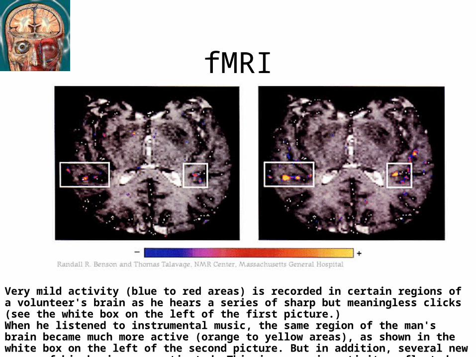
fMRI
Very mild activity (blue to red areas) is recorded in certain regions of a volunteer's brain as he hears a series of sharp but meaningless clicks (see the white box on the left of the first picture.)When he listened to instrumental music, the same region of the man's brain became much more active (orange to yellow areas), as shown in the white box on the left of the second picture. But in addition, several new areas of his brain were activated. This increase in activity reflected the richer meaning of the sounds.

Magnetoencephalography (MEG) and MRI

Electroencephalography (EEG)
QuickTime™ and aGIF decompressor
are needed to see this picture.