Embed Size (px)
Citation preview
1
Bortezomib and SAHA synergistically induce ROS-driven caspase-dependent apoptosis of nasopharyngeal carcinoma and block replication of Epstein-Barr virus Authors: K.F. Hui1, Benjamin H.W. Lam1, Dona N. Ho1, Sai Wah Tsao2,3 and Alan K.S. Chiang1,3 Author’s Affiliations: 1Department of Paediatrics and Adolescent Medicine, Li Ka Shing Faculty of Medicine, The University of Hong Kong, Queen Mary Hospital, Pokfulam, Hong Kong SAR, China; 2Department of Anatomy, Li Ka Shing Faculty of Medicine, The University of Hong Kong, Pokfulam, Hong Kong SAR, China; 3Center for Nasopharyngeal Carcinoma Research, The University of Hong Kong, Hong Kong SAR, China Note: K.F. Hui and Benjamin H.W. Lam contributed equally to the manuscript.
Running title: Bortezomib and SAHA on NPC apoptosis Keywords: Bortezomib; SAHA; nasopharyngeal carcinoma; Epstein-Barr virus; apoptosis Corresponding author: Alan KS Chiang, Department of Paediatrics and Adolescent Medicine, Li Ka Shing Faculty of Medicine, The University of Hong Kong, Queen Mary Hospital, Pokfulam, Hong Kong SAR, China. Tel: 852-22554485; Fax: 852-28551523; Email: [email protected] Author contributions
KFH and BHWL conceived and designed the study, carried out the experimental
work and wrote the manuscript. DNH carried out the experimental work. GSWT
generated NP and NPC cell lines for the study. AKSC conceived and designed the
study, interpreted the data and wrote the manuscript.
Grant support
This project is funded by NPC Area of Excellence (AoE/M 06/08 Center for
on May 1, 2020. © 2013 American Association for Cancer Research. mct.aacrjournals.org Downloaded from
Author manuscripts have been peer reviewed and accepted for publication but have not yet been edited. Author Manuscript Published OnlineFirst on March 8, 2013; DOI: 10.1158/1535-7163.MCT-12-0811
2
Nasopharyngeal Carcinoma Research), CRCG (#10401264) and Epstein-Barr virus
research (# 20004525) grants of A.K.S. Chiang.
Disclosure of potential conflicts of interest
The authors disclosed no potential conflicts of interest.
on May 1, 2020. © 2013 American Association for Cancer Research. mct.aacrjournals.org Downloaded from
Author manuscripts have been peer reviewed and accepted for publication but have not yet been edited. Author Manuscript Published OnlineFirst on March 8, 2013; DOI: 10.1158/1535-7163.MCT-12-0811
3
Abstract
A novel drug combination of a proteasome inhibitor, bortezomib and a histone
deacetylase inhibitor, suberoylanilide hydroxamic acid (SAHA) was tested in
nasopharyngeal carcinoma (NPC) both in vitro and in vivo. Dose-response of different
concentrations of bortezomib and SAHA on inhibition of cell proliferation of NPC
was determined. Mechanisms of apoptosis and effects on lytic cycle activation of
Epstein-Barr virus (EBV) were investigated. Combination of bortezomib and SAHA
(bortezomib/SAHA) synergistically induced killing of a panel of NPC cell lines.
Pronounced increase in sub-G1, annexin V-positive and TUNEL-positive cell
populations were detected after treatment with bortezomib/SAHA when compared
with either drug alone. Concomitantly, markedly augmented proteolytic cleavage of
PARP, caspase-3, -7, -8 and -9, reactive oxygen species (ROS) generation and
caspase-8-dependent histone acetylation were observed. ROS scavenger, N-acetyl
cysteine, diminished the apoptotic effects of bortezomib/SAHA while caspase
inhibitor, Z-VAD-FMK, significantly suppressed the apoptosis without decreasing the
generation of ROS. Bortezomib inhibited SAHA’s induction of EBV replication and
abrogated production of infectious viral particles in NPC cells. Furthermore,
bortezomib/SAHA potently induced apoptosis and suppressed the growth of NPC
xenografts in nude mice. In conclusion, the novel drug combination of bortezomib
and SAHA is highly synergistic in the killing of NPC cells in vitro and in vivo. The
major mechanism of cell death is ROS-driven caspase-dependent apoptosis.
Bortezomib antagonizes SAHA’s activation of EBV lytic cycle in NPC cells. This
study provides a strong basis for clinical testing of the combination drug regimen in
NPC patients.
on May 1, 2020. © 2013 American Association for Cancer Research. mct.aacrjournals.org Downloaded from
Author manuscripts have been peer reviewed and accepted for publication but have not yet been edited. Author Manuscript Published OnlineFirst on March 8, 2013; DOI: 10.1158/1535-7163.MCT-12-0811
4
Introduction:
Nasopharyngeal carcinoma (NPC) is concentrated in Southern Chinese populations in
Southeast Asia, North Africans, Greenlanders and Alaskan Eskimos but is rare in
other populations of the world. It has a predilection to affect young adult males. The
incidence rate reaches ~25 per 100,000 males in Southern Chinese (1). The disease
has uniquely strong association with Epstein-Barr virus (EBV) when compared with
other head and neck malignancies (2). Radiotherapy is the mainstay of NPC treatment
and confers cytotoxicity through generation of reactive oxygen species (ROS) (3).
Chemotherapy is required in locally advanced or metastatic cases and commonly used
chemotherapeutic drugs include cisplatin and docetaxel (4). Although NPC is both
radiosensitive and chemosensitive, the standard treatment regimen results in
significant long term sequelae. Moreover, the disease relapse rate is relatively high
with poor survival chance for recurrent or metastatic disease (5). Development of
novel therapeutic strategies against the disease is clearly needed.
Proteasome and histone deacetylase inhibitors are two classes of promising
therapeutic agents for cancer treatment. Bortezomib is a proteasome inhibitor
approved by FDA for the treatment of multiple myeloma and relapsed mantle cell
lymphoma (6). Its cytotoxicity is associated with the downstream effects of
proteasome inhibition including cyclin destabilisation, accumulation of tumor
suppressor p53 and inactivation of nuclear factor-�B (NF-�B) (7, 8). Suberoylanilide
hydroxamic acid (SAHA; also known as vorinostat) is a histone deacetylase inhibitor
approved by FDA for the treatment of cutaneous T-cell lymphoma. It alters gene
expression by histone acetylation and mediates various cellular effects, including cell
differentiation, cell cycle arrest and apoptosis, in different cancer cell types (9-12).
on May 1, 2020. © 2013 American Association for Cancer Research. mct.aacrjournals.org Downloaded from
Author manuscripts have been peer reviewed and accepted for publication but have not yet been edited. Author Manuscript Published OnlineFirst on March 8, 2013; DOI: 10.1158/1535-7163.MCT-12-0811
5
Combination of bortezomib and SAHA (bortezomib/SAHA) was shown to be
effective in the treatment of hematologic malignant cells such as multiple myeloma
(13), mantle cell lymphoma (14), cutaneous T-cell lymphoma (15) and leukemia (16,
17). Bortezomib/SAHA triggers apoptosis through caspase activation (14, 16-18) and
ROS generation (13-16, 19) in various types of cancers. Other cellular effects of
combined proteasome and histone deacetylase inhibitors in various cancer types
include histone acetylation (20, 21), aggresome disruption (22), NF-�B inactivation
(13, 14, 16, 17, 19), p53 and p21 up-regulation (13, 15, 16, 18), c-Jun NH2-terminal
kinase activation (13, 17) and mitochondrial membrane dysfunction (13, 16-18).
Proteasome and histone deacetylase inhibitors were also reported to induce EBV lytic
cycle in different EBV-associated malignancies and lead to specific therapeutic
effects against the cancer cells (11, 12, 23, 24). Bortezomib was reported to induce
EBV lytic cycle in EBV-positive Burkitt lymphoma and gastric carcinoma cells (25).
Induction of EBV lytic cycle by bortezomib could activate the radioisotope
[125I]2’-fluoro-2’-deoxy-β-D-5-iodouracil-arabinofuranoside to selectively suppress
the growth of Burkitt lymphoma xenografts in SCID mice (25). Our laboratory has
previously shown that SAHA could significantly induce viral lytic cycle in
EBV-positive gastric carcinoma and NPC cells and mediate enhanced apoptosis (11,
12). The lytic cycle induction and tumor growth suppression mediated by SAHA
could also be observed in NPC xenografts established in nude mice (12).
Since bortezomib and SAHA have synergistic action on various malignant cell types
and both drugs can induce viral lytic cycle in EBV-associated malignancies, we set
out to investigate the effects of combining bortezomib and SAHA in the treatment of
on May 1, 2020. © 2013 American Association for Cancer Research. mct.aacrjournals.org Downloaded from
Author manuscripts have been peer reviewed and accepted for publication but have not yet been edited. Author Manuscript Published OnlineFirst on March 8, 2013; DOI: 10.1158/1535-7163.MCT-12-0811
6
EBV-positive NPC. Specifically, we aimed to (i) determine the dose-response of
different concentrations of bortezomib/SAHA on inhibition of NPC cell proliferation
(ii) examine the effect of bortezomib/SAHA on apoptosis of NPC cells (iii) delineate
the mechanisms of apoptosis (iv) analyze the effect of bortezomib/SAHA on EBV
lytic cycle induction and its relationship with apoptosis (v) evaluate the in vivo
anti-tumor effect of bortezomib/SAHA on NPC xenografts in nude mice.
on May 1, 2020. © 2013 American Association for Cancer Research. mct.aacrjournals.org Downloaded from
Author manuscripts have been peer reviewed and accepted for publication but have not yet been edited. Author Manuscript Published OnlineFirst on March 8, 2013; DOI: 10.1158/1535-7163.MCT-12-0811
7
Materials and Methods:
Cell lines and drug treatment
HONE1 is an EBV-negative NPC cell line. HK1-EBV, HONE1-EBV, HA and
C666-1 are EBV-positive NPC cell lines (12). NP460 is a normal nasopharyngeal
epithelial cell line immortalized with human telomerase reverse transcriptase gene
(26). Human kidney 2 (HK2) is a normal kidney cell line immortalized with human
papillomavirus 16 E6/E7 genes (Gift from Prof. G.C.F. Chan, The University of Hong
Kong) (27). All the cell lines were cultured as previously described (12, 26, 27). In
most of the experiments in this study, NPC cells grown to 70% confluence were
treated with either 30 nM bortezomib (Selleck Chemicals, Houston, TX), 5 �M
SAHA (Cayman Chemicals, Ann Arbor, MI) or combination of them. To inhibit ROS
generation and caspase activation, cells were treated with 12 mM N-acetyl cysteine
(NAC; Sigma-Aldrich, St. Louis, MO), 50 �M Z-VAD-FMK (Torcris Bioscience,
Bristol, UK), 50 �M Z-IETD-FMK and 50 �M Z-LEHD-FMK (R&D Systems,
Minneapolis, MN) for 1 hr before treatment with bortezomib/SAHA. Cell lines were
authenticated with an AmpF/STR Identifiler PCR Amplication Kit (Applied
Biosystems, Foster City, CA), according to the manufacturer’s protocol. The data
were analyzed by GeneScan and GeneMapperTM ID Software (Applied Biosystems).
The STR profiles were compared with DSMZ database. Cells were tested on August
2011.
3-(4,5-Dimethylthiazol-2-yl)-2,5-diphenyltetrazolium bromide (MTT) assay
NPC cells, including HK1-EBV, HONE1-EBV, HA and C666-1, were seeded in
triplicates in 96-well plates and treated with various combinations of bortezomib (0,
7.5, 15, 30, 60 and 120 nM) and SAHA (0, 0.625, 1.25, 2.5, 5 and 10 μM) for 48 hr
except C666-1 for 72 hr. MTT (Invitrogen) assay was performed as previously
on May 1, 2020. © 2013 American Association for Cancer Research. mct.aacrjournals.org Downloaded from
Author manuscripts have been peer reviewed and accepted for publication but have not yet been edited. Author Manuscript Published OnlineFirst on March 8, 2013; DOI: 10.1158/1535-7163.MCT-12-0811
8
described (11). Absorbance at optical density (OD) 570 nm was measured with a
multimode detector (DTX 880; Beckman Coulter, Fullerton, CA). The percentage of
cell proliferation was calculated as (OD of treated cells)/(OD of untreated cells) X
100%.
Annexin V/propidium iodide assay
HA and C666-1 cells were incubated with drugs for 48 hr and 72 hr, respectively.
Supernatant and cells adhered to the plate were collected and washed with phosphate
buffered saline (PBS). Cells were diluted to 106 cells/ml in annexin V binding buffer
and stained with FITC annexin V and propidium iodide (BD PharmingenTM,
Heidelberg, Germany) according to the manufacturer’s protocol. The stained cells
were detected by flow cytometry (LSRII; BD Biosciences, San Jose, CA) and data
were analyzed by FlowJo software (Tree Star, San Carlos, CA).
Terminal deoxynucleotidyl transferase dUTP nick end labeling (TUNEL) assay
HA and C666-1 cells were incubated with drugs for 48 hr and 72 hr, respectively.
Following incubation, both floating and adherent cells were collected and washed
twice with PBS. TUNEL staining was then performed with APO-BrdUTM TUNEL
Assay Kit (Invitrogen) following manufacturer’s instructions. The stained cells were
detected by flow cytometry (LSRII, BD Biosciences) and data were analyzed by
FlowJo software (Tree Star).
Dichlorofluoescein diacetate (DCFH-DA) assay
Dichlorofluorescein diacetate (DCFH-DA; Sigma-Aldrich) was used to analyze the
intracellular reactive oxygen species level. HA and C666-1 cells were treated with
on May 1, 2020. © 2013 American Association for Cancer Research. mct.aacrjournals.org Downloaded from
Author manuscripts have been peer reviewed and accepted for publication but have not yet been edited. Author Manuscript Published OnlineFirst on March 8, 2013; DOI: 10.1158/1535-7163.MCT-12-0811
9
drugs for 24 hr and 48 hr, respectively. Adherent cells were then washed with PBS
and stained with 2 �M DCFH-DA diluted in PBS at 37 in the dark for 30 min (28).
The DCFH-DA dye oxidized by ROS can be excited by a 488-nm laser. The cells
were washed twice with PBS before flow cytometric analysis (LSRII, BD Biosciences)
and data were analyzed by FlowJo software (Tree Star).
JC-1 assay
HA and C666-1 cells were treated with drugs for 24 hr and 48 hr, respectively. One
million cells were collected and washed once with PBS. The cells with loss of
mitochondrial membrane potential, which was reflected by decreased JC-1 red signal,
were detected with Flow Cytometry Mitochondrial Membrane Potential Detection Kit
(BD Biosciences) following the manufacturer’s instructions. The stained cells were
detected by flow cytometry (LSRII, BD Biosciences). Data were analyzed by FlowJo
software (Tree Star).
Caspase-3, -7 in situ assay
HA and C666-1 cells were treated with drugs for 24 hr and 48 hr, respectively. One
million cells were collected and washed once with PBS. The cells with active caspase
were then detected with CaspaTag Caspase-3, -7 In Situ Assay Kit, Fluorescein
(Millipore, Billerica, MA) following the manufacturer’s instructions. The stained cells
were detected by flow cytometry (LSRII, BD Biosciences). Data were analyzed by
FlowJo software (Tree Star).
Immunocytochemistry
HA cells grown on cover slips coated with 0.1% gelatin were treated with drugs for 48
on May 1, 2020. © 2013 American Association for Cancer Research. mct.aacrjournals.org Downloaded from
Author manuscripts have been peer reviewed and accepted for publication but have not yet been edited. Author Manuscript Published OnlineFirst on March 8, 2013; DOI: 10.1158/1535-7163.MCT-12-0811
10
hr. Cells were fixed with acetone for 10 minutes at room temperature. The fixed cells
were then stained with cleaved caspase-3 or cleaved PARP rabbit polyclonal
antibodies antibodies (1:200; Cell Signaling Technology) overnight at 4oC.
Expression of the proteins was visualized with Alexa Fluor 488 F(ab’)2 fragment of
goat anti-rabbit IgG antibody (1:500; Invitrogen, Carlsbad, CA) under fluorescence
microscopy. Nuclei of cells were stained with 4',6-diamidino-2-phenylindole (DAPI)
(Roche, Mannheim, Germany).
Western blot analysis
HA and C666-1 cells were treated with drugs for 24 hr and 48 hr, respectively.
Protein from the cell cultures was extracted and western blot analysis was performed
as described previously (11). EBV proteins and apoptotic proteins were detected with
the antibodies reported previously (12). Cleavage of caspase-8 was detected with
cleaved caspase-8 antibody (1:1000; Cell Signaling Technology, Beverly, MA).
Histone acetylation was detected with acetyl-histone H3 and H4 rabbit polyclonal
antibodies (1:2000; Millipore, Temecula, CA). NK-κB signaling was detected with
p-p65, p65 and IKK-αβ rabbit polyclonal antibodies (1:1000; Cell Signaling
Technology, Beverly, MA). Tumor suppressor genes were detected with p-Rb, p-p53
and p53 rabbit polyclonal antibodies (1:1000; Cell Signaling Technology). Expression
of human β-actin was detected with β-actin antibody (1:5000; Sigma-Aldrich) as a
loading control.
Quantitative PCR assay
HA and C666-1 cells were treated with drugs for 48 hr and 72 hr, respectively. The
viral load analysis by quantitative PCR assay was performed as described previously
on May 1, 2020. © 2013 American Association for Cancer Research. mct.aacrjournals.org Downloaded from
Author manuscripts have been peer reviewed and accepted for publication but have not yet been edited. Author Manuscript Published OnlineFirst on March 8, 2013; DOI: 10.1158/1535-7163.MCT-12-0811
11
(11). EBV viral load was presented as number of viral genomes per cell. Data were
determined by triplicate wells in a 96-well plate format.
Infection assay
HONE1-EBV cells, which contain EBV with green fluorescent protein, were treated
with drugs for 5 days. The supernatant was collected and EBV infection assay was
performed as previously described (11). Daudi cells infected with EBV with green
fluorescent protein would give fluorescence under flow cytometric analysis (LSRII,
BD Biosciences). Data were analyzed by FlowJo software (Tree Star).
Nude mice experiment
C666-1 cells (5 X 10⁶) were re-suspended in medium containing 50 �l PBS plus 50 �l
matrigel (BD Biosciences). HONE1 and HA (1 X 10⁷) cells were re-suspended in 200
�l serum-free culture medium. The cells were subcutaneously injected at the right
flanks of female BALB/c nude (nu/nu) mice at 5-6 weeks of age. When the tumors
became palpable, 60 �g/kg bortezomib, 50 mg/kg SAHA or combination of them
dissolved in DMSO at a volume of 10 �l was administered to nude mice of treatment
group (n=5) by intraperitoneal (IP) injection 5 days per week for 4 weeks. Equal
volume of DMSO was administered by IP injection to nude mice of control group
(n=5). The size of the tumors were measured twice weekly using a caliper and the
tumor volume was estimated by the following formula: length X (width)2 X 3.14 / 6.
All mice were euthanized by IP injection of 200 mg/kg pentobarbital at the end of the
experiment (when mice of control group had tumors reaching 2 cm in diameter).
Tumors were dissected and weighed after euthanasia, followed by extraction of
protein for western blot analysis.
on May 1, 2020. © 2013 American Association for Cancer Research. mct.aacrjournals.org Downloaded from
Author manuscripts have been peer reviewed and accepted for publication but have not yet been edited. Author Manuscript Published OnlineFirst on March 8, 2013; DOI: 10.1158/1535-7163.MCT-12-0811
12
Statistical analysis
In vitro experiments were performed in triplicate and repeated at least 3 times. Data
were analyzed for statistical significance using unpaired Student t test. P value < 0.05
was considered statistically significant. To evaluate the synergism of bortezomib and
SAHA, isobolograms were generated from inhibitory concentrations plotted as a
function of the concentration of bortezomib versus the concentration of SAHA. Here,
the IC60 curves were plotted on the isobolograms and were compared with the
additive isoboles comparing the two IC60s of each drug. Curves that lie under the
additive isobole suggest synergism and vice versa (29). The combination index (CI)
was calculated using the Chou and Talalay method using Microsoft Excel software
(30). CI<1, =1 and >1 represents synergy, additivity and antagonism, respectively. All
statistical analyses were determined using GraphPad Prism Version 5.0 software.
on May 1, 2020. © 2013 American Association for Cancer Research. mct.aacrjournals.org Downloaded from
Author manuscripts have been peer reviewed and accepted for publication but have not yet been edited. Author Manuscript Published OnlineFirst on March 8, 2013; DOI: 10.1158/1535-7163.MCT-12-0811
13
Results
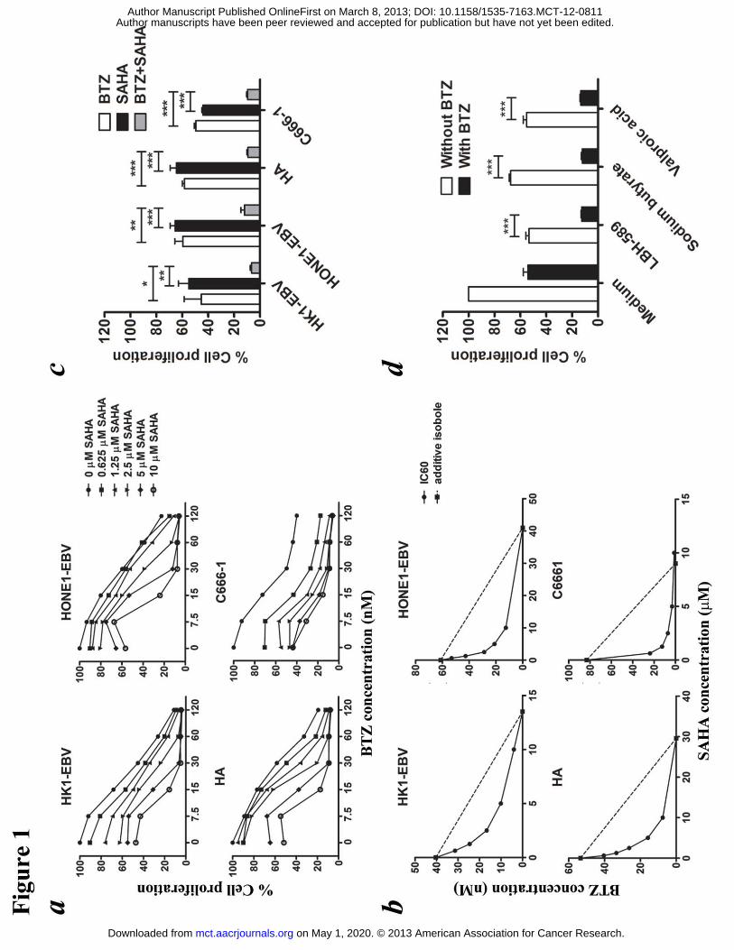
Bortezomib and SAHA inhibited NPC cell proliferation in a synergistic manner
HK1-EBV, HONE1-EBV, HA and C666-1 cells were treated with various
combinations of bortezomib (0, 7.5, 15, 30, 60 and 120 nM) and SAHA (0, 0.625,
1.25, 2.5, 5 and 10 μM) for 48 hr (for 72 hr in the case of C666-1 due to much slower
proliferation rate). The relative cell proliferation was determined by MTT assay and
the dose-response curves are shown in Fig. 1a. While each drug was able to reduce
NPC cell proliferation in a dose-dependent manner, the combination of bortezomib
and SAHA yielded much stronger anti-proliferative effect. The isobolograms of IC60s
are shown in Fig. 1b. The isoboles for IC60 lie below the additive isoboles for all
NPC cell lines, suggesting synergism of bortezomib/SAHA in their anti-proliferative
effects. Combination of 30 nM bortezomib and 5 �M SAHA induced a significantly
stronger inhibition of NPC cell proliferation when compared with either drug alone
(P<0.05 for HK1-EBV, P<0.01 for HONE1-EBV, P<0.001 for HA and C666-1 cells;
Fig. 1c). The combination indices of NPC cells treated with the combination of 30 nM
bortezomib and 5 �M SAHA are 0.3, 0.38, 0.33 and 0.04 for HK1-EBV, HONE1-EBV,
HA and C666-1 cells, respectively, when IC60s are compared. As the combination
indices are all <1, it suggests synergistic anti-proliferative effect. The synergistic
killing of NPC cells could also be observed upon treatment with combination of
bortezomib and other HDAC inhibitors, including LBH-589, sodium butyrate and
valproic acid (Fig. 1d). No synergistic killing was observed in NP460 normal
nasopharyngeal cells and HK2 normal kidney cells (Fig. S1).
Bortezomib/SAHA induced caspase-dependent apoptosis of NPC cells
HA and C666-1 NPC cells were treated with 30 nM bortezomib, 5 �M SAHA or their
combination, for 48 and 72 hr, respectively, and assayed for apoptosis by annexin V/
on May 1, 2020. © 2013 American Association for Cancer Research. mct.aacrjournals.org Downloaded from
Author manuscripts have been peer reviewed and accepted for publication but have not yet been edited. Author Manuscript Published OnlineFirst on March 8, 2013; DOI: 10.1158/1535-7163.MCT-12-0811
14
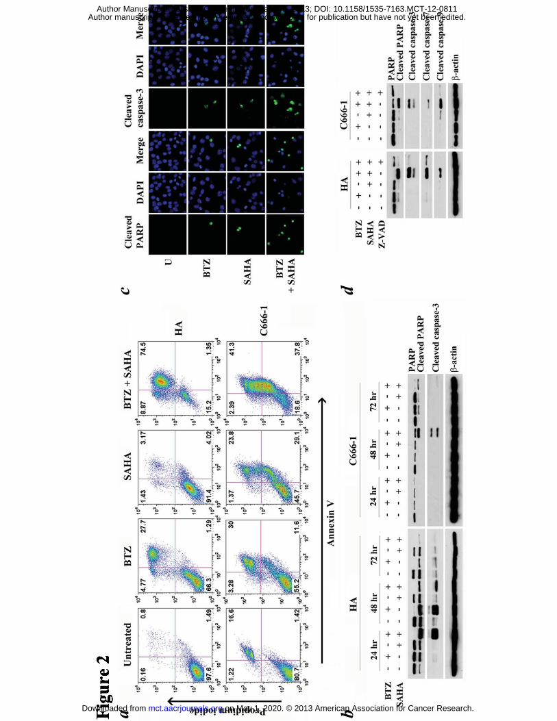
propidium iodide staining. Bortezomib/SAHA induced a higher percentage of
apoptotic cells, when compared with either drug alone, in both NPC cell lines. The
percentages of annexin V-positive populations upon treatment with bortezomib,
SAHA and bortezomib/SAHA increased to 29%, 7% and 76%, respectively in HA
cells and 42%, 53% and 79%, respectively in C666-1 cells (Fig. 2a). To study the
kinetics of apoptosis, HA and C666-1 cells were treated with 30 nM bortezomib, 5
�M SAHA or their combination for different time duration (24, 48 and 72 hr) and
assayed for proteolytic cleavage of poly (ADP-ribose) polymerase (PARP) and
caspase-3 by western blot analysis. Cleavage of PARP and caspase-3 was observed at
an earlier time point upon treatment with both drugs (24 hr in HA cells and 48 hr in
C666-1 cells) when compared with either drug alone (Fig. 2b). Upon treatment with
bortezomib/SAHA, increased number of NPC cells expressing cleaved PARP and
cleaved caspase-3 were also detected by immunofluoresent staining (Fig. 2c). The
apoptosis was caspase-dependent because a pan-caspase inhibitor, Z-VAD-FMK,
significantly suppressed the cleavage of PARP and caspase-3, -7 and -9 induced by
bortezomib/SAHA (Fig. 2d).
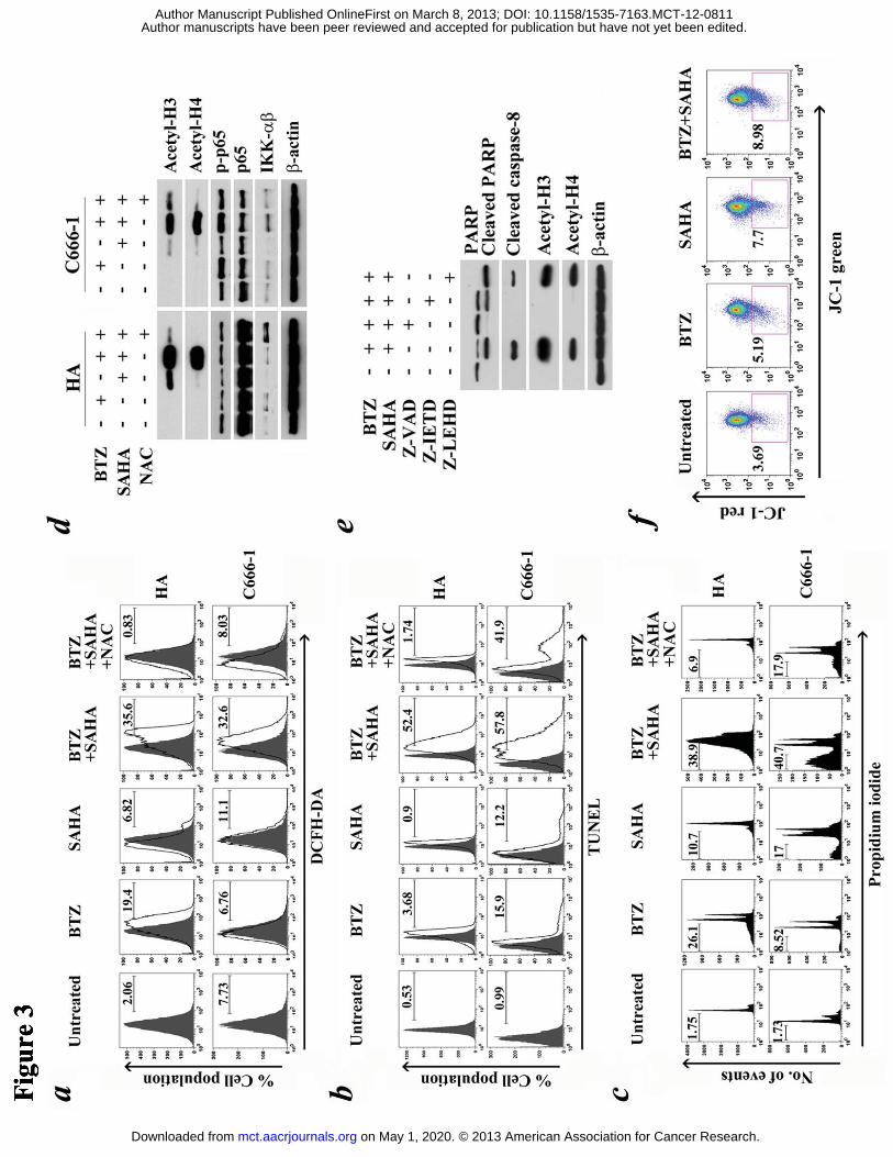
Bortezomib/SAHA’s cytotoxic effect was strongly associated with reactive oxygen
species (ROS)
Bortezomib/SAHA induced ROS generation in a wide variety of cancer cell lines
(13-16, 19). We, therefore, hypothesized that ROS generation were also involved in
bortezomib/SAHA-induced apoptosis in NPC cells. DCFH-DA assay demonstrated
enhanced ROS content in HA and C666-1 cells treated with bortezomib/SAHA at 24
hr and 48 hr post-treatment, respectively. N-acetyl cysteine (NAC), a ROS scavenger,
significantly reduced the generation of ROS (Fig. 3a). NAC also effectively
on May 1, 2020. © 2013 American Association for Cancer Research. mct.aacrjournals.org Downloaded from
Author manuscripts have been peer reviewed and accepted for publication but have not yet been edited. Author Manuscript Published OnlineFirst on March 8, 2013; DOI: 10.1158/1535-7163.MCT-12-0811
15
suppressed apoptosis, as indicated by the reduction of TUNEL-positive and sub-G1
populations in NPC cells upon treatment with bortezomib/SAHA. The
TUNEL-positive NPC cells dropped from 52.4% to 1.74% for HA and from 57.8% to
41.9% for C666-1 (Fig. 3b). The corresponding sub-G1 population dropped from
38.9% to 6.9% for HA and from 40.7% to 17.9% for C666-1 (Fig. 3c). We also
examined other possible mechanisms of cell death that would be induced by
bortezomib/SAHA. While SAHA mildly induced acetylation of histone H3 and H4,
addition of bortezomib resulted in a more significant increase in histone acetylation
(Fig. 3d). Meanwhile, the histone acetylation induced by bortezomib/SAHA reduced
upon co-incubation with either Z-VAD-FMK or NAC, indicating that the histone
acetylation was caspase- and ROS-dependent (Fig. 3d and Fig. S2). Specific
caspase-8 inhibitor Z-IETD-FMK, but not caspase-9 inhibitor Z-LEHD-FMK,
completely reduced the level of acetylated histone and partially reduced the cleavage
of PARP, showing that bortezomib/SAHA-induced cell death was partially related to
caspase-8-dependent histone acetylation (Fig. 3e). NK-κB activation and
mitochondrial membrane potential were shown to be closely associated with ROS
generation (31, 32). However, according to our results, no significant change in
expression of NF-κB-related proteins, including p-p65, p65 and IKK-α/β, was
observed after treatment with combined bortezomib/SAHA when compared with
either drug alone (Fig. 3d). The drug combination also did not result in significant loss
of mitochondrial membrane potential in the bortezomib/SAHA-treated cells (Fig. 3f).
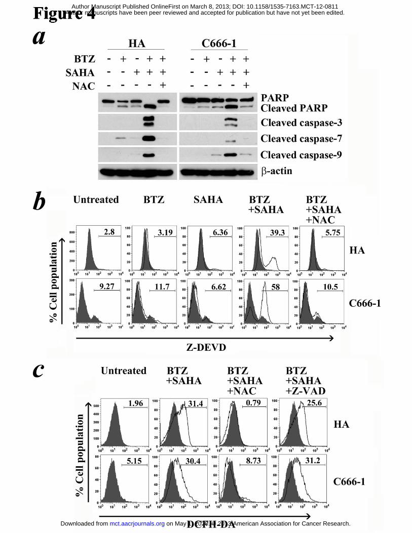
Caspase activation were regulated by ROS signaling in NPC cells
To further investigate the roles of caspases and ROS in NPC apoptosis, we analyzed
the effects of NAC and Z-VAD-FMK on caspase activation and ROS generation
on May 1, 2020. © 2013 American Association for Cancer Research. mct.aacrjournals.org Downloaded from
Author manuscripts have been peer reviewed and accepted for publication but have not yet been edited. Author Manuscript Published OnlineFirst on March 8, 2013; DOI: 10.1158/1535-7163.MCT-12-0811
16
mediated by bortezomib/SAHA. NAC effectively suppressed the cleavage of PARP
and caspase-3, -7 and -9 in both HA and C666-1 cells (Fig. 4a). Moreover, NAC
significantly decreased the percentage of cells containing active caspase-3/-7
following treatment with bortezomib/SAHA (Fig. 4b). The percentages decreased
from 39.3% to 5.75% in HA cells and from 58% to 10.5% in C666-1 cells. On the
other hand, Z-VAD-FMK, which effectively suppressed caspase activation, did not
reduce ROS generation induced by bortezomib/SAHA (Fig. 2d & 4c). These suggest
ROS induction by bortezomib/SAHA led to subsequent caspase activation in NPC
cells.
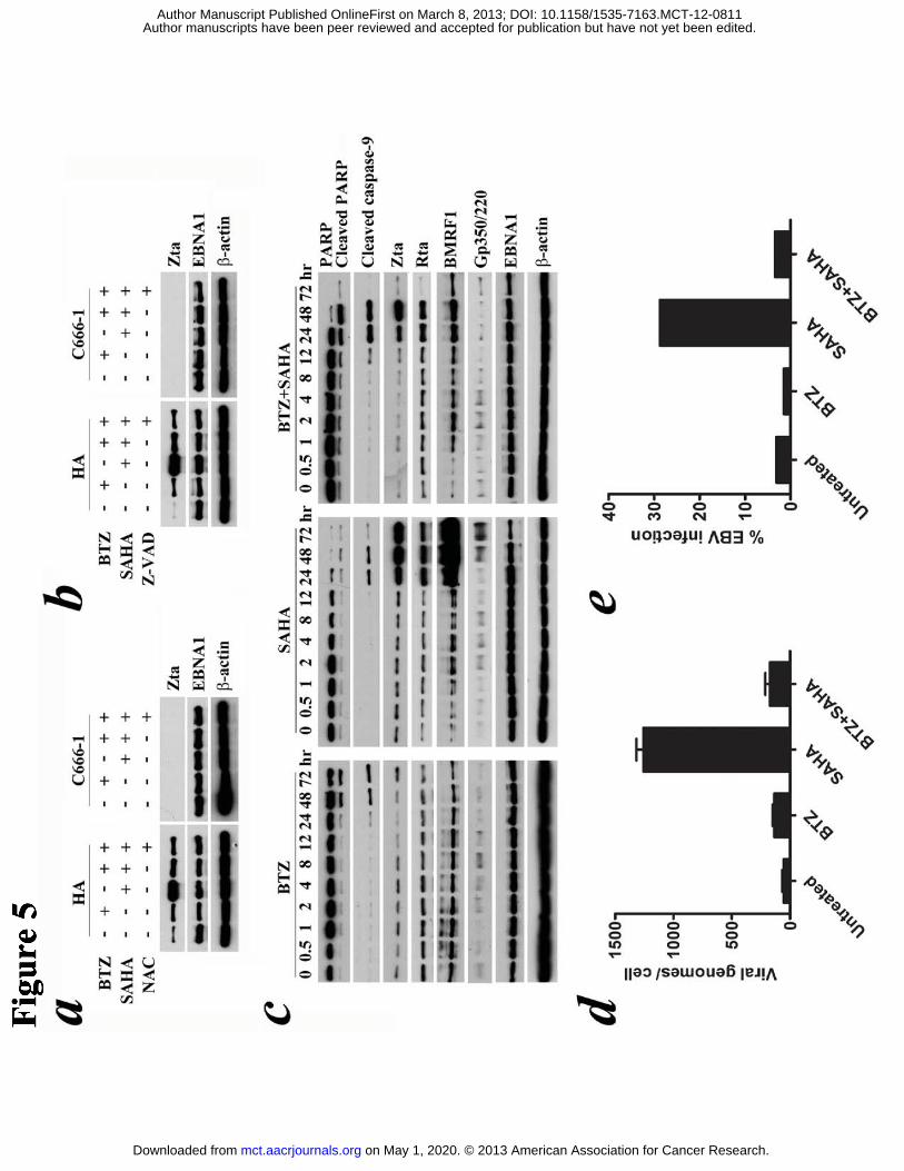
Bortezomib inhibited SAHA’s induction of EBV lytic protein expression and
abrogated production of infectious viral particles in NPC cells
Induction of EBV lytic cycle could lead to apoptosis of EBV-positive NPC cells (12).
We, therefore, investigated the expression of EBV immediate-early lytic (Zta) and
latent (EBNA1) proteins in HA and C666-1 cells after treatment with
bortezomib/SAHA for 24 hr and 48 hr, respectively. As expected, SAHA significantly
induced Zta expression in HA cells but not in C666-1 cells (in which Zta could only
be detected after treatment with SAHA for 3 days) (12) at these time points (Fig. 5a &
b). Addition of bortezomib effectively reduced the expression of SAHA-induced Zta
in HA cells (Fig. 5a & b). Time course experiment showed that bortezomib/SAHA
induced proteolytic cleavage of PARP and caspase-9 at an earlier time point (12 hr
post-treatment) when compared with either drug alone (24 hr post-treatment) (Fig. 5c).
Apoptosis was triggered at 12 hr post-treatment while expression of Zta was observed
later at ~24 hr post-treatment (Fig. 5c). Expression of EBV early lytic protein,
BMRF1, was suppressed to a greater extent than that of Zta protein and expression of
on May 1, 2020. © 2013 American Association for Cancer Research. mct.aacrjournals.org Downloaded from
Author manuscripts have been peer reviewed and accepted for publication but have not yet been edited. Author Manuscript Published OnlineFirst on March 8, 2013; DOI: 10.1158/1535-7163.MCT-12-0811
17
EBV late lytic protein, gp350/220, was totally abrogated after treatment with
bortezomib/SAHA when compared with SAHA alone (Fig. 5c). Bortezomib could
also abrogate SAHA-induced EBV DNA replication and infectious virus production.
At 48 hr post-treatment, the viral load was 1258 genomes per cell in HA treated with
SAHA and it dropped to 172 genomes per cell when HA was co-incubated with
bortezomib (Fig. 5d). The effect cannot be observed in C666-1 as SAHA was not
capable of activating EBV DNA replication in C666-1 as reported previously (data
not shown) (12). Supernatants from HONE1-EBV treated with SAHA could
effectively infect ~29% Daudi cells with EBV. However, when bortezomib was added,
only ~3% Daudi cells were infected with EBV (Fig. 5e). Moreover, the synergistic
killing by bortezomib/SAHA could be observed similarly in both EBV-negative and
EBV-positive NPC cells (Fig. S3). These data suggest that the synergism of
bortezomib/SAHA on apoptosis of NPC cells was independent of the presence of EBV.
However, bortezomib effectively abrogated SAHA-induced viral replication while
potentiating SAHA’s induction of apoptosis of NPC cells.
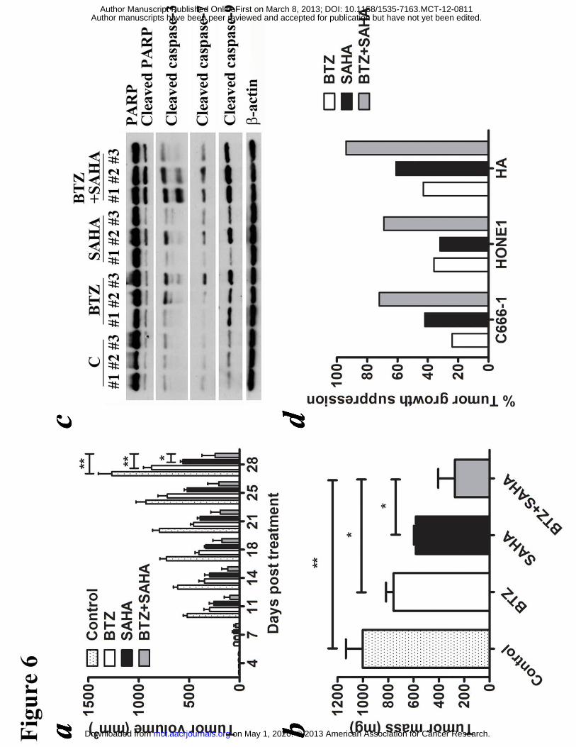
Bortezomib/SAHA significantly suppressed NPC tumor growth in vivo
We evaluated the in vivo effect of bortezomib/SAHA on growth suppression of NPC
xenografts established in nude mice. C666-1, HONE1 and HA cells were inoculated
subcutaneously at the right flanks of nude mice. The mice (n=5) were either treated
with DMSO (vehicle control), 60 �g/ml bortezomib, 50 mg/kg SAHA or combination
of them for 5 days per week over 4 weeks by intraperitoneal injection. Growth of
tumors was measured twice weekly during the experimental period. While either
bortezomib or SAHA alone suppressed the growth of NPC tumors, their combination
mediated a much stronger anti-tumor effect (Fig. 6 and Fig. S4). For instance, on day
on May 1, 2020. © 2013 American Association for Cancer Research. mct.aacrjournals.org Downloaded from
Author manuscripts have been peer reviewed and accepted for publication but have not yet been edited. Author Manuscript Published OnlineFirst on March 8, 2013; DOI: 10.1158/1535-7163.MCT-12-0811
18
28, the tumor mass of C666-1 in the control group increased to 1000 mg. The mass of
tumors treated with bortezomib and SAHA increased to 760 mg and 580 mg
respectively, while that treated with drug combination gave a much smaller increase
in tumor mass to 270 mg (Fig. 6b). Western blot analysis showed that
bortezomib/SAHA could induce a stronger proteolytic cleavage of PARP and caspases
in the tumors when compared with either drug alone (Fig. 6c). Similar anti-tumor
effects of bortezomib/SAHA could also be observed in HONE1 and HA xenografts
(Fig. 6d and S4). The weight of mice was recorded throughout the experiments.
Weight loss of ~5% was observed in mice treated with either the single drugs or their
combination (Fig. S4). No significant increase in weight loss was observed in the
mice treated with the combined drug regimen. Based on the observations in all three
mouse tumor models (C666-1, HONE1 and HA), the toxicity (generally mild) seems
to be similar for either the single drugs or their combination.
on May 1, 2020. © 2013 American Association for Cancer Research. mct.aacrjournals.org Downloaded from
Author manuscripts have been peer reviewed and accepted for publication but have not yet been edited. Author Manuscript Published OnlineFirst on March 8, 2013; DOI: 10.1158/1535-7163.MCT-12-0811
19
Discussion
We demonstrated that combination of bortezomib and SAHA could synergistically
inhibit proliferation of all four EBV-positive NPC cell lines employed in this study.
The synergistic killing was specific to NPC when compared with normal
nasopharyngeal and kidney cells and was mediated by increased apoptosis. We
showed that bortezomib/SAHA could significantly induce activation of caspase-3, -7
and -9 while addition of caspase inhibitor, Z-VAD-FMK, could effectively reduce the
cleavage of the caspases and PARP, suggesting a caspase-dependent apoptosis of the
NPC cells. We also observed a significant increase in ROS generation in NPC cells
after treatment with bortezomib/SAHA. The ROS generation likely played a critical
role in the induction of apoptosis of NPC cells because N-acetyl cysteine (NAC), a
ROS scavenger, could markedly reduce the sub-G1 cell populations, in line with
previously reported function of ROS generation in various cancer cell types upon
treatment with bortezomib/SAHA (13-16, 19). We sought to clarify the relationship
between ROS generation and caspase-dependent apoptosis in the NPC cells as
conflicting data on the link between ROS generation and caspase activation have been
reported in the literature (33, 34). Our data demonstrated that ROS generation resulted
in caspase activation and subsequent apoptosis of NPC cells because NAC effectively
reduced caspase activation while Z-VAD-FMK failed to reduce ROS production.
We have also investigated the potential roles of several signaling pathways which
could mediate apoptosis in the NPC cells upon treatment by bortezomib/SAHA.
Bortezomib was found to potentiate SAHA’s acetylation of histones H3 and H4 in the
NPC cells. Furthermore, the histone acetylation was ROS- and caspase-8-dependent
since both NAC and caspase-8 specific inhibitor, Z-IETD-FMK could markedly
on May 1, 2020. © 2013 American Association for Cancer Research. mct.aacrjournals.org Downloaded from
Author manuscripts have been peer reviewed and accepted for publication but have not yet been edited. Author Manuscript Published OnlineFirst on March 8, 2013; DOI: 10.1158/1535-7163.MCT-12-0811
20
reduce the acetylation of the histones. The results were similar to the induction of
caspase-8-dependent histone acetylation by combination of HDAC and proteasome
inhibitors in leukemic cells (21). One of the major effects mediated by histone
hyperacetylation is up-regulation of tumor suppressor genes (9). However, we did not
find any up-regulation of Rb or p53 in the bortezomib/SAHA-treated NPC cells (refer
to Fig. S2a). p53 expression was repressed by the combination treatment in HA cells
whereas such repression was not found in C666-1 cells. Since enhanced apoptosis was
observed in both HA and C666-1 cells, the cell death was unlikely related to the p53
pathway. It is unexpected that NF-κB inactivation, which was seen in vitro in various
cancer cell lines after treatment with bortezomib/SAHA, was not observed in the NPC
cells. However, this finding is in line with that in a clinical study on patients with
multiple myeloma where the clinical response to bortezomib/SAHA did not correlate
to the expression pattern of NF-κB p65 subunit protein in the bone marrow (35). Loss
of mitochondrial membrane potential was not significantly detected in the
bortezomib/SAHA-treated NPC cells, which was consistent with the finding that
caspase-9 specific inhibitor, Z-LEHD-FMK, could not suppress the apoptosis of NPC
cells. We postulated that the apoptosis was not mediated through the intrinsic
mitochondrial pathway but through the non-mitochondrial production of ROS via the
NADPH oxidase complex and endoplasmic reticulum system (32).
Our laboratory demonstrated that NPC cells proceeded to apoptosis following EBV
lytic cycle induction by SAHA (12). In this study, we critically investigated the
expression kinetics of EBV lytic proteins (Zta, Rta, BMRF1 and gp350/220) and
apoptotic markers (PARP and cleaved caspase-9) after treatment with either
bortezomib or SAHA alone or bortezomib/SAHA in the NPC cells. We found that
on May 1, 2020. © 2013 American Association for Cancer Research. mct.aacrjournals.org Downloaded from
Author manuscripts have been peer reviewed and accepted for publication but have not yet been edited. Author Manuscript Published OnlineFirst on March 8, 2013; DOI: 10.1158/1535-7163.MCT-12-0811
21
bortezomib/SAHA induced apoptosis of NPC cells at an earlier time point than either
drug alone and bortezomib reduced SAHA’s induction of EBV lytic cycle. Moreover,
cleavage of caspase-9 and PARP was detected much earlier than the expression of
EBV lytic proteins, indicating that bortezomib/SAHA potently and briskly induced
cell death through apoptotic pathways independent of EBV lytic cycle activation.
Interestingly, this drug combination will also serve to minimize the production of this
oncogenic virus from the NPC cells.
We examined the in vivo effect of bortezomib/SAHA in NPC xenografts in nude mice.
Our results demonstrated that combination of 60 μg/kg bortezomib and 50 mg/kg
SAHA could suppress the growth of NPC tumors, including C666-1, HONE1 and HA,
in a more-than-additive manner, when compared with either drug alone. Take C666-1
as an example, while bortezomib or SAHA resulted in reduction of tumor mass by
24% (P<0.05) and 42% (P<0.05), respectively, bortezomib/SAHA resulted in
reduction of tumor mass by 72% (P<0.01) when compared with the vehicle treated
group. Increased proteolytic cleavage of PARP and caspases was also observed in the
NPC xenografts. Of note, no apparent toxicity was observed in the mice treated with
the drug combination except weight loss of ~5%. Together, these in vivo data
complemented our in vitro data on the augmented anti-tumor effects of
bortezomib/SAHA in NPC over those of either bortezomib or SAHA alone. Indeed a
clinical study demonstrated possible efficacy of combined bortezomib and SAHA in
the treatment of relapsed and refractory multiple myeloma with acceptable toxicities
(35).
In addition to synergistic anti-tumor effect, combination of bortezomib and SAHA
on May 1, 2020. © 2013 American Association for Cancer Research. mct.aacrjournals.org Downloaded from
Author manuscripts have been peer reviewed and accepted for publication but have not yet been edited. Author Manuscript Published OnlineFirst on March 8, 2013; DOI: 10.1158/1535-7163.MCT-12-0811
22
could potentially reduce the side effects of either drug alone. Despite its efficacy in
multiple myeloma, 64% of patients receiving bortezomib experienced peripheral
neuropathy which is a dose-limiting factor of this drug in the treatment of this disease
(29, 36). The peripheral neuropathy is due to accumulation of ubiquitinated proteins
in neuron cells after inhibition of proteasome by bortezomib. Recent studies have
shown that SAHA can reduce bortezomib-induced neuropathy by increasing Schwann
cell autophagy (37). Radiotherapy is the mainstay of treatment for NPC patients and is
thought to confers cytotoxicity through ROS generation (3). Since bortezomib/SAHA
can enhance radiosensitivity in both gliomas and colorectal cancers (38, 39),
co-adminstration of the drug combination with radiotherapy could potentially result in
enhanced therapeutic effect.
In summary, combination of bortezomib and SAHA synergistically induced killing of
NPC cells. The major mechanism of cell death is ROS-driven caspase-dependent
apoptosis. Furthermore, bortezomib could effectively abrogate SAHA-induced EBV
replication in NPC cells. In vivo, bortezomib/SAHA potently induced apoptosis and
suppressed the growth of NPC xenografts in nude mice. Taken together, our study
provides a strong basis to progress to clinical testing of this drug combination regimen
in NPC patients.
Acknowledgements
We thank Prof. CH Tsai and Prof. JY Chen for providing the HA cell line and Prof.
GCF Chan for the HK2 cell line. We thank Prof. PJ Farrell, Prof. KH Chan and Prof.
JM Middeldorp for providing the EBV antibodies. We also thank Dr. AKL Cheung
and Dr. ST Cheung for their advice on nude mice experiments. We are also grateful to
on May 1, 2020. © 2013 American Association for Cancer Research. mct.aacrjournals.org Downloaded from
Author manuscripts have been peer reviewed and accepted for publication but have not yet been edited. Author Manuscript Published OnlineFirst on March 8, 2013; DOI: 10.1158/1535-7163.MCT-12-0811
23
Prof. ML Lung and Prof. KW Lo for their help in authentication of cell lines.
on May 1, 2020. © 2013 American Association for Cancer Research. mct.aacrjournals.org Downloaded from
Author manuscripts have been peer reviewed and accepted for publication but have not yet been edited. Author Manuscript Published OnlineFirst on March 8, 2013; DOI: 10.1158/1535-7163.MCT-12-0811
24
References:
1. Jia WH, Huang QH, Liao J, Ye W, Shugart YY, Liu Q, et al. Trends in incidence and mortality of nasopharyngeal carcinoma over a 20-25 year period (1978/1983-2002) in Sihui and Cangwu counties in southern China. BMC Cancer. 2006;6:178. 2. Young LS, Rickinson AB. Epstein-Barr virus: 40 years on. Nat Rev Cancer. 2004;4:757-68. 3. Shi L, Fang J. Implication of heme oxygenase-1 in the sensitivity of nasopharyngeal carcinomas to radiotherapy. J Exp Clin Cancer Res. 2008;27:13. 4. Posner MR, Hershock DM, Blajman CR, Mickiewicz E, Winquist E, Gorbounova V, et al. Cisplatin and fluorouracil alone or with docetaxel in head and neck cancer. N Engl J Med. 2007;357:1705-15. 5. Chan AT. Nasopharyngeal carcinoma. Ann Oncol. 2010;21 Suppl 7:vii308-12. 6. Moreau P, Richardson PG, Cavo M, Orlowski RZ, San Miguel JF, Palumbo A, et al. Proteasome inhibitors in multiple myeloma: 10 years later. Blood. 2012;120:947-59. 7. Ling YH, Liebes L, Zou Y, Perez-Soler R. Reactive oxygen species generation and mitochondrial dysfunction in the apoptotic response to Bortezomib, a novel proteasome inhibitor, in human H460 non-small cell lung cancer cells. J Biol Chem. 2003;278:33714-23. 8. Adams J. The proteasome: a suitable antineoplastic target. Nat Rev Cancer. 2004;4:349-60. 9. Marks PA. The mechanism of the anti-tumor activity of the histone deacetylase inhibitor, suberoylanilide hydroxamic acid (SAHA). Cell Cycle. 2004;3:534-5. 10. Marks PA. Discovery and development of SAHA as an anticancer agent. Oncogene. 2007;26:1351-6. 11. Hui KF, Chiang AK. Suberoylanilide hydroxamic acid induces viral lytic cycle in Epstein-Barr virus-positive epithelial malignancies and mediates enhanced cell death. Int J Cancer. 2010;126:2479-89. 12. Hui KF, Ho DN, Tsang CM, Middeldorp JM, Tsao GS, Chiang AK. Activation of lytic cycle of Epstein-Barr virus by suberoylanilide hydroxamic acid leads to apoptosis and tumor growth suppression of nasopharyngeal carcinoma. Int J Cancer. 2012;131:1930-40. 13. Pei XY, Dai Y, Grant S. Synergistic induction of oxidative injury and apoptosis in human multiple myeloma cells by the proteasome inhibitor bortezomib and histone deacetylase inhibitors. Clin Cancer Res. 2004;10:3839-52. 14. Heider U, von Metzler I, Kaiser M, Rosche M, Sterz J, Rotzer S, et al. Synergistic interaction of the histone deacetylase inhibitor SAHA with the proteasome
on May 1, 2020. © 2013 American Association for Cancer Research. mct.aacrjournals.org Downloaded from
Author manuscripts have been peer reviewed and accepted for publication but have not yet been edited. Author Manuscript Published OnlineFirst on March 8, 2013; DOI: 10.1158/1535-7163.MCT-12-0811
25
inhibitor bortezomib in mantle cell lymphoma. Eur J Haematol. 2008;80:133-42. 15. Heider U, Rademacher J, Lamottke B, Mieth M, Moebs M, von Metzler I, et al. Synergistic interaction of the histone deacetylase inhibitor SAHA with the proteasome inhibitor bortezomib in cutaneous T cell lymphoma. Eur J Haematol. 2009;82:440-9. 16. Yu C, Rahmani M, Conrad D, Subler M, Dent P, Grant S. The proteasome inhibitor bortezomib interacts synergistically with histone deacetylase inhibitors to induce apoptosis in Bcr/Abl+ cells sensitive and resistant to STI571. Blood. 2003;102:3765-74. 17. Zhang QL, Wang L, Zhang YW, Jiang XX, Yang F, Wu WL, et al. The proteasome inhibitor bortezomib interacts synergistically with the histone deacetylase inhibitor suberoylanilide hydroxamic acid to induce T-leukemia/lymphoma cells apoptosis. Leukemia. 2009;23:1507-14. 18. Emanuele S, Lauricella M, Carlisi D, Vassallo B, D'Anneo A, Di Fazio P, et al. SAHA induces apoptosis in hepatoma cells and synergistically interacts with the proteasome inhibitor Bortezomib. Apoptosis. 2007;12:1327-38. 19. Denlinger CE, Rundall BK, Jones DR. Proteasome inhibition sensitizes non-small cell lung cancer to histone deacetylase inhibitor-induced apoptosis through the generation of reactive oxygen species. J Thorac Cardiovasc Surg. 2004;128:740-8. 20. Sato A, Asano T, Ito K, Sumitomo M. Suberoylanilide hydroxamic acid (SAHA) combined with bortezomib inhibits renal cancer growth by enhancing histone acetylation and protein ubiquitination synergistically. BJU Int. 2012;109:1258-68. 21. Miller CP, Rudra S, Keating MJ, Wierda WG, Palladino M, Chandra J. Caspase-8 dependent histone acetylation by a novel proteasome inhibitor, NPI-0052: a mechanism for synergy in leukemia cells. Blood. 2009;113:4289-99. 22. Nawrocki ST, Carew JS, Pino MS, Highshaw RA, Andtbacka RH, Dunner K, Jr., et al. Aggresome disruption: a novel strategy to enhance bortezomib-induced apoptosis in pancreatic cancer cells. Cancer Res. 2006;66:3773-81. 23. Feng W-h, Kenney SC. Valproic Acid Enhances the Efficacy of Chemotherapy in EBV-Positive Tumors by Increasing Lytic Viral Gene Expression. Cancer Res %R 101158/0008-5472CAN-06-1006. 2006;66:8762-9. 24. Wildeman MA, Novalic Z, Verkuijlen SA, Juwana H, Huitema AD, Tan IB, et al. Cytolytic virus activation therapy for epstein-barr virus-driven tumors. Clin Cancer Res. 2012;18:5061-70. 25. Fu DX, Tanhehco Y, Chen J, Foss CA, Fox JJ, Chong JM, et al. Bortezomib-induced enzyme-targeted radiation therapy in herpesvirus-associated tumors. Nat Med. 2008;14:1118-22. 26. Li HM, Man C, Jin Y, Deng W, Yip YL, Feng HC, et al. Molecular and cytogenetic changes involved in the immortalization of nasopharyngeal epithelial
on May 1, 2020. © 2013 American Association for Cancer Research. mct.aacrjournals.org Downloaded from
Author manuscripts have been peer reviewed and accepted for publication but have not yet been edited. Author Manuscript Published OnlineFirst on March 8, 2013; DOI: 10.1158/1535-7163.MCT-12-0811
26
cells by telomerase. Int J Cancer. 2006;119:1567-76. 27. Ryan MJ, Johnson G, Kirk J, Fuerstenberg SM, Zager RA, Torok-Storb B. HK-2: an immortalized proximal tubule epithelial cell line from normal adult human kidney. Kidney Int. 1994;45:48-57. 28. McDonald JT, Kim K, Norris AJ, Vlashi E, Phillips TM, Lagadec C, et al. Ionizing radiation activates the Nrf2 antioxidant response. Cancer Res. 2010;70:8886-95. 29. Tallarida RJ. An overview of drug combination analysis with isobolograms. J Pharmacol Exp Ther. 2006;319:1-7. 30. Chou TC, Talalay P. Analysis of Combined Drug Effects - a New Look at a Very Old Problem. Trends Pharmacol Sci. 1983;4:450-4. 31. Bhalla S, Balasubramanian S, David K, Sirisawad M, Buggy J, Mauro L, et al. PCI-24781 induces caspase and reactive oxygen species-dependent apoptosis through NF-kappaB mechanisms and is synergistic with bortezomib in lymphoma cells. Clin Cancer Res. 2009;15:3354-65. 32. Trachootham D, Alexandre J, Huang P. Targeting cancer cells by ROS-mediated mechanisms: a radical therapeutic approach? Nat Rev Drug Discov. 2009;8:579-91. 33. Wang Y, He QY, Sun RW, Che CM, Chiu JF. GoldIII porphyrin 1a induced apoptosis by mitochondrial death pathways related to reactive oxygen species. Cancer Res. 2005;65:11553-64. 34. Choi K, Ryu SW, Song S, Choi H, Kang SW, Choi C. Caspase-dependent generation of reactive oxygen species in human astrocytoma cells contributes to resistance to TRAIL-mediated apoptosis. Cell Death Differ. 2010;17:833-45. 35. Badros A, Burger AM, Philip S, Niesvizky R, Kolla SS, Goloubeva O, et al. Phase I study of vorinostat in combination with bortezomib for relapsed and refractory multiple myeloma. Clin Cancer Res. 2009;15:5250-7. 36. Schiff D. Neurological side-effects caused by recently approved chemotherapy drugs Cancerworld. 2010. 37. Watanabe T, Nagase K, Chosa M, Tobinai K. Schwann cell autophagy induced by SAHA, 17-AAG, or clonazepam can reduce bortezomib-induced peripheral neuropathy. Br J Cancer. 2010;103:1580-7. 38. Labussiere M, Pinel S, Vandamme M, Plenat F, Chastagner P. Radiosensitizing properties of bortezomib depend on therapeutic schedule. Int J Radiat Oncol Biol Phys. 2011;79:892-900. 39. Folkvord S, Ree AH, Furre T, Halvorsen T, Flatmark K. Radiosensitization by SAHA in experimental colorectal carcinoma models-in vivo effects and relevance of histone acetylation status. Int J Radiat Oncol Biol Phys. 2009;74:546-52.
on May 1, 2020. © 2013 American Association for Cancer Research. mct.aacrjournals.org Downloaded from
Author manuscripts have been peer reviewed and accepted for publication but have not yet been edited. Author Manuscript Published OnlineFirst on March 8, 2013; DOI: 10.1158/1535-7163.MCT-12-0811
27
Figure legends:
Figure 1. Effects of bortezomib/SAHA on cell proliferation of EBV-positive NPC
cells. HK1-EBV, HONE1-EBV and HA cells were treated with various combinations
of BTZ and SAHA for 48 hr. C666-1 cells were treated for 72 hr. (A) Data are
presented as percentages of cell proliferation as determined by MTT assays. (B)
Synergisms of proliferation inhibition of different cell lines were analyzed by
isobologram analysis. (C) Percentages of cell proliferation of NPC cells upon
treatment with combination of 30 nM BTZ and 5 μM SAHA were compared to those
treated with either drug alone. (D) Percentages of cell proliferation of HA upon
treatment with combination of 30 nM BTZ and HDAC inhibitors including 100 nM
LBH-689, 3 mM sodium butyrate and 5 mM valproic acid for 48 hr were determined.
The results were analyzed for statistical significance using unpaired student’s t-test. P
value < 0.05 was considered statistically significant; ***p < 0.001, **p < 0.01 and *p
< 0.05. Error bars represent the standard error of mean (SEM) of data obtained in at
least three independent experiments.
Figure 2. Effects of bortezomib/SAHA on apoptosis of EBV-positive NPC cells. (A)
HA cells and C666-1 cells were treated with combination of 30 nM BTZ and 5 μM
SAHA or either drug alone for 48 hr and 72 hr, respectively. The treated cells were
assayed for apoptosis by annexin V/propidium iodide (AV/PI) staining. AV+/PI-
population represents early apoptotic cells while AV+/PI+ population represents late
apoptotic/ necrotic cells. (B) NPC cells were treated with combination of 30 nM BTZ
and 5 μM SAHA or either drug alone for 24, 48 or 72 hr followed by detection of
expression of PARP, cleaved PARP and cleaved caspase-3 by western blot analysis. (C)
After treatment for 24 hr, expression of cleaved caspase-3 and cleaved PARP (green
on May 1, 2020. © 2013 American Association for Cancer Research. mct.aacrjournals.org Downloaded from
Author manuscripts have been peer reviewed and accepted for publication but have not yet been edited. Author Manuscript Published OnlineFirst on March 8, 2013; DOI: 10.1158/1535-7163.MCT-12-0811
28
signals) in HA cells was detected by immunofluorescent staining. DAPI (blue signals)
stained the cell nuclei. (D) HA cells and C666-1 cells were pre-treated with 50 μM
Z-VAD-FMK for 1 hr followed by treatment with combination of 30 nM BTZ and 5
μM SAHA or either drug alone for 24 hr and 48 hr, respectively. Expression of PARP,
cleaved PARP and cleaved caspase-3, -7 and -9 was detected by western blot analysis.
β-actin served as loading control.
Figure 3. Role of reactive oxygen species (ROS) in bortezomib/SAHA-induced
apoptosis. (A) HA cells and C666-1 cells were pre-treated with 1 μM N-acetyl-cystein
(NAC) for 1 hr followed by treatment with combination of 30 nM BTZ and 5 μM
SAHA or either drug alone for 24 hr and 48 hr, respectively. Percentages of cells with
increased ROS level were detected by DCFH-DA assay. (B) Percentages of apoptotic
cells upon drug treatments were detected by TUNEL staining. (C) Cell cycle
distributions of cells upon drug treatments were analyzed by propidium iodide
staining. (D) Expression of acety-H3 and H4, p-p65, p65 and IKK-αβ proteins was
detected by western blotting. (E) Expression of PARP, cleaved caspase-8, acetyl-H3
and acetyl-H4 in HA cells after pre-treatment with either Z-VAD-FMK,
Z-IETD-FMK or Z-LEHD-FMK for 1 hr and treatment with BTZ and SAHA was
detected by western blotting. (F) Decreases in mitochondrial membrane potential in
HA cells were analyzed by JC-1 assay.
Figure 4. Effect of reactive oxygen species (ROS) on activation of caspases. HA and
C666-1 cells were pre-treated with either 1 μM N-acetyl-cystein (NAC) or 50 μM
Z-VAD-FMK for 1 hr followed by treatment with combination of 30 nM bortezomib
and 5 μM SAHA or either drug alone for 24 hr and 48 hr, respectively. (A) Expression
on May 1, 2020. © 2013 American Association for Cancer Research. mct.aacrjournals.org Downloaded from
Author manuscripts have been peer reviewed and accepted for publication but have not yet been edited. Author Manuscript Published OnlineFirst on March 8, 2013; DOI: 10.1158/1535-7163.MCT-12-0811
29
of PARP, cleaved PARP and cleaved caspase-3, -7 and -9 was detected by western blot
analysis. β-actin served as loading control. (B) Percentages of cells with active
caspase-3/-7 were detected by FLICA-Z-DEVD assay. (C) Percentages of cells with
increased ROS level were detected by DCFH-DA assay.
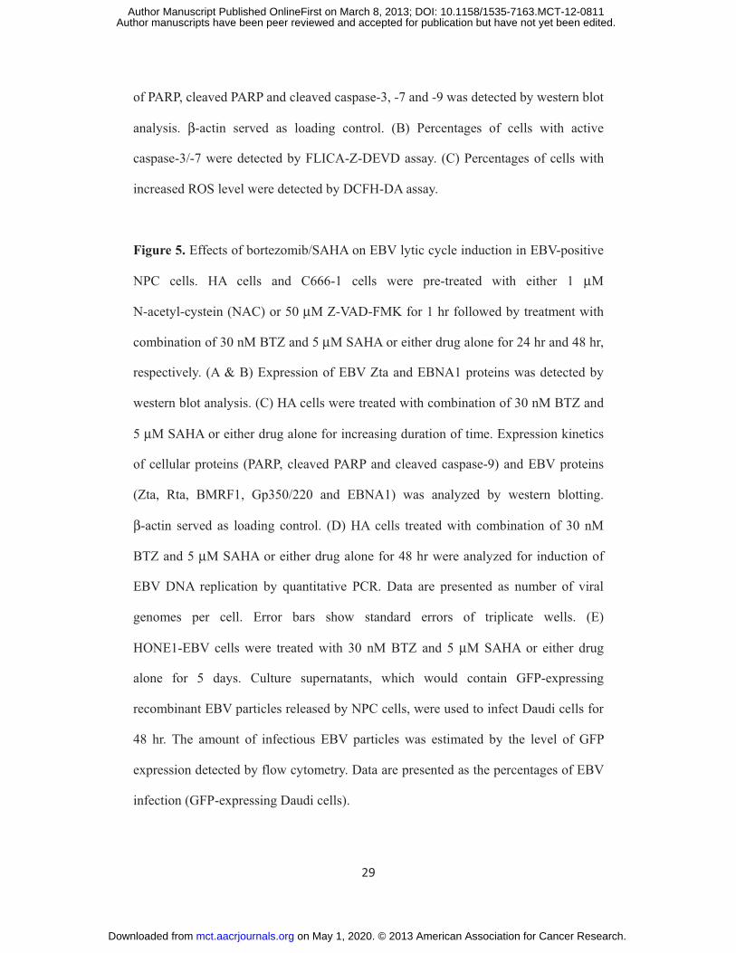
Figure 5. Effects of bortezomib/SAHA on EBV lytic cycle induction in EBV-positive
NPC cells. HA cells and C666-1 cells were pre-treated with either 1 μM
N-acetyl-cystein (NAC) or 50 μM Z-VAD-FMK for 1 hr followed by treatment with
combination of 30 nM BTZ and 5 μM SAHA or either drug alone for 24 hr and 48 hr,
respectively. (A & B) Expression of EBV Zta and EBNA1 proteins was detected by
western blot analysis. (C) HA cells were treated with combination of 30 nM BTZ and
5 μM SAHA or either drug alone for increasing duration of time. Expression kinetics
of cellular proteins (PARP, cleaved PARP and cleaved caspase-9) and EBV proteins
(Zta, Rta, BMRF1, Gp350/220 and EBNA1) was analyzed by western blotting.
β-actin served as loading control. (D) HA cells treated with combination of 30 nM
BTZ and 5 μM SAHA or either drug alone for 48 hr were analyzed for induction of
EBV DNA replication by quantitative PCR. Data are presented as number of viral
genomes per cell. Error bars show standard errors of triplicate wells. (E)
HONE1-EBV cells were treated with 30 nM BTZ and 5 μM SAHA or either drug
alone for 5 days. Culture supernatants, which would contain GFP-expressing
recombinant EBV particles released by NPC cells, were used to infect Daudi cells for
48 hr. The amount of infectious EBV particles was estimated by the level of GFP
expression detected by flow cytometry. Data are presented as the percentages of EBV
infection (GFP-expressing Daudi cells).
on May 1, 2020. © 2013 American Association for Cancer Research. mct.aacrjournals.org Downloaded from
Author manuscripts have been peer reviewed and accepted for publication but have not yet been edited. Author Manuscript Published OnlineFirst on March 8, 2013; DOI: 10.1158/1535-7163.MCT-12-0811
30
Figure 6. Effects of bortezomib/SAHA on tumor growth suppression of NPC
xenografts in nude mice. C666-1 cells were subcutaneously injected into the right
flanks of nude mice. When the tumors were palpable, the mice were either treated
with combination of 60 μg/kg bortezomib and 50 mg/kg SAHA or either drug alone
for 5 days/week over 4 weeks by intraperitoneal injection. (A) The size of tumors
during the period of experiment was measured twice weekly using a caliper. Data are
presented as the mean tumor volumes of mice in both treatment and control groups on
the days post-treatment. (B) Average tumor masses of mice of control and treated
groups were shown. Error bars represent the standard error of mean (SEM) of tumor
masses. (C) Protein samples extracted from the tumors were tested for expression of
PARP, cleaved PARP and cleaved caspase-3, -7 and -9 proteins by western blot
analysis. β-actin served as loading control. (D) The in vivo effects of bortezomib and
SAHA on the percentage of tumor growth suppression of C666-1, HONE1 and HA
xenografts were shown.
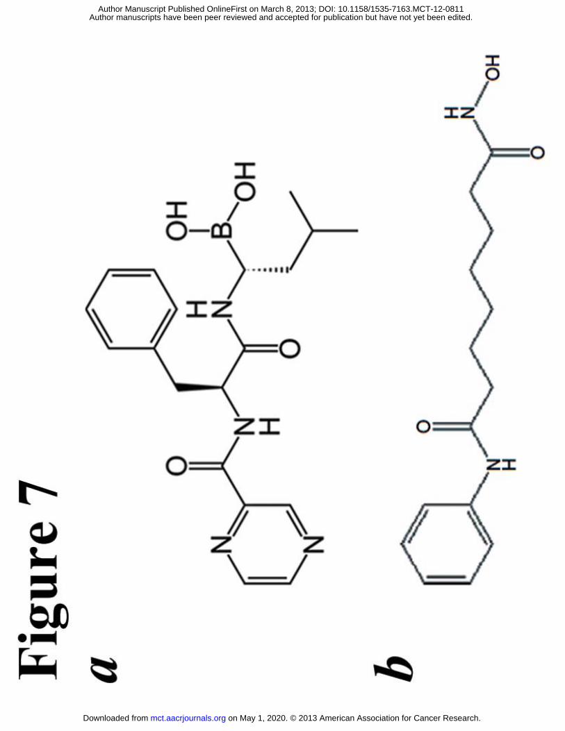
Figure 7. Chemical structures of (A) bortezomib and (B) SAHA.
on May 1, 2020. © 2013 American Association for Cancer Research. mct.aacrjournals.org Downloaded from
Author manuscripts have been peer reviewed and accepted for publication but have not yet been edited. Author Manuscript Published OnlineFirst on March 8, 2013; DOI: 10.1158/1535-7163.MCT-12-0811
on May 1, 2020. © 2013 American Association for Cancer Research. mct.aacrjournals.org Downloaded from
Author manuscripts have been peer reviewed and accepted for publication but have not yet been edited. Author Manuscript Published OnlineFirst on March 8, 2013; DOI: 10.1158/1535-7163.MCT-12-0811
on May 1, 2020. © 2013 American Association for Cancer Research. mct.aacrjournals.org Downloaded from
Author manuscripts have been peer reviewed and accepted for publication but have not yet been edited. Author Manuscript Published OnlineFirst on March 8, 2013; DOI: 10.1158/1535-7163.MCT-12-0811
on May 1, 2020. © 2013 American Association for Cancer Research. mct.aacrjournals.org Downloaded from
Author manuscripts have been peer reviewed and accepted for publication but have not yet been edited. Author Manuscript Published OnlineFirst on March 8, 2013; DOI: 10.1158/1535-7163.MCT-12-0811
on May 1, 2020. © 2013 American Association for Cancer Research. mct.aacrjournals.org Downloaded from
Author manuscripts have been peer reviewed and accepted for publication but have not yet been edited. Author Manuscript Published OnlineFirst on March 8, 2013; DOI: 10.1158/1535-7163.MCT-12-0811
on May 1, 2020. © 2013 American Association for Cancer Research. mct.aacrjournals.org Downloaded from
Author manuscripts have been peer reviewed and accepted for publication but have not yet been edited. Author Manuscript Published OnlineFirst on March 8, 2013; DOI: 10.1158/1535-7163.MCT-12-0811
on May 1, 2020. © 2013 American Association for Cancer Research. mct.aacrjournals.org Downloaded from
Author manuscripts have been peer reviewed and accepted for publication but have not yet been edited. Author Manuscript Published OnlineFirst on March 8, 2013; DOI: 10.1158/1535-7163.MCT-12-0811
on May 1, 2020. © 2013 American Association for Cancer Research. mct.aacrjournals.org Downloaded from
Author manuscripts have been peer reviewed and accepted for publication but have not yet been edited. Author Manuscript Published OnlineFirst on March 8, 2013; DOI: 10.1158/1535-7163.MCT-12-0811
Published OnlineFirst March 8, 2013.Mol Cancer Ther K.F. Hui, Benjamin H.W. Lam, Dona N. Ho, et al. and block replication of Epstein-Barr viruscaspase-dependent apoptosis of nasopharyngeal carcinoma Bortezomib and SAHA synergistically induce ROS-driven
Updated version
10.1158/1535-7163.MCT-12-0811doi:
Access the most recent version of this article at:
Material
Supplementary
http://mct.aacrjournals.org/content/suppl/2013/03/04/1535-7163.MCT-12-0811.DC1
Access the most recent supplemental material at:
Manuscript
Authoredited. Author manuscripts have been peer reviewed and accepted for publication but have not yet been
E-mail alerts related to this article or journal.Sign up to receive free email-alerts
Subscriptions
Reprints and
To order reprints of this article or to subscribe to the journal, contact the AACR Publications
Permissions
Rightslink site. Click on "Request Permissions" which will take you to the Copyright Clearance Center's (CCC)
.http://mct.aacrjournals.org/content/early/2013/03/08/1535-7163.MCT-12-0811To request permission to re-use all or part of this article, use this link
on May 1, 2020. © 2013 American Association for Cancer Research. mct.aacrjournals.org Downloaded from
Author manuscripts have been peer reviewed and accepted for publication but have not yet been edited. Author Manuscript Published OnlineFirst on March 8, 2013; DOI: 10.1158/1535-7163.MCT-12-0811