Embed Size (px)
Citation preview
Bone TumorsClues and CuesBone Tumors
Clues and Cues
William Herring, M.D. © 2002
In Slide Show mode, advance the slides by pressing the spacebarAll Photos Retain the Copyright of their Authors
Clues by Appearance
of Lesion
Patterns of Bone Destruction
Geographic
Moth-eaten
Permeative
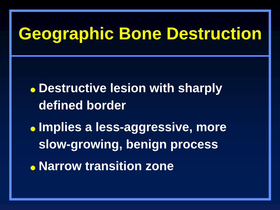
Geographic Bone Destruction
Destructive lesion with sharply defined border
Implies a less-aggressive, more slow-growing, benign process
Narrow transition zone
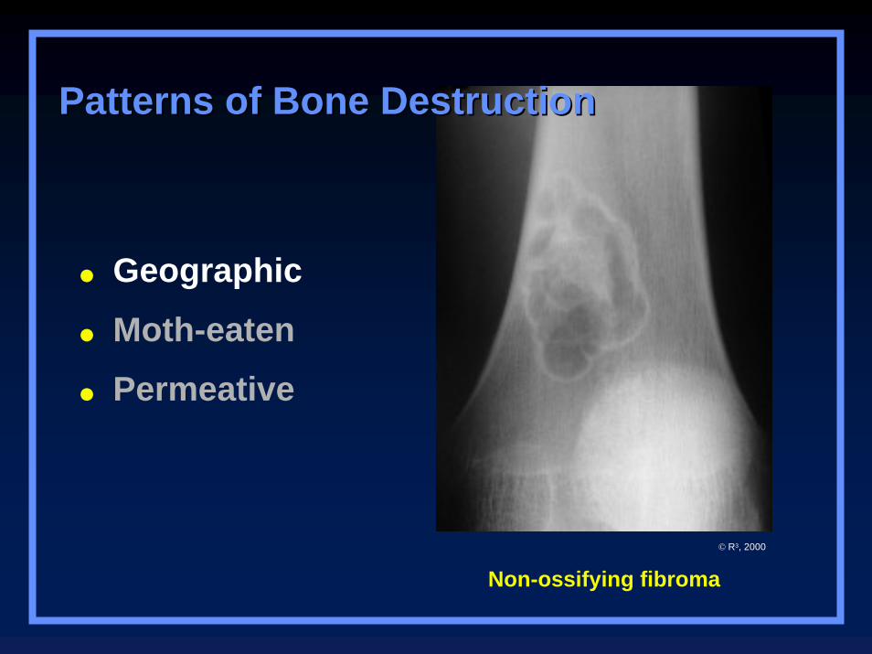
Non-ossifying fibroma
Geographic
Moth-eaten
Permeative
Patterns of Bone DestructionPatterns of Bone Destruction
© R3, 2000
Geographic LesionsExamples
Non-ossifying fibroma
Chondromyxoid fibroma
Eosinophilic granuloma
Moth-eaten Appearance
Areas of destruction with ragged borders
Implies more rapid growthProbably a malignancy
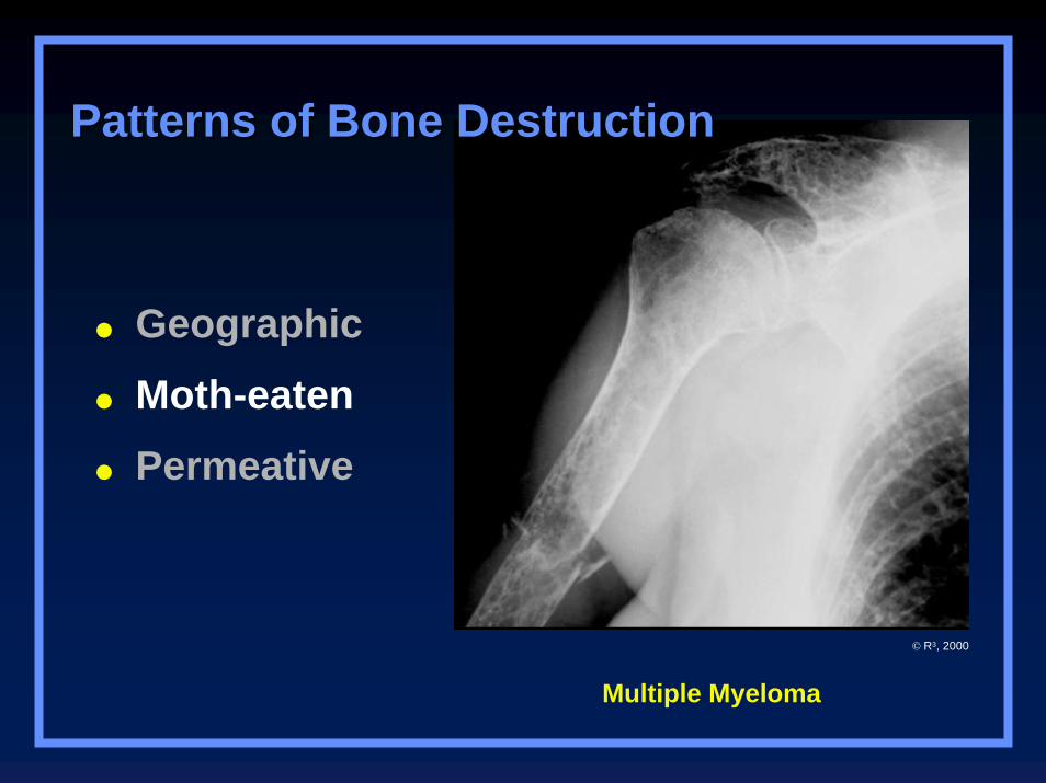
Multiple Myeloma
Patterns of Bone DestructionPatterns of Bone Destruction
Geographic
Moth-eaten
Permeative
© R3, 2000
Moth-eaten AppearanceExamples
Myeloma
Metastases
Lymphoma
Ewing’s sarcoma
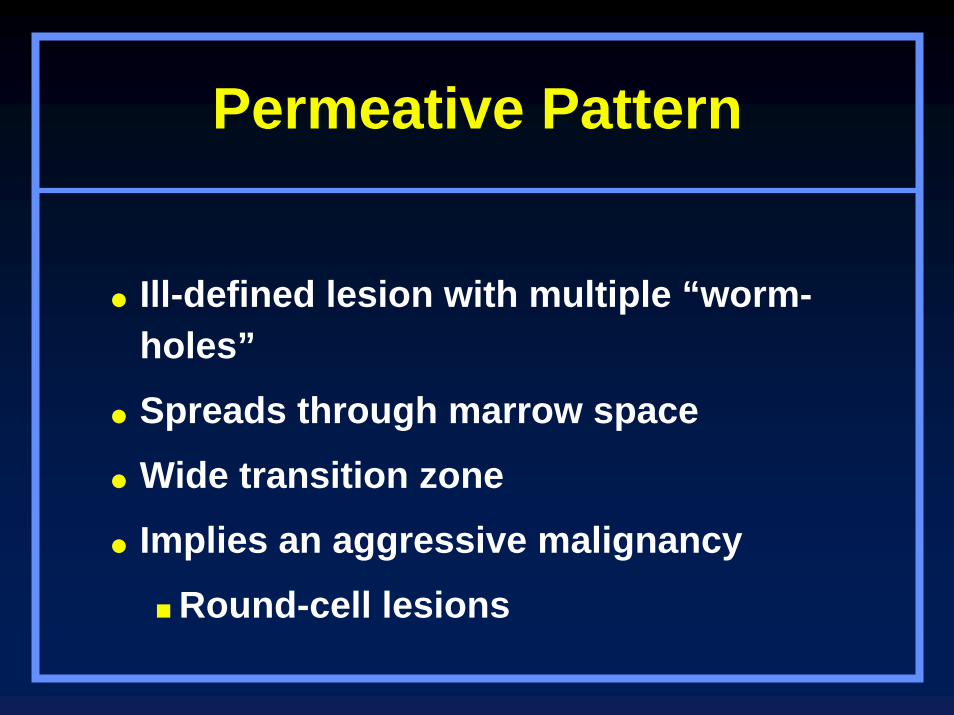
Permeative Pattern
Ill-defined lesion with multiple “worm-holes”
Spreads through marrow space
Wide transition zone
Implies an aggressive malignancy
Round-cell lesions
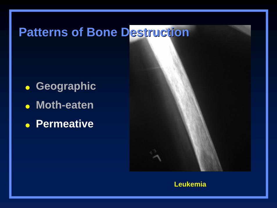
Leukemia
Patterns of Bone DestructionPatterns of Bone Destruction
Geographic
Moth-eaten
Permeative
Permeative PatternRound cell lesions
Lymphoma, leukemia
Ewing’s Sarcoma
Myeloma
Osteomyelitis
Neuroblastoma
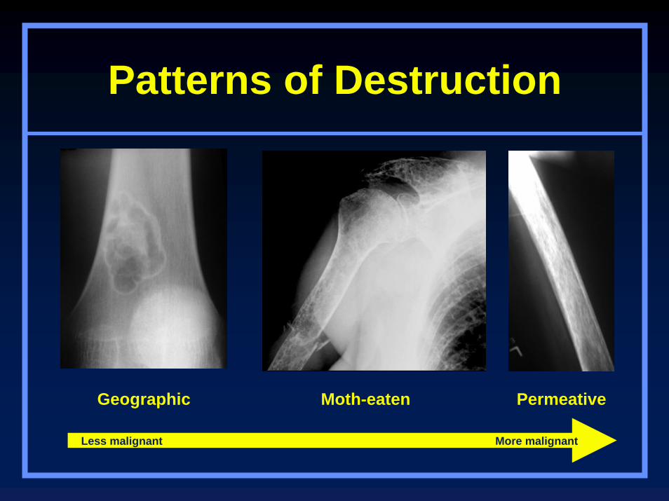
Patterns of Destruction
Geographic Moth-eaten Permeative
Less malignant More malignant

Periosteal Reactions
BenignNoneSolid
More aggressive or malignantLamellated or onion-skinningSunburstCodman’s triangle
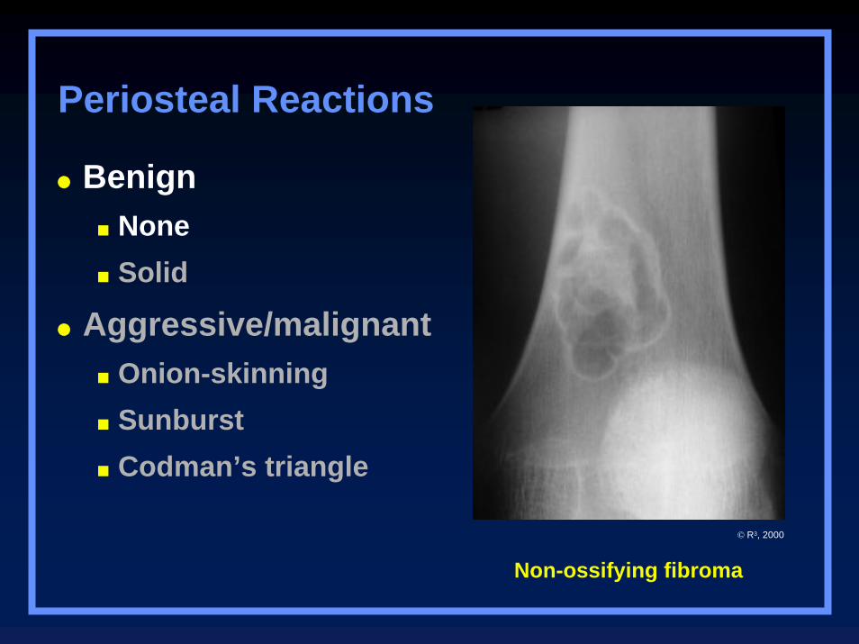
Non-ossifying fibroma
BenignNoneSolid
Aggressive/malignantOnion-skinningSunburstCodman’s triangle
Periosteal Reactions
© R3, 2000
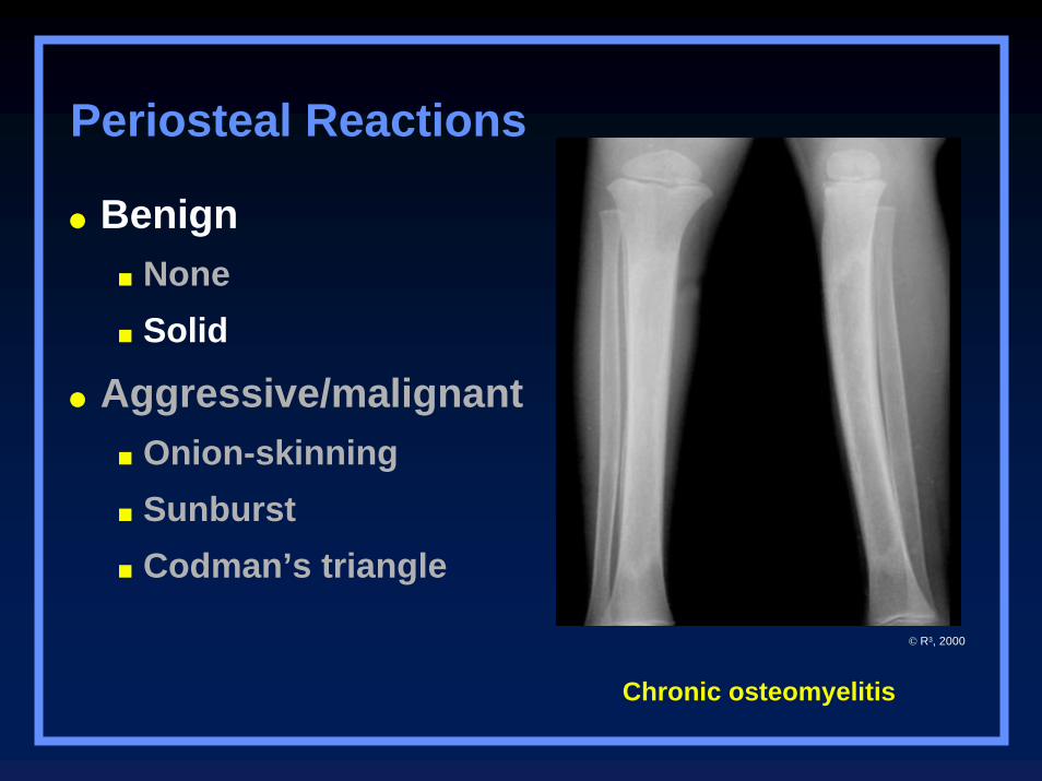
Chronic osteomyelitis
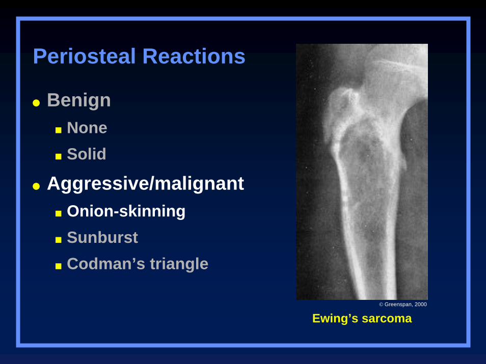
BenignNoneSolid
Aggressive/malignantOnion-skinningSunburstCodman’s triangle
Periosteal Reactions
© R3, 2000
BenignNoneSolid
Aggressive/malignantOnion-skinningSunburstCodman’s triangle
Periosteal Reactions
Ewing’s sarcoma© Greenspan, 2000
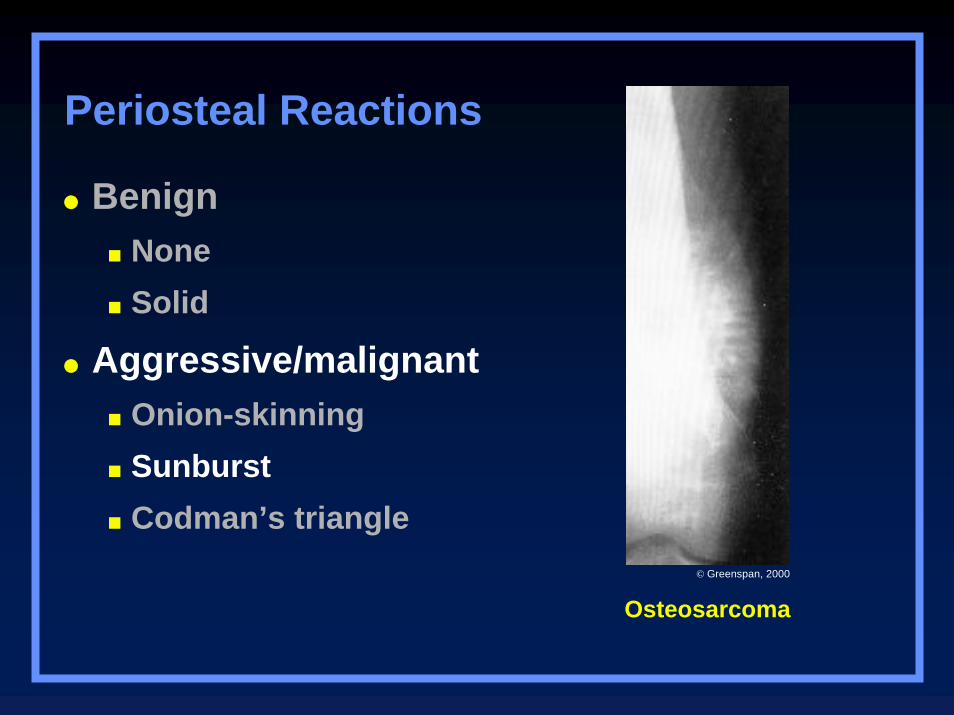
Osteosarcoma
BenignNoneSolid
Aggressive/malignantOnion-skinningSunburstCodman’s triangle
Periosteal Reactions
© Greenspan, 2000
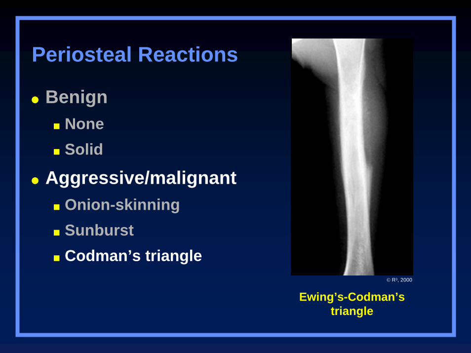
Ewing’s-Codman’striangle
BenignNoneSolid
Aggressive/malignantOnion-skinningSunburstCodman’s triangle
Periosteal Reactions
© R3, 2000
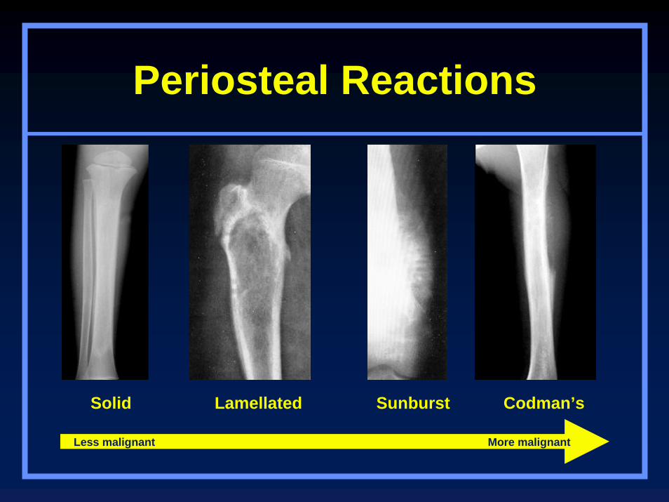
Periosteal Reactions
Solid Lamellated Sunburst Codman’s
Less malignant More malignant
Tumor Matrix
OsteoblasticFluffy, cotton-like or cloud-like densities
Osteosarcoma
CartilaginousComma-shaped, punctate, annular, popcorn-like
Enchondroma, chondrosarcoma, chondromyxoid fibroma
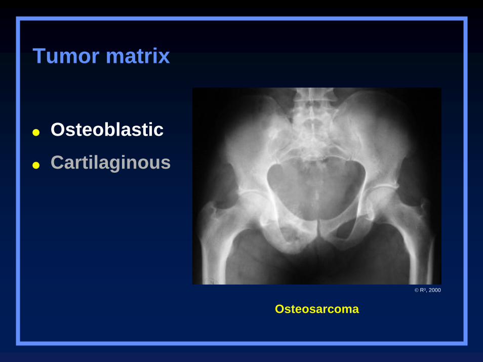
Osteoblastic
Cartilaginous
Tumor matrix
Osteosarcoma
© R3, 2000
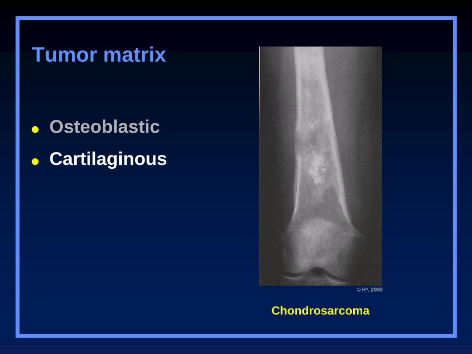
Osteoblastic
Cartilaginous
Tumor matrix
Chondrosarcoma
© R3, 2000
Expansile Lesions of Bone
Multiple myeloma Brown tumor
Mets Enchondroma
Aneurysmal bone cyst Lymphoma
Fibrous dysplasia
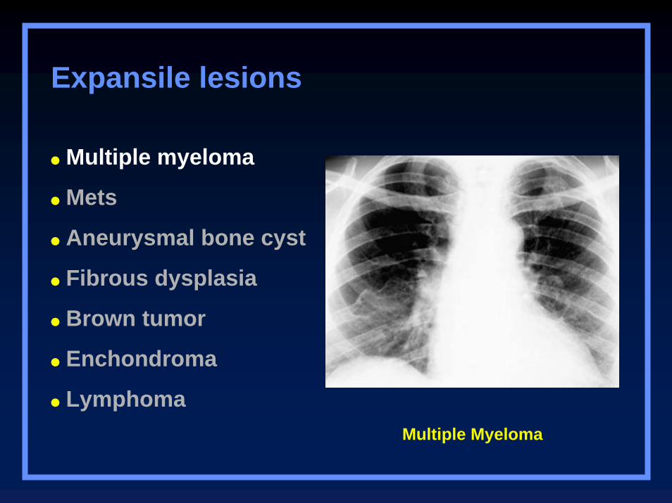
Multiple Myeloma
Expansile lesions
Multiple myeloma
Mets
Aneurysmal bone cyst
Fibrous dysplasia
Brown tumor
Enchondroma
Lymphoma
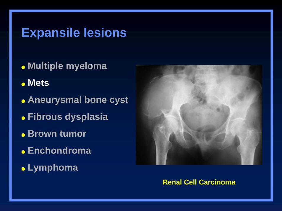
Renal Cell Carcinoma
Expansile lesions
Multiple myeloma
Mets
Aneurysmal bone cyst
Fibrous dysplasia
Brown tumor
Enchondroma
Lymphoma
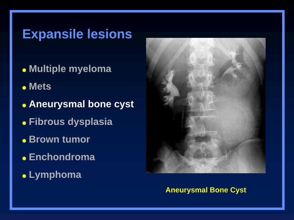
Aneurysmal Bone Cyst
Expansile lesions
Multiple myeloma
Mets
Aneurysmal bone cyst
Fibrous dysplasia
Brown tumor
Enchondroma
Lymphoma
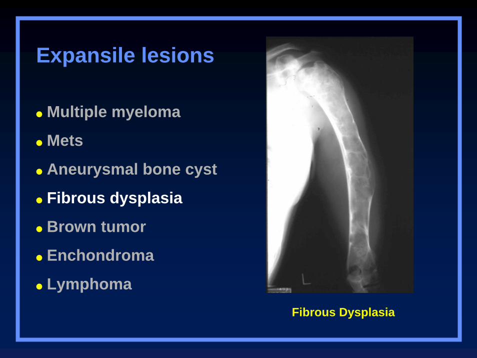
Fibrous Dysplasia
Expansile lesions
Multiple myeloma
Mets
Aneurysmal bone cyst
Fibrous dysplasia
Brown tumor
Enchondroma
Lymphoma
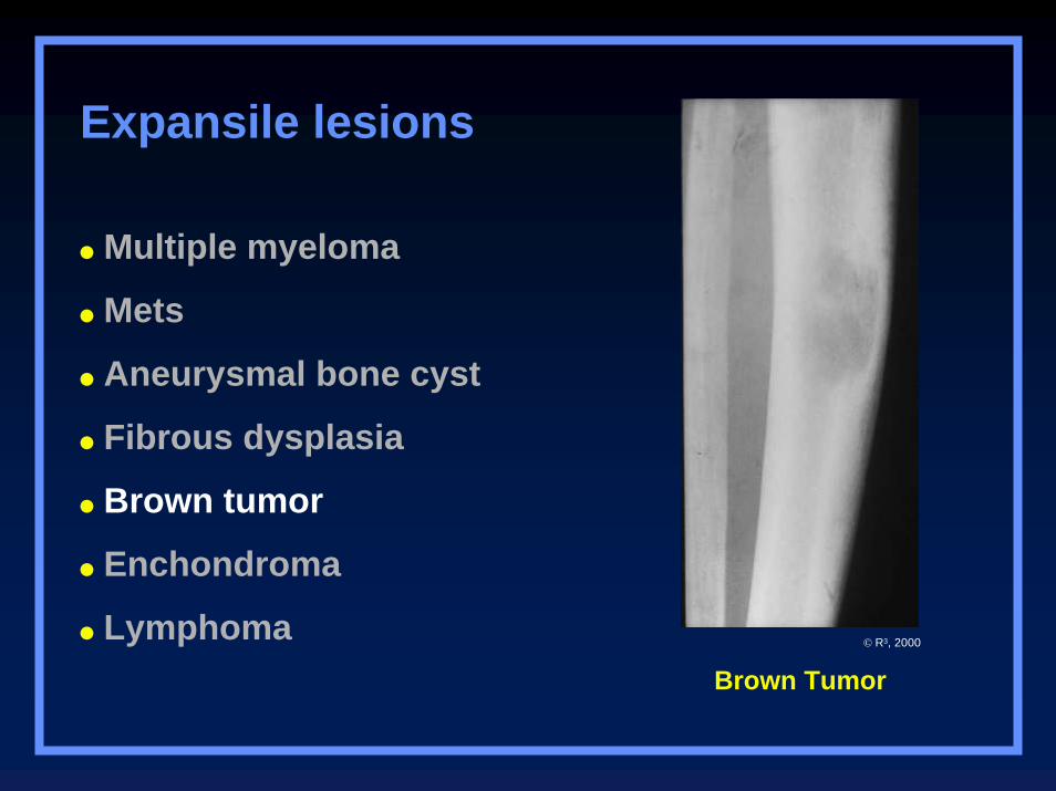
Brown Tumor
Expansile lesions
Multiple myeloma
Mets
Aneurysmal bone cyst
Fibrous dysplasia
Brown tumor
Enchondroma
Lymphoma © R3, 2000
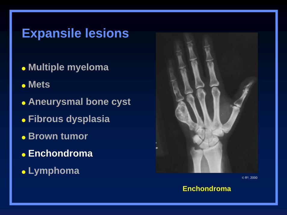
Enchondroma
Expansile lesions
Multiple myeloma
Mets
Aneurysmal bone cyst
Fibrous dysplasia
Brown tumor
Enchondroma
Lymphoma© R3, 2000
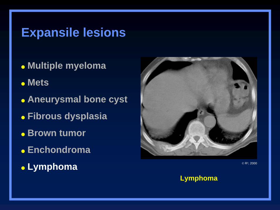
Lymphoma
Expansile lesions
Multiple myeloma
Mets
Aneurysmal bone cyst
Fibrous dysplasia
Brown tumor
Enchondroma
Lymphoma © R3, 2000
Clues by Location
of Lesion
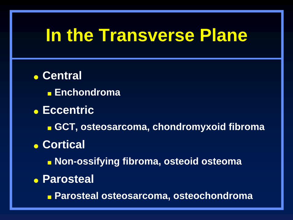
In the Transverse Plane
CentralEnchondroma
EccentricGCT, osteosarcoma, chondromyxoid fibroma
CorticalNon-ossifying fibroma, osteoid osteoma
ParostealParosteal osteosarcoma, osteochondroma
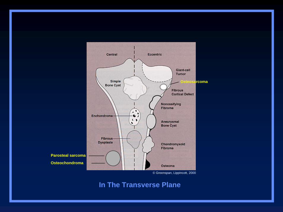
Parosteal sarcoma
Osteochondroma
Osteosarcoma
In The Transverse Plane
© Greenspan, Lippincott, 2000
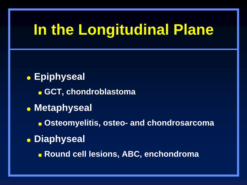
In the Longitudinal Plane
EpiphysealGCT, chondroblastoma
MetaphysealOsteomyelitis, osteo- and chondrosarcoma
DiaphysealRound cell lesions, ABC, enchondroma
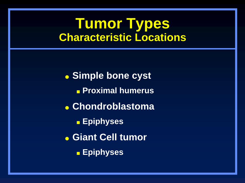
Tumor TypesCharacteristic Locations
Simple bone cystProximal humerus
ChondroblastomaEpiphyses
Giant Cell tumorEpiphyses
Simple bone cystProximal humerus
ChondroblastomaEpiphyses
Giant Cell tumorEpiphyses
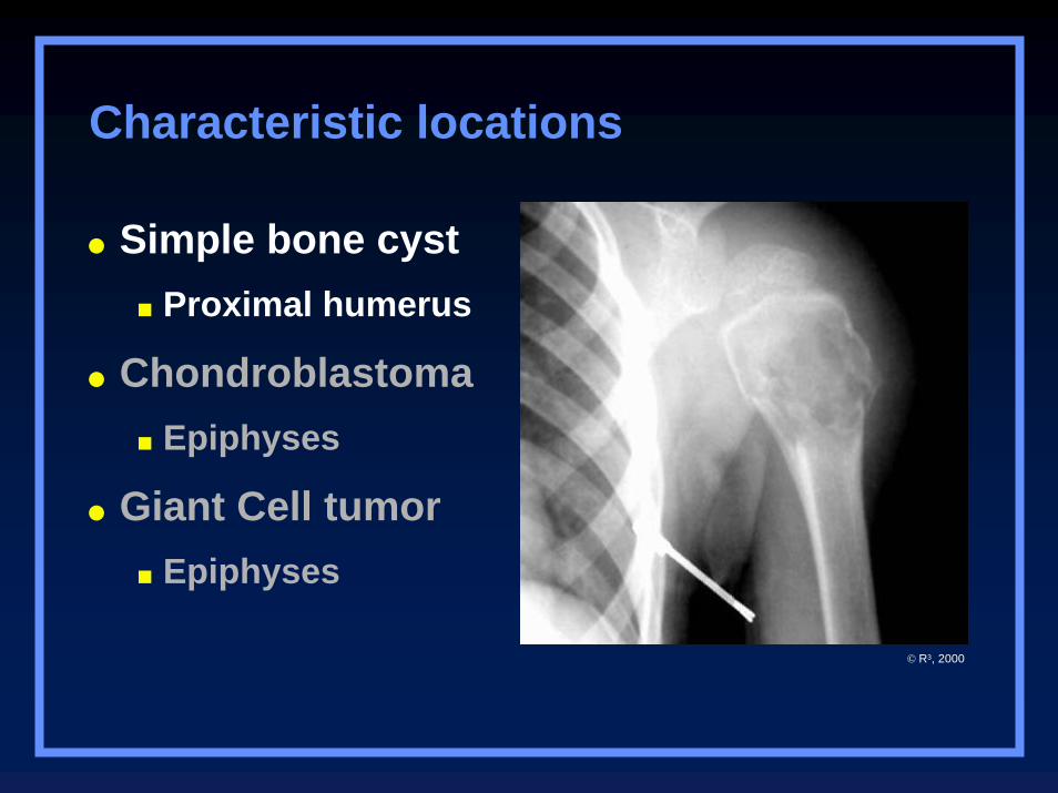
Characteristic locations
© R3, 2000
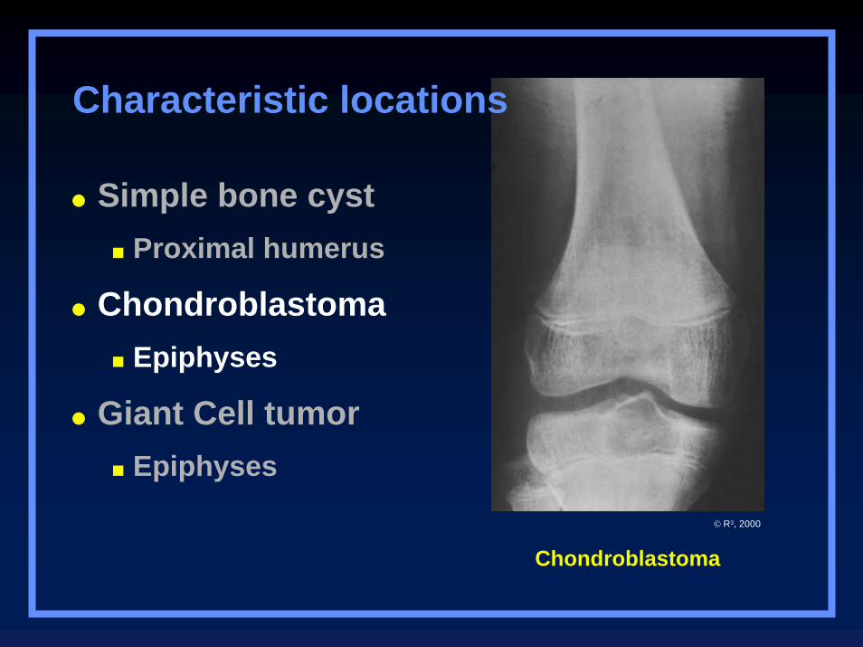
Chondroblastoma
Simple bone cystProximal humerus
ChondroblastomaEpiphyses
Giant Cell tumorEpiphyses
Characteristic locations
© R3, 2000
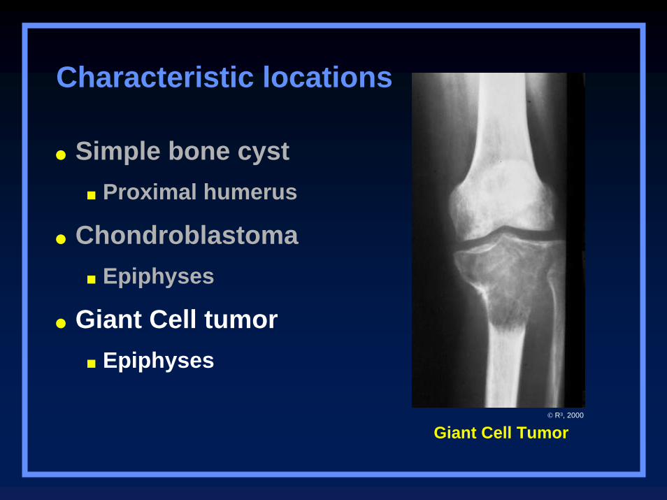
Giant Cell Tumor
Simple bone cystProximal humerus
ChondroblastomaEpiphyses
Giant Cell tumorEpiphyses
Characteristic locations
© R3, 2000
Tumor TypesCharacteristic Locations
AdamantinomaTibia
ChordomaSacrum, clivus
OsteoblastomaSpine, posterior
AdamantinomaTibia
ChordomaSacrum, clivus
OsteoblastomaSpine, posterior
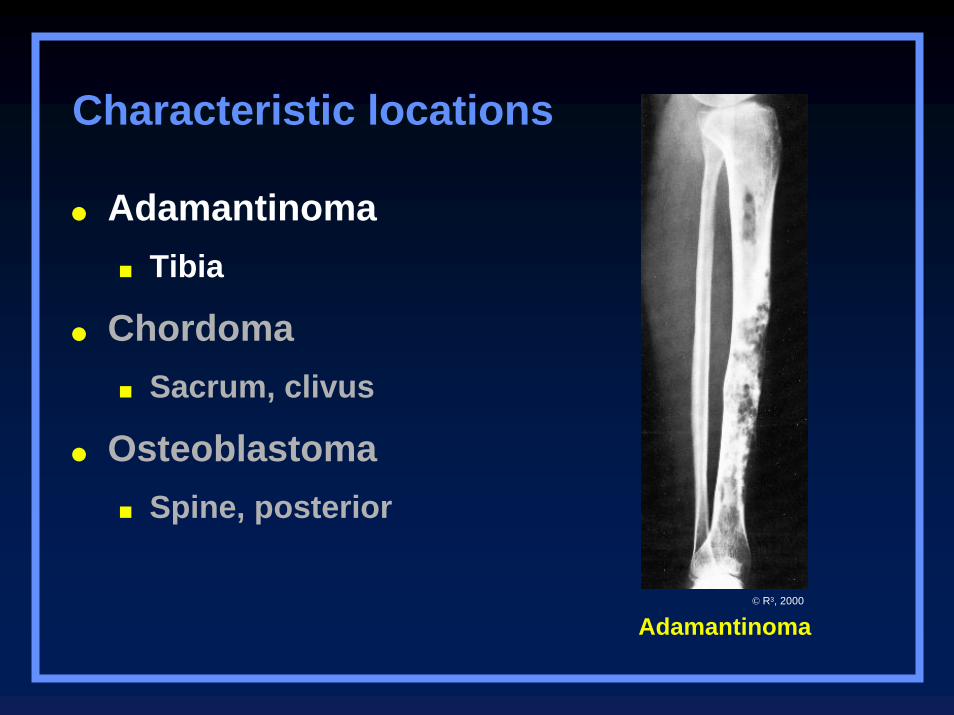
Characteristic locations
Adamantinoma© R3, 2000
AdamantinomaTibia
ChordomaSacrum, clivus
OsteoblastomaSpine, posterior
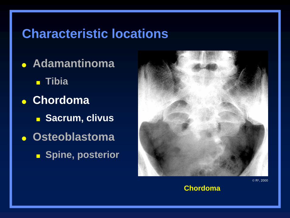
Chordoma
Characteristic locations
© R3, 2000
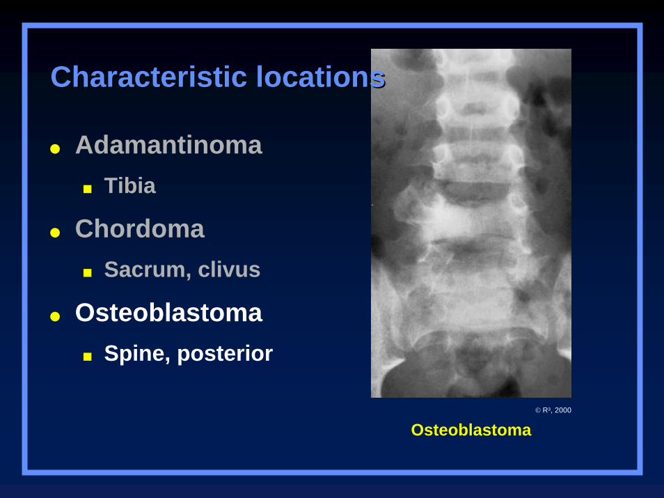
Osteoblastoma
AdamantinomaTibia
ChordomaSacrum, clivus
OsteoblastomaSpine, posterior
Characteristic locationsCharacteristic locations
© R3, 2000
Tumor TypesCharacteristic Locations
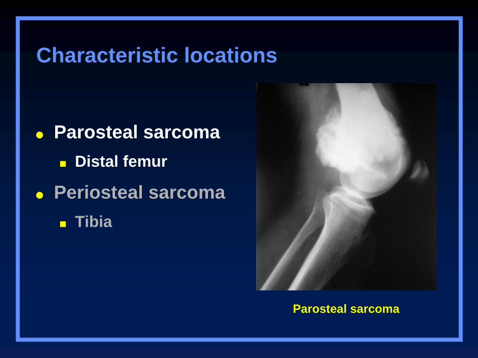
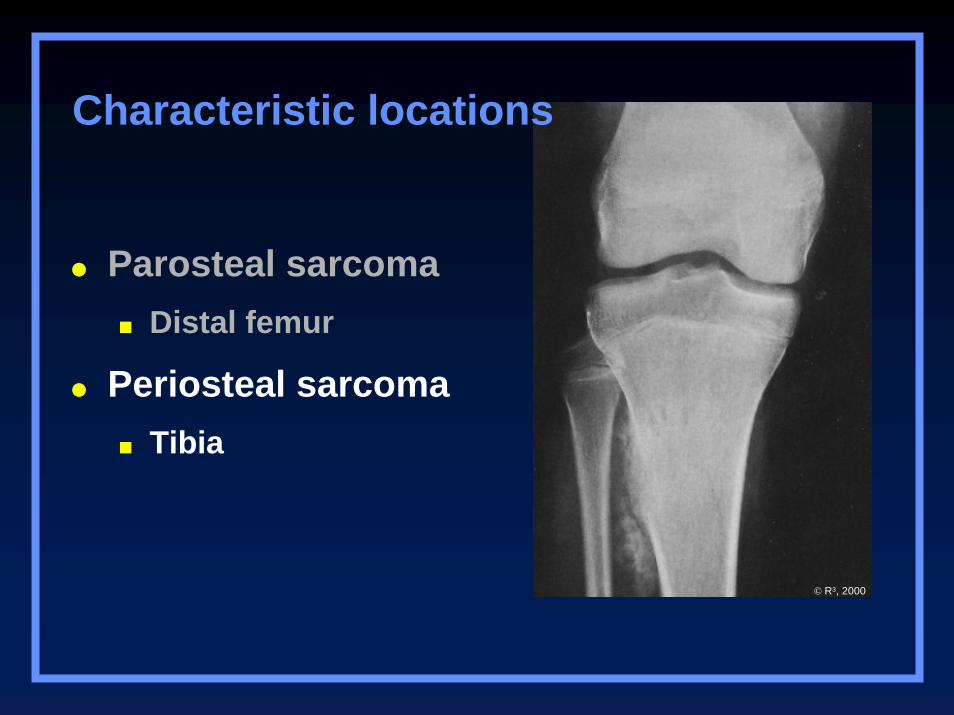
Parosteal sarcomaDistal femur
Periosteal sarcomaTibia
Characteristic locations
Parosteal sarcomaDistal femur
Periosteal sarcomaTibia
Parosteal sarcoma
Characteristic locations
Parosteal sarcomaDistal femur
Periosteal sarcomaTibia
© R3, 2000

Characteristic Tumors
By Body Site
Pelvic Lesions
Chondrosarcoma
Solitary plasmacytoma
Chordoma
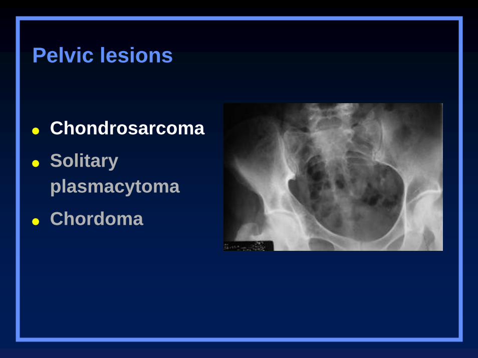
Chondrosarcoma
Solitary plasmacytoma
Chordoma
Pelvic lesions
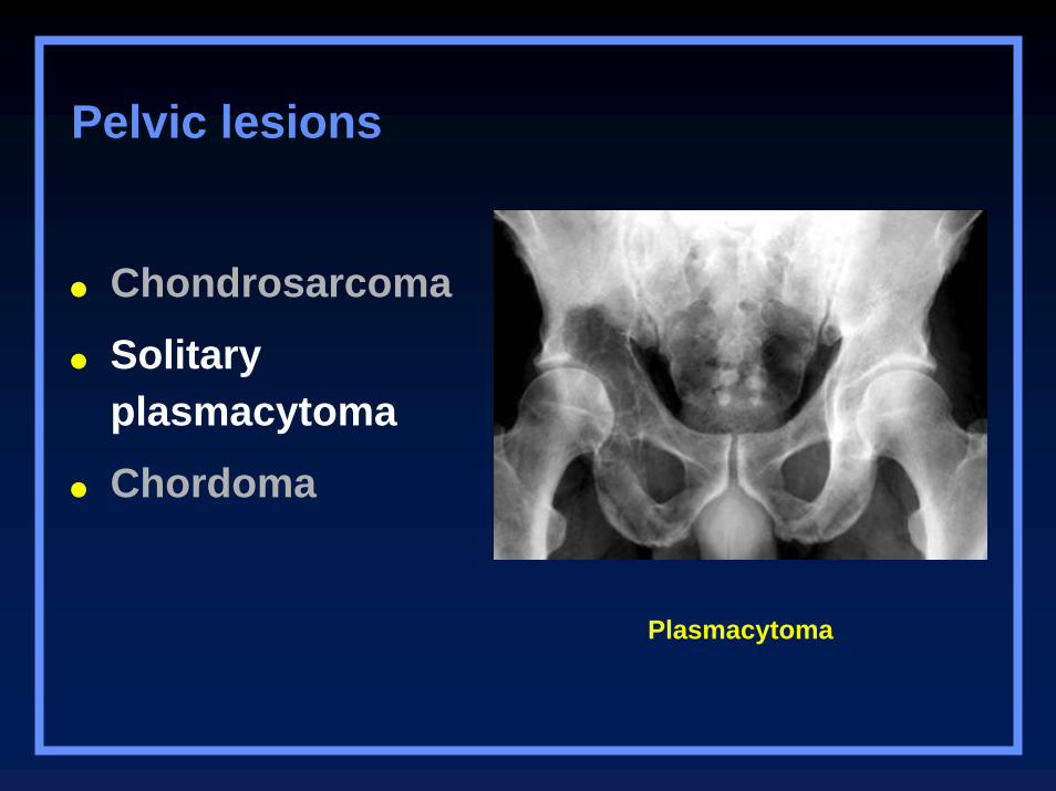
Plasmacytoma
Chondrosarcoma
Solitary plasmacytoma
Chordoma
Pelvic lesions
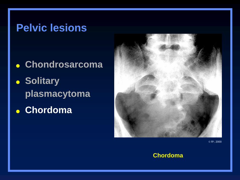
Chordoma
Chondrosarcoma
Solitary plasmacytoma
Chordoma
Pelvic lesions
© R3, 2000
Expansile Rib Lesions
Plasmacytoma
Metastases
Chondrosarcoma
Eosinophilic granuloma
Neurofibromatosis
Fibrous dysplasia
Plasmacytoma
Metastases
Chondrosarcoma
Eosinophilic granuloma
Neurofibromatosis
Fibrous dysplasia
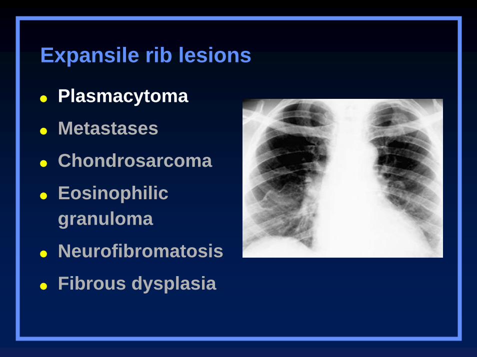
Expansile rib lesions
Plasmacytoma
Metastases
Chondrosarcoma
Eosinophilic granuloma
Neurofibromatosis
Fibrous dysplasia
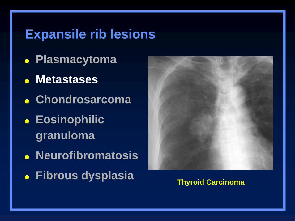
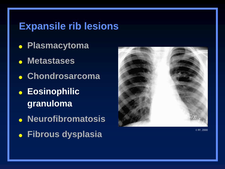
Expansile rib lesions
Thyroid Carcinoma
Plasmacytoma
Metastases
Chondrosarcoma
Eosinophilic granuloma
Neurofibromatosis
Fibrous dysplasia
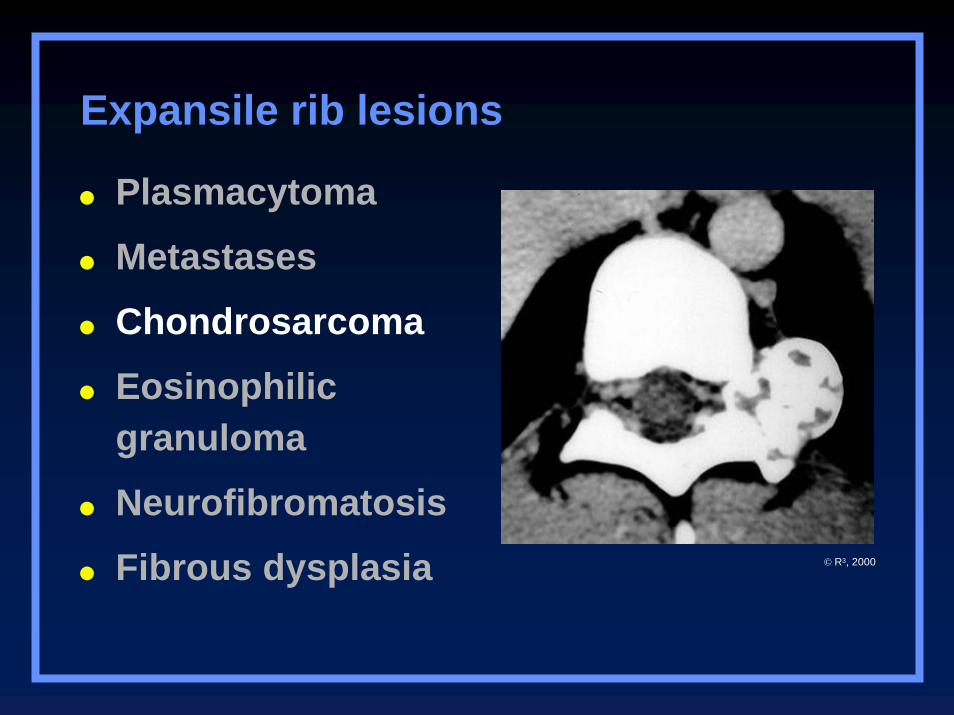
Expansile rib lesions
© R3, 2000
Plasmacytoma
Metastases
Chondrosarcoma
Eosinophilic granuloma
Neurofibromatosis
Fibrous dysplasia
Expansile rib lesions
© R3, 2000
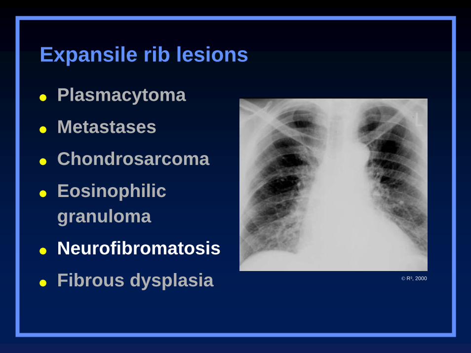
Plasmacytoma
Metastases
Chondrosarcoma
Eosinophilic granuloma
Neurofibromatosis
Fibrous dysplasia
Expansile rib lesions
© R3, 2000
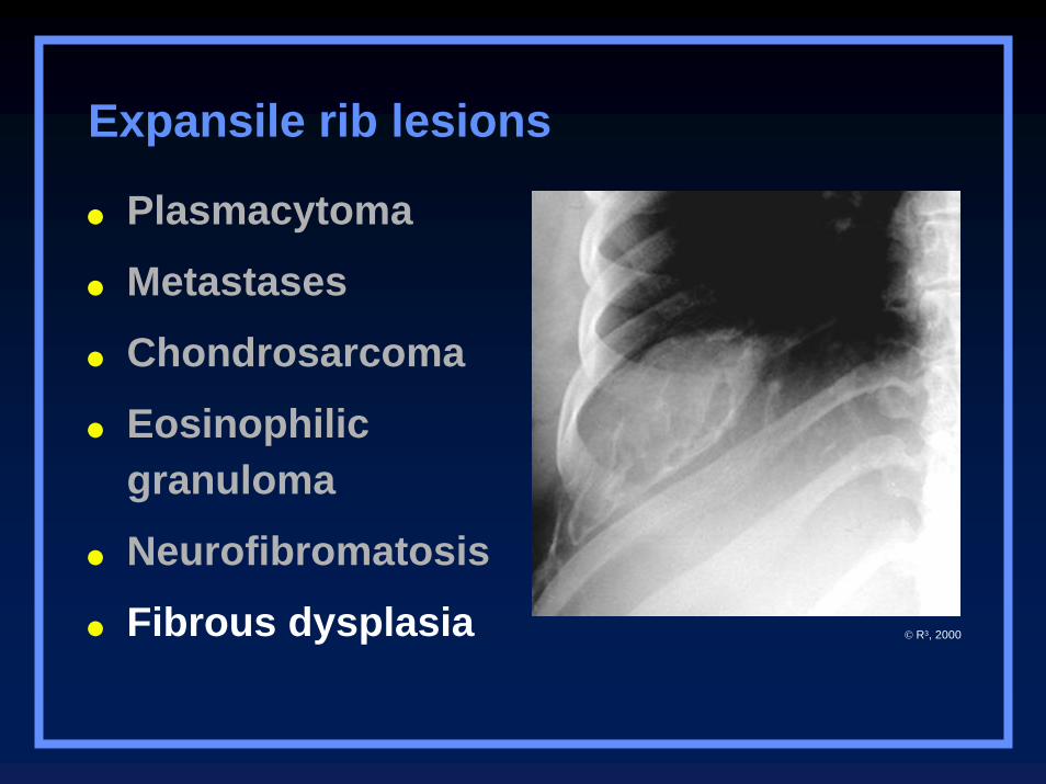
Plasmacytoma
Metastases
Chondrosarcoma
Eosinophilic granuloma
Neurofibromatosis
Fibrous dysplasia
Expansile rib lesions
© R3, 2000
Lesions of the Spine
OsteoblastomaExpansile, with punctate densities within
Chordoma
ABC
Metastatic disease
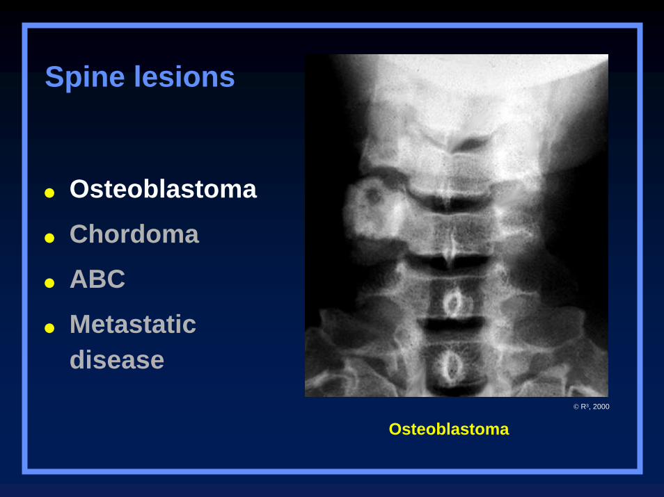
Osteoblastoma
Osteoblastoma
Chordoma
ABC
Metastatic disease
Spine lesions
© R3, 2000
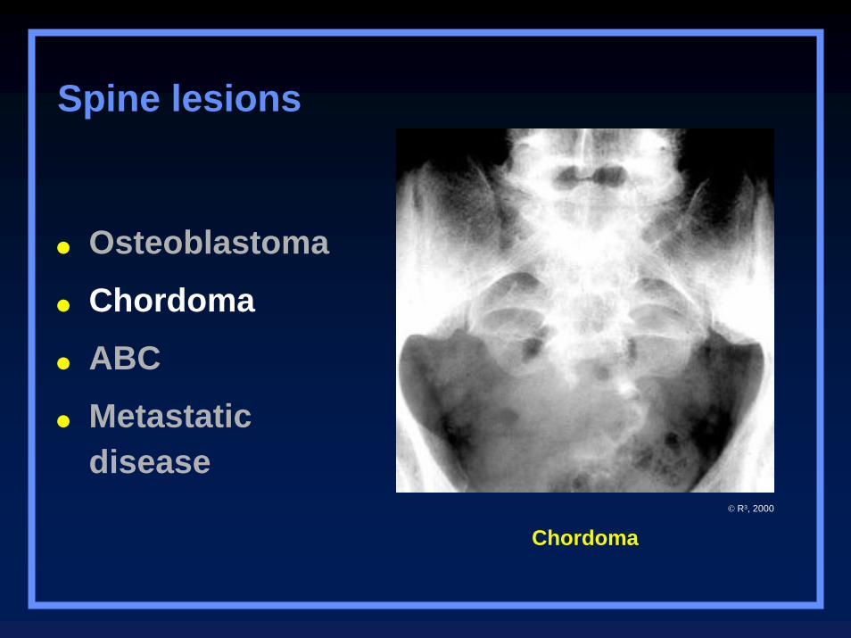
Chordoma
Osteoblastoma
Chordoma
ABC
Metastatic disease
Spine lesions
© R3, 2000
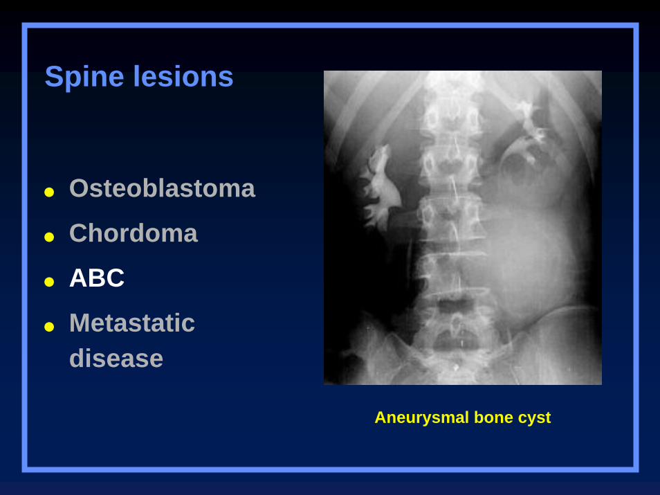
Aneurysmal bone cyst
Osteoblastoma
Chordoma
ABC
Metastatic disease
Spine lesions
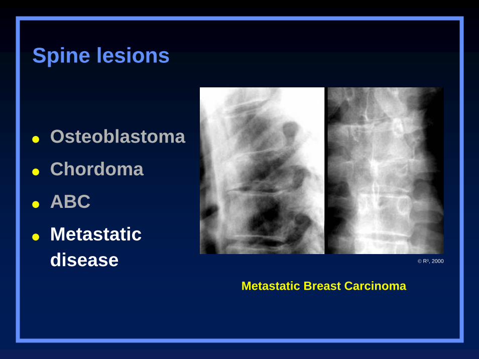
Metastatic Breast Carcinoma
Osteoblastoma
Chordoma
ABC
Metastatic disease
Spine lesions
© R3, 2000
Clues by Density
Of Lesion
Sclerotic Cortical Lesions
Osteoid osteoma
Brodie’s abscess
Stress fracture
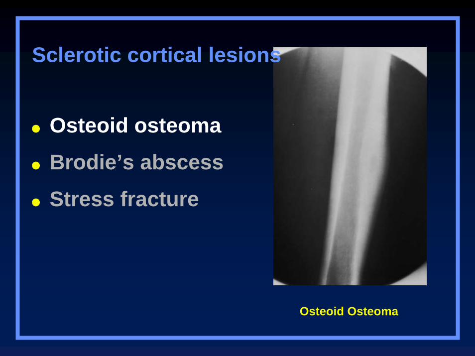
Osteoid Osteoma
Osteoid osteoma
Brodie’s abscess
Stress fracture
Sclerotic cortical lesions
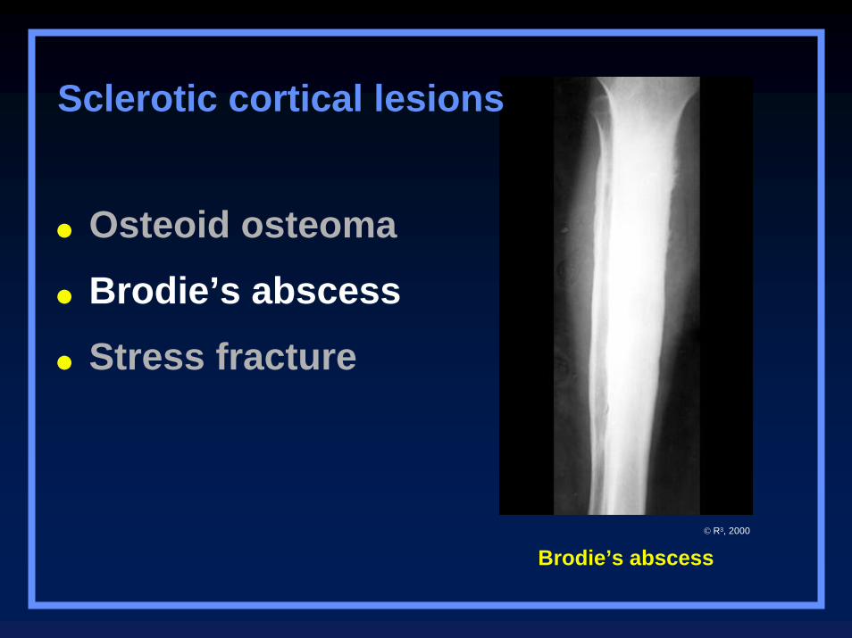
Brodie’s abscess
Osteoid osteoma
Brodie’s abscess
Stress fracture
Sclerotic cortical lesions
© R3, 2000
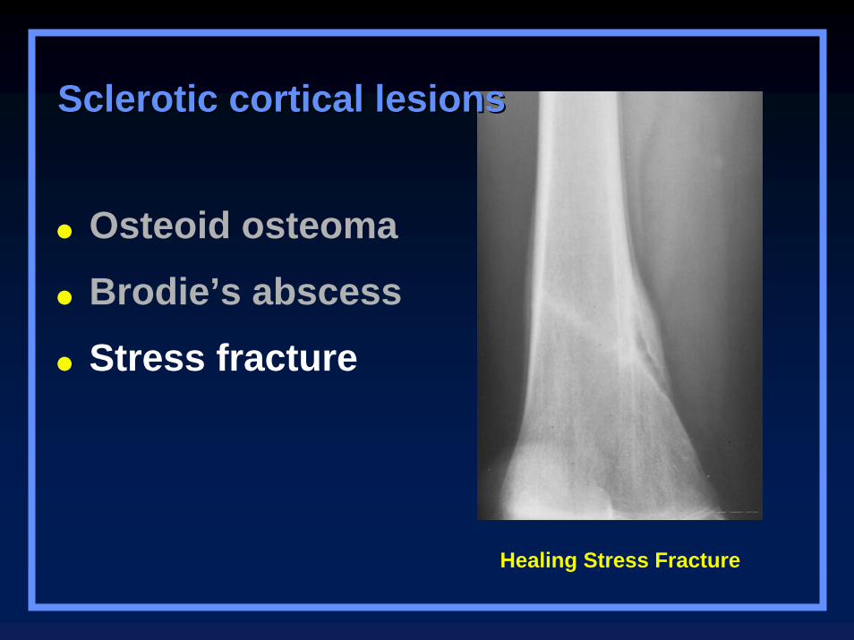
Healing Stress Fracture
Osteoid osteoma
Brodie’s abscess
Stress fracture
Sclerotic cortical lesionsSclerotic cortical lesions
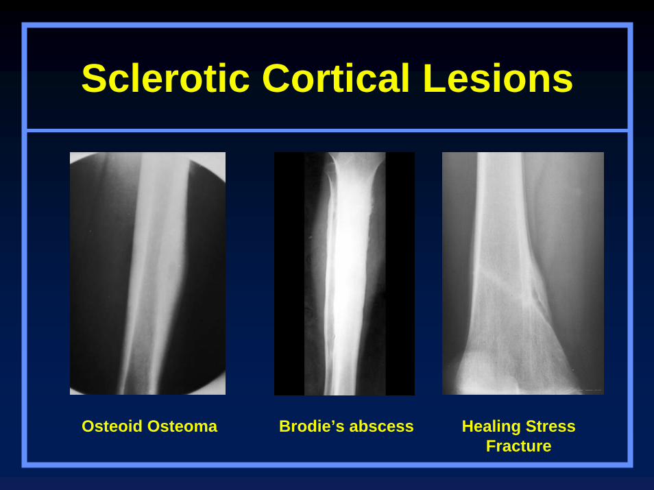
Sclerotic Cortical Lesions
Osteoid Osteoma Brodie’s abscess Healing Stress Fracture
Lytic Lesions in Children
Eosinophilic granuloma
Neuroblastoma
Leukemia
Eosinophilic granuloma
Neuroblastoma
Leukemia
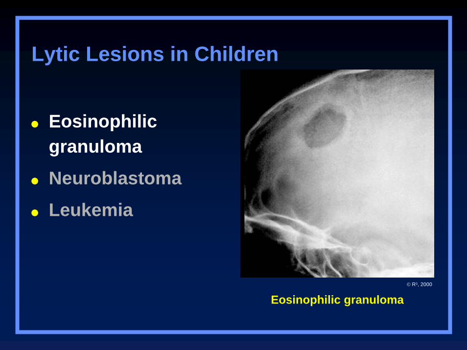
Lytic Lesions in Children
Eosinophilic granuloma© R3, 2000
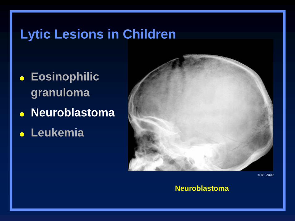
Neuroblastoma
Eosinophilic granuloma
Neuroblastoma
Leukemia
Lytic Lesions in Children
© R3, 2000
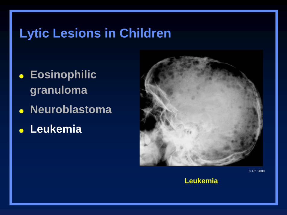
Leukemia
Eosinophilic granuloma
Neuroblastoma
Leukemia
Lytic Lesions in Children
© R3, 2000
Lytic Lesions in Adults
Metastatic lesionsLung
Renal
Thyroid
Multiple myeloma
Primary bone tumor
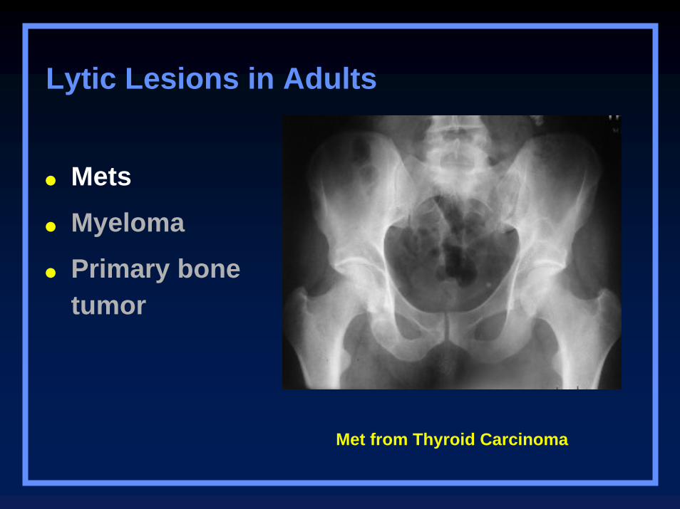
Met from Thyroid Carcinoma
Mets
Myeloma
Primary bone tumor
Lytic Lesions in Adults
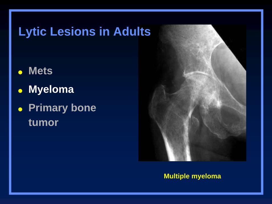
Multiple myeloma
Mets
Myeloma
Primary bone tumor
Lytic Lesions in Adults
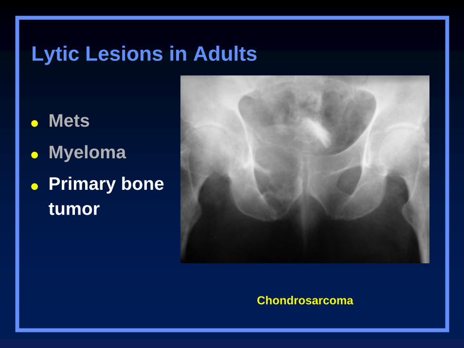
Chondrosarcoma
Mets
Myeloma
Primary bone tumor
Lytic Lesions in Adults
Blastic Lesions in Children
Medulloblastoma
Lymphoma
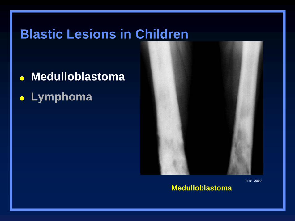
Medulloblastoma
Medulloblastoma
Lymphoma
Blastic Lesions in ChildrenBlastic Lesions in Children
© R3, 2000
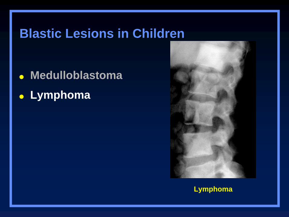
Lymphoma
Medulloblastoma
Lymphoma
Blastic Lesions in ChildrenBlastic Lesions in Children
Blastic Lesions in Adults
Metastatic diseaseBreast – female
Prostate – male
Lymphoma
Paget’s disease
Etcetera-mastocytosis, fluorosis
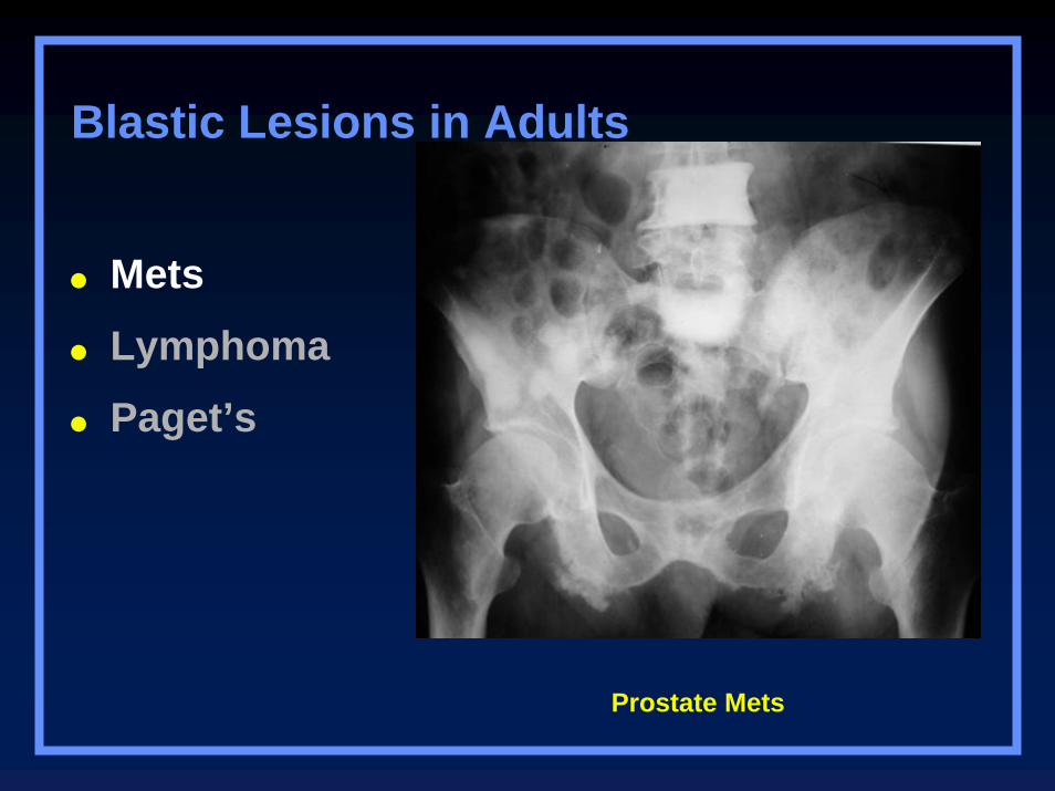
Prostate Mets
Mets
Lymphoma
Paget’s
Blastic Lesions in Adults
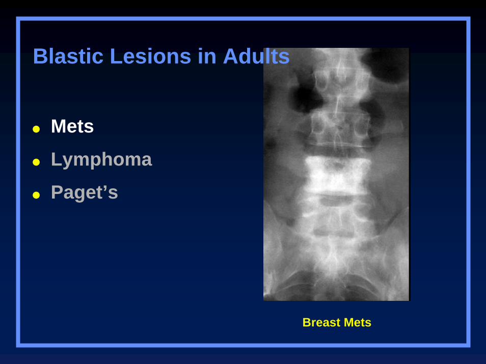
Breast Mets
Mets
Lymphoma
Paget’s
Blastic Lesions in Adults
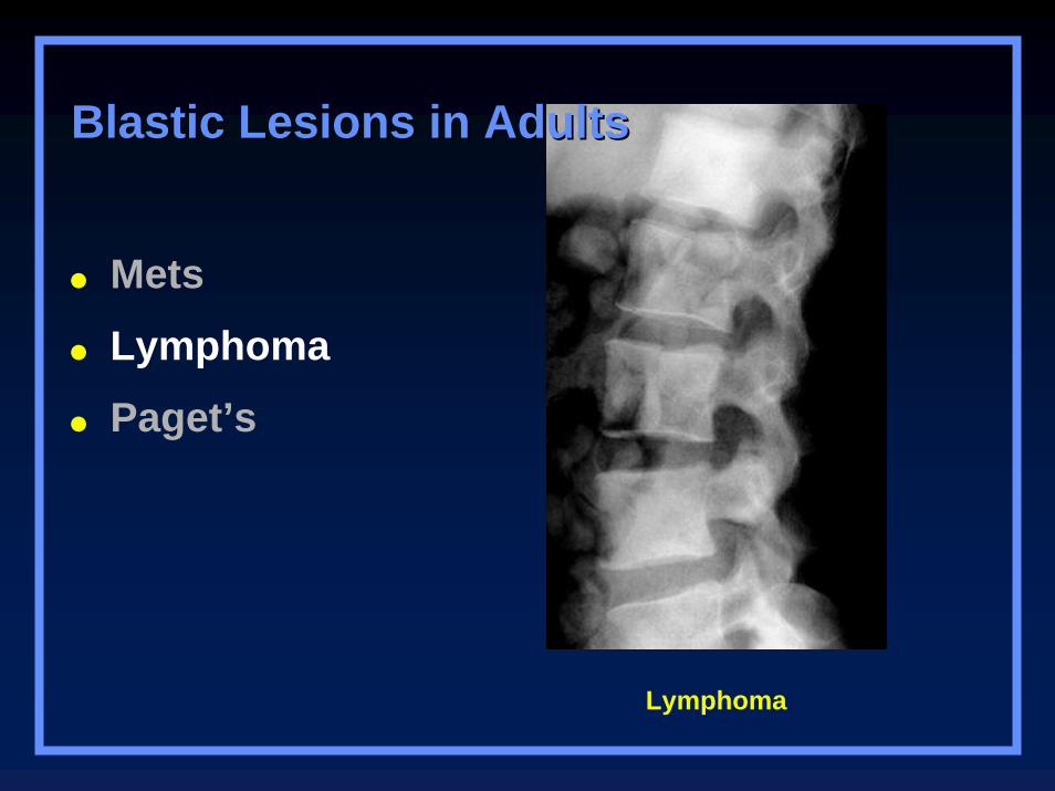
Lymphoma
Mets
Lymphoma
Paget’s
Blastic Lesions in AdultsBlastic Lesions in Adults
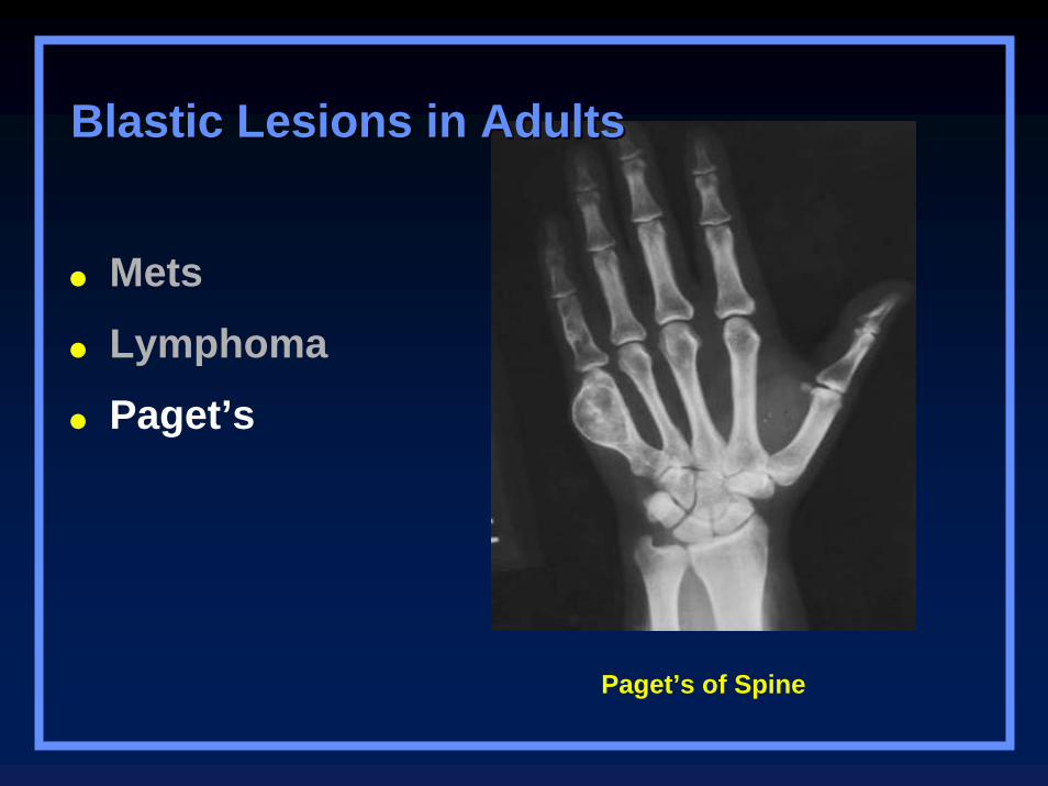
Paget’s of Spine
Mets
Lymphoma
Paget’s
Blastic Lesions in AdultsBlastic Lesions in Adults

Other Clues
Benign LesionsWithout Sclerotic Boarders
Giant Cell tumor
Brown tumor
Osteolytic phase of Paget’s Disease
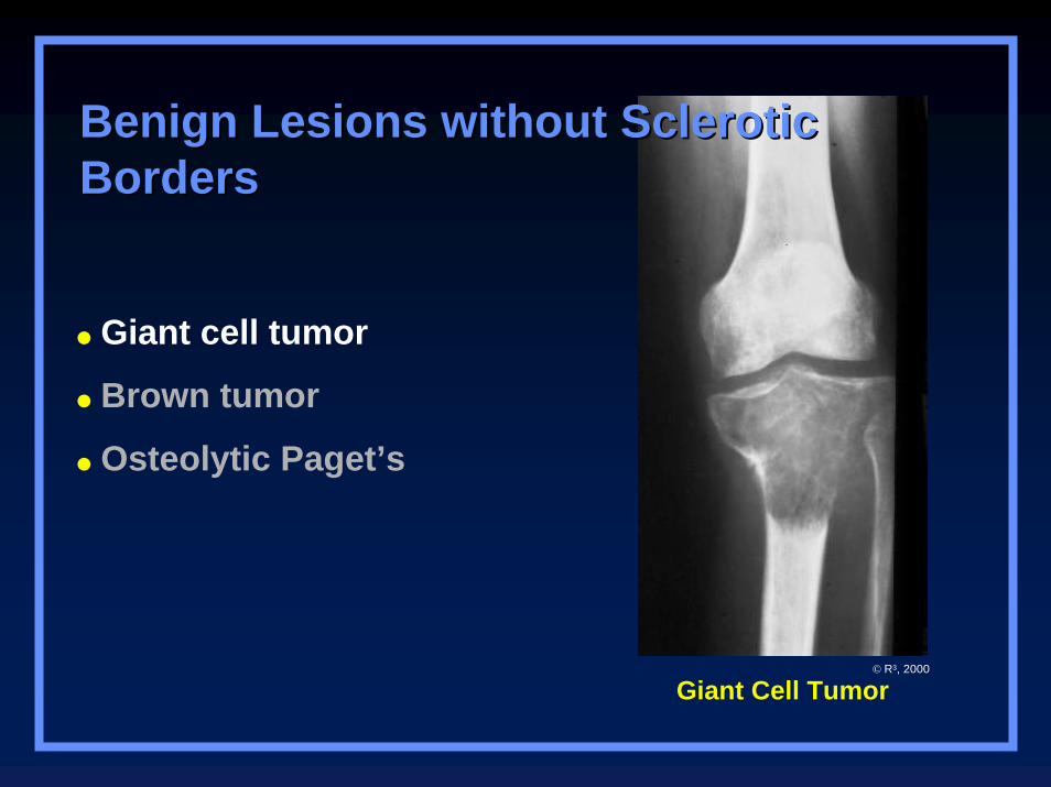
Benign Lesions without Sclerotic Benign Lesions without Sclerotic BordersBorders
Giant cell tumor
Brown tumor
Osteolytic Paget’s
Giant Cell Tumor© R3, 2000
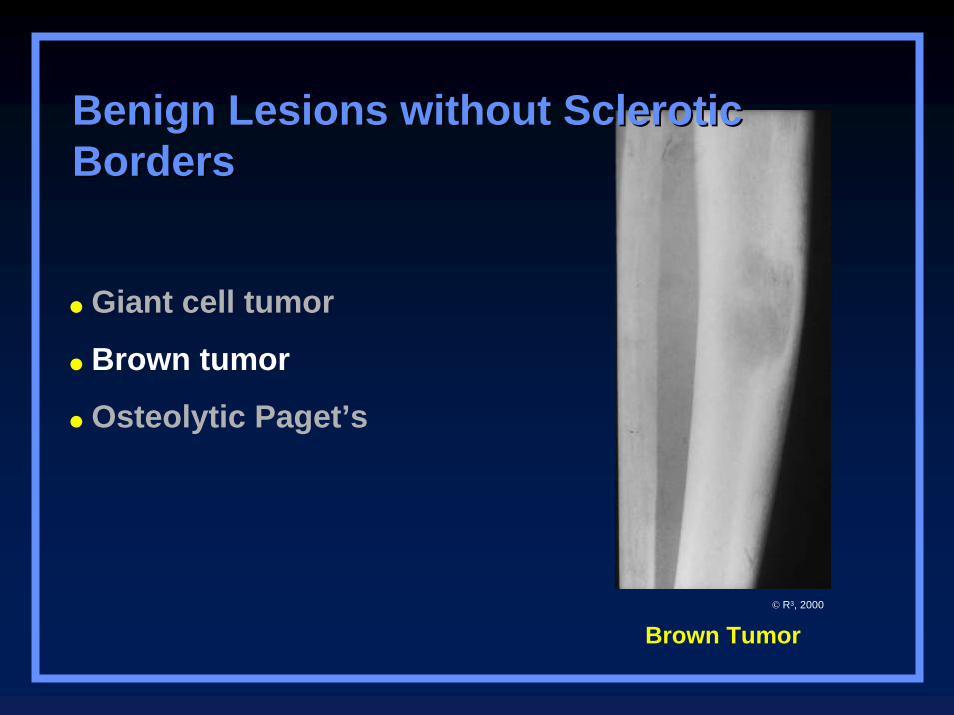
Brown Tumor
Benign Lesions without Sclerotic Benign Lesions without Sclerotic BordersBorders
Giant cell tumor
Brown tumor
Osteolytic Paget’s
© R3, 2000

Osteolytic Paget’s
Benign Lesions without Sclerotic Benign Lesions without Sclerotic BordersBorders
Giant cell tumor
Brown tumor
Osteolytic Paget’s
© R3, 2000
Soft Tissue Extension
Usually implies malignancyMore likely to form discrete soft tissue mass
Benign conditions with soft tissue extension
OsteomyelitisUsually infiltration of fat
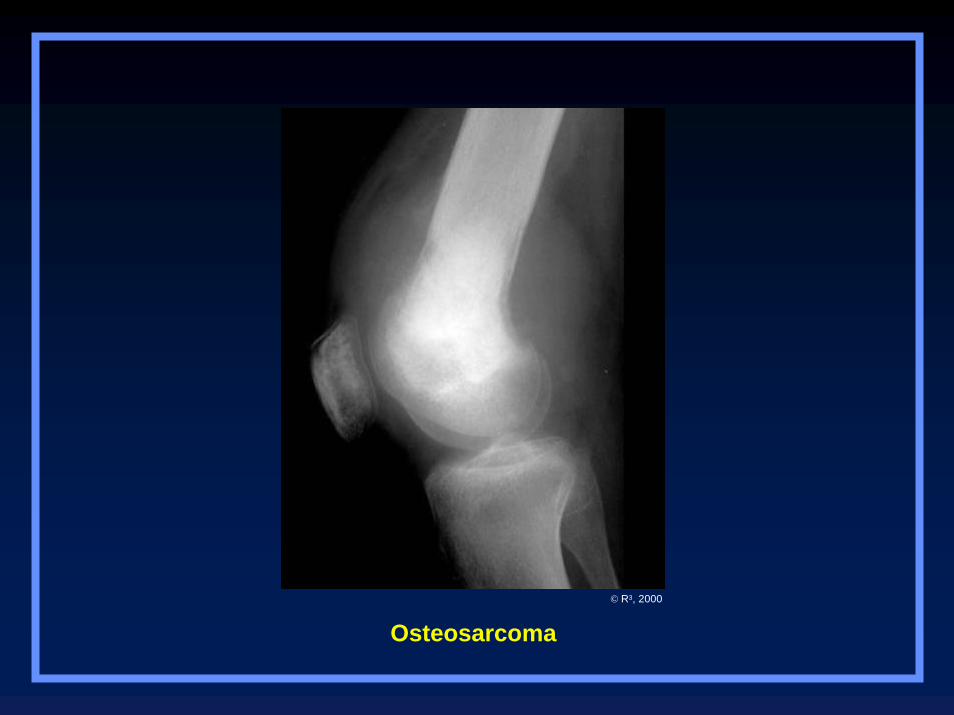
Osteosarcoma© R3, 2000
Multiple Lesions
More often benign
Malignancies with multiple lesionsMetastatic disease
Multiple myeloma
Lymphoma
Ewing’s sarcoma (rarely)
Osteosarcoma (rarely)
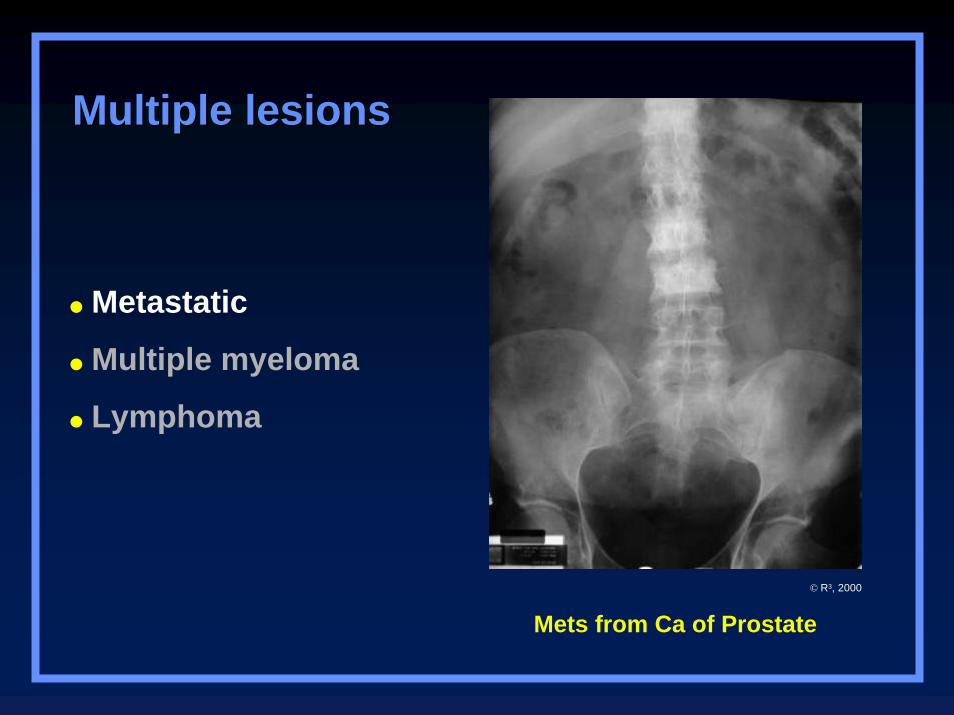
Mets from Ca of Prostate
Multiple lesionsMultiple lesions
Metastatic
Multiple myeloma
Lymphoma
© R3, 2000
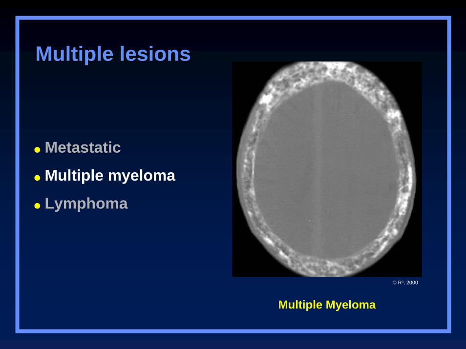
Multiple Myeloma
Multiple lesionsMultiple lesions
Metastatic
Multiple myeloma
Lymphoma
© R3, 2000
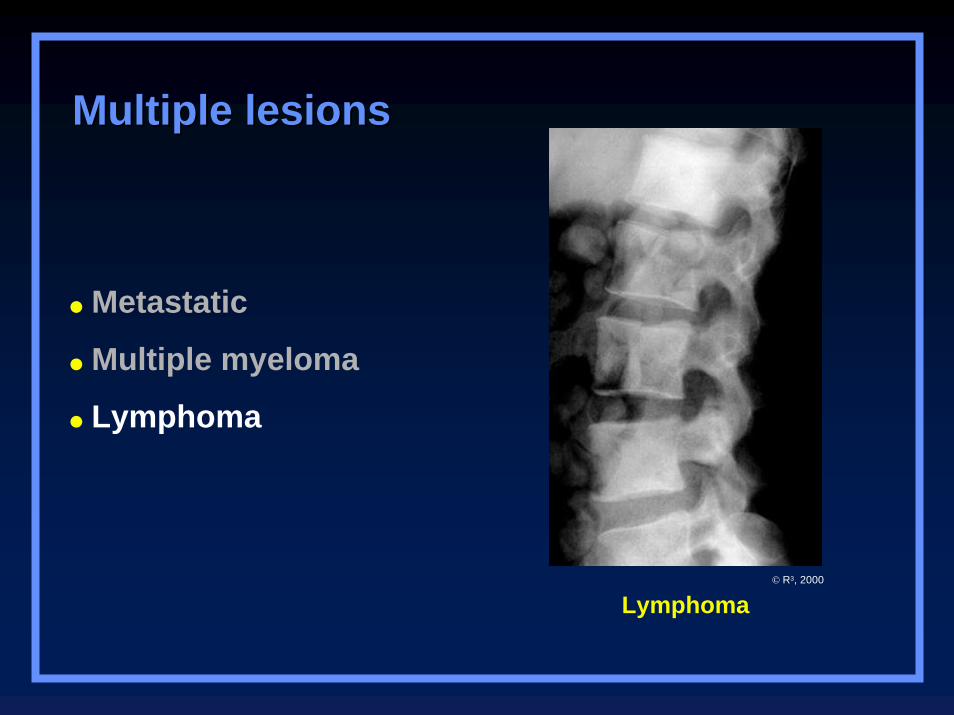
Lymphoma
Multiple lesionsMultiple lesions
Metastatic
Multiple myeloma
Lymphoma
© R3, 2000
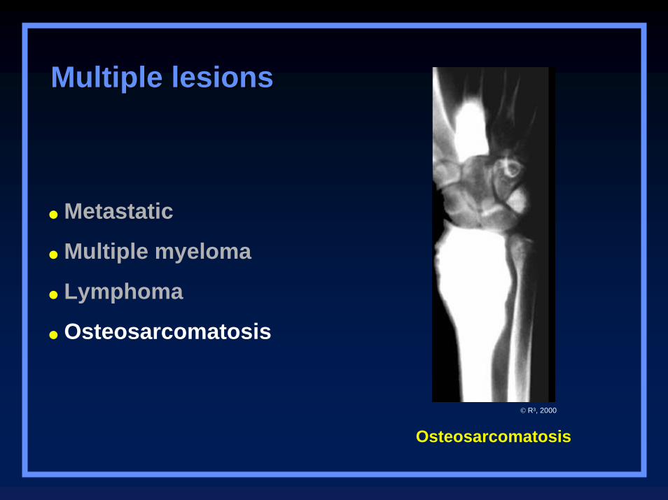
Osteosarcomatosis
Multiple lesionsMultiple lesions
Metastatic
Multiple myeloma
Lymphoma
Osteosarcomatosis
© R3, 2000
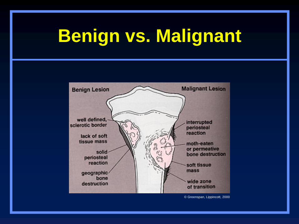
Benign vs. Malignant
© Greenspan, Lippincott, 2000

The End