Embed Size (px)
Citation preview
1
Bone regeneration mediated by a bioactive and
biodegradable ECM-like hydrogel based on elastin-like
recombinamers
Dante J. Coletta, MD,1# Arturo Ibáñez-Fonseca, MSc,2# Liliana R. Missana, PhD,3,4#
María V. Jammal, PhD,3,4 Ezequiel J. Vitelli, MD,1 Mariangeles Aimone, BSc,1 Facundo
Zabalza, BSc,1 João P. Mardegan Issa, PhD,5 Matilde Alonso, PhD,2 José Carlos
Rodríguez-Cabello, PhD,2 and Sara Feldman, PhD1
# These authors contributed equally to this work.
1 LABOATEM. Osteoarticular Biology, Tissue Engineering and Emerging Therapies
Laboratory, School of Medicine, National Rosario University, Rosario, Argentina.
2 BIOFORGE Lab, University of Valladolid, CIBER-BBN, Valladolid, Spain.
3 Experimental Pathology & Tissue Engineering Laboratory, Dental School, National
Tucumán University, Tucumán, Argentina.
4 Tissues Laboratory, Proimi-Biotechnology-Conicet, Tucumán, Argentina.
5 Ribeirão Preto School of Dentistry, University of São Paulo, São Paulo, Brazil.
Address correspondence to:
José Carlos Rodríguez-Cabello, PhD
BIOFORGE Lab
2
Universidad de Valladolid
Paseo de Belén, 19
47011 – Valladolid
SPAIN
E-mail: [email protected]
Phone: +34983184585
Abstract
The morbidity of bone fractures and defects is steadily increasing due to changes in the
age pyramid. As such, novel biomaterials that are able to promote the healing and
regeneration of injured bones are needed in order to overcome the limitations of auto-,
allo-, and xenografts, while providing a ready-to-use product that may help to minimize
surgical invasiveness and duration. In this regard, recombinant biomaterials, such as
elastin-like recombinamers (ELRs), are very promising as their design can be tailored by
genetic engineering, thus allowing scalable production and batch-to-batch consistency,
amongst others. Furthermore, they can self-assemble into physically cross-linked
hydrogels above a certain transition temperature, in this case body temperature, but are
injectable below this temperature, thereby markedly reducing surgical invasiveness.
Herein we have developed two bioactive hydrogel-forming ELRs, one including the
osteogenic and osteoinductive BMP-2 and the other the RGD cell-adhesion motif. The
combination of these two novel ELRs results in a BMP-2-loaded extracellular matrix-like
hydrogel. Moreover, elastase-sensitive domains were included in both ELR molecules,
3
thereby conferring biodegradation as a result of enzymatic cleavage and avoiding the need
for scaffold removal after bone regeneration. Both ELRs and their combination showed
excellent cytocompatibility, and the culture of cells on RGD-containing ELRs resulted in
optimal cell adhesion. In addition, hydrogels based on a mixture of both ELRs were
implanted in a pilot study involving a femoral bone injury model in New Zealand White
rabbits, showing complete regeneration in six out of seven cases, with the other showing
partial closure of the defect. Moreover, bone neo-formation was confirmed using different
techniques, such as radiography, computed tomography and histology. This hydrogel
system therefore displays significant potential in the regeneration of bone defects,
promoting self-regeneration by the surrounding tissue with no involvement of stem cells
or osteogenic factors other than BMP-2, which is released in a controlled manner by
elastase-mediated cleavage from the ELR backbone.
Keywords: bone regeneration, elastin-like recombinamers, bioactive hydrogels, BMP-2.
Introduction
It is well known that dental, maxillofacial, and other orthopedic surgeries often require
the use of different biomaterials for the treatment of injuries and other diseases through
tissue engineering, including osteoporosis (1-6). In addition, changes of the age pyramid
towards an older population have led to an increasing number of bone fractures (7-11).
Despite the availability of numerous biomaterials for tissue regeneration, autologous bone
is usually the first option for the replacement of injured bone tissue. However, a large
number of different types of biomaterials and bone grafts have been employed to date for
the healing of bone defects (12). These biomaterials should be: a) osteoinductive, hence
promoting stem cell differentiation to osteoblasts, b) osteoconductive, thereby inducing
growth of the surrounding healthy bone, and c) osseointegrative, merging with the nearby
4
bone (13-15). They should also stimulate an optimal cell response and be liable to be
replaced by de novo formed tissue, acting as a provisional substitute (1, 16, 17).
Engineered biomaterials, in combination with growth factors, have been shown to be an
effective approach in bone tissue engineering since they can act both as a scaffold and as
a drug-delivery system to promote bone repair and regeneration (18, 19). For instance,
the osteoinductive bone morphogenetic protein-2 (BMP-2) (20, 21) has been shown to
enhance the formation of bone tissue in situations that lead to bone degradation, such as
alcohol dependence (22) and osteometabolic diseases (23). Due to the high cost and rapid
release of BMP-2 when placed at the site of injury, it is often associated with carrier
matrices that act as drug-delivery systems to increase its half-life and to avoid the adverse
effects associated to high doses of BMP-2 (24-27).
On the other hand, protein-based recombinant biomaterials, such as resilin-, silk-,
collagen- and elastin-like polypeptides, have been developed over the last few decades
with the aim of improving the features of traditional biomaterials in terms of ease of
design and synthesis, biocompatibility and bioactivity (28). As an example, elastin-like
recombinamers (ELRs), thus named due to their polymeric and recombinant nature (29),
have been shown to be a potential tool for the development of biomedical devices for
regenerative medicine due to their thermosensitivity. This smart behavior is a result of
their composition, which is based on repetitions of the Val-Pro-Gly-X-Gly (VPGXG)
pentapeptide, in which X (guest residue) is any amino acid except L-proline. Moreover,
it is characterized by a transition temperature (Tt) which itself depends on the polarity of
the side chain in the guest residue. Thus, in an aqueous medium, the ELR chains remain
soluble below their Tt while above that Tt (e.g. physiologic temperature) the ELR self-
assembles hydrophobically, undergoing a phase transition (30). In this study, two
different ELRs have been developed, based on a previously described hydrogel-forming
5
ELR (31). Taking advantage of the recombinant nature of these biomolecules, one of the
novel ELRs designed in this work has been genetically engineered to include Arg-Gly-
Asp (RGD) motifs to enhance cell adhesion via cell-membrane integrins (32), whereas
the other ELR was designed to include BMP-2. Both ELRs also contain elastase-sensitive
domains resulting from repetition of the VGVAPG hexapeptide (33) to improve the
enzymatic biodegradability of the biomaterial.
In order to study the potential of these novel ELRs in bone regeneration, we have used a
previously developed model of femoral bone injury (FBI) in New Zealand White rabbits.
This involves the creation of a defect 6 mm in diameter in a femoral condyle 8 mm in
diameter (34, 35). This animal model allows the study of the defect by computed
tomography (CT) and by radiological studies given the size of the bone.
The aim of this work was to evaluate whether novel bioactive ELRs are cytocompatible
and degradable, while being able to form extracellular matrix (ECM)-like hydrogels and
promoting bone regeneration after implantation into an FBI in rabbits, as a preliminary
step for their use in humans. For this purpose, the cytocompatibility and biodegradation
ability were assessed in vitro, and a highly reproducible model was subsequently used to
carry out a pilot in vivo study.
Materials and methods
Ethical approval
Experimental procedures regarding the use of animals were approved by the Bioethics
Committee of Rosario National University (Resolution No. 150/2015). Its regulations
include well-established guidelines for animal care and manipulation to decrease pain and
suffering of the animal, according to the 3Rs (replacement, reduction and refinement),
and are in accordance with international laws concerning the use of animals.
6
ELR biosynthesis and characterization
The genetic construction of the ELRs used in this work was performed as described
elsewhere (36). Briefly, their DNA sequences were obtained by genetic engineering
techniques and cloned into a pET-25b(+) vector for expression in Escherichia coli. ELRs
were biosynthesized in a 15-L bioreactor and purified by several cooling and heating
purification cycles (Inverse Transition Cycling) taking advantage of the ability of these
recombinamers to precipitate above their transition temperature. Further centrifugation
steps led to a pure product, which was dialyzed against ultra-pure water, filtered through
0.22 μm filters (Nalgene, Thermo Fisher, USA) to obtain a sterile solution, and freeze-
dried prior to storage. The ELRs were found to contain less than two endotoxin units
(EU)/mg of ELR, as determined using the limulus amebocyte lysate assay with the
Endosafe®-PTS system (Charles River Laboratories). This process allowed the
production of two different ELRs, both of which were derived from a previously
synthesized block co-recombinamer (31). Further information can be obtained in
Supplementary Methods (Supplementary Information).
The characterization techniques used included sodium dodecyl sulfate polyacrylamide gel
electrophoresis (SDS-PAGE) and matrix-assisted laser desorption/ionization time-of-
flight (MALDI-TOF) spectrometry for purity and molecular weight (Mw) evaluation
compared to the theoretical values of 113,556 Da for ELR-E-RGD and 107,752 Da for
ELR-E-BMP-2; differential scanning calorimetry (DSC) to determine the transition
temperature; HPLC to determine the amino acid composition of both ELRs (Table S1 and
Table S2, Supplementary Information), and nuclear magnetic resonance (NMR) to
provide recombinamer fingerprint data (Fig. S3 and S4; Table S3 and Table S4,
Supplementary Information). The procedure for the measurement of the mechanical
7
properties of ELR-based hydrogels is described in Supplementary Methods
(Supplementary Information).
Elastase-mediated cleavage of the ELR in solution
Different quantities (1.2, 1.8 and 2.4 U) of porcine pancreas elastase (4 mg/mL, 6.8 U/mg)
(Sigma-Aldrich, USA) were added to solutions of the mixture of both ELRs (98% (w/w)
ELR-E-RGD and 2% (w/w) ELR-E-BMP-2) at a final concentration of 1 mg/mL
dissolved in ultra-pure water in order to evaluate the biodegradation rate for each quantity
of enzyme. The quantity of elastase used was 2000-, 3000- and 4000-times the amount
needed to cleave the mixture of ELRs used as substrate in 30 minutes, since preliminary
experiments showed that larger quantities than those calculated are required to observe
an actual effect of the enzyme in vitro. Samples were incubated at 37 ºC and collected at
various time points (10, 20, 30, 45, 60, 90 and 120 minutes), then stored frozen at –20 ºC
until further use. A negative control, namely an ELR molecule lacking elastase-sensitive
sequences but with the same elastin-like structure as the two ELRs designed for this work
(31), was also treated with 1.2 U of elastase for further comparisons. Methods concerning
the evaluation of the biodegradation are explained in Supplementary Methods
(Supplementary Information).
In vitro cell culture
Bone marrow derived human mesenchymal stem cells (hMSCs) were extracted and
isolated as described elsewhere (37) and were generously provided as a gift by Citospin
S.L. (Spain). They were cultured for expansion in DMEM low glucose (1 g/L) (Gibco,
USA) supplemented with 10% fetal bovine serum (FBS) (Gibco, USA) and 1%
Penicillin/Streptomycin (P/S) (Gibco, USA).
8
All cells were used at passage 3-5 in subsequent experiments. They were detached from
the wells using a Trypsin-EDTA solution (0.25%, Gibco, USA) and counted using a
hematocytometer.
Cell viability
Human MSCs were used to determine the in vitro viability using the Calcein AM assay
(Molecular Probes, USA) when cultured in DMEM supplemented with 10 mg/mL of the
different ELRs or the mixture of them (98% (w/w) ELR-E-RGD and 2% (w/w) ELR-E-
BMP-2) for 3 days. This assay was performed in a black, 96-well plate with clear-bottom
(Greiner Bio One, USA) according to the manufacturer’s instructions and the
fluorescence intensity measured at 530 nm using a plate reader (SpectraMax M2e,
Molecular Devices, USA). The intensity measured at this wavelength, corresponding to
live cells, was then used to calculate cell numbers by using calibration curves obtained
with different known quantities of cells (from 1000 to 10,000 cells per well) seeded on
96-well plates 24 h before the measurement. Each condition was performed in triplicate,
with four experiments for each (n = 4).
Cell adhesion on ELR-coated tissue culture plates
96-well plates were used for the coating of different wells with both ELRs separately and
combined (98% (w/w) ELR-E-RGD and 2% (w/w) ELR-E-BMP-2). Briefly, a 5 mg/mL
solution of the recombinamers in ultra-pure water was placed in the well and allowed to
adsorb to the surface for 24 h at 4 ºC. The wells were washed twice with 1x PBS (Gibco)
and blocked with 1% BSA for 2 h at 37 ºC, then rinsed again and, finally, 3000 cells per
well were seeded onto the modified surfaces to study cell adhesion after 24 hours. The
number of cells in each well was determined using the Calcein AM assay as described
above.

9
Dissolution of the ELRs for the in vivo experiments
A mixture of both ELRs (98% (w/w) ELR-E-RGD and 2% (w/w) ELR-E-BMP-2) was
prepared and dissolved in sterile tubes (one per animal) at 300 mg/mL with 1x sterile
phosphate buffered saline (PBS) (Gibco, USA) by incubation at 4 ºC for 24 h. A 2%
(w/w) ELR-E-BMP-2 solution at 300 mg/mL gives a similar amount of BMP-2 in our
device (5.57·10-5 M) as in INFUSE® Bone Graft (5.77·10-5 M, Medtronic, USA) (38).
This solution was kept in an ice bath during surgery until implantation.
In vivo experiments
Adult female New Zealand white rabbits (n = 7) with an average weight of 3.5 kg were
used for the creation and treatment of bone defects. These animals were kept in individual
cages with food (ACA Cooperativas, Argentina) and water ad libitum.
Antibiotic prophylaxis, anesthetic treatment and surgical techniques were performed
according to a previously described procedure (34, 35). Further details regarding the
surgical procedure can be found in Supplementary Methods (Supplementary
Information). Three months post-surgery, the animals were euthanized using three doses
of anaesthesia, as previously described (34, 39). The femora were then collected to
perform different experiments to assess bone regeneration (see below).
Multi-slice computed tomography (MSCT)
Multi-slice computed tomography was performed on the seven right femurs of the rabbits
using a Toshiba Alexion apparatus with 16 detectors and a thickness of 0.5 mm. Coronal,
sagittal and axial slices were obtained and the images were processed using Alexion
Advance Edition software with the Adaptive Iterative Dose Reduction (AIDR 3D)
algorithm, thus obtaining the 3D reconstruction for every sample. All images were
analyzed together for an optimal comparison.
10
Bone histopathology
Femoral bone samples were evaluated by way of radiographic studies using a
conventional dental X-ray machine with dental occlusal films (Eastman Kodak, USA) to
determine the implant position to guide the histological procedures. The femoral
epiphysis was cut 4 cm below the metaphysis using a carborundum-disk cutter (Dochem,
China) attached to a dental drill under irrigation with distilled water. The implanted area
was marked with Indian ink. Two samples were selected for decalcification using
modified Morse solution (Okayama University Dental School) and embedded in paraffin
following well-established protocols. The samples were serial cut (7 μm thick) using a
manual rotary microtome (Micron-Zeiss, Germany), and stained with Hematoxylin &
Eosin (H&E). All specimens were examined by light microscopy and evaluated by a
single pathologist. Subsequently, another pathologist (certified by the Argentinean
Ministry of Health No. 31455) performed an independent review to verify microscopic
observations. The reported results reflect the mutually-agreed-upon diagnoses by both
pathologists. Photomicrographs were taken from slides of each specimen using a Sony
digital camera fitted to an Olympus CH30 microscope with an Olympus stereo zoom
SZ51.
Statistical analysis
Data for the in vitro experiments are reported as mean ± SD (n = 4). Statistical analysis
of data following a normal distribution was performed using a one-way analysis of
variance and the Holm–Sidak method. A p-value of less than 0.05 was considered to be
statistically significant, while p > 0.05 indicates no significant differences (n.s.d.). (*) p
< 0.05, (**) p < 0.01.
11
Results
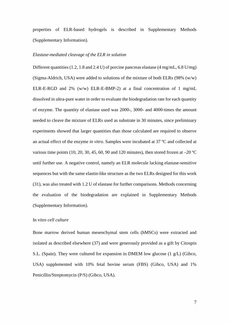
ELR Biosynthesis and characterization
Both ELRs were obtained as a lyophilized product in a yield of approximately 200 mg/L
(ELR/culture volume). Their molecular weights and purities were confirmed as
satisfactory by SDS-PAGE and MALDI-TOF (Fig. 1), while the Tt calculated by DSC
for the ELRs dissolved in PBS (pH 7.4) was found to be 15.8 ºC and 15.3 ºC for ELR-E-
RGD and ELR-E-BMP-2, respectively (Fig. S2, Supplementary Information).
FIG. 1. Molecular weight and purity assessment by SDS-PAGE and MALDI-TOF mass
spectrometry for ELR-E-RGD and ELR-E-BMP2. MALDI-TOF spectra represent non-
quantitative intensity (a.u.) against m/z (mass divided by net charge of the molecule) of
the ELRs.
12
As regards the mechanical characterization, the storage modulus (G’) of the ELR-based
hydrogel at a concentration of 300 mg/mL (98% ELR-E-RGD and 2% ELR-E-BMP-2)
was found to be approximately 1600 Pa at 37ºC (Fig. S5, Supplementary Information). In
addition, hydrogels were formed above the Tt, as observed macroscopically (Fig. S6,
Supplementary Information).
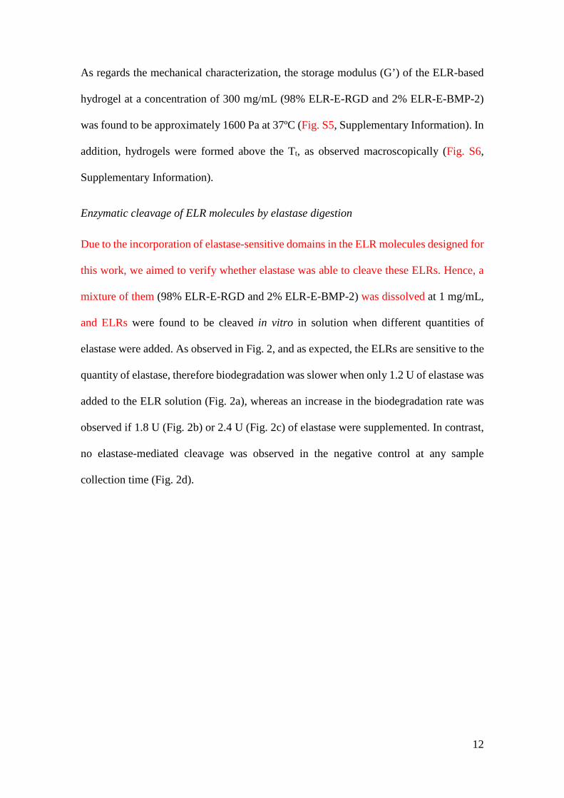
Enzymatic cleavage of ELR molecules by elastase digestion
Due to the incorporation of elastase-sensitive domains in the ELR molecules designed for
this work, we aimed to verify whether elastase was able to cleave these ELRs. Hence, a
mixture of them (98% ELR-E-RGD and 2% ELR-E-BMP-2) was dissolved at 1 mg/mL,
and ELRs were found to be cleaved in vitro in solution when different quantities of
elastase were added. As observed in Fig. 2, and as expected, the ELRs are sensitive to the
quantity of elastase, therefore biodegradation was slower when only 1.2 U of elastase was
added to the ELR solution (Fig. 2a), whereas an increase in the biodegradation rate was
observed if 1.8 U (Fig. 2b) or 2.4 U (Fig. 2c) of elastase were supplemented. In contrast,
no elastase-mediated cleavage was observed in the negative control at any sample
collection time (Fig. 2d).
13
FIG. 2. SDS-PAGE images showing the biodegradation of the mixture of ELR-E-RGD
(98%) and ELR-E-BMP-2 (2%) in solution at 1 mg/mL mediated by a) 1.2 U, b) 1.8 U
and c) 2.4 U of elastase, at different sample collection times, as indicated above each
picture (0, 10, 20, 30, 45, 60, 90 and 120 minutes after addition of the specific quantity
of elastase). Picture d) shows the lack of elastase-mediated biodegradation in the case of
the non-sensitive ELR. M represents the protein molecular weight marker.
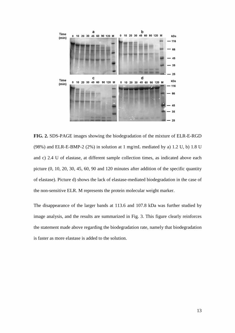
The disappearance of the larger bands at 113.6 and 107.8 kDa was further studied by
image analysis, and the results are summarized in Fig. 3. This figure clearly reinforces
the statement made above regarding the biodegradation rate, namely that biodegradation
is faster as more elastase is added to the solution.
14
FIG. 3. Graph showing the elastase-mediated cleavage rate of the highest molecular
weight band with data obtained from analysis of the SDS-PAGE gels from Fig. 2. The
net intensity of this double band at 113.6 and 107.8 kDa is represented at different
sampling times.
As regards the nascent bands observed by SDS-PAGE (Fig. 2), we expected to obtain
bands in three different molecular weight ranges, namely 65.5-66.5, 46.7-48.2, and 12-
12.9 kDa, as by-products of ELR-E-RGD/BMP-2 digestion since there are two different
elastase-sensitive domains at different points in the ELR-E-BMP-2 molecule. However,
the molecular weight of the higher bands was found to be 80.8 and 54.2 kDa, respectively,
while the band at 12-12.9 kDa could not be observed due to the limitations of SDS-PAGE
in terms of resolution. Nevertheless, these results correlate well with previous studies that
reported a 20% increase in the apparent molecular weight for different ELRs (40, 41). As
such, we estimated the Mw plus 20% and the values showed good agreement with those
found empirically, with the experimental values for the nascent bands being 80.8 and 54.2
kDa, while the expected values of Mw + 20% were 78.6-79.8 and 56.0-57.8, respectively
(Table S5, Supplementary Information).
15
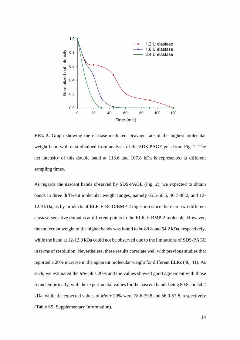
hMSCs viability and integrin-mediated cell adhesion
The viability of the cells after culture for 3 days in media supplemented with the ELRs
was found to be similar to that for the negative control, i.e. medium without
supplementation, as can be observed in Fig. 4. Since no significant differences were
observed, we can conclude that the ELRs alone, or the mixture thereof, do not affect cell
viability.
FIG. 4. Graph showing hMSCs viability results after 3 days of culture in terms of cell
number as measured using the Calcein AM assay for different ELR supplements in
medium at 10 mg/mL: ELR-E-RGD (represented as RGD), ELR-E-BMP-2 (BMP-2), the
mixture of both (98% (w/w) ELR-E-RGD and 2% (w/w) ELR-E-BMP-2, RGD/BMP-2)
and supplement-free medium (medium only). No significant differences (p > 0.05) were
found in any case.
Furthermore, the evaluation of cell adhesion in ELR-coated tissue culture plates (TCP)
showed good results in the case of ELR-E-RGD and the mixture of both. This finding
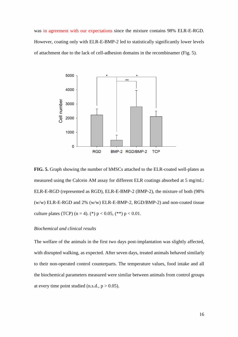
16
was in agreement with our expectations since the mixture contains 98% ELR-E-RGD.
However, coating only with ELR-E-BMP-2 led to statistically significantly lower levels
of attachment due to the lack of cell-adhesion domains in the recombinamer (Fig. 5).
FIG. 5. Graph showing the number of hMSCs attached to the ELR-coated well-plates as
measured using the Calcein AM assay for different ELR coatings absorbed at 5 mg/mL:
ELR-E-RGD (represented as RGD), ELR-E-BMP-2 (BMP-2), the mixture of both (98%
(w/w) ELR-E-RGD and 2% (w/w) ELR-E-BMP-2, RGD/BMP-2) and non-coated tissue
culture plates (TCP) (n = 4). (*) p < 0.05, (**) p < 0.01.
Biochemical and clinical results
The welfare of the animals in the first two days post-implantation was slightly affected,
with disrupted walking, as expected. After seven days, treated animals behaved similarly
to their non-operated control counterparts. The temperature values, food intake and all
the biochemical parameters measured were similar between animals from control groups
at every time point studied (n.s.d., p > 0.05).
17
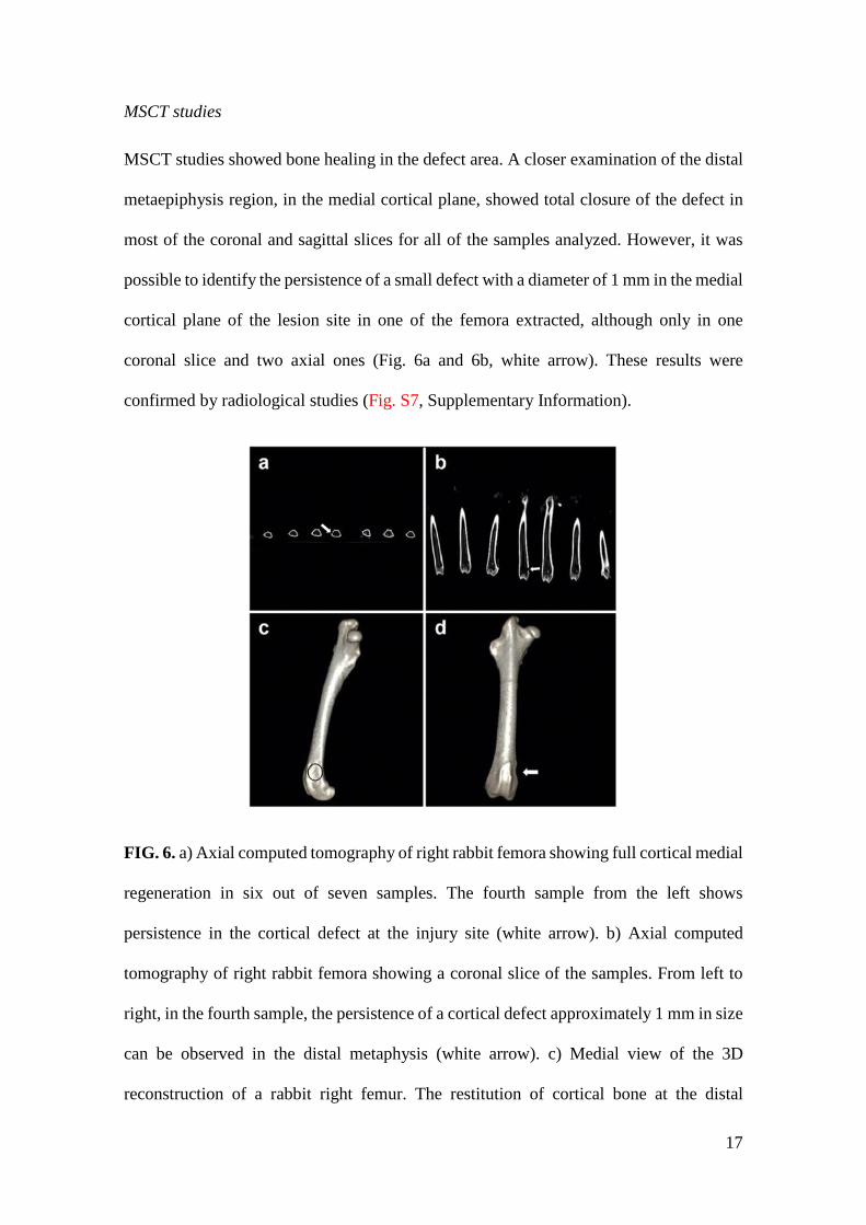
MSCT studies
MSCT studies showed bone healing in the defect area. A closer examination of the distal
metaepiphysis region, in the medial cortical plane, showed total closure of the defect in
most of the coronal and sagittal slices for all of the samples analyzed. However, it was
possible to identify the persistence of a small defect with a diameter of 1 mm in the medial
cortical plane of the lesion site in one of the femora extracted, although only in one
coronal slice and two axial ones (Fig. 6a and 6b, white arrow). These results were
confirmed by radiological studies (Fig. S7, Supplementary Information).
FIG. 6. a) Axial computed tomography of right rabbit femora showing full cortical medial
regeneration in six out of seven samples. The fourth sample from the left shows
persistence in the cortical defect at the injury site (white arrow). b) Axial computed
tomography of right rabbit femora showing a coronal slice of the samples. From left to
right, in the fourth sample, the persistence of a cortical defect approximately 1 mm in size
can be observed in the distal metaphysis (white arrow). c) Medial view of the 3D
reconstruction of a rabbit right femur. The restitution of cortical bone at the distal
18
metaphysis can be observed (black circumference). d) Medial view of the 3D
reconstruction of a rabbit right femur. In this case, the sample with the remaining partial
defect shows a small hollow with continuity (white arrow).
Bone restitution in the distal femoral metaphysis was also observed in the 3D
reconstructions of all samples, and the created defect could not be detected (injury site
indicated with a black circle; Fig. 6c), even in the case of the sample that showed a small
defect remaining in the axial and coronal slices. This 3D reconstruction showed a tiny
hollow (white arrow), but the processed signal correlates to cortical bone (Fig. 6d).
Histopathology results
The histological analytical results obtained for experimental samples showed de novo
bone formation in the experimental femoral injury (EFI) region. The new bone formed
was thick and comprised lamellar bone. In addition, it showed numerous vascular
channels of different calibers and was surrounded by various osteoblast layers (Fig. 7a).
Each bone layer was deposited on the remaining ELR hydrogel in a disorganized fashion
(Fig. 7b), resembling pagetoid-like bone (Fig. 7c), in which cellular activity produces a
mosaic pattern rather than the normal linear lamellar pattern.
19
FIG. 7. Microphotographs taken from decalcified femoral bone sections stained with
hematoxylin and eosin. a) Thick lamellar bone showing numerous vascular blood
channels (black asterisk), remnants of the ELR hydrogel (blue asterisk), hematopoietic
bone marrow and osteoblast layers (green asterisk) are observed in the EFI region.
Magnification 46.6x, bar = 1 mm. b) The high magnification images show the interface
between new lamellar bone (black asterisk) and the ELR hydrogel (red asterisk), with a
network aspect acting as a guide for cells. Magnification 233.4x, bar = 500 μm. c) At high
magnification, a few vascular channels are observed in new lamellar bone at the EFI.
Each layer of bone is deposited in the form of a “mosaic pattern”, resembling pagetoid-
like bone. Magnification 700.2x, bar= 250 μm. d) At high magnification the ELR
hydrogel shows mineralized amorphous regions surrounded by osteoblast-like cells
(black arrows). Micro-hemorrhage (red asterisk) and congestive vessels (grey asterisk)
are also observed. Magnification 700.2x bar= 250 μm. e) At low magnification, a
20
panoramic microphotography of the femoral epiphysis and metaphysis shows new bone
formed in the EFI region (black asterisk). This bone is surrounded by hematopoietic bone
marrow (green asterisk) with a few trabeculae (orange asterisk). Magnification 80x, bar
= 2 mm. f) A few bone nodules surrounding the EFI region are lined by several layers of
prominent osteoblasts (black arrows). Osteocytes (green arrows) are observed inside a
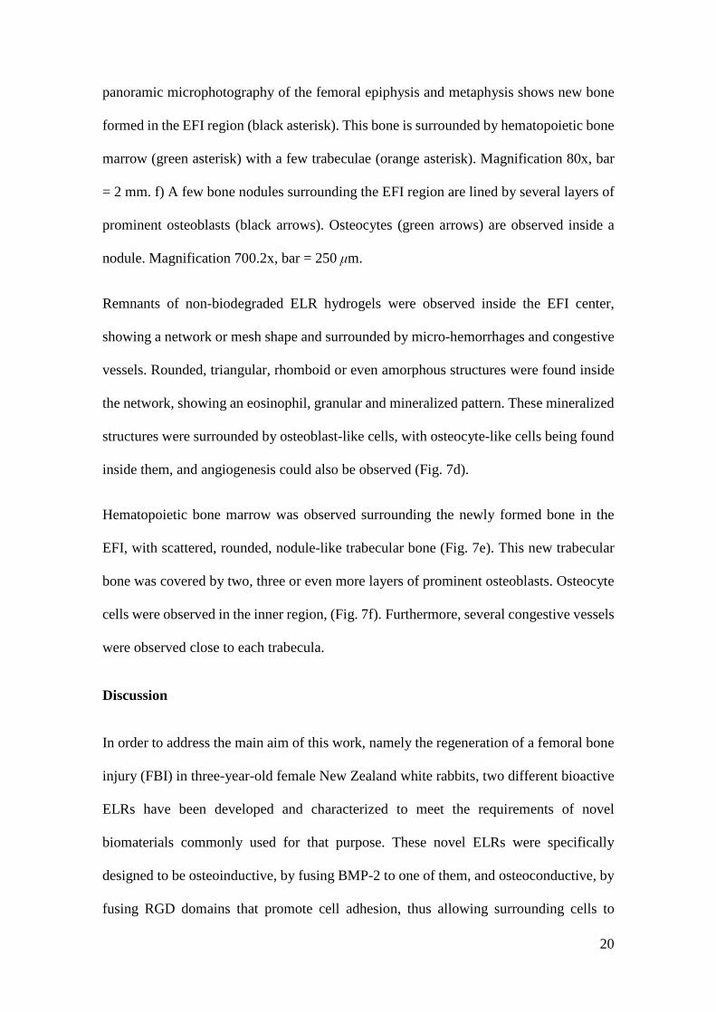
nodule. Magnification 700.2x, bar = 250 μm.
Remnants of non-biodegraded ELR hydrogels were observed inside the EFI center,
showing a network or mesh shape and surrounded by micro-hemorrhages and congestive
vessels. Rounded, triangular, rhomboid or even amorphous structures were found inside
the network, showing an eosinophil, granular and mineralized pattern. These mineralized
structures were surrounded by osteoblast-like cells, with osteocyte-like cells being found
inside them, and angiogenesis could also be observed (Fig. 7d).
Hematopoietic bone marrow was observed surrounding the newly formed bone in the
EFI, with scattered, rounded, nodule-like trabecular bone (Fig. 7e). This new trabecular
bone was covered by two, three or even more layers of prominent osteoblasts. Osteocyte
cells were observed in the inner region, (Fig. 7f). Furthermore, several congestive vessels
were observed close to each trabecula.
Discussion
In order to address the main aim of this work, namely the regeneration of a femoral bone
injury (FBI) in three-year-old female New Zealand white rabbits, two different bioactive
ELRs have been developed and characterized to meet the requirements of novel
biomaterials commonly used for that purpose. These novel ELRs were specifically
designed to be osteoinductive, by fusing BMP-2 to one of them, and osteoconductive, by
fusing RGD domains that promote cell adhesion, thus allowing surrounding cells to
21
interact with the hydrogel and possibly promote bone formation even from inside the
scaffold.
Initially it was shown that the Tt is lower than body temperature, which may permit the
formation of hydrogels once the ELR solution is injected into the body. In addition, this
Tt is similar to that described previously for the non-bioactive ELR, which was found to
be 13.0 ºC (42), although an increase of 2.8 and 2.3 ºC was observed for ELR-E-RGD
and ELR-E-BMP-2, respectively. This can be explained by the lower hydrophobicity of
the ELR molecule when other, more hydrophilic peptides or proteins containing charged
residues are fused to it (43, 44).
As regards the rheological data, although this system is intended to be used for bone
regeneration and the storage modulus is very low in comparison with bone tissue, this
hydrogel was designed to be able to promote cell invasion and proliferation inside itself,
acting as a temporary soft tissue that promotes optimal regeneration in a manner whereby
the implanted scaffold is substituted by host tissue. As such, although it may not be useful
on its own for treating large bone defects, it has been shown to be very suitable in the FBI
model used in this work since the hydrogel remains free from significant mechanical
stress (45).
Biodegradation of the ELR molecules in solution has been confirmed in vitro, thus
showing that this process can also be controlled by varying the quantity of elastase used.
Although this fails to imitate in vivo conditions, it sheds light onto the biodegradation
kinetics. The use of elastase-sensitive sequences should also allow the slow release of
BMP-2 from the ELR molecule to exert its biological effect. Consequently, the ELR-
based hydrogel acts as a drug-delivery system. Despite the fact that there are other
examples in which rhBMP-2 and ELRs are combined as an encapsulation system (46),
22
this approach allows a more efficient production and application by taking advantage of
recombinant DNA technology.
The excellent cell adhesion found on surfaces coated with ELR-E-RGD was similar to
that obtained in other studies using RGD-containing ELRs (47). As such, this work
demonstrates that the inclusion of RGD sequences in the final ELR molecule by genetic-
engineering methods promotes cell attachment and therefore provides a more ECM-
mimetic environment that is also osteoconductive. ELR-E-BMP-2-coated substrates did
not support cell adhesion due to the absence of cell-adhesion motifs in the ELR itself and
in BMP-2. With regard to cell viability, the lack of differences between the negative
control (medium only) and the media supplemented with the recombinamers is in
agreement with previous studies in which a cell-culture medium was supplemented with
ELRs (48).
As regards the clinical and biochemical results of the implant process, although initially
affected by the surgery per se, animal gait recovered rapidly to normal conditions. The
lack of change in the biochemical parameters showed that neither the surgical procedure
nor the subsequent possible matrix biodegradation had any effect on the animals, thus
showing good biocompatibility.
The images obtained in the tomographic study with 3D reconstruction of the samples
show promising results since the signal patterns processed in this work are correlated to
bone tissue with similar characteristics to the surrounding tissue, with complete closure
of the defect being achieved in six out of seven samples. Although a defect approximately
1 mm in diameter was still visible in the remaining animal, this was only the case in three
tomographic slices and may simply be a consequence of a lack of time for the regeneration
process in this particular animal. However, the bone formed had the same characteristics
23
as the other samples, therefore it can be concluded that these ELR-based matrices have a
high osteogenic potential to restitute a bone defect of 6 mm diameter and 6 mm depth ad
integrum in 90 days, most probably due to fusion of the BMP-2 protein to the ELR, which
results in a BMP-2-loaded hydrogel.
The histological analyses showed that the FBI was replaced by dense, new lamellar bone.
Although a few remnants of the ELR were observed at 90 days post-implantation, they
were surrounded by congestive vessels and dense laminar bone. This supports the
accepted knowledge whereby new bone is only formed in the presence of blood irrigation
(49). This new bone is arranged randomly, with an irregular arrangement in various
different directions, thus suggesting that the ELR-based hydrogels act as a carrier for
BMP-2, with osteoprogenitor cells colonizing these hydrogels, depositing osteoid matrix
and mineralizing as pagetoid-like bone, probably driven by the network arrangement of
the ELR-based hydrogels (50). The new trabeculae obtained show a peculiar shape, as if
they were obtained by the confluence of rounded isolated bone formations. The numerous
layers of prominent osteoblasts and various shapes observed, which appear to simulate
pseudo-stratification, could be a result of the activity of BMP-2 (22, 51, 52).
As observed in vivo from the microscopic results, the ELR-based hydrogel was found to
be biodegraded as bone formation occurred since the cells involved in this phenomenon
were stimulated by the BMP-2 released into the microenvironment, probably slowly
enough to allow the differentiation of stem and progenitor cells. As such, in this situation,
elastase (MMP-12) secretion by osteoclasts might be increased as a consequence of
matrix remodeling due to the formation of de novo bone tissue, as suggested before (53).
This could lead to degradation of the ELR-based hydrogel, which is sensitive to MMP-
12 as a result of inclusion of the VPVAPG sequence, as described previously (54). On
the other hand, ELRs without cleavable domains are not supposed to be biodegraded. In
24
this regard, Sallach et al. reported a long-term stability (up to 1 year) of a physically cross-
linked ELR-based hydrogel, similar to the one used in our work, when implanted in vivo
(55). In our case, biodegradation might happen simultaneously with bone regeneration,
thus resulting in a resorbable matrix that maintains bone integrity until full regeneration.
In addition, the peptides resulting from the degradation of VPVAPG have been reported
to exhibit a strong cell-proliferation activity that may promote tissue repair, as described
previously (56). Furthermore, RGD sequences provide anchoring points for cells that help
them to migrate and proliferate inside the scaffold, thereby promoting self-regeneration
of the damaged tissue.
Although several approaches have been developed in the field of tissue engineering, to
the best of our knowledge this is the first work describing the use of ELR-based hydrogels
for the successful regeneration of a bone defect in vivo. Previous examples make use of
ELRs in combination with other materials (57, 58), and most of them have only been
tested in vitro, although with promising results (59). Besides, the ELR-based hydrogel
described in this study overcomes different issues regarding the use of biomaterials in
bone tissue engineering. For instance, BMP-2 is not only loaded inside the hydrogel, but
it is part of it. Hence, there is no need to add this osteogenic factor during the preparation
of the scaffold, in contrast to other works (60), reducing its cost. In addition, this acellular
system has shown to be able to promote optimal bone regeneration, while other studies
report good outcomes only in the presence of mesenchymal stromal cells (61, 62). On the
other hand, another acellular scaffold has been described, showing its usefulness in bone
regeneration (63). However, this system is not injectable, and thus requires the use of
invasive methods for its implantation. Moreover, the adaptation of this scaffold to the
shape of the defect depends on the mold used in its development, reducing its versatility.
25
In conclusion, this work shows that a mixture of the originally designed ELRs is able to
self-assemble into an appropriate BMP-2 carrier, namely an injectable and biodegradable
hydrogel, which allows the slow release of this osteogenic factor, thereby stimulating
progenitor and stem cell differentiation and osteoblast proliferation. Furthermore, the
resulting ELR-based hydrogels also demonstrated an osteoconductive behavior since they
provide an ECM-like environment as a result of the inclusion of RGD sequences. These
two bioactivities (RGD and BMP-2), together with elastase-sensitiveness were easily
included in the final ELR molecules in a controlled manner, due to their recombinant
nature. Endogenous cells were able to migrate and proliferate into these hydrogels,
thereby favouring bone neo-formation at the femoral injury, as confirmed by computed
tomography, radiography and histology.
Acknowledgments
The authors are grateful for funding from the European Commission (NMP-2014-
646075, HEALTH-F4-2011-278557, PITN-GA-2012-317306 and MSCA-ITN-2014-
642687), the MINECO of the Spanish Government (MAT2013-42473-R and MAT2013-
41723-R), the Junta de Castilla y León (VA244U13 and VA313U14) and the Centro en
Red de Medicina Regenerativa y Terapia Celular de Castilla y León. Dante J. Coletta has
been funded by the Consejo Nacional de Investigaciones de Ciencia y Tecnología de la
Nación (CONICET, Argentina). We would also like to thank Dr. Pedro Esbrit, from the
Jiménez Díaz Foundation.
Author Disclosure Statement
No competing financial interests exist.
26
References
1. Amini, A.R., Laurencin, C.T., and Nukavarapu, S.P. Bone Tissue Engineering: Recent
Advances and Challenges. Critical reviews in biomedical engineering 40, 363, 2012.
2. Khademhosseini, A., and Langer, R. A decade of progress in tissue engineering. Nat
Protocols 11, 1775, 2016.
3. Yoseph, B.-C. Biomimetics. Biomimetics: CRC Press; 2005. pp. 495.
4. Costa, H.S., Mansur, A.A.P., Pereira, M.M., and Mansur, H.S. Engineered Hybrid
Scaffolds of Poly(vinyl alcohol)/Bioactive Glass for Potential Bone Engineering
Applications: Synthesis, Characterization, Cytocompatibility, and Degradation. Journal
of Nanomaterials 2012, 16, 2012.
5. Santolini, E., West, R., and Giannoudis, P.V. Risk factors for long bone fracture non-
union: a stratification approach based on the level of the existing scientific evidence.
Injury 46 Suppl 8, S8, 2015.
6. Chrcanovic, B.R., Albrektsson, T., and Wennerberg, A. Dental implants inserted in
male versus female patients: a systematic review and meta-analysis. Journal of oral
rehabilitation 42, 709, 2015.
7. Cointry, G.R., Capozza, R.F., Feldman, S., Reina, P., and Ferretti, J.L. Estructura y
biomecánica ósea. In: E. A., ed. Osteoporosis en Iberoamérica (2ª Ed). México DF
(México): El Manual Moderno; 2012. pp. 33.
8. Compston, J. Osteoporosis: social and economic impact. Radiologic clinics of North
America 48, 477, 2010.
27
9. Curran, D., Maravic, M., Kiefer, P., Tochon, V., and Fardellone, P. Epidemiology of
osteoporosis-related fractures in France: a literature review. Joint, bone, spine : revue du
rhumatisme 77, 546, 2010.
10. Cointry, G.R., Capozza, R.F., Feldman, S., Reina, P., Grappiolo, I., Ferretti, S.E.,
Mortarino, P., Chiappe, M.A., and Ferretti, J.L. Los huesos son estructuras genéticas,
metabólicas, biomecánicas, o todo a la vez. Actualizaciones en Osteología 5, 184, 2009.
11. Melton, L.J., 3rd, Achenbach, S.J., Atkinson, E.J., Therneau, T.M., and Amin, S.
Long-term mortality following fractures at different skeletal sites: a population-based
cohort study. Osteoporosis international : a journal established as result of cooperation
between the European Foundation for Osteoporosis and the National Osteoporosis
Foundation of the USA 24, 1689, 2013.
12. Kremenetzky, A., Kremenetzky, L., and Feldman, S. Aplicación de aloinjerto óseo
como cemento biológico. Revista de la Asociación Argentina de Ortopedia y
Traumatología 71, 61, 2006.
13. Alghazali, K.M., Nima, Z.A., Hamzah, R.N., Dhar, M.S., Anderson, D.E., and Biris,
A.S. Bone-tissue engineering: complex tunable structural and biological responses to
injury, drug delivery, and cell-based therapies. Drug metabolism reviews 47, 431, 2015.
14. Zhang, S., Zhang, X., Cai, Q., Wang, B., Deng, X., and Yang, X. Microfibrous beta-
TCP/collagen scaffolds mimic woven bone in structure and composition. Biomedical
materials 5, 065005, 2010.
15. Arealis, G., and Nikolaou, V.S. Bone printing: new frontiers in the treatment of bone
defects. Injury 46 Suppl 8, S20, 2015.
28
16. Salgado, A.J., Oliveira, J.M., Martins, A., Teixeira, F.G., Silva, N.A., Neves, N.M.,
Sousa, N., and Reis, R.L. Chapter One - Tissue Engineering and Regenerative Medicine:
Past, Present, and Future. In: Stefano Geuna I.P.P.T., Bruno B., eds. International Review
of Neurobiology: Academic Press; 2013. pp. 1.
17. Detsch, R., and Boccaccini, A.R. The role of osteoclasts in bone tissue engineering.
Journal of tissue engineering and regenerative medicine 9, 1133, 2015.
18. van der Stok, J., Lozano, D., Chai, Y.C., Amin Yavari, S., Bastidas Coral, A.P.,
Verhaar, J.A.N., Gómez-Barrena, E., Schrooten, J., Jahr, H., Zadpoor, A.A., Esbrit, P.,
and Weinans, H. Osteostatin-Coated Porous Titanium Can Improve Early Bone
Regeneration of Cortical Bone Defects in Rats. Tissue Engineering Part A 21, 1495, 2015.
19. Romagnoli, C., D’Asta, F., and Brandi, M.L. Drug delivery using composite scaffolds
in the context of bone tissue engineering. Clinical Cases in Mineral and Bone Metabolism
10, 155, 2013.
20. Inoda, H., Yamamoto, G., and Hattori, T. Histological investigation of osteoinductive
properties of rh-BMP2 in a rat calvarial bone defect model. Journal of Cranio-
Maxillofacial Surgery 32, 365, 2004.
21. Chen, D., Zhao, M., and Mundy, G.R. Bone morphogenetic proteins. Growth factors
22, 233, 2004.
22. G. dos S. Kotake, B., M. P. Salzedas, L., Ervolino, E., A. J. Calzzani, R., Sebald, W.,
and P. M. Issa, J. Bone Recuperation After rhBMP-2 Insertion in Alcoholic Animals-
Experimental Study. Current Pharmaceutical Design 21, 3557, 2015.
23. Siéssere, S., de Sousa, L.G., Issa, J.P.M., Iyomasa, M.M., Pitol, D.L., Barbosa,
A.P.A., Semprini, M., Sebald, W., Bentley, M.V.B., and Regalo, S.C.H. Application of
29
Low-Level Laser Irradiation (LLLI) and rhBMP-2 in Critical Bone Defect of
Ovariectomized Rats: Histomorphometric Evaluation. Photomedicine and Laser Surgery
29, 453, 2011.
24. Abdala, P.M.F., Iyomasa, M.M., Sato, S., Bentley, M.V.L.B., Pitol, D.L., Regalo,
S.C.H., Siéssere, S., and Issa, J.P.M. Osteoinductivity potential of rhBMP-2 associated
with two carriers in different dosages. Anatomical Science International 85, 181, 2010.
25. Issa, J.P.M., do Nascimento, C., Bentley, M.V.L.B., Del Bel, E.A., Iyomasa, M.M.,
Sebald, W., and de Albuquerque Jr, R.F. Bone repair in rat mandible by rhBMP-2
associated with two carriers. Micron 39, 373, 2008.
26. Haidar, Z.S., Hamdy, R.C., and Tabrizian, M. Delivery of recombinant bone
morphogenetic proteins for bone regeneration and repair. Part A: Current challenges in
BMP delivery. Biotechnology Letters 31, 1817, 2009.
27. Schmidmaier, G., Schwabe, P., Strobel, C., and Wildemann, B. Carrier systems and
application of growth factors in orthopaedics. Injury 39, S37, 2008.
28. Girotti, A., Orbanic, D., Ibáñez-Fonseca, A., Gonzalez-Obeso, C., and Rodríguez-
Cabello, J.C. Recombinant Technology in the Development of Materials and Systems for
Soft-Tissue Repair. Advanced Healthcare Materials 4, 2423, 2015.
29. Rodríguez-Cabello, J.C., Martín, L., Alonso, M., Arias, F.J., and Testera, A.M.
“Recombinamers” as advanced materials for the post-oil age. Polymer 50, 5159, 2009.
30. Urry, D.W. Molecular Machines: How Motion and Other Functions of Living
Organisms Can Result from Reversible Chemical Changes. Angewandte Chemie
International Edition in English 32, 819, 1993.
30
31. Martin, L., Arias, F.J., Alonso, M., Garcia-Arevalo, C., and Rodriguez-Cabello, J.C.
Rapid micropatterning by temperature-triggered reversible gelation of a recombinant
smart elastin-like tetrablock-copolymer. Soft Matter 6, 1121, 2010.
32. Ruoslahti, E., and Pierschbacher, M.D. Arg-Gly-Asp: a versatile cell recognition
signal. Cell 44, 517, 1986.
33. Lombard, C., Arzel, L., Bouchu, D., Wallach, J., and Saulnier, J. Human leukocyte
elastase hydrolysis of peptides derived from human elastin exon 24. Biochimie 88, 1915,
2006.
34. Goy, D.P., Gorosito, E., Costa, H.S., Mortarino, P., Pedemonte, N.A., Toledo, J.,
Mansur, H.S., Pereira, M.M., Battaglino, R., and Feldman, S. Hybrid matrix grafts to
favor tissue regeneration in rabbit femur bone lesions. The open biomedical engineering
journal 6, 85, 2012.
35. Coletta, D.J., Lozano, D., Rocha-Oliveira, A.A., Mortarino, P., Bumaguin, G.E.,
Vitelli, E., Vena, R., Missana, L., Jammal, M.V., Portal-Nunez, S., Pereira, M., Esbrit,
P., and Feldman, S. Characterization of Hybrid Bioactive Glass-polyvinyl Alcohol
Scaffolds Containing a PTHrP-derived Pentapeptide as Implants for Tissue Engineering
Applications. The open biomedical engineering journal 8, 20, 2014.
36. Rodríguez-Cabello, J.C., Girotti, A., Ribeiro, A., and Arias, F.J. Synthesis of
Genetically Engineered Protein Polymers (Recombinamers) as an Example of Advanced
Self-Assembled Smart Materials. In: Navarro M., Planell J.A., eds. Nanotechnology in
Regenerative Medicine: Methods and Protocols. Totowa, NJ: Humana Press; 2012. pp.
17.
31
37. Orozco, L., Munar, A., Soler, R., Alberca, M., Soler, F., Huguet, M., Sentis, J.,
Sanchez, A., and Garcia-Sancho, J. Treatment of knee osteoarthritis with autologous
mesenchymal stem cells: a pilot study. Transplantation 95, 1535, 2013.
38. McKay, W.F., Peckham, S.M., and Badura, J.M. A comprehensive clinical review of
recombinant human bone morphogenetic protein-2 (INFUSE® Bone Graft). International
Orthopaedics 31, 729, 2007.
39. Feldman, S., Cointry, G.R., Leite Duarte, M.a.E., Sarrió, L., Ferretti, J.L., and
Capozza, R.F. Effects of hypophysectomy and recombinant human growth hormone on
material and geometric properties and the pre- and post-yield behavior of femurs in young
rats. Bone 34, 203, 2004.
40. Meyer, D.E., and Chilkoti, A. Genetically Encoded Synthesis of Protein-Based
Polymers with Precisely Specified Molecular Weight and Sequence by Recursive
Directional Ligation: Examples from the Elastin-like Polypeptide System.
Biomacromolecules 3, 357, 2002.
41. McPherson, D.T., Xu, J., and Urry, D.W. Product Purification by Reversible Phase
Transition Following Escherichia coli Expression of Genes Encoding up to 251 Repeats
of the Elastomeric Pentapeptide GVGVP. Protein Expression and Purification 7, 51,
1996.
42. Fernandez-Colino, A., Arias, F.J., Alonso, M., and Rodriguez-Cabello, J.C. Self-
organized ECM-mimetic model based on an amphiphilic multiblock silk-elastin-like
corecombinamer with a concomitant dual physical gelation process. Biomacromolecules
15, 3781, 2014.
32
43. Christensen, T., Hassouneh, W., Trabbic-Carlson, K., and Chilkoti, A. Predicting
Transition Temperatures of Elastin-Like Polypeptide Fusion Proteins.
Biomacromolecules 14, 1514, 2013.
44. Ibáñez-Fonseca, A., Alonso, M., Arias, F.J., and Rodríguez-Cabello, J.C. Förster
Resonance Energy Transfer-Paired Hydrogel Forming Silk-Elastin-Like Recombinamers
by Recombinant Conjugation of Fluorescent Proteins. Bioconjugate Chemistry 2017.
45. Gibbs, D.M.R., Black, C.R.M., Dawson, J.I., and Oreffo, R.O.C. A review of
hydrogel use in fracture healing and bone regeneration. Journal of tissue engineering and
regenerative medicine 10, 187, 2016.
46. Bessa, P.C., Machado, R., Nurnberger, S., Dopler, D., Banerjee, A., Cunha, A.M.,
Rodriguez-Cabello, J.C., Redl, H., van Griensven, M., Reis, R.L., and Casal, M.
Thermoresponsive self-assembled elastin-based nanoparticles for delivery of BMPs.
Journal of controlled release : official journal of the Controlled Release Society 142, 312,
2010.
47. de Torre, I.G., Wolf, F., Santos, M., Rongen, L., Alonso, M., Jockenhoevel, S.,
Rodriguez-Cabello, J.C., and Mela, P. Elastin-like recombinamer-covered stents:
Towards a fully biocompatible and non-thrombogenic device for cardiovascular diseases.
Acta biomaterialia 12, 146, 2015.
48. Pina, M.J., Girotti, A., Santos, M., Rodriguez-Cabello, J.C., and Arias, F.J.
Biocompatible ELR-Based Polyplexes Coated with MUC1 Specific Aptamers and
Targeted for Breast Cancer Gene Therapy. Molecular pharmaceutics 13, 795, 2016.
49. Tomlinson, R.E., and Silva, M.J. Skeletal Blood Flow in Bone Repair and
Maintenance. Bone Research 1, 311, 2013.
33
50. Missana, L., Nagai, N., and Kuboki, Y. Comparative histological studies of bone and
cartilage formations induced by various BMP-carrier composites. Journal of Oral
Biosciences 36, 9, 1994.
51. Issa, J.P.M., Defino, H.L.A., Netto, J.C., Volpon, J.B., Regalo, S.C.H., Iyomasa,
M.M., Siéssere, S., and Tiossi, R. Evaluation of rhBMP-2 and Natural Latex as Potential
Osteogenic Proteins in Critical Size Defects by Histomorphometric Methods. The
Anatomical Record: Advances in Integrative Anatomy and Evolutionary Biology 293,
794, 2010.
52. Issa, J.P.M., Do Nascimento, C., Lamano, T., Iyomasa, M.M., Sebald, W., and De
Albuquerque Jr, R.F. Effect of recombinant human bone morphogenetic protein-2 on
bone formation in the acute distraction osteogenesis of rat mandibles. Clinical Oral
Implants Research 20, 1286, 2009.
53. Hou, P., Troen, T., Ovejero, M.C., Kirkegaard, T., Andersen, T.L., Byrjalsen, I.,
Ferreras, M., Sato, T., Shapiro, S.D., Foged, N.T., and Delaissé, J.-M. Matrix
metalloproteinase-12 (MMP-12) in osteoclasts: new lesson on the involvement of MMPs
in bone resorption. Bone 34, 37, 2004.
54. Taddese, S., Weiss, A.S., Jahreis, G., Neubert, R.H.H., and Schmelzer, C.E.H. In
vitro degradation of human tropoelastin by MMP-12 and the generation of matrikines
from domain 24. Matrix Biology 28, 84, 2009.
55. Sallach, R.E., Cui, W., Balderrama, F., Martinez, A.W., Wen, J., Haller, C.A., Taylor,
J.V., Wright, E.R., Long Jr, R.C., and Chaikof, E.L. Long-term biostability of self-
assembling protein polymers in the absence of covalent crosslinking. Biomaterials 31,
779, 2010.
34
56. Rodriguez-Cabello, J., Ribeiro, A., Reguera, J., Girotti, A., and Testera, A. 14 -
Elastin-like systems for tissue engineering. Natural-Based Polymers for Biomedical
Applications: Woodhead Publishing; 2008. pp. 374.
57. Tejeda-Montes, E., Klymov, A., Nejadnik, M.R., Alonso, M., Rodriguez-Cabello,
J.C., Walboomers, X.F., and Mata, A. Mineralization and bone regeneration using a
bioactive elastin-like recombinamer membrane. Biomaterials 35, 8339, 2014.
58. Prieto, S., Shkilnyy, A., Rumplasch, C., Ribeiro, A., Arias, F.J., Rodríguez-Cabello,
J.C., and Taubert, A. Biomimetic Calcium Phosphate Mineralization with Multifunctional
Elastin-Like Recombinamers. Biomacromolecules 12, 1480, 2011.
59. Vila, M., García, A., Girotti, A., Alonso, M., Rodríguez-Cabello, J.C., González-
Vázquez, A., Planell, J.A., Engel, E., Buján, J., García-Honduvilla, N., and Vallet-Regí,
M. 3D silicon doped hydroxyapatite scaffolds decorated with Elastin-like
Recombinamers for bone regenerative medicine. Acta biomaterialia 45, 349, 2016.
60. Perez, R.A., Kim, J.-H., Buitrago, J.O., Wall, I.B., and Kim, H.-W. Novel therapeutic
core–shell hydrogel scaffolds with sequential delivery of cobalt and bone morphogenetic
protein-2 for synergistic bone regeneration. Acta biomaterialia 23, 295, 2015.
61. Jo, S., Kim, S., Cho, T.H., Shin, E., Hwang, S.J., and Noh, I. Effects of recombinant
human bone morphogenic protein-2 and human bone marrow-derived stromal cells on in
vivo bone regeneration of chitosan–poly(ethylene oxide) hydrogel. Journal of Biomedical
Materials Research Part A 101A, 892, 2013.
62. Ren, Z., Wang, Y., Ma, S., Duan, S., Yang, X., Gao, P., Zhang, X., and Cai, Q.
Effective Bone Regeneration Using Thermosensitive Poly(N-Isopropylacrylamide)

35
Grafted Gelatin as Injectable Carrier for Bone Mesenchymal Stem Cells. ACS Applied
Materials & Interfaces 7, 19006, 2015.
63. Nguyen, T.B.L., and Lee, B.-T. A Combination of Biphasic Calcium Phosphate
Scaffold with Hyaluronic Acid-Gelatin Hydrogel as a New Tool for Bone Regeneration.
Tissue Engineering Part A 20, 1993, 2014.