Embed Size (px)
Citation preview

Kidney regeneration
Serena M Bagnasco, MD
Department of Pathology, Johns Hopkins School of Medicine
Baltimore, Maryland, USA

• 1- There is no commercial or financial interest related to my presentation / our research.
• 2- My presentation does not comprise any discussion or advertising beyond its purpose.
28th National Congress of Pathology - Declaration of Interest

No formation of new nephrons occurs after birth
The term “regenerative medicine” is used to describe anybiomedical approach to the replacement or regeneration
of human tissues or organs for therapeutic purposes.

No formation of new nephrons occurs after birth
The term “regenerative medicine” is used to describe anybiomedical approach to the replacement or regeneration
of human tissues or organs for therapeutic purposes.
Potential approaches to kidney regeneration
• Models for artificial kidney structures• Bio-artificial kidney: cells grown on artificial support • Decellularized extracellular renal matrix scaffolds with repopulation by renal cells• Microphysiological engineering of endothelial and tubular channels• Embrional/fetal stem cells infusion• Pluripotent stem cells (iPSC) induced from somatic cells • Organoids

Sochol et al. Curr Transplant Rep. 3:82-92,2016
Models of artificial kidney structures
a, b single-layer microfluidic systemsc microfluidic systems with two chambers
separated by a permeable membraned singular, straight tubular structure

Humes et al. Tissue engineering of a bioartificial renal tubule assist device: In vitro transport and metabolic characteristics.Kidney International, Vol. 55 (1999), pp. 2502–2514
• high-flux hollow-fiber hemofiltration cartridges (Fresenius, membrane surface areas of 97 cm2 or 0.4 m2), • coated with pronectin-L, • seeded with pig kidney proximal tubular primary cultured cells
Functional characterization of renal cellular activity: • presence of NaK-ATPase dependent transport, • bicarbonate and glucose reabsorption from lumen, • γ-glutamyltranspeptidase activity , • anion (PAH) transport to lumen, • ammoniagenesis, • vitamin D activation

Fig. 1 Schematic of an extracorporeal circuit of a bioartificialkidney (BAK) used to treat patients with acute kidney injury. The first cartridge is a hemofiltration cartridge in series with a renal tubule cellassist device (RAD). The ultrafiltrate is delivered to the luminal compartment of the RAD, which contains the cells and the post-filtered blood is pumped into the extracapillary space of the RAD. The processed luminal ultrafiltrate from the RAD is discarded to waste and the processed blood is returned to the patient
Cellassist device: human tubular cells from discarded donor kidneys grown as confluent monolayer on semipermeable hollow fibers coated with extracellular biomatrix packaged into cartridges
Renal tubule-assist device (RAD)
Ultrafitrate
Post-filtered blood
Luminal ultrafitratediscarded
Processed bloodReturned to patient
Blood

Phase I and II clinical experience with a human renal tubule–assist (RAD) device
Food and Drug Administration approved an Investigational New Drug application to study the renal tubule cellassist device RAD containing human cells in patients with AKI.
• Phase I trial demonstrated that this experimental treatment can be delivered safely under study protocol guidelines in this critically ill patient population for up to 24 h when used in conjunction with continuous venovenous hemofiltration (CVVH).
• Phase II trial in 58 patients with AKI requiring CVVH in the ICU were randomized (2:1) to receive CVVH + RAD (n=40) or CVVH alone (n=18).
• RAD treatment for up to 72 h promoted a statistically significant survival advantage over 180 days of follow-up: RAD 67 % survival rate versus control 39 % and demonstrated an acceptable safety profile.
Technological hurdles to development and commercialization of cell-based RAD
• Need for a reliable and consistent source of cells to manufacture thousands of these cell devices • Limited life span of cells • Reliable performance• Cost-effective manufacturing, storage, and distribution process for these devices
Humes et al. Pediatric Nephrology 29: 342, 2014

2016

Microvascular and microtubular bioengineered systems
Artificial microvascular systems
J Am Soc Nephrol 27: 2370–2381, 2016.

FIG. 1. Decellularization and matrix characterization of human kidney cortex. (A) Overview of the decellularization processbeginning with whole tissue (A.1), sectioning (A.2), and chemical processing at 1 h (A.3), 24h (A.4), and 120h (A.5), thenrinsing with water for 24 h (A.6) and 120 h (A.7). (B, C) Histological evaluation of untreated and decellularized kidneysections. (B) Hematoxylin and eosin staining reveals the removal of nuclei, but preservation of structural integrity of the ECMfollowing decellularization, including glomerular structures (asterisks). Scale bar, 100 mm. (C) Matrix protein expression ofFN, HSPG, LAM, Col-IV, and VN. Scale bar, 50 mm. Col-IV, collagen-IV; ECM, extracellular matrix; FN, fibronectin; HSPG,heparan sulfate proteoglycan; LAM, laminin; VN, versican. Color images available online at www.liebertpub.com/tea
Nagao et al. Tissue engineering, 2016
Matrix proteinswith cells
Matrix proteins with NO cells
• Decellularized matrix lyophilized for storage
• Reconstituted matrix gel• used to make microchannel
structures
• Microchannel structures seeded with cells

Zheng, Y. et al. Proc Natl Acad Sci U S A 109, 9342, 2012.
Schema for microvessels fabrication with organic matrix

Nagao et al. Tissue engineering, 2016
Reconstructed human kidney peritubular microvessels were fenestrated in vitroLigresti et al. J Am Soc Nephrol , 2016

Development of a microphysiological model of human kidney proximal tubule function.Elijah J. Weber, Alenka Chapron, Brian D. Chapron, Jenna L. Voellinger, Kevin A. Lidberg, Catherine K. Yeung, Zhican Wang, Yoshiyuki Yamaura, Dale W. Hailey, Thomas Neumann,Danny D. Shen, Kenneth E. Thummel, Kimberly A. Muczynski, Jonathan Himmelfarb and Edward J. Kelly
Kidney International (2016) 90, 627–637
Figure 1 (a) Scheme depicting construction of human proximal tubular epithelial cells (PTECs) in a microphysiologicalsystem (MPS). (A1) Cell isolation from human kidney cortex. (A2) Cell culture in 2 dimensions. (A3) Cell seeding and culture in 3-dimensional (3D) MPS. (A4) Phase contrast and viability of PTECs in MPS at day 28.
cell grown on collagen matrix, in perfused120 µm tubular channels

Weber et al. Kidney International (2016) 90, 627–637
Tests for proximal tubules phenotype in PTEC cells growing in microtubules, maintained for up to 28 days
Structural features:• Ultrastructural evidence of tight junctions, microvilli on the apical surface, basolateral interdigitations between
neighboring cells.• Evidence of polarization by localization of the tight junction protein ZO-1 to the luminal (apical) aspect, and
localization of Na+/K+ adenosine triphosphatase (ATPase) in basolateral border• Cilia formation in response to fluid shear stress• Surface expression of CD13 (aminopeptidase-N), E-cadherin, aquaporin-1
Functional features:• γ-glutamyl transpeptidase (GGT) immunolocalization in the luminal aspect of the PTEC cells,
and evidence of catalytic activity of GGT• Glucose transporter SGLT2 immunolocalization in the apical surface,
and evidence of cellular uptake of fluorescent glucose analog• Evidence that ATP is generated mostly by oxidative phosphorylation• Evidence of secretion of ammonia in response to decreased pH in the perfusate• Evidence of metabolization of 25-OH vitamin D3 (calcidiol) into bioactive 1α,25-(OH)2 vitamin D3 (calcitriol)• Evidence for presence of organic anion transporters: transport of organic anion paraminohippurate (PAH) and
anionic uremic solute indoxyl sulfate into luminal fluid
• Could not demonstrate Na+H+ exchange and NA+K+ ATPase activity
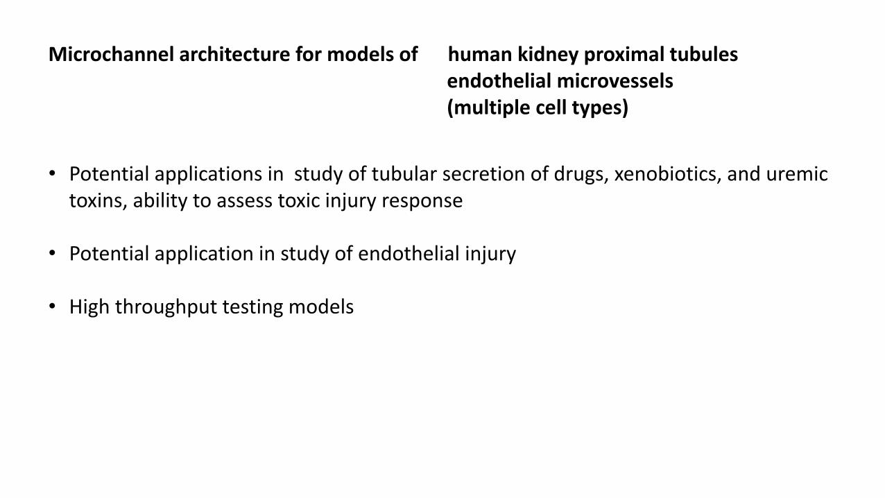
Microchannel architecture for models of human kidney proximal tubulesendothelial microvessels(multiple cell types)
• Potential applications in study of tubular secretion of drugs, xenobiotics, and uremic toxins, ability to assess toxic injury response
• Potential application in study of endothelial injury
• High throughput testing models

Sochol et al. Curr Transplant Rep. 3:82-92,2016
3D printing
More complex models of artificial kidney microstructures
Non-planar physical structures are difficult or impossible to reconstitute via conventional micromachining processes.
Submillimeter-scale 3D printing techniques are available with characteristics relevant to kidney-on-a-chip systems Stereolithography-Based Microsystems (SLA)
Fugitive ink 3D vascular network (i) micronozzles are used to 3D print a fugitive organic ink
network, (ii) the fugitive ink scaffold is infiltrated with a surrounding
liquid resin, (iii) the surrounding resin is cured, (iv) heat is applied to the fugitive organic ink to enable
vacuum-based extraction, thereby leaving behind a 3D network of hollow, interconnected microfluidic channels.
Resin/matrix
ink

Sochol et al. Curr Transplant Rep. 3:82-92,2016

Koletsky et al. 3D bioprinting of vascularized, heterogeneous cell-laden tissue constructs.Advanced Material 26:3124-3130, 2014
Poly(dimethyl siloxane) (PDMS) inks are removed leaving a 3D vascular network embedded in a collagen based matrix (GelMA), compatible with cell culture.

Cells seeded into 3D printer microstructures:• Red fluorescence protein expressing human
umbilical vein endothelial cell (HUVEC)• Green fluorescent protein expressing human
neonatal dermal fibroblasts (HNDFs)• 10T1/2 fibroblast cells
Koletsky et al. 3D bioprinting of vascularized, heterogeneous cell-laden tissue constructs.Advanced Material 26:3124-3130, 2014

Regenerative medicine in kidney diseaseMelissa H. Little and Pamela KairathKidney International (2016) 90: 289–299
Cell types potentially involved in repair/regeneration
• Renal progenitor cells (RPCs) expressing CD24 and CD133 in the human kidney can proliferate to repair acute tubular injury
• Mesenchymal stem cells (MSCs) from the bone marrow can reduce kidney damage, probably through immunomodulatory effects and release of growth factors, can have potential maldifferentiation during chronic injury
• Macrophages: M1 type pro-inflammatory: release of proinflammatory cytokines, reactive oxygen species, and nitric oxideM2 type anti-inflammatory: reparative role in ischemia reperfusion
• Human fetal stem cells: embryonic stem cells (ESCs) or induced pluripotent stem cells (iPSCs)iPSCs can be derived from adult somatic cells, potential source of autologous regenerative treatments
Cells

iPSC are pluripotent cells derived from somatic cells in which overexpression of 4 transcription factors Oct3/4, Sox2, Klf14, and c-myc has been induced, after
transfection with retrovirus *
Takahashi K, Yamanaka S. et al.
Induction of pluripotent stem cells from mouse embryonic and adult fibroblast cultures by defined factors. Cell. 2006;126:663–676.
Takahashi K, Tanebe K, Ohnuki M, et al.
Induction of pluripotent stem cells from adult human fibroblasts by defined factors. Cell. 2007;131:861–872.
iPSC (induced pluripotent stem cells )

Culturing iPSCs in a medium with stepwise addition of specific growth factors regulating kidney development can produce rudimentary
nephron-like structures termed “organoids”
Takasato M, Er PX, Chiu HS, et al.
Kidney organoids from human iPS cells contain multiple lineages and model human
nephrogenesis.
Nature. 2015;526:564–568.
Organoids from iPSC
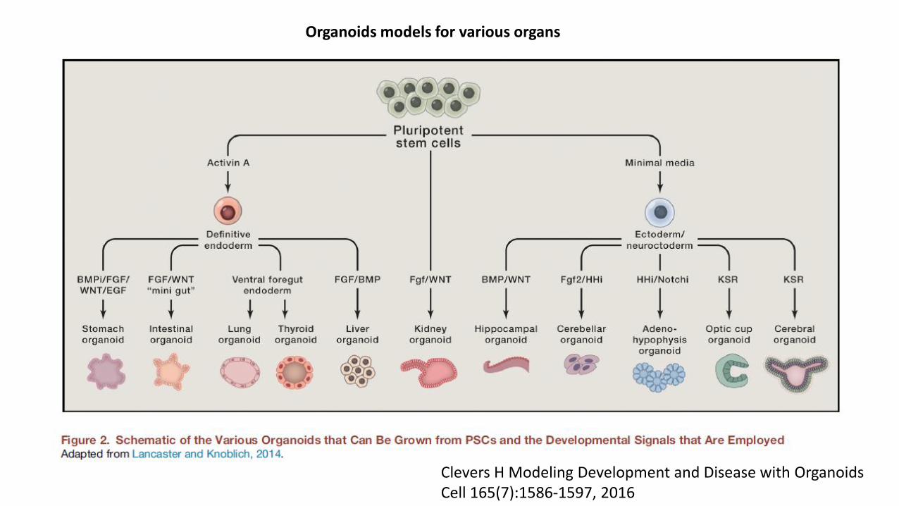
Clevers H Modeling Development and Disease with OrganoidsCell 165(7):1586-1597, 2016
Organoids models for various organs

A strategy for generating kidney organoids: Recapitulating the development in human pluripotent stem cells Minoru Takasato, Melissa H.LittleDevelopmental Biology 420: (2016) 210–220
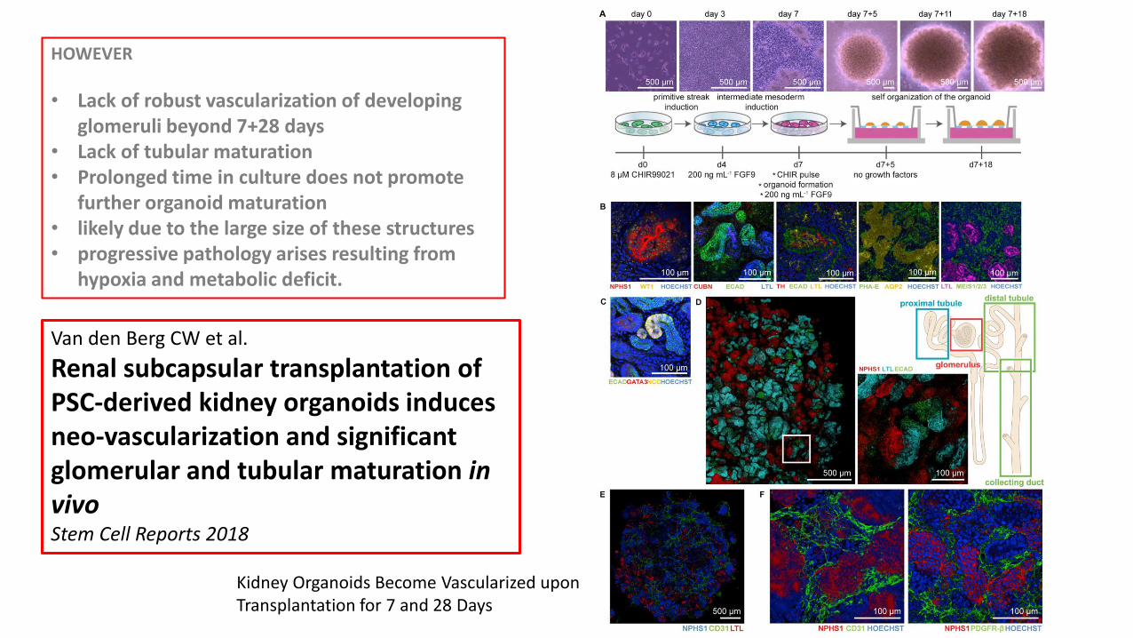
Directed differentiationThe stepwise differentiation begins with 4 days of CHIR99021 treatment for induction of primitive streak (PS) (CHIR99021 a GSK3 inhibitor that forcibly activates canonical Wnt signalling ) Followed by 3 days of FGF9 treatment for simultaneous induction of anterior and posterior intermediate mesoderm (IM).
At day 7, IM cells are trypsinized and cultured in an aggregate. Aggregates are stimulated by a CHIR99021 pulse to induce nephrogenesis, then cultured in medium with FGF9 for 5 days.
Growth factors are eventually withdrawn from the medium to allow cells to undergo self-organization forming kidney organoids in another 13 days (a phase contrast image: scale=1 mm).
(B) Immunofluorescence shows the presence of each kidney cell type in kidney organoids, including collecting ducts (ECAD, PAX2, GATA3), distal tubules (ECAD-positive, GATA3 and LTL-negative), loops of Henle (UMOD), proximal tubules (LTL, CUBN), glomeruli (NPHS1, WT1) with basement membrane (LAM), endothelialnetworks (CD31, SOX17) and renal interstitial cells (MEIS1). Cell nuclei (blue) were labeled with DAPI. Scale=50 µm.

Kidney Organoids Become Vascularized upon Transplantation for 7 and 28 Days
HOWEVER
• Lack of robust vascularization of developing glomeruli beyond 7+28 days
• Lack of tubular maturation• Prolonged time in culture does not promote
further organoid maturation • likely due to the large size of these structures • progressive pathology arises resulting from
hypoxia and metabolic deficit.
Van den Berg CW et al.
Renal subcapsular transplantation of PSC-derived kidney organoids induces neo-vascularization and significant glomerular and tubular maturation in vivoStem Cell Reports 2018

Van den Berg CW et al. Stem Cell Reports 2018
Bisected day 7 + 18 kidney organoidswere transplanted under the renal capsule of recipient immunocompromised mice for up to28 days.
(A) Concentration of VEGF (pg mL) in the supernatant of three cultured organoids measured weekly fromday 7 + 10 until day 7 + 52.
(B) Transplanted human kidney organoid under renal capsule of mice on the day of transplantation (tx), after 7 and 28 days showing growth upon vascularization.
C) Toluidine blue staining of organoid under the renal capsule after 28 days of transplantation. Boxed areas highlight glomerular andtubular structures displayed on the right.
(D) Scanning electron microscopy images suggests blood vessels in the kidney organoid after transplantation and inside a glomerularstructure. Close-up views of boxed areas are displayed.

Van den Berg CW et al. Stem Cell Reports 2018
ELeft -Human nuclei and LAMININ (LAM) in the organoid under the renal capsule of a mouse kidney
Right -integration of mouse endothelial cells (MECA-32+) in the organoid and glomerular structures
Fmouse endothelial cells MECA-32+ in glomeruli NPHS1+, WT1+
GPeritubular vascularization with mouse MECA-32+ endothelial cells aligning tubular (CUBN+) structures.
Vascularization of subcapsular human organoids by mouse endothelial cells
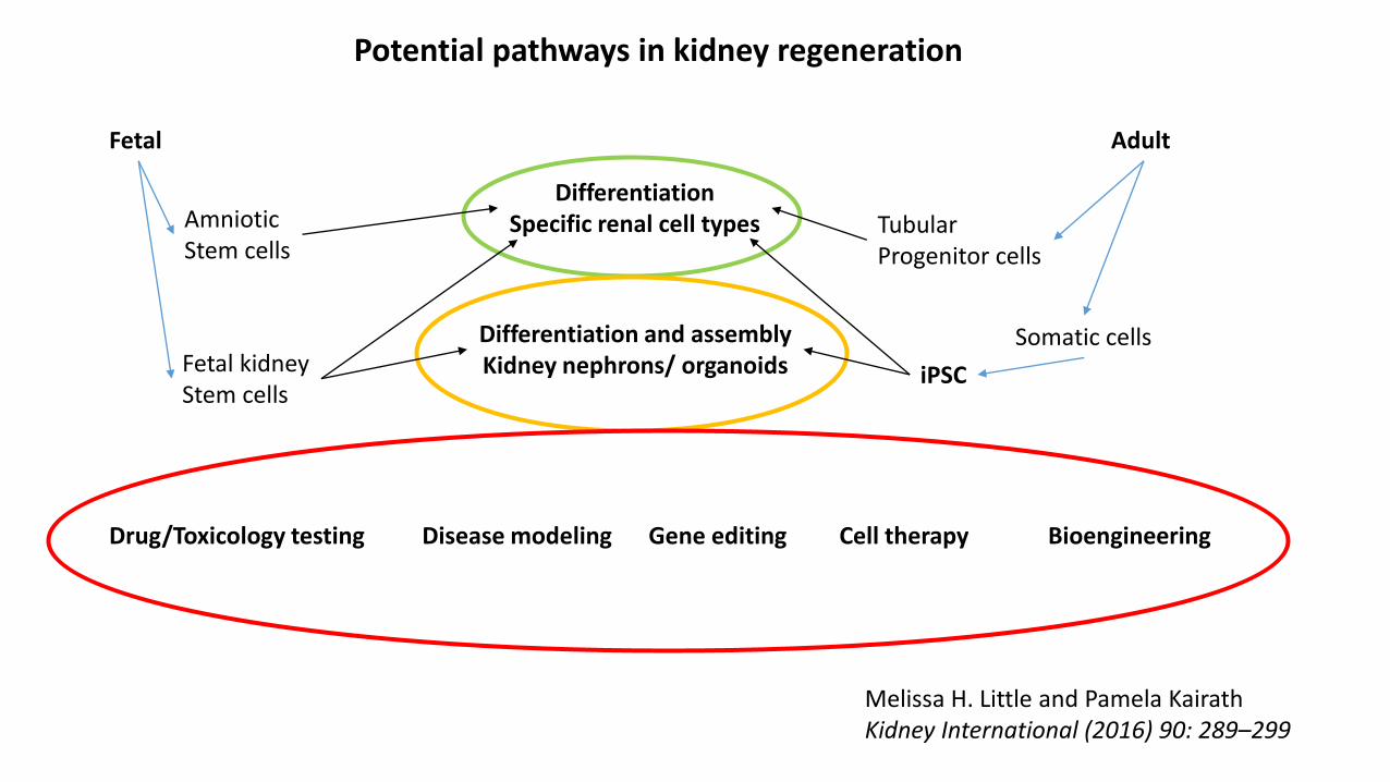
Fetal Adult
Tubular Progenitor cells
Somatic cells
iPSC
Amniotic Stem cells
DifferentiationSpecific renal cell types
Differentiation and assemblyKidney nephrons/ organoidsFetal kidney
Stem cells
Drug/Toxicology testing Disease modeling Gene editing Cell therapy Bioengineering
Melissa H. Little and Pamela KairathKidney International (2016) 90: 289–299
Potential pathways in kidney regeneration

Modeling disease in organoids
Infectious diseases• Human stomach organoids infected with Helicobacter Pylori with upregulation of IL8 (Bartfield et al. Gastroenterology 148:126,
2015)
• Forebrain human organoids infected with Zika virus show decrease in neuronal cell-layer volume, resembling microcephaly (Quian et al Cell 5:1238, 2016)
Hereditary disease• Human lung organoids from a Cystic Fibrosis patient lacked surface epithelial expression of the CFTR chloride channel
(Wong et al Nature 20:1310, 2012)• Human liver organoids from patients with alpha 1-atitrypsin deficiency show mutant protein accumulation in the
hepatocytes (Huch et al Cell 160:299, 2015)
CancerOrganoids from human prostatic cancer display histological patterns, markers and genetic mutations present in the original patient samples (Gao et al Cell 159:176, 2014)
ToxicologyCisplatin-induced damage of proximal tubular cells in human organoids (Takasato et al. Nature 526:564, 2015)

Genetic manipulation• CF mutation corrected with CRISPR/Cas9 in iPSC cells restored normal CFTR function in iPSC-derived airway epithelial
cells (Firth et al Cell Reports 12:1385, 2015)
• Introducing cancerogenous gene mutants in normal human pancreatic organoids results in development of atypical histologic features (Huang et al. Nature 21:1364,2015)
Drug screening Antineoplastic drugs show different sensitivity on neoplastic cell proliferation in pancreatic tumor organoids from different individual patients with pancreatic cancer (Huang et al. Nature 21:1364,2015)
Modeling disease in organoids

Broutier et al. Human primary liver cancer-derived organoid cultures for disease modeling and drug screening.Nature Med 23:1424-1435, 2017
c - Drug screening on liver cancer-derived organoids(tumoroids).
d - Effects of indicated compounds on the formation and viability of organoid in an organoid-formation assay.
e - In vivo activity of the ERK inhibitor (SCH772984) inCC-1_O tumoroids grafted subcutaneously in mice.
f, g - Histological analysis of the antitumor efficacy of SCH772984 on CC-1_O tumors was assessed 24 d after starting the treatment.

Dekkers et al. Characterizing responses to CFTR-modulating drugs using rectal organoids derived from subjects with cystic fibrosis. Science Transl Med 8:344, 2016
• Rectal organoid cultures werederived from the rectal epithelia of 71 individuals expressing 28 different CFTR mutations. • Forskolin-induced swelling (FIS) assay and steady-state lumen area (SLA) of organoids in the absence of forskolin were
used to test drug effect.
In vitro responses to drugs VX-809 and VX-770 in rectal organoids positively correlated with published outcome data from clinical trials with VX-809 and VX-770.
Testing drugs effect on organoids showed potential predictive value for CF patients carrying rare CFTR mutations to respond to drug therapy.

Schwank et al. Functional Repair of CFTR by CRISPR/Cas9 in Intestinal Stem Cell Organoids of Cystic Fibrosis PatientsCell 13:653, 2013
CFTR mutant organoids showedimpaired forskolin induced swelling
CRIPR/Cas9 repair of the CFTR gene mutation
restored forskolin induced swelling in organoids from CF patients

Bredenoord et al. Human tissues in a dish: The research and ethical implications of organoid technology.Science 355:6322, 2017
Organoids as alternative or complement to animal models in research:Good for animals Good for humans
Use of fetal material (embryonal stem cells) for research is controversial but may have therapeutic potential
• Total de-identification of stored tissue/organoidsfrom patients with disease may not be possible for need to link organoids with diseased patients.
• Privacy concerns • Who owns those organoidspatients, researchers, funding agencies, industry……
• Specific patient consent to use their tissue for possible commercial applications (pharma- industries).
• Rights of patient, researcher, industry (use and potential profits).
• Privacy concerns• Discoveries may negatively affect
patients (denied medical insurance for certain conditions.)
• Develop new research standards
• Responsibility and accurate representation to the public
• 2016 ISSCR Guidelines for Stem Cell Research and Clinical Translation

Kidney Regeneration…….any time soon?
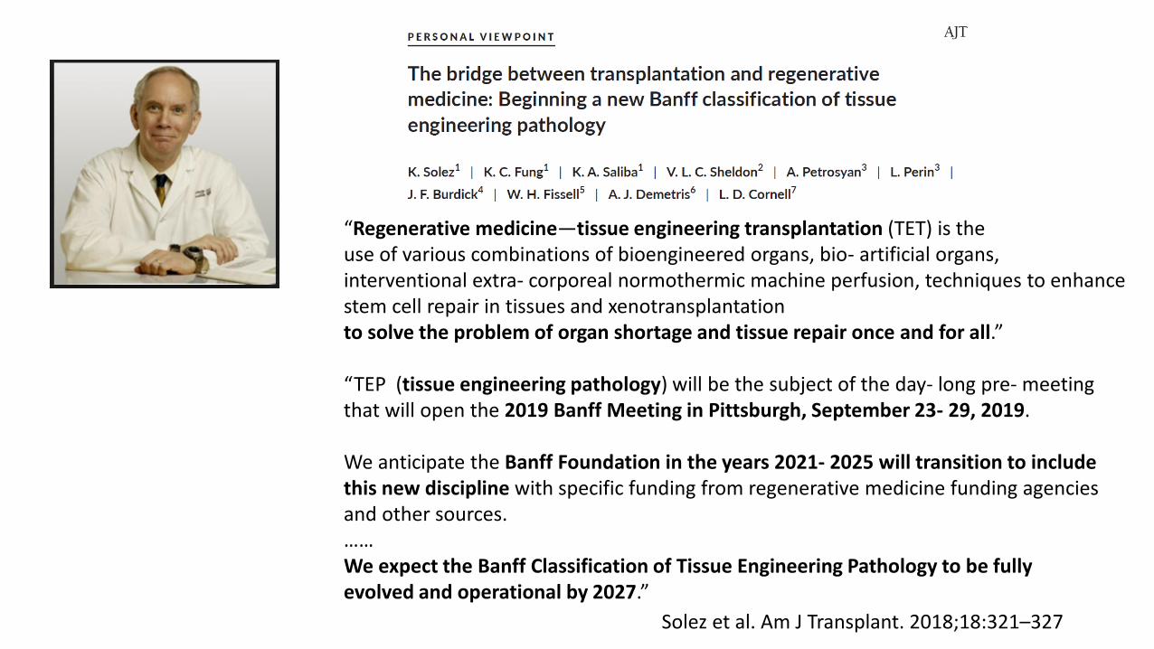
“Regenerative medicine—tissue engineering transplantation (TET) is theuse of various combinations of bioengineered organs, bio- artificial organs,interventional extra- corporeal normothermic machine perfusion, techniques to enhance stem cell repair in tissues and xenotransplantationto solve the problem of organ shortage and tissue repair once and for all.”
“TEP (tissue engineering pathology) will be the subject of the day- long pre- meeting that will open the 2019 Banff Meeting in Pittsburgh, September 23- 29, 2019.
We anticipate the Banff Foundation in the years 2021- 2025 will transition to include this new discipline with specific funding from regenerative medicine funding agencies and other sources.……We expect the Banff Classification of Tissue Engineering Pathology to be fullyevolved and operational by 2027.”
Solez et al. Am J Transplant. 2018;18:321–327


Sochol et al. Curr Transplant Rep. 3:82-92,2016
Quasi-static
Microchannels
3D printing

ah = Channel Heightw = Channel Widthl = Channel Lengtht = Membrane Thicknessp = Membrane Pore Size
bPDMS = Poly(dimethylsiloxane)PES/PVP = Polyethersulfone/Polyvinylpyrrolidone
Sochol et al. Curr Transplant Rep. 3:82-92,2016

Ng et al. A Fibrin-Based Tissue-Engineered Renal Proximal Tubule for Bioartificial Kidney Devices: Development, Characterization and In Vitro Transport Study- International Journal of Tissue Engineering 2013:10
Synthetic hollow fibers were constructed (polyethersulfone, polyvinylpyrrolidone, N-methyl-2-pyrrolidone), perfused with fibrinogen followed by thrombin to generate fibrin coating, then seeded with Human primary renal proximal tubule epithelial cells (RPTEC), perfused with culture medium and studied for 7-14 days.
Confluent RPTEC in the fiber expressed proximal tubule markers (CD13, megalin, γ-GTP, numerous mitochondria) and ion transporters (AQP-1, Na+K+ATPase, SGLT2).
Functional tests showed Na+K+ATPase-dependent active transport, glucose, and urea transport, but no creatinine transport.