Embed Size (px)
Citation preview

Original Article Brunei Int Med J. 2011; 7 (3): 148-156
Bone marrow study in patients
with Human Immune Deficiency Virus and Acquired Immune
Deficiency Syndrome Kenchappa RUDRESH 1, Tirthankar MUKHERJEE 2, Amit BHASIN 3, Vijaya Viswanath
MYSOREKAR 4, Nalini MODEPALLI 5, Aparna AHUJA 6 1 Department of Medicine, M.S. Ramaiah Medical College, Bangalore, 2 Department of Medicine, Kempegowda Institute of Medical Sciences, Bangalore, 3 Department of Medicine, Lady Hardinge Medical College, New Delhi, 4 Department of Pathology, M.S. Ramaiah Medical College, Bangalore, 5 Department of Pathology, Kempegowda Institute of Medical Sciences, Bangalore, 6 Department of Radio-diagnosis, Maulana Azad Medical College, New Delhi, India
ABSTRACT
Introduction: Human Immunodeficiency Virus/Acquired Immune Deficiency Syndrome (HIV/AIDS)
can involve almost any organ system. The study was aimed to assess the various bone marrow abnor-
malities seen in HIV/AIDS patients with haematologic abnormalities. Materials and Methods: 43 HIV
infected patients were included in the study. Baseline haematological investigations included full blood
count, CD4 positive lymphocyte counts, and absolute lymphocyte count. Bone marrow aspiration and
trephine biopsy were done in all patients. Bone marrows of these patients were carefully evaluated for
any abnormalities. HIV positive patients were classified into AIDS group (76%) and non-AIDS (24%)
group using National AIDS Control Organisation (NACO) criteria. Results: Normocytic normochromic
anaemia was the most common peripheral haematological finding occurring in 72% of patients. The
AIDS group had statistically significant lower platelet counts (p=0.004) but no differences in the other
parameters. Bone marrow was normocellular in 63.6% in the AIDS group and 100% in the non-AIDS
group. Dysplasia was observed in 37.2% of patients, predominantly affecting granulocytic series.
Myelodysplasia was statistically associated with a low platelet count. Reduced marrow lymphoid pre-
cursors (CD4+) were seen in 37.2% of patients. Conclusions: Bone marrow abnormalities were com-
mon in HIV/AIDS patients with haematological abnormalities. The AIDS group had a statistically sig-
nificant lower platelet count. Myelodysplasia was found in 37.2% of patients with HIV disease and was
also statistically associated with a lower CD4+ lymphocyte count.
Keywords: AIDS related complex, bone marrow diseases, bone marrow
examination, HIV infections
Correspondence author: Amit BHASIN,
Lady Hardinge Medical College,
New Delhi, INDIA
Tel: +91-9868658195
E mail: [email protected]
INTRODUCTION
Human Immunodeficiency Virus (HIV) infec-
tion can influence all the haematopoietic cell
lineages, resulting in a broad spectrum of

RUDRESH et al. Brunei Int Med J. 2011; 7 (3): 149
haematological abnormalities. Direct invasion
of the haematopoietic cells by HIV together
with infectious, inflammatory and neoplastic
processes explain the various haematological
alterations, which are seen during the course
of infection.
The consequences of these haema-
tologic problems are twofold. First, they are
associated with morbidity in themselves that
can adversely alter the patient’s quality of life
such as from anaemia (fatigue and dysp-
noea), leucopaenia (infections) and thrombo-
cytopaenia (bleeding). Second, they hinder
treatment of the primary viral infection, sec-
ondary infections and neoplastic complica-
tions. The poor haematopoietic tolerance of
therapies often necessitates dose reductions,
alteration of drug regimens or interruption of
therapies. There are no clear guidelines about
the optimal management of these haema-
tologic disorders. Use of marrow stimulants
such as granulocyte-colony stimulating factor
(G-CSF) and erythropoietin in addition to
Highly Active Anti-Retroviral Therapy
(HAART) have been shown to have a role in
reducing infective complications. 1, 2
The study aimed to assess the various
bone marrow abnormalities seen in HIV/AIDS
patients with haematological abnormalities.
out on a cohort of patients aged 16 years and
above (either sex) presenting to our hospital
who were Enzyme-linked immunosorbent as-
say (ELISA) positive for HIV and who had
peripheral haematological abnormalities like
anaemia, leucopaenia, neutropaenia, lympho-
paenia and thrombocytopaenia over a period
of three years. A total of 43 patients were
studied. Detailed clinical history was obtained
and physical examination was carried out.
The following haematological investi-
gations were carried out for all patients: hae-
moglobin, total leucocyte count (TLC), differ-
ential leucocyte count (DLC), erythrocyte
sedimentation rate (ESR), platelet count,
mean corpuscular volume (MCV), mean cor-
puscular haemoglobin concentration (MCHC),
mean corpuscular haemoglobin (MCH),
packed cell volume (PCV), reticulocyte count,
peripheral smear for blood picture, bleeding
time, clotting time, and serum urea, serum
creatinine. Absolute lymphocyte count was
calculated as ALC (/mL) = TLC x DLC (%).
Diagnosis of HIV was made in these
patients by ELISA and Western Blot (2 ELISA
using different kits or 1 ELISA and 1 Western
Blot). CD4 count was carried out on all pa-
tients. The bone marrow aspirate was used to
assess the cellularity. Bone marrow aspiration
and biopsy: aspirate was stained using
Leishman’s stain, whereas trephine biopsy
was stained with haematoxylin and eosin
stain. Ziehl–Neelsen (ZN) staining for Acid
Fast Bacillus (AFB) was done in selected
cases. All the bone marrow aspiration and
biopsy specimens were examined and inter-
preted by the same pathologist, to avoid in-
ter-observer variation.
MATERIALS AND METHODS
The study was approved by the local ethics
committee and has therefore been performed
in accordance with the ethical standards laid
down in the 1964 Declaration of Helsinki. All
persons gave their informed consent prior to
their inclusion in the study.
An observational study was carried

RUDRESH et al. Brunei Int Med J. 2011; 7 (3): 150
Patients were classified into two
groups – AIDS and non-AIDS according to
National AIDS Control Organisation (NACO)
criteria and all the patients were categorised
based on the Center for Disease Control
(CDC) and Prevention clinical category of HIV
Infection as shown in graph (Figure 1). 3, 4
Patients who were on antiretroviral therapy
were excluded. This was done to reduce the
confounding effect of antiretroviral drug in-
duced bone marrow suppression.
Statistical Analysis: Proportions were com-
pared using chi-square test of significance.
Student t test was done as indicator of statis-
tical significance. Data analysis was carried
out using Statistical Package for Social Sci-
ence (SPSS Version 10.5, Chicago, Il. USA.)
In five cases, adequate bone marrow could
not be aspirated, in two cases it was grossly
diluted with blood. In such patients, the tre-
phine biopsy was relied upon for cellu larity.
There was no statistically significant differ-
ence in cellularity between the two groups
(p=0.08). The results of marrow cellularity
are depicted in Table 3. Overall, the bone
marrow biopsy was normal in all non– AIDS
group but only in 27 (81.8%) AIDS group.
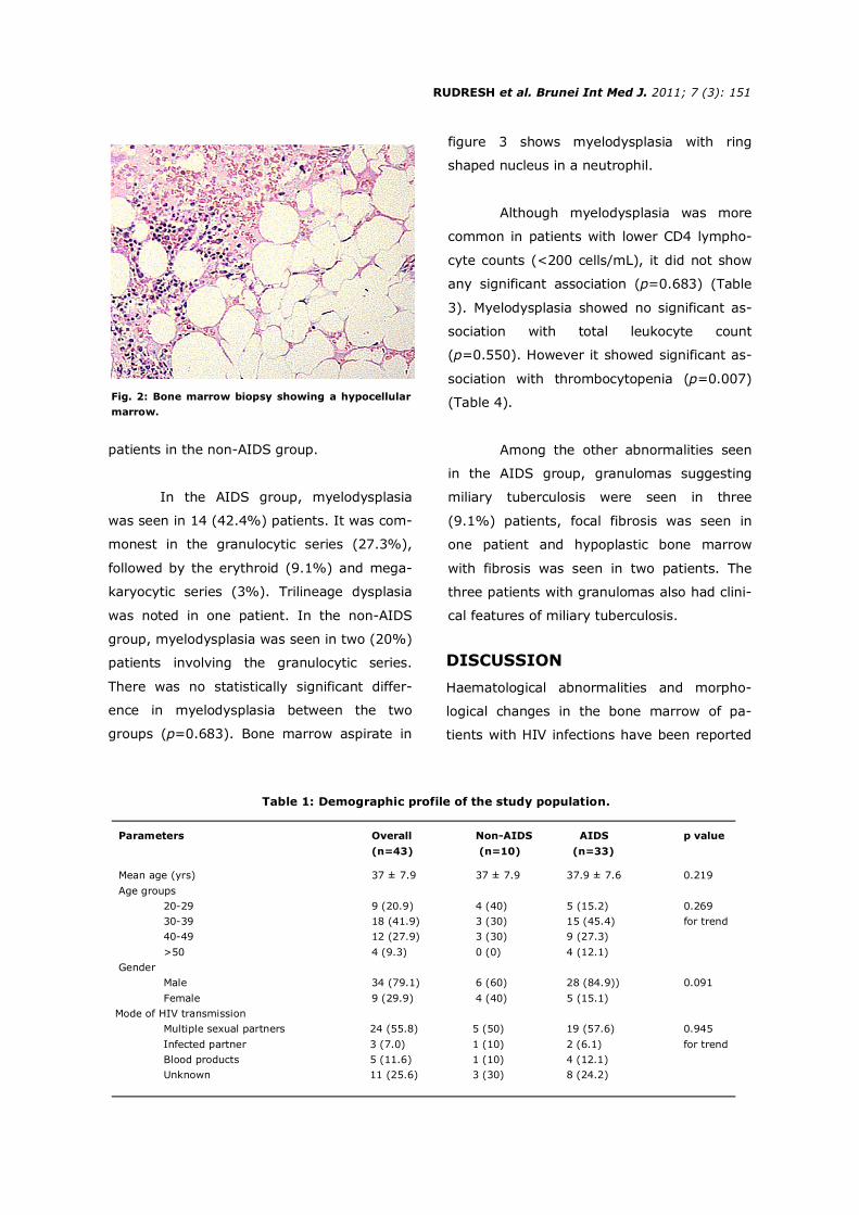
Figure 2 shows a hypocellular bone marrow.
In both groups, the majority of pa-
tients had normal lymphoid precursors. In the
AIDS group decreased lymphoid cells were
seen in 13 (39.4%) patients and was in-
creased in two (6.1%) patients. In the non-
AIDS group decreased lymphoid cells were
seen in three (30%) patients whereas these
were increased in one (10%) patient. There
was no statistically significant difference in
the number of lymphoid cells between the
two groups (p=0.821).
Increased plasma cells were observed
in 48.5% patients in the AIDS group and 60%
RESULTS
The demographic data of the patients are
shown in Table 1.
The distribution of mean values of
laboratory parameters among study groups is
shown in Table 2.
Fig. 1: Distribution of patients based on the Center for Disease Control (CDC) and Prevention classification for
HIV infection (Refer to supplementary text for Classification).
Percentage
0 0
12.1
0 0
21.2
0 0
66.7
20
10
0
10 10
0
30
20
00
10
20
30
40
50
60
70
80
A1 A2 A3 B1 B2 B3 C1 C2 C3 Categories
Non-AIDS AIDS
RUDRESH et al. Brunei Int Med J. 2011; 7 (3): 151
patients in the non-AIDS group.
In the AIDS group, myelodysplasia
was seen in 14 (42.4%) patients. It was com-
monest in the granulocytic series (27.3%),
followed by the erythroid (9.1%) and mega-
karyocytic series (3%). Trilineage dysplasia
was noted in one patient. In the non-AIDS
group, myelodysplasia was seen in two (20%)
patients involving the granulocytic series.
There was no statistically significant differ-
ence in myelodysplasia between the two
groups (p=0.683). Bone marrow aspirate in
figure 3 shows myelodysplasia with ring
shaped nucleus in a neutrophil.
Although myelodysplasia was more
common in patients with lower CD4 lympho-
cyte counts (<200 cells/mL), it did not show
any significant association (p=0.683) (Table
3). Myelodysplasia showed no significant as-
sociation with total leukocyte count
(p=0.550). However it showed significant as-
sociation with thrombocytopenia (p=0.007)
(Table 4).
Among the other abnormalities seen
in the AIDS group, granulomas suggesting
miliary tuberculosis were seen in three
(9.1%) patients, focal fibrosis was seen in
one patient and hypoplastic bone marrow
with fibrosis was seen in two patients. The
three patients with granulomas also had clini-
cal features of miliary tuberculosis.
Fig. 2: Bone marrow biopsy showing a hypocellular
marrow.
Table 1: Demographic profile of the study population.
Parameters Overall Non-AIDS AIDS p value
(n=43) (n=10) (n=33)
Mean age (yrs) 37 ± 7.9 37 ± 7.9 37.9 ± 7.6 0.219
Age groups
20-29 9 (20.9) 4 (40) 5 (15.2) 0.269
30-39 18 (41.9) 3 (30) 15 (45.4) for trend
40-49 12 (27.9) 3 (30) 9 (27.3)
>50 4 (9.3) 0 (0) 4 (12.1)
Gender
Male 34 (79.1) 6 (60) 28 (84.9)) 0.091
Female 9 (29.9) 4 (40) 5 (15.1)
Mode of HIV transmission
Multiple sexual partners 24 (55.8) 5 (50) 19 (57.6) 0.945
Infected partner 3 (7.0) 1 (10) 2 (6.1) for trend
Blood products 5 (11.6) 1 (10) 4 (12.1)
Unknown 11 (25.6) 3 (30) 8 (24.2)
DISCUSSION
Haematological abnormalities and morpho-
logical changes in the bone marrow of pa-
tients with HIV infections have been reported

RUDRESH et al. Brunei Int Med J. 2011; 7 (3): 152
cytopenias are often treatable and even cor-
rectable.
In our study, 43 HIV positive patients
were studied of which 33 had AIDS. Inade-
quate aspiration of the bone marrow was
seen in 11% cases. It may be a consequence
of focal fibrosis, which was seen in 14% of
cases. This finding is in agreement with the
report of Tripathi et al. and Sitalakshmi et al.
who observed marrow fibrosis in 54% HIV
infected cases, whereas Karcher et al. have
documented marrow fibrosis in 20% cases. 7-9
Various peripheral haematological
abnormalities were observed in isolation or in
combination. Normocytic anaemia was the
most common haematological abnormality,
occurring in 76% patients (54.5% AIDS pa-
tients and 60% of non-AIDS patients) and
this is in agreement with the published litera-
ture. 7, 8 Normocytic anaemia probably re-
flected anaemia of chronic disorders like re-
current pneumonia, tuberculosis and oppor-
tunistic infections involving the bone marrow.
Myelodysplastic changes in the bone marrow
can also cause normocytic anaemia. 10, 11 Mi-
crocytic anaemia was observed in 20% of the
patients in our study (21% the AIDS patients
and 20% of the non-AIDS patients). It was
in many studies. These abnormalities are
seen not only in patients with advanced dis-
ease, but also during primary infection and
during the clinical latency phase. 5 They pro-
gress in frequency and severity from asymp-
tomatic carrier state to AIDS-Related Complex
(ARC) to symptomatic AIDS. Low blood
counts are common in patients with HIV infec-
tion and AIDS, with anaemia, leucopenia and
thrombocytopenia each developing in more
than half of infected patients. 6 Isolated
thrombocytopenia that resembles idiopathic
thrombocytopenic purpura (ITP) sometimes
occurs early in the illness. Cytopenias often
cause symptoms and contribute to the com-
plications suffered by patients with AIDS like
infections, anaemia and bleeding. These
Table 2: Distribution of mean values of laboratory parameters among study groups.
Haematological parameters Overall Non-AIDS AIDS p value
(n=43) (n=10) (n=33)
Haemoglobin (gm/dL) 9.8 ± 1.63 10.4 ± 1.3 9.6 ± 1.7 0.122
Neutrophil (%) 68.8 ± 9.7 67.6 ± 8.5 69.2 ± 10.1 0.621
Lymphocytes (%) 27.8 ± 8.8 28.7 ± 8.1 27.6 ± 9.1 0.722
Esosinophil (%) 2.0 ± 1.25 1.8 ± 0.9 2.1 ± 1.3 0.492
Monocytes (%) 1.7 ± 1.3 1.6 ± 1.2 1.8 ± 1.4 0.675
Basophils (%) 0.1 ± 0.3 0.2 ± 0.4 0.3 ± 0.4 0.122
Reticulocytes (%) 0.9 ± 1.1 0.9 ± 0.2 0.8 ± 0.3 0.329
Platelets (109/L) 1.84 ± 0.47 2.20 ± 0.32 1.77 ± 0.45 0.004
Fig. 3: Bone marrow aspirate showing myelodysplasia
with ring shaped nucleus in a neutrophil (Arrow).

RUDRESH et al. Brunei Int Med J. 2011; 7 (3): 153
more common in women. Lymphopenia was
seen in 12% of patients belonging to the
AIDS group as compared to 27% AIDS pa-
tients in another study. 7 Lymphopenia is
probably a result of direct attack of lympho-
cytes by HIV through CD4 binding sites. 12 We
observed leukopenia in 14% of patients (12%
patients of AIDS group and 20% patients in
non-AIDS group). Tripathi et al. reported leu-
kopenia in 6% of patients (7% patients in
AIDS group and 5% patients in the non-AIDS
group). 7 This difference in percentage can be
attributed to the difference in the sample size
of the study groups. Leukocytosis was seen in
six percent of patients with AIDS. These pa-
tients had sepsis secondary to pneumonia or
urinary tract infection.
CD4 lymphocyte
count
Table 3: Correlation between myelodysplasia and CD4 lymphocyte count.
Myelodysplasia
Erythroid Granulocytic Megakaryocytic Nil Trilineage Total
<200 3 (9.1) 9 (27.3) 1 (3.0) 19 (57.6) 1 (3.0) 33 (100)
≥200 0 (0) 2 (20.0) 0 (0) 8 (80.0) 0 (0) 10 (100)
Total 3 (7.0) 11 (25.6) 1 (2.3) 27 (62.8) 1 (2.3) 43 (100)
0
10
20
30
40
50
60
70
80
90
100
Overall Non-AIDS AIDS
Fig. 3: Cellularity of the bone
marrow (p=0.008 for trend).
Percentages
Patients
Pancytopenia was observed in three
percent of patients in the AIDS group. Studies
have reported pancytopenia in 23% of HIV
positive patients. 8 ITP was observed in three
percent of patients in the AIDS group which is
comparable with other studies. 7, 8 We also
observed a statistically significant lower plate-
let count in the AIDS group compared to the
non-AIDS group (p<0.05).
We studied bone marrow for cellular-
ity, dysplasia, plasma cell numbers, lymphoid
precursor numbers, dysplastic changes, fibro–
sis and granulomas. The majority of HIV in-
fected patients (63% in the AIDS group and
100% in the non-AIDS group) had a normo-
cellular bone marrow. However mixed reports
Normocellular
Hypocellular
Hypercellular
Overall Non-AIDS AIDS

RUDRESH et al. Brunei Int Med J. 2011; 7 (3): 154
cells in the bone marrow in 27% of HIV in-
fected patients (49% patients of AIDS group
and 60% patients of non-AIDS group). Other
studies have reported plasmacytosis in 25%
and 22% patients respectively. 9, 16
Reduced bone marrow lymphoid cells
were seen in 37% HIV infected patients (39%
patients of AIDS group and 30% patients of
non-AIDS group). These findings were similar
to another study. 10
Interestingly, granulomas were seen
in 9% of patients in the AIDS group who also
had clinical features suggestive military tu-
berculosis. In all these cases, bone marrow
stained negatively for acid-fast bacilli. Cas-
tella et al. and Calore et al. reported granulo-
mas in 16% and 12% cases respectively. 14, 17
Treatment of haematological abnor-
malities aims primarily at reducing replication
of HIV, thereby diminishing suppression of
haematopoiesis by the virus, and at control-
ling the opportunistic infections during the
course of the disease. The possibility of bone
marrow suppression mediated by a toxic drug
effect should be considered in these patients.
In our study, we only studied patients who
had not been previously treated with any HIV
therapy. These haematologic complications of
HIV infection will undoubtedly decrease as
infectious complications are better controlled,
Platelet
count
Table 4: Correlation between myelodysplasia and platelets count.
Myelodysplasia
Erythroid Granulocytic Megakaryocytic Nil Trilineage Total
S Low 3 (9.1) 9 (27.3) 1 (3.0) 19 (57.6) 1 (3.0) 33 (100)
Normal 0 (0) 2 (20.0) 0 (0) 8 (80.0) 0 (0) 10 (100)
Total 3 (7.0) 11 (25.6) 1 (2.3) 27 (62.8) 1 (2.3) 43 (100)
are available on this finding, as some studies
concur while others disagree with our data. 7,
9, 13, 14 A hypercellular marrow may be seen in
early stages of the disease, but it is more
likely to be normocellular or hypocellular in
advanced disease. 15
Myelodysplasia was also one of the
parameters assessed in the bone marrow as-
pirates of our study population as various
studies have reported it to be a common fea-
ture in AIDS patients. 9 Myelodysplasia was
observed in 37% patients (42.4% patients in
the AIDS group and 20% patients in the non-
AIDS group). Studies have reported myelo-
dysplasia in 32.3% patients (37% patients in
the AIDS group and 21% patients in the non-
AIDS group), which had a significant associa-
tion with a low CD4 lymphocyte count. 7 How-
ever Karcher et al. reported myelodysplasia in
69% of HIV positive patients. 9 In our study,
dysplastic changes predominantly involved
the granulocytic series followed by the
erythroid and megakaryocytic series. Triline-
age dysplasia was seen in 3% cases, all of
which belonged to the AIDS group. Our find-
ings agreed with another study observation. 7
Although we found clustering of myelodyspla–
sia cases with low CD4 lymphocyte counts,
we could not establish any statistical associa-
tion.
We also observed increased plasma

RUDRESH et al. Brunei Int Med J. 2011; 7 (3): 155
These abnormalities become more frequent
as the disease progresses. Bone marrow
study is an important investigation in HIV in-
fected patients with peripheral haematological
abnormalities. It is a relatively safe and low
cost procedure that can contribute to a com-
prehensive evaluation of cytopenias which
lead to various complications.
REFERENCES
1: Henry DH, Beall GN, Benson CA et al. Recombi-
nant human erythropoietin in the treatment of ane-
mia associated with human immunodeficiency virus
(HIV) infection and Zidovudine therapy: overview
of four clinical trials. Ann Intern Med 1992;
117:739-48.
2: Kuritzkes DR, Parenti D, Ward DJ, et al. Filgas-
trim prevents severe neutropenia and reduces in-
fective morbidity in patients with advanced HIV
infection: Results of a randomized, multicenter,
controlled trial. G-CSF 930101 study group. AIDS
1998; 12:65-74.
3: National AIDS Control Organization. Specialist’s
training and reference module; 1999. CDC. 1993
Revised classification system for HIV infection and
expanded surveillance case definition for AIDS
among adolescents and adults. MMWR 1992; 41
(No. RR-17).
4: Patwardhan MS, Golwilkar AS, Abhyanakar, Atre
MC. Hematological profile of HIV positive patients.
Indian J Pathol Microbiol 2002; 45:147-50.
5: Treacy M, Lai L, Costello C, Clark A. Peripheral
blood and bone marrow abnormalities in patients
with HIV related disease. Br J Haematol 1987;
65:289-94.
6: Tripathi AK, Kalra P, Misra R, Kumar A, Gupta N.
Study of bone marrow abnormalities in patients
with HIV disease. JAPI 2005; 53:105-10.
7: Sitalakshmi S, Srikrishna A, Damodar P et al.
Hematological changes in HIV infection. Indian J
Pathol Microbiol 2003; 46:180-83.
8: Karcher DS, Frost AR.The bone marrow in hu-
man immunodeficiency virus-related disease-
morphology and clinical correlation. Am J Clin
Pathol 1991; 95:63-71.
resulting in longer life spans.
The finding of cytopenias in conjunc-
tion with a hypercellular marrow with dyspoi-
etic features which are described in myelo-
dysplasia, increased megakaryocytes forming
clusters and fibrosis which are consistent with
myeloproliferative disease, increased plasma
cells, eosinophils, increased iron and fibrosis
which are suggestive of chronic infection are
all encountered in HIV infection and hence
pose diagnostic confusion. With the continu-
ing rise in prevalence of HIV infection world-
wide, it is important for the haemato-
pathologist to recognise the haematological
abnormalities and morphological changes in
the bone marrow associated with HIV infec-
tion.
The exact mechanisms of HIV induced
peripheral haematologic and bone marrow
abnormalities are not known. The aetiology of
these findings are possibly either direct ef-
fects of HIV, nutritional deficiencies, oppor-
tunistic infections of marrow or the use of
marrow suppressive agents. Further research
on haematological complications of HIV dis-
ease will lead to effective management of
cases and reduce the morbidity and mortality
from this dreaded disease
There are several limitations with our
study. First the sample size was small. More
over we did not culture the bone marrow as-
pirate for detecting opportunistic pathogens
and HIV RNA estimation was not done in our
patients.
In conclusion, peripheral and bone
marrow abnormalities are common in HIV
infected individuals and patients with AIDS.

RUDRESH et al. Brunei Int Med J. 2011; 7 (3): 156
9: Ryu T, Ikeda M, Okazaki Y. Myelodysplasia asso-
ciated with acquired immunodeficiency syndrome.
Intern Med 2001; 40:795-801
10: Henry K, Costello C. HIV-associated bone mar-
row changes. Curr Diag Pathol 1994;1:131-41.
11: Richman DD, Fischl MA, Grieco MH . The toxic-
ity of azidothymidine in the treatment of patients
with AIDS and AIDS-related complex: a double
blind, placebo-controlled trial. N Engl J Med 1987;
317:192-97.
12: Geller S, Muller R, Greenberg ML, Siegal FP.
Acquired immunodeficiency syndrome-distinctive
features of bone marrow biopsies. Arch Pathol Lab
Med 1985; 109:138-41.
13: Castella A, Croxson TS, Mildvan D, Witt DH,
Zalusky R. The bone marrow in AIDS-a histologic,
hematologic, and microbiologic study. Am J Clin
Pathol 1985; 84:425-32.
14: Katasrou C. Terpos E, Pastouris E, et al. Myelo-
dysplastic features in patients with long term HIV
infection and hemophilia. Hemophilia 2001;7: 47-
52.
15: Osborne BM, Guarda LA, Butler JJ, et al. Bone
marrow biopsies in patients with the acquired im-
munodeficiency syndrome. Human Pathology
1984;15:1048-53.
16: Calore EE, Tanaka PY, Perez NM, Almeida LVl.
Bone marrow pathology in AIDS. Pathology-
Research and Practice 2004; 200:591-97.
Image of the Week Educational materials
For more information, visit
the journal website @
www.bimjonline.com
BIMJ welcome submissions to the
Image of the Week
Note: Image of the Week is only available from the BIMJ website as education materials