Embed Size (px)
Citation preview
Am. J. Trop. Med. Hyg., 89(1), 2013, pp. 78–92doi:10.4269/ajtmh.12-0719Copyright © 2013 by The American Society of Tropical Medicine and Hygiene
Body Size and Wing Shape Measurements as Quality Indicators of Aedes aegypti
Mosquitoes Destined for Field Release
Heng Lin Yeap,* Nancy M. Endersby, Petrina H. Johnson, Scott A. Ritchie, and Ary A. Hoffmann
Bio21 Institute and the Department of Genetics, University of Melbourne, Melbourne, Victoria, Australia; School of Public Health,Tropical Medicine and Rehabilitation Sciences, James Cook University, Cairns, Queensland, Australia; Tropical Population Health Unit,
Queensland Health, Cairns, Queensland, Australia
Abstract. There is increasing interest in rearing modified mosquitoes for mass release to control vector-bornediseases, particularly Wolbachia-infected Aedes aegypti for suppression of dengue. Successful introductions requirerelease of high quality mosquitoes into natural populations. Potential indicators of quality are body size and shape. Wetested to determine if size, wing/thorax ratio, and wing shape are associated with field fitness of Wolbachia-infectedAe. aegypti. Compared with field-collected mosquitoes, released mosquitoes were larger in size, with lower size varianceand different wing shape but similar in wing-thorax ratio and its associated variance. These differences were largelyattributed to nutrition and to a minor extent to wMel Wolbachia infection. Survival potential of released femalemosquitoes was similar to those from the field. Females at oviposition sites tended to be larger than those randomlycollected from BG-Sentinel traps. Rearing conditions should thus aim for large size without affecting wing/thorax ratios.
INTRODUCTION
There is increasing interest in modifying mosquitoes forpotential release into field populations to suppress disease,including modifications by the introduction of Wolbachia tointerfere with disease transmission,1,2 and modifications togenerate sterility and other changes.3–6 As in mass releaseswith other insects, the field success of these programs willultimately depend on the release of high quality material,and on developing ways of measuring and maintaining qual-ity. For example, biocontrol of agricultural moth pests usingTrichogramma spp. has been ongoing for many years and anumber of studies have assessed effects of mass rearing onquality of these wasps, based on measurements of life historytraits under laboratory7,8 and field8–10 conditions. A variety ofquality indicators have been proposed and developed for suchinsect systems including measures of body size.11,12
Aedes aegypti, a highly anthropophilic mosquito species,13,14
and the primary vector of dengue virus,15 has been successfullyinfected with Wolbachia2,16 and released in the field.1 Theprospects of using this approach to reduce dengue transmis-sion2,17–19 and/or reduce adult life span20 may lead to large-scale releases of these mosquitoes akin to releases beingundertaken with other biocontrol agents.To date, mass-rearing of high quality Wolbachia-infected
mosquitoes for field release has relied on backcrossing schemesto introduce field nuclear backgrounds and thereby minimizelaboratory adaptation, and mass rearing under semi-field con-ditions to control for environmental effects.1 Mosquitoes arereared under high nutrition conditions to ensure the produc-tion of a large number of mosquitoes in a synchronized man-ner. Based on past research, high nutrition produces largemosquitoes that are potentially fitter in terms of fecundity,sperm quantity, and survival (e.g., References 21–24) butmight lack other beneficial attributes. However, few fieldstudies on the effects of Ae. aegypti size on field fitness havebeen performed, even though these could be completed with
a mark-release-recapture design,25 and through comparisonsof resting and host-seeking females.26,27
Apart from size, field fitness may also be affected by thewing loading and shape of insects. Wing loading as measuredby weight to wing span has been shown to affect dispersal ininsects such as butterflies and damselflies.28–31 In Drosophila,there is evidence for an association between wing loading andresource finding ability that contributes to an antagonisticinteraction between wing size and thorax length.32 An associ-ation between wing shape and fitness has also been suggestedin insects including parasitoids (e.g., Reference 10), althoughsome researchers have suggested that shape may be pheno-typically invariable and unlikely to affect fitness.33
Here, we first analyze variation among populations of lab-oratory and field females for adult size, based on measure-ments of both wing size and thorax length. We also considerdifferences in wing loading and wing shape. Based on mos-quito surveillance and Wolbachia releases, using differenttrapping methods and parity assessment, we assess whetherwing size, thorax length, wing size/thorax length, and wingshape affect the ability of mosquitoes to locate breeding sitesand blood feed under field conditions. The successful invasionof Ae. aegypti populations in Cairns, Queensland, Australiaby Wolbachia in 20111 provided an opportunity to monitorchanges in morphometric measures associated with Wolbachiainvasion. We use these analyses to make recommendationsabout quality indicators and the generation of high fitnessmosquitoes in mass release programs.
MATERIALS AND METHODS
Three laboratory lines of Ae. aegypti were used in thisstudy. Uninfected C67 and C89 were lines established fromseveral hundred eggs collected from Cairns, Queensland,Australia, around January 2010 and August 2010, respectively.A wMel-infected line2 was generated through outcrossing toF1 C89 males and successive backcrossing female offspringwith F1 C89 males for an additional three generations (Aug–Nov 2010); this line is identified as wC89.Colony maintenance (University of Melbourne). All life
stages of the uninfected, and Wolbachia-infected lines weremaintained in a controlled 12:12 L:D laboratory environment
*Heng Lin Yeap, Department of Genetics, Bio21 Institute Universityof Melbourne, 30 Flemington Rd, Parkville VIC 3052 Australia.E-mail: [email protected]
78

at 26°C and 75–85% humidity. Eggs were hatched in plastictrays (20 cm +28.5 cm +9 cm) containing 3 L of RO (reverseosmosis) water, yeast (~0.09 mg), and one crushed tablet(300 mg) of TetraMin Tropical Fish Food tablets Rich Mix(Tetra Holdings Inc. [US], made in Germany). Density wascontrolled to 200 individuals at the second instar stage andlarvae were transferred to shallow plastic trays (42.7 cm +31.2 cm + 7.2 cm) (Modulab Systems, Gratnell Ltd., UnitedKingdom). The trays contained 4 L RO water (1 larva/20 mL)and were supplied with TetraMin in excess.Pupae were sexed by size (females are larger than males)
and sex was confirmed by checking eclosed adults. Adultswere housed in plastic containers with mesh sides (20 cm +20 cm + 30 cm, covered in a plastic bag to maintain highhumidity) and allowed constant access to a wick attached toa 20 mL vial of 10% sucrose solution. Females were givenaccess to a human arm for blood feeding within 1 week ofemergence and provided with sandpaper strips (11.8 m +3.83 cm) for oviposition inside a plastic cup containing 150 mLRO water. Oviposition strips were collected and replaceddaily for 1 week following blood feeding. Eggs were allowedto embryonate by leaving strips wet for 2 days after collec-tion, partially drying them on paper towel for 30 s and leav-ing them moist for one more day. Strips were then driedunder rearing temperature conditions until only traces ofmoisture by touch were apparent (i.e., strips were not com-pletely dried) on the smooth side of the strip and stored insealed bags with moist cotton wool to maintain high humidityand prevent desiccation.Colony maintenance (James CookUniversity [JCU], Cairns).
The wC89 line (see above) was established in two semi-fieldcages (8.0 m + 9.0 m + 4.1 m) described elsewhere34 at theend of 2010. The cage contains free flying adults (800–1,600females) and males hatched from eggs of field collected eggsare regularly released into this cage to allow for further out-crossing that minimizes laboratory adaptation.1 Females wereblood fed with human volunteers almost daily in this colony(JCU Human Ethics Approval H2250). Eggs were collectedusing red flannel cloth half submerged in ovibuckets and lar-vae were reared in a second field cage to simulate naturalconditions. Eggs were hatched with yeast solution, fed excessTetraMin. Larvae were reared in white bucket 205 mm indiameter with ~1,500 mL water. Density was controlled to150 larvae per bucket. Adults were provided access to 50%honey water solution before release.Field release. There were two field releases between January
andMarch2011over10weeks inYorkeysKnobandGordonvale,as described by Hoffmann and others1; during the releaseperiod, more than 10,000 mosquitoes were released each week.Sampling. Three types of traps were used in this study:
BioGent-Sentinel (BGS) traps (Biogents AG, Regensburg,Germany), double sticky ovitraps, and felt-cloth ovitraps. TheBGS traps are useful for surveillance of all physiologicalstages of adult Ae. aegypti, although teneral and blood-fedfemales were undersampled.35–37 BioGent-Sentinel trappingwas conducted without olfactory cues. Trapped mosquitoeswere identified as Ae. aegypti and stored in ethanol at −20°C.In some cases, ovarian parity was assessed before preserva-tion in ethanol. Double sticky ovitraps (referred to here assticky ovitraps) composed of an internal removable stickypanel placed above a container of water. Because females areattracted to the water source to lay their eggs, sticky traps are
effective in catching egg-laying (gravid) female Ae. aegypti,with 99% of females being parous.38,39 Ovipositing femalesare likely to be inseminated because insemination triggersoviposition and increases fecundity.40–42 In contrast, virginblood-fed females do not readily lay eggs (< 10% 1 week afterblood feeding (Yeap HL, unpublished data). The stickyovitraps were used to determine whether there were morpho-metric differences between egg-laying females and those in theoverall population. Mosquitoes were retrieved 1–3 + per weekand Ae. aegypti were stored in a dry vial at 4°C. Ovarian paritystatus was determined before storing these mosquitoes inethanol at −20°C. Finally, felt cloth ovitraps are effective incollecting Ae. aegypti eggs from the field,34,43 and can behatched under laboratory conditions. For these traps, red feltcloth was used as the egg-laying substrate in the trap, 0.5 g oflucerne was added as an oviposition attractant, and the feltwas collected 1 week after setting the trap. The cloth wasdried to condition eggs and to prevent immediate hatching.Ovarian parity. Abdomens of cold-anaesthetized female
mosquitoes were dissected in diluted detergent and assessedfor parity status according to Detinova44 and Clements andBoocock.45 Three ovarian parity states were noted: nulliparous,gravid, and parous. We defined gravid females as those witheggs developing past Christopher’s Stage II,45 ranging fromovarian tracheoles almost blocked from sight, to ovariescompletely engorged with fully developed eggs. Because weused ovarian parity as a sign of blood feeding, we classifiedfemales that had traces of blood as gravid, particularly as bloodfeeding results in the development of mature ovules within3 days.46 Parous females were defined here as completely emp-tied of eggs with some tracheolation, whereas nulliparousfemales still had uncoiled tracheoles. Unlike gravid females,parous females would not have fed within the last 3 days.Wolbachia testing. The BGS-trap samples from field
releases in Yorkeys Knob, Gordonvale, and Machans Beachwere tested for Wolbachia by real-time polymerase chainreaction (PCR) using a Roche Applied Science (Australia)LightCycler 480.47 Whole mosquitoes or their abdomens werehomogenized in 250 mL or 150 mL of 5% Chelex 100 resin(Bio-Rad Laboratories, Hercules, CA), respectively. Homog-enized tissue was subjected to 45–60 minutes at 65°C withProteinase K (Roche Diagnostics Australia Pty. Ltd., CastleHill NSW, Australia), and then 10–15 minutes at 90°C to lyseProteinase K. An aliquot of 10 mL of each sample was thendiluted 10 + or 5 + for whole mosquito or abdomen respec-tively in a 96-well plate.Three PCR reactions were performed for each sample. Three
sets of primers were used47: 1) universal primer pair (mRpS6_F:5¢-AGTTGAACGTATCGTTTCCCGCTAC; mRpS6_R: 5¢-GAAGTGACGCAGCTTGTGGTCGTCC), which target theconserved region of the RpS6 gene, to detect presence ofmosquito DNA; 2) Ae. aegypti specific primers (aRpS6_F: 5¢-ATCAAGAAGCGCCGTGTCG; aRpS6_R: 5¢- CAGGTGCAGGATCTTCATGTATTCG), which target the Ae. aegypti-specific polymorphisms within the highly variable region ofRpS6, to distinguish between Ae. aegypti and non-Ae. aegypti
specimens; or 3) a pair of Wolbachia-specific primers (w1_F:5¢- AAAATCTTTGTGAAGAGGTGATCTGC; w1_R: 5¢-GCACTGGGATGACAGGAAAAGG) to detect the pres-ence ofWolbachiaDNA.The PCR cycling conditions were as follows: 95°C for 10 min-
utes, 40 cycles of 95°C for 5 seconds, 58°C for 15 seconds, and
SIZE AND SHAPE IN RELEASED MOSQUITOES 79

72°C for 15 seconds. Products were heated to 95°C for1 minute, cooled to 40°C for 20 seconds, and then raised to65°C. As temperature increases gradually from 65°C to 95°C,fluorescence data were acquired continuously. The amplifica-tion and melting profiles of each PCR were subsequently usedto determine the crossing point (Cp) values and meltingtemperatures (Tm) using the Absolute Quantification andTm calling modules of the LightCyler 480 software package(Roche Applied Science).Morphometrics. Left wings were detached and mounted on
a slide with Hoyer’s solution. A photograph was taken ofeach wing at 11.25 + magnification using a camera (NikonSMZ1500, Japan) mounted on a microscope. A 2 mm grati-cule was photographed to standardize size measurements.Photos were digitized with tPSUtil48 and tPSDig2 version2.16.49 Fifteen landmarks were located on the wings to cap-ture shape and size (Figure 1). This includes extra landmarkscompared with an earlier study50 to allow wing shape to beaccurately represented, but fewer points than used in Vargasand others51 because we found that some of their specifiedlandmarks were difficult to locate accurately and reliably inour specimens. Wing centroid size is the square root of thesum of squares of the Euclidean distances between landmarksto the centroid.52
Wing length and centroid sizes are highly correlated toother measures of body size.53,54 Thorax length was also mea-sured from the scutellum to the most anterior point of thethorax. The ratio of wing centroid size to thorax length wascomputed as the inverse of wing load, the body size to wingspan ratio.Repeatibility tests on measurements were initially performed
by twice landmarking 10 samples from 25 groups of wingphotos (250 images). One-way analysis of variance (ANOVA)was performed for each x- and y-coordinate, including cen-troid size, with individual as the fixed factor, thus the ran-dom error corresponds to within- individual or repeatibilityerrors.55 Repeatibility (R) was computed as the ratio of betweenindividual variance to the sum of variance of between indi-vidual and within individual. All repeatibility values were> 0.99. The correlations between all repeated landmarks forall x-coordinates, y-coordinates, and overall centroid size wereall high (> 0.99).
Comparisons and experiments. Field and laboratory samples.
Samples were collected from BGS traps from within therelease sites Yorkeys Knob (YK) and Gordonvale (GV)(Figure 2) at two time points from November to December2009 and January to February 2010 before the release oflaboratory-reared Wolbachia-infected Ae. aegypti. Approxi-mately 80–100 female mosquitoes from each site and each timepoint were measured. Males were also obtained, but numberswere lower. These samples were considered representative ofthe uninfected field population during the wet season beforethe release. During the release period in January–March 2011,we collected more uninfected field samples from 20 BGS-traps placed in Holloways Beach (HB) and Machans Beach(MB) close to YK, and we also collected mosquitoes from13 BGS traps in Pyramid Estate and Edmonton close to GVin January–February 2011 (Figure 2).The C67 and C89 samples were obtained in May 2010 and
May/June 2011, respectively (after both strains had beenreared eight generations in the laboratory) to provide samplesof uninfected mosquitoes reared under the laboratory envi-ronment. We also collected C67 and C89 at the same timesfrom low nutrition conditions, by rearing them under identicalconditions but providing larvae a quarter of the food givenunder the culture conditions described previously. The wMel-infected mosquitoes, wC89 were reared alongside uninfectedC89 under both high and low nutrition conditions and used toexamine the influence of infection status on measurements.The C67 laboratory strain was used in comparisons with fieldmosquitoes. Mosquitoes were stored in ethanol at −20°C afterone gonotrophic cycle or directly after eclosion.Field eggs from HB, MB, Edmonton, and Pyramid Estate
were collected with ovitraps at the time of BGS trapping inthose locations. Eggs were hatched under the laboratoryconditions described previously and Ae. aegypti identified atthe 3rd or 4th instar stage. These were then reared at onelarva per 20 mL density with excess TetraMin. A day aftereclosion, adults were stored in ethanol at −20°C. These F1individuals from field females were compared with laboratory-reared mosquitoes.Field comparisons. The oviposition success of mosquitoes
of different sizes was investigated by comparing mosquitoesretrieved from BGS traps and sticky ovitraps, as BGS-trapswere expected to capture all mosquitoes, whereas stickyovitraps captured ovipositing females. Eighty BGS traps wereplaced in the Cairns Central Business District (CBD), Cairnsinner suburbs, and Machans Beach areas from March 8–18,2011. Twenty of the traps with consistently high mosquitonumbers were inspected daily. The others were checked oncea week. Females were collected from sticky traps and BGStraps from Cairns CBD, Cairns inner suburbs, and MachansBeach in March 2011. Ovarian parity was assessed for allfemales from the BGS traps before preservation in ethanol.Size and shape were compared between females of differentparity status. We compared gravid (and parous) females thatwould have survived long enough to feed, with nulliparousfemales that represented a population that had not yet under-gone selection for feeding and survival.All females caught in the double sticky ovitraps were par-
ous or gravid. Parous females laid eggs onto the trap or insidethe vials where they were stored. This “death stress oviposi-tion” phenomenon leads to identification of captured gravidfemales as gravid rather than parous, and has been noted inFigure 1. Position of 15 landmarks on the wings ofAedes aegypti.
80 YEAP AND OTHERS
previous double sticky ovitrap evaluations.38 From the BGStraps, we obtained 13 nulliparous, 17 parous, and 79 gravidfemales, along with 17 non-gravid females whose status wasundefined because the ovarial tracheolation could not be visu-alized clearly.Monitoring during release. Infected mosquitoes were col-
lected from YK and GV from 30 BGS traps in areas whereweekly releases were undertaken.1 We assessed mosquitoesobtained in the third week of the release (week beginning17 January), 2 weeks after the final release (week beginning21 March) and in the last 2 weeks of May after sites weresuccessfully invaded. Wolbachia-infected mosquitoes fromthe third week of the release are almost all likely to be fieldcage material, as there is insufficient time for a field genera-tion to reach adulthood from the egg stage in two weeks.Infected females reared in the laboratory were tested at thesame time as a representative sample of released material.Statistical analyses. Size measures from the samples were
subjected to Shapiro-Wilk tests to check for normality. Vari-ances were compared with check for the assumption ofhomoscedascity before performing ANOVAs or t-tests. TheR2.11 program and SPSS version 20 (SPSS, Inc., Chicago, IL)were used for most of the analyses. Because of multiple com-parisons in the tests, P values were corrected by the Dunn-Sidak procedure where appropriate. Size measures were almostalways normally distributed and we used parametric tests,but in those rare cases where data were not normally distrib-uted, we also confirmed any significant differences with non-parametric tests. Where variances differed, we used theunequal variance Welch t tests to compare groups. We alsoran non-parametric tests to compare samples for wing thorax
ratios. For all the analyses, we only considered femalesbecause sample sizes for males were small.For analyses of wing shape, Procrustes superimposition was
undertaken on landmark coordinates to remove orientation,location, and size effects.56 An initial exploratory analysis ofshape variation observed within the datasets was undertakenwith Principal Component Analyses (PCA) and/or ProcrustesANOVA. This was followed by Canonical Variate Analysis(CVA) with 10,000 permutations to test for pairwise distancesand/or Discriminant Function Analysis (DFA) with 1,000 per-mutations on Procrustes distances to compare shape betweengroups. Canonical variates were graphed, although distancesdo not always reflect significance, because DFA and CVAmaximize differences between groups. Shape variables wereregressed with size to test for allometry. All shape analyseswere performed in MorphoJ version 1.05a.57 We then gener-ated shape changes for pairs of groups that were significantlydifferent to visualize changes in shape.Morphometric variation in laboratory and field populations.
To compare the field and laboratory samples of uninfectedC67 mosquitoes, we first tested if the samples were homoge-neous across the areas where field samples were obtained(YK and GV collected in 2010 before the release, or the fourouter suburbs collected in 2011 during the release) usingANOVA and t-tests. We then compared the field samples tothe laboratory-reared sample by unequal variance t-tests. Forwing/thorax ratios, we used Mann-Whitney U tests to com-pare distributions among collections. We also used statisticaltests to examine differences in coefficient of variation (CoV)between field samples, and between field and laboratorysamples.58 For the shape analysis, in cases where Procrustes
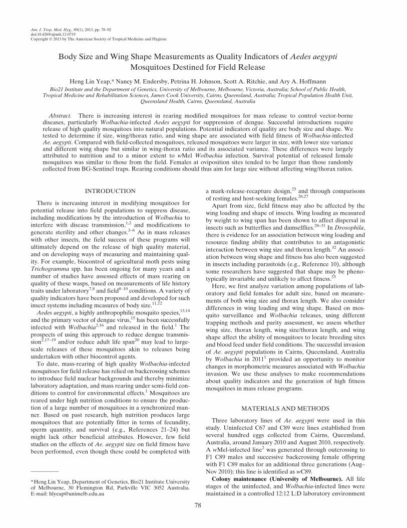
Figure 2. Map of collection sites of Aedes aegypti and other sites mentioned in the work. This map was produced using MapConnect webMapping http://www.ga.gov.au/mapconnect with GlobalMap 1 million data.
SIZE AND SHAPE IN RELEASED MOSQUITOES 81
ANOVA indicated differences in shape between groups, weperformed CVA on standardized shape variables to furtherelucidate the nature of these differences. Shape changes werevisualized for pairs of groups that were significantly differentafter correction for multiple comparisons.Field comparison. Wing centroid size, thorax length, and
wing/thorax ratio were compared between mosquitoes with adifferent parity states using t-tests, ANOVA, or non-parametrictests, whichever was appropriate. We also compared stickyovitrap data and BGS-trap data to test for differences betweenovipositing females collected in sticky ovitraps and a randomsample of field mosquitoes (BGS traps) (Table 1). Gravid andparous females from BGS traps were compared with nullip-arous to investigate size effects on blood feeding success.Gravid and parous females were also compared. Because BGStraps represent the general population, we acknowledge thatthe comparison between sticky ovitrap and BGS-trap samplesmay be prone to type II error, i.e., true difference will beunderestimated and missed. This also applies to parity statesbecause nulliparous mosquitoes will eventually give rise togravid and then parous females; the parous state is thereforea subset of gravid females, whereas both are subsets of nullip-arous mosquitoes.We investigated shape variables for the BGS traps and
sticky ovitraps with Procrustes to test for differences betweengroups. The CVA with pairwise comparisons was then per-formed to compare shape for the different parity status andsticky ovitrap-caught females. Principal component scoresof standardized shape variables were obtained by PCA. TheANOVA was performed on principal components to identifyif there was a significant effect of change in shape on ability tolocate oviposition site.We examined the relationship between ability to find an
oviposition site and blood feeding success with each morpho-metric estimate and shape variables in the form of principalcoordinates or canonical variates. For this, we used the cubicspline analysis using the “gam” or generalized additive modelswith integrated smoothness estimation function, from the“mgcv” or multiple smoothing parameter estimation by thegeneral cross-validation or unbiased risk estimator (UBRE)package in R2.11.59 The cubic spline analyses involve selec-tion of a smoothing parameter that minimizes the generalcross-validation score to maximize the predictive ability of amodel. The smoothed spline was plotted along with Bayesianprediction standard errors.Monitoring the release.With the samples collected at differ-
ent time points at the release sites (Table 1), we examinedhow size changed over time. For all captured females, weseparatedwMel-infected and uninfected females and performed
CoV to compare captured female mosquitoes with laboratory-reared and other field females. Cleveland plots were used tovisualize the change in variance over different sampling timepoints with a modified command60 in R2.11. We also comparedshape of these samples. Procrustes ANOVA was used toexplore overall difference between groups. The CVA withpairwise comparison tests of groups were then run to explorehow shape of captured female mosquitoes during the releaseover the three time points differed from laboratory-reared andfield female mosquitoes before the release. We also comparedcaptured infected females with other groups using DFA toobtain discriminant scores when comparing groups.
RESULTS
Comparison between field populations. The 2011 BGS-trapcollections of field mosquitoes from Holloways Beach andMachans Beach (within 5 km of YK) and Edmonton andPyramid Estate (within 10 km of GV) did not differ signifi-cantly for wing centroid size (range of means 2.75–2.84 mm)or for thorax length (range of means 1.23–1.25 mm). Fieldmosquitoes from YK and GV collected in January–February2010 (1 year before the wMel trial release) also did not differfrom each other for wing centroid size (means of 2.75–2.85 mm)or thorax length (means of 1.21–1.27 mm). We thereforepooled into two datasets, field mosquitoes from 2011 and2010, respectively (Figure 3). The 2010 and 2011 samples werenot significantly different from each other by ANOVA withrespect to size (Table 2). The CoVs for wing centroid size andthorax length did not differ significantly among the sites oryears (Z < 1.3, P > 0.195), and neither did wing size/thoraxratios (range 2.23–2.26) (Figure 3).Procrustes ANOVA and CVA of Procrustes superimposed
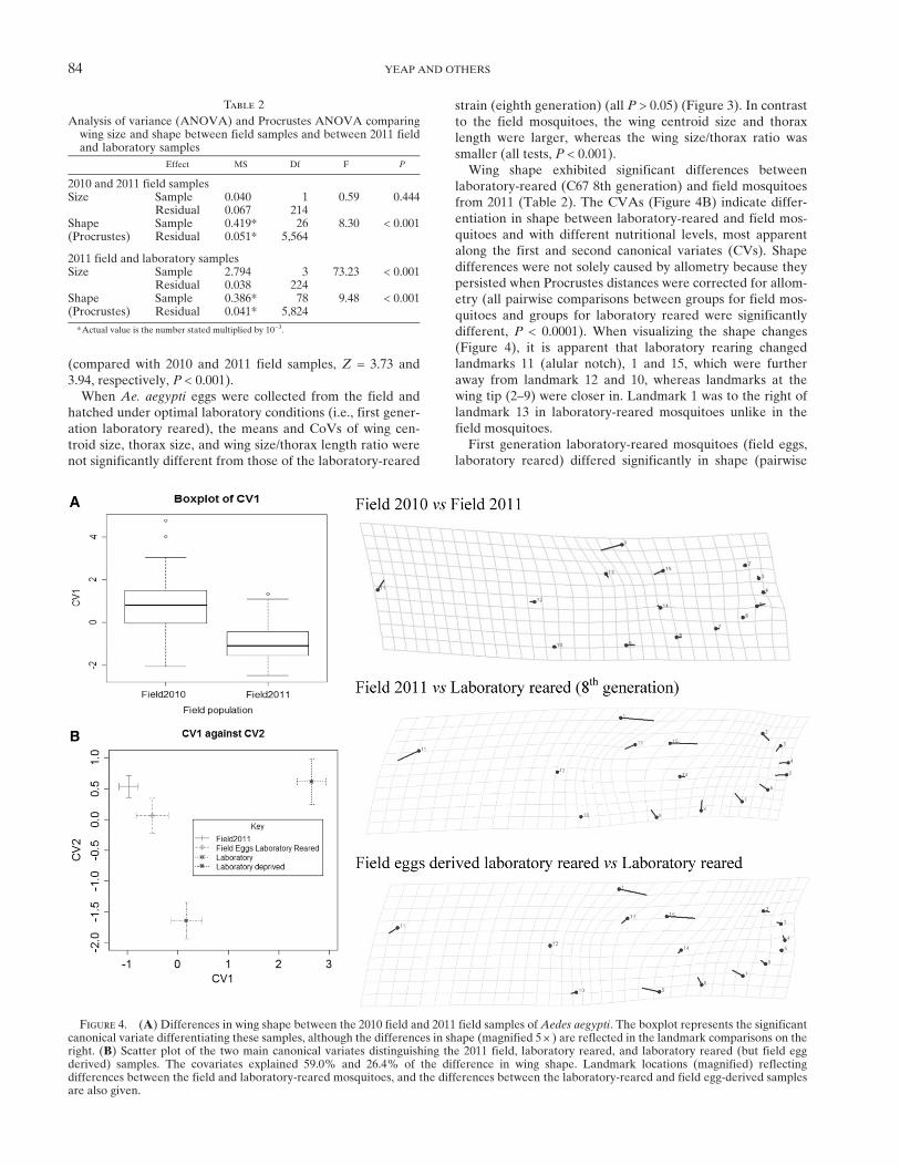
shape variables showed that GV and YK samples collectedin 2010 could be classified as one group, as could the 2011samples collected in the surrounding suburbs (all P > 0.05).However, in the Procrustes ANOVA of the pooled 2010and 2011 samples, there were significant shape differences(Table 2) as evident from the boxplot of the canonical vari-ate (Figure 4A). The difference in wing shape between yearswas associated with changes in landmarks 1, 15, and 11 (alularnotch) (Figure 4), with landmark 1 in 2011 samples on aver-age positioned toward the left of landmark 13. Further com-parisons of shape were therefore only made between samplescollected within the same year.Comparison between laboratory and field. Laboratory-
reared mosquitoes (C67) were substantially larger on averagethan field mosquitoes (Figure 3, Table 2). Mean size wassignificantly different when compared with 2010 GV and YK
Table 1
Summary of samples included in main comparisons*
Comparisons
2010 2011 During† Post† May† BGS Sticky JCU Laboratory
YK/GV HB/MB ED/PE YK/GV YK/GV YK/GV Cairns/MB Cage Uninfected wMel
Laboratory vs. field X X X Field eggs/C67Infection C89 XField X XMonitoring X X
X XX X
All field collected mosquitoes (i.e., NOT JCU and Laboratory) are caught in BGS traps except for Sticky (sticky ovitrap).*YK = Yorkeys Knob; GV = Gordonvale; MB = Machans Beach; HB = Holloways Beach; ED = Edmonton; PE = Pyramid Estate.†During = Third week of wMel-infected release; Post = Two weeks post-final release; May = Samples from May.
82 YEAP AND OTHERS
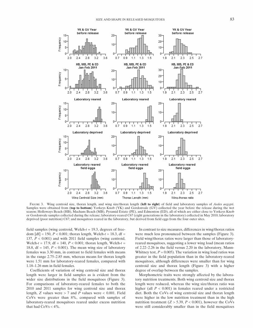
field samples (wing centroid, Welch-t = 19.3, degrees of free-dom [df] = 150, P < 0.001; thorax length, Welch-t = 18.3, df =137, P < 0.001) and with 2011 field samples (wing centroid,Welch-t = 17.9, df = 140, P < 0.001; thorax length, Welch-t =18.8, df = 145, P < 0.001). The mean wing size of laboratoryfemales was 3.30 mm, in contrast to field females with meansin the range 2.75–2.85 mm, whereas means for thorax lengthwere 1.51 mm for laboratory-reared females, compared with1.18–1.26 mm in field females.Coefficients of variation of wing centroid size and thorax
length were larger in field samples as is evident from thewider size distributions in the field mosquitoes (Figure 3).For comparisons of laboratory-reared females to both the2010 and 2011 samples for wing centroid size and thoraxlength, Z values were > 7 and P values were < 0.001. FieldCoVs were greater than 8%, compared with samples oflaboratory-reared mosquitoes reared under excess nutritionthat had CoVs < 4%.
In contrast to size measures, differences in wing/thorax ratioswere much less pronounced between the samples (Figure 3).Field wing/thorax ratios were larger than those of laboratory-reared mosquitoes, suggesting a lower wing load (mean ratiosof 2.22–2.26 in the field versus 2.20 in the laboratory, Mann-Whitney test, P = 0.005). The variation in wing load ratios wasgreater in the field population than in the laboratory-rearedmosquitoes, although differences were smaller than for wingcentroid size and thorax length (Figure 3) with a higherdegree of overlap between the samples.Morphometric traits were strongly affected by the labora-
tory nutrition treatments. Both wing centroid size and thoraxlength were reduced, whereas the wing size/thorax ratio washigher (all P < 0.001) in females reared under a restricteddiet. Both the CoVs of wing centroid size and thorax lengthwere higher in the low nutrition treatment than in the highnutrition treatment (Z > 5.39, P < 0.001), however the CoVswere still considerably smaller than in the field mosquitoes
Figure 3. Wing centroid size, thorax length, and wing size/thorax length (left to right) of field and laboratory samples of Aedes aegypti.Samples were obtained from (top to bottom) Yorkeys Knob (YK) and Gordonvale (GV) collected the year before the release during the wetseason; Holloways Beach (HB), Machans Beach (MB), Pyramid Estate (PE), and Edmonton (ED), all of which are either close to Yorkeys Knobor Gordonvale samples collected during the release; laboratory-reared C67 (eight generations in the laboratory) collected in May 2010; laboratorydeprived (poor nutrition) C67; and mosquitoes reared in the laboratory, but derived from field eggs from the four outer sites.
SIZE AND SHAPE IN RELEASED MOSQUITOES 83
(compared with 2010 and 2011 field samples, Z = 3.73 and3.94, respectively, P < 0.001).When Ae. aegypti eggs were collected from the field and
hatched under optimal laboratory conditions (i.e., first gener-ation laboratory reared), the means and CoVs of wing cen-troid size, thorax size, and wing size/thorax length ratio werenot significantly different from those of the laboratory-reared
strain (eighth generation) (all P > 0.05) (Figure 3). In contrastto the field mosquitoes, the wing centroid size and thoraxlength were larger, whereas the wing size/thorax ratio wassmaller (all tests, P < 0.001).Wing shape exhibited significant differences between
laboratory-reared (C67 8th generation) and field mosquitoesfrom 2011 (Table 2). The CVAs (Figure 4B) indicate differ-entiation in shape between laboratory-reared and field mos-quitoes and with different nutritional levels, most apparentalong the first and second canonical variates (CVs). Shapedifferences were not solely caused by allometry because theypersisted when Procrustes distances were corrected for allom-etry (all pairwise comparisons between groups for field mos-quitoes and groups for laboratory reared were significantlydifferent, P < 0.0001). When visualizing the shape changes(Figure 4), it is apparent that laboratory rearing changedlandmarks 11 (alular notch), 1 and 15, which were furtheraway from landmark 12 and 10, whereas landmarks at thewing tip (2–9) were closer in. Landmark 1 was to the right oflandmark 13 in laboratory-reared mosquitoes unlike in thefield mosquitoes.First generation laboratory-reared mosquitoes (field eggs,
laboratory reared) differed significantly in shape (pairwise
Figure 4. (A) Differences in wing shape between the 2010 field and 2011 field samples of Aedes aegypti. The boxplot represents the significantcanonical variate differentiating these samples, although the differences in shape (magnified 5 +) are reflected in the landmark comparisons on theright. (B) Scatter plot of the two main canonical variates distinguishing the 2011 field, laboratory reared, and laboratory reared (but field eggderived) samples. The covariates explained 59.0% and 26.4% of the difference in wing shape. Landmark locations (magnified) reflectingdifferences between the field and laboratory-reared mosquitoes, and the differences between the laboratory-reared and field egg-derived samplesare also given.
Table 2
Analysis of variance (ANOVA) and Procrustes ANOVA comparingwing size and shape between field samples and between 2011 fieldand laboratory samples
Effect MS Df F P
2010 and 2011 field samplesSize Sample 0.040 1 0.59 0.444
Residual 0.067 214Shape Sample 0.419* 26 8.30 < 0.001(Procrustes) Residual 0.051* 5,564
2011 field and laboratory samplesSize Sample 2.794 3 73.23 < 0.001
Residual 0.038 224Shape Sample 0.386* 78 9.48 < 0.001(Procrustes) Residual 0.041* 5,824
*Actual value is the number stated multiplied by 10−3.
84 YEAP AND OTHERS

Procrustes distance) from laboratory-reared mosquitoes atthe 8th generation, nutrition-deprived mosquitoes and fieldmosquitoes (all P < 0.001). This may reflect a subtle changein genetic variation for shape during laboratory rearing,although the sample size is small (N = 38). For the comparisonwith field mosquitoes from 2011, laboratory-reared mosqui-toes at the 8th generation also had landmark 1 to the right oflandmark 13, compared with mosquitoes at the first genera-tion of laboratory rearing (Figure 4). In addition, landmarks1, 15, and 11 were relatively further from landmark 12 and10 in the well-established laboratory lines.Under laboratory conditions, wMel-infected females were
not significantly different in wing size compared withuninfected mosquitoes (C89) under low nutrition (2.29 mmversus 2.33 mm) and under high nutrition (uninfected versusinfected, 2.95 mm versus 2.98 mm, P = 0.041) after adjustingfor multiple comparison (Table 3). Procrustes ANOVA ofshape variables (Table 3) indicated that the effect of infectionstatus on shape was significant under high nutrition but notunder low nutrition (Table 3). The shape change in infectedcompared with uninfected females under control conditionsinvolved landmark 12 moving closer to landmark 11, away
from the other inner landmarks (13–15). Unsurprisingly, therewere also large effects of nutrition on size (P < 0.0001) consis-tent with effects observed in the other experiments (Figure 4).There was also an effect of nutrition on shape in both theinfected and uninfected individuals (P < 0.001) (Table 3), asevident from differences in CV1 computed from the CVA(Figure 5). The shape differences between the high to lownutrition conditions involved the alular notch (landmark 11)moving closer to landmarks 12 and 10, whereas landmark 1was located toward the left of landmark 13 (Figure 5).Field comparison. Within the BGS traps, gravid and parous
females not infected withWolbachia together (mean wing size,2.87 mm; thorax length, 1.27 mm) were not significantly differ-ent (P > 0.17) from nulliparous females (mean wing size,2.76 mm; thorax length, 1.23 mm). If we separate gravid andparous females, parous females were smaller than gravid females(see Table 4) and this differencewas significant (t = 2.34, df= 86,P < 0.05), but the number of parous females was low (Table 4).Females lacking Wolbachia and collected from sticky
ovitraps tended to be larger than females collected in the BGStraps (Table 4), a difference that was just non-significant forwing centroid size (t = 1.93, df = 171, P = 0.06) and significant
Table 3
Analysis of variance (ANOVA) and Procrustes ANOVA comparing wing size and shape between infected and uninfected mosquitoes scoredunder control (high) and low nutrition laboratory conditions; and comparing overall effect of nutrition levels
Effect MS df F P
High nutrition Size Infection 0.059 1 4.22 0.041Residual 0.014 283
Shape Infection 0.133* 26 3.38 < 0.0001(Procrustes) Residual 0.039* 7,358
Low nutrition Size Infection 0.042 1 2.02 0.160Residual 0.021 77
Shape Infection 0.049* 26 1.21 0.213(Procrustes) Residual 0.040* 2002
Overall laboratory Size Nutrition 27.042 1 1,721.98 < 0.001Residual 0.016 362
Shape Nutrition 0.666* 26 16.69 < 0.001(Procrustes) Residual 0.040* 9,412
*Actual value is the number stated multiplied by 10−3.
Figure 5. (Left) The main canonical variates explaining differences between wMel and uninfected Aedes aegypti females at two differentnutrition conditions (CV1, 77.3%; CV2, 15.3%). Non-overlapping error bars do not imply significant difference; (top right) shape change fromuninfected to wMel infected; (bottom right) shape change from high nutrition to deprived nutrition. All shape changes were magnified 5 +.
SIZE AND SHAPE IN RELEASED MOSQUITOES 85

for thorax length (t = 2.32, df = 198, P = 0.02). The BGS-trapsample included gravid females that had similar means tothose from the sticky ovitraps (see means in Table 4) and didnot differ significantly from them (P > 0.38).Turning to wing size/thorax ratio, sticky ovitrap females
had a lower ratio (higher wing load) compared with BGS-trapped females (Table 4) and this difference was significant(Mann-Whitney U = 3739, P < 0.01). Unlike wing size andthorax length, gravid females from BGS traps were not signif-icantly different from all other parity states for wing/size tho-rax ratio (Mann-Whitney U = 1377, P = 0.98), whereas gravid
females in BGS-traps had a higher ratio compared with stickyovitrap females (Table 4) (Mann-WhitneyU = 2323. P < 0.01).We investigated how relative fitness changed with morpho-
metric measures fewer than two assumptions. First, weassumed sticky ovitrap females were relatively fitter thanBGS-trap females, thereby testing for oviposition success.Second, we also compared gravid and parous females to nul-liparous, to investigate possible differences between groupsdiffering in blood feeding. As we have noted, these are likelyto be conservative comparisons as we are comparing groupsfor which one is a subset of the other. We found that the
Table 4
Summary statistics (sample size, mean, SD, and coefficient of variation (CoV) of wing centroid size, thorax length and wing size/thorax ratio ofmosquitoes obtained from BGS or sticky ovitraps in March 2011*
Wing centroid size (mm) Thorax length (mm) Wing size/thorax ratio
N Mean SD CoV n Mean SD CoV n Median SD CoV
BGS trapsOverall 117 2.834 0.27 9.50% 124 1.258 0.139 11.00% 108 2.294 0.075 3.30%Gravid 71 2.902 0.238 8.20% 79 1.287 0.124 9.70% 67 2.292 0.073 3.20%Parous (1) 17 2.751 0.242 8.80% 16 1.206 0.107 8.80% 15 2.290 0.067 2.90%Nulliparous (2) 12 2.765 0.331 12.00% 13 1.240 0.184 14.80% 12 2.294 0.086 3.80%Non-gravid (3) 17 2.685 0.302 11.30% 16 1.184 0.159 13.40% 14 2.317 0.08 3.50%(1)+(2)+(3) 46 2.73 0.285 10.50% 45 1.208 0.149 12.30% 41 2.296 0.077 3.40%
Sticky ovitraps 56 2.921 0.286 9.80% 76 1.306 0.144 11.00% 53 2.257 0.073 3.20%
*Median of wing size/thorax ratio because we used Mann-Whitney U comparison of medians to test difference.
Figure 6. Fitness function plots using cubic spline with Bayesian standard error based on wing centroid size, thorax length, and wing size/thorax ratio of Aedes aegypti. Fitness curves on the left assume only egg laying females from sticky ovitraps are fit. On the right, fitness curvesconsider successful blood-feeding as high fitness, which includes gravid, parous and sticky ovitrap females.
86 YEAP AND OTHERS

likelihood of belonging to the ovipositing group increased withsize (particularly for wing centroid size) (Figure 6). Likelihoodof belonging to the ovipositing group tended to decrease withincreasing wing size/thorax ratios, but there was no noticeabledifference in likelihood between ratios of 2.1–2.3, which com-prised 63–67% of all individuals in the analysis.As in the case of wing/thorax ratio, we found a significant
effect of trap type on wing shape. Based on ProcrustesANOVA, we found that sticky ovitrap females were signifi-cantly different from all categories of BGS-trapped females(P < 0.0001). When the BGS samples were separated andcompared against the sticky ovitrap samples, the CVAs(Figure 7) indicated that the gravid, parous, and nulliparousfemales from BGS traps clustered as one group, and thesewere not different in pairwise comparisons of Procrustes dis-tances (P > 0.05). We therefore only visualized how shapevariables relate to ability to locate oviposition sites based ona comparison of sticky ovitrap versus BGS-trap females. ThePCA was performed to obtain six orthogonal principal com-ponents (PCs) explaining > 5% of the variance: PC1 explained24.6%, PC2 18.9%, PC3 12.5%, PC4 7.8%, PC5 6.5%, andPC6 5.3%. The ANOVAs on PCs indicated that after correc-tion for multiple comparisons, only PC1 differed betweengravid versus other parity states (F(1,145) = 5.61, P = 0.01) andits association was more pronounced when it was assumedthat only egg laying females were fitter than BGS-trappedfemales (F(1,145) = 15.64, P < 0.0001), with trends plotted in
Figure 7. Shape differences between the BGS-trapped andsticky ovitrap females involved central landmarks rather thanthose at the wing tip, with landmark 1 tending toward theright of landmark 13, whereas landmark 15 was closer tolandmark 13 (Figure 7).Changes during the release. The release of large field cage
mosquitoes was expected to produce an influx of largeinfected mosquitoes in the field populations, with contributionsfrom field-reared infected mosquitoes increasing over time asthe Wolbachia frequency steadily increased throughout therelease and immediate post-release periods. In YorkeysKnob, Wolbachia frequency increased from 61% to 76.9%and then to 89.7% when comparing the three time points(Week 3, 2 weeks after final release and May), whereas inGordonvale, frequencies were 53.3%, 65%, and finally 73.2%.1
Based on the comparison of laboratory and field reared mos-quitoes, we expected some shape differences as well.Third week of release. Captured wMel-infected females
were not significantly different in size from the JCU cage-reared mosquitoes (mean wing centroid size: 3.17 versus3.22 mm, Welch-t = 1.85, df = 55, P > 0.05, also see Table 5;thorax length: 1.44 versus 1.49 mm, Welch-t = 2.37, df = 62,P > 0.01). There was also no significant difference betweenwing size to thorax length ratio (median: 2.19 versus 2.17,P > 0.2). Because we expected almost all the mosquitoesfrom this period to be reared in the cage, the lack of signifi-cant differences in sizes suggest that there is no evidence of
Figure 7. (Top left) Main canonical variates differentiating mosquitoes from sticky ovitrap and BGS traps with parity states before sizecorrection, CV1 and CV2 explained 56.8% and 23.0% of difference; (top right) main principal component versus fitness (ability to seek ovipositionsite) with Bayesian standard errors; (bottom) shape change between BGS-trapped female mosquitoes and sticky ovitrap female mosquitoes(magnified 5 +).
SIZE AND SHAPE IN RELEASED MOSQUITOES 87

variation in survival between different sized mosquitoes fromthe field cage.The CoVs of wing centroid size (overall: 4.6%) and thorax
length (overall: 5.9%) of captured wMel-infected femaleswere significantly larger than those of JCU field cage-rearedmosquitoes (CoV: wing centroid size, 2.7%; thorax length,3.3%) (Z > 1.96, P < 0.05) (Figure 8). When compared withcaptured uninfected females (overall CoV: wing centroid size,9.8%; thorax length, 10.8%), the magnitude of the disparitywas much greater and statistically significant (Z > 1.96, P <0.05) (Figure 8). The CoVs of captured uninfected femalesfrom both sites were not significantly different from field mos-
quitoes captured the year before the release (for all compari-sons: Z < 1.96, P > 0.05). As expected, these patterns reflect thefact that captured wMel-infected females represent releasematerial, but they also point to mosquitoes with diverse sizessurviving in the field at least during the wet season.Procrustes ANOVAs (Table 5) revealed that captured
infected females differ significantly from JCU field cage femalesand field mosquitoes from 2011. Pairwise permutation tests onProcrustes distances also confirmed these results with P < 0.05for both comparisons. Canonical variate analysis suggests thatcaptured infected females are still more similar to JCU fieldcage mosquitoes than to the field mosquitoes.
Figure 8. Cleveland plots of wing centroid size and thorax length of Aedes aegypti. YK = Yorkeys Knob; GV = Gordonvale; JCU = JamesCook University cage-reared mosquitoes.
88 YEAP AND OTHERS

Two weeks post-final release. By this stage, the frequency ofwMel infection in the two sites was high. Captured wMel-infected females had significantly reduced wing size and tho-rax length when compared with JCU field cage mosquitoes:wing centroid size (2.90 versus 3.22 mm;Welch-t = 7.44, df = 63,P < 0.0001), thorax length (1.28 versus 1.49 mm,Welch-t = 9.67,df = 76, P < 0.0001). Wing/thorax ratios of captured wMel-infected females were significantly higher than those of theJCU field cage females (2.27 versus 2.17, P < 0.0001).As expected, field females had significantly larger CoVs
compared with JCU field cage mosquitoes: wing centroid size(10.3% versus 2.7%) and thorax length (12.0% versus 3.3%)(all comparisons: Z > 1.96, P < 0.05) (Figure 8). The CoVswere similar to those from field female mosquitoes capturedthe year before the release (2010 sample) (all comparisons:Z < 1.96, P > 0.05) (Figure 8). Procrustes ANOVAs (Table 5)indicate that captured infected females were significantlydifferent from JCU field cage mosquitoes in shape. Pairwisepermutation tests on Procrustes distances and canonicalvariates also suggested a marginally significant (P < 0.05)difference in shape between captured infected females andfield mosquitoes.Because of the significant differences in size and shape
between field cage and field mosquitoes, we estimated thesurvival of released females using individuals in the popula-tion with a large wing size and shape similar to those of therelease sample. On the basis of wing centroid size only, weestimated that ~22.5% of the field female mosquitoes (2010and 2011 samples) were within the narrow size range observedin the release sample (JCU field cage). This compares with~34.2% (GV) and 31.2% (YK) of female mosquitoes’ post-final release, which had wing centroid sizes within the rangeof the JCU field cage mosquitoes. We can estimate algebrai-cally the frequency of individuals originating from the fieldcage to be 15.1% and 11.3% after 2 weeks from GV and YK,
respectively. These translate into 87.4% and 85.6% survivalper day. We also estimated survival in the same way byassigning mosquitoes based on shape instead of size using adiscriminant analysis contrasting field cage and field mosqui-toes, but daily survival estimates were 89.4% in YK and90.3% in GV. Combining both size and shape constraints, thedaily survival rate in YK was estimated to be 87% and in GVto be 79%. All of this was made possible by assuming nounderlying Wolbachia effect on survival and size, constantpopulation size of infected mosquitoes over the 2 weeks postrelease, and negligible survival of released mosquitoes beforethe last release.Samples from May. In May, ambient temperature in Cairns
was on average 5°C lower than in January to March. Thischange could have an impact on morphological traits of mos-quitoes.61,62 Wing centroid sizes of females from this samplingperiod were larger (2.92 versus 2.81 mm) than field femalessampled in the warmer months (t = 2.25, df = 151, P = 0.03).There was, however, no evidence of a difference in thoraxlength (t = 0.98, df = 160, P = 0.33). The CoV for both mea-sures were similar to field females from the year before therelease. With regards to wing size/thorax ratios, these weresignificantly higher in May compared with field samples fromthe wet season (January–March) (median ratio = 2.33 versus2.28, Mann-Whitney U = 3023, P values = 0.004), reflectingthe fact that wing size changed without a concomitant changein thorax length.Captured infected females in May were significantly differ-
ent in shape from JCU field cage-reared females and whencompared with field mosquitoes from 2011 (Table 5). How-ever, based on canonical variate analysis, we found that cap-tured infected females were more similar to field mosquitoesfrom 2011 than to JCU field cage mosquitoes. This is similarto the difference observed between infected females from2 weeks post-final release and JCU field cage females.
Table 5
Analysis of variance (ANOVA) of wing size and Procrustes ANOVA of wing shape of captured third week of release, two weeks post release andtwo months post release (May) compared with field cage-reared and field mosquitoes from 2011
Effect MS df F P
Third week of release wMel captured vs. field cage Size Origin 0.051 1 3.63 0.060Residual 0.014 75
Shape Individual 0.127* 26 3.50 < 0.001Residual 0.036* 1,950
wMel captured vs. field 2011 Size Individual 3.437 1 60.28 < 0.001Residual 0.057 134
Shape Individual 0.093* 26 2.43 < 0.001Residual 0.038* 3,484
2 weeks post-release wMel captured vs. field cage Size Individual 2.371 1 44.41 < 0.001Residual 0.053 92
Shape Individual 0.187* 26 5.56 < 0.001Residual 0.034* 2,392
wMel captured vs. field 2011 Size Individual 0.291 1 3.82 0.053Residual 0.076 151
Shape Individual 0.099* 26 3.73 < 0.001Residual 0.036* 3926
May wMel captured vs. field cage Size Individual 1.905 1 33.71 < 0.001Residual 0.057 82
Shape Individual 0.534* 26 14.42 < 0.001Residual 0.037* 2,132
wMel captured vs. field 2011 Size Individual 0.368 1 4.62 0.033Residual 0.080 141
Shape Individual 0.112* 26 2.90 < 0.001Residual 0.038* 3,666
*Actual value is the number stated multiplied by 10−3.
SIZE AND SHAPE IN RELEASED MOSQUITOES 89
DISCUSSION
The findings in this study suggest that morphometric traitsare not only strongly affected by environmental conditionsbut also linked to the likelihood of being collected from ovi-position sites of Ae. aegypti females in the field. These traitscan be used to assess the fitness of laboratory-reared coloniesdestined for release, and to assess survival of released individ-uals in the field. Large size seems to reflect oviposition sitelocation in females. The effects may have been underestimatedas a result of comparing non-exclusive groups, particularly asBGS-traps contained > 50% gravid females. This result mayreflect the fact that large females have an increased fecun-dity.63–66 Larger females may also have a better flight range,higher survival, increased host finding, and blood feeding suc-cess, and improved ability to locate oviposition sites.67,68
Larger also means higher energy reserves,67 and by having ahigher mass/surface area ratio, they might be less prone todesiccation than small mosquitoes.69 These results suggest thatthe strategy of rearing large mosquitoes in the mass-releases ofwMel-infected mosquitoes1 may have contributed to the suc-cess of these releases. It is unclear if the large size of males alsoinfluences fitness in the field, although laboratory studies sup-port the idea that larger males are fitter.22,70,71
We found no difference in measurements between gravidand parous versus nulliparous females. Field studies on bloodfeeding success suggests a negative association with size25,72
or no strong evidence of association.72 In contrast, we did finda significant difference in size between gravid and parousfemales and several factors might contribute to this difference.First, larger females may be more likely to have acquired ablood meal recently, particularly when the presence of matureovules indicates successful blood feeding within at least the last3 days.46 Second, blood-feeding rate may not depend on sizebut larger females may stay gravid for a longer time because ofhigher fecundity and a lengthy period of oviposition arisingfrom skip oviposition behavior. Third, smaller females may livelong enough to undergo complete oviposition, in contrast tothe larger females. Finally, smaller females may be more likelyto be inseminated. Because there is no strong evidence thatsmaller females have higher survival in smaller females,25,67
we suspect that the third reason is not likely to be important.We also suspect that there is unlikely to be an inseminationbias given the results of laboratory studies22; the results maytherefore reflect success in blood feeding or egg retention, butsorting this out requires further work.Although the wing size to thorax length ratio also differed
between the laboratory and field samples, this difference wasmuch less than the difference in size. Given that previous studieshave suggested that this trait may influence dispersal ability,32
and given the lower ratio in females from the sticky ovitraps, itwould seem prudent to try to match ratios in released materialwith those from the field. Temperature manipulations may needto be monitored closely with changing seasons, as there is someevidence from our May sampling data and previous studies73,74
that temperature could affect wing load.The shape analyses suggest that the available wing shape
variation is associated with environmental effects and fitnessbased on ability to seek oviposition sites but not blood-feedingsuccess. Effects of nutrition and other variables on wing shapehave previously been documented in insects,53,75 and in thecurrent study there was a distinct change in wing shape asso-
ciated with low nutrition. Given the multivariate nature ofshape, it is difficult to determine precisely the likely implica-tions of this shape change on field fitness. Changes in land-marks 1 and 15 are affected by nutrition conditions and differbetween BGS and sticky samples, so changes in these aspectsof wing shape may influence field fitness.Any effects of Wolbachia releases on morphometric varia-
tion are likely to be transient; because once releases werecompleted the morphometric traits and their variances con-verged rapidly on those of field populations before the release.This convergence also provides evidence that mass-rearingprotocols and the backcrossing scheme did not markedly influ-ence morphometric traits, with rearing in the laboratory andfield cage environments likely to exert effects through pheno-typic plasticity. Field environments presumably result in vari-ability because of the wide array of environmental conditionsavailable for juvenile stages.76,77
Finally, size and shape appeared useful in discrimination ofreleased mosquitoes from field-reared mosquitoes. By com-bining the morphometrics with an assay of Wolbachia status,we were able to assess the survival potential of the releasedmosquitoes, which appeared similar to estimates of survival ofAe. aegyptimosquitoes in the field,78 corroborating the resultsof laboratory studies of wMel-infected mosquitoes,2 whichindicated that survival of wMel-infected mosquitoes in thelaboratory was not significantly impaired by the infection.Despite some previous studies suggesting that shape could beused to discriminate different environmental conditions,79,80
we suspect that it should be applied alongside size to discrim-inate populations from different conditions.
Received November 30, 2012. Accepted for publication March 28, 2013.
Published online May 28, 2013.
Acknowledgments: We thank members of the Eliminate DengueProject team who released mosquitoes and collected and identifiedBGS samples. We thank James Cook University staff, particularlyChris Paton, Clare Omodei and Gavin Omodei who organized mos-quito rearing. We thank members of the Eliminate Dengue team forhelping with BGS collections, Sharron Long and Karel von Herckfrom Queensland Health for assisting with sticky ovitrap collections.We also thank Jason K. Axford and Ashley G. Callahan for theassistance given in some of the laboratory based experiments andrearing. We are grateful to the residents of Cairns, Yorkeys Knoband Gordonvale for allowing us to set traps at their properties.
Financial support: This project was funded by a grant from the Foun-dation for the National Institutes of Health through the Grand Chal-lenges in Global Health Initiative of the Bill and Melinda GatesFoundation, the National Health and Medical Research Council,Australia, and the Urban Health Cluster of the CSIRO ClimateChange Flagship program. AAH was funded by a Fellowship fromthe Australian Research Council.
Authors’ addresses: Heng Lin Yeap, Nancy M. Endersby, and Ary A.Hoffmann, Department of Genetics, Parkville, VIC 3052, Australia,E-mails: [email protected], [email protected], and [email protected]. Petrina H. Johnson, School of Biological Sciences,Faculty of Science, Monash University, Australia, E-mail: [email protected]. Scott A. Ritchie, Tropical Medicine andRehabilitation Sciences, James Cook University, Cairns, Queensland,Australia, E-mail: [email protected].
REFERENCES
1. Hoffmann AA, Montgomery BL, Popovici J, Iturbe-Ormaetxe I,Johnson PH, Muzzi F, Greenfield M, Durkan M, Leong YS,Dong Y, Cook H, Axford J, Callahan AG, Kenny N, Omodei C,
90 YEAP AND OTHERS

McGraw EA, Ryan PA, Ritchie SA, Turelli M, O’Neill SL,2011. Successful establishment of Wolbachia in Aedes popula-tions to suppress dengue transmission. Nature 476: 454–457.
2. Walker T, Johnson PH, Moreira LA, Iturbe-Ormaetxe I, FrentiuFD, McMeniman CJ, Leong YS, Dong Y, Axford J, KriesnerP, Lloyd AL, Ritchie SA, O’Neill SL, Hoffmann AA, 2011.The wMel Wolbachia strain blocks dengue and invades cagedAedes aegypti populations. Nature 476: 450–453.
3. Bellini R, Calvitti M, Medici A, Carrieri M, Celli G, Maini S,2007. Use of the sterile insect technique against Aedesalbopictus in Italy: first results of a pilot trial. Vreysen MJ,Robinson AS, Hendrichs J, eds. Area-Wide Control of InsectPests: From Research to Field Implementation. Springer,Dordrecht, The Netherlands, 505–515.
4. de Valdez MR, Nimmo D, Betz J, Gong HF, James AA, AlpheyL, Black WC, 2011. Genetic elimination of dengue vector mos-quitoes. Proc Natl Acad Sci USA 108: 4772–4775.
5. Helinski ME, Hassan MM, El-Motasim WM, Malcolm CA, KnolsBG, El-Sayed B, 2008. Towards a sterile insect technique fieldrelease ofAnopheles arabiensismosquitoes in Sudan: irradiation,transportation, and field cage experimentation.Malar J 7: 65.
6. Alphey L, Benedict M, Bellini R, Clark GG, Dame DA, ServiceMW, Dobson SL, 2010. Sterile-insect methods for control ofmosquito-borne diseases: an analysis. Vector Borne ZoonoticDis 10: 295–311.
7. Cerutti F, Bigler F, 1995. Quality assessment of Trichogramma-brassicae in the laboratory. Entomol Exp Appl 75: 19–26.
8. Dutton A, Bigler F, 1995. Flight activity assessment of the eggparasitoid Trichogramma brassicae (Hym: Trichogrammatidae)in laboratory and field conditions. Entomophaga 40: 223–233.
9. Dutton A, Cerutti F, Bigler F, 1996. Quality and environmentalfactors affecting Trichogramma brassicae efficiency under fieldconditions. Entomol Exp Appl 81: 71–79.
10. Kolliker-Ott UM, Blows MW, Hoffmann AA, 2003. Are wingsize, wing shape and asymmetry related to field fitness ofTrichogramma egg parasitoids? Oikos 100: 563–573.
11. Kazmer DJ, Luck RF, 1991. Female body size, fitness and bio-logical control quality: field experiments with Trichogrammapretiosum. Colloques de l’INRA 56: 37–40.
12. Navarro-Campos C, Martinez-Ferrer MT, Campos JM, Fibla JM,Alcaide J, Bargues L, Marzal C, Garcia-Mari F, 2011. Theinfluence of host fruit and temperature on the body size ofadult Ceratitis capitata (Diptera: Tephritidae) under laboratoryand field conditions. Environ Entomol 40: 931–938.
13. Montgomery BL, Ritchie SA, 2002. Roof gutters: a key containerfor Aedes aegypti and Ochlerotatus notoscriptus (Diptera:Culicidae) in Australia. Am J Trop Med Hyg 67: 244–246.
14. Montgomery BL, Ritchie SA, Hart AJ, Long SA, Walsh ID, 2004.Subsoil drain sumps are a key container for Aedes aegypti inCairns, Australia. J Am Mosq Control Assoc 20: 365–369.
15. Wilder-Smith A, Ooi E-E, Vasudevan S, Gubler D, 2010. Updateon dengue: epidemiology, virus evolution, antiviral drugs, andvaccine development. Curr Infect Dis Rep 12: 157–164.
16. McMeniman CJ, Lane AM, Fong AW, Voronin DA, Iturbe-Ormaetxe I, Yamada R, McGraw EA, O’Neill SL, 2008. Hostadaptation of a Wolbachia strain after long-term serial passagein mosquito cell lines. Appl Environ Microbiol 74: 6963–6969.
17. Kambris Z, Blagborough AM, Pinto SB, Blagrove MS, GodfrayHCJ, Sinden RE, Sinkins SP, 2010. Wolbachia stimulatesimmune gene expression and inhibits Plasmodium develop-ment in Anopheles gambiae. PLoS Pathog 6: e1001143.
18. Moreira LA, Iturbe-Ormaetxe I, Jeffery JA, Lu GJ, Pyke AT,Hedges LM, Rocha BC, Hall-Mendelin S, Day A, Riegler M,Hugo LE, Johnson KN, Kay BH, McGraw EA, van den HurkAF, Ryan PA, O’Neill SL, 2009. A Wolbachia symbiont inAedes aegypti limits infection with Dengue, Chikungunya, andPlasmodium. Cell 139: 1268–1278.
19. Pan XL, Zhou GL, Wu JH, Bian GW, Lu P, Raikhel AS, Xi ZY,2012. Wolbachia induces reactive oxygen species (ROS)-dependent activation of the Toll pathway to control denguevirus in the mosquito Aedes aegypti. Proc Natl Acad Sci USA109: E23–E31.
20. McMeniman CJ, Lane RV, Cass BN, Fong AW, Sidhu M, WangYF, O’Neill SL, 2009. Stable introduction of a life-shorteningWolbachia infection into the mosquito Aedes aegypti. Science323: 141–144.
21. Harrington LC, Connors KJ, Cator LJ, Helinski ME, 2009. Assor-tative mating in the dengue vector mosquito, Aedes aegypti.Am J Trop Med Hyg 81: 1017.
22. Ponlawat A, Harrington LC, 2009. Factors associated with malemating success of the dengue vector mosquito, Aedes aegypti.Am J Trop Med Hyg 80: 395–400.
23. Xue RD, Barnard DR, Muller GC, 2010. Effects of body size andnutritional regimen on survival in adult Aedes albopictus(Diptera: Culicidae). J Med Entomol 47: 778–782.
24. Armbruster P, Hutchinson RA, 2002. Pupal mass and wing lengthas indicators of fecundity in Aedes albopictus and Aedesgeniculatus (Diptera: Culicidae). J Med Entomol 39: 699–704.
25. Maciel-De-Freitas R, Codego CT, Lourenco-De-Oliveira R,2007. Body size-associated survival and dispersal rates ofAedes aegypti in Rio de Janeiro.Med Vet Entomol 21: 284–292.
26. Nasci RS, 1986. Relationship between adult mosquito (Diptera,Culicidae) body size and parity in field populations. EnvironEntomol 15: 874–876.
27. Scott TW, Morrison AC, Lorenz LH, Clark GG, Strickman D,Kittayapong P, Zhou H, Edman JD, 2000. Longitudinalstudies of Aedes aegypti (Diptera: Culicidae) in Thailandand Puerto Rico: population dynamics. J Med Entomol 37:77–88.
28. Breuker CJ, Brakefield PM, Gibbs M, 2007. The associationbetween wing morphology and dispersal is sex-specific in theglanville fritillary butterfly Melitaea cinxia (Lepidoptera:Nymphalidae). Eur J Entomol 104: 445–452.
29. Corbet SA, 2000. Butterfly nectaring flowers: butterfly morphol-ogy and flower form. Entomol Exp Appl 96: 289–298.
30. Hassall C, Thompson DJ, Harvey IF, 2008. Latitudinal variationin morphology in two sympatric damselfly species with con-trasting range dynamics (Odonata: Coenagrionidae). Eur JEntomol 105: 939–944.
31. Kemp DJ, 2002. Butterfly contests and flight physiology: why doolder males fight harder? Behav Ecol 13: 456–461.
32. Hoffmann AA, Ratna E, Sgro CM, Barton M, Blacket M, HallasR, De Garis S, Weeks AR, 2007. Antagonistic selectionbetween adult thorax and wing size in field releasedDrosophilamelanogaster independent of thermal conditions. J Evol Biol20: 2219–2227.
33. Santos M, Iriarte PF, Cespedes W, 2005. Genetics and geometryof canalization and developmental stability in Drosophilasubobscura. BMC Evol Biol 5: 7.
34. Ritchie SA, Johnson PH, Freeman AJ, Odell RG, Graham N,Dejong PA, Standfield GW, Sale RW, O’Neill SL, 2011. Asecure semi-field system for the study of Aedes aegypti. PLoSNegl Trop Dis 5: e988.
35. Ball TS, Ritchie SR, 2010. Sampling biases of the BG-Sentinel trapwith respect to physiology, age, and body size of adult Aedesaegypti (Diptera: Culicidae). J Med Entomol 47: 649–656.
36. Maciel-de-Freitas R, Eiras AE, Lourenco-de-Oliveira R, 2006.Field evaluation of effectiveness of the BG-Sentinel, a newtrap for capturing adult Aedes aegypti (Diptera: Culicidae).Mem Inst Oswaldo Cruz 101: 321–325.
37. Williams CR, Long SA, Russell RC, Ritchie SA, 2006. Fieldefficacy of the BG-sentinel compared with CDC backpackaspirators and CO2-baited EVS traps for collection of adultAedes aegypti in Cairns, Queensland, Australia. J Am MosqControl Assoc 22: 296–300.
38. Chadee DD, Ritchie SA, 2010. Efficacy of sticky and standardovitraps for Aedes aegypti in Trinidad, West Indies. J VectorEcol 35: 395–400.
39. Chadee DD, Ritchie SA, 2010. Oviposition behavior and parityrates of Aedes aegypti collected in sticky traps in Trinidad,West Indies. Acta Trop 116: 212–216.
40. Hiss EA, Fuchs MS, 1972. Effect of matrone on oviposition inmosquito, Aedes aegypti. J Insect Physiol 18: 2217.
41. Judson CL, 1967. Feeding and oviposition behavior in Aedesaegypti (L). I. Preliminary studies of physiological controlmechanisms. Biol Bull 133: 369–378.
42. Lavoipierre MMJ, 1958. Biting behavior of mated and unmatedfemales of an African strain of Aedes aegypti. Nature 181:1781–1782.
43. Ritchie SA, Rapley LP, Williams C, Johnson PH, Larkman M,Silcock RM, Long SA, Russell RC, 2009. A lethal ovitrap-based mass trapping scheme for dengue control in Australia: I.
SIZE AND SHAPE IN RELEASED MOSQUITOES 91

Public acceptability and performance of lethal ovitraps. MedVet Entomol 23: 295–302.
44. Detinova TS, 1962. Age-grouping methods in Diptera of medicalimportance with special reference to some vectors of malaria.Monogr Ser World Health Organ 47: 13–191.
45. Clements AN, Boocock MR, 1984. Ovarian development in mos-quitoes: stages of growth and arrest and follicular resorption.Physiol Entomol 9: 1–8.
46. Gwadz RW, Spielman A, 1973. Corpus allatum control of ovariandevelopment in Aedes aegypti. J Insect Physiol 19: 1441–1448.
47. Lee SF, White VL, Weeks AR, Hoffmann AA, EndersbyNM, 2012. High-throughput PCR assays to monitor Wolbachiainfection in the dengue mosquito (Aedes aegypti) andDrosophila simulans. Appl Environ Microbiol 78: 4740–4743.
48. Rohlf FJ, 2004. tpsUtil, File Utility Program, Version 1.26.Department of Ecology and Evolution, State University ofNew York at Stony Brook.
49. Rohlf FJ, 2010. tpsDig, Digitize Landmarks and Outlines, Ver-sion 2.16. Department of Ecology and Evolution, State Univer-sity of New York at Stony Brook.
50. Yeap HL, Mee P, Walker T, Weeks AR, O’Neill SL, Johnson P,Ritchie SA, Richardson KM, Doig C, Endersby NM,Hoffmann AA, 2011. Dynamics of the “popcorn” Wolbachiainfection in outbred Aedes aegypti informs prospects formosquito vector control. Genetics 187: 583–595.
51. Vargas RE, Ya-umphan P, Phumala-Morales N, KomalamisraN, Dujardin JP, 2010. Climate associated size and shapechanges in Aedes aegypti (Diptera: Culicidae) populationsfrom Thailand. Infect Genet Evol 10: 580–585.
52. Bookstein FL, 1991. Morphometric Tools for Landmark DataGeometry and Biology. New York: Cambridge University Press.
53. Jirakanjanakit N, Leemingsawat S, Thongrungkiat S,Apiwathnasorn C, Singhaniyom S, Bellec C, Dujardin JP,2007. Influence of larval density or food variation on the geom-etry of the wing of Aedes (Stegomyia) aegypti. Trop Med IntHealth 12: 1354–1360.
54. Siegel JP, Novak RJ, Lampman RL, Steinly BA, 1992. Statisticalappraisal of the weight wing length relationship of mosquitoes.J Med Entomol 29: 711–714.
55. Arnqvist G, Martensson T, 1998. Measurement error in geomet-ric morphometrics: Empirical strategies to assess and reduce itsimpact on measures of shape. Acta Zoologica AcademiaeScientiarum Hungaricae 44: 73–96.
56. Klingenberg CP, McIntyre GS, 1998. Geometric morphometricsof developmental instability: analyzing patterns of fluctuatingasymmetry with procrustes methods. Evolution 52: 1363–1375.
57. Klingenberg CP, 2011. MorphoJ: an integrated software packagefor geometric morphometrics. Mol Ecol Res 11: 353–357.
58. Miller GE, 1991. Asymptotic test statistics for coefficients of var-iation. Comm Statist Theory Methods 20: 3351–3363.
59. Schluter D, 1988. Estimating the form of natural-selection on aquantitative trait. Evolution 42: 849–861.
60. Kozak M, 2010. dotplot.errors, a new R function to ease the painof creating dotplots. Commun Biometry Crop Sci 5: 69–77.
61. Padmanabha H, Lord CC, Lounibos LP, 2011. Temperatureinduces trade-offs between development and starvationresistance in Aedes aegypti (L.) larvae. Med Vet Entomol25: 445–453.
62. Mohammed A, Chadee DD, 2011. Effects of different tempera-ture regimens on the development of Aedes aegypti (L.)(Diptera: Culicidae) mosquitoes. Acta Trop 119: 38–43.
63. Bader CA, Williams CR, 2012. Mating, ovariole number andsperm production of the dengue vector mosquito Aedes aegypti
(L.) in Australia: broad thermal optima provide the capacity forsurvival in a changing climate. Physiol Entomol 37: 136–144.
64. Briegel H, 1990. Metabolic relationship between female bodysize, reserves and fecundity of Aedes aegypti. J Insect Physiol36: 165–172.
65. Naksathit AT, Scott TW, 1998. Effect of female size on fecundityand survivorship of Aedes aegypti fed only human blood ver-sus human blood plus sugar. J Am Mosq Control Assoc 14:148–152.
66. Steinwascher K, 1982. Relationship between pupal mass andadult survivorship and fecundity for Aedes aegypti. EnvironEntomol 11: 150–153.
67. Briegel H, Knusel I, Timmermann SE, 2001. Aedes aegypti: size,reserves, survival, and flight potential. J Vector Ecol 26: 21–31.
68. Nasci RS, 1991. Influence of larval and adult nutrition on bitingpersistence inAedes aegypti (Diptera, Culicidae). JMed Entomol28: 522–526.
69. Mogi M, Miyagi I, Abadi K, Syafruddin, 1996. Inter- and intra-specific variation in resistance to desiccation by adult Aedes(Stegomyia) spp. (Diptera: Culicidae) from Indonesia. J MedEntomol 33: 53–57.
70. Helinski MEH, Harrington LC, 2011. Male mating history andbody size influence female fecundity and longevity of the den-gue vector Aedes aegypti. J Med Entomol 48: 202–211.
71. Ponlawat A, Harrington LC, 2007. Age and body size influencemale sperm capacity of the dengue vector Aedes aegypti(Diptera: Culicidae). J Med Entomol 44: 422–426.
72. Scott TW, Amerasinghe PH, Morrison AC, Lorenz LH, ClarkGG, Strickman D, Kittayapong P, Edman JD, 2000. Longi-tudinal studies of Aedes aegypti (Diptera: Culicidae) inThailand and Puerto Rico: blood feeding frequency. J MedEntomol 37: 89–101.
73. Loeschcke V, Bundgaard J, Barker JS, 1999. Reaction normsacross and genetic parameters at different temperatures for tho-rax and wing size traits in Drosophila aldrichi and D-buzzatii.J Evol Biol 12: 605–623.
74. Reiskind MH, Zarrabi AA, 2012. Is bigger really bigger? Differ-ential responses to temperature in measures of body size of themosquito, Aedes albopictus. J Insect Physiol 58: 911–917.
75. Hoffmann AA, Woods RE, Collins E, Wallin K, White A,McKenzie JA, 2005. Wing shape versus asymmetry as an indi-cator of changing environmental conditions in insects. Aust JEntomol 44: 233–243.
76. Schneider JR, Morrison AC, Astete H, Scott TW, Wilson ML,2004. Adult size and distribution of Aedes aegypti (Diptera:Culicidae) associated with larval habitats in Iquitos, Peru.J Med Entomol 41: 634–642.
77. Tun-Lin W, Burkot TR, Kay BH, 2000. Effects of temperatureand larval diet on development rates and survival of the den-gue vector Aedes aegypti in north Queensland, Australia. MedVet Entomol 14: 31–37.
78. Muir LE, Kay BH, 1997. Aedes aegypti as a vector of dengueviruses in northern Queensland: what have we learnt? Arbovi-rus research in Australia Proceedings Seventh ArbovirusResearch in Australia Symposium and Second Mosquito Con-trol Association of Australia Conference. Surfers Paradise,Australia, 25–29 November, 1996, 190–193.
79. Jirakanjanakit N, Dujardin J-P, 2005. Discrimination of Aedesaegypti (Diptera: Culicidae) laboratory lines based on wing geom-etry. Southeast Asian J Trop Med Public Health 36: 858–861.
80. Jirakanjanakit N, Leemingsawat S, Dujardin JP, 2008. The geom-etry of the wing ofAedes (Stegomyia) aegypti in isofemale linesthrough successive generations. Infect Genet Evol 8: 414–421.
92 YEAP AND OTHERS