Embed Size (px)
Citation preview
4467Research Article
IntroductionThe discovery of adult stem cells in the central nervous system(CNS) raised many hopes for stem cell therapy (Reynolds andWeiss, 1992). For therapeutic approaches stem cells have to becharacterized with respect to proliferation potential, stateof determination and morphogenetic behavior. To test themorphogenetic behavior of stem cells, specific experimental invivo models are required. Our approach was to transplant adultmouse CNS stem cells into the microenvironment of the avianembryo.
Subventricular zone stem cells (SVZ cells) are wellcharacterized in vitro. In general, after isolation from thesubventricular zone cells are grown in coated culture dishes.After a phase of proliferation with the mitogen FGF2, stemcells are transferred into non-coated culture dishes, wherefree-floating neurospheres form. Neurospheres are highlydynamic structures with distinct radial gradients of cellproliferation, cell survival zones, apoptosis and phagocytosis(Bez et al., 2003). On FGF2 withdrawal SVZ stem cells leavethe phase of proliferation and differentiate in vitro. SVZ cellsgenerate CNS neurons, astrocytes, and oligodendrocytes (Joheet al., 1996; Reynolds and Weiss, 1996). BMP-2 and FGF2treatment during proliferation induces neural crest cell fates(measured by expression of smooth muscle �-actin, SMA)upon withdrawal of growth factors (Shah et al., 1996; Mujtabaet al., 1998; Molne et al., 2000; Sailer et al., 2005). Whethertheir smooth muscle cells are identical to the smooth musclein vivo or those derived from neural crest stem cells remains
a matter of discussion (Shah et al., 1996; Tsai and McKay,2000).
The role of BMPs was studied in the embryo and in SVZcells in vitro. In the chick embryo, BMP-2 and BMP-4 mRNAis expressed in the dorsal neural tube between stages 12and 20 (Sela-Donenfeld and Kalcheim, 1999). In the mouseembryo, BMPs are capable of inducing dorsal precursor fatesin the neural tube, including roof plate and neural crest byinducing the expression of the helix-loop-helix proteinMASH1 (Shah et al., 1996; Liem et al., 1997). BMPs expressedin the epidermal ectoderm and by dorsal neural cells appear toprovide a secondary source of dorsalizing signals that mightoperate at a time when the epidermal ectoderm is no longer incontact with the neural epithelium (Liem et al., 1995; Sela-Donenfeld and Kalcheim, 1999). Myb is an important mediatorin the BMP-4-induced formation of the neural crest (Karafiatet al., 2005).
Basch et al. (Basch et al., 2006) implanted beads coated withWnt or BMP inhibitors into the presumptive Pax7 expressiondomain of the stage 3-4 chick embryo. Pax7 is an early markerrequired for neural crest formation in avian embryos. Theywere able to show a decrease in Pax7 expression, indicatingthat Wnt and BMP proteins are required for induction of neuralcrest cell fates during gastrulation.
Sox9 is essential for BMP signal-mediated induction ofSnail2 and subsequent epithelial-mesenchymal transformation(EMT) in the avian neural crest (Sakai et al., 2006). Co-electroporation of Sox9 and SLUG or a forced expression of
Central nervous system (CNS) stem cells isolated fromthe subventricular zone (SVZ) show a remarkabledifferentiation potential into neural derivatives.Surprisingly adult SVZ cells can also be induced in vitro todifferentiate into neural crest cell fates. This fate switch isdependent on the combination of fibroblast growth factor2 (FGF2) and bone morphogenetic proteins (BMPs). Herewe transplanted adult SVZ stem cells from GFP mice asneurospheres into the trunk neural tube of chick and quailembryos. Only neurospheres pre-exposed to BMP-2and FGF2 formed close contacts with the dorsalneuroepithelium corresponding to the neural crest area.GFP-positive cells emigrated from the neurosphere andwere identified in the roof plate, the dorsal neuroepithelium
and among emigrating neural crest cells adjacent to theneural tube. Neurospheres not treated with BMP-2 did notintegrate into the neuroepithelium. Our data demonstratethat adult CNS stem cells can be efficiently preparedin vitro for integration into the embryonic neural crest.BMP-2 treatment conveys the necessary morphogeneticcapabilities to adult stem cells for future clinicaltransplantation strategies.
Supplementary material available online athttp://jcs.biologists.org/cgi/content/full/119/21/4467/DC1
Key words: BMP-2, SVZ cells, Neurospheres, Chick embryo, mL1,EMT
Summary
BMP-2-dependent integration of adult mousesubventricular stem cells into the neural crest of chickand quail embryosChristian Busch, Matthias Oppitz, Martin H. Sailer, Lothar Just, Marco Metzger and Ulrich Drews*Anatomisches Institut, Oesterbergstr. 3, D-72074 Tübingen, Germany*Author for correspondence (e-mail: [email protected])
Accepted 8 August 2006Journal of Cell Science 119, 4467-4474 Published by The Company of Biologists 2006doi:10.1242/jcs.03205
Jour
nal o
f Cel
l Sci
ence
JCS ePress online publication date 10 October 2006
4468
FoxD3 resulted in EMT, observed by ectopic HNK-1expression in the chick embryo neural tube (Cheung et al.,2005). Both SLUG and Snail are repressors of E-cadherin andhave been shown to cause EMT (reviewed by Hay, 2005).
Most of the known actions of BMPs during neural precursordevelopment can be attributed to the distinct actions oftwo BMP receptors: BMPR1A and BMPR1B. This wasdemonstrated in the knockout caBmpr-1A transgenic mouse(Panchision et al., 2001). BMP-2 causes proliferation viaBMPR1A in all proliferative zones of the neural ectoderm aftergastrulation, regardless of location along the anterior-posterioror dorsal-ventral axis. The expression of BMP receptorsBMPR1A and BMPR1B has been shown in adult mouse SVZcells (Lim et al., 2000).
Brustle et al. (Brustle et al., 1995; Brustle et al., 1997;Brustle et al., 1998) injected human embryonic stem cells(ES cells) into the cerebral ventricles of embryonic rats. Thecells incorporated individually at random into all majorcompartments of the brain (olfactory bulb, cortex,hippocampus, striatum, septum, tectum, thalamus and brainstem). After incorporation, the cells differentiated into neurons,astrocytes and oligodendrocytes, which populated the hostfore-, mid- and hindbrain (Brustle et al., 1995; Brustle et al.,1997; Brustle et al., 1998). In these experiments, the possiblefates of the injected cells were tested after specific homing in,in various parts of the brain. By contrast, the goal of ourexperiments was to directly observe the behavior ofneurospheres in a defined micro-location.
The objective of the current study was to assess the effect ofBMP-2 treatment of neurospheres from adult GFP mice ontheir behavior in the embryonic neural crest environment.Therefore we transplanted BMP-2-treated neurospheres intothe neural tube of the 2-day-old chick embryo and comparedthem with untreated neurospheres of the same batch. After afurther incubation period of 12 and 24 hours and explantationof the embryos from the windowed egg, we studied theintegration of the neurospheres into the neuroepitheliumby immunofluorescence studies of living cells. Forcharacterization on the cellular level, immunohistochemicalstaining for anti-GFP, HNK-1 and nestin, and in situhybridization of the mouse-cell-specific mL1 sequence wasperformed. We were able to detect and evaluate the graftedmouse cells in the chick embryo. The experiments with quailembryos were conducted to exclude graft and host cell fusionby combining quail-selective immunostaining and mL1 in situhybridization.
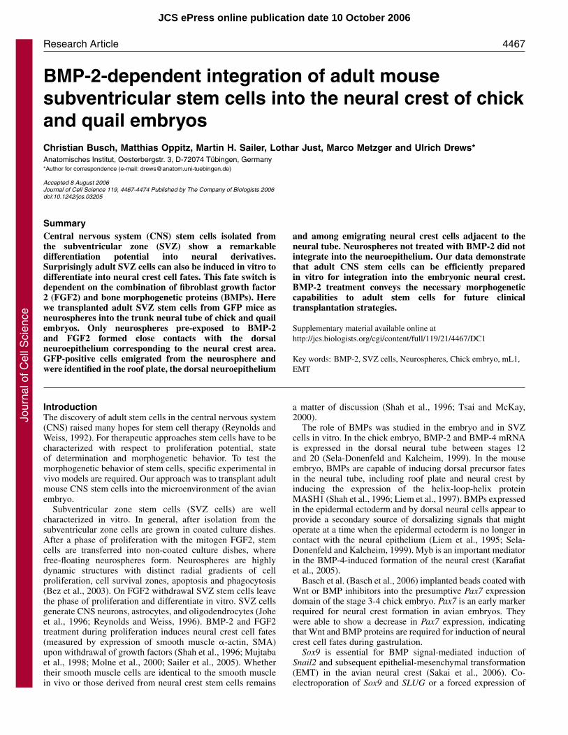
ResultsNeurospheresNeurospheres from adult GFP mice were used fortransplantation experiments. Under the chicken �-actinpromoter and cytomegalovirus enhancer, 90% of the cells inneurospheres expressed the enhanced GFP (EGFP)-actinfusion protein (Okabe et al., 1997). Neurospheres were looseaggregates of apparently undifferentiated cells (Fig. 1A-F).Nestin was expressed in a minimum of 70% of cells in BMP-2treated and non-treated neurospheres (mean ± s.d. 75±5%,evaluated by microscopical counting of ten sections ofneurospheres using Analysis image-processing software),whereas mL1 DNA sequence was detected in all cells (Fig. 1B-D). Staining of neurospheres with neural-crest-specific HNK-
Journal of Cell Science 119 (21)
1 showed that SVZ neurosphere cells showed a strongreactivity, which has not been reported before. Surprisingly,there was no difference in HNK-1 staining between BMP-2-treated and untreated neurospheres (Fig. 1E,F). Neurospheresremaining after transplantation were cultured with FGF2 butwithout BMP-2. There was no difference in the growthproperties (DAPI staining, GFP epifluorescence) betweenBMP-2-treated and untreated groups. Proliferation of culturedneurosphere cells was measured by BrdU uptake and alsoshowed no difference between the two groups of neurospheres.Viability of SVZ cells without BMP-2 treatment 24 hours aftertransplantation was assessed with the TUNEL reaction(supplementary material Fig. S1). Only two TUNEL-positiveSVZ cells were found in a parallel section of the non-integratedneurosphere aggregate shown in Fig. 3B. The TUNEL stainingis included as supplementary material Fig. S1.
Transplantation into the chick embryoNeurospheres were transplanted into the neural tube. Thepositioning of the neurosphere transplant (a small entireneurosphere, or parts of larger ones per embryo) was evaluatedby epifluorescence in the fenestrated egg using astereomicroscope. The location was at the transition from thesegmented to the non-segmented somite mesoderm. This
Fig. 1. (A) SVZ neurospheres stained with Nile Blue sulphateimmediately before injection. (B) Paraffin section of BMP-2 pre-treated neurosphere: nestin expression was observed in 70% of thecells evaluated by microscopical counting of ten sections ofneurospheres using image processing software (Analysis, SIS,Germany). (C) Untreated neurosphere: nestin expression wasobserved in 70% of the cells. (D) Neurosphere after hybridizationwith the mL1 probe. Strong HNK-1 reactivity was observed in bothBMP-2-treated (E) and untreated (F) neurosphere cells.
Jour
nal o
f Cel
l Sci
ence
4469BMP-2-dependent integration
corresponded to the 16th and 19th pair of somites of the 2-day-old embryo equal to stage 12 to 13 (Hamburger and Hamilton,1992; Graham and Meier, 1975).
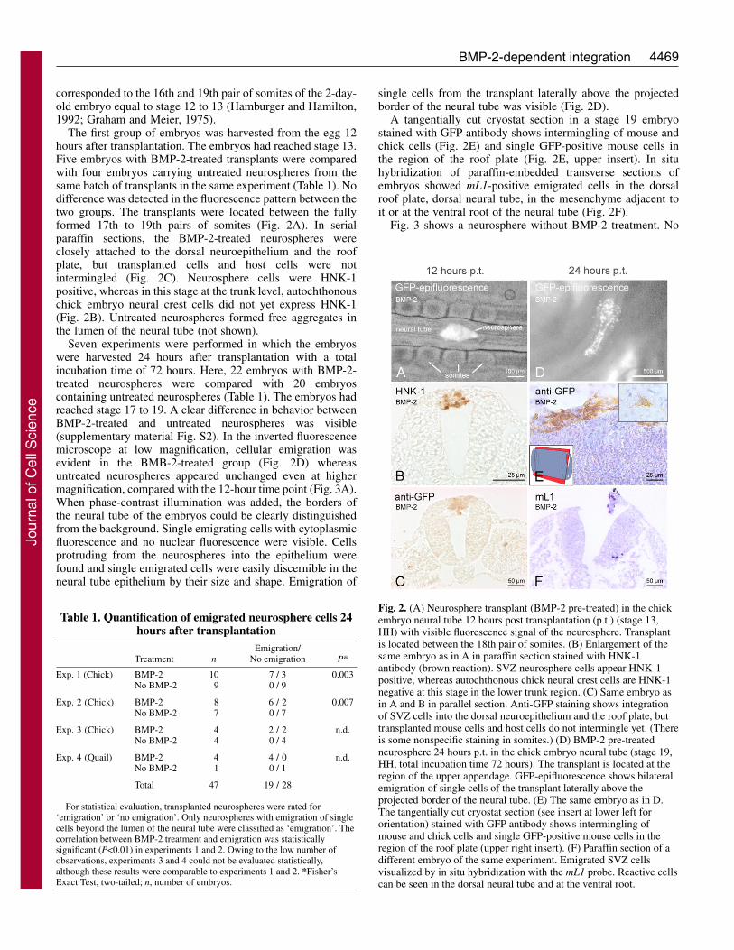
The first group of embryos was harvested from the egg 12hours after transplantation. The embryos had reached stage 13.Five embryos with BMP-2-treated transplants were comparedwith four embryos carrying untreated neurospheres from thesame batch of transplants in the same experiment (Table 1). Nodifference was detected in the fluorescence pattern between thetwo groups. The transplants were located between the fullyformed 17th to 19th pairs of somites (Fig. 2A). In serialparaffin sections, the BMP-2-treated neurospheres wereclosely attached to the dorsal neuroepithelium and the roofplate, but transplanted cells and host cells were notintermingled (Fig. 2C). Neurosphere cells were HNK-1positive, whereas in this stage at the trunk level, autochthonouschick embryo neural crest cells did not yet express HNK-1(Fig. 2B). Untreated neurospheres formed free aggregates inthe lumen of the neural tube (not shown).
Seven experiments were performed in which the embryoswere harvested 24 hours after transplantation with a totalincubation time of 72 hours. Here, 22 embryos with BMP-2-treated neurospheres were compared with 20 embryoscontaining untreated neurospheres (Table 1). The embryos hadreached stage 17 to 19. A clear difference in behavior betweenBMP-2-treated and untreated neurospheres was visible(supplementary material Fig. S2). In the inverted fluorescencemicroscope at low magnification, cellular emigration wasevident in the BMB-2-treated group (Fig. 2D) whereasuntreated neurospheres appeared unchanged even at highermagnification, compared with the 12-hour time point (Fig. 3A).When phase-contrast illumination was added, the borders ofthe neural tube of the embryos could be clearly distinguishedfrom the background. Single emigrating cells with cytoplasmicfluorescence and no nuclear fluorescence were visible. Cellsprotruding from the neurospheres into the epithelium werefound and single emigrated cells were easily discernible in theneural tube epithelium by their size and shape. Emigration of
single cells from the transplant laterally above the projectedborder of the neural tube was visible (Fig. 2D).
A tangentially cut cryostat section in a stage 19 embryostained with GFP antibody shows intermingling of mouse andchick cells (Fig. 2E) and single GFP-positive mouse cells inthe region of the roof plate (Fig. 2E, upper insert). In situhybridization of paraffin-embedded transverse sections ofembryos showed mL1-positive emigrated cells in the dorsalroof plate, dorsal neural tube, in the mesenchyme adjacent toit or at the ventral root of the neural tube (Fig. 2F).
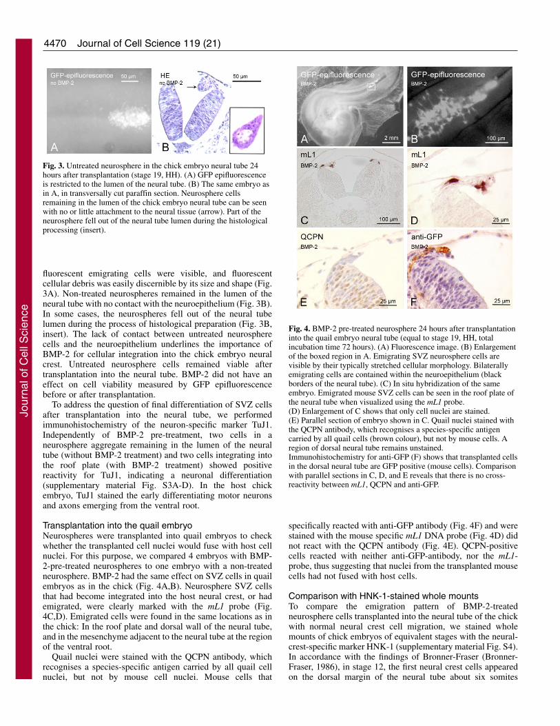
Fig. 3 shows a neurosphere without BMP-2 treatment. No
Table 1. Quantification of emigrated neurosphere cells 24hours after transplantation
Emigration/Treatment n No emigration P*
Exp. 1 (Chick) BMP-2 10 7 / 3 0.003No BMP-2 9 0 / 9
Exp. 2 (Chick) BMP-2 8 6 / 2 0.007No BMP-2 7 0 / 7
Exp. 3 (Chick) BMP-2 4 2 / 2 n.d.No BMP-2 4 0 / 4
Exp. 4 (Quail) BMP-2 4 4 / 0 n.d.No BMP-2 1 0 / 1
Total 47 19 / 28
For statistical evaluation, transplanted neurospheres were rated for‘emigration’ or ‘no emigration’. Only neurospheres with emigration of singlecells beyond the lumen of the neural tube were classified as ‘emigration’. Thecorrelation between BMP-2 treatment and emigration was statisticallysignificant (P<0.01) in experiments 1 and 2. Owing to the low number ofobservations, experiments 3 and 4 could not be evaluated statistically,although these results were comparable to experiments 1 and 2. *Fisher’sExact Test, two-tailed; n, number of embryos.
Fig. 2. (A) Neurosphere transplant (BMP-2 pre-treated) in the chickembryo neural tube 12 hours post transplantation (p.t.) (stage 13,HH) with visible fluorescence signal of the neurosphere. Transplantis located between the 18th pair of somites. (B) Enlargement of thesame embryo as in A in paraffin section stained with HNK-1antibody (brown reaction). SVZ neurosphere cells appear HNK-1positive, whereas autochthonous chick neural crest cells are HNK-1negative at this stage in the lower trunk region. (C) Same embryo asin A and B in parallel section. Anti-GFP staining shows integrationof SVZ cells into the dorsal neuroepithelium and the roof plate, buttransplanted mouse cells and host cells do not intermingle yet. (Thereis some nonspecific staining in somites.) (D) BMP-2 pre-treatedneurosphere 24 hours p.t. in the chick embryo neural tube (stage 19,HH, total incubation time 72 hours). The transplant is located at theregion of the upper appendage. GFP-epifluorescence shows bilateralemigration of single cells of the transplant laterally above theprojected border of the neural tube. (E) The same embryo as in D.The tangentially cut cryostat section (see insert at lower left fororientation) stained with GFP antibody shows intermingling ofmouse and chick cells and single GFP-positive mouse cells in theregion of the roof plate (upper right insert). (F) Paraffin section of adifferent embryo of the same experiment. Emigrated SVZ cellsvisualized by in situ hybridization with the mL1 probe. Reactive cellscan be seen in the dorsal neural tube and at the ventral root.
Jour
nal o
f Cel
l Sci
ence
4470
fluorescent emigrating cells were visible, and fluorescentcellular debris was easily discernible by its size and shape (Fig.3A). Non-treated neurospheres remained in the lumen of theneural tube with no contact with the neuroepithelium (Fig. 3B).In some cases, the neurospheres fell out of the neural tubelumen during the process of histological preparation (Fig. 3B,insert). The lack of contact between untreated neurospherecells and the neuroepithelium underlines the importance ofBMP-2 for cellular integration into the chick embryo neuralcrest. Untreated neurosphere cells remained viable aftertransplantation into the neural tube. BMP-2 did not have aneffect on cell viability measured by GFP epifluorescencebefore or after transplantation.
To address the question of final differentiation of SVZ cellsafter transplantation into the neural tube, we performedimmunohistochemistry of the neuron-specific marker TuJ1.Independently of BMP-2 pre-treatment, two cells in aneurosphere aggregate remaining in the lumen of the neuraltube (without BMP-2 treatment) and two cells integrating intothe roof plate (with BMP-2 treatment) showed positivereactivity for TuJ1, indicating a neuronal differentiation(supplementary material Fig. S3A-D). In the host chickembryo, TuJ1 stained the early differentiating motor neuronsand axons emerging from the ventral root.
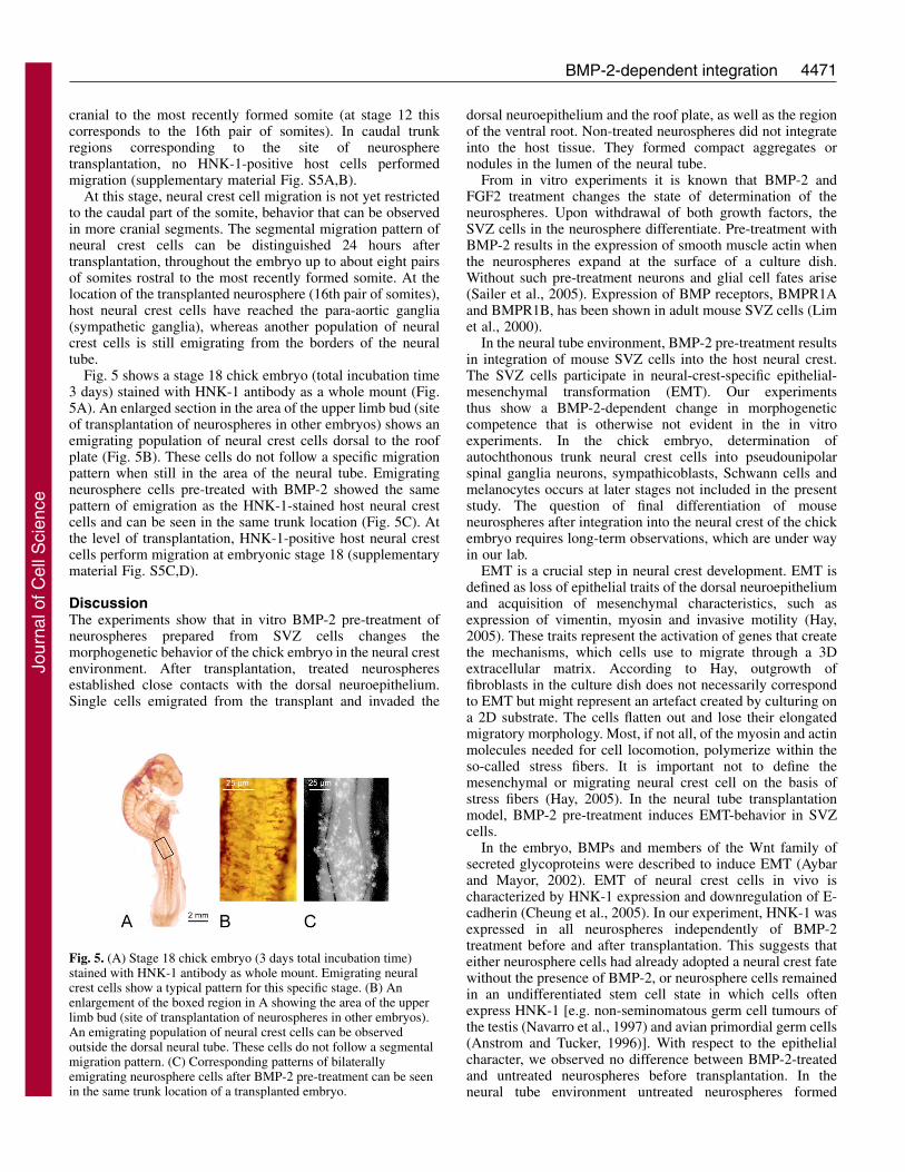
Transplantation into the quail embryoNeurospheres were transplanted into quail embryos to checkwhether the transplanted cell nuclei would fuse with host cellnuclei. For this purpose, we compared 4 embryos with BMP-2-pre-treated neurospheres to one embryo with a non-treatedneurosphere. BMP-2 had the same effect on SVZ cells in quailembryos as in the chick (Fig. 4A,B). Neurosphere SVZ cellsthat had become integrated into the host neural crest, or hademigrated, were clearly marked with the mL1 probe (Fig.4C,D). Emigrated cells were found in the same locations as inthe chick: In the roof plate and dorsal wall of the neural tube,and in the mesenchyme adjacent to the neural tube at the regionof the ventral root.
Quail nuclei were stained with the QCPN antibody, whichrecognises a species-specific antigen carried by all quail cellnuclei, but not by mouse cell nuclei. Mouse cells that
specifically reacted with anti-GFP antibody (Fig. 4F) and werestained with the mouse specific mL1 DNA probe (Fig. 4D) didnot react with the QCPN antibody (Fig. 4E). QCPN-positivecells reacted with neither anti-GFP-antibody, nor the mL1-probe, thus suggesting that nuclei from the transplanted mousecells had not fused with host cells.
Comparison with HNK-1-stained whole mountsTo compare the emigration pattern of BMP-2-treatedneurosphere cells transplanted into the neural tube of the chickwith normal neural crest cell migration, we stained wholemounts of chick embryos of equivalent stages with the neural-crest-specific marker HNK-1 (supplementary material Fig. S4).In accordance with the findings of Bronner-Fraser (Bronner-Fraser, 1986), in stage 12, the first neural crest cells appearedon the dorsal margin of the neural tube about six somites
Journal of Cell Science 119 (21)
Fig. 3. Untreated neurosphere in the chick embryo neural tube 24hours after transplantation (stage 19, HH). (A) GFP epifluorescenceis restricted to the lumen of the neural tube. (B) The same embryo asin A, in transversally cut paraffin section. Neurosphere cellsremaining in the lumen of the chick embryo neural tube can be seenwith no or little attachment to the neural tissue (arrow). Part of theneurosphere fell out of the neural tube lumen during the histologicalprocessing (insert).
Fig. 4. BMP-2 pre-treated neurosphere 24 hours after transplantationinto the quail embryo neural tube (equal to stage 19, HH, totalincubation time 72 hours). (A) Fluorescence image. (B) Enlargementof the boxed region in A. Emigrating SVZ neurosphere cells arevisible by their typically stretched cellular morphology. Bilaterallyemigrating cells are contained within the neuroepithelium (blackborders of the neural tube). (C) In situ hybridization of the sameembryo. Emigrated mouse SVZ cells can be seen in the roof plate ofthe neural tube when visualized using the mL1 probe.(D) Enlargement of C shows that only cell nuclei are stained.(E) Parallel section of embryo shown in C. Quail nuclei stained withthe QCPN antibody, which recognises a species-specific antigencarried by all quail cells (brown colour), but not by mouse cells. Aregion of dorsal neural tube remains unstained.Immunohistochemistry for anti-GFP (F) shows that transplanted cellsin the dorsal neural tube are GFP positive (mouse cells). Comparisonwith parallel sections in C, D, and E reveals that there is no cross-reactivity between mL1, QCPN and anti-GFP.
Jour
nal o
f Cel
l Sci
ence
4471BMP-2-dependent integration
cranial to the most recently formed somite (at stage 12 thiscorresponds to the 16th pair of somites). In caudal trunkregions corresponding to the site of neurospheretransplantation, no HNK-1-positive host cells performedmigration (supplementary material Fig. S5A,B).
At this stage, neural crest cell migration is not yet restrictedto the caudal part of the somite, behavior that can be observedin more cranial segments. The segmental migration pattern ofneural crest cells can be distinguished 24 hours aftertransplantation, throughout the embryo up to about eight pairsof somites rostral to the most recently formed somite. At thelocation of the transplanted neurosphere (16th pair of somites),host neural crest cells have reached the para-aortic ganglia(sympathetic ganglia), whereas another population of neuralcrest cells is still emigrating from the borders of the neuraltube.
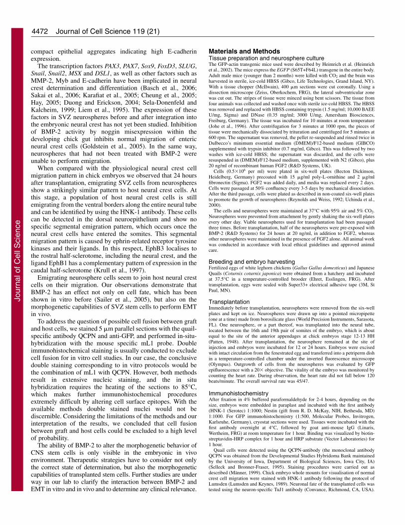
Fig. 5 shows a stage 18 chick embryo (total incubation time3 days) stained with HNK-1 antibody as a whole mount (Fig.5A). An enlarged section in the area of the upper limb bud (siteof transplantation of neurospheres in other embryos) shows anemigrating population of neural crest cells dorsal to the roofplate (Fig. 5B). These cells do not follow a specific migrationpattern when still in the area of the neural tube. Emigratingneurosphere cells pre-treated with BMP-2 showed the samepattern of emigration as the HNK-1-stained host neural crestcells and can be seen in the same trunk location (Fig. 5C). Atthe level of transplantation, HNK-1-positive host neural crestcells perform migration at embryonic stage 18 (supplementarymaterial Fig. S5C,D).
DiscussionThe experiments show that in vitro BMP-2 pre-treatment ofneurospheres prepared from SVZ cells changes themorphogenetic behavior of the chick embryo in the neural crestenvironment. After transplantation, treated neurospheresestablished close contacts with the dorsal neuroepithelium.Single cells emigrated from the transplant and invaded the
dorsal neuroepithelium and the roof plate, as well as the regionof the ventral root. Non-treated neurospheres did not integrateinto the host tissue. They formed compact aggregates ornodules in the lumen of the neural tube.
From in vitro experiments it is known that BMP-2 andFGF2 treatment changes the state of determination of theneurospheres. Upon withdrawal of both growth factors, theSVZ cells in the neurosphere differentiate. Pre-treatment withBMP-2 results in the expression of smooth muscle actin whenthe neurospheres expand at the surface of a culture dish.Without such pre-treatment neurons and glial cell fates arise(Sailer et al., 2005). Expression of BMP receptors, BMPR1Aand BMPR1B, has been shown in adult mouse SVZ cells (Limet al., 2000).
In the neural tube environment, BMP-2 pre-treatment resultsin integration of mouse SVZ cells into the host neural crest.The SVZ cells participate in neural-crest-specific epithelial-mesenchymal transformation (EMT). Our experimentsthus show a BMP-2-dependent change in morphogeneticcompetence that is otherwise not evident in the in vitroexperiments. In the chick embryo, determination ofautochthonous trunk neural crest cells into pseudounipolarspinal ganglia neurons, sympathicoblasts, Schwann cells andmelanocytes occurs at later stages not included in the presentstudy. The question of final differentiation of mouseneurospheres after integration into the neural crest of the chickembryo requires long-term observations, which are under wayin our lab.
EMT is a crucial step in neural crest development. EMT isdefined as loss of epithelial traits of the dorsal neuroepitheliumand acquisition of mesenchymal characteristics, such asexpression of vimentin, myosin and invasive motility (Hay,2005). These traits represent the activation of genes that createthe mechanisms, which cells use to migrate through a 3Dextracellular matrix. According to Hay, outgrowth offibroblasts in the culture dish does not necessarily correspondto EMT but might represent an artefact created by culturing ona 2D substrate. The cells flatten out and lose their elongatedmigratory morphology. Most, if not all, of the myosin and actinmolecules needed for cell locomotion, polymerize within theso-called stress fibers. It is important not to define themesenchymal or migrating neural crest cell on the basis ofstress fibers (Hay, 2005). In the neural tube transplantationmodel, BMP-2 pre-treatment induces EMT-behavior in SVZcells.
In the embryo, BMPs and members of the Wnt family ofsecreted glycoproteins were described to induce EMT (Aybarand Mayor, 2002). EMT of neural crest cells in vivo ischaracterized by HNK-1 expression and downregulation of E-cadherin (Cheung et al., 2005). In our experiment, HNK-1 wasexpressed in all neurospheres independently of BMP-2treatment before and after transplantation. This suggests thateither neurosphere cells had already adopted a neural crest fatewithout the presence of BMP-2, or neurosphere cells remainedin an undifferentiated stem cell state in which cells oftenexpress HNK-1 [e.g. non-seminomatous germ cell tumours ofthe testis (Navarro et al., 1997) and avian primordial germ cells(Anstrom and Tucker, 1996)]. With respect to the epithelialcharacter, we observed no difference between BMP-2-treatedand untreated neurospheres before transplantation. In theneural tube environment untreated neurospheres formed
Fig. 5. (A) Stage 18 chick embryo (3 days total incubation time)stained with HNK-1 antibody as whole mount. Emigrating neuralcrest cells show a typical pattern for this specific stage. (B) Anenlargement of the boxed region in A showing the area of the upperlimb bud (site of transplantation of neurospheres in other embryos).An emigrating population of neural crest cells can be observedoutside the dorsal neural tube. These cells do not follow a segmentalmigration pattern. (C) Corresponding patterns of bilaterallyemigrating neurosphere cells after BMP-2 pre-treatment can be seenin the same trunk location of a transplanted embryo.
Jour
nal o
f Cel
l Sci
ence
4472
compact epithelial aggregates indicating high E-cadherinexpression.
The transcription factors PAX3, PAX7, Sox9, FoxD3, SLUG,Snail, Snail2, MSX and DSL1, as well as other factors such asMMP-2, Myb and E-cadherin have been implicated in neuralcrest determination and differentiation (Basch et al., 2006;Sakai et al., 2006; Karafiat et al., 2005; Cheung et al., 2005;Hay, 2005; Duong and Erickson, 2004; Sela-Donenfeld andKalcheim, 1999; Liem et al., 1995). The expression of thesefactors in SVZ neurospheres before and after integration intothe embryonic neural crest has not yet been studied. Inhibitionof BMP-2 activity by noggin misexpression within thedeveloping chick gut inhibits normal migration of entericneural crest cells (Goldstein et al., 2005). In the same way,neurospheres that had not been treated with BMP-2 wereunable to perform emigration.
When compared with the physiological neural crest cellmigration pattern in chick embryos we observed that 24 hoursafter transplantation, emigrating SVZ cells from neurospheresshow a strikingly similar pattern to host neural crest cells. Atthis stage, a population of host neural crest cells is stillemigrating from the ventral borders along the entire neural tubeand can be identified by using the HNK-1 antibody. These cellscan be detected in the dorsal neuroepithelium and show nospecific segmental emigration pattern, which occurs once theneural crest cells have entered the somites. This segmentalmigration pattern is caused by ephrin-related receptor tyrosinekinases and their ligands. In this respect, EphB3 localises tothe rostral half-sclerotome, including the neural crest, and theligand EphB1 has a complementary pattern of expression in thecaudal half-sclerotome (Krull et al., 1997).
Emigrating neurosphere cells seem to join host neural crestcells on their migration. Our observations demonstrate thatBMP-2 has an effect not only on cell fate, which has beenshown in vitro before (Sailer et al., 2005), but also on themorphogenetic capabilities of SVZ stem cells to perform EMTin vivo.
To address the question of possible cell fusion between graftand host cells, we stained 5 �m parallel sections with the quail-specific antibody QCPN and anti-GFP, and performed in-situ-hybridization with the mouse specific mL1 probe. Doubleimmunohistochemical staining is usually conducted to excludecell fusion for in vitro cell studies. In our case, the conclusivedouble staining corresponding to in vitro protocols would bethe combination of mL1 with QCPN. However, both methodsresult in extensive nucleic staining, and the in situhybridization requires the heating of the sections to 85°C,which makes further immunohistochemical proceduresextremely difficult by altering cell surface epitopes. With theavailable methods double stained nuclei would not bediscernible. Considering the limitations of the methods and ourinterpretation of the results, we concluded that cell fusionbetween graft and host cells could be excluded to a high levelof probability.
The ability of BMP-2 to alter the morphogenetic behavior ofCNS stem cells is only visible in the embryonic in vivoenvironment. Therapeutic strategies have to consider not onlythe correct state of determination, but also the morphogeneticcapabilities of transplanted stem cells. Further studies are underway in our lab to clarify the interaction between BMP-2 andEMT in vitro and in vivo and to determine any clinical relevance.
Materials and MethodsTissue preparation and neurosphere cultureThe GFP-actin transgenic mice used were described by Heimrich et al. (Heimrichet al., 2002). The mice express the EGFP (S65T+F64L) transgene in the entire body.Adult male mice (younger than 2 months) were killed with CO2 and the brain washarvested in sterile, ice-cold HBSS (Gibco, Life Technologies, Grand Island, NY).With a tissue chopper (McIlwain), 400 �m sections were cut coronally. Using adissection microscope (Zeiss, Oberkochem, FRG), the lateral subventricular zonewas cut out. The stripes of tissue were minced using bent scissors. The tissue fromfour animals was collected and washed once with sterile ice-cold HBSS. The HBSSwas removed and replaced with HBSS containing trypsin (1.5 mg/ml; 10,000 BAEEU/mg, Sigma) and DNase (0.35 mg/ml; 3000 U/mg, Amersham Biosciences,Freiburg, Germany). The tissue was incubated for 10 minutes at room temperature(Johe et al., 1996). After centrifugation for 3 minutes at 1000 rpm, the pieces oftissue were mechanically dissociated by trituration and centrifuged for 5 minutes at600 rpm. The supernatant was removed, the pellet re-suspended and rinsed twice inDulbecco’s minimum essential medium (DMEM)/F12-based medium (GIBCO)supplemented with trypsin inhibitor (0.7 mg/ml, Gibco). This was followed by twowashes with ice-cold HBSS; the supernatant was discarded, and the cells wereresuspended in (DMEM)/F12-based medium, supplemented with N2 (Gibco), plus20 ng/ml of recombinant human FGF2 (R&D Systems, UK).
Cells (0.5�106 per ml) were plated in six-well plates (Becton Dickinson,Heidelberg, Germany) precoated with 15 �g/ml poly-L-ornithine and 2 �g/mlfibronectin (Sigma). FGF2 was added daily, and media was replaced every 2 days.Cells were passaged at 50% confluency every 3-5 days by mechanical dissociation.After the third passage, cells were plated as described in non-coated six-well platesto promote the growth of neurospheres (Reynolds and Weiss, 1992; Uchinda et al.,2000).
The cells and neurospheres were maintained at 37°C with 95% air and 5% CO2.Neurospheres were prevented from attachment by gently shaking the six-well platesevery other day. Viable neurospheres used for transplantation had been passagedthree times. Before transplantation, half of the neurospheres were pre-exposed withBMP-2 (R&D Systems) for 24 hours at 20 ng/ml, in addition to FGF2, whereasother neurospheres were maintained in the presence of FGF2 alone. All animal workwas conducted in accordance with local ethical guidelines and approved animalcare.
Breeding and embryo harvestingFertilized eggs of white leghorn chickens (Gallus Gallus domesticus) and JapaneseQuails (Coturnix coturnix japonica) were obtained from a hatchery and incubatedat 37.5°C in a temperature-controlled brooder (Ehret, Esslingen, FRG). Aftertransplantation, eggs were sealed with Super33+ electrical adhesive tape (3M, StPaul, MN).
TransplantationImmediately before transplantation, neurospheres were removed from the six-wellplates and kept on ice. Neurospheres were drawn up into a pointed micropipette(one at a time) made from borosilicate glass (World Precision Instruments, Sarasota,FL). One neurosphere, or a part thereof, was transplanted into the neural tube,located between the 16th and 19th pair of somites of the embryo, which is aboutequal to the site of the anterior appendages at chick embryos stage 12-13 HH(Patten, 1948). After transplantation, the neurosphere remained at the site ofinjection and embryos were incubated for 12 or 24 hours. Embryos were excisedwith intact circulation from the fenestrated egg and transferred into a petriperm dishin a temperature-controlled chamber under the inverted fluorescence microscope(Olympus). Outgrowth of cells from the neurospheres was evaluated by GFPepifluorescence with a 20� objective. The vitality of the embryo was monitored bycounting the heart rate. During observation, the heart rate did not fall below 120beats/minute. The overall survival rate was 45/47.
ImmunohistochemistryAfter fixation in 4% buffered paraformaldehyde for 2-4 hours, depending on thesize, embryos were embedded in paraplast and incubated with the first antibody(HNK-1 (Serotec) 1:1000; Nestin (gift from R. D. McKay, NIH, Bethesda, MD)1:1000. For GFP immunohistochemistry (1:500, Molecular Probes, Invitrogen,Karlsruhe, Germany), cryostat sections were used. Tissues were incubated with thefirst antibody overnight at 4°C, followed by goat anti-mouse IgG (Linaris,Wertheim, FRG) at room temperature for 1 hour. Binding was visualized by biotin-streptavidin-HRP complex for 1 hour and HRP substrate (Vector Laboratories) for1 hour.
Quail cells were detected using the QCPN-antibody (the monoclonal antibodyQCPN was obtained from the Developmental Studies Hybridoma Bank maintainedby the University of Iowa, Department of Biological Sciences, Iowa City, IA)(Selleck and Bronner-Fraser, 1995). Staining procedures were carried out asdescribed (Männer, 1999). Chick embryo whole mounts for visualisation of normalcrest cell migration were stained with HNK-1 antibody following the protocol ofLumsden (Lumsden and Keynes, 1989). Neuronal fate of the transplanted cells wastested using the neuron-specific TuJ1 antibody (Convance, Richmond, CA, USA).
Journal of Cell Science 119 (21)
Jour
nal o
f Cel
l Sci
ence
4473BMP-2-dependent integration
Tissues were incubated with the first antibody (1:500) overnight at 4°C, followedby goat anti-mouse IgG (Linaris, Wertheim, Germany) at room temperature for 1hour. Binding was visualized by incubation with biotin-streptavidin-HRP complexfor 1 hour and HRP substrate (Vector Laboratories) for 1 hour.
To detect apoptotic cells in untreated neurospheres, we conducted the TUNELassay on parallel sections using a commercially available kit (TUNEL apoptosisdetection kit, Chemicon, Temecula, CA) using the procedure described by themanufacturer.
In situ hybridizationIn situ hybridization for mL1 sequences was carried out with a digoxigenin (DIG)-labeled, 416 bp antisense DNA probe corresponding to the consensus sequence ofmouse mL1 (Hatano et al., 1998). Probe labeling was performed by PCR with thePCR DIG Probe Synthesis Kit (Roche, Mannheim, Germany). The in situhybridization protocol is a modification of a method described for the hALUsequence (Just et al., 2003). Sections were incubated with TE buffer (100 mM Tris-HCl, 50 mM EDTA, pH 8) containing 2 �g/ml proteinase K for 10 minutes at roomtemperature. To reduce nonspecific background, slides were acetylated with TEAbuffer (0.1 M triethanolamine, pH 8.0) containing 0.25% (v/v) acetic anhydride(Sigma) twice for 5 minutes. After pre-hybridization with hybridization buffer [50%formamide (Sigma), 10% dextran sulfate, 5 mM EDTA, 20 mM Tris-HCl pH 8, 10mM DTT, 1� Denhardt’s solution, 0.05% tRNA, 300 mM NaCl] for 3 hours at85°C, slides were incubated with fresh hybridisation buffer containing the denaturedDIG-labeled DNA-L1 probe (100 ng/ml) for 2 hours at 85°C. Slides wereimmediately transferred to ice for 10 minutes and then incubated overnight at 37°C.After hybridization, slides were briefly rinsed in 2� SSC at room temperature andthree times in 0.1� SSC for 15 minutes at 37°C. Detection of DIG-labeled RNAprobe was performed according to the protocol of the DIG nucleic acid detectionkit (Roche). The tissues were blocked for 30 minutes with blocking buffer (1%blocking reagent (Roche) in maleic acid buffer (0.1 M maleic acid, 0.15 M NaCl,pH 7.5), and then incubated with alkaline phosphatase-conjugated antibody solution[anti-DIG antibody (1:2500 Roche) in blocking buffer containing 0.1% Triton® X-100] for 1 hour. Following four washes with maleic acid buffer for 15 minutes, slideswere equilibrated for 5 minutes in Tris buffer pH 9.5 (0.1 M Tris-HCl, pH 9.5, 0.1M NaCl, 50 mM MgCl2). The color development was carried out with substratesolution [nitro blue tetrazolium salt (NBT) and 5-bromo-4-chloro-3-indolylphosphate (X-phosphate) (Roche) in Tris buffer pH 9.5]. After three washes withPBS, slides were rinsed in distilled water, dried and coverslips were sealed withKaiser’s gelatine (Merck, Darmstadt, Germany).
Statistical analysisFisher’s Exact Test was applied for statistical analysis. Detected emigration ofneurosphere cells into the surrounding tissue was categorized into ‘emigration’ or‘no emigration’. The criterion was the observation of fluorescent neurosphere cellsextending beyond the neural tube lumen (supplementary material Fig. S2).Statistical evaluation of all experiments is shown in Table 1. We found that in twoof the four experiments performed, emigration was significantly (P<0.01) observedin embryos that had been transplanted with BMP-2-treated neurospheres.
In experiment 1, BMP-2-treated and untreated neurospheres were transplanted inexperiments on different days, but the same batch of neurospheres was used. Inexperiments 2, 3 and 4, BMP-2-treated and untreated neurospheres from the samebatch were transplanted on the same day.
The authors would like to express their gratitude to R. D. McKayfor the opportunity given to the first author for gaining technicalexperience at the NINDS, National Institutes of Health, Bethesda,MD, and to Ravi Saini for proofreading the manuscript. The �-actin-GFP transgenic mice were kindly provided by M. Okabe, GenomeInformation Research Centre, University of Osaka, Japan. Cell cultureexperiments were performed by S. Conrad (Institute of Anatomy,University of Tübingen, Germany).
ReferencesAnstrom, K. K. and Tucker, R. P. (1996). Tenascin-C lines the migratory pathways of
avian primordial germ cells and haematopoietic progenitor cells. Dev. Dyn. 206, 437-446.
Aybar, M. and Mayor, R. (2002). Early induction of enural crest cells: lessons learnedfrom frog, fish and chick. Curr. Opin. Genet. Dev. 12, 452-458.
Basch, M. L., Bronner-Fraser, M. and Garcia-Castro, M. I. (2006). Specification ofthe neural crest occurs during gastrulation and requires Pax7. Nature 441, 218-222.
Bez, A., Corsini, E., Curti, D., Biggiogera, M., Colombo, A., Nicosia, R. F., Pagano,S. F. and Parati, E. A. (2003). Neurosphere and neurosphere-forming cells:morphological and ultrastructural characterization. J. Brain Res. 993, 18-29.
Bronner-Fraser, M. (1986). Analysis of the early stages of trunk neural crest migrationin avian embryos using monoclonal antibody HNK-1. Dev. Biol. 115, 44-55.
Brustle, O., Maskos, U. and McKay, R. D. (1995). Host-guided migration allowstargeted introduction of neurons into the embryonic brain. Neuron 15, 1275-1285.
Brustle, O., Spiro, A. C., Karram, K., Choudhary, K., Okabe, S. and McKay, R. D.(1997). In vitro-generated neural precursors participate in mammalian braindevelopment. Proc. Natl. Acad. Sci. USA 94, 14809-14814.
Brustle, O., Choudhary, K., Karram, K., Huttner, A., Murray, K., Dubois-Dalcq, M.and McKay, R. D. (1998). Chimeric brains generated by intraventriculartransplantation of fetal human brain cells into embryonic rats. Nat. Biotechnol. 16,1040-1044.
Cheung, M., Chaboissier, M.-C., Mynett, A., Hirst, E., Schedl, A. and Briscoe, J.(2005). The transcriptional control of trunk neural crest induction, survival, anddelamination. Dev. Cell 8, 179-192.
Duong, T. D. and Erickson, C. A. (2004). MMP-2 plays an essential role in producingepithelial-mesenchymal transformations in the avian embryo. Dev. Dyn. 229, 42-53.
Goldstein, A. M., Brewer, K. C., Doyle, A. M., Nagy, N. and Roberts, D. J. (2005).BMP signaling is necessary for neural crest cell migration and ganglion formation inthe enteric nervous system. Mech. Dev. 122, 821-833.
Graham, D. L. and Meier, G. W. (1975). Standards of morphological development ofthe quail, Coturnix Coturnix Japonica, embryo. Growth 39, 389-400.
Hamburger, V. and Hamilton, H. L. (1992). A series of normal stages in thedevelopment of the chick embryo (1951). Dev. Dyn. 195, 231-272.
Hatano, H., Tokunaga, K., Ogose, A., Hotta, T., Yamagiwa, H., Hayami, T., Endo,N. and Takahashi, H. E. (1998). Origin of bone-forming cells in human osteosarcomastransplanted into nude mice – which cells produce bone, human or mouse? J. Pathol.185, 204-211.
Hay, E. D. (2005). The mesenchymal cell, its role in the embryo, and the remarkablesignaling mechanisms that create it. Dev. Dyn. 233, 706-720.
Heimrich, B., Vogt, J., Simbürger, E., Skutella, T. and Glumm, R. (2002). Axonguidance and the formation of specific connections in the hippocampus.Neuroembryology 1, 154-160.
Johe, K. K., Hazel, T. G., Muller, T., Dugich-Djordjevic, M. M. and McKay, R. D.(1996). Single factors direct the differentiation of stem cells from the fetal and adultcentral nervous system. Genes Dev. 10, 3129-3140.
Just, L., Timmer, M., Tinius, J., Stahl, F., Deiwick, A., Nikkhah, G. and Bader, A.(2003). Identification of human cells in brain xenografts and in neural co-cultures ofrat by in situ hybridisation with Alu probe. J. Neurosci. Methods 126, 69-77.
Karafiat, V., Dvorakova, M., Krejci, E., Kralova, J., Pajer, P., Snajdr, P., Mandikova,S., Bartunek, P., Grim, M. and Dvorak, M. (2005). Transcription factor c-Myb isinvolved in the regulation of the epithelial-mesenchymal transition in the avian neuralcrest. Cell. Mol. Life Sci. 62, 2516-2525.
Krull, C. E., Lansford, R., Gale, N. W., Collazo, A., Marcelle, C., Yancopoulos, G.D., Fraser, S. E. and Bronner-Fraser, M. (1997). Interactions of Eph-related receptorsand ligands confer rostrocaudal pattern to trunk neural crest migration. Curr. Biol. 7,571-580.
Liem, K. F., Tremml, G., Roelink, H. and Jessell, T. M. (1995). Dorsal differentiationof neural plate cells induced by BMP-mediated signals from epidermal ectoderm. Cell82, 969-979.
Liem, K. F. J., Tremml, G. and Jessell, T. M. (1997). A role for the roof plate and itsresident TGF�-related proteins in neuronal patterning in the dorsal spinal cord. Cell91, 127-138.
Lim, D. A., Tramontin, A. D., Trevejo, J. M., Herrera, D. G., Garcia-Verdugo, J. M.and Avarez-Buylla, A. (2000). Noggin antagonizes BMP signalling to create a nichefor adult neurogenesis. Neuron 28, 713-726.
Lumsden, A. and Keynes, R. (1989). Segmental patterns of neuronal development in thechick hindbrain. Nature 337, 424-428.
Männer, J. (1999). Does the subepicardial mesenchyme contribute myocardioblasts ofthe chick embryo heart? A quail-chick chimera study tracing the fate of the epicardialprimordium. Anat. Rec. 255, 212-226.
Molne, M., Studer, L., Tabar, V., Ting, Y. T., Eiden, M. V. and McKay, R. D. (2000).Early cortical precursors do not undergo LIF-mediated astrocytic differentiation. J.Neurosci. Res. 59, 301-311.
Mujtaba, T., Mayer-Proschel, M. and Rao, M. S. (1998). A common neural progenitorfor the CNS and the PNS. Dev. Biol. 200, 1-15.
Navarro, S., Noguera, R., Peydro-Olaya, A. and Llombart-Bosch, A. (1997). Bipolar(neural and myoblastic) phenotype in cell lines derived from human germ cell tumoursof testis. Virchows Arch. 430, 291-300.
Okabe, M., Ikawa, M., Kominami, K., Nakanishi, T. and Nishimune, Y. (1997).‘Green mice’ as a source of ubiquitous green cells. FEBS Lett. 407, 313-319.
Panchision, D. M., Pickel, J. M., Studer, L., Lee, S. H., Turner, P. A., Hazel, T. G.and McKay, R. D. (2001). Sequential actions of BMP receptors control neuralprecursor cell production and fate. Genes Dev. 15, 2094-2110.
Patten, B. M. (1948). The Early Embryology of the Chick. Philadelphia, Toronto:Blakiston.
Reynolds, B. A. and Weiss, S. (1992). Generation of neurons and astrocytes from isolatedcells of the adult mammalian central nervous system. Science 255, 1707-1710.
Reynolds, B. A. and Weiss, S. (1996). Clonal and population analyses demonstrate thatan EGF-responsive mammalian embryonic CNS precursor is a stem cell. Dev. Biol.175, 1-13.
Reynolds, B. A., Tetzlaff, W. and Weiss, S. (1992). A multipotent EGF-responsivestriatal embryonic progenitor cell produces neurons and astrocytes. J. Neurosci. 12,4565-4574.
Sailer, M. H., Hazel, T. G., Panchision, D. M., Hoeppner, D. J., Schwab, M. E. andMcKay, R. D. (2005). BMP2 and FGF2 cooperate to induce neural-crest-like fatesfrom fetal and adult CNS stem cells. J. Cell Sci. 118, 5849-5860.
Sakai, D., Suzuki, T., Osumi, N. and Wakamatsu, Y. (2006). Cooperative action of
Jour
nal o
f Cel
l Sci
ence

4474
Sox9, Snail2 and PKA signalling in early neural crest development. Development 133,1323-1333.
Sela-Donnenfeld, D. and Kalcheim, C. (1999). Regulation of the onset of neural crestmigration by coordinated activity of BMP4 and Noggin in the dorsal neural tube.Development 126, 4749-4762.
Selleck, A. J. and Bronner-Fraser, M. (1995). Origins of the avian neural crest: the roleof neural-plate-epidermal interactions. Development 121, 525-538.
Shah, N. M., Groves, A. K. and Anderson, D. J. (1996). Alternative neural crest cellfates are instructively promoted by TGF� superfamily members. Cell 85, 331-343.
Tsai, R. Y. and McKay, R. D. (2000). Cell contact regulates fate choice by cortical stemcells. J. Neurosci. 20, 3725-3735.
Uchida, N., Buck, D. W., He, D., Reitsma, M. J., Masek, M., Phan, T. V., Tsukamoto,A. S., Gage, F. H. and Weissman, I. L. (2000). Direct isolation of human centralnervous system stem cells. Proc. Natl. Acad. Sci. USA 97, 14720-14725.
Journal of Cell Science 119 (21)
Jour
nal o
f Cel
l Sci
ence


![BMC Systems Biology - microsoft.comneuroblasts migrate radially toward the subventricular zone (SVZ) along the glial fiber, which serves as a their scaffold [1]. On exit from the VZ,](https://img.pdfslide.us/doc/110x75/5f1242dc180a0020b15da8b4/bmc-systems-biology-neuroblasts-migrate-radially-toward-the-subventricular-zone.jpg)