Embed Size (px)
Citation preview
BioMed CentralBMC Evolutionary Biology
ss
Open AcceResearch articleOldest record of Trimeniaceae from the Early Cretaceous of northern JapanToshihiro Yamada*1, Harufumi Nishida2, Masayoshi Umebayashi1, Kazuhiko Uemura3 and Masahiro Kato4Address: 1Division of Life Sciences, Graduate School of Natural Science and Technology, Kanazawa University, Kanazawa 920-1192, Japan, 2Faculty of Science and Engineering, Chuo University, Tokyo 112-8551, Japan, 3Department of Geology and Paleontology, National Museum of Nature and Science, 3-23-1 Hyakunincho, Tokyo 169-0073, Japan and 4Department of Botany, National Museum of Nature and Science, Tsukuba 305-0005, Japan
Email: Toshihiro Yamada* - [email protected]; Harufumi Nishida - [email protected]; Masayoshi Umebayashi - [email protected]; Kazuhiko Uemura - [email protected]; Masahiro Kato - [email protected]
* Corresponding author
AbstractBackground: Molecular phylogenetic analyses have identified Trimeniaceae, a monotypic familydistributed only in Oceania, as among the earliest diverging families of extant angiosperms.Therefore, the fossils of this family are helpful to understand the earliest flowering plants.Paleobotanical information is also important to track the historical and geographical pathways toendemism of the Trimeniaceae. However, fossils of the family were previously unknown from theEarly Cretaceous, the time when the angiosperm radiated. In this study, we report a seed from thelate Albian (ca. 100 million years ago) of Japan representing the oldest known occurrence ofTrimeniaceae and discuss the character evolution and biogeography of this family.
Results: A structurally preserved seed was collected from the early Late Albian HikagenosawaFormation of the Yezo Group, which was deposited in palaeolatitudes of 35 to 40°N. The seed hasa multilayered stony exotesta with alveolate surface, parenchymatous mesotesta, and operculateinner integument, which are characteristic to extant trimeniaceous seeds. However, the seeddiffers from extant seeds, i.e., in its well-developed endosperm and absence of antiraphal vascularbundle. Thus, the seed would be a new genus and species of Trimeniaceae.
Conclusion: The fossil seed indicates that seed coat characters were conserved for 100 millionyears or more in Trimeniaceae. It also suggests that the antiraphal vascular bundle and perispermyoriginated secondarily in Trimeniaceae as previously inferred from the phylogeny and characterdistribution in the extant basalmost angiosperms. The fossil seed provides the first evidence thatTrimeniaceae was distributed in a midlatitude location of the Northern Hemisphere during theEarly Cretaceous, when angiosperms radiated extensively, supporting a hypothesis that the extantaustral distribution is relict.
Published: 8 May 2008
BMC Evolutionary Biology 2008, 8:135 doi:10.1186/1471-2148-8-135
Received: 22 February 2008Accepted: 8 May 2008
This article is available from: http://www.biomedcentral.com/1471-2148/8/135
© 2008 Yamada et al; licensee BioMed Central Ltd. This is an Open Access article distributed under the terms of the Creative Commons Attribution License (http://creativecommons.org/licenses/by/2.0), which permits unrestricted use, distribution, and reproduction in any medium, provided the original work is properly cited.
Page 1 of 7(page number not for citation purposes)
BMC Evolutionary Biology 2008, 8:135 http://www.biomedcentral.com/1471-2148/8/135
BackgroundTrimeniaceae is a small family consisting of only thegenus Trimenia, with eight species known from Celebes toeastern Australia and the southwest Pacific [1,2]. Trimeniaconsists of shrubs and lianas that have unisexual or bisex-ual flowers with numerous tepals and stamens and a sin-gle carpel [1-4]. A solitary pendant ovule is enclosed in theovary and develops into a seed with a stony seed coat con-tained in a berry [1,4-7]. The family was placed in the Lau-rales based on floral structures, such as the single carpeland ovule per flower [3,4], but molecular phylogeneticstudies have identified it as one of the earliest divergingfamilies of extant angiosperms, along with the Amborel-laceae, Nymphaeales [Cabombaceae and Nymphae-aceae], Hydatellaceae, Austrobaileyaceae, and Illiciaceae[8-11]. Thus, clarifying the primitive character states ofTrimeniaceae would be helpful in understanding the ear-liest flowers. In particular, information on key innova-tions of angiosperms, for example, fruit, bitegmic seeds,and endosperm, would provide clues to solving the originof angiosperms, a central mystery of plant evolution[12,13].
Great efforts have been made to find evidence of earlyangiosperms from the Early to mid-Cretaceous, when theangiosperms radiated extensively [14-20]. The early emer-gence of Nymphaeales [18,21] and Illiciaceae [16] in thefossil record partly supports the phylogenetic frameworkinferred from molecular data. However, direct evidencethat the other earliest diverging families emerged in theearly phase of angiosperm radiation [20] has not beenfound, although paleobotanical records [16,18], as well asphylogeny [8-11], imply that these families should havediverged by the mid-Cretaceous [22].
From the austral distribution of Trimeniaceae, and thesimilar distribution of Amborellaceae and Austrobailey-aceae [1], neobotanists have inferred the Gondwana ori-gin of these families [23,24], but accumulatedpalynological data have indicated that a different explana-tion is needed for this distribution. These data indicatethat angiosperms originated in low paleolatitudes (20°Nto 20°S) no later than the Hauterivian, Early Cretaceous,about 132 million years ago [25,26] and spread towardthe poles over time [26-28]. Pollen records provide tem-poral and spatial distribution patterns of angiosperms,with the implication that the Trimeniaceae could havemigrated to Laurasia, a continental mass in the NorthernHemisphere. However, pollen is difficult to assign toextant families due to the frequent convergent evolutionof pollen morphology, such as the number and positionof apertures [20,29]. Moreover, extant Trimenia pollensare eurypalynos with regard to the number of apertureeven in a same species [29], obscuring diagnostic pollencharacters in familiar level. Thus, findings from other
reproductive structures, such as flowers, fruits, and seeds,are needed to confirm the past existence of the Trimeni-aceae in the Northern Hemisphere.
We report the oldest seed of Trimeniaceae from the EarlyCretaceous Yezo Group in Hokkaido, northern Japan, anddiscuss character evolution and biogeography of the Tri-meniaceae.
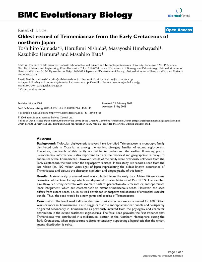
Geological settings and other paleobotanical data of collection siteA structurally preserved seed was found within a calcare-ous siltstone nodule collected at Pombetsu, Mikasa City,Hokkaido, Japan (Figure 1). The Hikagenosawa Forma-tion of the Yezo Group outcrops in the area and containsammonoids indicating a date in the early Late Albian[30,31]. Palaeomagnetic studies show that these sedi-ments were deposited in palaeolatitudes of 35 to 40°Nand that the sedimentary basin was located on the easternside of Laurasia [30,32]. Because the formation mainlyconsists of offshore sediments, there are few palaeobotan-ical records within it [33-35], but a subtropical climate
Geological map of Ponbetsu area and locality of the trimenia-ceous seedFigure 1Geological map of Ponbetsu area and locality of the trimeniaceous seed. Ponbetsu area are boxed in large-scale map of Hokkaido on the left-bottom corner. Geological map is redrawn from Narita et al. 31.
Page 2 of 7(page number not for citation purposes)
BMC Evolutionary Biology 2008, 8:135 http://www.biomedcentral.com/1471-2148/8/135
with dry seasons seems to have prevailed in the area, assuggested by the occurrence of a cheirolepidiaceous coni-fer [34] and a variety of ephedroid palynomorphs [33].
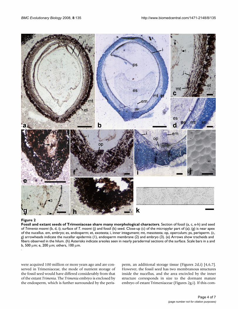
ResultsThe seed is ellipsoid, 3 mm long and 2.2 mm thick. It hasan inner integument and an outer seed coat (testa) (Fig-ures 2a,c,e). The micropyle is formed by both inner integ-ument and testa (endostome and exostome, respectively)and adjacent to the hilum, which contains tracheids andsclerenchymatous fibers, i.e., the seed is anatropous (Fig-ures 2e,f). These tracheids and fibers are found only in sec-tions through the raphal side, thus the vascular bundledoes not extend to the antiraphal side beyond the chalaza(Figure 2e). The inner integument is almost crushed,except in the endostomic region, where the inner integu-ment is thickened to form the operculum (Figures 2c,f).The testa consists of an outer (exotesta) and inner (mesot-esta) part. The exotesta comprises one to five layers of iso-diametric cells, while the mesotesta is made up of four tofive layers of longitudinally elongated cells. The exotesta islignified to give mechanical strength to the seed coat (Fig-ure 2c). In a nearly tangential section through the seedsurface, there are polygonal openings in the exotesta, indi-cating the presence of polygonal areoles on the exotestalsurface (asterisks in Figure 2h). This configuration wasfurther examined by reconstructing a three-dimensionalimage of the seed surface by compiling serial peel sec-tions, revealing that the exotestal surface is alveolate (Fig-ure 2k). Inside the inner integument are threemembranous structures. The outermost structure, con-nected to the chalaza, is the nucellar epidermis (Figure 2aand arrowheads in Figures 2c,g). The innermost structureis observed only in sections through the center of the seedand encircles an area 200 μm in length and 40 μm inwidth (arrowhead 3 in Figure 2g).
DiscussionRelationships of the fossil seedAlthough seed structures have not been examined in allTrimenia species, available information on four species (T.moorei, T. neocaledonica, T. papuana, and T. weinmanniifo-lia) suggests that Trimeniaceae is uniform family in regardof ovule and seed structures [1,4-7]. The seed of extant Tri-menia is characterized by the testa, consisting of a lignifiedmultilayered exotesta with an alveolate surface and a non-lignified multilayered mesotesta (Figures 2b,d,i,j) [1,4,7].Although many families possess seed coats with a hard-ened exotesta, none other than Trimeniaceae is known topossess a multilayered exotesta [1,4,7,36]. The seeds ofsome taxa are similar in appearance to the Trimeniaceaeseed in that the testa consists of outer sclerotic and innernon-sclerotic cell layers. These taxa include the genusNuphar (Nymphaeaceae) [37,38], the families Buxaceae[36], Simmondsiaceae [39], Melianthaceae [40], Myrta-
ceae [36,41], and Theaceae [36], and the order Sapindales[36,42]. However, in these taxa, the stony outer layer iscomposed of a single-layered columnar exotesta and theouter part of the isodiametric mesotestal cells, whereas theexotesta of Trimenia is composed uniformly of scleroticisodiametric cells, which are derivatives of the outer epi-dermis of the outer integument [7]. Thus, to our knowl-edge, no other seeds can be compared to the fossil seed.
The fossil seed is not distinguishable from that of extantTrimenia in terms of shape, size, anatropy, bitegmy, endoand exostomic micropyle, crushed inner integument withan operculum, lignified multilayered exotesta composedof isodiametric cells, alveolate surface of the exotesta, ornonlignified multilayered mesotesta (Figure 2). Theseremarkable similarities unequivocally indicate the closeaffinity of the fossil seed to the Trimeniaceae. The fossilseed differs from Trimenia seed in the absence of antirap-hal vascular bundle (Figure 2e) and in the size of membra-nous structures surrounding the nucellus (Figures 2c,g; seebelow for details). Thus, a new genus of Trimeniaceaeshould be assigned to the fossil seed. The seed also differsfrom Trimenia seed in having a stalklike structure at thenucellar base (Figure 2a). This stalklike structure mightrepresent a diagnostic feature of the seed, but the structurecould be artificially formed by shrinkage.
Divergence time of TrimeniaceaeMolecular phylogeny indicates that the Trimeniaceaediverged after the Nymphaeales and before the Illiciaceae[8-11]. Thus, diversification of the Trimeniaceae in theEarly to earliest Late Cretaceous (from 125 to 90 millionyears ago) is indicated [22] by the Late Barremian flowerof the Nymphaeales from Portugal [18] and the Cenoma-nian to Turonian seeds of the Illiciaceae from Kazakhstan[16]. Although Trimeniaceae-like triporate or polyforatepollen was collected in the Albian to Cenomanian of Bra-zil [43,44], the Barremian of Portugal [15,17] and theCampanian to Maastrichitian (83–65 million years ago)of Australia and Antarctica [44,45], the possibility couldnot be ruled out that these are assigned to other families[20,29,44,46]. Longstrethia varidentata, a foliar speciesreported from the Cenomanian of Nebraska, is similar toleaves of Trimeniaceae in presence of an intermarginalvein, but L. varidentata would represent a stem-grouptaxon of Trimeniaceae-Illiciaceae clade because somecharacters are also shared with Illiciaceae [47]. Therefore,no unequivocal record of Trimeniaceae in the Cretaceousexisted. The fossil seed provides the first unequivocal evi-dence of Trimeniaceae 100 million years ago that fills thegap between molecular data and paleobotanical records.
Character evolution in TrimeniaceaeThe fossil seed coat structures are strikingly similar tothose of Trimenia (Figure 2). Although these characters
Page 3 of 7(page number not for citation purposes)
BMC Evolutionary Biology 2008, 8:135 http://www.biomedcentral.com/1471-2148/8/135
were acquired 100 million or more years ago and are con-served in Trimeniaceae, the mode of nutrient storage ofthe fossil seed would have differed considerably from thatof the extant Trimenia. The Trimenia embryo is enclosed bythe endosperm, which is further surrounded by the peris-
perm, an additional storage tissue (Figures 2d,i) [4,6,7].However, the fossil seed has two membranous structuresinside the nucellus, and the area encircled by the innerstructure corresponds in size to the dormant matureembryo of extant Trimeniaceae (Figures 2g,i). If this com-
Fossil and extant seeds of Trimeniaceae share many morphological charactersFigure 2Fossil and extant seeds of Trimeniaceae share many morphological characters. Section of fossil (a, c, e-h) and seed of Trimenia moorei (b, d, i); surface of T. moorei (j) and fossil (k) seed. Close-up (c) of the micropylar part of (a); (g) is near apex of the nucellus. em, embryo; es, endosperm; et, exotesta; i, inner integument; mt, mesotesta; op, operculum; ps, perisperm. (c, g) arrowheads indicate the nucellar epidermis (1), endosperm membrane (2) and embryo (3). (e) Arrows show tracheids and fibers observed in the hilum. (h) Asterisks indicate areoles seen in nearly paradermal sections of the surface. Scale bars in a and b, 500 μm; e, 200 μm; others, 100 μm.
Page 4 of 7(page number not for citation purposes)
BMC Evolutionary Biology 2008, 8:135 http://www.biomedcentral.com/1471-2148/8/135
parison is correct, the outer structure may be comparableto the endosperm membrane. In albuminous seeds with-out a perisperm, the epidermis of the nucellus is eithercompletely disintegrated by the enlarged endosperm orvestigially retained as a crushed layer, whereas in perisper-mous seeds, it is retained as the epidermis of the perisp-erm [6,7,36]. The fossil has a relatively well preservednucellar epidermis, which may imply the presence of aperisperm. The putative endosperm in the fossil occupiesa large area inside the nucellus, and thus the endospermwould have played a major storage role. Among the earli-est families to diverge, only Nymphaeales [38], Hydatel-laceae [48], and Trimeniaceae [4,6,7] have a diploidmaternal perisperm, which occupies a larger area than thediploid or polyploid fertilized endosperm. This charactervariation implies that the storage function was largelytaken over by the perisperm secondarily [49,50] and thatthe fossil seed may have been on the way toward perisp-ermy.
The fossil seed vasculature is different from that of extantTrimenia. In Trimenia, the vascular bundle supplied fromthe fruit wall enters the raphe of the seed and extends tothe antiraphal side beyond the chalaza [4]. Lack of anti-raphal vascular bundles in the fossil seed suggests that theantiraphal bundle is a derived character in the Trimeni-aceae. This is consistent with the hypothesis, based oncharacter distribution in extant basalmost angiosperms,that the antiraphal bundle is a derived character in theangiosperm seed [6,51].
So far, we know only the seed of this trimeniaceous fossil,but the areoles sculptured in the seed surface may implythat the fossil seed was contained in a berry because thepressure of berry endocarp cells forms areoles on seeds ofextant Trimenia [1-7].
BiogeographyExtant Trimeniaceae species are distributed in eastern Aus-tralia and an island chain stretching from Celebes to theMoluccas, New Guinea, New Caledonia, Fiji, Samoa, andthe Marquesas [1,2,4]. Along with the Trimeniaceae, otherbasalmost families (Amborellaceae, Hydatellaceae, Aus-trobaileyaceae) and many eumagnoliid families (e.g.,Degeneriaceae, Monimiaceae, Winteraceae) now growonly in derivative fragments of Gondwanaland [1,4,52].Contrary to their extant austral distribution, palynologicalrecords suggest that early angiosperms originated in thelow latitudes [25,26] and migrated both northward andsouthward [26-28]. Thus, the paleobotanical data indicatethat the current austral distributions do not reflect the cra-dle of angiosperms but the area of conservation [26-28].At the same time, the poleward migration model predictsthe past occurrence of these basalmost angiosperm fami-lies in the Northern Hemisphere. Some pollen grains, ten-
tatively assigned to the Amborellaceae [53] orTrimeniaceae [15,17,20], are reported from the NorthernHemisphere, but familial assignations of pollen are diffi-cult [20,29,46]. The fossil seed reveals that Trimeniaceaeoccurred in the eastern margin of Laurasia in the NorthernHemisphere during the Albian period and indicates areduction to the relict area subsequent to the hypothe-sized bipolar migration.
ConclusionFossil seed of Trimeniaceae is described from the EarlyCretaceous (ca. 100 million years ago) Yezo Group inHokkaido, northern Japan. The seed, which is the oldestyet found for the family, indicates; 1) Some seed coatstructures of Trimeniaceae have been conserved for about100 million years, including the multilayered stony exot-esta with alveolate surface, parenchymatous mesotesta,and operculate inner integument. 2) The secondary ori-gins of the perisperm and antiraphal vascular bundle. 3)Trimeniaceae was distributed in a midlatitude location ofthe Northern Hemisphere during the Early Cretaceous,when angiosperms radiated extensively, supporting thatthe extant austral distribution is relict.
MethodsMaterials (Yamada 001002) and methods for plastic sec-tions and SEM micrograph of Trimenia moorei (extant)were described previously [7]. The fossil seed was sec-tioned in planes tangential to the raphe, and the extantseed was sectioned in planes parallel to the raphe. A partof the fossil seed, 1.5 mm thick, was cut into 38 successivepeel sections, i.e., one section is ca. 40 μm thick. Forreconstruction, the outline of the seed surface was traced,and surface views were compiled manually by overlyingsuccessive camera lucida drawings. The slides of serial sec-tions and the nodule containing the seed are stored in theNational Museum of Nature and Science, Tokyo, Japan, asNSM-PP-9176.
Authors' contributionsTY performed the field survey, found the fossil and col-lected data on the fossil and extant seeds. HN performedthe field survey and provided facilities for making peelsections. MU drew the reconstruction of the fossil. KU col-lected data on fossils and provided facilities for makingpeel sections. MK organised the study and collected dataon extant seeds. All authors discussed the results and com-mented on the manuscript.
AcknowledgementsWe thank K. Kobayashi and N. Kobayashi for kindly providing us lodgings. The Sorachi Forest Management Office permitted us to conduct our field survey within its boundary. This research was supported by Grants-in-Aid for scientific research from the Japan Society for the Promotion of Science to TY and MK.
Page 5 of 7(page number not for citation purposes)
BMC Evolutionary Biology 2008, 8:135 http://www.biomedcentral.com/1471-2148/8/135
References1. Kubitzki K, Rohwer JG, Bittrich V: The Families and Genera of Vascular
Plants. Flowering Plants. Dicotyledons Volume II. Springer, Berlin; 1993. 2. Wanger WL, Lorence DH: A revision of Trimenia Seem. (Trime-
niaceae) in the Marquesas Islands with description of a newspecies, Trimenia nukuhivensis. Adansonia 1999, 21:225-230.
3. Money LI, Bailey IW, Swamy BGL: The morphology and relation-ships of the Monimiaceae. J Arnold Arbor 1950, 31:372-404.
4. Endress PK, Sampson FB: Floral structure and relationships ofthe Trimeniaceae (Laurales). J Arnold Arbor 1983, 64:447-473.
5. Endress PK, Igersheim A: Gynoecium diversity and systematicsof the Laurales. Bot J Linn Soc 1997, 125:93-168.
6. Prakash N: An embryological study of Piptocalyx moorei (Tri-meniaceae). In Plant form and function Edited by: Bhatia B, ShuklaAK, Sharma HL. New Delhi: Angkor Publishers; 1998:207-216.
7. Yamada T, Imaichi R, Prakash N, Kato M: Developmental mor-phology of ovules and seeds of Austrobaileyales. Aust J Bot2003, 51:555-564.
8. Qiu YL, Lee J, Bernasconi-Quadroni F, Soltis DE, Soltis PS, Zanis M,Zimmer EA, Chen ZD, Savolainen V, Chase MW: The earliestangiosperms: evidence from mitochondrial, plastid andnuclear genomes. Nature 1999, 402:404-407.
9. Jansen RK, Cai Z, Raubeson LA, Daniell H, dePamphilis CW, Leebens-Mack J, Müller KF, Guisinger-Bellian M, Haberle RC, Hansen AK,Chumley TW, Lee SB, Peery R, McNeal JR, Kuehl JV, Boore JL: Anal-ysis of 81 genes from 64 plastid genomes resolves relation-ships in angiosperms and identifies genome-scaleevolutionary patterns. Proc Natl Acad Sci 2007, 104:19369-19374.
10. Moore MJ, Bell CD, Soltis PS, Soltis DE: Using plastid genome-scale data to resolve enigmatic relationships among basalangiosperms. Proc Natl Acad Sci 2007, 104:19363-19368.
11. Saarela JM, Rai HS, Doyle JA, Endress PK, Mathews S, Marchant AD,Briggs BG, Graham SW: Hydatellaceae identified as a newbranch near the base of the angiosperm phylogenetic tree.Nature 2007, 446:312-315.
12. Friedman WE: Embryological evidence for developmentallability during early angiosperm evolution. Nature 2006,441:337-340.
13. Frohlich MW: Recent developments regarding the evolution-ary origin of flowers. Adv Bot Res 2006, 44:63-127.
14. Sun G, Dilcher DL, Zheng S, Zhou Z: In search of the first flower:a Jurassic angiosperm, Archaefructus, from northeast China.Science 1998, 282:1692-1695.
15. Friis EM, Pedersen KR, Crane PR: Early angiosperm diversifica-tion: the diversity of pollen associated with angiospermreproductive structures in early Cretaceous floras from Por-tugal. Ann Mo Bot Gard 1999, 86:259-296.
16. Frumin S, Friis EM: Magnoliid reproductive organs from theCenomanian-Turonian of north western Kazakhstan: Mag-noliaceae and Illiciaceae. Plant Syst Evol 1999, 216:265-288.
17. Friis EM, Pedersen KR, Crane PR: Reproductive structure andorganization of basal angiosperms from the Early Creta-ceous (Barremian or Aptian) of western Portugal. Int J PlantSci 2000, 161(Suppl 6):S169-S182.
18. Friis EM, Pedersen KR, Crane PR: Fossil evidence of water lilies(Nymphaeales) in the Early Cretaceous. Nature 2001,410:357-360.
19. Sun G, Ji Q, Dilcher DL, Zheng S, Nixon KC, Wang X: Archaefruc-taceae, a new basal angiosperm family. Science 2002,296:899-904.
20. Friis EM, Pedersen KR, Crane PR: Cretaceous angiosperm flow-ers: Innovation and evolution in plant reproduction. Palaeoge-ogr Palaeoclimatol Palaeoecol 2006, 232:251-293.
21. Friis EM, Doyle JA, Endress PK, Leng Q: Archaefructus-angiospermprecursor or specialized early angiosperm? Trends Plant Sci2003, 8:369-373.
22. Schneider H, Schuettpelz E, Pryer KM, Cranfill R, Magallón S, Lupia R:Ferns diversified in the shadow of angiosperms. Nature 2004,428:553-557.
23. Bailey IW: Origin of angiosperms: Need for a broadened out-look. J Arnold Arbor 1949, 30:64-70.
24. Cronquist A: The Evolution and Classification of Flowering Plants 2nd edi-tion. New York Botanical Garden, New York; 1988.
25. Hughes NF, McDougall AB, Chapman JL: Exceptional new recordsof Cretaceous Hauterivian angiospermid pollen from south-ern England. J Micropalaeontol 1991, 10:75-82.
26. Crane PR, Lidgard S: Angiosperm diversification and paleolati-tudinal gradients in Cretaceous floristic diversity. Science1989, 246:675-678.
27. Axelrod DI: Poleward migration of early angiosperm floras.Science 1954, 130:203-207.
28. Doyle JA: Evolutionary, geographic, and ecological aspects ofthe rise of angiosperms. Proc 27th Int Geol Congr, Paleontol 1984,2:23-33.
29. Sampson FB: Variation and similarities in pollen features insome basal angiosperms, with some taxonomic implications.Plant Syst Evol 2007, 263:59-75.
30. Takashima R, Kawabe F, Nishi H, Moriya K, Wani R, Ando H: Geol-ogy and stratigraphy of forearc basin sediments in Hokkaido,Japan: Cretaceous environmental events on the north-westPacific margin. Cret Res 2004, 25:365-390.
31. Narita A, Yamada T, Matsumoto M: Platanoid leaves from Ceno-manian to Turonian Mikasa Formation, northern Japan andtheir mode of occurrence. Paleontol Res 2008, 12:81-88.
32. Kodama K, Maeda H, Shigeta Y, Kase T, Takeuchi T: Integratedbiostratigraphy and magnetostratigraphy of the Upper Cre-taceous System along the River Naiba in southern Sakhalin,Russia. J Geol Soc Japan 2002, 108:366-384.
33. Takahashi M, Takai K, Saiki K: Ephedroid fossil pollen from theLower Cretaceous (Upper Albian) of Hokkaido, Japan. J PlantRes 1995, 108:11-15.
34. Saiki K: Frenelopsis pombetsuensis : a new cheirolepidiaceousconifer from the Lower Cretaceous (Albian) of Hokkaido,Japan. Paleontol Res 1997, 1:126-131.
35. Takahashi K, Suzuki M: Dicotyledonous fossil wood flora andearly evolution of wood characters in the Cretaceous ofHokkaido, Japan. IAWA J 2003, 24:269-309.
36. Corner EJH: The seeds of dicotyledons Volume 1,2. Cambridge Univ.Press, Cambridge; 1976.
37. Collinson ME: Recent and Tertiary seeds of the Nymphae-aceae sensu lato with a revision of Brasenia ovula (Brong.)Reid et Chandler. Ann Bot 1980, 46:603-632.
38. Yamada T, Imaichi R, Kato M: Developmental morphology ofovules and seeds of Nymphaeales. Amer J Bot 2001, 88:963-974.
39. Tobe H, Yasuda S, Oginuma K: Seed coat anatomy, karyomor-phology, and relationships of Simmondsia (Simmondsiaceae).Bot Mag Tokyo 1992, 105:529-538.
40. Doweld AB: The systematic relevance of fruit and seed struc-ture in Bersama and Melianthus (Melianthaceae). Plant Syst Evol2001, 227:75-103.
41. Landrum LR, Sharp WP: Seed coat characters of some Ameri-can Myrtinae (Myrtaceae): Psidium and related genera. SystBot 1989, 14:370-376.
42. Doweld AB: The systematic relevance of fruit and seed anat-omy and morphology of Akania (Akaniaceae). Bot J Linn Soc1996, 120:379-389.
43. Herngreen GFW: Palynology of Albian-Cenomanian strata ofBorehole 1-QS-1-MA, state of Maranhao, Brazil. Pollen Spores1973, 15:515-555.
44. Dettmann ME, Jarzen DM: The Antarctic/Australian rift valley:Late Cretaceous cradle of northeastern Australian relicts?Rev Palaeobot Palynol 1990, 65:131-144.
45. Dettmann ME: Cretaceous vegetation: the microfossil record.In History of the Australian Vegetation: Cretaceous to Recent Edited by:Hill RS. Cambridge: Cambridge Univ. Press; 1994:143-170.
46. Walker JW, Doyle JA: Ultrastructure and relationships of mid-Cretaceous polyforate and triporate pollen from NorthernGondwana. In Ultrastructure of fossil spores and pollen Edited by: Kur-mann K, Doyle JA. London: Royal Botanic Gardens, Kew;1994:161-172.
47. Upchurch GR, Dilcher DL: Cenomanian angiosperm leaf mega-fossils, Dakota Formation, Rose Creek Locality, JeffersonCounty, southeastern Nebraska. U S Geol Surv Bull 1990,1915:1-55.
48. Rudall PJ, Sokoloff DD, Remizowa MV, Conran JG, Davis JI, Macfar-lane TD, Stevenson DW: Morphology of Hydatellaceae, ananomalous aquatic family recently recognized as an early-divergent angiosperm lineage. Amer J Bot 2007, 94:1073-1092.
49. Floyd SK, Friedman WF: Evolution of endosperm developmen-tal patterns among basal flowering plants. Int J Plant Sci 2000,161(Suppl 6):S57-S81.
Page 6 of 7(page number not for citation purposes)
BMC Evolutionary Biology 2008, 8:135 http://www.biomedcentral.com/1471-2148/8/135
Publish with BioMed Central and every scientist can read your work free of charge
"BioMed Central will be the most significant development for disseminating the results of biomedical research in our lifetime."
Sir Paul Nurse, Cancer Research UK
Your research papers will be:
available free of charge to the entire biomedical community
peer reviewed and published immediately upon acceptance
cited in PubMed and archived on PubMed Central
yours — you keep the copyright
Submit your manuscript here:http://www.biomedcentral.com/info/publishing_adv.asp
BioMedcentral
50. Doyle JA, Endress PK: Morphological phylogenetic analysis ofbasal angiosperms: comparison and combination withmolecular data. Int J Plant Sci 2000, 161(Suppl 6):S121-S153.
51. Tobe H, Jaffré T, Raven PH: Embryology of Amborella (Ambore-llaceae): descriptions and polarity of character states. J PlantRes 2000, 113:271-260.
52. Hamann U: Hydatellaceae. In The Families and Genera of VascularPlants IV. Flowering Plants. Monocotyledons. Alismatanae and Commel-inanae (except Gramineae) Edited by: Kubitzki K. Berlin: Springer;1998:231-234.
53. Hughes NF, McDougall AB: Records of angiospermid pollenentry into the english early cretaceous succession. Rev Palae-obot Palynol 1987, 50:255-272.
Page 7 of 7(page number not for citation purposes)

![BMC Evolutionary Biology BioMed Central · 2016-08-01 · BMC Evolutionary Biology Research article Open Access ... the horizontal transfer events are strongly doc-umented [16-18]](https://img.pdfslide.us/doc/110x75/5eb4152e96adee2c1d7bc8db/bmc-evolutionary-biology-biomed-central-2016-08-01-bmc-evolutionary-biology-research.jpg)


![BMC Evolutionary Biology BioMed Centralhub.hku.hk/bitstream/10722/89340/1/content.pdf · MULTIDIVTIME [23], based on Bayesian dating methods. BEAST (Bayesian Evolutionary Analysis](https://img.pdfslide.us/doc/110x75/5f0ab6fc7e708231d42cfb7c/bmc-evolutionary-biology-biomed-multidivtime-23-based-on-bayesian-dating-methods.jpg)