Embed Size (px)
Citation preview
RESEARCH ARTICLE Open Access
Blunting type 1 insulin-like growth factorreceptor expression exacerbates neuronalapoptosis following hypoxic/ischemic injuryWen Liu, Joseph A D’Ercole and Ping Ye*
Abstract
Background: Abundant experimental data have implicated an important role for insulin-like growth factor (IGF) inprotecting neuronal cells from injury, including hypoxia/ischemia (H/I) injury, a major cause of neuron death. Whilethe specific interaction of IGFs with neuronal or glial type 1 IGF receptors (IGF1R) has been shown to be essentialto IGF actions during development, the same has not been directly demonstrated following H/I injury. To directlyexamine the role of neuronal IGF1R following H/I injury, we utilized conditional mutant nes-igf1r-/Wt mice anddetermined the impact of IGF1R haplodeficiency specifically in nestin-expressing neuronal precursors and theirprogeny on H/I-induced neuronal damage and apoptosis in hippocampus.
Results: H/I induced significant damage to the cerebral hemisphere and hippocampus ipsilateral to the ligatedright common carotid artery both in control and nes-igf1r-/Wt mice at postnatal day 10. Blunting IGF1R expression,however, markedly exacerbated H/I-induced damage and appeared to increase mortality. In the ipsilateralhemisphere and hippocampus, nes-igf1r-/Wt mice had infarct areas double the size of those in controls. The size ofthe ipsilateral hemisphere and hippocampus in nes-igf1r-/Wt mice were 15% to 17% larger than those in controls,reflecting more severe edema. Consistent with its effects on infarct area, IGF1R haplodeficiency causes a greaterdecrease in neurons in the ipsilateral hippocampus of nes-igf1r-/Wt mice. The reduction in neurons was largely dueto increases in neuronal apoptosis. Judged by pyknotic nuclei, TUNEL and caspase-3 labeling, nes-igf1r-/Wt mice hadsignificantly more apoptotic cells than that in controls after injury. To determine possible mechanisms of IGF1Ractions, the mRNA expression of the pro-survival proteins IAP-1 and XIAP was determined. Compared to controls,the abundance of cIAP-1 and XIAP mRNA was markedly suppressed in mice with blunted IGF1R or IGF-I expression,while was increased in the brain of IGF-I overexpressing transgenic mice.
Conclusion: IGF1R in neuronal cells is critically important for their survival following H/I injury, and IGF-upregulatedexpression of neuronal cIAP-1 and XIAP likely in part contributes to IGF-IGF1R protection against neuronalapoptosis following H/I injury.
BackgroundHypoxia/ischemia (H/I) during prenatal brain develop-ment is a major cause of neural cell loss, and conse-quently morbidity and mortality in infants and children[1]. It is estimated that 0.1% - 0.2% of full-term infantsand ~50% of surviving preterm infants suffer braindamage caused by H/I injury [2], leading to cerebralpalsy, epilepsy, cognitive deficits and growth retardationin affected children.
Several lines of experimental evidence implicate insu-lin-like growth factor (IGF) in protecting neurons frominjury-induced cell death and in promoting neural repairduring recovery: 1) IGF-I mRNA abundance falls sharplyin injured and surrounding areas during the first 24 hrfollowing H/I injury [3-5], concurrent with significantneuronal apoptosis during this time [6]; and thenincreases during recovery [4,5]. 2) Injection of exogenousIGF-I into the lateral ventricle immediately or shortlyafter H/I injury attenuates H/I-induced brain damageand neuron loss [7-9], and oligodendrocyte precursordamage [10]. Locally administered IGF-I also promotes
* Correspondence: [email protected] of Pediatrics, University of North Carolina at Chapel Hill, NC27599-7039, USA
Liu et al. BMC Neuroscience 2011, 12:64http://www.biomedcentral.com/1471-2202/12/64
© 2011 Liu et al; licensee BioMed Central Ltd. This is an Open Access article distributed under the terms of the Creative CommonsAttribution License (http://creativecommons.org/licenses/by/2.0), which permits unrestricted use, distribution, and reproduction inany medium, provided the original work is properly cited.
neural tissue recovery [8,9,11]. 3) More recently, periph-erally administered IGF-I (through subcutaneous injec-tion [12] or nasal insufflation [13]) mitigates H/I braininjury significantly, in part by promoting the survival ofneuronal cells and the proliferation of neural precursorcells. Notably, IGF-I remains effective when given by sub-cutaneous injection 24 and 48 hrs after H/I injury [12], afinding that supports its potential clinical utility in treat-ing ischemic neural injury.The type 1 IGF receptor (IGF1R) is essential in mediat-
ing IGF actions during neural cell development [14-16].Whether the IGF1R also plays a key role in IGF’s neuro-protection and/or promotion of neural regeneration fol-lowing H/I injury, however, has not been directlydemonstrated. In an earlier study, the data of Guan et al[17] raised the possibility that these IGF-I actions involvemechanisms in addition to those mediated by the IGF1R.des-IGF-I, an IGF-I analog lacking the N-terminal threepeptides, retains the high affinity of the native peptide forthe IGF1R, but has greatly reduced affinity for IGF bind-ing proteins (IGFBPs). Consequently, it is generally morepotent than native IGF-I, because its actions are notinhibited by IGFBPs. Nonetheless, Guan et al. [17]showed that des-IGF-I was much less effective thannative IGF-I in mitigating H/I-induced brain damage,suggesting that IGF-I could exert its effects independentof IGF1R and/or that it requires IGFBPs.To directly address the role of the IGF1R in neuropro-
tection following H/I, we studied neuronal cell survivalfollowing H/I in conditional mutant mice in which theIGFIR expression is halved specifically in nestin-expres-sing neuronal precursors and their progeny (nes-igf1r-/Wt
[15]). We demonstrated that signaling through IGF1R iscritically important for neuronal cell survival in develop-ing brains following H/I injury.
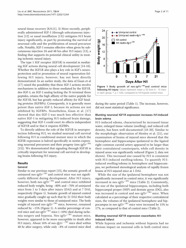
ResultsGeneralSimilar to our previous report [15], the somatic growth ofuninjured nes-igf1r-/Wt and control mice was not signifi-cantly different during development. After H/I injury,however, both nes-igf1r-/Wt and control mice exhibitedreduced body weight, being ~80% and ~70% of uninjuredmice from 1 to 3 days after injury (DAI) and at 7 DAI,respectively (Figure 1). Somatic growth of injured controlmice gradually caught up such that by 21 DAI their bodyweights were similar to those of uninjured mice. The bodyweight of injured nes-igf1r-/Wt mice, however, remainedreduced by ~15% (Figure 1). A similar percentage of con-trol mice and nes-igf1r-/Wt mice (~10%) died during ische-mia surgery and hypoxia. Nes-igf1r-/Wt mutant mice,however, appeared to be more susceptible to death afterH/I injury. About 16% of nes-ifg1r -/Wt mice died within48 hr after surgery, while only ~8% of control mice died
during the same period (Table 1). The increase, however,did not meet statistical significance.
Blunting neuronal IGF1R expression increases H/I-inducedbrain edemaH/I-induced edema, characterized by increased tissuewater, enlarged tissue volume (swelling), and reduced celldensity, has been well-documented [18-20]. Similar tothe morphologic observation of Menley et al. [21], ourexamination of brains of injured mice showed that thehemisphere and hippocampus ipsilateral to the ligatedright common carotid artery appeared to be larger thantheir contralateral counterparts, while cell density ininjured areas was significantly reduced (Figure 2, data notshown). This increased size caused by H/I is consistentwith H/I-induced swelling/edema. To quantify H/I-induced swelling/edema in hemisphere and hippocam-pus, we performed stereological analysis of rapidly frozenbrains of H/I-injured mice at 1 DAI.While the size of the ipsilateral hemisphere was not
significantly increased in control mice, it was significantlyincreased in nes-igf1r-/Wt mice (Table 2). Consistently,the size of the ipsilateral hippocampus, including bothhippocampal proper (HIP) and dentate gyrus (DG), alsowas increased in control and nes-igf1r-/Wt mice. Whencalculated as a percentage of their respective contralateralsizes, the volumes of the ipsilateral hemisphere and hip-pocampus in nes-igf1r-/Wt mice were increased by 15% to17%, as compared to that of control mice (Table 2).
Blunting neuronal IGF1R expression exacerbates H/I-induced infarctWhile hypoxia and ischemia without hypoxia had noobvious impact on neuronal cells in both control mice
Figure 1 Body growth of nes-igf1r-/Wtand control micefollowing H/I injury. Values represent mean ± SE from 6 - 15 micein each group. *, p < 0.05, compared to H/I-injured control mice.
Liu et al. BMC Neuroscience 2011, 12:64http://www.biomedcentral.com/1471-2202/12/64
Page 2 of 11
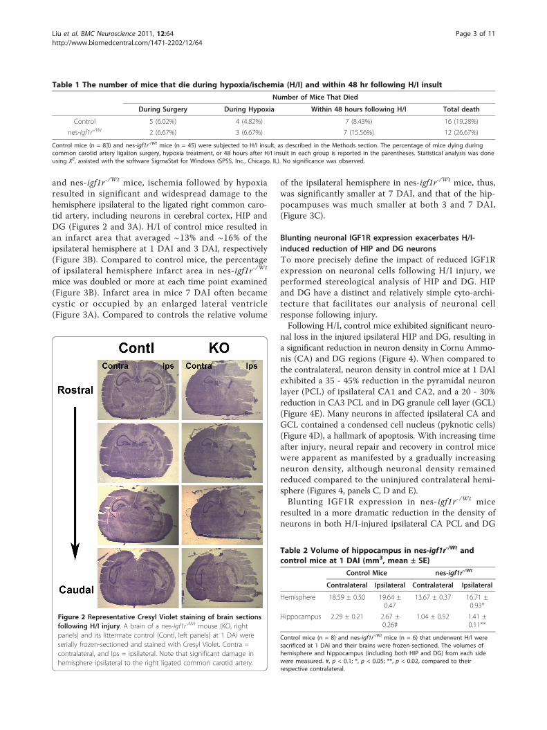
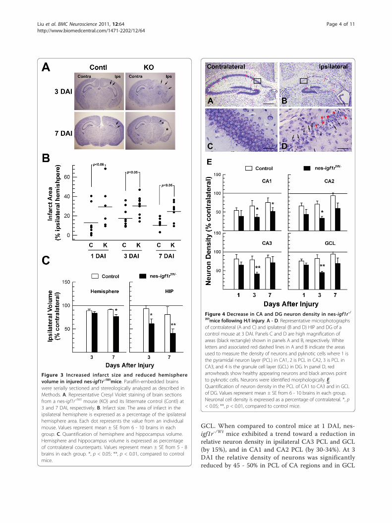
and nes-igf1r-/Wt mice, ischemia followed by hypoxiaresulted in significant and widespread damage to thehemisphere ipsilateral to the ligated right common caro-tid artery, including neurons in cerebral cortex, HIP andDG (Figures 2 and 3A). H/I of control mice resulted inan infarct area that averaged ~13% and ~16% of theipsilateral hemisphere at 1 DAI and 3 DAI, respectively(Figure 3B). Compared to control mice, the percentageof ipsilateral hemisphere infarct area in nes-igf1r-/Wt
mice was doubled or more at each time point examined(Figure 3B). Infarct area in mice 7 DAI often becamecystic or occupied by an enlarged lateral ventricle(Figure 3A). Compared to controls the relative volume
of the ipsilateral hemisphere in nes-igf1r-/Wt mice, thus,was significantly smaller at 7 DAI, and that of the hip-pocampuses was much smaller at both 3 and 7 DAI,(Figure 3C).
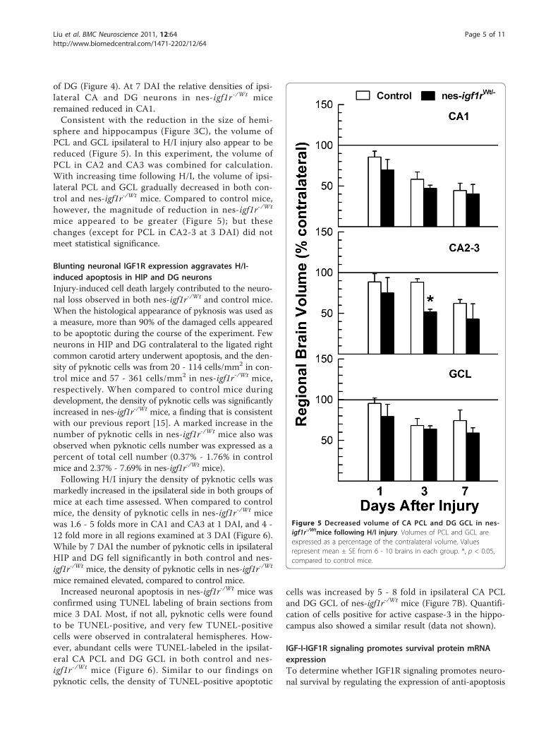
Blunting neuronal IGF1R expression exacerbates H/I-induced reduction of HIP and DG neuronsTo more precisely define the impact of reduced IGF1Rexpression on neuronal cells following H/I injury, weperformed stereological analysis of HIP and DG. HIPand DG have a distinct and relatively simple cyto-archi-tecture that facilitates our analysis of neuronal cellresponse following injury.Following H/I, control mice exhibited significant neuro-
nal loss in the injured ipsilateral HIP and DG, resulting ina significant reduction in neuron density in Cornu Ammo-nis (CA) and DG regions (Figure 4). When compared tothe contralateral, neuron density in control mice at 1 DAIexhibited a 35 - 45% reduction in the pyramidal neuronlayer (PCL) of ipsilateral CA1 and CA2, and a 20 - 30%reduction in CA3 PCL and in DG granule cell layer (GCL)(Figure 4E). Many neurons in affected ipsilateral CA andGCL contained a condensed cell nucleus (pyknotic cells)(Figure 4D), a hallmark of apoptosis. With increasing timeafter injury, neural repair and recovery in control micewere apparent as manifested by a gradually increasingneuron density, although neuronal density remainedreduced compared to the uninjured contralateral hemi-sphere (Figures 4, panels C, D and E).Blunting IGF1R expression in nes-igf1r-/Wt mice
resulted in a more dramatic reduction in the density ofneurons in both H/I-injured ipsilateral CA PCL and DG
Table 1 The number of mice that die during hypoxia/ischemia (H/I) and within 48 hr following H/I insult
Number of Mice That Died
During Surgery During Hypoxia Within 48 hours following H/I Total death
Control 5 (6.02%) 4 (4.82%) 7 (8.43%) 16 (19.28%)
nes-igf1r-/Wt 2 (6.67%) 3 (6.67%) 7 (15.56%) 12 (26.67%)
Control mice (n = 83) and nes-igf1r-/Wt mice (n = 45) were subjected to H/I insult, as described in the Methods section. The percentage of mice dying duringcommon carotid artery ligation surgery, hypoxia treatment, or 48 hours after H/I insult in each group is reported in the parentheses. Statistical analysis was doneusing X2, assisted with the software SigmaStat for Windows (SPSS, Inc., Chicago, IL). No significance was observed.
Figure 2 Representative Cresyl Violet staining of brain sectionsfollowing H/I injury. A brain of a nes-igf1r-/Wt mouse (KO, rightpanels) and its littermate control (Contl, left panels) at 1 DAI wereserially frozen-sectioned and stained with Cresyl Violet. Contra =contralateral, and Ips = ipsilateral. Note that significant damage inhemisphere ipsilateral to the right ligated common carotid artery.
Table 2 Volume of hippocampus in nes-igf1r-/Wt andcontrol mice at 1 DAI (mm3, mean ± SE)
Control Mice nes-igf1r-/Wt
Contralateral Ipsilateral Contralateral Ipsilateral
Hemisphere 18.59 ± 0.50 19.64 ±0.47
13.67 ± 0.37 16.71 ±0.93*
Hippocampus 2.29 ± 0.21 2.67 ±0.26#
1.04 ± 0.52 1.41 ±0.11**
Control mice (n = 8) and nes-igf1r-/Wt mice (n = 6) that underwent H/I weresacrificed at 1 DAI and their brains were frozen-sectioned. The volumes ofhemisphere and hippocampus (including both HIP and DG) from each sidewere measured. #, p < 0.1; *, p < 0.05; **, p < 0.02, compared to theirrespective contralateral.
Liu et al. BMC Neuroscience 2011, 12:64http://www.biomedcentral.com/1471-2202/12/64
Page 3 of 11
GCL. When compared to control mice at 1 DAI, nes-igf1r-/Wt mice exhibited a trend toward a reduction inrelative neuron density in ipsilateral CA3 PCL and GCL(by 15%), and in CA1 and CA2 PCL (by 30-34%). At 3DAI the relative density of neurons was significantlyreduced by 45 - 50% in PCL of CA regions and in GCL
Figure 3 Increased infarct size and reduced hemispherevolume in injured nes-igf1r-/Wtmice. Paraffin-embedded brainswere serially sectioned and stereologically analyzed as described inMethods. A. Representative Cresyl Violet staining of brain sectionsfrom a nes-igf1r-/Wt mouse (KO) and its littermate control (Contl) at3 and 7 DAI, respectively. B. Infarct size. The area of infarct in theipsilateral hemisphere is expressed as a percentage of the ipsilateralhemisphere area. Each dot represents the value from an individualmouse. Values represent mean ± SE from 6 - 10 brains in eachgroup. C. Quantification of hemisphere and hippocampus volume.Hemisphere and hippocampus volume is expressed as percentageof contralateral counterparts. Values represent mean ± SE from 5 - 8brains in each group. *, p < 0.05; **, p < 0.01, compared to controlmice.
Figure 4 Decrease in CA and DG neuron density in nes-igf1r-/Wtmice following H/I injury. A - D. Representative microphotographsof contralateral (A and C) and ipsilateral (B and D) HIP and DG of acontrol mouse at 3 DAI. Panels C and D are high magnification ofareas (black rectangle) shown in panels A and B, respectively. Whiteletters and associated red dashed lines in A and B indicate the areasused to measure the density of neurons and pyknotic cells where 1 isthe pyramidal neuron layer (PCL) in CA1, 2 is PCL in CA2, 3 is PCL inCA3, and 4 is the granule cell layer (GCL) in DG. In panel D, redarrowheads show healthy appearing neurons and black arrows pointto pyknotic cells. Neurons were identified morphologically. E.Quantification of neuron density in the PCL of CA1 to CA3 and in GCLof DG. Values represent mean ± SE from 6 - 10 brains in each group.Neuronal cell density is expressed as a percentage of contralateral. *, p< 0.05; **, p < 0.01, compared to control mice.
Liu et al. BMC Neuroscience 2011, 12:64http://www.biomedcentral.com/1471-2202/12/64
Page 4 of 11
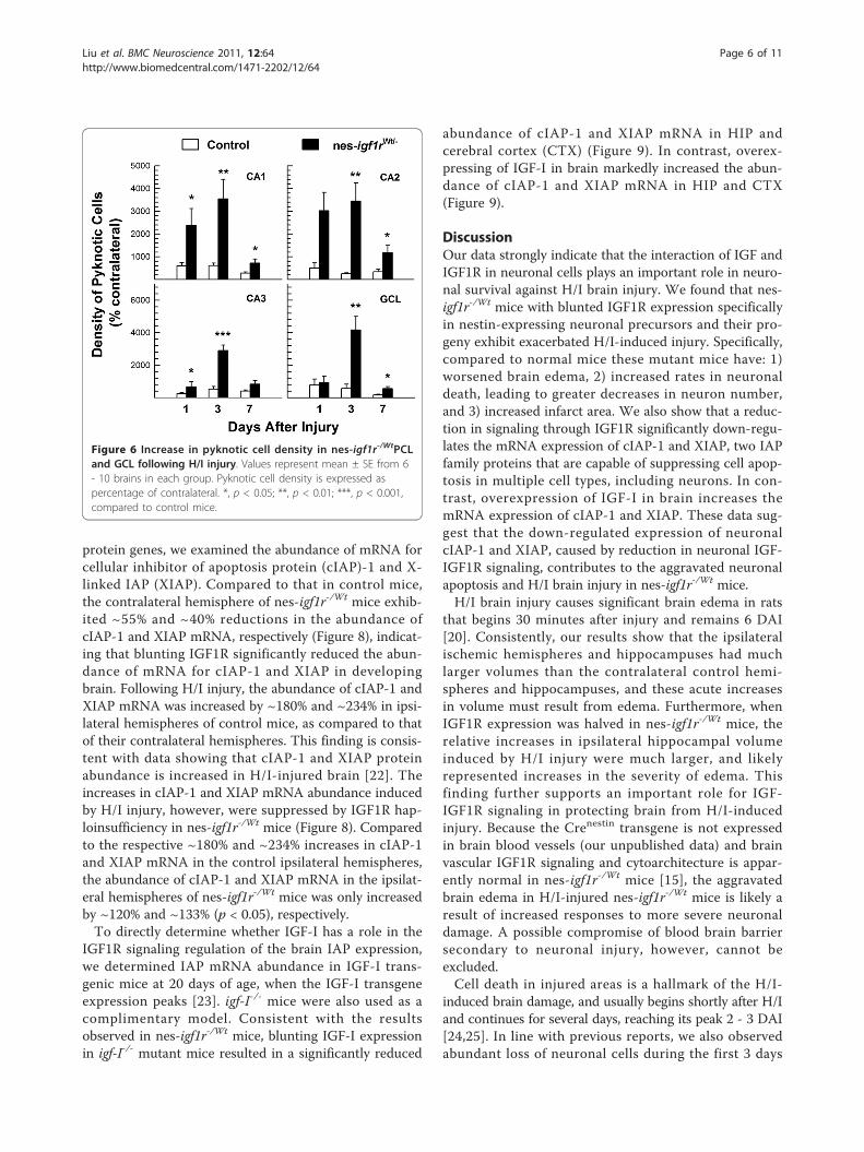
of DG (Figure 4). At 7 DAI the relative densities of ipsi-lateral CA and DG neurons in nes-igf1r-/Wt miceremained reduced in CA1.Consistent with the reduction in the size of hemi-
sphere and hippocampus (Figure 3C), the volume ofPCL and GCL ipsilateral to H/I injury also appear to bereduced (Figure 5). In this experiment, the volume ofPCL in CA2 and CA3 was combined for calculation.With increasing time following H/I, the volume of ipsi-lateral PCL and GCL gradually decreased in both con-trol and nes-igf1r-/Wt mice. Compared to control mice,however, the magnitude of reduction in nes-igf1r-/Wt
mice appeared to be greater (Figure 5); but thesechanges (except for PCL in CA2-3 at 3 DAI) did notmeet statistical significance.
Blunting neuronal IGF1R expression aggravates H/I-induced apoptosis in HIP and DG neuronsInjury-induced cell death largely contributed to the neuro-nal loss observed in both nes-igf1r-/Wt and control mice.When the histological appearance of pyknosis was used asa measure, more than 90% of the damaged cells appearedto be apoptotic during the course of the experiment. Fewneurons in HIP and DG contralateral to the ligated rightcommon carotid artery underwent apoptosis, and the den-sity of pyknotic cells was from 20 - 114 cells/mm2 in con-trol mice and 57 - 361 cells/mm2 in nes-igf1r-/Wt mice,respectively. When compared to control mice duringdevelopment, the density of pyknotic cells was significantlyincreased in nes-igf1r-/Wt mice, a finding that is consistentwith our previous report [15]. A marked increase in thenumber of pyknotic cells in nes-igf1r-/Wt mice also wasobserved when pyknotic cells number was expressed as apercent of total cell number (0.37% - 1.76% in controlmice and 2.37% - 7.69% in nes-igf1r-/Wt mice).Following H/I injury the density of pyknotic cells was
markedly increased in the ipsilateral side in both groups ofmice at each time assessed. When compared to controlmice, the density of pyknotic cells in nes-igf1r-/Wt micewas 1.6 - 5 folds more in CA1 and CA3 at 1 DAI, and 4 -12 fold more in all regions examined at 3 DAI (Figure 6).While by 7 DAI the number of pyknotic cells in ipsilateralHIP and DG fell significantly in both control and nes-igf1r-/Wt mice, the density of pyknotic cells in nes-igf1r-/Wt
mice remained elevated, compared to control mice.Increased neuronal apoptosis in nes-igf1r-/Wt mice was
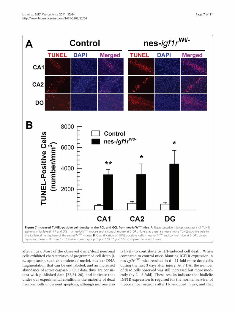
confirmed using TUNEL labeling of brain sections frommice 3 DAI. Most, if not all, pyknotic cells were foundto be TUNEL-positive, and very few TUNEL-positivecells were observed in contralateral hemispheres. How-ever, abundant cells were TUNEL-labeled in the ipsilat-eral CA PCL and DG GCL in both control and nes-igf1r-/Wt mice (Figure 6). Similar to our findings onpyknotic cells, the density of TUNEL-positive apoptotic
cells was increased by 5 - 8 fold in ipsilateral CA PCLand DG GCL of nes-igf1r-/Wt mice (Figure 7B). Quantifi-cation of cells positive for active caspase-3 in the hippo-campus also showed a similar result (data not shown).
IGF-I-IGF1R signaling promotes survival protein mRNAexpressionTo determine whether IGF1R signaling promotes neuro-nal survival by regulating the expression of anti-apoptosis
Figure 5 Decreased volume of CA PCL and DG GCL in nes-igf1r-/Wtmice following H/I injury. Volumes of PCL and GCL areexpressed as a percentage of the contralateral volume. Valuesrepresent mean ± SE from 6 - 10 brains in each group. *, p < 0.05,compared to control mice.
Liu et al. BMC Neuroscience 2011, 12:64http://www.biomedcentral.com/1471-2202/12/64
Page 5 of 11
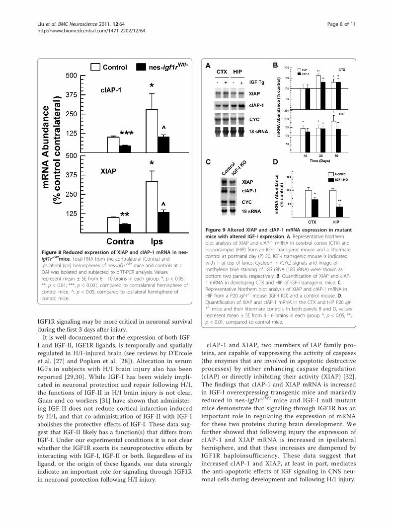
protein genes, we examined the abundance of mRNA forcellular inhibitor of apoptosis protein (cIAP)-1 and X-linked IAP (XIAP). Compared to that in control mice,the contralateral hemisphere of nes-igf1r-/Wt mice exhib-ited ~55% and ~40% reductions in the abundance ofcIAP-1 and XIAP mRNA, respectively (Figure 8), indicat-ing that blunting IGF1R significantly reduced the abun-dance of mRNA for cIAP-1 and XIAP in developingbrain. Following H/I injury, the abundance of cIAP-1 andXIAP mRNA was increased by ~180% and ~234% in ipsi-lateral hemispheres of control mice, as compared to thatof their contralateral hemispheres. This finding is consis-tent with data showing that cIAP-1 and XIAP proteinabundance is increased in H/I-injured brain [22]. Theincreases in cIAP-1 and XIAP mRNA abundance inducedby H/I injury, however, were suppressed by IGF1R hap-loinsufficiency in nes-igf1r-/Wt mice (Figure 8). Comparedto the respective ~180% and ~234% increases in cIAP-1and XIAP mRNA in the control ipsilateral hemispheres,the abundance of cIAP-1 and XIAP mRNA in the ipsilat-eral hemispheres of nes-igf1r-/Wt mice was only increasedby ~120% and ~133% (p < 0.05), respectively.To directly determine whether IGF-I has a role in the
IGF1R signaling regulation of the brain IAP expression,we determined IAP mRNA abundance in IGF-I trans-genic mice at 20 days of age, when the IGF-I transgeneexpression peaks [23]. igf-I-/- mice were also used as acomplimentary model. Consistent with the resultsobserved in nes-igf1r-/Wt mice, blunting IGF-I expressionin igf-I-/- mutant mice resulted in a significantly reduced
abundance of cIAP-1 and XIAP mRNA in HIP andcerebral cortex (CTX) (Figure 9). In contrast, overex-pressing of IGF-I in brain markedly increased the abun-dance of cIAP-1 and XIAP mRNA in HIP and CTX(Figure 9).
DiscussionOur data strongly indicate that the interaction of IGF andIGF1R in neuronal cells plays an important role in neuro-nal survival against H/I brain injury. We found that nes-igf1r-/Wt mice with blunted IGF1R expression specificallyin nestin-expressing neuronal precursors and their pro-geny exhibit exacerbated H/I-induced injury. Specifically,compared to normal mice these mutant mice have: 1)worsened brain edema, 2) increased rates in neuronaldeath, leading to greater decreases in neuron number,and 3) increased infarct area. We also show that a reduc-tion in signaling through IGF1R significantly down-regu-lates the mRNA expression of cIAP-1 and XIAP, two IAPfamily proteins that are capable of suppressing cell apop-tosis in multiple cell types, including neurons. In con-trast, overexpression of IGF-I in brain increases themRNA expression of cIAP-1 and XIAP. These data sug-gest that the down-regulated expression of neuronalcIAP-1 and XIAP, caused by reduction in neuronal IGF-IGF1R signaling, contributes to the aggravated neuronalapoptosis and H/I brain injury in nes-igf1r-/Wt mice.H/I brain injury causes significant brain edema in rats
that begins 30 minutes after injury and remains 6 DAI[20]. Consistently, our results show that the ipsilateralischemic hemispheres and hippocampuses had muchlarger volumes than the contralateral control hemi-spheres and hippocampuses, and these acute increasesin volume must result from edema. Furthermore, whenIGF1R expression was halved in nes-igf1r-/Wt mice, therelative increases in ipsilateral hippocampal volumeinduced by H/I injury were much larger, and likelyrepresented increases in the severity of edema. Thisfinding further supports an important role for IGF-IGF1R signaling in protecting brain from H/I-inducedinjury. Because the Crenestin transgene is not expressedin brain blood vessels (our unpublished data) and brainvascular IGF1R signaling and cytoarchitecture is appar-ently normal in nes-igf1r-/Wt mice [15], the aggravatedbrain edema in H/I-injured nes-igf1r-/Wt mice is likely aresult of increased responses to more severe neuronaldamage. A possible compromise of blood brain barriersecondary to neuronal injury, however, cannot beexcluded.Cell death in injured areas is a hallmark of the H/I-
induced brain damage, and usually begins shortly after H/Iand continues for several days, reaching its peak 2 - 3 DAI[24,25]. In line with previous reports, we also observedabundant loss of neuronal cells during the first 3 days
Figure 6 Increase in pyknotic cell density in nes-igf1r-/WtPCLand GCL following H/I injury. Values represent mean ± SE from 6- 10 brains in each group. Pyknotic cell density is expressed aspercentage of contralateral. *, p < 0.05; **, p < 0.01; ***, p < 0.001,compared to control mice.
Liu et al. BMC Neuroscience 2011, 12:64http://www.biomedcentral.com/1471-2202/12/64
Page 6 of 11
after injury. Most of the observed dying/dead neuronalcells exhibited characteristics of programmed cell death (i.e., apoptosis), such as condensed nuclei, nuclear DNAfragmentation that can be end labeled, and an increasedabundance of active caspase-3. Our data, thus, are consis-tent with published data [22,24-26], and indicate thatunder our experimental conditions the majority of deadneuronal cells underwent apoptosis, although necrosis also
is likely to contribute to H/I-induced cell death. Whencompared to control mice, blunting IGF1R expression innes-igf1r-/Wt mice resulted in 6 - 11 fold more dead cellsduring the first 3 days after injury. At 7 DAI the numberof dead cells observed was still increased but more mod-estly (by 2 - 3 fold). These results indicate that biallelicIGF1R expression is required for the normal survival ofhippocampal neurons after H/I-induced injury, and that
Figure 7 Increased TUNEL-positive cell density in the PCL and GCL from nes-igf1r-/Wtmice. A. Representative microphotographs of TUNELstaining in ipsilateral HIP and DG in a nes-igf1r-/Wt mouse and a control mouse at 3 DAI. Note that there are many more TUNEL-positive cells inthe ipsilateral hemisphere of the nes-igf1r-/Wt mouse. B. Quantification of TUNEL-positive cells in nes-igf1r-/Wt and control mice at 3 DAI. Valuesrepresent mean ± SE from 6 - 10 brains in each group. *, p < 0.05; **, p < 0.01, compared to control mice.
Liu et al. BMC Neuroscience 2011, 12:64http://www.biomedcentral.com/1471-2202/12/64
Page 7 of 11
IGF1R signaling may be more critical in neuronal survivalduring the first 3 days after injury.It is well-documented that the expression of both IGF-
I and IGF-II, IGF1R ligands, is temporally and spatiallyregulated in H/I-injured brain (see reviews by D’Ercoleet al. [27] and Popken et al. [28]). Alteration in serumIGFs in subjects with H/I brain injury also has beenreported [29,30]. While IGF-I has been widely impli-cated in neuronal protection and repair following H/I,the functions of IGF-II in H/I brain injury is not clear.Guan and co-workers [31] have shown that administer-ing IGF-II does not reduce cortical infarction inducedby H/I, and that co-administration of IGF-II with IGF-Iabolishes the protective effects of IGF-I. These data sug-gest that IGF-II likely has a function(s) that differs fromIGF-I. Under our experimental conditions it is not clearwhether the IGF1R exerts its neuroprotective effects byinteracting with IGF-I, IGF-II or both. Regardless of itsligand, or the origin of these ligands, our data stronglyindicate an important role for signaling through IGF1Rin neuronal protection following H/I injury.
cIAP-1 and XIAP, two members of IAP family pro-teins, are capable of suppressing the activity of caspases(the enzymes that are involved in apoptotic destructiveprocesses) by either enhancing caspase degradation(cIAP) or directly inhibiting their activity (XIAP) [32].The findings that cIAP-1 and XIAP mRNA is increasedin IGF-I overexpressing transgenic mice and markedlyreduced in nes-igf1r-/Wt mice and IGF-I null mutantmice demonstrate that signaling through IGF1R has animportant role in regulating the expression of mRNAfor these two proteins during brain development. Wefurther showed that following injury the expression ofcIAP-1 and XIAP mRNA is increased in ipsilateralhemisphere, and that these increases are dampened byIGF1R haploinsufficiency. These data suggest thatincreased cIAP-1 and XIAP, at least in part, mediatesthe anti-apoptotic effects of IGF signaling in CNS neu-ronal cells during development and following H/I injury.
Figure 8 Reduced expression of XIAP and cIAP-1 mRNA in nes-igf1r-/Wtmice. Total RNA from the contralateral (Contra) andipsilateral (Ips) hemispheres of nes-igf1r-/Wt mice and controls at 1DAI was isolated and subjected to qRT-PCR analysis. Valuesrepresent mean ± SE from 6 - 10 brains in each group. *, p < 0.05;**, p < 0.01; ***, p < 0.001, compared to contralateral hemisphere ofcontrol mice. ^, p < 0.05, compared to ipsilateral hemisphere ofcontrol mice.
Figure 9 Altered XIAP and cIAP-1 mRNA expression in mutantmice with altered IGF-I expression. A. Representative Northernblot analysis of XIAP and cIAP-1 mRNA in cerebral cortex (CTX) andhippocampus (HIP) from an IGF-I transgenic mouse and a littermatecontrol at postnatal day (P) 20. IGF-I transgenic mouse is indicatedwith + at top of lanes. Cyclophilin (CYC) signals and image ofmethylene blue staining of 18S rRNA (18S rRNA) were shown asbottom two panels, respectively. B. Quantification of XIAP and cIAP-1 mRNA in developing CTX and HIP of IGF-I transgenic mice. C.Representative Northern blot analysis of XIAP and cIAP-1 mRNA inHIP from a P20 igf-I-/- mouse (IGF-I KO) and a control mouse. D.Quantification of XIAP and cIAP-1 mRNA in the CTX and HIP P20 igf-I-/- mice and their littermate controls. In both panels B and D, valuesrepresent mean ± SE from 4 - 6 brains in each group. *, p < 0.05; **,p < 0.01, compared to control mice.
Liu et al. BMC Neuroscience 2011, 12:64http://www.biomedcentral.com/1471-2202/12/64
Page 8 of 11
ConclusionsTaken together, our data demonstrated that bluntingIGF1R expression specifically in neuronal cells exacerbatesH/I-induced injury, and thus, are consistent with the pro-position that IGF-IGF1R interaction in neuronal cells iscritically important for their survival following H/I injury.In addition, our data showing that IGF-IGF1R signalingup-regulates the expression of neuronal cIAP-1 and XIAPduring development and following H/I, strongly suggestthat cIAP-1 and XIAP are two candidate targets for thera-peutic treatment of H/I-induced brain injuries.
MethodsAnimals and H/I InjuryNes-igf1r-/Wt conditional mutant mice, generated as pre-viously described [15], were bred as heterozygotes. Aspreviously reported, igf1rlox/Wt and nestin-Cre transgenicmice have normal postnatal growth, and our preliminarystudies show that these mice exhibit similar changes tothose of wildtype (Wt) mice in responding to H/I injury.Thus, these mutant mice were grouped and consideredcontrols. Transgenic mice that express a human IGF-IcDNA driven by a metallothionein-I promoter (IGF-Itransgenic mice) [23], and mice with a global IGF-I nullmutation (igf-I-/- [14,33] also have been described else-where. All procedures used were consistent with theguidelines of the National Institutes of Health andapproved by the institutional review committees of theUniversity of North Carolina at Chapel Hill.H/I injury was done using a modified method [34,35]
that was adapted from a neonatal rat method [18]. Briefly,mice at postnatal day 10 (weighting 4 to 5 grams) weresubjected to artery ligation surgery or sham surgery underisoflurane anesthesia (4% for induction and 1.5-2% formaintenance). After the skin was sterilized with 75% etha-nol and 10% povidone-iodine, a midline neck incision (0.5- 1 cm in length) was made, and the right common carotidartery was carefully isolated and ligated with a bipolar coa-gulator. The wound was then closed with interruptednylon sutures. During the procedure, which typically tookno longer than 10 minutes, body temperature was main-tained using a heating lamp. After ligation or sham sur-gery, mice were allowed to recover from anesthesia in achamber partially submerged in a 37°C water bath. After~10 minutes, mice were returned to their dam. One hourafter surgery, mice were individually placed in a chamberthat was partially submerged in a water bath (36 ± 0.2°C),subjected to hypoxia (10% O2, balanced with N2) for 45minutes, and then were returned to their dam. Both con-trol mice and nes-igf1r-/Wt mice resumed normal activityand feeding within 5-10 minutes after return to their dam.Mice were sacrificed 1, 3 or 7 DAI. Similar to previously
reported data [18,36], control mice or nes-igf1r-/Wt mice,which underwent hypoxia alone, ischemia alone, sham
surgery, or sham surgery followed by hypoxia, exhibitedsimilar body growth, behavioral and brain morphologicalcharacteristics as their intact untreated counterparts. H/Iinjury resulted in a suboptimal somatic growth (seeResults and Figure 1). As we previously reported, reduc-tion in IGF1R expression causes brain growth retardation[15], and therefore, all changes are expressed as% of thecontralateral brain hemisphere, except where indicated.
Histology and Stereological AnalysisAfter being fixed by submersion in 4% paraformaldehydeovernight, brains were paraffin-embedded, and coronallysectioned at 8 μm in thickness. Two to three sets of serialsections that comprised very 10th section were collected.To estimate the volume of brain regions, one series ofsections were stained with cresyl violet, and the area ofbrain regions on each section (corresponding to platesbetween number 41 and 52 [37]) was measured under amicroscope, assisted with Stereo Investigator software(Microbrightfield, Colchester, VT). The volume (V) wasthen estimated using the formula: V = ΣA × T × I, whereΣA = sum of area measured on each section, T = sectionthickness, and I = section intervals.To determine the density of pyramidal neurons in CA
PCL regions and granule neurons in DG GCL, stained sec-tion corresponding to plate 50 [37] from each brain wasselected, and cell nuclei within delineated areas werecounted, assisted with Stereo Investigator software. In eachsection the cell density in each brain region was calculatedby dividing the regional cell count by its respective area.To determine infarct area, sections corresponding to
plate 50 [37] from each paraffin-embedded brain werestained with cresyl violet. The area of infarct in the ipsi-lateral hemisphere was then measured using StereoInvestigator, as described above.To evaluate possible brain swelling/edema after H/I
injury, some brains were fresh-frozen in liquid N2 to pre-serve water content and brain structures. Serial sections(20 μm in thickness) were cut coronally on a cryostat.Two to three sets of serial sections that comprised every6th section were collected. One series of sections (alsocorresponding to plates between number 41 and 52)were fixed with 4% paraformaldehyde, and stained withcresyl violet. The areas of ipsilateral and contralateralhemisphere and hippocampus were measured, and theirvolumes were calculated as described above.
Terminal uridine nucleotide end labeling (TUNEL)After paraffin-sections (corresponding to plate [37])were dewaxed and rehydrated, TUNEL-positive apopto-tic cells were detected using an In Situ Cell DeathDetection Kit (Roche Applied Science, Indianapolis, IN),following the manufacturer’s protocol. The sectionswere then counterstained with the nuclear dye 6-
Liu et al. BMC Neuroscience 2011, 12:64http://www.biomedcentral.com/1471-2202/12/64
Page 9 of 11
diamidine-2’-phenylindole dihydrochloride (DAPI, Invi-trogen, Carlsbad, CA). Images of TUNEL-positive cellswithin CA and DG regions were digitally captured usinga microscope and a Spot Jr. digital camera (DiagnosticInstruments, Sterling Heights, MI). Within delineatedareas (measured and assisted with a Stereo Investigatorsoftware), TUNEL-positive cells were counted, and thentheir densities were calculated.
Quantitative Real Time-PCR (qRT-PCR) and Northern BlotHybridization AnalysisSlides containing coronal frozen-sections (20 μm in thick-ness, and corresponding to plate 48 - 50) were briefly air-dried. Contralateral and ipsilateral hemisphere tissueswere scraped from the slides and collected, and total RNAwas isolated using a RNA plus kit (Qiagen, Valencia, CA).cDNA reverse transcription was performed, and the resul-tant mRNA-derived cDNA was quantified by qRT-PCRusing specific primer sets. The sequences of the primersused are as follows: cIAP-1 (Genbank accession number:BC145985), forward primer: 5’-gatggtggcttgagatgttgg-3’,reverse primer, 5’-tttctccagggccaaaatgc-3’; XIAP (Genbankaccession number: NM_009688), forward primer, 5’-ctattggatgagaaggggcaag-3’, reverse primer, 5’-ataga-tagctgctcccggatg-3’. Primers for 18S rRNA were obtainedfrom Invitrogen. The abundance of mRNA was deter-mined, based on a standard curve for each target mRNAand 18S rRNA, as described [38].Northern blot hybridization and quantification of
mRNA abundance on blots were performed as previouslydescribed [23]. Total RNA was isolated using the acidicguanidinium thiocyanate-phenol-chloroform method [39].32P-labeled single stranded cDNA probes were generatedas previously described [23], using plasmid containingXIAP (also known as MIHA) or cIAP-1 (also known asBIRC2 or MIHB) cDNA. XIAP and cIAP-1 plasmids weregenerously provided by Dr. Uren [40]. PCR-amplifiedXIAP and cIAP-1 cDNA fragments correspond to basepairs (bp) 584-870 (Genbank accession number: U36842)and bp 1685-1898 (Genbank accession number: U37547),respectively.
StatisticsAll experiments were repeated at least two times. Student-t test or one-way ANOVA was used to test statistical sig-nificance among the groups, followed by comparison ofeach group mean with the Newman-Keuls-Student testassisted with the software SigmaStat for Windows (SPSS,Inc., Chicago, IL).
AcknowledgementsThis work was supported by NIH grants RO1 HD008299 and RO1 NS48868.The authors wish to thank Dr. William Lagard for his help in the setup of H/Iinstruments.
Authors’ contributionsPY and AJD designed the study. WL and PY performed the experiments andcollected data. PY and AJD analyzed the data and wrote the manuscript. Allauthors read and approved the manuscript.
Competing interestsThe authors declare that they have no competing interests.
Received: 11 May 2011 Accepted: 30 June 2011 Published: 30 June 2011
References1. Vannucci RC: Experimental biology of cerebral hypoxia-ischemia: relation
to perinatal brain damage. Pediatr Res 1990, 27:317-326.2. Volpe JJ: Neurology of the Neonate Saunders; 2000.3. Beilharz EJ, Klempt ND, Klempt M, Sirimanne E, Dragunow M, Gluckman PD:
Differential expression of insulin-like growth factor binding proteins(IGFBP) 4 and 5 mRNA in the rat brain after transient hypoxic-ischemicinjury. Mol Brain Res 1993, 18:209-215.
4. Lee WH, Clemens JA, Bondy CA: Insulin-like growth factors in theresponse to cerebral ischemia. Mol Cell Neurosci 1992, 3:36-43.
5. Lee WH, Wang GM, Seaman LB, Vannucci SJ: Coordinate IGF-I and IGFBP5gene expression in perinatal rat brain after hypoxia-ischemia. J CerebBlood Flow Metab 1996, 16:227-236.
6. Clawson TF, Vannucci SJ, Wang GM, Seaman LB, Yang XL, Lee WH:Hypoxia-ischemia-induced apoptotic cell death correlates with IGF-ImRNA decrease in neonatal rat brain. Biol Signals Receptors 1999,8:281-293.
7. Brywe KG, Mallard C, Gustavsson M, Hedtjarn M, Leverin AL, Wang XY,Blomgren K, Isgaard J, Hagberg H: IGF-I neuroprotection in the immaturebrain after hypoxia-ischemia, involvement of Akt and GSK3 beta? Eur JNeurosci 2005, 21:1489-1502.
8. Gluckman P, Klempt N, Guan J, Mallard C, Sirmanne E, Dragunow M,Klempt M, Singh K, Williams C, Nikolics K: A role for IGF-I in the rescue ofCNS neurons following hypoxic-ischemic injury. Biochem Biophys ResCommun 1992, 182:593-599.
9. Guan J, Miller OT, Waugh KM, McCarthy DC, Gluckman PD: Insulin-likegrowth factor-1 improves somatosensory function and reduces theextent of cortical infarction and ongoing neuronal loss after hypoxia-ischemia in rats. Neuroscience 2001, 105:299-306.
10. Lin S, Fan LW, Pang Y, Rhodes PG, Mitchell HJ, Cai Z: IGF-1 protectsoligodendrocyte progenitor cells and improves neurological functionsfollowing cerebral hypoxia-ischemia in the neonatal rat. Brain Res 2005,1063:15-26.
11. Guan J, Bennet TL, George S, Waldvogel HJ, Faull RL, Gluckman PD,Keunen H, Gunn AJ: Selective neuroprotective effects with insulin-likegrowth factor-1 in phenotypic striatal neurons following ischemic braininjury in fetal sheep. Neuroscience 2000, 95:831-839.
12. Zhong J, Zhao L, Du Y, Wei G, Yao WG, Lee WH: Delayed IGF-1 treatmentreduced long-term hypoxia-ischemia-induced brain damage andimproved behavior recovery of immature rats. Neurol Res 2009,31:483-489.
13. Lin S, Fan LW, Rhodes PG, Cai Z: Intranasal administration of IGF-1attenuates hypoxic-ischemic brain injury in neonatal rats. Exp Neurol2009, 217:361-370.
14. Liu JP, Baker J, Perkins AS, Robertson EJ, Efstratiadis A: Mice carrying nullmutations of the genes encoding Insulin-like Growth Factor I (Igf-1) andtype 1 IGF receptor. Cell 1993, 75:59-72.
15. Liu W, Ye P, O’Kusky JR, D’Ercole AJ: Type 1 insulin-like growth factorreceptor signaling is essential for the development of the hippocampalformation and dentate gyrus. J Neurosci Res 2009, 87:2821-2832.
16. Zeger M, Popken G, Zhang J, Xuan S, Lu QR, Schwab MH, Nave KA,Rowitch D, D’Ercole AJ, Ye P: Insulin-like growth factor type 1 receptorsignaling in the cells of oligodendrocyte lineage is required for normalin vivo oligodendrocyte development and myelination. Glia 2007,55:400-411.
17. Guan J, Williams CE, Skinner SJ, Mallard EC, Gluckman PD: The effects ofinsulin-like growth factor (IGF)-1, IGF-2, and des-IGF-1 on neuronal lossafter hypoxic-ischemic brain injury in adult rats: evidence for a role forIGF binding proteins. Endocrinology 1996, 137:893-898.
18. Rice JE III, Vannucci RC, Brierley JB: The influence of immaturity onhypoxic-ischemic brain damage in the rat. Ann Neurol 1981, 9:131-141.
Liu et al. BMC Neuroscience 2011, 12:64http://www.biomedcentral.com/1471-2202/12/64
Page 10 of 11
19. Van Reempts J: The hypoxic brain: histological and ultrastructuralaspects. Behav Brain Res 1984, 14:99-108.
20. Mujsce DJ, Christensen MA, Vannucci RC: Cerebral blood flow and edemain perinatal hypoxic-ischemic brain damage. Pediatr Res 1990, 27:450-453.
21. Menley GT, Binder DK, Papadopoulos MC, Verkman AS: New insights intowatertransport and edema in the central nervous system fromphenotype analysis of Aquaporin-4 null mice. Neuroscience 2004,129:983-991.
22. Tanaka H, Yokota H, Jover T, Cappuccio I, Calderone A, Simionescu M,Bennett MV, Zukin RS: Ischemic preconditioning: neuronal survival in theface of caspase-3 activation. J Neurosci 2004, 24:2750-2759.
23. Ye P, Carson J, D’Ercole AJ: In vivo actions of insulin-like growth factor-I(IGF-I) on brain myelination: studies of IGF-I and IGF binding protein-1(IGFBP-1) transgenic mice. J Neurosci 1995, 15:7344-7356.
24. Beilharz EJ, Williams CE, Dragunow M, Sirimanne ES, Gluckman PD:Mechanisms of delayed cell death following hypoxic-ischemic injury inthe immature rat: evidence for apoptosis during selective neuronal loss.Brain Res Mol Brain Res 1995, 29:1-14.
25. Zhu C, Wang X, Xu F, Bahr BA, Shibata M, Uchiyama Y, Hagberg H,Blomgren K: The influence of age on apoptotic and other mechanisms ofcell death after cerebral hypoxia-ischemia. Cell Death Differ 2005,12:162-176.
26. Liu CL, Siesjo BK, Hu BR: Pathogenesis of hippocampal neuronal deathafter hypoxia-ischemia changes during brain development. Neuroscience2004, 127:113-123.
27. D’Ercole AJ, Ye P, Calikoglu AS, Gutirerrze-Ospina G: The role of the Insulin-like Growth Factors in central nervous system. Mol Neurobiol 1996,13:227-255.
28. Popken G, Dechert-Zeger M, Ye P, D’Ercole AJ: Chapter 6, BrainDevelopment.Edited by: Varela-Nieto I, Chowan J. Springer ScienceBusiness Media, Inc., New York; 2005:187-220, The Growth Hormone-Insulin-like Growth Factor Axis during Development.
29. Satar M, Ozcan K, Yapicioglu H, Narli N: Serum insulin-like growth factor 1and growth hormone levels of hypoxic-ischemic newborns. Biol Neonate2004, 85:15-20.
30. Scheepens A, Sirimanne E, Beilharz E, Breier BH, Waters MJ, Gluckman PD,Williams CE: Alterations in the neural growth hormone axis followinghypoxic-ischemic brain injury. Brain Res Mol Brain Res 1999, 68:88-100.
31. Guan J, Bennet L, Gluckman PD, Gunn AJ: Insulin-like growth factor-1 andpost-ischemic brain injury. Prog Neurobiol 2003, 70:443-462.
32. Eckelman BP, Salvesen GS, Scott FL: Human inhibitor of apoptosisproteins: why XIAP is the black sheep of the family. EMBO Rep 2006,7:988-994.
33. Ye P, Li L, Richards RG, DiAugustine RP, D’Ercole AJ: Myelination is alteredin insulin-like growth factor-I null mutant mice. J Neurosci 2002,22:6041-6051.
34. Liu XH, Kwon D, Shielde G, Yang GY, Silverstein FS, Barks JD: Mice deficientin interleukin-1 converting enzyme are resistant to neonatal hypoxic-ischemic brain damage. J Cereb Blood Flow Metab 1999, 19:1099-1108.
35. Plane JM, Liu R, Wang TW, Silverstein FS, Parent JM: Neonatal hypoxic-ischemic injury increases forebrain subventricular zone neurogenesis inthe mouse. Neurobiol Dis 2004, 16:585-595.
36. Olson EE, McKeon RJ: Characterization of cellular and neurologicaldamage following unilateral hypoxia/ischemia. J Neurol Sci 2004, 227:7-19.
37. Franklin KBJ, Paxinos G: The mouse brain in stereotaxic coordinates.Academic Press; 1997.
38. Liu H, Hu Q, D’Ercole AJ, Ye P: Histone deacetylase 11 regulatesoligodendrocyte-specific gene expression and cell development in OL-1oligodendroglial cells. Glia 2009, 57:1-12.
39. Chomczynski P, Sacchi N: Single-step method of RNA isolation by acidguanidinium thiocyanate-phenol-chloroform extraction. Anal Biochem1987, 162:156-159.
40. Uren AG, Pakusch M, Hawkins CJ, Puls KL, Vaux D: Cloning and expressionof apoptosis inhibitory protein homologs that function to inhibitapoptosis and/or bind tumor necrocic factor receptor-associated factors.Proc Natl Acad Sci USA 1996, 93:4974-4978.
doi:10.1186/1471-2202-12-64Cite this article as: Liu et al.: Blunting type 1 insulin-like growth factorreceptor expression exacerbates neuronal apoptosis following hypoxic/ischemic injury. BMC Neuroscience 2011 12:64.
Submit your next manuscript to BioMed Centraland take full advantage of:
• Convenient online submission
• Thorough peer review
• No space constraints or color figure charges
• Immediate publication on acceptance
• Inclusion in PubMed, CAS, Scopus and Google Scholar
• Research which is freely available for redistribution
Submit your manuscript at www.biomedcentral.com/submit
Liu et al. BMC Neuroscience 2011, 12:64http://www.biomedcentral.com/1471-2202/12/64
Page 11 of 11