Embed Size (px)
Citation preview

ORIGINAL ARTICLE
Blueberry husks, rye bran and multi-strain probiotics affect theseverity of colitis induced by dextran sulphate sodium
ASA HAKANSSON1, CAMILLA BRANNING2, DIYA ADAWI3, GORAN MOLIN1,
MARGARETA NYMAN2, BENGT JEPPSSON3 & SIV AHRNE1
1Food Hygiene, 2Division of Applied Nutrition and Food Chemistry, Lund University, Lund, Sweden, and 3Department of
Surgery, Lund University, Malmo University Hospital, Malmo, Sweden
AbstractObjective. The enteric microbiota is a pivotal factor in the development of intestinal inflammation in humans butprobiotics, dietary fibres and phytochemicals can have anti-inflammatory effects. The aim of this study was to evaluate thetherapeutic effect of multi-strain probiotics and two conceivable prebiotics in an experimental colitis model. Material andmethods. Sprague-Dawley rats were fed a fibre-free diet alone or in combination with Lactobacillus crispatus DSM 16743,L. gasseri DSM 16737 and Bifidobacterium infantis DSM 15158 and/or rye bran and blueberry husks. Colitis was induced by5% dextran sulphate sodium (DSS) given by oro-gastric tube. Colitis severity, inflammatory markers, gut-load of lactobacilliand Enterobacteriaceae, bacterial translocation and formation of carboxylic acids (CAs) were analysed. Results. The diseaseactivity index (DAI) was lower in all treatment groups. Viable counts of Enterobacteriaceae were reduced and correlatedpositively with colitis severity, while DAI was negatively correlated with several CAs, e.g. butyric acid. The addition ofprobiotics to blueberry husks lowered the level of caecal acetic acid and increased that of propionic acid, while rye bran incombination with probiotics increased caecal CA levels and decreased distal colonic levels. Blueberry husks with probioticsreduced the incidence of bacterial translocation to the liver, colonic levels of myeloperoxidase, malondialdehyde and seruminterleukin-12. Acetic and butyric acids in colonic content correlated negatively to malondialdehyde. Conclusions. Acombination of probiotics and blueberry husks or rye bran enhanced the anti-inflammatory effects compared with probioticsor dietary fibres alone. These combinations can be used as a preventive or therapeutic approach to dietary amelioration ofintestinal inflammation.
Key Words: Blueberry husks, dietary fibre, experimental colitis, multi-strain probiotics, rye bran
Introduction
Inflammatory bowel disease (IBD) is believed to be a
result of an abnormal gastrointestinal (GI) immune
response leading to inflammation, triggered by one
or more environmental risk factors in genetically
predisposed individuals. The enteric microbiota is
implicated increasingly as a pivotal factor in the
development of intestinal inflammation in humans
and experimental animals [1,2], but which bacterial
characteristics drive the inflammatory reaction re-
mains to be elucidated. Bacteria inhabiting the
interface between the lumen and the mucosa may
play a role in the inflammatory process, since
dysfunctions in innate immunity are related to
intestinal inflammation [2,3].
A hallmark of IBD is the infiltration of large
numbers of phagocytic leucocytes into the mucosal
interstitium [4] and a majority of the cells in the active
lesions of ulcerative colitis (UC) are activated neu-
trophils and macrophages [5,6], causing production
and release of reactive oxygen species (ROS). This is
associated with an increased lipid peroxidation ob-
served in the mucosa of UC and in experimental
colitis models [7,8]. Overproduction of ROS has
been demonstrated in colonic tissue from
IBD patients, suggesting that colonic inflammation
Correspondence: Siv Ahrne, PhD, Division of Applied Nutrition and Food Chemistry, Chemical Center, P.O. Box 124, SE 221 00 Lund, Sweden. Tel: �46 46
2228 327. Fax: �46 46 2224 532. E-mail: [email protected]
Scandinavian Journal of Gastroenterology, 2009; 44: 1213�1225
(Received 25 March 2009; accepted 8 July 2009)
ISSN 0036-5521 print/ISSN 1502-7708 online # 2009 Informa UK Ltd.
DOI: 10.1080/00365520903171268
Scan
d J
Gas
troe
nter
ol D
ownl
oade
d fr
om in
form
ahea
lthca
re.c
om b
y C
ase
Wes
tern
Res
erve
Uni
vers
ity o
n 10
/31/
14Fo
r pe
rson
al u
se o
nly.

produces high levels of oxidants that probably exceed
the low antioxidant capacity in IBD patients [9]. By
secretion of pro-inflammatory products, polymor-
phonuclear leucocytes can injure epithelial cells [10]
and transepithelial migration can affect the epithelial
permeability. This leads to deterioration of the
epithelial barrier function and exposure of the under-
lying tissue and bloodstream to harmful luminal
components, e.g. bacteria and endotoxins [11]. As a
consequence, direct stimulation of epithelial cytokine
production can occur [12]. Myeloperoxidase (MPO),
a constituent of the primary granules of neutrophils,
has been widely used as a marker for the presence and
activation of neutrophil granulocytes [13].
Alterations of the composition of the GI micro-
biota may alter host immunity and the course of
inflammation [14]. Administration of probiotics
and/or fermentable dietary fibres, i.e. potential
prebiotics, could be strategies to affect the balance
of the microbiota in a positive direction. Certain
strains of Bifidobacterium or Lactobacillus can be
given orally as probiotics and may alleviate intestinal
inflammatory response and halt the vicious circle of
inflammation [15]. The human diet consists of a
large variety of fruits, vegetables and cereals that
contain dietary fibres and phytochemicals, with the
potential to reach the colon and be metabolized by
the microbiota [16,17]. Carbohydrates are the most
important substrate for the colonic microbiota and
are degraded mainly to carboxylic acids (CAs).
Among the CAs, butyrate most effectively protects
the intestinal mucosa against injury and promotes
mucosal healing [18]. Blueberry and rye bran are
especially rich sources of dietary fibres and phyto-
chemicals. Diet can also affect the composition of
the GI microbiota [19]. Specific intestinal bacteria
can metabolize phenolics to varying degrees, produ-
cing a variety of aromatic metabolites which can
either be retained by the bacterial cell or released in
the lumen [20]. Antimicrobial effects of polyphe-
nols, as well as antioxidant activities, have been
demonstrated [21].
In view of the experimental limitations imposed by
studies in humans, animal models are appealing, and
in a model reproducing some of the UC character-
istics, dextran sulphate sodium (DSS) is used to
induce colitis. The DSS model has become a
frequently applied research tool for the pathogenesis
of UC and the development of therapeutic agents
[22]. In the present study the DSS model was
applied in rat, the aim being to clarify the therapeutic
effect of a multi-strain probiotics alone or together
with blueberry husks and rye bran (two conceivable
prebiotics). Colitis severity, inflammatory markers,
the gut-load of lactobacilli and Enterobaceriaceae,
bacterial translocation and formation of CAs along
the hindgut have all been investigated. The idea
behind the treatment regimens was that both pro-
biotics and dietary fibres can exercise positive effects
on the intestinal environment and mitigate inflam-
mation, and that they can possibly complement each
other.
Material and methods
Animals
Female Sprague-Dawley rats (n�36), initial weight
of 21392 g, were purchased from Mollegard (Viby,
Denmark). The animals were housed individually at
a constant room temperature and humidity in a 12 h
light/dark cycle and acclimatized for 1 week before
commencement of the trial. They were given free
access to water, while the feed intake was restricted
to 23 g (dwb, dry weight basis) per day. Manipula-
tion and experimental procedures followed general
guidelines in the care and use of laboratory animals,
and the protocol was approved by the Animal Ethics
Committee at Lund University.
Blueberry husks, rye bran and probiotics
Husks from wild-growing, low-bush, blueberries of
Vaccinium myrtillus L were derived from pressed
berries and freeze dried (Probi AB, Lund, Sweden),
and the bran derived from commercial Swedish rye
was supplied by Lantmannen (Jarna, Sweden).
A mixture of three bacterial strains representing
two Lactobacillus species and one Bificobacterium
species was used: The strains L. crispatus DSM
16743 and L. gasseri DSM 16737 (Probi AB)
originate from the healthy vaginas of fertile women.
Bifidobacterium infantis DSM 15158 has been iso-
lated from the stool of a healthy, breast-fed infant
(Probi AB).
The bacterial preparations of lactobacilli were
made from fresh cultures in LCM (Lactobacillus-
carrying medium) [23]. The cultures were incubated
anaerobically at 378C for 48 h, harvested by cen-
trifugation and resuspended in bacterial suspension
medium (sodium chloride, 8.5 g/l; bacteriological
peptone (Oxoid, Unipath Ltd., Basingstoke, Hamp-
shire, England), 1 g/l; Tween 80, 1 g/l; L-cysteine
hydrochloride monohydrate (Merck, Darmstadt,
Germany) 0.2 g/l), before being given to the rats.
The frozen bacterial culture of B. infantis (Probi
AB) was allowed to thaw, the cells were harvested by
centrifugation and resuspended in bacterial suspen-
sion medium. The amounts of bacteria in the multi-
strain probiotics given daily to the rats were 2�1010
CFU (colony-forming units) for B. infantis DSM
15158 and 5�1010�1�1011 CFU for L. gasseri
DSM 16737 and L. crispatus DSM 16743.
1214 A. Hakansson et al.
Scan
d J
Gas
troe
nter
ol D
ownl
oade
d fr
om in
form
ahea
lthca
re.c
om b
y C
ase
Wes
tern
Res
erve
Uni
vers
ity o
n 10
/31/
14Fo
r pe
rson
al u
se o
nly.
Treatment groups and study design
Five test diets and one control diet were prepared as
described in Table I. The rats were randomly
allocated to one of the six treatment groups, each
group comprising 6 animals: colitis control (C
group), probiotics (P group), rye bran (R group),
rye bran and probiotics (RP group), blueberry husks
(B group) and blueberry husks and probiotics (BP
group).
Rye bran and blueberry husks were included at a
level of 80 g dietary fibre/kg in the diets (dwb). The
dry matter content of the diets was adjusted with
wheat starch. The wheat starch has been shown to be
completely digested and hence does not contribute
to any CA formation [24]. The content of dietary
fibre in rye bran was 48.0 g/100 g (dwb), while the
content of dietary fibre in blueberry husks was
40.8 g/100 g (dwb). Of the dietary fibre in rye
bran, 5.1 g/100 g (dwb) could be considered as
Klason lignin, i.e. components not soluble in 12 M
H2SO4, whereas the amount of Klason lignin in
blueberry husks was 14.1 g/100 g (dwb). The non-
starch polysaccharides in rye bran mainly consisted
of xylose (39%), glucose (30%) and arabinose
(21%), and in blueberry husks the non-starch
polysaccharides mostly consisted of glucose (39%),
uronic acids (25%) and xylose (20%).
Probiotics were mixed daily with the feed at
feeding time. The rats were allowed to adapt to the
diets for 7 days after which a 7-day experimental
period followed. Daily food intake was measured for
all animals. Colitis was induced by 5% (w/w) DSS
(MW�36,000�50,000; ICN Biomedicals Inc., Aur-
ora, Ohio, USA) dissolved in water and given once a
day by oro-gastric tube. The mean starting weight of
the rats was 22393 g. On day 7, post-induction of
colitis, the animals were anaesthetized by subcuta-
neous injection of a mixture (1:1:2) of Hypnorm
(Division of Janssen-Cilag Ltd., Janssen Pharmaceu-
tica, Beerse, Belgium), Dormicum (F. Hoffmann-La
Roche AG, Basel, Switzerland) and water at a dose
of 0.15 ml/100 g. Under an aseptic technique, a
laparotomy was performed through a midline inci-
sion, arterial blood was withdrawn for cytokine
assay, samples from the caudate lobe of the liver
were taken for bacteriological analysis and the
caecum and colon were dissected. The luminal
content was gently removed, the tissues were rinsed
with isotonic saline and samples were collected for
bacteriological analysis, as well as for CAs (caecal,
proximal and distal colon; stored at �408C) and
assay activity of MPO and malondialdehyd (MDA).
pH was measured in the caecal content before
storage at �408C. Samples for bacteriological eva-
luation were placed in sterile tubes containing
freezing media, and all samples were immediately
frozen at �708C for later determination.
Dietary fibres and carboxylic acids
A gravimetrically method by Asp et al. [25] was used
to determine soluble and insoluble dietary fibre in
the raw materials. The composition of the insoluble
and soluble fibre residues was analysed by gas-liquid
chromatography (GLC) for the neutral sugars as
their alditol acetates and spectrophotometrically for
the uronic acids [26].
The amount of CAs (acetic, propionic, isobutyric,
butyric, isovaleric, valeric, caproic, heptanoic,
succinic and lactic acid) was analysed by a GLC
method [27]. The caecal and colonic contents were
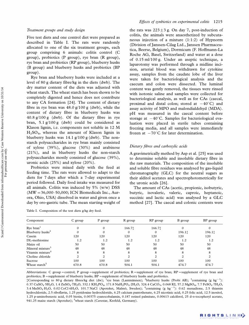
Table I. Composition of the test diets g/kg dry feed.
Component C group P group R group RP group B group BP group
Rye bran1 0 0 166.7% 166.7% 0 0
Blueberry husks2 0 0 0 0 196.1% 196.1%Casein 120 120 120 120 120 120
DL-methionine 1.2 1.2 1.2 1.2 1.2 1.2
Maize oil 50 50 50 50 50 50
Mineral mixture3 48 48 48 48 48 48
Vitamin mixture4 8 8 8 8 8 8
Choline chloride 2 2 2 2 2 2
Sucrose 100 100 100 100 100 100
Wheat starch5 670.8 670.8 504.1 504.1 474.7 474.7
Abbreviations: C group�control; P group�supplement of probiotics; R�supplement of rye bran; RP�supplement of rye bran and
probiotics; B�supplement of blueberry husks; BP�supplement of blueberry husks and probiotics.
%Corresponding to 80 g dietary fibres/kg diet (dw); 1rye bran (Lantmannen); 2blueberry husks (Probi AB); 3containing (g kg�1):
0.37 CuSO4 �5H2O, 1.4 ZnSO4 �7H2O, 332.1 KH2PO4, 171.8 NaH2PO4 �2H2O, 324.4 CaCO3, 0.068 KI, 57.2 MgSO4, 7.7 FeSO4 �7H2O,
3.4 MnSO4 �H2O, 0.02 CoCl �6H2O, 101.7 NaCl (Apoteket, Malmo, Sweden); 4containing (g kg�1): 0.62 menadione, 2.5 thiamin
hydrochloride, 2.5 riboflavin, 1.25 pyridoxine hydrochloride, 6.25 calcium pantothenate, 6.25 nicotinic acid, 0.25 folic acid, 12.5 inositol,
1.25 p-aminobenzoic acid, 0.05 biotin, 0.00375 cyanocobalamin, 0.187 retinol palmitate, 0.00613 calciferol, 25 d-a-tocopheryl acetate,
941.25 maize starch (Apoteket); 5wheat starch (Cerestar, Krefeld, Germany).
Effects of synbiotics on experimental colitis 1215
Scan
d J
Gas
troe
nter
ol D
ownl
oade
d fr
om in
form
ahea
lthca
re.c
om b
y C
ase
Wes
tern
Res
erve
Uni
vers
ity o
n 10
/31/
14Fo
r pe
rson
al u
se o
nly.

homogenized with an Ultra Turrax† T25 basic
(IKA†-Werke, Staufen, Germany) after adding in-
ternal standard (2-ethylbutyric acid; Sigma Chemi-
cal Company, St Louis, Mo., USA).
After complete derivatisation, the samples were
injected onto an HP-5 column (Hewlett Packard,
GLC, HP 6890, Wilmington, Del., USA) and Chem
Station software (Hewlett Packard) was used for the
analysis.
Assessment of the severity of colitis
The severity of colitis was assessed daily from day 0
to day 7 using a clinical Disease Activity Index (DAI)
that has been validated [28] and shown to correlate
histologically with pathological findings [29]. DAI
values ranging from 0 to 4 were calculated using:
stool consistency (normal, loose, diarrhoea), pre-
sence or absence of faecal blood (test slides with
Hemoccult II (SmithKline Diagnostic, USA) and
macroscopic evaluation of the anus) and weight loss.
The arithmetical mean of the three scores deter-
mines the DAI value. Weight changes were based on
the starting weight of each rat at initiation of
treatment. Weight-loss scores were determined as
0�no weight loss; 1��0�4.99% weight loss; 2�5.00�9.99% weight loss; 3�10.00�19.99% weight
loss; 4��20.00% weight loss. Stool scores were
determined as 0�normal stool, well-formed pellets;
2�loose stool, pasty stool that does not stick to the
anus; 4�diarrhoea, liquid stool that sticks to the
anus. Bleeding scores were determined as 0�no
bleeding; 2�positive Hemoccult test, 4�gross
bleeding.
Myeloperoxidase and malondialdehyde activity
MPO was estimated in the whole colonic tissue [30],
which contains mucosa and muscle layers. Colonic
tissues were collected, weighed prior to storage at
�708C until time of assay [31].
Colonic content of malondialdehyde (MDA) was
determined as an index of lipid peroxidation, using
MDA 586 (Oxis International Inc., Portland, Oreg.,
USA), a colorimetric assay designed to quantify
MDA [32]. Whole colonic tissues were collected,
rinsed in ice cold Dulbecco’s phosphate buffered
saline (PBS), weighed and then frozen immediately
at �708C for later evaluation.
Interleukin-12 (Rt IL-12)
Serum IL-12 production was assayed by solid-phase
sandwich ELISA. The commercially available IL-12
ELISA kit was purchased from BioSource Interna-
tional, Inc., USA, and the assay recognized both
natural and recombinant Rt IL-12 (p70), as well as
the free p40 subunit. The analysis of cytokine
protein expression was done according to the pro-
cedures described in the protocol and all samples
were analysed in duplicate.
Viable count
Liver samples (0.36190.14 g) of the caudate lobe
were placed in room temperature for 5 days to
stimulate multiplication of possible existing bacteria
translocated from the intestinal tract. Before cultur-
ing, the liver samples were placed in an ultrasonic
bath for 5 min and swirled for 2 min on Chiltern
(Therma-Glas, Gothenburg, Sweden). Viable
counts were obtained from violet-red bile glucose
(VRBG) agar (Oxoid), incubated aerobically at 378Cfor 24 h (Enterobacteriaceae count), from Rogosa agar
(Oxoid), incubated anaerobically (Gas Pack System,
Gas Pack; Becton-Dickinson Microbiology Systems,
Cockeynsville, Md., USA) at 378C for 72 h (lacto-
bacilli count), and from brain heart infusion (BHI)
agar (Difco, Detroit, Mich., USA), incubated both
aerobically and anaerobically, as described above,
at 378C for 72 h (aerobic and anaerobic count,
respectively).
Caecal tissues were placed in an ultrasonic bath
for 5 min and swirled for 1 min on Chiltern. Viable
counts were obtained from Rogosa agar (Oxoid) that
was incubated anaerobically, as described above, at
378C for 72 h (lactobacilli count) and from VRBG
(Oxoid) incubated aerobically at 378C for 24 h
(Enterobacteriaceae count). The number of colonies
formed on each plate was counted and corrected for
the weight of the original tissue.
Randomly amplified polymorphic DNA (RAPD)
Quantitatively predominant colonies were randomly
picked, and re-cultured on Rogosa agar. Crude
cell extracts were made from pure colonies, and
one microlitre was used in the polymerase chain
reaction (PCR) [33]. Agarose gel electrophoresis
was run; the gels were stained with ethidium
bromide and photographed under ultraviolet (UV)
illumination.
16S rRNA gene sequencing
Amplification of the 16S rRNA genes was carried
out using the universal primers ENV1 (5?-AGA
GTT TGA TII TGG CTC AG-3?, Escherichia coli
numbering 8 to 27) and ENV2 (5?-CGG ITA CCT
TGT TAC GAC TT-3?, E. coli numbering 1511 to
1492) [34]. The PCR mixture contained 0.2 mM of
each primer, 2 ml template DNA, 5 ml 10�PCR
buffer (100 mM Tris-HCL, 500 mM KCl, pH 8.3),
200 mM of each deoxyribonucleotide triphosphate,
1216 A. Hakansson et al.
Scan
d J
Gas
troe
nter
ol D
ownl
oade
d fr
om in
form
ahea
lthca
re.c
om b
y C
ase
Wes
tern
Res
erve
Uni
vers
ity o
n 10
/31/
14Fo
r pe
rson
al u
se o
nly.

2.5 mM MgCl2 and 2.5 U of Taq DNA polymerase
(Roche Diagnostics, Mannheim, Germany) in a final
volume of 50 ml. PCR was performed in a PCR
Mastercycle 5333 (Eppendorf) with the following
profile: 1 cycle at 948C for 3 min, followed by
30 cycles of 968C for 15 s, 508C for 30 s, and 728Cfor 90 s, with an additional extension at 728C for
10 min. The amplification products were checked by
running the products on 1.5% (w/v) agarose gel
(Type III, High EEO; Sigma) after ethidium bro-
mide staining and were visualized under UV light.
Amplicons were single-strand sequenced by MWG
(Biotech, Ebersberg, Germany) and the 16S rDNA
sequences (mostly around 500 bp) generated from
the mucosal samples were subjected to BLAST
search against GenBank [35] for approximate phy-
logenetic affiliation and aligned to 16S rDNA
encoding sequences of selected Lactobacillus strains
(type strains) retrieved from the Ribosomal Data
Base (RDP-II) [36].
Statistics
DAI scores, MPO activity, lipid peroxidation, Rt
IL-12 and lactobacilli and Enterobacteriaceae counts
(Figures 1�6 and Table IV) were presented as
medians with 25 and 75 percentiles. The statistics
were conducted in SigmaStat† version 3.0 (SPSS
Inc., Chicago, Ill., USA). Differences between
all groups were evaluated by Kruskal-Wallis
test one-way ANOVA on ranks followed by all
pairwise multiple comparison procedures (Student-
Newman-Keuls method), if appropriate. When com-
paring only two groups, the Mann-Whitney rank
sum test was used. The correlation between expecta-
tions of benefit was ascertained using Pearson’s
correlation coefficient. Calculation of the incidence
of translocation was conducted in QuickStat version
2.6 and was evaluated by the Fisher exact test.
Caecal pools of CAs were calculated as the
concentrations of each acid (mmol/g) multiplied by
the caecal amount. The values were extrapolated to a
complete intake of dietary fibre during the experi-
mental period. The proportion of individual CAs
was calculated as a percentage of total CAs for each
rat before statistical evaluation. Feed intake, body-
weight change, caecal content, caecal tissue weight,
caecal pH and CAs (Tables II and III) were
presented as means9SEM (standard error of
the mean). Data from rats given diets containing
probiotics were compared with the control or the
corresponding fibre diets. Rats given rye bran or
blueberry husks, without any probiotics, were com-
pared with those given the control diet. For statis-
tical evaluation of the differences between samples,
two-way ANOVA was used to determine the effects
of dietary fibre (Fibre), probiotics (Pro) and their
interactions (Fibre�Pro). The probiotic and the
dietary fibre effects were then evaluated by using
one-way ANOVA followed by Tukey’s procedure.
The Minitab statistical software (Release 14) was
used to make these evaluations. P-values of less than
0.05 were considered significant.
Results
Colitis severity
The products were well tolerated by all animals, with
no adverse side effects during the period of intake.
There was no mortality among the experimental
groups.
DAI score increased with each treatment day and
differences between groups reached statistical sig-
nificance on day 3 of the DSS regimen ( pB0.05)
(Figure 1). By days 6 and 7, the DAI score was
significantly lower in all treatment groups compared
with the colitis control group (C group) ( pB0.01).
On the seventh day, rats in the C group had
diarrhoea, weight loss, gross rectal bleeding, erosion
and/or superficial ulcer of the colonic mucosa in the
distal part.
The rats consumed approximately half of the feed
provided during the experimental period (72�105 g)
(Table II) and the body-weights were corrected for
the feed intake. Compared with the C group, the
body-weight change was positive in all treatment
groups ( pB0.05), with the exception of the R
group.
The caecal tissue weights were significantly higher
in the C group than in the R and B groups ( p�0.01
and p�0.03, respectively; Table II).
Carboxyolic acids in the hindgut
The pH of the caecal content was significantly
lower in groups R and B compared with the C group
( pB0.001; Table II).
The caecal pools and levels of CAs were sig-
nificantly higher in the B group compared with the
C group ( p�0.002 and p�0.03, respectively),
and also for the pools compared with the R group
( p�0.013; Table III). The higher caecal amount of
CAs was due to acetic acid and butyric acid. The R
group exhibited significantly higher CA levels than
the C group in the distal part of colon ( p�0.04;
Table III).
In general, the proportion of acetic acid was
significantly higher throughout the hindgut in the
B group than in the R group ( p�0.026). In
the caecum, the proportion of butyric acid was
significantly higher in the R group than in the C
group, while the proportion of propionic acid and
Effects of synbiotics on experimental colitis 1217
Scan
d J
Gas
troe
nter
ol D
ownl
oade
d fr
om in
form
ahea
lthca
re.c
om b
y C
ase
Wes
tern
Res
erve
Uni
vers
ity o
n 10
/31/
14Fo
r pe
rson
al u
se o
nly.

minor acids was significantly lower in the B group
compared with the C group ( p�0.02 and p�0.005,
respectively). In the distal part of colon, the propor-
tion of lactic acid was significantly lower in the R
group and B group compared with the C group ( p�0.027 and p�0.026, respectively; Table III).
Concerning the effects of probiotics, the caecal
pool of CAs was significantly higher in the RP group
than in the R group ( p�0.002), whereas the level in
the distal part of colon was lower ( pB0.05; Table
III). The caecal pool of CAs was significantly lower
in the P group than in the C group ( pB0.05),
Figure 1. Effects of 5% dextran sulphate sodium (DSS) on the time-course of changes in the disease activity index (DAI) over the 7-day
experimental period. DAI scores are expressed as medians (25 and 75 percentiles). The asterisk (*) denotes pB0.05 and asterisks (**)
denote pB0.01 compared with colitis control. Day 3: *Control (C) versus treatment with rye bran (R), blueberry husks (B) or blueberry
husks and bacteria mixture (BP). Day 4: *Control versus bacteria mixture (P), **control versus rye bran, rye bran and bacteria mixture
(RP), blueberry or blueberry husks and bacteria mixture. Days 5 and 6: The same as day 4. Day 7: **Control versus all treatment groups.
Figure 2. Effects of dextran sulphate sodium on colon mucosal
myeloperoxidase activity. Data are expressed as medians (25 and
75 percentiles). The asterisk (*) denotes pB0.05 compared with
all groups; the hash (#) denotes pB0.01 between the rye bran
group (R) and the rye bran and bacteria mixture group (RP).
Figure 3. Effects on colon mucosal lipid peroxidation by induc-
tion of colitis by dextran sulphate sodium. Data are expressed as
medians (25 and 75 percentiles). The asterisk (*) denotes pB0.05
and asterisks (**) denote pB0.01 compared with the colitis
control group (C).
1218 A. Hakansson et al.
Scan
d J
Gas
troe
nter
ol D
ownl
oade
d fr
om in
form
ahea
lthca
re.c
om b
y C
ase
Wes
tern
Res
erve
Uni
vers
ity o
n 10
/31/
14Fo
r pe
rson
al u
se o
nly.

whereas the level in the distal part of colon was
higher, resulting in a significantly increased propor-
tion of butyric acid ( pB0.05; Table III).
Inflammatory markers
As assessed by elevated MPO activity in the colonic
tissue, administration of DSS for 7 days caused
significantly lower levels of neutrophil infiltration
in the BP group compared with all other groups
( pB0.05), while MPO in the RP group was lower
than that in the R group ( pB0.05; Figure 2).
Compared with the C group, MDA levels were
significantly lower in all treatment groups ( pB0.01),
with the exception of the P group (Figure 3).
Serum IL-12 was significantly lower in the B
group, and the BP group, as compared with the C
group ( pB0.05; Figure 4).
Translocation to liver
With the exception of the R group, the incidence of
translocation to the caudate lobe of the liver,
measured as incidence of positive plate counts,
decreased significantly in all treatment groups com-
pared with the C group ( pB0.01; Table IV).
Lactobacilli and Enterobacteriaceae counts in caecal
tissue
The viable count of lactobacilli was significantly
higher in the R group, compared with the C group
( pB0.05; Figure 5). When comparing the groups
receiving the probiotic mixture, the viable count was
significantly higher in the RP group than in the P
group and the BP group, and the count of the BP
group was significantly higher than the B group
( pB0.05; Figure 5). The caecal counts of Enter-
obacteriaceae significantly decreased in all treatment
groups compared with the C group ( pB0.05; Figure
6). A significant decrease was also found between the
R group and RP group, and between the R group
and B group. The Enterobacteriaceae count of the BP
Figure 4. Interleukin-12 concentrations in serum are expressed as
medians (25 and 75 percentiles). Asterisks (*) denote pB0.05
compared with the colitis control group (C) and with the groups
treated with bacteria mixture (P), rye bran (R), and rye bran and
bacteria mixture in combination (RP).
Figure 5. Total lactobacilli counts are expressed as medians (25
and 75 percentiles). The hash (#) denotes pB0.05 between the
rye bran group (R) and the control (C) and blueberry husk group
(B), and also the rye bran and bacteria group (RP) compared with
the bacteria group (P) and the blueberry husks and bacteria group
(BP). The asterisk (*) denotes pB0.05 between the blueberry
husk group and the blueberry husks and bacteria group.
Figure 6. Total Enterobacteriaceae counts are expressed as medians
(25 and 75 percentiles). The asterisk (*) denotes pB0.05 between
the control group (C) and all the other test groups. The hash
(#) denotes pB0.05 when comparing the rye bran group (R)
versus rye bran and bacteria group (RP), the rye bran
group versus blueberry husks group (B) and the blueberry husks
group versus blueberry husks and bacteria mixture group (BP).
Effects of synbiotics on experimental colitis 1219
Scan
d J
Gas
troe
nter
ol D
ownl
oade
d fr
om in
form
ahea
lthca
re.c
om b
y C
ase
Wes
tern
Res
erve
Uni
vers
ity o
n 10
/31/
14Fo
r pe
rson
al u
se o
nly.

Table II. Body-weight change, caecal content and caecal pH in rats fed a fibre-free diet (C group) supplemented with probiotics (P group),
rye bran (R group), rye bran and probiotics (RP group), blueberry husks (B group) or blueberry husks and probiotics (BP group)1,2.
p-value
C group P group R group RP group B group BP group Fibre Pro Fibre�Pro
Feed intake, g/d 10.391.1a 12.991.0 13.491.1a,b 13.090.9 15.090.4b 13.590.5* 0.016 NS NS
Body-weight
change, g/g
feed3
�0.2890.15a 0.0390.05* �0.0090.04a 0.1390.03* 0.1190.02b 0.0490.02 0.003 0.014 0.005
Caecal content, g 1.690.5 1.190.3 1.690.2 2.190.4 2.690.3 2.290.4 0.028 NS NS
Caecal tissue
weight, g
1.490.1a 1.290.1 1.090.1b 1.290.1 1.090.1b 1.190.1 0.018 NS NS
Caecal pH 7.290.1a 7.390.1 6.690.1b 6.590.2 6.590.1b 6.590.1 B0.001 NS NS
1Values are expressed as means9SEM, n�6. Asterisks indicate statistically significant difference from rats fed the same diet but without
bacteria: *pB0.05, **pB0.01, ***pB0.001; 2between control, rye bran and blueberry husks, i.e. those without any added probiotics,
without a common letter differ, pB0.05; 3body-weight change during the experimental period divided by the total feed intake during the
experimental period.
Table III. Carboxylic acids in the hindgut of rats fed a fibre-free diet (C group) supplemented with probiotics (P group), rye bran (R group),
rye bran and probiotics (RP group), blueberry husks (B group) or blueberry husks and probiotics (BP group)1,2.
p-value
C group P group R group RP group B group BP group Fibre Pro Fibre�Pro
Caecum3
Acetic 5193a 5892 6091b 5991 6892c 6192* B0.001 NS 0.004
Propionic 1491a 1291 1191a 1290 1091b 1391* NS NS 0.026
Butyric 791a 991 1191b 1292 1091a,b 1191 0.031 NS NS
Lactic 1094 992 891 691 591 591 NS NS NS
Succinic 694 290 391 391 391 391 NS NS NS
Minor4 1291a 1091 992a 891 591b 790 B0.001 NS NS
Total
mmol/g 4498a 5993 6896a,b 7994 7597b 8898 B0.001 0.015 NS
mmol 10299a 59910* 135931a 344936** 223924b 299971 B0.001 NS B0.001
Proximal5
Acetic 3695a 5392** 5394a 5592 6693b 6392 B0.001 B0.05 B0.01
Propionic 591 991* 691 892 692 891 NS B0.05 NS
Butyric 391 791** 692 791 591 791 NS B0.05 NS
Lactic 3494a 1693** 2197a 1692 1191b 1092 B0.001 B0.01 NS
Succinic 591 891** 894 591 391 591 NS NS NS
Minor4 1794 891* 891 991 991 491* B0.01 B0.05 NS
Total
mmol/g 7492a 5194 5796b 6696 4294b 4694 B0.001 NS B0.01
Distal6
Acetic 4795a 5092 5693a,b 5193 6292b 5993 0.002 NS NS
Propionic 991 891 1091 991 791 791 NS NS NS
Butyric 591 890* 891 991 792 691 NS NS NS
Lactic 1895a 1692 992b 1292 891b 1093 0.004 NS NS
Succinic 893 692 1294 1192 491 793 NS NS NS
Minor4 1393a 1294 391b 991** 1291a 593* 0.027 NS 0.043
Total
mmol/g 6296a 102911** 98913b 5895* 6596a,b 7897 NS NS B0.001
1Values are expressed as the means of levels (mmol/g) or pools (mmol/caecum) and standard errors or percentages of the total CA, n�6.
Asterisks indicate statistically significant difference from rats fed diets without probiotics: *pB0.05, **pB0.01, ***pB0.001; 2values of
control, rye bran and blueberry husks, i.e. those without any added probiotics, without a common letter are statistically different, pB0.05;3rats fed the fibre-free diet, rye bran diet, blueberry husks diet or the blueberry husks and probiotics diet; n�5; 4formic, isobutyric,
isovaleric, valeric, caproic and heptanoic acid; 5rats fed the rye bran diet; n�3; 6rats fed the rye bran and probiotics diet; n�5.
1220 A. Hakansson et al.
Scan
d J
Gas
troe
nter
ol D
ownl
oade
d fr
om in
form
ahea
lthca
re.c
om b
y C
ase
Wes
tern
Res
erve
Uni
vers
ity o
n 10
/31/
14Fo
r pe
rson
al u
se o
nly.

group was significantly higher than that of the B
group ( pB0.05).
Identification of caecal lactobacilli
A total of 108 isolates from caecum tissue, picked
from countable Rogosa agar plates, and representing
18 isolates per group, were subjected to RAPD
typing, and the RAPD types dominating each group,
based on recurrent band pattern, were identified to
species level. L. reuteri represented the majority of
isolates from the C group, and in 4 out of 6 groups
L. reuteri and L. animalis constitute a dominant part
of the lactobacilli flora of the caecum tissue (group
C, 72% L. reuteri and 22% L. animalis; group R,
67% L. reuteri and 17% L. animalis; group RP, 67%
L. reuteri and 22% L. animalis; group BP, 69%
L. reuteri and 23% L. animalis). The composition of
the lactobacilli flora was different in group P where
28% of the isolates were L. gasseri (67% L. reuteri), and
in group B it was dominated by L. animalis (91%).
Correlations
A linear relationship was observed between the DAI
at day 7 and Enterobacteriaceae counts (r�0.54;
pB0.001). There was also a positive correlation
between the DAI score and MPO activity (r�0.39;
pB0.05) and between body-weight change during
the last 7 days and the total level of CAs in the
caecum (r�0.61; pB0.001). The DAI at day 7
correlated negatively to the total caecal concentra-
tion of CAs (r��0.55; p�0.001), acetic acid (r��0.63; pB0.001) and butyric acid (r��0.54; p�0.001). A negative correlation was found between
the MDA and the acetic acid (r��0.40, p�0.02)
and the butyric acid (r��0.41, p�0.01) concen-
trations in the distal part of the colon.
Discussion
In the present study, we have shown that probiotics,
rye bran and blueberry husks together or alone were
able to attenuate the inflammatory response of DSS.
During the course of the experiment, treated rats
had an attenuated inflammatory response, evidenced
by lower DAI values from day 3 to 7. The original
scoring of DAI by Cooper et al. [29] was used, but
the limits of body-weight change have been more
precisely defined in the present study. With the
modified scoring limits the judgement between
scores 1 and 2, and 2 and 3, may be objectively
facilitated.
Body-weight loss is clinically relevant and is
included in the DAI. Patients with IBD are exposed
to nutritional risk since malnutrition and loss of
body-weight are predominant clinical features [37],
and except for poor nutritional intake and malab-
sorption, the inflammatory reactions affect the
catabolism [38]. By supplementation of probiotics,
rye bran or blueberry husks, it was feasible to
prevent the body-weight loss of the animals during
progression of colitis.
Viable count of the caecal tissue verified an
increase in lactobacilli in the R, RP and BP groups.
Lactobacilli are regarded as beneficial and a decrease
in numbers has been found in the mucosa of patients
with UC [39]. It has been suggested that the
differential physiology of the intestinal mucosa
caused by inflammation may influence the composi-
tion of lactobacilli [40]. In contrast, all treatments
decreased the Enterobacteriaceae counts as compared
with the C group, although the B group showed the
strongest reduction. Blueberry husks have previously
been shown to have an antimicrobial effect, sug-
gested to be mediated by the polyphenols [41], and a
positive correlation was observed in the present
study between the Enterobacteriaceae count and
DAI. The Gram-negative, sometimes pathogenic or
opportunistically pathogenic Enterobacteriaceae are
increasingly linked with IBD [42] and experimental
colitis [43]. This family includes species such as
E. coli and Klebsiella pneumonia, but all members of
the Enterobacteriaceae family have lipopolysaccharides
(LPS) associated with the cell wall. E. coli strains
associated with IBD and with colitis in dogs [44]
share the ability to replicate in cultured epithelial cells
and macrophages and belong to a group of adherent
and invasive E. coli. Cats with signs of intestinal
inflammation had an increased mucosal Enterobacter-
iaceae load [45]. It has been shown that a subset
of bacteria comprising E. coli, other species of
Enterobacteriaceae and Clostridium correlated with
abnormalities in mucosal architecture, up-regulation
Table IV. Incidence of translocation to the caudate lobe of the
liver.
VRBG BHI anaerobic BHI aerobic Rogosa
C group 5/6 5/6 5/6 2/6
P group 0/6** 0/6** 0/6** 0/6**
R group 2/6 2/6 2/6 2/6
RP group 0/6** 0/6** 0/6** 0/6**
B group 0/6** 0/6** 0/6** 0/6**
BP group 0/6** 0/6** 0/6** 0/6**
Abbreviations: C group�control; P group�supplement of pro-
biotics; R�supplement of rye bran; RP�supplement of rye bran
and probiotics; B�supplement of blueberry husks; BP�supple-
ment of blueberry husks and probiotics; VRBG�violet-red bile
glucose; BHI�brain heart infusion.
**Denotes pB0.01 compared with the control group.
Effects of synbiotics on experimental colitis 1221
Scan
d J
Gas
troe
nter
ol D
ownl
oade
d fr
om in
form
ahea
lthca
re.c
om b
y C
ase
Wes
tern
Res
erve
Uni
vers
ity o
n 10
/31/
14Fo
r pe
rson
al u
se o
nly.

of pro-inflammatory cytokines (IL-12, IL-8 and IL-
1) and the clinical disease activity [45].
In the present study, IL-12 in plasma was mea-
sured as an inflammatory mediator with systemic
effects, and a decrease in IL-12 was found in the
groups given blueberry husks in the diet. The
antimicrobial effects from polyphenols in blueberry
may suppress pro-inflammatory components of the
intestinal microbiota, and hence reduce the stimula-
tion of macrophages, resulting in a subsequent less-
pronounced induction of IL-12. The role of different
cytokines in the hierarchy of inflammatory mediators
orchestrating the inflammatory process in IBD is not
fully elucidated. In contrast, the importance of IL-12
in active IBD has been shown experimentally by
demonstrating that the IL-12 cytokine profile in
colonic tissue is up-regulated due to DSS treatment
[46] and that antibodies to IL-12 can abrogate
experimental colitis [47]. Further, clinical data
show that gene transcription of IL-12 is up-regulated
in active UC [48]. No significant effects of probiotics
on the IL-12 levels could be recorded in the present
study, even if Lactobacillus, in a strain-specific
manner, can modulate a host immune response by
altering the profiles of cytokines released in the gut
by epithelial and immune cells [49], and Lactobacillus
has been shown to modify and potentiate the
macrophage activity and response to stimulants
[50]. A proposed mechanism of DSS action includes
increased macrophage activation [51]. It is impor-
tant to point out that as well as the possible effects of
the Lactobacillus strains in the probiotic mixture, the
resident Lactobacillus flora might exercise effects.
L. murinus and L. reuteri usually comprise a con-
siderable part of the intestinal Lactobacillus popula-
tion of rats [52,53]. With the exception of two of the
treatment groups, this is consistent with results from
the present study. However, in the P group, the
dominating species were L. gasseri and L. reuteri, and
L. animalis was the major component of the dom-
inating lactobacilli of the B group. Bacteria with
tannase activity from faeces of mice have been
identified as L. animalis or L. murinus (i.e. L.
animalis is relatively closely related to L. murinus)
[54]. This might suggest that the fibre-free diet did
not provide optimal survival and persistence condi-
tions for L. animalis, while blueberry husks improved
growth performance and competing ability.
In the present study bacterial translocation to the
caudate lobe of the liver was significantly inhibited in
all groups receiving probiotics and also blueberry
husks alone. It has previously been shown that
Gram-negative bacteria can be found in portal blood
and liver biopsies from patients with UC undergoing
colectomy [55], and circulating and agglutinating
antibodies against various enteric bacteria have been
identified in active UC [56]. The indigenous flora
and an intact mucosa are vital components of body
defences against luminal pathogenic bacteria, and a
part of the therapeutic strategy for UC aims at
restoring the intestinal integrity and function, as
increased intestinal permeability might facilitate the
translocation of bacteria from the gut and give rise to
potential infection and inflammation [57].
A positive correlation between DAI values and
MPO was found in the present study and the
mucosal activity of MPO was significantly decreased
in the BP group. Up-regulation of MPO has been
described in DSS-induced colitis [58] and DAI has
been shown to be correlated with MPO [29]. MPO
is a potent pro-oxidative enzyme, and is one of the
chief mediators of neutrophil-dependent tissue da-
mage [59]. There is a direct relationship between
MPO activity and the number of neutrophils in
tissue [60]. A positive correlation between the
amounts of macrophages and neutrophils in colonic
tissue of patients suffering from UC has been found,
suggesting an active role of macrophages in neutro-
phil recruitment in UC [5].
MDA, which is an indicator of the degree of lipid
peroxidation induced by reactive oxygen species [61],
was decreased in the colonic tissue of groups given
blueberry husks or rye bran. Oxidants produced by
activated neutrophils are involved in the initiation and
perpetuation of the inflammatory process by increas-
ing the number of neutrophils and macrophages that
induce a self-sustaining loop [62].
Lipid peroxidation is mediated by oxygen free
radicals and is believed to be an important factor for
the damage of cell membranes [63]. The protective
effect of the dietary supplements may be due to the
scavenging effects of the polyphenols, in rye bran
and blueberry husks, to the oxidative damage caused
by ROS at the induction of colitis. Serum MDA
levels increase in patients with UC, which suggests
that lipid peroxidation could have an important role
in the pathogenesis of UC [64].
In the present study, the distal colonic concentra-
tion of acetic and butyric acids correlated negatively
to MDA in colonic tissue, and rye bran gave
comparatively higher levels of CAs in the distal
colon than the control diet. Since UC appears
distally, dietary fibres that release their fermentation
products in this part of the colon may offer an
advantage. A slow fermentation of rye has been
obtained by others [65] and may be caused by
arabinoxylan, the main fibre component in rye. On
the one hand, in the present study the caecal levels of
CAs increased, whereas the level in the distal colon
decreased with the addition of probiotics to the rye
bran diet. On the other hand, the damage induced
by DSS has been reported to affect the distal colon
1222 A. Hakansson et al.
Scan
d J
Gas
troe
nter
ol D
ownl
oade
d fr
om in
form
ahea
lthca
re.c
om b
y C
ase
Wes
tern
Res
erve
Uni
vers
ity o
n 10
/31/
14Fo
r pe
rson
al u
se o
nly.
and caecum preferentially [66], with less damage
evident in the proximal colon [66] and in the ileum
[67]. Therefore, an increased production in the
caecum would be favourable in this model. With
this in mind, it is interesting to observe that the RP
group yielded the highest amounts of butyric acid.
The B group had a higher proportion of acetic
acid in the caecum compared with the C group or
the R group, which can be explained by the
comparatively high content of uronic acids in blue-
berry husks. Interestingly, the proportion of acetic
acid decreased, whereas that of propionic acid
increased when probiotics were added. This may
be a consequence of a change in the composition of
the microbiota, in this case indicated by the higher
count of lactobacilli in the caecum in the groups
supplemented with probiotics. The phenomenon
can possibly be of some clinical value since propionic
acid is decreased in patients with IBD [68], and both
propionate and butyrate have been shown to de-
crease cytokine release from human neutrophils to a
higher extent than acetate [69]. Indeed, a negative
correlation was found between DAI and several
specific CAs, including butyric acid. This is in line
with the observation that severe colitis is character-
ized by low CA levels and impaired utilization of
butyric acid [70,71].
Several studies have proposed that dietary manip-
ulations can be beneficial for the management of
intestinal inflammatory conditions like UC. This is
interesting in view of the fact that most pharmaceu-
ticals currently available, although effective, are not
devoid of potentially serious side effects, limiting
their chronic use [72]. The present study demon-
strates that administration of especially blueberry
husks supplemented with a mixture of probiotic
strains attenuates DSS-induced colonic injury and
inflammation in rats.
Acknowledgements
This study was funded by the Functional Food
Science Centre (FFSC) at Lund University. We
thank Lantmannen (Jarna, Sweden) and Probi AB
(Lund, Sweden) for kindly supplying the rye bran,
the freeze-dried blueberry husks and probiotics.
None of the authors has any conflicts of interest to
declare, but Siv Ahrne, Goran Molin and Bengt
Jeppsson are minority stockholders in Probi AB. All
of the co-writers, took part in the design of the study,
the evaluation of the results and the writing of the
manuscript. Asa Hakansson and Camilla Branning
contributed equally to the work, performed on the
animal experiments and the experimental studies.
References
[1] Elson CO, Cong Y, McCracken VJ, Dimmitt RA, Lorenz
RG, Weaver CT. Experimental models of inflammatory
bowel disease reveal innate, adaptive, and regulatory me-
chanisms of host dialogue with the microbiota. Immunol Rev
2005;/206:/260�76.
[2] Sartor RB. Mechanisms of disease: pathogenesis of Crohn’s
disease and ulcerative colitis. Nat Clin Pract Gastroenterol
Hepatol 2006;/3:/390�407.
[3] Hanauer SB. Inflammatory bowel disease: epidemiology,
pathogenesis, and therapeutic opportunities. Inflamm Bowel
Dis 2006;/12:/3�9.
[4] Reifen R, Nissenkorn A, Matas Z, Bujanover Y. 5-ASA and
lycopene decrease the oxidative stress and inflammation
induced by iron in rats with colitis. J Gastroenterol 2004;/
39:/514�9.
[5] Kayo S, Ikura Y, Suekane T, Shirai N, Sugama Y, Ohsawa
M, et al. Close association between activated platelets and
neutrophils in the active phase of ulcerative colitis in
humans. Inflamm Bowel Dis 2006;/12:/727�35.
[6] Simmonds NJ, Rampton DS. Inflammatory bowel disease: a
radical view. Gut 1993;/34:/865�8.
[7] Yoshikawa T, Yamaguchi T, Yoshida N, Yamamoto H,
Kitazumi S, Takahashi S, et al. Effect of Z-103 on TNB-
induced colitis in rats. Digestion 1997;/58:/464�8.
[8] Ahnfelt-Ronne I, Nielsen OH, Christensen A, Langholz E,
Binder V, Riis P. Clinical evidence supporting the radical
scavenger mechanism of 5-aminosalicylic acid. Gastroenter-
ology 1990;/98:/1162�9.
[9] Buffinton GD, Doe WF. Depleted mucosal antioxidant
defences in inflammatory bowel disease. Free Radical Biol
Med 1995;/19:/911�8.
[10] Kubes P, McCafferty DM. Nitric oxide and intestinal
inflammation. Am J Med 2000;/109:/150�8.
[11] Permodo JJ, Cavaillon J-M, Huerre M, Ohayon H, Sanso-
netti PJ. Acute inflammation causes epithelial invasion and
mucosal destruction in experimental Shigellosis. J Exp Med
2001;/180:/1307�19.
[12] Stadnyk AW, Dollard CD, Issekutz AC. Neutrophil migra-
tion stimulates epithelial cell cytokines during T. spiralis
infection of the rat. J Leukoc Biol 2000;/68:/821�7.
[13] Olofsson T, Olsson I, Venge P, Elgefors B. Serum myeloper-
oxidase and lactoferrin in neutropenia. Scand J Haematol
1977;/18:/73�80.
[14] Hooper LV, Wong MH, Thelin A, Hansson L, Falk PG,
Gordon JI. Molecular analysis of commensal host-microbial
relationships in the intestine. Science 2001;/291:/881�4.
[15] Isolauri E, Kirjavainen PV, Salminen S. Probiotics: a role in
the treatment of intestinal infection and inflammation. Gut
2002;/50:/54�9.
[16] Russell WR, Labat A, Scobbie L, Duncan S. Availability of
blueberry phenolics for microbial metabolism in the colon
and the potential inflammatory implications. Mol Nutr Food
Res 2007;/51:/726�31.
[17] Andreasen MF, Landbo A-K, Christensen LP, Hansen A,
Meyer AS. Antioxidant effects of phenolic rye (Secale cereale
L.). Extracts, monomeric hydroxycinnamates, and ferulic
acid dehydrodimers on human low-density lipoproteins. J
Agric Food Chem 2001;/49:/4090�6.
[18] Medina V, Afonso JJ, Alvarez-Arguelles H, Hernandez C,
Gonzalez F. Sodium butyrate inhibits carcinoma develop-
ment in a 1,2-dimethylhydrazine-induced rat colon cancer.
JPEN J Parenter Enteral Nutr 1998;22:14�7.
[19] Volker M. Dietary modification of the intestinal microbiota.
Nutr Rev 2004;/62:/235�42.
Effects of synbiotics on experimental colitis 1223
Scan
d J
Gas
troe
nter
ol D
ownl
oade
d fr
om in
form
ahea
lthca
re.c
om b
y C
ase
Wes
tern
Res
erve
Uni
vers
ity o
n 10
/31/
14Fo
r pe
rson
al u
se o
nly.

[20] Lee HC, Jenner AM, Low CS, Lee YK. Effect of tea
phenolics and their aromatic fecal bacterial metabolites on
intestinal microbiota. Res Microbiol 2006;/157:/876�84.
[21] Heinonen M. Antioxidant activity and antimicrobial effect of
berry phenolics: a Finnish perspective. Mol Nutr Food Res
2007;/51:/684�91.
[22] Kimura I, Nagahama S, Kawasaki M, Kataoka M, Sato M.
Study on the experimental ulcerative colitis (UC) model
induced by dextran sulfate sodium (DSS) in rats (3). Folia
Pharmacol Jpn 1996;/108:/259�66.
[23] Efthymiou C, Hansen CA. An antigenic analysis of Lacto-
bacillus acidophilus. J Infect Dis 1962;/110:/258�67.
[24] Bjorck I, Nyman M, Pedersen B, Siljestrom M, Asp N-G,
Eggum B. Formation of enzyme resistant starch during
autoclaving of wheat starch: studies in vitro and in vivo. J
Cereal Sci 1987;/6:/159�72.
[25] Asp N-G, Johansson C-G, Hallmer H, Siljestrom M. Rapid
enzymatic assay of insoluble and soluble dietary fiber. J Agric
Food Chem 1983;/31:/476�82.
[26] Theander O, .Aman P, Westerlund E, Andersson R, Petters-
son D. Total dietary fiber determined as neutral sugar
residues, uronic acid residues, and Klason lignin (the
Uppsala method): collaborative study. J AOAC Int 1995;/
78:/1030�44.
[27] Richardson A, Calder A, Stewart C, Smith A. Simultaneous
determination of volatile and non-volatile acidic fermenta-
tion products of anaerobes by capillary gas chromatography.
Lett Applied Microbiol 1989;/9:/5�8.
[28] Murthy S, Murthy NS, Coppola D, Wood DL. The
efficiency of BAY y 1015 in dextran sulfate model of mouse
colitis. Inflamm Res 1997;/46:/224�33.
[29] Cooper HS, Murthy SN, Shah RS, Sedergran DJ. Clinico-
pathologic study of dextran sulphates sodium experimental
murine colitis. Lab Invest 1993;/69:/238�49.
[30] Khan I, Al-Awadi FM. Colonic muscle enhances the
production of interleukin-1( messenger RNA in experimen-
tal colitis. Gut 1997;/40:/307�12.
[31] Osman N, Adawi D, Ahrne S, Jeppsson B, Molin G.
Probiotic strains of Lactobacillus and Bifidobacerium affect
the translocation and intestinal load of Enterobacteriaceae
differently after D-galactosamine-induced liver injury. Mi-
crobiol Ecol Health Dis 2005;/17:/40�6.
[32] Hakansson A, Stene C, Mihaescu A, Molin G, Ahrne S,
Thorlacius H, et al. Rosehip and Lactobacillus plantarum
DSM 9843 reduce ischemia/reperfusion injury in the mouse
colon. Dig Dis Sci 2006;/51:/2094�101.
[33] Quednau M, Ahrne S, Peterson A, Molin G. Identification
of clinically important Species of Enterococcus within 1 day
with randomly amplified polymorphic DNA (RAPD). Curr
Microbiol 1998;/36:/332�6.
[34] Brosius J, Palmer ML, Kennedy PJ, Noller HF. Complete
nucleotide sequence of a 16S ribosomal RNA gene from
Escherichia coli. Proc Natl Acad Sci USA 1978;/75:/4801�5.
[35] Altschul SF, Gish W, Miller W, Myers EW, Lipman DJ.
Basic local alignment search tool. J Mol Biol 1990;/215:/
403�10.
[36] Cole JR, Chai B, Marsh TL, Farris RJ, Wang Q, Kulam SA,
et al. The Ribosomal Database Project (RDP-II): previewing
and a new autoaligner that allows regular updates and the
new prokaryotic taxonomy. Nucleic Acids Res 2003;/31:/
442�3.
[37] Silk DB, Payne-James J. Inflammatory bowel disease: nutri-
tional implications and treatment. Proc Nutr Soc 1989;/48:/
355�61.
[38] Bistrian BR. Role of the systemic inflammatory response in
the development of protein-energy malnutrition in inflam-
matory bowel disease. Nestle Nutr. Workshop Ser Clin
Perform Programme. 1999;2:1�6.
[39] Ott SJ, Musfeldt M, Wenderoth DF, Hampe J, Brant O,
Folisch UR, et al. Reduction in diversity of the colonic
mucosa associated bacterial microflora in patients with active
inflammatory bowel disease. Gut 2004;/53:/685�93.
[40] Zhang M, Liu B, Zhang Y, Wei H, Lei Y, Zhao L. Structural
shifts of mucosa-associated lactobacilli and Clostridium
leptum subgroups in patients with ulcerative colitis. J Clin
Microbiol 2007;/45:/496�500.
[41] Branning C, Hakansson A, Ahrne S, Jeppsson B, Molin G,
Nyman M. Blueberry husks and multi-strain probiotics
affect colonic fermentation and weight gain in healthy rats.
Br J Nutr 2009;/101:/859�70.
[42] Barnich N, Darfeuille-Michau A. Adherent-invasive Escher-
ichia coli and Crohn’s disease. Curr Opin Gastroenterol
2007;/23:/16�20.
[43] Lupp C, Robertson ML, Wickham ME, Sekirov I, Cham-
pion OL, Gaynor EC, et al. Host-mediated inflammation
disrupts the intestinal microbiota and promotes the over-
growth of Enterobacteriaceae. Cell Host Microbe 2007;/2:/
119�29.
[44] Simpson KW, Dogan B, Rishniw M, Goldstein RE, Klaessig
S, McDonough PL, et al. Adherent and invasive Escherichia
coli is associated with granulomatous colitis in boxer dogs.
Infect Immun 2006;/74:/4778�92.
[45] Janeczko S, Atwater D, Bogel E, Greiter-Wilke A, Gerold A,
Baumgart M, et al. The relationship of mucosal bacteria to
duodenal histopathology, cytokine mRNA, and clinical
disease activity in cats with inflammatory bowel disease.
Vet Microbiol 2008;/128:/178�93.
[46] Egger B, Bajaj-Elliot M, McDonald TT, Inglin R, Eysselein
VE, Buhler MW. Characterisation of acute murine dextran
sodium sulphate colitis: cytokine profile and dose depen-
dency. Digestion 2000;/62:/240�8.
[47] Neurath MF, Fuss I, Kelsall BL, Stuber E, Strober W.
Antibodies to interleukin 12 abrogate established experi-
mental colitis in mice. J Exp Med 1995;/182:/1281�90.
[48] Nielsen OH, Kirman I, Rudiger N, Hendel J, Vainer B. Up-
regulation of interleukin-12 and -17 in active inflammatory
bowel disease. Scand J Gastroenterol 2003;/2:/180�5.
[49] Pena JA, Li SY, Wilson PH, Thibodeau SA, Szary AJ,
Versalovic J. Genotypic and phenotypic studies of intestinal
lactobacilli: species differences in mice with and without
colitis. Appl Environ Microbiol 2004;/70:/558�68.
[50] Perdigon G, DeMacias MEN, Alvarez S, Oliver G, DeRuiz
Holgado AAP. Effect of perorally administered lactobacilli
on macrophage activation in mice. Infect Immun 1986;/53:/
404�10.
[51] Vowinkel T, Kalogeris TJ, Mori M, Krieglstein CF, Granger
DN. Impact of dextran sulfate sodium load on the severity of
inflammation in experimental colitis. Dig Dis Sci 2004;/49:/
556�64.
[52] Hemme D, Raibaud P, Ducluzeau R, Galpin JV, Sicard P,
Van Heijenoort J. Lactobacillus murinus n. sp., a new species
of the autochthonous dominant flora of the digestive tract of
rat and mouse. Ann Microbiol 1980;/131:/297�308.
[53] Molin G, Johansson M-L, Stahl M, Ahrne S, Andersson R,
Jeppsson B, et al. Systematics of the Lactobacillus population
on rat intestinal mucosa with special reference to Lactoba-
cillus reuteri. Antonie van Leeuwenhoek 1992;/61:/175�83.
[54] Sasaki E, Shimada T, Osawa R, Nishitani Y, Spring S, Lang
E. Isolation of tannin-degrading bacteria isolated from feces
of the Japanese large wood mouse, Apodemus speciosus,
feeding on tannin-rich acorns. Syst Appl Microbiol 2005;/
28:/358�65.
1224 A. Hakansson et al.
Scan
d J
Gas
troe
nter
ol D
ownl
oade
d fr
om in
form
ahea
lthca
re.c
om b
y C
ase
Wes
tern
Res
erve
Uni
vers
ity o
n 10
/31/
14Fo
r pe
rson
al u
se o
nly.

[55] Eade MN, Brooke BN. Portal bacteraemia in cases of
ulcerative colitis submitted to colectomy. Lancet 1969;/1:/
1008�9.
[56] Heddle RJ, Shearman DJC. Serum antibodies to Escherichia
coli in subjects with ulcerative colitis. Clin Exp Immunol
1979;/38:/22�30.
[57] Gardiner KR, Halliday MI, Barclay GR, Milne L, Brown D,
Stephens S, et al. Significance of systemic endotoxaemia in
inflammatory bowel disease. Gut 1995;/36:/897�901.
[58] Pelissier MA, Muller C, Hill M, Morfin R. Protection
against dextran sodium sulfate-induced colitis by dehydroe-
piandrosterone and 7alpha-hydroxy-dehydroepiandroster-
one in the rat. Steroids 2006;/71:/240�8.
[59] Weiss SJ. Tissue destruction by neutrophils. N Engl J Med
1989;/320:/365�76.
[60] Bradeley PP, Priebat DA, Christensen RD, Rothstein G.
Measurement of cutaneous inflammation: Estimation of
neutrophil content with an enzyme marker. J Clin Dermatol
1982;/78:/206�9.
[61] Marnett LJ. Oxy radicals, lipid peroxidation and DNA
damage. Toxicology 2002;/181�182:/219�22.
[62] Dal Sasso M, Culici M, Guffanti EE, Bianchi T, Fonti E,
Braga PC. A combination of budesonide and the SH-
metabolite I of erdosteine acts synergistically in reducing
chemiluminescence during human neutrophil respiratory
burst. Pharmacology 2005;/74:/127�34.
[63] Stark G. Functional consequences of oxidative membrane
damage. J Membr Biol 2005;/205:/1�16.
[64] Baskol G, Baskol M, Yurci A, Ozbakir O, Yucesoy M. Serum
paraoxonase 1 activity and malondialdehyde levels in
patients with ulcerative colitis. Cell Biochem Funct 2006;/
24:/283�6.
[65] Karppinen S, Kiiliainen K, Liukkonen K, Forssell P,
Poutanen K. Extraction and in vitro fermentation of rye
bran fractions. J Cereal Sci 2001;/34:/269�78.
[66] Geier MS, Tenikoff D, Yazbeck R, McCaughan GW, Abbott
CA, Howarth GS. Development and resolution of experi-
mental colitis in mice with targeted deletion of dipeptidyl
peptidase iv. J Cell Physiol 2005;/204:/687�92.
[67] Geier MS, Smith CL, Butler RN, Howarth GS. Small-
intestinal manifestations of dextran sulphate sodium con-
sumption in rats and assessment of the effects of Lactobacillus
fermentum BR11. Dig Dis Sci 2009;54:1222�8.
[68] Takaishi H, Matsuki T, Nakazawa A, Takada T, Kado S,
Asahara T, et al. Imbalance in intestinal microflora constitu-
tion could be involved in the pathogenesis of inflammatory
bowel disease. Int J Med Microbiol 2008;/298:/463�72.
[69] Tedelind S, Westberg F, Kjerrulf M, Vidal A. Anti-inflam-
matory properties of the short-chain fatty acids acetate and
propionate: a study with relevance to inflammatory bowel
disease. World J Gastroenterol 2007;/13:/2826�32.
[70] Vernia P, Marcheggiano A, Caprilli R, Frieri G, Corrao G,
Valpiani, et al. Short-chain fatty acid topical treatment in
distal ulcerative colitis. Aliment Pharmacol Ther 1995;
9:309�13.
[71] Hallert C, Bjorck I, Nyman M, Pousette A, Granno C,
Svensson H. Increasing fecal butyrate in ulcerative colitis
patients by diet: controlled pilot study. Inflamm Bowel Dis
2003;/9:/116�21.
[72] Stein RB, Hanauer SB. Comparative tolerability of treat-
ments for inflammatory bowel disease. Drug Safe 2000;/23:/
429�48.
Effects of synbiotics on experimental colitis 1225
Scan
d J
Gas
troe
nter
ol D
ownl
oade
d fr
om in
form
ahea
lthca
re.c
om b
y C
ase
Wes
tern
Res
erve
Uni
vers
ity o
n 10
/31/
14Fo
r pe
rson
al u
se o
nly.