Embed Size (px)
Citation preview
Blood-oxygen-level-dependent-functional magnetic resonance imaging and diffusion tensor imaging in traumatic brain injury research
Thesis for the degree of Philosophiae Doctor
Trondheim, February 2010
Norwegian University of Science and TechnologyFaculty of MedicineDepartment of Circulation and Medical Imaging
Jian Xu

NTNUNorwegian University of Science and Technology
Thesis for the degree of Philosophiae Doctor
Faculty of MedicineDepartment of Circulation and Medical Imaging
© Jian Xu
ISBN 978-82-471-2017-0 (printed ver.)ISBN 978-82-471-2018-7 (electronic ver.)ISSN 1503-8181
Doctoral theses at NTNU, 2010:31
Printed by NTNU-trykk

1
1 SAMMENDRAG Magnet resonans tomografi baserte teknikker som diffusjon tensor avbildning og blod-oksygen-
nivå-avhengig funksjonell avbildning er moderne undersøkelsesmetoder for henholdsvis
mikrostruktur i hvitsubstans og hjerneaktivitet. Ved å utvikle tilpassede paradigmer og
analysemetoder kan disse to avbildningsteknikker gi oss ny innsikt og forståelse av hjernens
struktur og funksjon. I dette arbeidet er fokus applikasjon av diffusjon tensor avbildning og blod-
oksygen-nivå-avhengig funksjonell avbildning i personer som har vært utsatt for alvorlig traumatisk
hjerneskade.
Hos pasienter med traumatisk hjerneskade, kan diffusjon tensor avbildning påvise diffus aksonal
skade i hjernens hvite substans som ikke er synlige med konvensjonell magnet ressonans tomografi
teknikker. Ved å bruke avanserte postprosesseringsteknikker som traktografi, kan store hvit
substans baner i hjernen visualiseres og undersøkes for å vise effekt av traumatisk hjerneskade. Ved
å ta i bruk blod-oksygen-nivå-avhengig funksjonell avbildning, er det funnet et mer utbredt
aktiveringsmønster som involverer ekstra hjerneområder hos pasienter sammenlignet med friske i
planlegging, arbeidshukommelse og dobbeloppgavehåndtering. Denne metoden ble også brukt til å
undersøke romslig navigasjon hos friske. Nevral aktivitet i flere hjerneområder inkludert medial
temporal lappen ble observert. I tillegg ble det funnet ingen korrelasjon mellom signaler fra blod-
oksygen-nivå-avhengig funksjonell avbildning og diffusjon tensor avbildning målinger.
2
2 SUMMARY Magnetic resonance imaging techniques (MRI) techniques such as diffusion tensor imaging (DTI)
and blood oxygen level dependent functional imaging (BOLD fMRI) are modern tools for mapping
brain structure and function, respectively. In this work, the focus is on the application of DTI and
BOLD fMRI in chronic severe traumatic brain injury (TBI) survivors.
In TBI survivors, DTI can detect diffuse axonal injury in white matter which may not be visible
using conventional MRI methods. By using advanced post processing techniques such as
tractography, major white matter tracts in the brain can be visualized and investigated for damage
and deformity following injury. By using BOLD fMRI, a more dispersed activation pattern
involving additional cerebral areas was found in patients when compared to healthy controls in
planning, working memory and dual tasking. This method was also used to study spatial navigation
in healthy controls. Neural activity in multiple cerebral areas including the medial temporal lobe
was observed. In addition no correlation was found between signal in BOLD fMRI and DTI
measurements.
3
3 TABLE OF CONTENTS 1 SAMMENDRAG
2 SUMMARY
3 TABLE OF CONTENTS
4 ACKNOWLEDGEMENTS
5 ABBREVIATIONS
6 LIST OF PAPERS
7 INTRODUCTION 7.1 History of medical imaging
7.2 MRI
7.3 Diffusion Tensor Imaging (DTI) 7.3.1 Tractography
7.4 DTI limitations and considerations
7.5 Blood Oxygen Level Dependent Functional Magnetic Resonance Imaging
7.6 BOLD fMRI limitations and considerations 7.6.1 BOLD signal
7.6.2 Measurement and analysis of BOLD signal
7.6.3 Paradigm design
7.7 BOLD fMRI and DTI in TBI survivors 7.7.1 Epidemiology of TBI
7.7.2 Clinical findings in TBI survivors
7.7.3 Imaging DAI and TBI survivors
7.7.4 Cognitive deficits in TBI survivors
7.7.5 Neuroplasticity following TBI
8 AIMS

4
9 MATERIALS AND METHODS
9.1 Participants
9.2 Ethical approval
9.3 MRI scanning
9.4 Stimulus presentation
9.5 Data analysis 9.5.1 DTI
9.5.2 BOLD fMRI
9.5.3 Behavioral data
10 SYNOPSIS OF PAPERS 10.1 Paper 1
10.2 Paper 2
10.3 Paper 3
10.4 Paper 4
10.5 Paper 5
11 DISCUSSION 11.1 DTI in TBI survivors
11.2 fMRI in TBI survivors
11.2.1 fMRI paradigm design
11.2.3 Behavior and performance
11.2.4 Neuroplasticity in the executive network
12 CONCLUSION
13 REFERENCES
14 INDIVIDUAL PAPERS
5
4 ACKNOWLEDGEMENTS This thesis was carried out at the Department of Circulation and Medical Imaging, Norwegian
University of Science and Technology (NTNU) in collaboration with St Olav´s University Hospital.
It is financed through research medical student research program (foskerlinja) at the faculty of
medicine (NTNU), MI Lab (NTNU) and competence center for fMRI (St Olavs Hospital)
I want to thank all my supervisors through time, Professor Olav Haraldseth who introduced me to
the wonderful world of research; Dr. Asta Kristine Håberg who encouraged and guided me with her
enthusiastic visions, and Dr. Inge André Rasmussen jr. who worked together with me on numerous
projects through countless nights. My appreciation goes also to Dr. Torgil Vangberg who taught me
essentials of diffusion tensor imaging and data analysis and to my colleagues who helped me during
various challenges during my career: Carl Pintzka, Erik Berntsen, Hallvard Røe Evensmoen, Hanne
Lehn and Ida Antonsen.
I also wish to thank Dr. Jim Lagopoulos and Dr. Gin S. Malhi at the Black Dog Institute in Sydney,
Australia for their support and guidance in the many aspects of my thesis. In addition I am grateful
for the clinical guidance provided to me by radiologist Dr. Kjell Arne Kvistad, neurosurgeon Dr.
Geirmund Unsgård and ophthalmologist Dr. Ola Morten Rygh. I would also like to thank
radiographers at MR-center for instructing and assisting me in understanding and operating MR
scanners. Finally this work could not be accomplished without the support of my parents and
friends, particularly Linn Ida Hjelmeland, Christian Iversen and Lars Gunnar Aabak Angvik.
Trondheim, November 2009
Jian Xu

6
5 ABBREVIATIONS ADC: Apparent Diffusion Coefficient
BOLD: Blood Oxygen Level Dependent
CAT: Computer Aided Tomography
CBA: Cortical Based Alignment
CBF: Cerebral Blood Flow
DAI: Diffuse Axonal Injury
DTI: Diffusion Tensor Imaging
DWI: Diffusion Weighted Imaging
EEG: Electroencefahalography
FA: Fractional Anisotropy
FACT: Fiber Assignment by Continuous Tracking
FDG: Fluorodeoxyglucose
FDR: False Discovery Rate
FLAIR: Fluid Attenuated Inversion Recovery
fMRI: functional Magnetic Resonance Imaging
GCS: Glasgow Coma Scale
GE: Gradient Echo
GLM: General Linear Model
HC: Healthy Control
HDR: HemoDynamic Response
ICA: Individual Component Analysis
LFP: Local Field Potential
MEG: MagnetoEncephaloGraphy
MNI: Montreal Neurological Institute
MRI: Magnetic Resonance Imaging
MTL: Media Temporal Lobe
NMR: Nuclear Magnetic Resonance
PD: Proton Density
PET: Positron Emission Tomography

7
RBG: Red Blue Green
RFX: Random Effects
ROI: Region of Interest
SNR: Signal to Noise Ratio
TBI: Traumatic Brain Injury
ToL: Tower of London
VBM: Volume Based Morphometry
VLPFC: VentroLateral PreFrontal Cortex
WM: White Matter

8
6 LIST OF PAPERS 1. Rasmussen IA, Antonsen IK, Berntsen EM, Xu J, Lagopoulos J, Haberg AK: Brain activation
measured using functional magnetic resonance imaging during the Tower of London task. ACTA
NEUROPSYCHIATRICA 18 (5), 216-225, OCT 2006
2. Xu J, Rasmussen IA, Lagopoulos J, Haberg A: Diffuse axonal injury in severe traumatic brain
injury visualized using high-resolution diffusion tensor imaging. JOURNAL OF
NEUROTRAUMA 24 (5), 753-765, MAY 2007
3. Rasmussen IA, Xu J, Antonsen IK, Brunner J, Skandsen T, Axelson DE, Berntsen EM, Lydersen
S, Haberg A: Simple dual tasking recruits prefrontal cortices in chronic severe traumatic brain
injury patients, but not in controls. JOURNAL OF NEUROTRAUMA 25 (9), 1057-1070, SEP
2008
4. Xu J, Evensmoen HR, Lehn H, Pintzka CWS, Haberg AK: Persistent posterior and trasient
anterior medial temporal lobe activity during navigation. SUBMITTED NEUROIMAGE 2009
5. Palmer HS, Garzon B, Xu, J, Berntsen EM, Håberg A: Reduced fractional anisotropy does not
change the shape of the hemodynamic response in survivors of severe traumatic brain injury
SUBMITTED JOURNAL OF NEUROTRAUMA 2009
In the text to follow, papers will be referred to as paper 1, 2, 3, 4 and 5
Other publications not included in thesis Xu J, Rasmussen IA, Berntsen EM, Moss K, Shnier R, Lagopoulos J, Malhi GS: A growth in
bipolar disorder? ACTA PSYCHIATRICA SCANDINAVICA 115 (3) 246-250 MAY 2007
Rasmussen IA, Lindseth F, Rygh OM, Berntsen EM, Selbekk T, Xu J, Hernes TAN, Harg E,
Haberg A, Unsgaard G: Functional neuronavigation combined with intra-operative 3D ultrasound.
ACTA NEUROCHIRURGICA 149 (4) 365-378 APR 2007

9
7 INTRODUCTION 7.1 History of Neuroimaging Neuroimaging is the science of imaging and studying the brain’s structure and function in humans
and animals. The first step towards present-day neuroimaging was made by Wilhelm Röntgen with
the discovery of X-ray in 1895 (Röntgen 1896). The same year, he published an X-ray image of his
wife’s hand with a ring (figure 1). Using X-ray for brain imaging, cerebral pathology could be
detected if they contained calcifications and/or dislocated calcified landmarks due to the lower X-
ray penetrability of the calcifications. In the following decades, several methods for imaging the
brain using X-rays were explored and put into clinical practice. In 1918, the American
neurosurgeon Walter Dandy pioneered a procedure called ventriculography (Dandy 1918) which
imaged the ventricular system with X-ray by first filling them with air via the spinal canal. It was an
extremely painful procedure, but provided vital information about axial shift of the brain that might
reflect potential intracranial hemorrhage or tumor growth. In 1927, the Portuguese neurologist Egas
Moniz successfully imaged the internal carotid artery using a technique called cerebral angiography
(Moniz 1931). It was done by injecting iodine as a contrast agent in the internal carotid artery and
then imaging the brain using X-rays.
Figure 1: Hand mit Ringen, print of Wihelm Röntgen’s x-ray image of his wife’s hand
Another big step in neuroimaging was the invention of computer aided tomography (CAT) by
Godfrey Hounsfield (Hounsfield 1973) and Allan McLeod Cormack (Cormack 1976) in the 1970s.
A CAT scan works by taking a sequence of X-ray images of an object, for instance a brain, from
different angles and then using a computer for calculation and generation of a virtual 3D
representation of the object. The scanned and digitalized brain can then be cut into thin slices giving
doctors and neuroscientists a chance to virtually browse through it. Using CAT scan, the contour of

10
the cerebral parenchyma can be visualized. Despite the limited quality of these early CAT images
compared to those of later innovations, the combination of multiple X-ray images in CAT still
provided better anatomical details than one single X-ray image.
In addition to CAT, another technique, positron emission tomography (PET) also became available
for studying the brain in the 1970s. The concept of transmission tomography was introduced by
David Kuhl and Roy Edwards (Kuhl and Edwards 1963), and medical imaging based on
annihilation radiation was first demonstrated by Gordon Brownell (Brownell and Sweet 1953).
Similar to CAT, PET also relies on computers to calculate 3D representation of an object based on
multiple 2D images, but instead of using X-rays, PET utilizes radioactive tracers. Radiotracers are
chemical compounds such as glucose, water or neurotransmitter substances tagged with radioactive
isotopes with short half life such as 11carbon, 13nitrogen or 15oxygen. As a result of their radioactive
properties, the radiotracers emit positrons. In the body, these particles collide with electrons,
thereby annihilate each other, producing two beams of gamma-ray radiating in opposite directions.
Using gamma-ray cameras, the beams can be detected and subsequently used for image generation.
One of the most used radiotracer is fluorodeoxyglucose (FDG) (Ido, Wan et al. 1978), an analog to
glucose. It has been used to describe the close coupling between cerebral activity and glucose
metabolism (Sokoloff 1977). Alternatively radiolabeled water containing oxygen-15 can be used as
a diffusible tracer for studying cerebral blood flow (Raichle, Martin et al. 1983). In addition,
neurotransmitters can be radiolabeled. This method allows the detection and study of changes in the
serotonergic, dopaminergic and GABAergic systems in the brain.
In the 1970s, a new imaging technique called magnetic resonance imaging (MRI) emerged. In
contrast to other imaging methods, MRI does not require ionizing radiation or the use of a
radiotracers. Instead, it is based on the physical phenomenon called nuclear magnetic resonance
(NMR) first discovered by Isidor Rabi in 1938 (Rabi 1938), later refined by Felix Bloch (Bloch
1946) and Edward Mill Purcell (Purcell 1946). Block and Purcell received the Nobel Price in
physics in 1952 for their development of new methods for nuclear magnetic precession
measurements, but it was not until the 1970s that the NMR was adapted for medical imaging by the
combined efforts of Paul Lauterbur (Lauterbur 1973) and Peter Mansfield (Mansfield and Maudsley
1977), who also received a Nobel Prize in medicine in 2003.
The advent of MRI scanners marked a new chapter in neuroimaging. It enabled the distinction
between different cerebral tissues such as white and gray matter, and allowed for the manipulation
of contrasts through the use of various imaging sequences. For studying brain functions, MRI

11
scanning also provided increased spatial resolution compared to PET. Today MRI has established
itself as an indispensable tool in modern image diagnostics and brain research for investigating
many different properties of tissues. It can be used to detect structural pathology in multiple
sclerosis (Guo, MacFall et al. 2002), volumetric change in Alzheimer (Medina, DeToledo-Morrell
et al. 2006; Duara, Loewenstein et al. 2008), white matter integrity in traumatic injury (Arfanakis,
Haughton et al. 2002; Huisman, Schwamm et al. 2004), biophysical properties such as cerebral
blood flow (Ogawa, Lee et al. 1990) and many other aspects of the brain anatomy and physiology.
7.2 MRI
MRI relies on the physical phenomenon called nuclear magnetic resonance (NMR). It is based on
the quantum mechanical magnetic properties of an atom’s nucleus. All nuclei that contain odd
numbers of protons and neutrons have an intrinsic magnetic moment called spin, and this
phenomenon is utilized in MRI. The most commonly measured spin in MRI is that originating from
hydrogen (H+) (the proton) which can be found abundantly in water and all organic molecules.
When exposed to a powerful static magnetic field, the spin directions of the protons align
themselves with the external magnetic field. The protons can either be in parallel or anti-parallel
alignment with the external magnetic field. The distribution is almost at equilibrium, but there is a
slight excess of nuclei parallel to the external magnetic field at room temperature. This small
alignment imbalance is the source of MRI signal.
By applying a radio frequency pulse at a particular frequency, the Larmor frequency, the spin
direction of protons can be changed in a process called excitation. The Larmor frequency depends
on the strength of the static magnetic field and the type of the nucleus to be excited. At a field
strength of 3 Tesla (T), the Larmor frequency for hydrogen nucleus is 127.74 MHz (formula 1).
0BF �� �
Formula 1: F = Larmor frequencies; �: gyro magnetic ratio; B0: field strength
After excitation the protons are not aligned parallel to the magnetic field as they are in a high
energy state. This state is unstable and the nuclei will return to the more stable low-energy state by
realigning with the external magnetic field either in parallel or anti-parallel after the radio frequency
pulse is removed. This process is called relaxation. During relaxation, excessive energy is given
either to neighboring protons or to the lattice as a whole, and as a result the magnetization in the
system changes. The lattice is the magnetic and thermal environment through which nuclei
exchange energy. The changing magnetic field will induce voltage changes in a coil and these
voltage changes are the signal from which the MRI images are made (figure 2).

12
Figure 2: A typical MRI scanner (www.magnet.fsu.edu)
Several different types of images can be generated from the same biological material utilizing
different contrast mechanisms in different MRI sequences. Contrast is the relative differences
between the signal intensities in two adjacent voxels of an image. In MRI, contrast is based mainly
on three intrinsic features of the tissues (Bloembergen and Purcell 1948). First the proton density
(PD), which is the number of excitable spins per unit volume, determines the maximum obtainable
signal from a given tissue. Second the T1 or spin-lattice relaxation time which is the time it takes
for excited spins to recover and be available for next excitation. Third the T2 or spin-spin relaxation
time which is the decay rate of the MR signal after excitation. In term of quantum mechanics, the
T1 reflects recovery of longitudinal magnetization, while T2 describes decay of transverse
magnetization.
Different tissues have different PD, T1 and T2 properties. They form the basis for contrasts between
tissue types and make tissue differentiation possible. In T1-weighted MRI images, fat has relatively
high signal intensity and appears bright, whereas water has low signal intensity and appears dark. In
T2-weighted images, fat is dark and water is bright. Besides T1 and T2 weighted imaging, endless
other contrasts may be generated through careful manipulation of gradients and relaxation
phenomena. Each contrast reflects a different property of the underlying tissue. Two types of
contrast generating mechanisms are of particular interest for this work: Diffusion-weighted contrast,
which explores the microscopic water diffusion and blood-oxygen-level-dependent contrast
mechanism, which is a T2*-weighted contrast based on susceptibility variations in the blood caused
by changes in ratio between oxygenated and deoxygenated hemoglobin.
13
7.3 Diffusion Tensor Imaging (DTI) DTI (Basser, Mattiello et al. 1994) is a further development of diffusion-weighted imaging (DWI).
The diffusion weighted images are T2-weighted images based on a spin echo sequence and
sensitized to diffusion by the application of diffusion gradients for example those demonstrated by
Stetsjkal and Tanner (Stejskal and Tanner 1965).
7.3.1 Diffusion
All molecules in a fluid (or gas) that has temperatures above zero degrees Kelvin undergo a
constant random thermal motion, called Brownian motion, or diffusion. The mean displacement (in
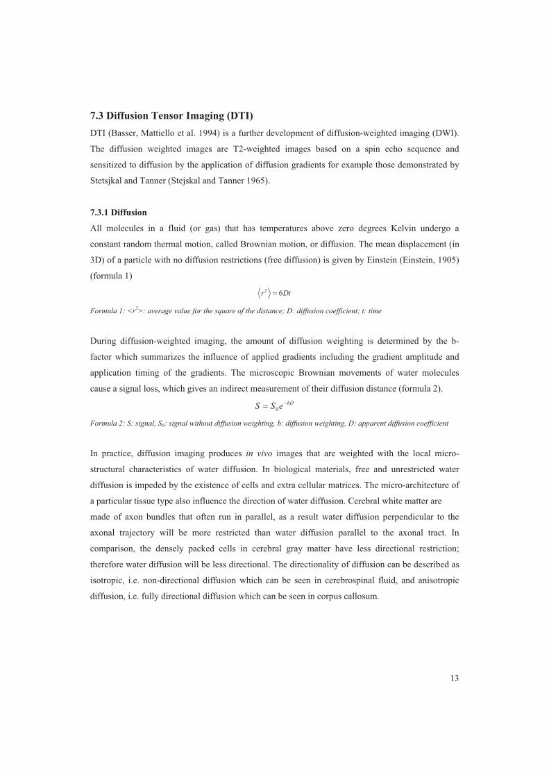
3D) of a particle with no diffusion restrictions (free diffusion) is given by Einstein (Einstein, 1905)
(formula 1) 2 6r Dt�
Formula 1: <r2>: average value for the square of the distance; D: diffusion coefficient; t: time
During diffusion-weighted imaging, the amount of diffusion weighting is determined by the b-
factor which summarizes the influence of applied gradients including the gradient amplitude and
application timing of the gradients. The microscopic Brownian movements of water molecules
cause a signal loss, which gives an indirect measurement of their diffusion distance (formula 2). bDeSS �� 0
Formula 2: S: signal, S0: signal without diffusion weighting, b: diffusion weighting, D: apparent diffusion coefficient
In practice, diffusion imaging produces in vivo images that are weighted with the local micro-
structural characteristics of water diffusion. In biological materials, free and unrestricted water
diffusion is impeded by the existence of cells and extra cellular matrices. The micro-architecture of
a particular tissue type also influence the direction of water diffusion. Cerebral white matter are
made of axon bundles that often run in parallel, as a result water diffusion perpendicular to the
axonal trajectory will be more restricted than water diffusion parallel to the axonal tract. In
comparison, the densely packed cells in cerebral gray matter have less directional restriction;
therefore water diffusion will be less directional. The directionality of diffusion can be described as
isotropic, i.e. non-directional diffusion which can be seen in cerebrospinal fluid, and anisotropic
diffusion, i.e. fully directional diffusion which can be seen in corpus callosum.

14
Figure 3: the difference between isotropic diffusion (free diffusion) and anisotropic diffusion
7.3.2 ADC
Based on diffusion weighted images, we can calculate the apparent diffusion coefficient (formula
3). The ADC is a measure of diffusivity or freedom of diffusion. It describes molecular motion of
water molecules in a given environment such as the brain where cellular size and integrity may
interfere. In gray matter ADC is low because neurons are densely packed therefore making an
efficient omnidirectional diffusion barrier. In white matter ADC is higher in some directions
because axons are organized in parallel bundles. As a result water diffusion perpendicular to the
axons will be more restricted than diffusion along the axons (figure 3). ADC can only be measured
in the direction of which the diffusion gradients are applied. But by averaging ADC in all gradient
direction applied a better estimate of diffusivity can be obtained, called ADCmean.
bSS
ADCD)/ln( 0��
Formula 3: D: apparent diffusion coefficient, S: signal with diffusion weighting, S0: signal without diffusion weighting,
b: diffusion weighting
7.3.3 Tensor
DWI is sufficient to describe isotropic diffusion, but DTI is required to measure the anisotropy of
diffusion in order to estimate the largest diffusion direction. In DTI, at least six gradient directions
are used for computing a diffusion tensor (formula 4). It can be described using a fully
diagonalizable 3×3 matrix; as a result only six measurements are needed. The eigenvectors and
eigenvalues (�1, �2, �3) of the tensor describes the three perpendicular axes in an ellipsoid with the
longest axes (�1) in parallel with the main diffusion direction of the underlying voxel.

15
���
�
�
�
�
zzzyzx
yzxyyx
xzxyxx
DDDDDDDDD
D
Formula 4: A 3×3 matrix describing the diffusion tensor.
7.3.3 Mean diffusivity
Based on the tensor model, mean diffusivity can be calculated. It is similar but not equal to
ADCmean (formula 5).
1 2 3
3MD
� � � �
Formula 5: MD: mean diffusivity, �: eigenvalue of the tensor matrix D
7.3.4 Fractional Anisotropy
The FA (formula 6) is a measure of the “directionality” of water diffusion, it is assigned a value
between 0 and 1 (Basser and Pierpaoli 1996). A FA value of 0 reflects isotropic diffusion, and a FA
value of 1 reflects maximally anisotropic diffusion. FA values close to 1 can be observed in tightly
packed neuronal bundles such as the corpus callosum. In an isotropic medium, such as a glass of
water, water molecules move randomly according to Brownian motion (Brown 1828; Einstein
1905). In biological tissues, however, the diffusion is restricted and is anisotropic. For example a
water molecule inside the axon has a low probability of crossing the myelin sheets and therefore the
water molecule will move along the axon and thus making the main direction of diffusion parallel
to the axonal trajectory (figure 3).
23
22
21
23
22
21 )ˆ()ˆ()ˆ(
23
���
������
� � ��FA
Formula 6: FA: fractional anisotropy, �: eigenvalue of the tensor matrix D
For practical and visualization purposes FA-maps can be colored coded using red, green and blue
(RGB) to present the direction of the principal eigenvectors, red indicating main diffusion along the
X axis: right-left, green indicating diffusion along the Y axis: posterior-anterior and blue indicating
diffusion along the Z axis: superior-inferior (figure 4C). By using color-coded FA-map radiologists
can more easily identify individual neuronal bundles, or tracts, in the brain. Different tracts run in
different direction, thus giving them separate color-coding, as shown in figure 5. In figure 5A, the
difference between cerebrospinal fluid (white) and brain parenchyma (grey) can clearly be seen,
while the boundary between white and grey matter within the brain parenchyma is harder to spot. In
figure 5B, the difference between white matter (white) and grey matter (grey) is more clearly

16
visible. In figure 5C, three major tracts have been identified (corpus callosum in red, corticospinal
tract in blue and superior longitudinal fasciculus in green).
Figure 4: different contrasts that can be achieved using DTI (ADC: apparent diffusion coefficient; FA: Fractional
anisotropy; RBG-Color-coded FA-map
Both ADC and FA are frequently used as parameters for probing white matter properties such as
restriction, hindrance, tortuosity and multiple compartments (LeBihan 1995). In healthy white
matter DTI can be used to follow cerebral maturation in children and adolescence as increment in
FA (Barnea-Goraly, Menon et al. 2005). In pathologic conditions structural barriers to water
diffusion in white matter might be subjected to alterations of permeability or geometry, as a result
ADC and FA might be changed when compared to unaffected and healthy white matter. After
traumatic brain injury, diffuse axonal injury might occur and cause lower FA and higher ADC.
These measurements may indicate histological abnormalities such as cytoskeletal misalignment,
lobulation and axonal disconnection (Arfanakis, Haughton et al. 2002). Higher ADC and lower FA
values are also seen in multiple sclerosis caused by edema, demyelination, inflammation and axonal
loss (Filippi, Cercignani et al. 2001), and in Alzheimer’s disease which is likely caused by
Wallerian degeneration and gliosis (Medina, DeToledo-Morrell et al. 2006).
7.3.1 Tractography Tractography is a visualization technique for cerebral axonal bundles based on DTI measurements
(Bihan, Mangin et al. 2001; Mori, Frederiksen et al. 2002). Based on the tensor for each voxel, three
perpendicular eigenvectors can be calculated, each describing diffusion in one direction. The largest
eigenvector is considered to represent the primary diffusion direction of the underlying axons in
voxels in white matter. By sequentially piecing together discrete and connecting estimates of the
principal eigenvectors, the axon bundles may be visualized.

17
In recent years, several tracking algorithms have been developed such as probabilistic tractography
(Behrens, Woolrich et al. 2003; Parker, Haroon et al. 2003) and deterministic tractography (Mori,
Crain et al. 1999). The goal of probabilistic tractography is to obtain a connectivity index along
white matter pathways that reflects fiber organization (figure 5A) giving a statistical likelihood for
the connection from a certain area in the brain to another predetermined region. Deterministic
tractography, on the other hand, follows the direction of the largest eigenvector in each voxel, and
virtually reconstructs a tract. One of the deterministic tracking algorithms is the fiber assignment by
continuous tracking (FACT) algorithm (Mori, Crain et al. 1999) (figure 5B). It utilizes a method
called fast marching tractography (Basser and Pierpaoli 1996) to find the axonal bundles in the
brain. FACT initiates tracking in all voxels in a given data set at once and does not require a seed
point to proceed. The reconstructed tracts can be used as a mask to select a region of white matter
for analysis. In the current work, a deterministic tractography method was used.
Figure 5A: probabilistic tracking of the optical radiation showing the probability of connection between the lateral gen
body and the visual cortex. The brighter color indicates higher statistic likelihood of connection. 5B: Deterministic
tracking of Inge’s corpus callosum, shows the spatial location of the tract inside a head.
During FACT initial tracking, initiation and termination criteria are required. The initiation criterion
is the lowest FA-value of a voxel in which tracking will proceed. Tracking terminates if the FA-
value in a voxel falls below or the angle between two eigenvectors in two adjacent voxels rise
above predetermined values. The initial tracking results in all traceable fiber bundles in the brain
being reconstructed. Next, Boolean operators are used to manually isolate the desired fiber bundles.
Usable operators for fiber selection include the OR, AND and NOT. The OR is the first operator to
be used, which selects all fibers that comes through a marked region. After “OR-ing”, a
combination of AND and NOT are used to manually fine tune and trim the selection based on visual

18
inspection. The AND operator discards fibers that do not go through the marked region, and NOT-
operator rejects all fibers that pass through the marked region (figure 6). It is therefore relatively
straightforward to segment and virtually reconstruct prominent white matter structures such as the
corpus callosum, the corticospinal tract, the optic radiation and the longitudinal fascicles (figure
5B).
Figure 6: procedure for selecting the desired fiber bundle using Boolean operators. The colors of the ring depict
different operator. Green: OR; Yellow: AND; Red: NOT.
7.4 DTI limitations and considerations DTI together with T2-weighted FLAIR and T2* imaging methods are tools for in vivo study of
white matter anatomy and structural connectivity in a non-invasive manner. Previously axonal
structures can only be studied using a technique pioneered by Klingler (Klingler 1935) which
involved repeatedly freezing and thawing the brain post mortem before dissection for axonal sub-
structures. DTI as a method is imperfect; limitations exist and will be discussed briefly in the
following section.
DTI-MRI measurements are extremely prone to motion related artifacts caused by head movement
and physiological noise such as cardiac pulsations and respiratory movements (Wirestam, Greitz et
al. 1996). Also, the DTI sequence itself gives rise to image distortions since it relies on heavy
gradient pulses which induce eddying currents in the antenna coils. Furthermore, magnetic field
inhomogeneity is a concern in regions with tissues of differing magnetic susceptibility such as in
regions with soft tissue and air interfaces (Frahm, Merboldt et al. 1988). Several solutions to these
problems have been suggested. The duration of the experiment should be kept at minimum as
lengthy experiments increase the risk of head movements. During scanning light physical
constraints should be applied and cardiac and respiratory gating may be used for minimizing
physiological noise (Skare and Andersson 2001). Intra-scan head-motion and eddy current artifacts

19
can be corrected using mathematical algorithms (Rohde, Barnett et al. 2004). It is possible to reduce
susceptibility artifacts by placing diamagnetic passive shims in the roof of the mouth (Wilson,
Jenkinson et al. 2002) or more elegantly by using B0-field map correction (Anderson and Gore
1994; Jezzard and Balaban 1995).
Limitations also apply to DTI data analysis. In tractography, the common voxel size is a cube a few
cubic millimeters large, which might contain tens of thousands of axonal sections. Tractography is
therefore an inaccurate method in regions with crossing fibers and for small and winding pathways
(Johansen-Berg and Behrens 2006). One way to solve the crossing fiber problem (Mori and van Zijl
2002) is to use advanced diffusion imaging techniques such as high-angular (Tuch, Reese et al.
2002) and Q-ball imaging (Tuch, Reese et al. 2003; Tuch 2004). In addition to imaging related
artifacts, brain pathology such as lesions and edema makes tractography even more challenging.
Although tractography allows for virtual dissection of white matter tracts, it must not be confused
with anatomical dissection as substantial difference in tract locations are observed between tracts
derived from DTI and histology (Dauguet, Peled et al. 2007). It should also be noted that
tractography is a subjective procedure still missing a standardized approach, and therefore highly
dependent on the analyst’s experience and competence. The interpretation of the results is also
dependent on the observer’s understanding of the shortcomings of the method.
Another challenge in DTI data analysis is brain size variations among subjects (Allen, Damasio et
al. 2002) particularly in voxel based morphometry (VBM) where the image volume is compared
across brains at every voxel (Ashburner and Friston 2000). Therefore, before any group-wise
statistical analysis is carried out, the subjects’ brains have to be made spatially compatible in a
process called normalization. One normalization approach is spatial transformation and
registeration of subjects’ brains to a template brain (Friston, Ashburner et al. 1995). The template
can be an average of brains of multiple subjects such as the Montreal Neurological Institute (MNI)
template (Montreal, Quebec, Canada) or a single subject defined as being “standard” such as the
Talairach template (Talairach and Tournoux 1988). The accuracy of normalization is often
jeopardized by the presence of cerebral pathology. Therefore it can be advantageous to improve
precision by making a customized template. First, subjects’ brains are normalized to pre-made
templates such as MNI-template, then the normalized brains are averaged in order to create a
custom template which serves as the new target brain for the subjects’ brains during the second
normalization (Ashburner and Friston 2000). Despite all efforts, no normalization process is
perfect, and therefore any group-wise co-localization is inherently pseudo-accurate and this may
reduce the chance of detecting statistically significant difference between groups. It is possible to
20
use other methods for statistic inference which do not rely on normalization, one being region of
interest (ROI) analysis. The ROIs can be selected manually as 3D geometric figures according to
predetermined anatomical localization criteria in each individual, or be chosen semi-automatically
through for instance tractography where each region corresponds to a white matter tract or a section
of it. It should be emphasized that any manual or semi-automatic region selection is subjective and
depends on the analyst’s experience and competence. Furthermore, using a ROI approach, only
predetermined regions are investigated, this might lead to other regions with significant group
differences being overlooked.
7.5 Blood Oxygen Level Dependent Functional Magnetic Resonance Imaging BOLD fMRI is based on a presumed coupling between neural activity and cerebral blood flow
(CBF) (Raichle 1987). Neuronal activity can be recorded electrophysiologically using invasive
electrodes placed in neural tissue. The input and local processing in the neurons can be observed as
local field potentials (LFP) which integrate signals over a couple of millimeters (Legatt, Arezzo et
al. 1980). The output from the neurons can be recorded as multi-unit spiking activities which
combine signals over a few hundred micrometers. Studies have shown that BOLD fMRI signals
correlate strongly with LFP and to a lesser extend with spiking activity (Logothetis, Pauls et al.
2001; Mukamel, Gelbard et al. 2005), therefore the BOLD signals predominantly reflects the input
and local processing rather than output from the neurons. Neural activity also increases CBF and
causes an oversupply of oxygenated hemoglobin that exceeds local metabolic requirement. The
lowering of the amount of deoxygenated hemoglobin is detectable using susceptibility-weighted
MRI (Ogawa, Lee et al. 1990) since deoxygenated hemoglobin acts as an endogenous paramagnetic
contrast agent (Pauling and Coryell 1936). The most commonly used BOLD-fMRI technique is
based on a T2*-weighted gradient echo sequence combined with echo planar imaging (Mansfield
1977) which can sample the whole brain in a few seconds. It is similar to T2-weighted images as
both measure the spin-spin relaxation or decay rate of a MR signal after exication, but in T2* the
inhomogeneities of the local magnetic field is also taken into consideration. As a result T2* time is
shorter than T2, and T2* weighting is more sensitive to field inhomogeneities caused by for
example changes in oxygenated/deoxygenated hemoglobin ratio. The possibility to indirectly detect
changes in neural activity using BOLD fMRI was rapidly embraced by neuroscientists and the
method is now widely used.
Most commonly during BOLD fMRI experiments, subjects perform certain tasks inside the scanner,
and the difference in the BOLD signal during performance of the task and baseline, or task A and
task B, can subsequently be analyzed. In task-dependent fMRI the tasks, often called paradigms,

21
can be motor tasks, e.g. hand movements, or cognitive tasks, such as planning, memory or spatial
navigation. The tasks are usually presented and stimulus collected using a software program like E-
prime, or in-house designed programs. The participants view the task on an LCD screen or via a
projector mounted outside the scanner bore. The subjects can view the screen through a mirror
placed on the head coil or in goggles. Most commonly the stimuli are presented according to an
epoch-related design (Deyoe, Bandettini et al. 1994) inspired by earlier works on PET (Raichle
1987), or event-related design inspired by ElectroEncephaloGraphy (EEG) and
MagnetoEncephaloGraphy (MEG) studies (Picton, Lins et al. 1995). The epoch-related design is
easy to implement and analyze and have a high signal-to-noise ratio (SNR). Each individual task
stimulus usually lasts 14-50 seconds and they are interleaved with control conditions of varying
length. The event-related design is more complicated to implement and analyze and have a lower
SNR. Each individual task stimulus usually lasts 1-10 seconds spaced apart with control and/or
baseline task periods of varying length. Compared to epoch-related design, event-related design
yields higher specificity in the neural correlates of the cognitive task being investigated, but with
lower SNR. It is also possible to implement self-paced tasks, in which duration of each stimulus is
not predetermined. Furthermore, predetermined timing of each task condition can be avoided by
employing alternative model free analysis methods such as individual component analysis (ICA).
It should be noted that there is also task independent fMRI, i.e. resting state fMRI, where the person
is resting during fMRI scanning. However, this method was not used in this work and will not be
discussed further see the work by Gusnard and colleagues for details (Gusnard and Raichle 2001).
During data analysis, the collected BOLD fMRI data is first preprocessed using digital filters such
as motion correction algorithms and noise-removal filters to improve detection of the true BOLD
signal (see also section 7.5 for more details). Thereafter the BOLD fMRI data-set is aligned to a T1-
weighted image of the brain. If group-wise comparison involving multiple subjects is needed, the
T1-weighted images of the brains have to be normalized. It is commonly implemented using whole
brain template based methods, similarly as in DTI group analysis (see also section 7.5 for more
details). It is also possible to do cortex based alignment (CBA) (Dale, Fischl et al. 1999; Goebel,
Esposito et al. 2006). The CBA utilizes the hemispherical curvature information to minimize the
spatial difference between the subjects’ individual brains. CBA is a time-consuming technique
requiring segmentation and reconstruction of each subject’s hemispheres (Dale, Fischl et al. 1999).
The reconstructions are then inflated and transformed to a sphere, which serves as the starting point
for the alignment process. Upon completion of the alignment, the spheres are transformed and
deflated back to its original shape. Alternatively the spheres can be transformed back without
deflation and cut and flattened to form a flat map of the hemispheres (Fischl, Sereno et al. 1999).
22
Finally, the BOLD signal variations are convolved with a hemodynamic response (HDR) function
which reflects the assumed temporal fluctuation of the BOLD signal due to changing neural
activity.
The HDR also introduces temporal smoothing and delays when compared to the actual neural
activity that is supposed to arise in response to the presented stimuli/task performance. Then a
statistical parametric map based on the general linear model is calculated from the measured BOLD
signal changes convolved with the HDR (Friston, Holmes et al. 1994). The calculations are often
done using two main approaches, the single-voxel approach which tests each voxel separately, and
the region of interest (ROI) approach which performs statistical analysis on time course of a ROI.
Alternatively, using model free analysis methods, such as ICA (Comon 1994; McKeown, Makeig et
al. 1998), no assumptions of the underlying BOLD signal fluctuations are made, therefore there is
no need to implement an HDR . Instead, ICA explores the data and tries to identify spatio-temporal
patterns in a data driven manner.
7.6 BOLD fMRI Limitations and considerations BOLD fMRI has rapidly become a standard method for studying brain activity. Still, the method
has several limitations and shortcomings that must to be taken into consideration to properly
interpret results. In the following text, methodological issues will be discussed
7.6.1 BOLD signal
The measured BOLD signal changes are not a direct reflection of neural activity. Instead it depicts
regions with increased blood flow presumed to be caused by increased neural activity. The signal
maximum is delayed from the onset of stimulus due to the time required for production and
diffusion of vascular signal substances which dilates the vascular bed and causes a washout of
deoxygenated hemoglobin (Marota, Ayata et al. 1999). Therefore, temporal resolution in BOLD
fMRI is inferior compared to EEG and MEG. On the other side BOLD fMRI has better spatial
localization than EEG and MEG, thus being a complementary brain studying technique. Patterns of
neural activity derived from BOLD fMRI experiments only show the relative differences in neural
activity between task conditions. When a task condition is compared to a non-task or baseline
condition, the results describe the neural activity that is statistically different from the latter. The
baseline condition reflects resting state neural activity. Different task conditions can also be
compared in order to identify regions subserving specific components of for example a cognitive
task. The theoretical model for this approach is called cognitive subtraction and was first described
by Donders (Donders 1868). It assumes that cognitive processes happen sequentially and
23
individually without any mutual interference. The idea of independent cognitive processes or pure
insertion has been a subject of substantial skepticism, as the brain is a highly nonlinear system and
does not conform to additive or linear principles (Friston, Price et al. 1996). Alternatively, event-
related paradigm design, which does not completely rely on cognitive subtraction can be used
(Postle, Zarahn et al. 2000). Another confounding phenomenon is the underlying task-independent
differences in measured BOLD signal among different subject groups. These differences can be
caused by cerebrovascular disease (Roc, Wang et al. 2006), white matter inflammation (Langkilde,
Frederiksen et al. 2002), age-related changes in cerebrovasculature and autoregulatory mechanisms
(D'Esposito, Zarahn et al. 1999), pharmacological effects (Liu, Behzadi et al. 2004) and psycho-
stimulant drug use (Friedman, Turner et al. 2008). These BOLD signal differences might make
group-wise comparisons between patients and healthy controls inaccurate since an inherent signal
differences are already present independent of the task. These factors should be taken into account
in BOLD fMRI experiments where subjects belonging to different groups, for instance a healthy
control group versus a group with pervasive brain pathology, are compared directly.
7.6.2 Measurement and analysis of BOLD signal
The ability to detect BOLD signal changes is often measured using signal to noise ration (SNR)
which is the relationship or power ratio between the signal and the background noise. The
magnitude of BOLD signal changes induced by brain activity is weak usually in the range of 1-6%
of the total signal. It is more robust for primary visual, motor and sensory functions than in higher
cognitive functions such as memory, planning etc. (Huettel and Song 2003). BOLD sequence is
based on a T2*-weighted gradient echo imaging sequence which is vulnerable to distortions and
artifacts caused by several factors. First susceptibility artifacts may arise in regions close to air
filled spaces or sinuses. These regions include orbitofrontal cortex, parahippocampal/hippocampal
cortices and the temporal lobes. Second, motion or physiology related artifacts can be caused by
subject motion, cardiac pulsation or respiration. Third artifacts or distortions may be the results of
field inhomogeneity of the scanner. Some methods for combating these problems have been briefly
discussed previously (see section 7.5)
SNR can be increased with higher static magnetic field strength which yields higher net
magnetization and thereby larger BOLD signals change (Yang 1999; Krasnow 2003). It also
increases possible spatial resolution and reduces partial volume effect by allowing smaller voxels
and at the same time maintaining sufficient SNR for signal detection. In addition higher static field
alters T2*-relaxation time and causes BOLD signals to increases faster in the extravascular
components of small vessel than larger vessel. Smaller vessels are more likely to be colocalized

24
with the studied neural activity. Therefore increased statistic magnetic field improves the spatial
specificity of the BOLD signal (Huettel, Song et al. 2003).
During data analysis, the ability to detect BOLD signal related to neural activity can be improved
by several means. Motion correction can partially removes the effects of subject motion and the
associated signal variability. Spatial smoothing with a Gaussian filter can facilitate the detection of
true BOLD signal in statistic analysis by reducing noise (Oppenheim 1978) and improves the fit of
the data to the general linear model (Adler 1981). High and low-pass filter (Friston, Holmes et al.
1995) can remove noise in temporal domain such as physiological noise. Alternatively, cardiac
pulsation and respiration can be monitored and modeled as effects of non-interest during data
analysis (Biswal, DeYoe et al. 1996). The ability to detect BOLD signal is further affected by
statistical analysis method. Activation maps calculated from single-voxel based analysis are
inherently limited by the SNR of the individual voxel. In ROI based approach some of the low SNR
can be overcome, but at the cost of possible overlooking activities in other brain regions than those
pre-defined. Also, it is essential to ensure adequate normalization of brains during group-wise
comparisons using single-voxel based analyze methods. The normalization can be done using
template based approach or CBA. Comparing these two methods, CBA provides better overlap of
functional areas with similar sulci topology across subjects such as the visual and motor areas than
template based methods (Fischl, Sereno et al. 1999), while other areas, such as subcortical grey
matter, may have no “sulcal” topology, which makes the advantage of CBA less obvious (Brett,
Johnsrude et al. 2002).
7.8.3 Paradigm Design
The performance of any tasks inside the scanner should not involve movement of large muscle
groups since any excessive motion will lead to head motion and motion related artifacts. The
difficulty of the paradigm has to be adapted to suit the cognitive and motor ability of the test
subjects to ensure adequate success rate. The duration of each paradigm should be kept short to
prevent subject fatigue. Lengthy experiments can be divided into separate sessions to allow proper
restitution in-between. By doing so new problem might be introduced, but these topics are outside
the scope of this thesis. The equipment required for task completion such as response buttons and
screen for viewing the task has to be MRI compatible in order to function properly, safely and
without disturbing the MRI signal significantly. In term of sensory modalities, it is easiest to present
visual stimuli and difficult to receive oral response from the test subject. As a result standard
neuropsychological tasks such as the Wisconsin card sorting (Berg 1948) and Tower of London
(Shallice 1982) have to be adapted and carried out virtually, which alters the task from its original

25
intended version. In addition, all subjects are scanned in the supine position, which is an uncommon
position for performance of most tasks. Indeed, this position might cause nausea when combined
with visual stimuli such as spatial navigation (Slater, Usoh M et al. 1995).
In an fMRI paradigm, the stimuli or task is the independent variable and the measured BOLD signal
is the dependent variable. Additional variables might be present in the paradigm and may correlate
with the dependent and independent variable. These variables are called confounding factors and
might cause incorrect data interpretation. Methods to minimize these effects include
counterbalancing and randomization. In counterbalanced experiments, the confounding factors are
present in all conditions and will cancel each other out during comparison. For example during
visual experiments which involve pictures in task conditions, a scrambled version of the same
picture containing the exact same number of pixels of each color can be presented during the rest
conditions. In randomized experiments, individual conditions are presented randomly to mitigate
the effect of habituation, a psychological process in which psychological and behavioral response
decreases as a result of repeated exposure to same or similar task condition over long time
(Thompson and Spencer 1966; Sokolov 1990). It has for instance been shown in humans that
habitation causes reduced neural activities in amygdala (Fischer, Furmark et al. 2000; Wright,
Fischer et al. 2001). Despite the advantages of randomization, there are factors which advice
against its usage. In BOLD fMRI experiments containing task conditions of varying difficulties, it
might be favorable to perform the most challenging task first to avoid fatigue or if the result of that
first task condition serves as the input of the next one. Particular attention should be paid to patients
with brain disorders who often experiences difficulties in understanding and following instructions.
Another factor that needs consideration during paradigm design is the timing of individual task and
rest conditions. In epoch based and event related paradigm designs, timing is predetermined and
therefore remains constant across subjects. Timing in epoch based paradigms can also be allowed to
vary between subjects by terminating the task conditions automatically upon completion thus
making the conditions self-paced. By doing so, the onset of the conditions will vary with TR and
data sampling will be distributed in time contributing to reduced bias and increased sensitivity in
the final results (Veltman, Mechelli et al. 2002). Also self-pacing reduces neuropsychological
effects such as fatigue and habituation by making individual task conditions more different and
perhaps more interesting. Other favorable effects of self-pacing include the increased likelihood of
achieving similar performance in two groups with differences in for instance processing speed. This
is done by allowing subjects in each group to use different but sufficient amounts of time to
compete the tasks. As a result, this reduces the impact of performance as a possible confounding

26
factor and ensures comparable neural processes taking place in both groups. The difference in the
duration of the task conditions reflects subject performance can be used as regressor in later data
analysis. Self-paced conditions also involve technical challenges. First the task itself have to be
“self-paceable” which means that the completion of the task can be monitored using algorithm
incorporated in the paradigm software itself, or recorded by allowing subjects to respond when they
are finished for example by pressing a button. During analysis, self-paced conditions require
individual HDR reflecting the assumed fluctuation in BOLD signal to be made before convolving
with the real observed BOLD signal variations. It is a time consuming step prone to human errors.
Alternatively to epoch based and event related design, ICA can be used to completely avoid the
need for timing.
7.7 BOLD- and DTI in TBI survivors 7.7.1 Epidemiology of TBI
Traumatic brain injury (TBI) is a common cause of disability. In Norway, 7-8% of all patients
treated for injury in the emergency room or hospital have head injuries (NEL 2009). While the
majority of these patients only sustain concussion or mild head injury, there are still 450-500 head
injury related fatalities annually. Men are twice as likely as women to experience head injury and
young people under 30 years are at particular risk. Each year 10.000 are admitted to Norwegian
hospitals with a head injury. In total, these amount to 80.000 days of hospitalization and contribute
to a considerable health expense (NEL 2009). The total annual cost for a bed at a specialized
rehabilitation center is estimated to be 3 million NOK (Sosial-_og_helsedirektoratet 2005).
7.7.2 Clinical findings in TBI survivors
Trauma leading to TBI can be either penetrating such as those caused by firearms or edged
weapons, or non-penetrating such as those caused by motor vehicle accidents with extreme
acceleration and deceleration forces, falls, or blunt weapons. The type of injury can be divided into
focal, diffuse and a combination of both. The primary mechanism for focal injury is direct impact of
the brain. For diffuse injury, it is the shear-strain deformation, a change in brain shape but without
volume change (Arfanakis, Haughton et al. 2002). Focal brain injury can manifest as epidural,
subdural, contusion and traumatic intra-cerebral hematomas. Diffuse injury can result in diffuse
axonal injury, diffuse brain edema and hypoxic brain injury.
The clinical outcome following TBI ranges from no functional deficit to death. The severity of the
traumatic brain injury is initially commonly assessed using the Glasgow coma scale (GCS)
(Teasdale and Jennett 1974), and measures consciousness level according to verbal and motor
27
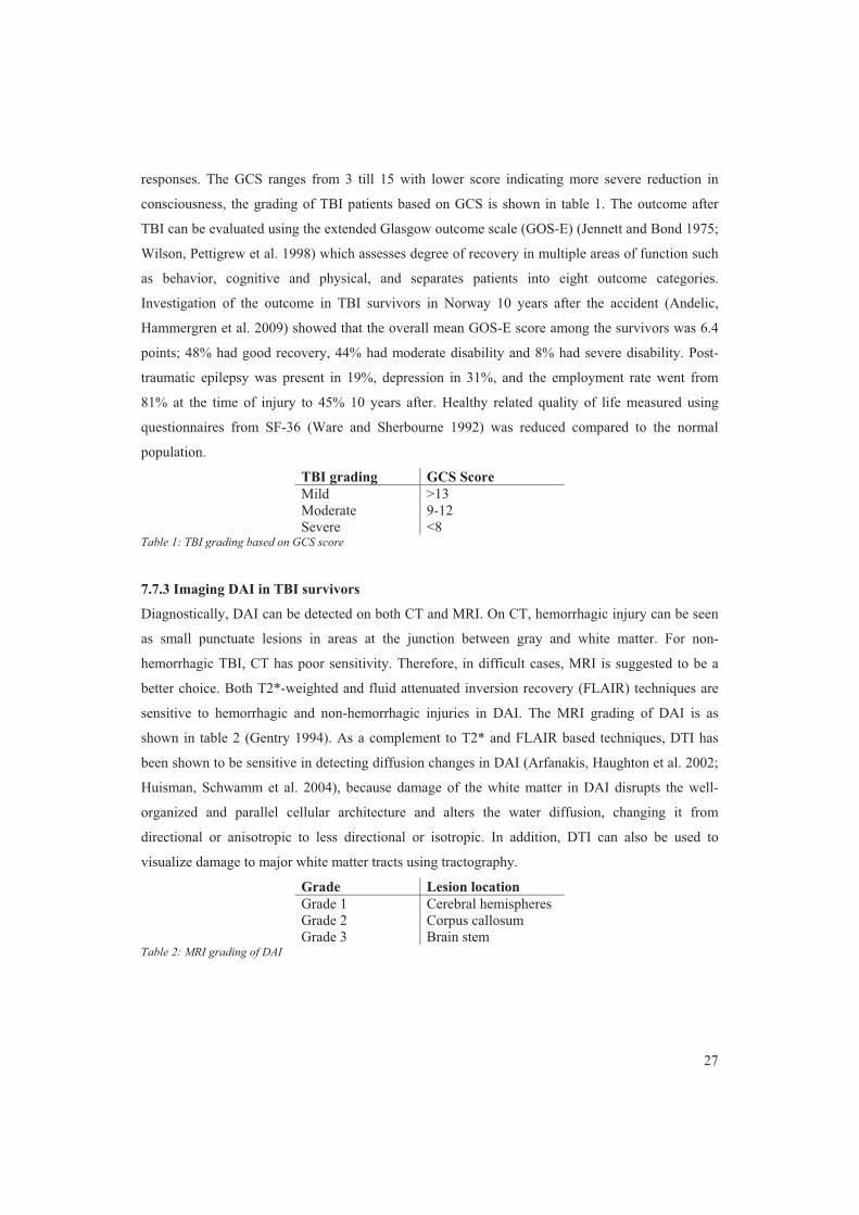
responses. The GCS ranges from 3 till 15 with lower score indicating more severe reduction in
consciousness, the grading of TBI patients based on GCS is shown in table 1. The outcome after
TBI can be evaluated using the extended Glasgow outcome scale (GOS-E) (Jennett and Bond 1975;
Wilson, Pettigrew et al. 1998) which assesses degree of recovery in multiple areas of function such
as behavior, cognitive and physical, and separates patients into eight outcome categories.
Investigation of the outcome in TBI survivors in Norway 10 years after the accident (Andelic,
Hammergren et al. 2009) showed that the overall mean GOS-E score among the survivors was 6.4
points; 48% had good recovery, 44% had moderate disability and 8% had severe disability. Post-
traumatic epilepsy was present in 19%, depression in 31%, and the employment rate went from
81% at the time of injury to 45% 10 years after. Healthy related quality of life measured using
questionnaires from SF-36 (Ware and Sherbourne 1992) was reduced compared to the normal
population.
TBI grading GCS Score Mild >13 Moderate 9-12 Severe <8
Table 1: TBI grading based on GCS score
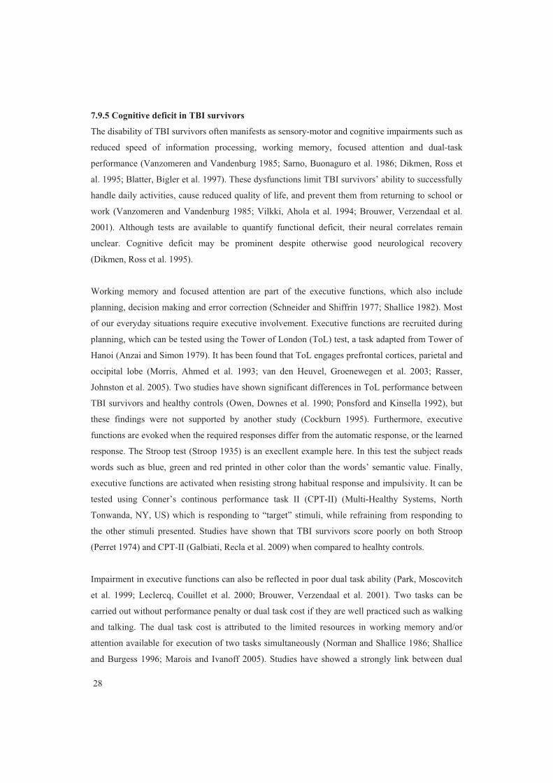
7.7.3 Imaging DAI in TBI survivors
Diagnostically, DAI can be detected on both CT and MRI. On CT, hemorrhagic injury can be seen
as small punctuate lesions in areas at the junction between gray and white matter. For non-
hemorrhagic TBI, CT has poor sensitivity. Therefore, in difficult cases, MRI is suggested to be a
better choice. Both T2*-weighted and fluid attenuated inversion recovery (FLAIR) techniques are
sensitive to hemorrhagic and non-hemorrhagic injuries in DAI. The MRI grading of DAI is as
shown in table 2 (Gentry 1994). As a complement to T2* and FLAIR based techniques, DTI has
been shown to be sensitive in detecting diffusion changes in DAI (Arfanakis, Haughton et al. 2002;
Huisman, Schwamm et al. 2004), because damage of the white matter in DAI disrupts the well-
organized and parallel cellular architecture and alters the water diffusion, changing it from
directional or anisotropic to less directional or isotropic. In addition, DTI can also be used to
visualize damage to major white matter tracts using tractography.
Grade Lesion location Grade 1 Cerebral hemispheres Grade 2 Corpus callosum Grade 3 Brain stem
Table 2: MRI grading of DAI
28
7.9.5 Cognitive deficit in TBI survivors
The disability of TBI survivors often manifests as sensory-motor and cognitive impairments such as
reduced speed of information processing, working memory, focused attention and dual-task
performance (Vanzomeren and Vandenburg 1985; Sarno, Buonaguro et al. 1986; Dikmen, Ross et
al. 1995; Blatter, Bigler et al. 1997). These dysfunctions limit TBI survivors’ ability to successfully
handle daily activities, cause reduced quality of life, and prevent them from returning to school or
work (Vanzomeren and Vandenburg 1985; Vilkki, Ahola et al. 1994; Brouwer, Verzendaal et al.
2001). Although tests are available to quantify functional deficit, their neural correlates remain
unclear. Cognitive deficit may be prominent despite otherwise good neurological recovery
(Dikmen, Ross et al. 1995).
Working memory and focused attention are part of the executive functions, which also include
planning, decision making and error correction (Schneider and Shiffrin 1977; Shallice 1982). Most
of our everyday situations require executive involvement. Executive functions are recruited during
planning, which can be tested using the Tower of London (ToL) test, a task adapted from Tower of
Hanoi (Anzai and Simon 1979). It has been found that ToL engages prefrontal cortices, parietal and
occipital lobe (Morris, Ahmed et al. 1993; van den Heuvel, Groenewegen et al. 2003; Rasser,
Johnston et al. 2005). Two studies have shown significant differences in ToL performance between
TBI survivors and healthy controls (Owen, Downes et al. 1990; Ponsford and Kinsella 1992), but
these findings were not supported by another study (Cockburn 1995). Furthermore, executive
functions are evoked when the required responses differ from the automatic response, or the learned
response. The Stroop test (Stroop 1935) is an execllent example here. In this test the subject reads
words such as blue, green and red printed in other color than the words’ semantic value. Finally,
executive functions are activated when resisting strong habitual response and impulsivity. It can be
tested using Conner’s continous performance task II (CPT-II) (Multi-Healthy Systems, North
Tonwanda, NY, US) which is responding to “target” stimuli, while refraining from responding to
the other stimuli presented. Studies have shown that TBI survivors score poorly on both Stroop
(Perret 1974) and CPT-II (Galbiati, Recla et al. 2009) when compared to healhty controls.
Impairment in executive functions can also be reflected in poor dual task ability (Park, Moscovitch
et al. 1999; Leclercq, Couillet et al. 2000; Brouwer, Verzendaal et al. 2001). Two tasks can be
carried out without performance penalty or dual task cost if they are well practiced such as walking
and talking. The dual task cost is attributed to the limited resources in working memory and/or
attention available for execution of two tasks simultaneously (Norman and Shallice 1986; Shallice
and Burgess 1996; Marois and Ivanoff 2005). Studies have showed a strongly link between dual
29
tasking and prefrontal cortex activity (D'Esposito, Detre et al. 1995; Koechlin, Basso et al. 1999).
The idea that prefrontal cortex is the primary site for dual tasking is challenged by another
hypothesis, which suggests that dual tasking recruits additional brain regions already activated by
each individual task, and does not need additional activation of the executive system (Smith, Geva
et al. 2001; Erickson, Colcombe et al. 2005). This controversy may be explained by the lack of
standardized clinical test for evaluating dual task performance.
In addition, TBI survivors may show spatial navigation deficits (Skelton, Ross et al. 2006;
Livingstone and Skelton 2007) as a result to injury to the medial temporal lobe (MTL). Successful
navigation is a complex task requiring several cognitive components. Initially the environment has
to be learned by making a mental representation either allocentrically which is view point
independent, or egocentrically which is view point dependent (Jordan, Schadow et al. 2004). When
required to navigate, this previously acquired representation is retrieved from memory and
interpreted for route calculation. This sequence of cognitive processes can be divided into phases
including self-localization, target localization and route execution (Spiers and Maguire 2006;
Shipman and Astur 2008). Animal studies have shown the importance of the MTL in spatial
navigation by detecting place cell (Okeefe and Dostrovs.J 1971), grid cell (Fyhn, Molden et al.
2004), head direction cell (Sargolini, Fyhn et al. 2006) and border cell (Solstad, Boccara et al. 2008)
in that region. Modern neuroimaging studies have shown that an extended cortical and subcortical
network is engaged during spatial navigation with the MTL playing a pivotal role (Jordan, Schadow
et al. 2004; Spiers and Maguire 2006; Shipman and Astur 2008).
7.9.6 Neuroplasticity in TBI survivors
The neural correlates of cognitive deficit and impairment detected using BOLD fMRI have been
shown as difference in activity pattern between TBI survivors and healthy controls (McAllister,
Saykin et al. 1999; Christodoulou, DeLuca et al. 2001; Scheibel, Pearson et al. 2003). The source of
the differences is believed to be primarily caused by neuroplastic changes in the brain after injury
(Johansen-Berg, Dawes et al. 2002). The principle of neuroplasticity was first hypothesized by
William James in 1890. It is the brain’s ability to make structural and functional changes to better
adapt to the environment and increase survivability. These changes are influenced by experience,
learning, aging or pathology (Emerit, Riad et al. 1992; Nitsche, Liebetanz et al. 2005). In the cortex
two neurotransmitters are of particularly importance, they are glutamate and GABA, which induce
morphological and structural changes in the synapses by promoting neural sprouting and increasing
the number of synaptic buttons (Gil-Loyzaga 2009). It should also be noted that the global
projection neurons, containing the monoamine neurotransmitters (serotonin, noradrenalin,
30
acetylcholine and dopamine), play a role in neuroplasticity as seen in for example memory and
learning (Rasmusson 2000). Following TBI there is an improvement of cognitive function even as
the structural changes continue to develop negatively, for instance increasing atrophy (Wilde,
Bigler et al. 2007; Sidaros, Skimminge et al. 2009). This could be viewed as a paradox, and can be
regarded as an internally driven “brain repair” process aimed at regaining a certain functional level
by altering brain processing. Several types of changes have been shown to take place that may play
a larger or smaller role in these functionally adaptive changes seen after TBI. Axonal sprouting and
synaptogenesis (Laurberg and Zimmer 1981), unmasking or reorganization (Bachyrita 1981),
diaschisis (Von Monakow 1914) and neurogenesis (Eriksson, Perfilieva et al. 1998). Although adult
neurongenesis exists, as demonstrated by neuronal progenitor cells in the dentate gyrus of adult
humans which can divide and generate new neurons (Eriksson, Perfilieva et al. 1998) its role in
neuroplasticity remains elusive. In comparison, other modes of neuroplastic repair are considered to
be more frequent. In collateral sprouting, uninjured axons branches to assume territory of injured
axons. In reorganization or unmaksing, healthy neural structures not formerly used for a given
purpose are reassigned to do functions formerly subserved by the injured area. Similarity can be
drawn to redundant design in engineering where critical components of a system are duplicated to
increase the reliability of the system in the case of a backup or fail safe. At a cellular level,
unmasking happens by activation of previously “silent” synapses after injury to primary functional
synapses. As a result an alternative neuronal route is established indicating that neural circuitry is
not hardwired and can to some extent be rerouted. In diaschisis, damage to one specific location in
the brain causes functional deficits in another distant but undamaged site since the “normal” input
to the distant site is lost. But gradually, the distant site may recover its function. The neuroplastic
potential is also dependent on the type of tissue. In visual cortex, the talamo-cortical neuroplasticity
is extremely limited following injury to the early components of the visual system. The major
contributor to functional improvement is cortico-cortical neuroplasticity (Dariansmith and Gilbert
1994; Chow, Groszer et al. 2009). Also different cortical regions have different degrees of ability to
reorganize, the motor cortex is for instance much less plastic than the somatosensory cortex
(Castroalamancos, Donoghue et al. 1995). The structural adaptations in the brain are reflected
functionally through substitution and compensation. In substitution, additional cortical areas within
the same functional network are recruited. In compensation, additional areas outside the same
functional network are recruited.

31
8 AIMS The aims of the studies were to apply and evaluate two modern MRI imaging techniques; DTI and
BOLD fMRI for studying axonal microstructure and cognitive functions in TBI survivors.
The DTI study (paper 2) was performed in order to explore the potential of DTI in mapping changes
in white matter following TBI. The BOLD fMRI studies (paper 1 and paper 3) investigated neural
correlates for executive functions such as planning (paper 1) and dual-tasking (paper 3) which are
known to be impaired in severe TBI survivors. The purpose of these two papers was to study the
neural correlates behind planning and dual-tasking, explore the neuroplasticity following TBI and
evaluate the feasibility of tasks for differentiation of TBI from healthy controls. Also a spatial
navigation study using virtual reality (VR) (paper 4) was carried out in healthy controls to
investigate the neural correlates of MTL during varying navigational scenarios. The ultimate
purpose of this study is to implement a variant of the task in TBI survivors to study neural
correlates in the brain, particularly the MTL, during spatial navigation at a later time point. Finally
DTI and BOLD fMRI were combined (paper 5) to investigate the effect of axonal damage on the
HDR and to validate the fMRI BOLD findings in the other papers (paper 1 and paper 3)
32
9 MATERIALS AND METHODS 9.1 Participants Paper 1
Ten male patients with chronic TBI were recruited from an outpatient rehabilitation follow-up
group at Munkvoll Rehabilitation Center (St. Olav’s Hospital, Trondheim, Norway). All patients
had initial GCS below 8 indicating severe injury. For controls ten healthy volunteers were included.
Neither of the controls had a history of head trauma or neurological disorders, nor a history of
DSM-IV axis I diagnosis of psychiatric illness.
Paper 2
Nine male patients with chronic TBI were recruited from an outpatient rehabilitation follow-up
group at Munkvoll Rehabilitation Center (St. Olav’s Hospital, Trondheim, Norway). All patients
had initial GCS below 8 indicating severe injury. For controls eleven healthy volunteers were
included. None of the controls had a history of trauma to the head or neurological disorders, nor a
history of DSM-IV axis I diagnosis of psychiatric illness.
Paper 3
Ten male patients with chronic TBI were recruited from an outpatient rehabilitation follow-up
group at Munkvoll Rehabilitation Center (St. Olav’s Hospital, Trondheim, Norway). Nine patients
were severe TBI (GCS<8) and one had moderate TBI (GCS=9). For controls, eleven age-matched
healthy male volunteers were recruited among the patients’ friends and first siblings. None of the
controls had a history of neurological disorders or current DSM-IV axis I diagnosis of psychiatric
illness.
Paper 4
Twenty male healthy volunteers with no history of neurological disorders, head trauma, or current
DSM-IV axis I diagnosis of psychiatric illness were recruited from the NTNU university campus.
Paper 5
Ten male patients with chronic TBI were recruited from an outpatient rehabilitation follow-up
group at Munkvoll Rehabilitation Center (St, Olav’s Hospital, Trondheim, Norway). All patients
were severe TBI survivors with GCS below eight, and had white matter abnormalities diagnosed as
DAI. For controls, nine age-matched healthy male volunteers were recruited among the patient’s

33
friends and first siblings. None of the controls had a history of neurological disorders or current
DSM-IV axis I diagnosis of psychiatric illness.
9.2 Ethical Approval All studies were approved by the local ethical committee for clinical research. All patients and
healthy controls gave their written informed consent after the procedure had been explained and
opportunities to ask questions given. All image data, questionnaires and other collected data were
made anonymous and handled according to the Helsinki convention.
9.3 MRI Scanning MRI scanning was performed on two different scanners at St Olavs Hospital, Trondheim, Norway.
Only scan parameters for BOLD fMRI and DTI sequences will be listed here.
Scanner I (Paper 1, 2, 3 and 5)
Scanning was performed on a Philips 3 Tesla MRI Scanner (Philips Medical Best, Netherlands),
with a quasar dual gradient system yielding a maximum of 80 mT/m and a SENSE head coil using
parallel imaging (MRI Devices/InVivo, Orlando, Florida, USA).
All BOLD fMRI was done using single shot echo-planar-imaging (EPI) sequence with SENSE
reduction factor = 2.2. Scan parameters for paper 1 were: 41 slices, TR=3000 ms, TE=35 ms and
2.4×2.4×2.4mm voxel size; for paper 3: 40 slices, TR=3000 ms, TE=35 ms and 1.8×1.8×2.5mm
voxel size; and for paper 5: 23 slices, TR=1500 ms, TE=35 ms and 1.8×1.8×2.3mm voxel size. All
DTI imaging was done using a spin-echo EPI sequence with SENSE reduction factor = 1.5. Full
brain volumes were collected using 55 slices with 32 spatial independent directions. Cardiac
triggering was applied with a systolic trigger delay of 150 msec with TR of 15 R-R intervals giving
a TR of 13-16 sec. Other major scan parameters were b-factor = 800 sec/mm2, TE=50 msec and
1.80×1.80×1.72mm voxel size.
Scanner II (Paper 4)
Scanning was performed on a Siemens Trio 3 Tesla MRI Scanner (Siemens, Erlangen, Germany),
with a 12 channel head matrix coil (Siemens, Erlangen, Germany). BOLD fMRI was done using an
echo-planar imaging pulse sequence. The scan parameters were: TR=2600 ms, TE=30ms and
3.0×3.0×3.0 mm voxel size. The slices were positioned as close to 90o on the anterior-posterior
direction of the hippocampus as possible without causing fold-in from the neck.

34
9.4 fMRI Stimulus design, presentation and response collection Stimuli for papers 1, 3 and 5 were compiled in E-Prime 1.1 (Psychology Software Tools,
Pittsburgh, Pennsylvania, USA). For paper 4 it is made using Torque Game Engine (Garage Games,
Eugene, Oregon, USA) in close collaboration with Terra vision (TerraVission, Trondheim,
Norway). All stimuli were presented in a block design. In paper 1, the block duration was
predetermined and remained constant. In paper 3 and 4 the block duration varied depending on the
performance of the subject. A time limit was applied to control the maximum duration.
For papers 1, 3 and 5, the stimuli were presented using a liquid crystal display (Philips Medical
Best, Netherlands) placed behind the magnet bore which the subjects viewed through a tilted mirror
mounted on the head coil. The responses were collected using response grips (Nordic Neuro lab,
Bergen, Norway). For paper 4, an organic light-emitting diode based video goggle (Nordic Neuro
lab, Bergen, Norway) was used for better subject visual field of view coverage giving a more
submerged experience. The subject carried out the experiment using a joystick (Current Designs,
Philadelphia, USA) and the task performance related data was logged by the Torque Game Engine.
9.5 Data Analysis 9.5.1 DTI
Analysis of DTI images involved several software packages. Pre-processing was done using FSL
3.3 (Analysis Group, FMRIB, Oxford, UK) (Smith et al., 2004), creation of FA-maps, ADC-maps
and tractography was done using DTI-Studio (Laboratory of Brain Anatomical MRI, John Hopkins
Medical Institute, Baltimore, USA) (Jiang et al., 2006) based on the fiber assignment by continuous
tracking (FACT) algorithms (Mori et al., 1999). Group-based analysis of FA and ADC-maps was
done using SPM2 (Wellcome Department of Imaging Neuroscience, London, UK). Skull stripping
and ROI drawing was done with MRIcro (Rorden & Brett, 2000).
9.5.2 BOLD fMRI
Analysis of structural and BOLD fMRI image data for paper 1 and 3 was performed using Brain
Voyager QX 1.4.10 (Brain Innovation, Maastricht, The Nederlands). Using this software suite, the
BOLD fMRI image data were corrected for motion, smoothed over a 4-mm full width at half
maximum kernel, removed for linear trend and high-pass filtered. Then all brains were segmented
and reconstructed based on the structural MRI data. The reconstructed brains formed the basis
normalization using CBA (see section 6 for details). All statistical analyses of BOLD fMRI image
data were carried out using contrasts according to the GLM and group activation maps generated
using random effect analysis. Conditions were modeled according to a boxcar stimulus function

35
convolved with a two-gamma HDR function. The group statistical parametric maps were corrected
for false positives using the false discovery rate (for details see papers).
For paper 4 and 5, the analysis of structural and BOLD fMRI image data was performed using FSL
4.0 (Analysis Group, FMRIB, Oxford, UK) (Smith et al., 2004). First, the BOLD image data were
corrected for motion, smoothed over a 5-mm full width at half maximum kernel and high pass
filtered. Then all brains were segmented based on the structural MRI data. The resulting brains
formed the basis for normalization using template based approach (see section 7.5 for details) with
MNI standard template. All statistical analyses of BOLD fMRI image data were carried out using
contrasts according to the GLM and group activation maps generated using a mixed effects model
of variance, as implemented in FLAME1 (FMRIB’s Local Analysis of Mixed Effects). Conditions
were modeled according to a boxcar stimulus function convolved with a two-gamma HDR function
(for details see papers).
9.5.3. Behavioral data
Excel (Microsoft, Redmond, Washington, USA) and SPSS (SPSS, Chicago, Illinois, USA) were
used to perform statistical analyses of group characteristics and behavioral performance.

36
10 SYNOPSES OF PAPERS 10.1 Paper 1 Brain activation measured using functional magnetic resonance imaging during the Tower of
London task
Rasmussen I-A Jr, Antonsen IK, Berntsen EM, Xu J, Lagopoulos J, Håberg AK.
ACTA NEUROPSYCHIATRICA 18 (5) 216-225 OCT 2006
The aim of this study was to assess the patterns of regional brain activation in response to the
Tower of London task (ToL) in chronic TBI survivors using BOLD fMRI. ToL is a well-described
executive task considered to specifically tap planning which is frequently impaired in TBI.
In this paper ten patients and ten age-matched controls underwent fMRI while performing a
modified ToL task. The analysis of the performance data indicated no difference in response
accuracy between the groups. The statistic analysis of the BOLD data was done using RFX GLM
model and FDR corrected. The results revealed that TBI patients recruited additional and larger
cerebral regions than healthy controls. These regions included additional functional area in the
parietal and frontal lobes, and a larger increased right-lateralization of activity especially in the
prefrontal lobe. In addition a parametric analysis with task difficulty as independent variable was
conducted. It did not show any significant between group differences.
The results of this study pointed to a cortical reorganization inside the executive system of vigilance
and working memory in survivors of severe TBI. Both parietal and prefrontal areas were recruited
to compensate for damaged brain tissue. The TBI patients also showed a greater degree of right
lateralization. The lack of between group differences in the parametric model might indicate that
both groups recruited similar brain regions as task difficulty increases.
37
10.3 Paper 2 Diffuse axonal injury in severe traumatic brain injury visualized using high-resolution
diffusion tensor imaging
Xu J, Rasmussen IA, Lagopoulos J, Håberg A
JOURNAL OF NEUROTRAUMA 24 (5) 753-765 MAY 2007
The aim of this study was to investigate whether diffusion tensor imaging (DTI) offered additional
information as to the extent of damage not visualized with standard magnetic resonance imaging
(MRI) in patients with severe traumatic brain injury (TBI).
In this paper nine chronic severe TBI patients and eleven matched controls were scanned using
DTI. Based on the DTI data, FA- and ADC-maps were calculated and compared between the
groups. The comparisons were carried out first using voxel based morphometry (VBM), followed
by a region of interest based approach (ROI), and finally complemented by a tractography based
method. The ROI were placed in the following regions: anterior and posterior corpus callosum,
anterior and posterior periventricular, deep frontal, medial orbitofronal, occipital and posterior limb
of internal capsule. The tractograms included the corpus callosum and corticospinal tracts. Results
from VBM revealed significantly reduced FA and increased ADCmean in major white matter tracts
in TBI patients when compared to healthy controls. These findings were confirmed by both ROI
and tractography based analyses in the investigated regions.
The results of this study suggested widespread white matter injuries following severe TBI. In
addition it also illustrated that DTI holds great promise as a diagnostic tool to identify and quantify
the degree of white matter injury in TBI patients.
38
10.5 Paper 3 Simple dual tasking recruits prefrontal cortices in chronic severe traumatic brain injury
patients, but not in controls
Rasmussen IA, Xu J, Antonsen IK, Brunner J, Skandsen T, Axelson DE, Berntsen EM, Lydersen S,
Håberg A
JOURNAL OF NEUROTRAUMA 25 (9) 1057-1070 SEP 2008
The aim of this study was to investigate the cost of dual tasking in chronic severe TBI patients
using BOLD-imaging. Dual tasking is the ability to carry out two tasks simultaneously. Its cost is
the decreased performance on one or both tasks. The dual task ability is specifically impaired after
traumatic brain injury.
In this paper, ten TBI patients and eleven matched healthy controls were scanned using BOLD
techniques while performing two tasks. They were a visual search task and a simple two-button
press motor task. Analysis of the performance data demonstrated significant dual task interference
in both groups, and increased performance variability in TBI patients. Analysis of the BOLD data
showed significantly reduced activation in TBI patients in the single task conditions compared to
healthy controls. For both single task conditions, the activity was reduced in the occipital and
posterior cingulated cortices, and for the visual task also in the thalami. A reversed pattern was
observed when comparing BOLD data from dual task condition. Significantly increased activity
was observed in TBI patients in prefrontal-anterior midline-parietal network, predominantly
lateralized to the left. The increase in activation occurred within regions shown by other studies to
be associated with increased dual task cost in healthy controls.
The results of this study pointed to neural substitution and more effortful processing in TBI
survivors while carrying out two tasks simultaneously. Recruitment of additional prefrontal regions
may be connected to serial rather than parallel processing in low level dual tasking. Therefore in
severe TBI survivors, low level dual task performance depends on increased attentional and
executive guidance.

39
10.6 Paper 4 Persistent posterior and transient anterior medial temporal lobe activity during navigation
Xu J, Evensmoen HR, Lehn H, Pintzka CWS, Håberg AK
SUBMITTED NEUROIMAGE 2009
The aim of this study was to explore the activity within MTL subregions in the initial phase of
navigation, i.e. self-localization, target localization and path planning, compared to the execution
phase. In addition, the effect of environmental manipulations on MTL activity was investigated.
In this paper twenty male healthy controls were scanned using BOLD fMRI while navigating in a
virtual environment that resembled an office with numerous complex landmarks. In total three task
(normal, without, block) and one baseline (line) conditions were included. Each condition was
divided into two phases, initial and execution. The volunteers learned the environment before
scanning through free and structured exploration. The analysis of the data showed that different
regions within an extended neuronal network are recruited. The initial phase engaged anterior MTL
regions including bilateral rostral and caudal entorhinal cortex and bilateral anterior hippocampus.
Also right anterior parahippocampal cortex was significantly more active during the initial phase.
Activity in the very anterior aspect of the right hippocampus correlated positively with navigational
success. The whole navigation phase recruited right posterior hippocampus and parahippocampal
cortex. Hippocampal activity was only detected when the virtual environment remained unaltered in
condition normal. Navigational success was positively correlated with activity in the anterior
hippocampus for the whole block.
The results of this study indicated a functional segregation within the MTL with regard to
navigational phase, i.e. initial versus execution phase. Based on the current findings it appears that
the anterior part of MTL completes associations related to the environment at large, and the
posterior part keeps track of current location. Moreover, hippocampal activity depended on
environmental features, e.g. presence or absence of landmarks and blockings.

40
10.7 Paper 5 Reduced fractional anisotropy does not change the shape of the hemodynamic response in
survivors of severe traumatic brain injury
H. S. Palmer, B. Garzon, J. Xu, E.M. Berntsen, T. Skandsen, A. Håberg
SUBMITTED JOURNAL OF NEUROTRAUMA 2009
The aims of the present study were to describe the HDR in visual cortex, and to examine its
relationship with the microstructure of the optical radiation in severe TBI survivors and controls.
In this paper, ten TBI survivors without visual impairments, but with known diffuse axonal injury
and nine healthy controls were scanned using DTI and BOLD fMRI during brief visual stimuli at
randomized intervals. For each individual the optical radiations were identified from DTI using
diffusion tensor tractography. Fractional anisotropy (FA) and mean apparent diffusion coefficient
(ADCmean) values for these tracts were calculated. BOLD signal changes for each subject were
estimated in V1, and group HDR curves produced. Standard between-group analysis of BOLD
activation in V1+V2 was performed. The data analysis showed group HDR curves from visual
cortex were fully transposable between TBI survivors and healthy controls despite a significant
reduction in FA in the optical radiation in the TBI group. A significant correlation between BOLD
signal beta values in the visual cortex and FA values in the optical tract was present in controls, but
not TBIs. Between-group contrast showed TBI survivors had a greater area of activation, especially
in V2 during visual stimulation compared to controls.
The results of this study indicated an intact HDR in traumatic white matter damage. There was a
loss of thalamo-cortical input to the visual cortex, and the increase in area of activation in the visual
cortex in TBI probably stemmed from cortico-cortical neuroplasticity. This study supports the
validity of using standard fMRI methodology to study neuroplasticity in TBI.
41
11 DISCUSSION In this thesis DTI and BOLD fMRI methods were applied in studying axonal microstructure and
cognitive functions in severe chronic TBI survivors.
The DTI study (paper 2) showed that severe TBI survivors had widespread white matter axonal
abnormality when compared to healthy controls. These structural pathologies were reflected as
changes in FA and ADCmean in the major white matter tracts in the TBI survivors. Such white
matter injuries are considered to be a contributor to cognitive deficits (Sidaros, Engberg et al.
2008). The impact of TBI on brain activity in response to cognitive tasks was investigated in two
BOLD fMRI studies (paper 1 and paper 3). In these two studies, paradigms inferring executive
functions such as planning and dual-tasking were successfully designed and applied. Both
paradigms successfully differentiated TBI survivors from healthy controls in neural activity. Only
the paradigm in paper 3 produced significant different behavior separating the groups. The results
from the two BOLD fMRI studies provided evidence for neuroplastic changes in the brain in TBI
survivors in the chronic phase. These neural modifications could be seen as changes in cerebral
activation pattern, which included increased area of activation, recruitment of additional areas, and
shift in lateralization. The physiological effect of white matter pathologies on the HDR was
examined in paper 5. The results indicated that significantly altered white matter axonal structure
does not lead to changes in HDR. Thus strengthening the between-group BOLD fMRI results in the
two fMRI studies included in this thesis (paper 1 and paper 3).
Finally a spatial navigation task (paper 4) were designed and carried out in healthy controls.
Experiences from this study showed the feasibility of a spatial navigation task and provided
framework for understanding neural correlates during spatial navigation in future studies on TBI
survivors. Contusions in the anterior part of the MTL is frequent in TBI, and even more
interestingly hippocampal atrophy is seen even without the presence of focal injury to the MTL
(Wilde, Bigler et al. 2007). Paper 4 also demonstrated the possibility of differentiate neural activity
related to different phases of spatial navigation. By applying a similar experimental set-up to TBI
patients with focal and/or more generalized lesions in the MTL, it should be possible to gain
increased understanding of the neuronal correlates leading to behavioral deficits following TBI, the
compensatory mechanisms following various degree of focal and local injury, and finally to
evaluate hypotheses relating specific navigational functions to specific regions in the MTL by
specifically examine patients with different types of lesions to the MTL.
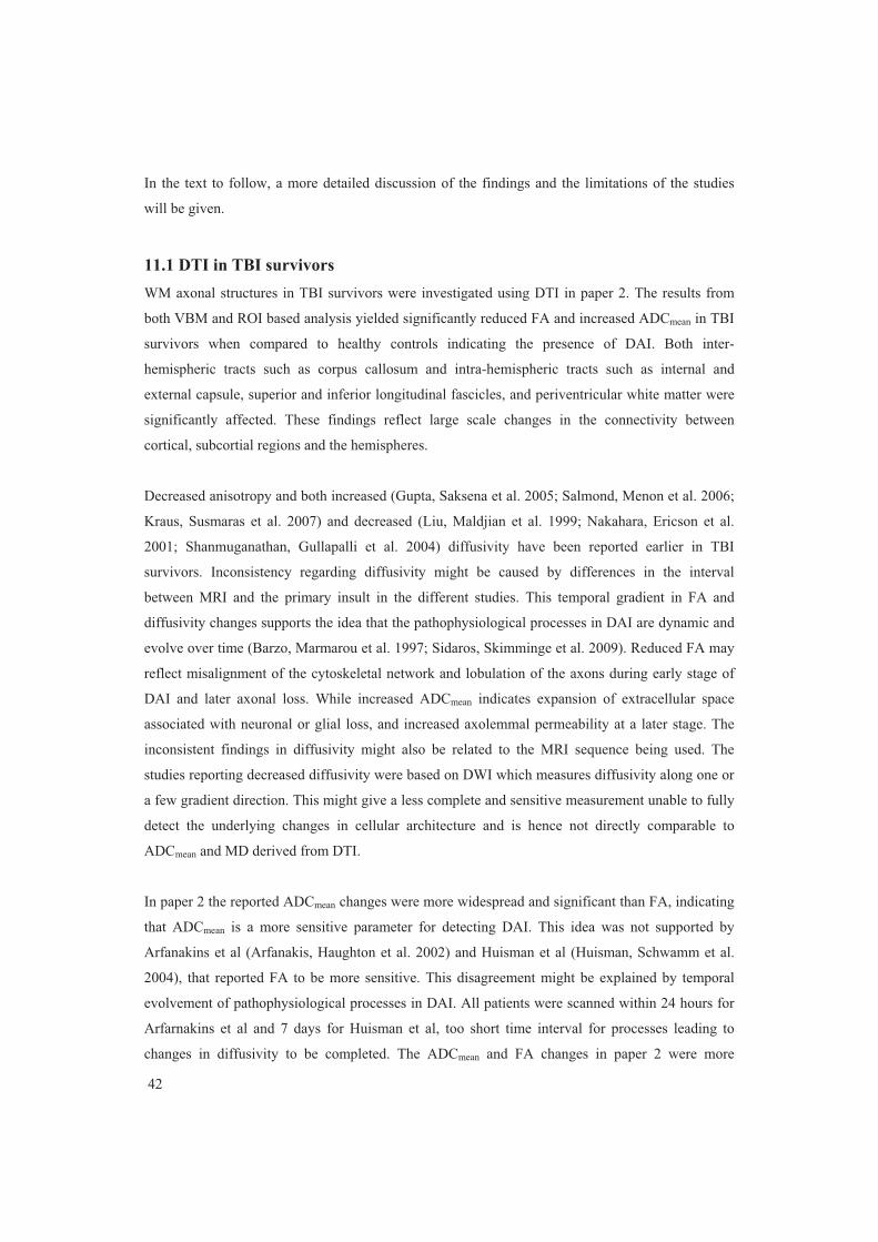
42
In the text to follow, a more detailed discussion of the findings and the limitations of the studies
will be given.
11.1 DTI in TBI survivors WM axonal structures in TBI survivors were investigated using DTI in paper 2. The results from
both VBM and ROI based analysis yielded significantly reduced FA and increased ADCmean in TBI
survivors when compared to healthy controls indicating the presence of DAI. Both inter-
hemispheric tracts such as corpus callosum and intra-hemispheric tracts such as internal and
external capsule, superior and inferior longitudinal fascicles, and periventricular white matter were
significantly affected. These findings reflect large scale changes in the connectivity between
cortical, subcortial regions and the hemispheres.
Decreased anisotropy and both increased (Gupta, Saksena et al. 2005; Salmond, Menon et al. 2006;
Kraus, Susmaras et al. 2007) and decreased (Liu, Maldjian et al. 1999; Nakahara, Ericson et al.
2001; Shanmuganathan, Gullapalli et al. 2004) diffusivity have been reported earlier in TBI
survivors. Inconsistency regarding diffusivity might be caused by differences in the interval
between MRI and the primary insult in the different studies. This temporal gradient in FA and
diffusivity changes supports the idea that the pathophysiological processes in DAI are dynamic and
evolve over time (Barzo, Marmarou et al. 1997; Sidaros, Skimminge et al. 2009). Reduced FA may
reflect misalignment of the cytoskeletal network and lobulation of the axons during early stage of
DAI and later axonal loss. While increased ADCmean indicates expansion of extracellular space
associated with neuronal or glial loss, and increased axolemmal permeability at a later stage. The
inconsistent findings in diffusivity might also be related to the MRI sequence being used. The
studies reporting decreased diffusivity were based on DWI which measures diffusivity along one or
a few gradient direction. This might give a less complete and sensitive measurement unable to fully
detect the underlying changes in cellular architecture and is hence not directly comparable to
ADCmean and MD derived from DTI.
In paper 2 the reported ADCmean changes were more widespread and significant than FA, indicating
that ADCmean is a more sensitive parameter for detecting DAI. This idea was not supported by
Arfanakins et al (Arfanakis, Haughton et al. 2002) and Huisman et al (Huisman, Schwamm et al.
2004), that reported FA to be more sensitive. This disagreement might be explained by temporal
evolvement of pathophysiological processes in DAI. All patients were scanned within 24 hours for
Arfarnakins et al and 7 days for Huisman et al, too short time interval for processes leading to
changes in diffusivity to be completed. The ADCmean and FA changes in paper 2 were more

43
prominent on the right side, consistent with similar right sided patterns of focal pathology observed
on conventional MRI scans of these TBI survivors. Previous studies have reported reduced
anisotropy in the right internal capsule and right optic radiation (Wieshmann, Symms et al. 1999;
Rugg-Gunn, Symms et al. 2001). One possible explanation to the right lateralized pattern might be
attributed to rotational forces charactering motor vehicle accidents happening in countries such as
Norway with right-side driving custom. In addition to detecting structural pathologies, several
studies have established significant correlations between DTI parameters and different aspects of
outcome for TBI survivors. FA has been shown to positively correlate with clinical outcome (Ptak,
Sheridan et al. 2003), cognitive function (Kraus, Susmaras et al. 2007) and negatively to injury
severity (Huisman, Schwamm et al. 2004). MD has been found to correlate negatively with memory
and learning (Salmond, Menon et al. 2006). To my knowledge no study has investigated the
correlation between executive functions and diffusion parameters in TBI survivors. But one study
on ischemic leukoaraiosis patients showed that diffusivity, and not FA, correlates with executive
function (O'Sullivan, Morris et al. 2004). Unfortunately, the TBI survivors included in the present
work did not complete the same battery of psychological tests due to the lack of test standards.
Therefore we were unable to look into the relationship between FA and diffusivity and higher
cognitive functions. It should also be noted that the relatively small group size in paper 2, and the
variation in location and amount of structural changes is not ideal for such a correlation analysis.
The results in paper 2 are consistent with the idea that TBI is accompanied by DAI. By using DTI
changes in WM microstructure could be detected in areas where it was not evident on conventional
CT or MR images. Despite the advantages of DTI in detecting WM pathology in TBI survivors, the
method is still not used for routine diagnostics due to its labor intensiveness and hard-to-use
software. Progresses are being made both in software and hardware to allow DTI to become part of
the clinical toolbox.
11.2 fMRI in TBI survivors 11.2.1 fMRI Paradigm Design
In paper 1 and paper 3, fMRI paradigms considered to tap functions known to be impaired in many
TBI survivors were selected and modified for investigating the neuronal correlates to these test
following TBI. The paradigms used here were in paper 1 based on a modified standard
neuropsychological test (paper 1), and in paper 3 custom built from scratch (paper 3).
In paper 1, the paradigm was designed according to ToL, a standard psychological test for planning.
By conforming to the standards, our results were compatible with other ToL experiments. In the

44
original version subjects physically manipulated the beads. In some computerized ToL experiments
(Morris, Downes et al. 1988; Owen, Downes et al. 1990), the physical movements have been
replaced by virtual movements. In our version no physical or virtual manipulation of the beads was
required. Instead subjects performed the transformation between the start and target configuration
mentally. They subsequently responded by choosing between three numbers indicating the least
required number of moves. By altering the ToL test in this manner, we believe that the requirements
on several cognitive processes might be altered and possibly increased. These processes include
working memory for keeping the number for moves and previous states online from start to task
completion, mental rotation for manipulation of the beads, and linguistic comprehension skill for
understanding instructions and keeping them available during task performance. Therefore to
ensure subject compliance, a training session was given and scanning did not proceed until
satisfactory training results were achieved. By choosing between three alternatives, there is 33%
chance of guessing the correct answer. Thankfully, the success rate was 83.6% for TBI survivors
and 87.5% for healthy controls, which was above chance. Furthermore, the between group
difference was not significant. The similarity in success rate indicates that all subjects understood
and were able to perform the paradigm, and any difference in neural activation was therefore not
dependent on success rate. In paper 1, the timing of the paradigm was predetermined, hence
collecting data on completion time was impossible. During image analysis, we detected significant
between group differences in the activation patterns. This supports the idea that our ToL paradigm
is able to detect and differentiate TBI survivors from healthy controls and describe the neural
correlates for planning following TBI.
In paper 3 we investigated dual task ability. There is no standard test for dual task function, the dual
task paradigms in the literature vary greatly from psychological refractory period experiments
(Tombu and Jolicoeur 2003) via two motor tasks (Bekkering, Adam et al. 1994) to two executive
tasks (Pashler 1994). Our paradigm was custom designed and composed of a visual search task and
motor task. Both components are non-executive and functionally unrelated. To ensure subject
compliance, pre scanning training was given. In addition the paradigm was not randomized in order
to ensure that the TBI survivors were not fatigued during the dual task performance. Behavioral
data revealed same success rate in both groups, but significant difference in completion time which
was collected in this self-paced paradigm design. The success rate indicated that sufficient subject
compliance was achieved, and the differences in completion time separated the groups and
indicated difference in dual task cost. Later during image analysis, we found significant between
group difference in neural activity, and correlation between neural activity and motor response
rhythm.
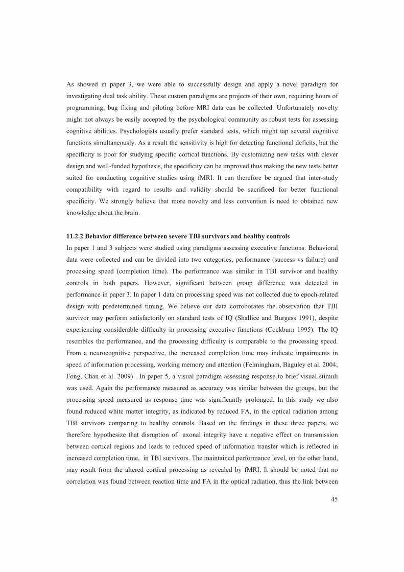
45
As showed in paper 3, we were able to successfully design and apply a novel paradigm for
investigating dual task ability. These custom paradigms are projects of their own, requiring hours of
programming, bug fixing and piloting before MRI data can be collected. Unfortunately novelty
might not always be easily accepted by the psychological community as robust tests for assessing
cognitive abilities. Psychologists usually prefer standard tests, which might tap several cognitive
functions simultaneously. As a result the sensitivity is high for detecting functional deficits, but the
specificity is poor for studying specific cortical functions. By customizing new tasks with clever
design and well-funded hypothesis, the specificity can be improved thus making the new tests better
suited for conducting cognitive studies using fMRI. It can therefore be argued that inter-study
compatibility with regard to results and validity should be sacrificed for better functional
specificity. We strongly believe that more novelty and less convention is need to obtained new
knowledge about the brain.
11.2.2 Behavior difference between severe TBI survivors and healthy controls
In paper 1 and 3 subjects were studied using paradigms assessing executive functions. Behavioral
data were collected and can be divided into two categories, performance (success vs failure) and
processing speed (completion time). The performance was similar in TBI survivor and healthy
controls in both papers. However, significant between group difference was detected in
performance in paper 3. In paper 1 data on processing speed was not collected due to epoch-related
design with predetermined timing. We believe our data corroborates the observation that TBI
survivor may perform satisfactorily on standard tests of IQ (Shallice and Burgess 1991), despite
experiencing considerable difficulty in processing executive functions (Cockburn 1995). The IQ
resembles the performance, and the processing difficulty is comparable to the processing speed.
From a neurocognitive perspective, the increased completion time may indicate impairments in
speed of information processing, working memory and attention (Felmingham, Baguley et al. 2004;
Fong, Chan et al. 2009) . In paper 5, a visual paradigm assessing response to brief visual stimuli
was used. Again the performance measured as accuracy was similar between the groups, but the
processing speed measured as response time was significantly prolonged. In this study we also
found reduced white matter integrity, as indicated by reduced FA, in the optical radiation among
TBI survivors comparing to healthy controls. Based on the findings in these three papers, we
therefore hypothesize that disruption of axonal integrity have a negative effect on transmission
between cortical regions and leads to reduced speed of information transfer which is reflected in
increased completion time, in TBI survivors. The maintained performance level, on the other hand,
may result from the altered cortical processing as revealed by fMRI. It should be noted that no
correlation was found between reaction time and FA in the optical radiation, thus the link between

46
increased response time and reduced white matter integrity may still require further research to
solidify, maybe with a larger sample size.
From a data analysis point of view, the statistical power of our studies is reinforced by the similar
performance and weakened by the differences in processing speed. We were able to avoid one
confounding factor, performance, but the other factor, completion time, remained. But difference in
behavior data can be can be exploited in a constructive way. In paper 3, significant correlation was
found when using motor response rhythm as a regressor.
11.2.3 Functional difference between severe TBI survivors and healthy controls
In paper 1 and 3 significant differences in neural activity between TBI survivors and healthy
controls were found using fMRI paradigms investigating executive functions. The functional
differences showed two characteristic patterns, i.e. increased dispersion and changes in
lateralization of activity.
Dispersion or increased spatial extend of the neural activity was evident in both paper 1 and paper
3. In paper 1 we used ToL, a planning task. TBI survivors showed a dispersed activation pattern in
the parietal and the ventrolateral prefrontal cortical areas compared to the healthy controls. The
ventrolateral prefrontal cortex (VLPFC) is generally associated with emotional, working memory
and higher order sensory processing. The VLPFC role in working memory includes the ability to
hold and compare sequences of items in short-term working memory (Owen 1997; Petrides 2000).
Based on this observation, we suggest that increased VLPFC in TBI survivors is the result of
inadequate ability in keeping track of information being held in spatial working memory, and leads
to recruiting additional cortical resources. In paper 3, we used a dual task paradigm including a
visual search and a simple motor task. Dispersion was again detected, but more differentiated than
in paper 1. First dispersion was detected in TBI survivors when compared to healthy controls only
during dual task conditions, and included prefrontal structures such as midline anterior superior
frontal gyrus and cingulate cortex. The pattern was actually reversed during single task conditions.
Second, dispersion during dual task conditions was also present in healthy controls when compared
to TBI survivors, but in different cortical areas. Healthy controls recruited additional neural
resources within the regions employed by each component tasks as seen in other studies in healthy
controls (Smith, Geva et al. 2001). TBI survivors recruited additional cortical resources outside the
regions already employed by each component tasks, but remained inside the same functional
network. These additional regions corresponded to regions used by healthy controls in more
difficult visual search tasks (Collette, Olivier et al. 2005).

47
Based on the results in paper 1 and paper 3, several hypothesizes about the cause of dispersion of
activation can be made. First, since dispersion was present in the dual task condition, but not single
task condition, reduced or impaired modulation of activity involving the global projecting neurons
may play a role. A similar pattern of increased activity in healthy controls compared to TBI
survivors during simple tasks and the opposite during more challenging tasks have been detected by
Bayer and colleagues (Braver, Cohen et al. 1997) during a visual N-back letter task. They suggested
that relative subtle group differences in frontal activation might reflect difficulty in modulating or
allocating cognitive resources according to working memory load in TBI survivors. Second,
dispersion points to substitution as the mechanism for functional reorganization in TBI survivors.
Substitution is the recruitment of additional cerebral regions within the same partially restored
functional network. The addition cerebral regions also might indicate inefficient processing going
from parallel to serial.
In addition to dispersion, the neural activity was also more lateralized in TBI survivors. The neural
activity was more prominent on the right side in paper 1 and on the left side in paper 3. Earlier
studies support these findings. One study showed that in mild TBI survivors effective verbal
working memory results in left lateralized BOLD activations, while poor performance causes a
more right-lateralized activation pattern (McAllister, Saykin et al. 1999). Other study reported that
in TBI survivors, there is significant negative correlation between diffusivity and memory function
lateralized to the left (Salmond, Menon et al. 2006). Based on these finding it is possible that TBI
survivors utilize a right-lateralized network also found in healthy controls, but not used by the
highly skilled or proficient individuals. In addition our ToL paradigm also required large amount of
mental rotation, which is predominantly lateralized to the right hemisphere (Hermsen, Haag et al.
2009). In paper 3, a dual task paradigm was used. One study has reported a similar left-sided fronto-
parietal network in healthy volunteers in response to dual task performance of two tasks not
dependent on prefrontal resources (Collette, Olivier et al. 2005). The disagreement in lateralization
between paper 1 and 3 is confusing, and no study has systematically investigated changes in
lateralization with regard to type of task and clinical data describing for instance severity of TBI,
time from injury, functional outcome etc. in TBI survivors. Therefore we propose two possible
mechanisms. First, changes in lateralization of function may be arbitrary, depending on lesion
location and time from initial incident. This idea is supported by Feydy and colleagues who found
that lateralization of motor function after stroke depends on the site of lesion and time after insult
(Feydy, Carlier et al. 2002). Second, the lateralization pattern may be connected to extent of white
matter injury in the corpus callosum and within the hemisphere(s). It is possible that damage to
major inter-hemispheric neural pathway leads to separation of previously connected inter-

48
hemispheric functional networks. After disconnection, information is no longer processed in
parallel with the entire network participating. Instead information is processed in a more serial
fashion by each hemisphere. It can be speculated that part of the network in one hemisphere will
assume the dominant role, and process the majority of the information, and as a result give rise to
more extended neural activity. Functional or material-specific lateralization in prefrontal areas is
well established principle. Neural activity is lateralized to the left for verbal tasks and to the right
for the non-verbal tasks (Wagner, Poldrack et al. 1998). It is also interesting no note that the
lateralization in neural activity seen in paper 1 and paper 3 does not agree with the right lateralized
prominence of white matter injury found in paper 2. This observation further strengthened the belief
that an internal driven “brain repair” independent of structural deterioration such as increasing
atrophy and reduction in FA.
The characteristic difference in neural activity between TBI survivors and healthy controls is likely
attributed to neuroplastic changes in the brain. After brain injury basic developmental neuronal
growth processes are released and potentiated due to disturbance in the dynamic balance between
growth-stimulating and growth-suppressing factors (Finger and Almli 1985). These changes in
signals will often results in plastic changes such as neural rearrangement involing axonal sprouting,
synaptogenesis and highly unlikely neoneurogenesis. These developmental changes together with
changes in input may lead to other more passive plastic changes such as unmasking and diaschisis.
All these plastic processes will take place during “brain repair”, therefore it is difficult if not
impossible to determine the exact correlation between repair mechanisms and neural activity
pattern. In paper 1 and paper 3 we observed dispersion and changes in lateralization of neural
activity. We believe dispersion is the result of unmasking and axonal sprouting in cortex after
injury. During this process, the sprouting neurons will interact with other neurons than before
injury, thus extend its spatial influence which can be observed as dispersion. Unmasking can be
connected to changes in local processing within a restricted region of the cortex due to changes in
for instance lateral inhibition and /or local input, but also to unmasking of white matter pathways of
lesser importance in the healthy brain. For instance, interruption of major intra- or inter-hemispheric
connection can lead to the use of alternative neural routes in an attempt to resume proper function.
The relationship between brain activity changes as revealed with fMRI, the underlying neuronal
plasticity responsible for these changes and their clinical significance are still undetermined.
The changes in processing speed and in distribution of activity in TBI survivors raise an interesting
question about the validity of the statistical comparisons used fMRI analyses. In order for a direct
comparison between the two groups the HDR has to be comparable in TBI survivors and healthy

49
controls. In paper 5 we explored the relationship between FA and the BOLD response in an attempt
to investigate the effects of structural pathology on the neurovascular coupling. Imaging results
based on DTI showed significant changes in FA and ADCmean of the optical radiation in TBI
survivors compared to healthy controls. Interestingly these changes were not reflected in HDR
measured in the visual cortex. It appears that white matter damage secondary to TBI is insufficient
to alter HDR. Therefore we are confident that HDR is robust even in the face of microstrucural
changes in white matter, and thus basic BOLD fMRI experiment using standard methods and
models will be valid in TBI survivors with known white matter pathologies such as DAI.
11.3 Navigation in VR In addition to executive functions, spatial navigation is another essential but often impaired ability
in TBI survivors, as the MTL injury is common (Umile, Sandel et al. 2002). Therefore in paper 4
we established a MRI compatible navigation task, using a complex virtual reality environment in
which subjects can move actively. Today no standard test for spatial navigation ability in humans
exist, some studies use passive viewing of images or movies (Aguirre, Detre et al. 1996; Maguire,
Burgess et al. 1998; Groen, Wunderlich et al. 2000), while others are based on active navigation
inside a virtual environment of varying complexity (Aguirre and Desposito 1997; Maguire,
Woollett et al. 2006). We believe that an environment rich in detail that resembles our daily
encounters with the world is needed to sufficiently reflect the complex nature of spatial navigation.
Our environment mimics the inside of a modern office building with rooms, corridors and open
areas of various sizes, but lacks exterior windows. Fifty-six distinct landmarks, made up of 195
objects and 60 pictures are placed at different locations. Wall structure, ceiling, carpeting and
lighting of the interior are similar throughout the environment, modeled to make it as realistic as
possible.
Unfortunately due to the time constraints, the environment was only tested in healthy controls, no
TBI survivors were included. But based on the image results, we achieved robust activation in the
MTL including caudal and rostral entorhinal cortex, anterior hippocampus and parahippocampal
cortex during the initial phase of navigation, which involves self-localization, target-localization
and route planning. For successful performance the components of initial navigation requires a
mental representation of the virtual environment at large. It is therefore reasonable to hypothesize
that poor spatial navigation in TBI survivors might be caused by the inability of making such a
comprehensive representation of previously learned environment due to injuries to the anterior
portion of the MTL. We plan to answer this question in future studies.

50
11.4 Limitations and consideration In the following text, several key limitations of the studies included in this thesis will be discussed.
11.4.1 Patient inclusion and number of subjects
One major limitation of this study is the small sample size, which makes it difficult to draw
generalized conclusions based on statistical significant findings using GLM in BOLD fMRI
(Schafer, Mostofsky et al. 2003). Still, the studies included in this thesis were able to detect
significant findings despite the small groups of 10-12 subjects using random effects. One reason
was probably the homogeneity of severity and injury mechanisms, predominantly DAI, among the
subjects. In addition the subjects were matched on sex, ethnicity and socioeconomical background,
and to some degree with regard to age, and time from accident The small sample size also preclude
analyzes of variation in brain activity with lesion location or type of lesion as regressor.
Other issues needing consideration are the criteria used for selecting TBI survivors for studies. In
this study we used GCS, which measures brain function at time of the accident. It does not
necessarily reflect degree of structural pathology and similar GCS can be caused by completely
different underlying pathology. By using GCS, comparison with other studies is made easier since
GCS is one of the most commonly used selection criteria. In addition GCS allows structurally
unbiased selection of subjects, favorable in structural studies such as paper 2. But the lack of
structural homogeneity in subjects may introduce challenges in data analysis such as difficulties in
normalization. Alternatively subjects can be selected based on similar radiological findings, or on
neuropsychological outcome data. Selecting participants based on similarity of lesion location and
extent is extremely difficult as these vary considerably. The option of selecting subjects by
neuropsychological outcome was not available as TBI survivors had not undergone the same battery
of tests, making it impossible to select on basis of such criteria.
11.4.2 Imaging data acquisition and processing
All essential issue in DTI and BOLD fMRI data acquisition is subject compliance which includes
the ability to remain physically still, to mentally comprehend the instruction, and to perform the
tasks at a certain level. All these abilities can be jeopardized in any subject, but TBI survivors will
be at higher risk. Non-compliance can result in excessive movements which lead to image distortion
and subsequent data exclusion. Movement related image artifacts can also interfere with data
analysis and increase the likelihood of showing false positives or negatives in the between group
analysis. Precautions were taken to ensure adequate subject compliance. In addition to compliance
related problems, structural variability may also directly or indirectly affect the results. First,

51
anatomical abnormality increases data variance in DTI studies and has a direct impact on results. In
paper 2, sub-cortical lesions such as enlarged ventricle, atrophies of the corpus callosum, and focal
or diffuse lesions were masked out in order to successfully and accurately drive spatial
normalization for the VBM-based-analysis method. Being one of the most frequently used approach
for DTI data analyses, the VBM-method provides a good overview of structural differences. One of
the major controversies of the VBM-method is the necessity of accurate spatial normalization.
Slight misalignment may produce spurious difference in the diffusion measures. Therefore to
complement the VBM approach, two ROI-methods were applied in our study. The ROIs were either
geometric representing a 7 voxel 3D cross or tractogram of the entire tract. Both ROI-methods
revealed white matter changes similar to the VBM results. Using ROI-methods, the inherent low
SNR in individual voxels can be overcome, and the number of statistical comparison can be
reduced and therefore making detection of false positive or negative less probable. The two ROI-
methods differentiate mainly in the way the ROIs are selected. Using tractography, the ROIs are
selected semi-automatically and usually involve larger volumes than conventionally marked ROIs.
This makes tractography based ROI-method less subjective and prone to measurement errors. In
addition tractograms can also help in understanding the extent of white matter injury in TBI
survivors by providing intuitive visual reconstructions. Each analyze method has its pros and cons.
The individual methodical weaknesses can be mitigated by combining all the methods which will
provide more solid and robust results that better reflect the underlying structural changes and
therefore substantiate the final conclusion.
Second, in BOLD fMRI studies structural variance may complicates spatial normalization and
indirectly cause inaccuracy in the resulting statistical map depicting neural activity (Crinion,
Ashbumer et al. 2007). The most commonly used methods for spatial normalization are the
template-methods (e.g. MNI or Talairach template). These methods rely on both cortical and sub-
cortical anatomy to align the brains. In TBI survivors, lesions often challenge template-methods,
making the results less accurate (Crinion, Ashbumer et al. 2007). The problem associated with
template-methods can to some extend be mitigated by CBA-method. CBA-method relies on cortical
anatomy; therefore sub-cortical abnormalities will not be considered during normalization, but
cortical lesions and imperfections need to be manually corrected and masked out before CBA-
method can be successfully run. In study 1 and 3 we were able to achieve good normalization
results using the CBA-method despite sub-cortical lesions. Also, using CBA-method, only the
cortex will be sufficiently aligned. Therefore BOLD fMRI analysis based on normalization using
CBA-method should be spatially confined to the cortex. In TBI survivors, DAI is a common
determinant for functional outcome (Fork, Bartels et al. 2005; Scheid, Walther et al. 2006),

52
therefore interaction between cortical and sub-cortical grey matter regions is of great interest. In an
attempt to answer this call, we normalized the BOLD fMRI data in paper 3 using both CBA and
template-method and then analyzed separately.

53
12 CONCLUSION In summary the work presented in the present thesis show that severe chronic TBI survivors have
significant changes in white matter integrity. The extent of such differences can be explored using
DTI, and this method allows for many different approaches with regard to analysis and presentation
of the information derived from the DTI. In addition to changes in white matter, the TBI survivors
had distinct changes in the brain activity pattern compared to healthy controls. These changes
indicated neuroplastic changes following traumatic injury to the brain. All paradigms used here
were able to successfully separate the groups on the basis of brain activity patterns. Both dual task
and brief visual stimuli at randomized intervals paradigms showed the well described slowing in
processing speed following TBI. Moreover we showed that changes in DTI parameters implying
altered white matter integrity did not affect the HDR validating between group difference in neural
activity detected using BOLD fMRI in the other papers. Finally, we successfully applied a virtual
navigation paradigm and detected strong neural correlates in several brain regions including the
MTL in healthy controls. In addition, the papers in this thesis also serve as technical demonstrations
illuminating issues concerning DTI and BOLD fMRI during data acquisition and analysis, and
suggest some applicable solutions.
54
13 REFERENCES Adler, R. J. (1981). The geometry of random fields. New York, Wiley.
Aguirre, G. K. and M. Desposito (1997). "Environmental knowledge is subserved by separable
dorsal/ventral neural areas." Journal of Neuroscience 17(7): 2512-2518.
Aguirre, G. K., J. A. Detre, et al. (1996). "The parahippocampus subserves topographical learning
in man." Cerebral Cortex 6(6): 823-829.
Allen, J. S., H. Damasio, et al. (2002). "Normal neuroanatomical variation in the human brain: An
MRI-volumetric study." American Journal of Physical Anthropology 118(4): 341-358.
Andelic, N., N. Hammergren, et al. (2009). "Functional outcome and health-related quality of life
10 years after moderate-to-severe traumatic brain injury." Acta Neurologica Scandinavica
120(1): 16-23.
Anderson, A. W. and J. C. Gore (1994). "Analysis and correction of motion artifacts in-diffusion
weighted imaging." Magnetic Resonance in Medicine 32(3): 379-387.
Anzai, Y. and H. A. Simon (1979). "Theory of learning by doing." Psychological Review 86(2):
124-140.
Arfanakis, K., V. M. Haughton, et al. (2002). "Diffusion tensor MR imaging in diffuse axonal
injury." American Journal of Neuroradiology 23(5): 794-802.
Ashburner, J. and K. J. Friston (2000). "Voxel-based morphometry - The methods." Neuroimage
11(6): 805-821.
Bachyrita, P. (1981). "Central Nervous-system lesions - sprouting and unmasking in rehabilitation."
Archives of Physical Medicine and Rehabilitation 62(9): 413-417.
Barnea-Goraly, N., V. Menon, et al. (2005). "White matter development during childhood and
adolescence: A cross-sectional diffusion tensor imaging study." Cerebral Cortex 15(12):
1848-1854.
Barzo, P., A. Marmarou, et al. (1997). "Contribution of vasogenic and cellular edema to traumatic
brain swelling measured by diffusion-weighted imaging." Journal of Neurosurgery 87(6):
900-907.
Basser, P. J., J. Mattiello, et al. (1994). "Estimation of the Effective Self-Diffusion Tensor from the
Nmr Spin-Echo." Journal of Magnetic Resonance Series B 103(3): 247-254.
Basser, P. J. and C. Pierpaoli (1996). "Microstructural and physiological features of tissues
elucidated by quantitative-diffusion-tensor MRI." Journal of Magnetic Resonance Series B
111(3): 209-219.
Behrens, T. E. J., M. W. Woolrich, et al. (2003). "Characterization and propagation of uncertainty
in diffusion-weighted MR imaging." Magnetic Resonance in Medicine 50(5): 1077-1088.

55
Bekkering, H., J. J. Adam, et al. (1994). "Reaction-time latencies of eye and hand movements in
single-task and dual-task conditions." Experimental Brain Research 97(3): 471-476.
Berg, E. A. (1948). "A simple objective technique for measuring flexibility in thinking." Journal of
General Psychology 39: 15-22.
Bihan, D. L., J.-F. Mangin, et al. (2001). "Diffusion Tensor Imaging: Concepts and Application."
Journal of Magnetic Resonance Series B 13: 534-546.
Biswal, B., E. A. DeYoe, et al. (1996). "Reduction of physiological fluctuations in fMRI using
digital filters." Magnetic Resonance in Medicine 35(1): 107-113.
Blatter, D. D., E. D. Bigler, et al. (1997). "MR-based brain and cerebrospinal fluid measurement
after traumatic brain injury: Correlation with neuropsychological outcome." American
Journal of Neuroradiology 18(1): 1-10.
Bloch, F. (1946). "Nuclear induction." Physical Review 70(7-8): 460-474.
Bloembergen, E. M. and R. V. Purcell (1948). "Relaxation Effects in Nuclear Magnetic Resonance
Absorption." Physical Review 73(7): 679-746.
Braver, T. S., J. D. Cohen, et al. (1997). "A parametric study of prefrontal cortex involvement in
human working memory." Neuroimage 5(1): 49-62.
Brett, M., I. S. Johnsrude, et al. (2002). "The problem of functional localization in the human
brain." Nature Reviews Neuroscience 3(3): 243-249.
Brouwer, W., M. Verzendaal, et al. (2001). Divided attention years after severe closed head injury:
The effect of dependencies between the subtasks, Academic Press Inc.
Brown, R. (1828). "A brief account of microscopical observations made in the months of June, July
and August, 1827, on the particles contained in the pollen of plants; and on the general
existence of active molecules in organic and inorganic bodies." Phil. Mag 4: 161-173.
Brownell, G. L. and W. H. Sweet (1953). "Localization of brain tumors with positron emitters."
Nucleonics 11(11): 40-45.
Castroalamancos, M. A., J. P. Donoghue, et al. (1995). "Different forms of synaptic plasticity in
somatosensory and motor areas of the neocortex." Journal of Neuroscience 15(7): 5324-
5333.
Chow, D. K., M. Groszer, et al. (2009). "Laminar and compartmental regulation of dendritic growth
in mature cortex." Nature Neuroscience 12(2): 116-118.
Christodoulou, C., J. DeLuca, et al. (2001). "Functional magnetic resonance imaging of working
memory impairment after traumatic brain injury." Journal of Neurology Neurosurgery and
Psychiatry 71(2): 161-168.
Cockburn, J. (1995). "Performance on the Tower of London test after severe head injury." J Int
Neuropsychol Soc 1(6): 537-44.

56
Collette, F., L. Olivier, et al. (2005). "Involvement of both prefrontal and inferior parietal cortex in
dual-task performance." Cognitive Brain Research 24(2): 237-251.
Comon, P. (1994). "Independent component analysis, a new concept." Signal Processing 36(3):
287-314.
Cormack, A. M. (1976). "Quantitative proton tomography - preliminary experiments." Physics in
Medicine and Biology 21(4): 560-569.
Crinion, J., J. Ashbumer, et al. (2007). "Spatial normalization of lesioned brains: Performance
evaluation and impact on fMRI analyses." Neuroimage 37(3): 866-875.
D'Esposito, M., J. A. Detre, et al. (1995). "The neural basis of the central executive system of
working memory." Nature 378(6554): 279-281.
D'Esposito, M., E. Zarahn, et al. (1999). "The effect of normal aging on the coupling of neural
activity to the bold hemodynamic response." Neuroimage 10(1): 6-14.
Dale, A. M., B. Fischl, et al. (1999). "Cortical surface-based analysis - I. Segmentation and surface
reconstruction." Neuroimage 9(2): 179-194.
Dandy, W. (1918). "Ventriculography following the injection of air into the cerebral ventricles."
Ann Surg 68: 5-11.
Dariansmith, C. and C. D. Gilbert (1994). "Axonal sprouting accompanies functional reorganization
in adult cat striate cortex." Nature 368(6473): 737-740.
Dauguet, J., S. Peled, et al. (2007). "Comparison of fiber tracts derived from in-vivo DTI
tractography with 3D histological neural tract tracer reconstruction on a macaque brain."
Neuroimage 37(2): 530-538.
Deyoe, E. A., P. Bandettini, et al. (1994). Functional magnetic-resonance-imaging (fMRI) of the
human brain, Elsevier Science Bv.
Dikmen, S. S., B. L. Ross, et al. (1995). "One year psychosocial outcome in head injury." J Int
Neuropsychol Soc 1(1): 67-77.
Donders, F. (1868). On the speed of mental processes. Anttention and performance II. Amsterdam:
412-431.
Duara, R., D. A. Loewenstein, et al. (2008). "Medial temporal lobe atrophy on MRI scans and the
diagnosis of Alzheimer disease." Neurology 71(24): 1986-1992.
Einstein, A. (1905). "Über die von der molekularkinetischen Theorie der Wärme geforderte
Bewegung von in ruhenden Flüssigkeiten suspendierten Teilchen." Ann. Phys 17: 549.
Emerit, M. B., M. Riad, et al. (1992). "Trophic effects of neurotransmitters during brain
maturation." Biology of the Neonate 62(4): 193-201.
Erickson, K. I., S. J. Colcombe, et al. (2005). "Neural correlates of dual-task performance after
minimizing task-preparation." Neuroimage 28(4): 967-979.

57
Eriksson, P. S., E. Perfilieva, et al. (1998). "Neurogenesis in the adult human hippocampus." Nature
Medicine 4(11): 1313-1317.
Felmingham, K. L., I. J. Baguley, et al. (2004). "Effects of diffuse axonal injury on speed of
information processing following severe traumatic brain injury." Neuropsychology 18(3):
564-571.
Feydy, A., R. Carlier, et al. (2002). "Longitudinal study of motor recovery after stroke -
Recruitment and focusing of brain activation." Stroke 33(6): 1610-1617.
Filippi, M., M. Cercignani, et al. (2001). "Diffusion tensor magnetic resonance imaging in multiple
sclerosis." Neurology 56(3): 304-311.
Finger, S. and C. R. Almli (1985). "Brain-damage and neuroplasticity - mechanisms of recovery or
development." Brain Research Reviews 10(3): 177-186.
Fischer, H., T. Furmark, et al. (2000). "Brain representation of habituation to repeated complex
visual stimulation studied with PET." Neuroreport 11(1): 123-126.
Fischl, B., M. I. Sereno, et al. (1999). "Cortical surface-based analysis - II: Inflation, flattening, and
a surface-based coordinate system." Neuroimage 9(2): 195-207.
Fong, K. N. K., M. K. L. Chan, et al. (2009). "Measuring processing speed after traumatic brain
injury in the outpatient clinic." Neurorehabilitation 24(2): 165-173.
Fork, M., L. Bartels, et al. (2005). "Neuropsychological sequelae of diffuse traumatic brain injury."
Brain Injury 19(2): 101-108.
Frahm, J., K. D. Merboldt, et al. (1988). "Direct flash MR imaging of magnetic-field
inhomogeneities by gradient compensation." Magnetic Resonance in Medicine 6(4): 474-
480.
Friedman, L., J. A. Turner, et al. (2008). "Chronic smoking and the BOLD response to a visual
activation task and a breath hold task in patients with schizophrenia and healthy controls."
Neuroimage 40(3): 1181-1194.
Friston, K. J., J. Ashburner, et al. (1995). "Spatial registration and normalization of images."
Human Brain Mapping 3(3): 165-189.
Friston, K. J., A. P. Holmes, et al. (1995). "Analysis of fMRI time-series revisisted." Neuroimage
2(1): 45-53.
Friston, K. J., A. P. Holmes, et al. (1994). "Statistical parametric maps in functional imaging: A
general linear approach." Human Brain Mapping 2(4): 189-210.
Friston, K. J., C. J. Price, et al. (1996). "The trouble with cognitive subtraction." Neuroimage 4(2):
97-104.
Fyhn, M., S. Molden, et al. (2004). "Spatial representation in the entorhinal cortex." Science
305(5688): 1258-1264.
58
Galbiati, S., M. Recla, et al. (2009). "Attention Remediation Following Traumatic Brain Injury in
Childhood and Adolescence." Neuropsychology 23(1): 40-49.
Gentry, L. (1994). "Imaging of closed head injury." Radiology 191: 1-17.
Gil-Loyzaga, P. (2009). "Biological bases of neuroplasticity - in vivo and in vitro studies: Interest
for the auditory system " Audiological Medicine 7(1): 7.
Goebel, R., F. Esposito, et al. (2006). Analysis of Functional Image Analysis Contest (FIAC) data
with BrainVoyager QX: From single-subject to cortically aligned group general linear
model analysis and self-organizing group independent component analysis, Wiley-Liss.
Groen, G., A. P. Wunderlich, et al. (2000). "Brain activation during human navigation: Gender-
different neural networks as substrate of performance." Nature Neuroscience 3(4): 404-408.
Guo, A. C., J. R. MacFall, et al. (2002). "Multiple sclerosis: Diffusion tensor MR Imaging for
evaluation of normal appearing white matter." Radiology 222(3): 729-736.
Gupta, R. K., S. Saksena, et al. (2005). "Diffusion tensor imaging in late posttraumatic epilepsy."
Epilepsia 46(9): 1465-1471.
Gusnard, D. A. and M. E. Raichle (2001). "Searching for a baseline: Functional imaging and the
resting human brain." Nature Reviews Neuroscience 2(10): 685-694.
Hermsen, A., A. Haag, et al. (2009). Hemispheric lateralization of mental rotation in patients with
TLE using functional transcranial doppler sonography, Wiley-Blackwell Publishing, Inc.
Hounsfield, G. (1973). "Computerized transverse axial scanning (tomography). 1. description of
system." British Journal of Radiology 46(552): 1016-1022.
Huettel, S. and A. Song (2003). Functional Magnetic Resonance Imaging. Sunderland, MA, USA,
Sinauer Associates Inc: 217-251.
Huettel, S., A. Song, et al. (2003). Signal and Noise in fMRI. Functional Magnetic Resonance
Imaging, Sinauer Associates: 239.
Huisman, T., L. H. Schwamm, et al. (2004). "Diffusion tensor imaging as potential biomarker of
white matter injury in diffuse axonal injury." American Journal of Neuroradiology 25(3):
370-376.
Ido, T., C. N. Wan, et al. (1978). "Labeled 2-deoxy-D-glucose analogs - F-18-labelled 2-deoxy-2-
fluoro-D-glucose, 2-deoxy-2-fluoro-D-mannose and C-14-2-deoxy-2-fluoro-D-glucose."
Journal of Labelled Compounds & Radiopharmaceuticals 14(2): 175-183.
Jennett, B. and M. Bond (1975). "Assessment of outcome after severe brain-damage - practical
scale." Lancet 1(7905): 480-484.
Jezzard, P. and R. S. Balaban (1995). "Correction for geometric distortion in echo-planar images
from B-0 field variations." Magnetic Resonance in Medicine 34(1): 65-73.
59
Johansen-Berg, H. and T. E. J. Behrens (2006). "Just pretty pictures? What diffusion tractography
can add in clinical neuroscience." Current Opinion in Neurology 19(4): 379-385.
Johansen-Berg, H., H. Dawes, et al. (2002). "Correlation between motor improvements and altered
fMRI activity after rehabilitative therapy." Brain 125: 2731-2742.
Jordan, K., J. Schadow, et al. (2004). "Different cortical activations for subjects using allocentric or
egocentric strategies in a virtual navigation task." Neuroreport 15(1): 135-140.
Klingler, J. (1935). "Facilitate the macroscopic preparation of the brain through the freezing
process." Schweizer Archiv fur Neurologie und Psychiatrie(36): 247.
Koechlin, E., G. Basso, et al. (1999). "The role of the anterior prefrontal cortex in human
cognition." Nature 399(6732): 148-151.
Krasnow, B. (2003). "Comparison of fMRI activation at 3 and 1.5 T during perceptual, cognitive,
and affective processing." Neuroimage 18(4): 813-826.
Kraus, M. F., T. Susmaras, et al. (2007). "White matter integrity and cognition in chronic traumatic
brain injury: a diffusion tensor imaging study." Brain 130: 2508-2519.
Kuhl, D. E. and R. Q. Edwards (1963). "Image separation radioisotope scanning." Radiology 80(4):
653-662.
Langkilde, A. R., J. L. Frederiksen, et al. (2002). "Functional MRI of the visual cortex and visual
testing in patients with previous optic neuritis." European Journal of Neurology 9(3): 277-
286.
Laurberg, S. and J. Zimmer (1981). "Lesion-induced sprouting of hippocampal mossy fiber
collaterals to the fascia dentata in developing and adult-rats." Journal of Comparative
Neurology 200(3): 433-459.
Lauterbur, P. (1973). "MRI - A New Way of Seeing." Nature 242: 190-191.
LeBihan, D. (1995). "Molecular diffusion, tissue microdynamics and microstructure." Nmr in
Biomedicine 8(7-8): 375-386.
Leclercq, M., J. Couillet, et al. (2000). "Dual task performance after severe diffuse traumatic brain
injury or vascular prefrontal damage." Journal of Clinical and Experimental
Neuropsychology 22(3): 339-350.
Legatt, A. D., J. Arezzo, et al. (1980). "Averaged multiple unit-activity as an estimate of phasic
changes in local neuronal-activity - effects of volume-conducted potentials." Journal of
Neuroscience Methods 2(2): 203-217.
Liu, A. Y., J. A. Maldjian, et al. (1999). "Traumatic brain injury: Diffusion-weighted MR imaging
findings." American Journal of Neuroradiology 20(9): 1636-1641.
Liu, T. T., Y. Behzadi, et al. (2004). "Caffeine alters the temporal dynamics of the visual BOLD
response." Neuroimage 23(4): 1402-1413.

60
Livingstone, S. A. and R. W. Skelton (2007). "Virtual environment navigation tasks and the
assessment of cognitive deficits in individuals with brain injury." Behavioural Brain
Research 185(1): 21-31.
Logothetis, N. K., J. Pauls, et al. (2001). "Neurophysiological investigation of the basis of the fMRI
signal." Nature 412(6843): 150-157.
Maguire, E. A., N. Burgess, et al. (1998). "Knowing where and getting there: A human navigation
network." Science 280(5365): 921-924.
Maguire, E. A., K. Woollett, et al. (2006). "London taxi drivers and bus drivers: A structural MRI
and neuropsychological analysis." Hippocampus 16(12): 1091-1101.
Mansfield, P. (1977). "Multi-planar image-formation using NMR spin echoes." Journal of Physics
C-Solid State Physics 10(3): L55-L58.
Mansfield, P. and A. A. Maudsley (1977). "Medical imaging by NMR." British Journal of
Radiology 50(591): 188-194.
Marois, R. and J. Ivanoff (2005). "Capacity limits of information processing in the brain." Trends in
Cognitive Sciences 9(6): 296-305.
Marota, J. J. A., C. Ayata, et al. (1999). "Investigation of the early response to rat forepaw
stimulation." Magnetic Resonance in Medicine 41(2): 247-252.
McAllister, T. W., A. J. Saykin, et al. (1999). "Brain activation during working memory 1 month
after mild traumatic brain injury - A functional MRI study." Neurology 53(6): 1300-1308.
McKeown, M. J., S. Makeig, et al. (1998). "Analysis of fMRI data by blind separation into
independent spatial components." Human Brain Mapping 6(3): 160-188.
Medina, D., L. DeToledo-Morrell, et al. (2006). "White matter changes in mild cognitive
impairment and AD: A diffusion tensor imaging study." Neurobiology of Aging 27(5): 663-
672.
Moniz, E. (1931). Diagnostic des tumeurs cérébrais et épreuve de l'encephalographie artérielle.
Paris, Masson.
Mori, S., B. J. Crain, et al. (1999). "Three-dimensional tracking of axonal projections in the brain
by magnetic resonance imaging." Annals of Neurology 45(2): 265-269.
Mori, S., K. Frederiksen, et al. (2002). "Brain white matter anatomy of tumor patients evaluated
with diffusion tensor imaging." Annals of Neurology 51(3): 377-380.
Mori, S. and P. C. M. van Zijl (2002). "Fiber tracking: principles and strategies - a technical
review." Nmr in Biomedicine 15(7-8): 468-480.
Morris, R. G., S. Ahmed, et al. (1993). "Neural correlates of planning ability - frontal-lobe
activation during the tower-of-london-test." Neuropsychologia 31(12): 1367-&.

61
Morris, R. G., J. J. Downes, et al. (1988). "Planning and spatial working memory in parkinsons-
disease." Journal of Neurology Neurosurgery and Psychiatry 51(6): 757-766.
Mukamel, R., H. Gelbard, et al. (2005). "Coupling between neuronal firing, field potentials, and
fMR1 in human auditory cortex." Science 309(5736): 951-954.
Nakahara, M., K. Ericson, et al. (2001). "Diffusion-weighted MR and apparent diffusion coefficient
in the evaluation of severe brain injury." Acta Radiologica 42(4): 365-369.
NEL, N. E. L. (2009). "Hodeskader." 2009.
Nitsche, M. A., D. Liebetanz, et al. (2005). "Pharmacological characterisation and modulation of
neuroplasticity in humans." Current Neuropharmacology 3(3): 217-229.
Norman, D. A. and T. Shallice (1986). Attention ot Action: Willed and Automatic control of
Behaior. New York, Plenum Press.
O'Sullivan, M., R. G. Morris, et al. (2004). "Diffusion tensor MRI correlates with executive
dysfunction in patients with ischaemic leukoaraiosis." Journal of Neurology Neurosurgery
and Psychiatry 75(3): 441-447.
Ogawa, S., T. M. Lee, et al. (1990). "Brain magnetic-resonance-imaging with contrast dependent on
blood oxygenation." Proceedings of the National Academy of Sciences of the United States
of America 87(24): 9868-9872.
Okeefe, J. and Dostrovs.J (1971). "Hippocampus as a spatial map - preliminary evidence from unit
activity in freely-moving rat." Brain Research 34(1): 171-&.
Oppenheim, A. V. (1978). Applications of Digital Signal Processing, Prentice Hall.
Owen, A. M. (1997). "Cognitive planning in humans: Neuropsychological, neuroanatomical and
neuropharmacological perspectives." Progress in Neurobiology 53(4): 431-450.
Owen, A. M., J. J. Downes, et al. (1990). "Planning and spatial working memory following frontal-
lobe lesions in man." Neuropsychologia 28(10): 1021-1034.
Park, N. W., M. Moscovitch, et al. (1999). "Divided attention impairments after traumatic brain
injury." Neuropsychologia 37(10): 1119-1133.
Parker, G. J. M., H. A. Haroon, et al. (2003). A framework for a streamline-based probabilistic
index of connectivity (PICo) using a structural interpretation of MRI diffusion
measurements, John Wiley & Sons Inc.
Pashler, H. (1994). "Graded capacity-sharing in dual-task interference." Journal of Experimental
Psychology-Human Perception and Performance 20(2): 330-342.
Pauling, L. and C. D. Coryell (1936). "The magnetic properties and structure of hemoglobin,
oxyhemoglobin and carbonmonoxyhemoglobin." Proc Nation Acad Sci V S A 22((4)): 210-
216.

62
Perret, E. (1974). "Left frontal lobe of man and suppression of habitual responses in verbal
categorical behavior." Neuropsychologia 12(3): 323-330.
Petrides, M. (2000). "Dissociable roles of mid-dorsolateral prefrontal and anterior inferotemporal
cortex in visual working memory." Journal of Neuroscience 20(19): 7496-7503.
Picton, T., O. Lins, et al. (1995). The recording and analysis of event-related potentials. Handbook
of Neuropsychology. Amsterdam, Elsevier. 10: 3-74.
Ponsford, J. and G. Kinsella (1992). "Attentional deficits following closed-head injury." Journal of
Clinical and Experimental Neuropsychology 14(5): 822-838.
Postle, B. R., E. Zarahn, et al. (2000). "Using event-related fMRI to assess delay-period activity
during performance of spatial and nonspatial working memory tasks." Brain Research
Protocols 5(1): 57-66.
Ptak, T., R. L. Sheridan, et al. (2003). "Cerebral fractional anisotropy score in trauma patients: A
new indicator of white matter injury after trauma." American Journal of Roentgenology
181(5): 1401-1407.
Purcell, E. M. (1946). "Spontaneous emission probabilities at radio frequencies." Physical Review
69(11-1): 681-681.
Rabi, I. (1938). "A New Method of Measuring Nuclear Magnetic Moment." Physical Review
53(318).
Raichle, M. E. (1987). Higher Functions of the Brain. Handbook of Physiology: The Nervous
System. Bethesda, American Physiology Society. 5: 643-674.
Raichle, M. E., W. R. W. Martin, et al. (1983). "Brain blood-flow measured with intravenous
(H2O)-O-15 .2. implementation and validation." Journal of Nuclear Medicine 24(9): 790-
798.
Rasmusson, D. D. (2000). "The role of acetylcholine in cortical synaptic plasticity." Behavioural
Brain Research 115(2): 205-218.
Rasser, P. E., P. Johnston, et al. (2005). "Functional MRI BOLD response to Tower of London
performance of first-episode schizophrenia patients using cortical pattern matching."
Neuroimage 26(3): 941-951.
Roc, A. C., J. J. Wang, et al. (2006). "Altered hemodynamics and regional cerebral blood flow in
patients with hemodynamically significant stenoses." Stroke 37(2): 382-387.
Rohde, G. K., A. S. Barnett, et al. (2004). "Comprehensive approach for correction of motion and
distortion in diffusion-weighted MRI." Magnetic Resonance in Medicine 51(1): 103-114.
Rugg-Gunn, F. J., M. R. Symms, et al. (2001). "Diffusion imaging shows abnormalities after blunt
head trauma when conventional magnetic resonance imaging normal." Journal of Neurology
Neurosurgery and Psychiatry 70(4): 530-533.

63
Röntgen, W. (1896). "On a new Kind of Rays: A preliminary communication." Würzburg's
Physical-Medical Society Journal.
Salmond, C. H., D. K. Menon, et al. (2006). "Diffusion tensor imaging in chronic head injury
survivors: Correlations with learning and memory indices." Neuroimage 29(1): 117-124.
Sargolini, F., M. Fyhn, et al. (2006). "Conjunctive representation of position, direction, and velocity
in entorhinal cortex." Science 312(5774): 758-762.
Sarno, M. T., A. Buonaguro, et al. (1986). "Characteristics of verbal impairment in closed head
injured patients." Archives of Physical Medicine and Rehabilitation 67(6): 400-405.
Schafer, J. G. B., S. H. Mostofsky, et al. (2003). "How many subjects are enough: Subsampling and
consensus in fMRI." Proc. Intl. Soc. Mag. Reson. Med 11: 1.
Scheibel, R. S., D. A. Pearson, et al. (2003). "An fMRI study of executive functioning after severe
diffuse TBI." Brain Injury 17(11): 919-930.
Scheid, R., K. R. Walther, et al. (2006). "Cognitive sequelae of diffuse axonal injury." Archives of
Neurology 63(3): 418-424.
Schneider, W. and R. M. Shiffrin (1977). "Controlled and automatic human information-processing
.1. detection, search, and attention." Psychological Review 84(1): 1-66.
Shallice, T. (1982). "Specific impairments of planning." Philosophical Transactions of the Royal
Society of London Series B-Biological Sciences 298(1089): 199-209.
Shallice, T. and P. Burgess (1996). "The domain of supervisory processes and temporal
organization of behaviour." Philosophical Transactions of the Royal Society B-Biological
Sciences 351(1346): 1405-1411.
Shallice, T. and P. W. Burgess (1991). "Deficits in strategy application following frontal-lobe
damage in man." Brain 114: 727-741.
Shanmuganathan, K., R. P. Gullapalli, et al. (2004). "Whole-brain apparent diffusion coefficient in
traumatic brain injury: Correlation with Glasgow coma scale score." American Journal of
Neuroradiology 25(4): 539-544.
Shipman, S. L. and R. S. Astur (2008). "Factors affecting the hippocampal BOLD response during
spatial memory." Behavioural Brain Research 187(2): 433-441.
Sidaros, A., A. Engberg, et al. (2008). "Diffusion tensor imaging during recovery from severe
traumatic brain injury and relation to clinical outcome: a longitudinal study." Brain 131:
559-572.
Sidaros, A., A. Skimminge, et al. (2009). "Long-term global and regional brain volume changes
following severe traumatic brain injury: A longitudinal study with clinical correlates."
Neuroimage 44(1): 1-8.

64
Skare, S. and J. L. R. Andersson (2001). "On the effects of gating in diffusion imaging of the brain
using single shot EPI." Magnetic Resonance Imaging 19(8): 1125-1128.
Skelton, R. W., S. P. Ross, et al. (2006). "Human spatial navigation deficits after traumatic brain
injury shown in the arena maze, a virtual Morris water maze." Brain Injury 20(2): 189-203.
Slater, M., Usoh M, et al. (1995). "The Influence of a Walking Technique on Presence in Virtual
Reality." ACM Transactions on Computer-Human Interaction 2(3): 201-219.
Smith, E. E., A. Geva, et al. (2001). "The neural basis of task-switching in working memory:
Effects of performance and aging." Proceedings of the National Academy of Sciences of the
United States of America 98(4): 2095-2100.
Sokoloff, L. (1977). "Relation between physiological function and energy-metabolism in central
nervous-system." Journal of Neurochemistry 29(1): 13-26.
Sokolov, E. N. (1990). "THE ORIENTING RESPONSE, AND FUTURE-DIRECTIONS OF ITS
DEVELOPMENT." Pavlovian Journal of Biological Science 25(3): 142-150.
Solstad, T., C. N. Boccara, et al. (2008). "Representation of geometric borders in the entorhinal
cortex." Science 322(5909): 1865-1868.
Sosial-_og_helsedirektoratet (2005). Et reddet liv skal også leves. Oslo, Sosial- og
helsedirektoratet: 8.
Spiers, H. J. and E. A. Maguire (2006). "Thoughts, behaviour, and brain dynamics during
navigation in the real world." Neuroimage 31(4): 1826-1840.
Stejskal, E. O. and J. E. Tanner (1965). "Spin diffusion measurments - spin echoes in presence of a
time-dependent field gradient." Journal of Chemical Physics 42(1): 288-&.
Stroop, J. R. (1935). "Studies of interference in serial verbal reactions." Journal of Experimental
Psychology 18: 643-662.
Talairach, J. and Tournoux (1988). Coplanar stereotaxic atlas of the human brain. New York,
Thieme Medical.
Teasdale, G. and B. Jennett (1974). "Assessment of coma and impaired consciousness - practical
scale." Lancet 2(7872): 81-84.
Thompson, R. F. and W. A. Spencer (1966). "Habituation - a model phenomenon for study of
neuronal substrates of behavior." Psychological Review 73(1): 16-&.
Tombu, M. and P. Jolicoeur (2003). "A central capacity sharing model of dual-task performance."
Journal of Experimental Psychology-Human Perception and Performance 29(1): 3-18.
Tuch, D. S. (2004). "Q-Ball imaging." Magnetic Resonance in Medicine 52(6): 1358-1372.
Tuch, D. S., T. G. Reese, et al. (2002). "High angular resolution diffusion imaging reveals
intravoxel white matter fiber heterogeneity." Magnetic Resonance in Medicine 48(4): 577-
582.
65
Tuch, D. S., T. G. Reese, et al. (2003). "Diffusion MRI of complex neural architecture." Neuron
40(5): 885-895.
Umile, E. M., M. E. Sandel, et al. (2002). "Dynamic imaging in mild traumatic brain injury:
Support for the theory of medial temporal vulnerability." Archives of Physical Medicine and
Rehabilitation 83(11): 1506-1513.
van den Heuvel, O. A., H. J. Groenewegen, et al. (2003). "Frontostriatal system in planning
complexity: a parametric functional magnetic resonance version of Tower of London task."
Neuroimage 18(2): 367-374.
Vanzomeren, A. H. and W. Vandenburg (1985). "Residual complaints of patients 2 years after
severe head-injury." Journal of Neurology Neurosurgery and Psychiatry 48(1): 21-28.
Veltman, D. J., A. Mechelli, et al. (2002). "The importance of distributed sampling in blocked
functional magnetic resonance imaging designs." Neuroimage 17(3): 1203-1206.
Vilkki, J., K. Ahola, et al. (1994). "Prediction of psychosocial recovery after head-injury with
cognitive tests and neurobehavioral ratings." Journal of Clinical and Experimental
Neuropsychology 16(3): 325-338.
Von Monakow, C. (1914). "Die lokalisatin im Grosshim und der Abbau der Funktion durch
kortikale Herde." Wiesbaden Bergmann.
Wagner, A. D., R. A. Poldrack, et al. (1998). "Material-specific lateralization of prefrontal
activation during episodic encoding and retrieval." Neuroreport 9(16): 3711-3717.
Ware, J. E. and C. D. Sherbourne (1992). "The MOS 36-item short-form healthy survey (SF-36) .1.
conceptual-framework and item selection." Medical Care 30(6): 473-483.
Wieshmann, U. C., M. R. Symms, et al. (1999). "Blunt-head trauma associated with widespread
water-diffusion changes." Lancet 353(9160): 1242-1243.
Wilde, E. A., E. D. Bigler, et al. (2007). "Hippocampus, amygdala, and basal ganglia
morphometrics in children after moderate-to-severe traumatic brain injury." Developmental
Medicine and Child Neurology 49(4): 294-299.
Wilson, J. L., M. Jenkinson, et al. (2002). "Optimization of static field homogeneity in human brain
using diamagnetic passive shims." Magnetic Resonance in Medicine 48(5): 906-914.
Wilson, J. T. L., L. E. L. Pettigrew, et al. (1998). "Structured interviews for the Glasgow Outcome
Scale and the extended Glasgow Outcome Scale: Guidelines for their use." Journal of
Neurotrauma 15(8): 573-585.
Wirestam, R., D. Greitz, et al. (1996). "Theoretical and experimental evaluation of phase-dispersion
effects caused by brain motion in diffusion and perfusion MR imaging." Jmri-Journal of
Magnetic Resonance Imaging 6(2): 348-355.

66
Wright, C. I., H. Fischer, et al. (2001). "Differential prefrontal cortex and amygdala habituation to
repeatedly presented emotional stimuli." Neuroreport 12(2): 379-383.
Yang, Y. H. (1999). "Comparison of 3D BOLD functional MRI with spiral acquisition at 1.5 and
4.0 T." Neuroimage 9(4): 446-451.

Paper I

Is not included due to copyright

Paper II

Is not included due to copyright

Paper III

Is not included due to copyright

Paper IV


Persistent posterior and transient anterior medial temporal lobe
activity during navigation
Jian Xu 1,2, Hallvard R. Evensmoen 2, Hanne Lehn 1, Carl W.S. Pintzka 1, Asta K. Håberg 2,3
1 Department of Circulation and Medical Imaging, Norwegian University of Science and Technology (NTNU), Trondheim, Norway 2 Department of Medical Imaging, St. Olav’s Hospital, Trondheim, Norway 3 Department of Neuromedicine, Norwegian University of Science and Technology (NTNU), Trondheim, Norway Corresponding author: Asta Kristine Håberg, Department of Neuromedicine, Faculty of Medicine, Norwegian University of Science and Technology (NTNU), 7489 Trondheim, Norway. Phone: +47 73551352 , Fax: +47 73551350 E-mail: [email protected] Running Title: Medial temporal lobe activity during navigation Key words: entorhinal cortex, parahippocampal cortex, hippocampus, self- localization, initial phase

2
ABSTRACT The aim of this study was to explore the activity within medial temporal lobe (MTL) subregions in the initial phase of navigation, i.e. self-localization, target localization and path planning, compared to the execution phase. In addition, the effect of environmental manipulations on MTL activity was investigated. To this end we combined fMRI at 3T and navigation in a learned large-scale virtual office landscape with numerous complex landmarks. The initial phase specifically engaged the anterior MTL. Increased activity was found in rostral and caudal entorhinal cortex bilaterally. This is, to our knowledge, the first report of entorhinal activity in virtual navigation detected in a direct comparison. Also bilateral anterior hippocampus and right anterior parahippocampal cortex were significantly more active during the initial phase. Activity in the very anterior aspect of the right hippocampus correlated positively with later navigational success. Activity lasting through out the navigational period was found in right posterior hippocampus and parahippocampal cortex. Hippocampal activity was only detected when the virtual environment remained unaltered. Navigational success was positively correlated with activity in the anterior hippocampus for the whole block. These results suggest a functional segregation within the MTL with regard to navigational phase, i.e. initial versus execution phase. Based on the current findings it appears that the anterior part of MTL completes associations related to the environment at large, and the posterior part keeps track of current location. Moreover, hippocampal activity depended on environmental features, e.g. presence or absence of landmarks and blockings. INTRODUCTION An extended cortical and subcortical network is engaged during spatial navigation in virtual environments (Spiers and Maguire, 2006). Neuroimaging studies have demonstrated that depending on navigational strategy (Doeller et al., 2008; Jordan et al., 2004), changes in navigational demands (Rauchs et al., 2008; Wolbers et al., 2007) and the phase of navigation (Shipman and Astur, 2008; Spiers and Maguire, 2006) different regions within this network are recruited. Regions in the medial temporal lobe (MTL), including the hippocampal formation and the parahippocampal, perirhinal and entorhinal cortices, are pivotal for the ability to navigate. It has been known for several decades that place cells in the hippocampus fire in response to certain locations within an environment (Ekstrom et al., 2003; O'keefe and Dostrovsky, 1971). More recently, entorhinal cortex has been attributed a central role in navigation, based on the discovery of the entorhinal grid cells (Fyhn et al., 2004), head direction cells (Sargolini et al., 2006), and border cells (Solstad et al., 2008). Furthermore, the parahippocampal cortex is considered important for topographical memory, perception of the current scene, and object-place associations (Epstein, 2008). Still, the specific roles of MTL subregions in different phases of navigation remain largely unexplored in humans. The aim of the current study was to identify the subregions within the MTL that support the initial phase of navigation, which involves self-localization, target localization and planning how to reach target (Jeffery, 2007), as compared to the execution phase of navigation. Two previous imaging studies of navigation have revealed activation in the anterior part of the hippocampus during planning and target localization (Shipman and Astur, 2008; Spiers and Maguire, 2006). Another region in the anterior part of the MTL, the entorhinal cortex, has been shown to be active during mental navigation (Ghaem et al., 1997; Mellet et al., 2000), which may share features with navigational planning. Moreover, activity in enthorhinal cortex has also been demonstrated to correlate positively with increasing distance to target (Spiers and Maguire, 2007). In addition, entorhinal activity was observed in retrieval of landmark sequence from learned routes within a virtual environment (Janzen and Weststeijn, 2007). Based on these reports we hypothesized that the anterior part of the MTL, i.e. the anterior hippocampus and entorhinal cortex, is specifically involved in the initial phase of navigation independent of upcoming navigational demands. In order to explore this hypothesis we designed a large-scale, realistic virtual office landscape with
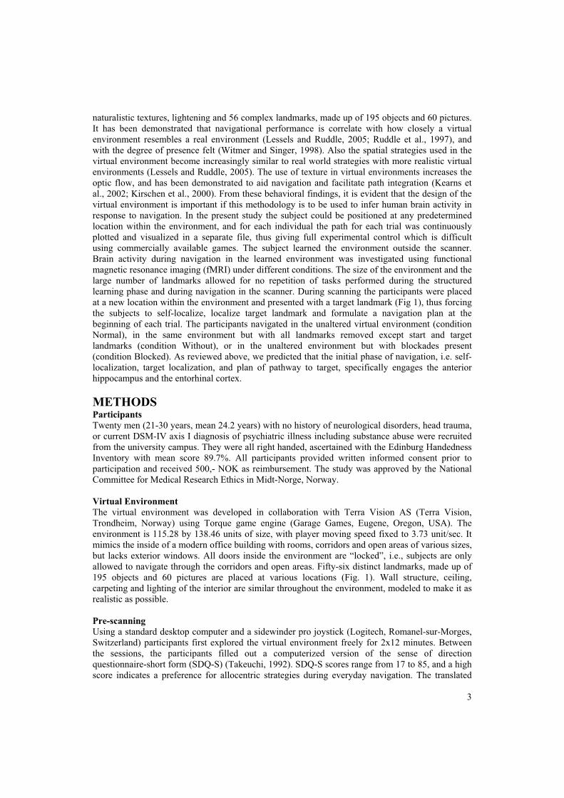
3
naturalistic textures, lightening and 56 complex landmarks, made up of 195 objects and 60 pictures. It has been demonstrated that navigational performance is correlate with how closely a virtual environment resembles a real environment (Lessels and Ruddle, 2005; Ruddle et al., 1997), and with the degree of presence felt (Witmer and Singer, 1998). Also the spatial strategies used in the virtual environment become increasingly similar to real world strategies with more realistic virtual environments (Lessels and Ruddle, 2005). The use of texture in virtual environments increases the optic flow, and has been demonstrated to aid navigation and facilitate path integration (Kearns et al., 2002; Kirschen et al., 2000). From these behavioral findings, it is evident that the design of the virtual environment is important if this methodology is to be used to infer human brain activity in response to navigation. In the present study the subject could be positioned at any predetermined location within the environment, and for each individual the path for each trial was continuously plotted and visualized in a separate file, thus giving full experimental control which is difficult using commercially available games. The subject learned the environment outside the scanner. Brain activity during navigation in the learned environment was investigated using functional magnetic resonance imaging (fMRI) under different conditions. The size of the environment and the large number of landmarks allowed for no repetition of tasks performed during the structured learning phase and during navigation in the scanner. During scanning the participants were placed at a new location within the environment and presented with a target landmark (Fig 1), thus forcing the subjects to self-localize, localize target landmark and formulate a navigation plan at the beginning of each trial. The participants navigated in the unaltered virtual environment (condition Normal), in the same environment but with all landmarks removed except start and target landmarks (condition Without), or in the unaltered environment but with blockades present (condition Blocked). As reviewed above, we predicted that the initial phase of navigation, i.e. self-localization, target localization, and plan of pathway to target, specifically engages the anterior hippocampus and the entorhinal cortex. METHODS Participants Twenty men (21-30 years, mean 24.2 years) with no history of neurological disorders, head trauma, or current DSM-IV axis I diagnosis of psychiatric illness including substance abuse were recruited from the university campus. They were all right handed, ascertained with the Edinburg Handedness Inventory with mean score 89.7%. All participants provided written informed consent prior to participation and received 500,- NOK as reimbursement. The study was approved by the National Committee for Medical Research Ethics in Midt-Norge, Norway. Virtual Environment The virtual environment was developed in collaboration with Terra Vision AS (Terra Vision, Trondheim, Norway) using Torque game engine (Garage Games, Eugene, Oregon, USA). The environment is 115.28 by 138.46 units of size, with player moving speed fixed to 3.73 unit/sec. It mimics the inside of a modern office building with rooms, corridors and open areas of various sizes, but lacks exterior windows. All doors inside the environment are “locked”, i.e., subjects are only allowed to navigate through the corridors and open areas. Fifty-six distinct landmarks, made up of 195 objects and 60 pictures are placed at various locations (Fig. 1). Wall structure, ceiling, carpeting and lighting of the interior are similar throughout the environment, modeled to make it as realistic as possible. Pre-scanning Using a standard desktop computer and a sidewinder pro joystick (Logitech, Romanel-sur-Morges, Switzerland) participants first explored the virtual environment freely for 2x12 minutes. Between the sessions, the participants filled out a computerized version of the sense of direction questionnaire-short form (SDQ-S) (Takeuchi, 1992). SDQ-S scores range from 17 to 85, and a high score indicates a preference for allocentric strategies during everyday navigation. The translated

4
questionnaire has previously been administrated to a group (n=51) of male students at NTNU, giving an score of 59.7�10.2, which is similar to the score reported in an equivalent Japanese male population, 58.6�15.6 (Ohnishi et al., 2006). Participants were then given structured navigation tasks in order to ensure that all participants had seen every landmark. In this task starting landmarks and target landmarks were all positioned in the east-west direction of each other, and the order of task was randomized between subjects. Participants were excluded if they were not able to finish the structured learning session within 60 min. Subsequently the participants performed three computer-based tests to ascertain their level of proficiency of the virtual environment: recognition of landmarks, judgments of distance, and judgments of direction between landmarks. Finally participants were given a brief demonstration of each task condition in the fMRI experiment, and practiced one of each task type. Before MRI participants were given a 30-min break. Scanning Procedure Scanning was performed on a 3T Siemens Trio scanner with a 12-channel Head Matrix Coil (Siemens AG, Erlangen, Germany). Foam pads were used to minimize head motion. The fMRI stimuli were presented using MRI compatible LCD goggles with 640x480 resolution (Nordic Neuron Lab, Bergen, Norway). Subjects moved inside the environment using a MRI compatible joystick (Current Designs, Philadelphia, USA). The participants were first allowed to familiarize themselves with the presentation equipment and joystick, and then completed four practice trials, one from each experimental condition. Scanning was commenced when complete task compliance was ensured. fMRI paradigm The fMRI paradigm was a variable length block design with alternating blocks of navigation (30±2 s) and rest (i.e. fixation; 10±2 s). There were three navigation and one baseline conditions. In the three navigation conditions participants navigated towards specific targets. All combinations of starting positions and targets were unique for the fMRI experiment, and had not been presented during the learning phase. In condition Normal, the environment was unchanged, in condition Without all landmarks except start and target landmarks were removed, and in condition Block all landmarks were in place, but some corridors very close to the target landmark were blocked. The blockage, a stop sign, was not visible before the subject came upon it. In all navigation conditions, participants were placed at a different landmark at the start of each block and an image of a target landmark was inserted at the bottom center of the screen (Fig. 1). The participants were instructed to move as fast and accurately as possible to this landmark. If the participant reached the landmark before the block ended, a new target landmark was presented. Based on pilot studies, the tasks were designed so that arrival at the first landmark could be achieved well within the time limit of the block, while the second landmark was always beyond range. The baseline condition was, condition Line, designed to control for motor and visual components of navigation. Here participants were asked to move in the environment by following a yellow line on the floor. In this condition, all landmarks were removed from the environment, including the start and target landmarks. This was done to avoid that subjects used this condition to try to learn the environment better, which was observed during pilot studies. The four navigation conditions were separated by 10s (� 2 s) of fixation, white cross on black screen. Each participant completed three experimental runs, with 20 blocks (five of each condition) and 20 fixation blocks in each run. The order of runs was randomized between participants. Performance data was logged throughout the experiment and extracted with in-house developed software written in Python (Python Software Foundation, Hampton, NH, USA). Success rate was computed as % of possible targets reached within each block. Position data of the participants’ movements inside the environment were logged with a time interval of 30 ms, and can be displayed as a trace (Fig. 1).

5
Imaging parameters T2* weighted, blood-oxygen-level-dependent (BOLD) sensitive images were acquired using an echo-planar imaging pulse sequence (TR=2600 ms, TE=30 ms, FOV=244 mm, slice thickness=3.0 mm, slice number=47, matrix=80x80 giving an in-plane resolution of 3x3 mm). Each functional run contained 327 volumes, with slices positioned as close to 90� on the anterior-posterior direction of the hippocampus as possible without causing fold-in from the neck. For anatomical reference one T1 weighted 3D volume was acquired with an MPRage sequence (TR=2300 ms, TE=30 ms, FOV=256 mm, slice thickness=1.0 mm, matrix 256x256, given an in-plane resolution of 1.0x1.0 mm). Post-scanning After scanning the volunteers were presented for a random sample of tasks performed in the scanner, and asked to indicate when the initial phase; self localization, target localization and path planning were completed. The participants also completed a study specific strategy questionnaire (SSSQ) based on the SDQ-S, but pertaining specifically to the current environment. The score range was 8-40, with score over 24 indicating a more allocentric knowledge of the virtual environment. Data Analysis Behavioral data Behavioral data were analyzed in SPSS 14.0 (SPSS Inc., Chicago, Illinois, USA). The analysis included the total scores (sum of all ratings) on the SDQ-S and SSSQ, the number of correct answers on the tests of recognition, judgment of direction and judgment of distance. The analysis also included success-rate calculated separately for conditions Normal, Without and Block. ANOVA analyses was followed by paired t-tests for within-subjects comparisons. All values are given as Mean ± SD. MRI data analysis Imaging data were analyzed using FSL 4.0 (Analysis Group, FMRIB, Oxford, UK). First, non-brain tissue was removed from the T1-weighted anatomical images using BET (Brain Extraction Tool, FMRIB, Oxford, UK), and the resulting images were transformed to the MNI 152 1x1x1 mm template (Montreal Neurological Institute, Montreal, QC, Canada) with FLIRT (FMRIB, OXFORD UK). The fMRI data was motion corrected using FLIRT, with the median volume of each run as reference. Then each functional run was co-registered to the corresponding anatomical T1 image and transformed into MNI space by using the transformation matrix obtained with the T1 image. The functional data was smoothed with a 5mm full-width at half-maximum Gaussian filter, and temporally high-pass filter with a cutoff time of 250 seconds. The statistical analysis of the fMRI data was carried out in FEAT (FEAT, FMRIB, Oxford, UK). Conditions were modeled according to a boxcar stimulus function convolved with a two-gamma hemodynamic response function. The effect of each condition was estimated with GLM using FLAME 1 (FMRIB’s Local Analysis of Mixed Effects). A whole brain analysis was performed using first a statistical threshold of Z�4 (P�0.000032) for each voxel, and then a cluster threshold of p=0.05. The conditions Normal-Line following, Without-Line following and Block-Line following, and differences between conditions Normal, Without and Blocked were explored. Since the region of interest for this study was the medial temporal lobe (MTL), a brain mask created by combining the probabilistic maps of the Harvard Oxford Structural Atlases and the Juelich Histological Atlas (part of FSL; http://www.fmrib.ox.ac.uk/fsl/fslview/atlas-descriptions.html#ho) (Flitney et al., 2007), using max probability >50 % as threshold, was applied. In total the mask encompassed 16 180 1mm voxels. The entorhinal cortex and the perirhinal cortex were segregated
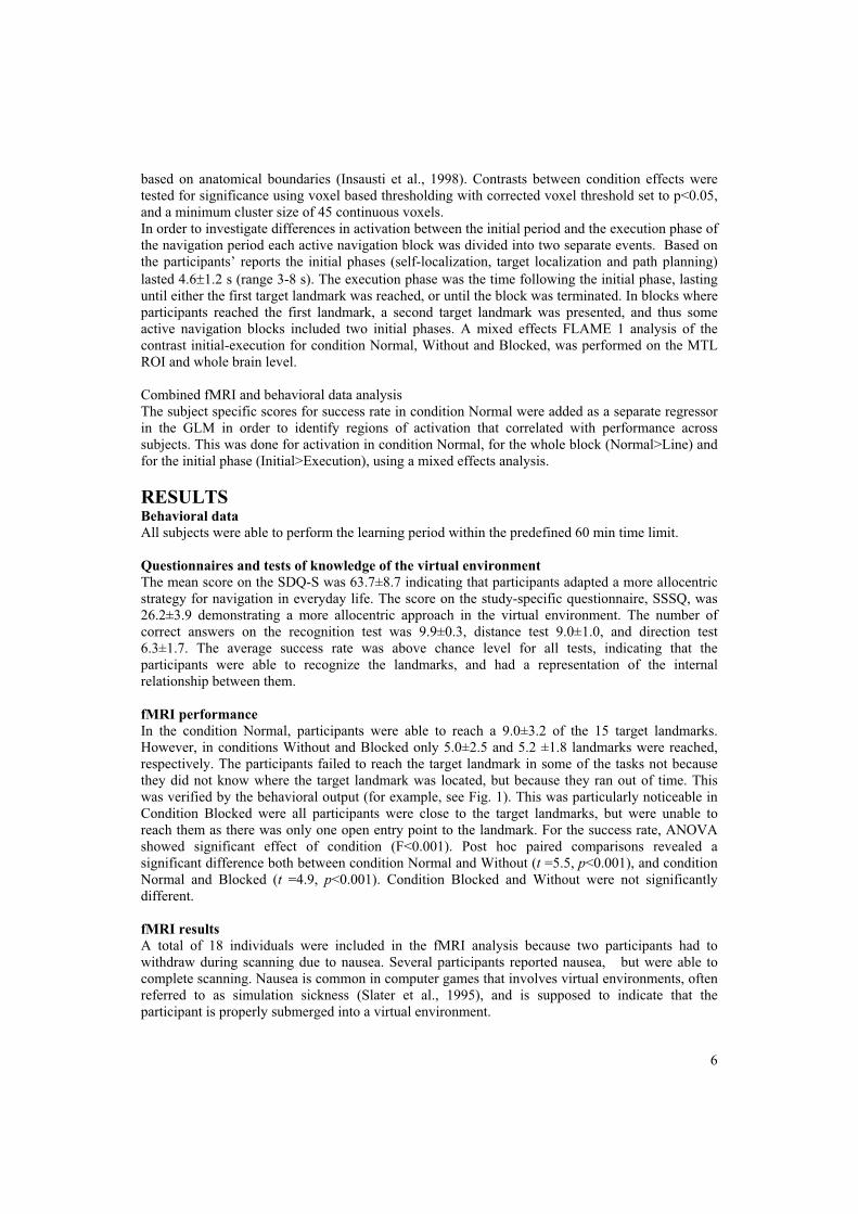
6
based on anatomical boundaries (Insausti et al., 1998). Contrasts between condition effects were tested for significance using voxel based thresholding with corrected voxel threshold set to p<0.05, and a minimum cluster size of 45 continuous voxels. In order to investigate differences in activation between the initial period and the execution phase of the navigation period each active navigation block was divided into two separate events. Based on the participants’ reports the initial phases (self-localization, target localization and path planning) lasted 4.6�1.2 s (range 3-8 s). The execution phase was the time following the initial phase, lasting until either the first target landmark was reached, or until the block was terminated. In blocks where participants reached the first landmark, a second target landmark was presented, and thus some active navigation blocks included two initial phases. A mixed effects FLAME 1 analysis of the contrast initial-execution for condition Normal, Without and Blocked, was performed on the MTL ROI and whole brain level. Combined fMRI and behavioral data analysis The subject specific scores for success rate in condition Normal were added as a separate regressor in the GLM in order to identify regions of activation that correlated with performance across subjects. This was done for activation in condition Normal, for the whole block (Normal>Line) and for the initial phase (Initial>Execution), using a mixed effects analysis. RESULTS Behavioral data All subjects were able to perform the learning period within the predefined 60 min time limit. Questionnaires and tests of knowledge of the virtual environment The mean score on the SDQ-S was 63.7±8.7 indicating that participants adapted a more allocentric strategy for navigation in everyday life. The score on the study-specific questionnaire, SSSQ, was 26.2±3.9 demonstrating a more allocentric approach in the virtual environment. The number of correct answers on the recognition test was 9.9±0.3, distance test 9.0±1.0, and direction test 6.3±1.7. The average success rate was above chance level for all tests, indicating that the participants were able to recognize the landmarks, and had a representation of the internal relationship between them. fMRI performance In the condition Normal, participants were able to reach a 9.0±3.2 of the 15 target landmarks. However, in conditions Without and Blocked only 5.0±2.5 and 5.2 ±1.8 landmarks were reached, respectively. The participants failed to reach the target landmark in some of the tasks not because they did not know where the target landmark was located, but because they ran out of time. This was verified by the behavioral output (for example, see Fig. 1). This was particularly noticeable in Condition Blocked were all participants were close to the target landmarks, but were unable to reach them as there was only one open entry point to the landmark. For the success rate, ANOVA showed significant effect of condition (F<0.001). Post hoc paired comparisons revealed a significant difference both between condition Normal and Without (t =5.5, p<0.001), and condition Normal and Blocked (t =4.9, p<0.001). Condition Blocked and Without were not significantly different. fMRI results A total of 18 individuals were included in the fMRI analysis because two participants had to withdraw during scanning due to nausea. Several participants reported nausea, but were able to complete scanning. Nausea is common in computer games that involves virtual environments, often referred to as simulation sickness (Slater et al., 1995), and is supposed to indicate that the participant is properly submerged into a virtual environment.
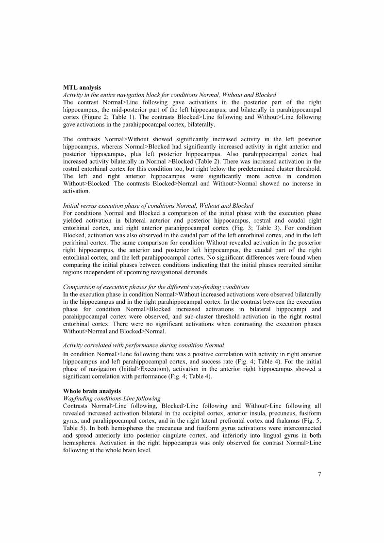
7
MTL analysis Activity in the entire navigation block for conditions Normal, Without and Blocked The contrast Normal>Line following gave activations in the posterior part of the right hippocampus, the mid-posterior part of the left hippocampus, and bilaterally in parahippocampal cortex (Figure 2; Table 1). The contrasts Blocked>Line following and Without>Line following gave activations in the parahippocampal cortex, bilaterally. The contrasts Normal>Without showed significantly increased activity in the left posterior hippocampus, whereas Normal>Blocked had significantly increased activity in right anterior and posterior hippocampus, plus left posterior hippocampus. Also parahippocampal cortex had increased activity bilaterally in Normal >Blocked (Table 2). There was increased activation in the rostral entorhinal cortex for this condition too, but right below the predetermined cluster threshold. The left and right anterior hippocampus were significantly more active in condition Without>Blocked. The contrasts Blocked>Normal and Without>Normal showed no increase in activation. Initial versus execution phase of conditions Normal, Without and Blocked For conditions Normal and Blocked a comparison of the initial phase with the execution phase yielded activation in bilateral anterior and posterior hippocampus, rostral and caudal right entorhinal cortex, and right anterior parahippocampal cortex (Fig. 3; Table 3). For condition Blocked, activation was also observed in the caudal part of the left entorhinal cortex, and in the left perirhinal cortex. The same comparison for condition Without revealed activation in the posterior right hippocampus, the anterior and posterior left hippocampus, the caudal part of the right entorhinal cortex, and the left parahippocampal cortex. No significant differences were found when comparing the initial phases between conditions indicating that the initial phases recruited similar regions independent of upcoming navigational demands. Comparison of execution phases for the different way-finding conditions In the execution phase in condition Normal>Without increased activations were observed bilaterally in the hippocampus and in the right parahippocampal cortex. In the contrast between the execution phase for condition Normal>Blocked increased activations in bilateral hippocampi and parahippocampal cortex were observed, and sub-cluster threshold activation in the right rostral entorhinal cortex. There were no significant activations when contrasting the execution phases Without>Normal and Blocked>Normal.
Activity correlated with performance during condition Normal In condition Normal>Line following there was a positive correlation with activity in right anterior hippocampus and left parahippocampal cortex, and success rate (Fig. 4; Table 4). For the initial phase of navigation (Initial>Execution), activation in the anterior right hippocampus showed a significant correlation with performance (Fig. 4; Table 4). Whole brain analysis Wayfinding conditions-Line following Contrasts Normal>Line following, Blocked>Line following and Without>Line following all revealed increased activation bilateral in the occipital cortex, anterior insula, precuneus, fusiform gyrus, and parahippocampal cortex, and in the right lateral prefrontal cortex and thalamus (Fig. 5; Table 5). In both hemispheres the precuneus and fusiform gyrus activations were interconnected and spread anteriorly into posterior cingulate cortex, and inferiorly into lingual gyrus in both hemispheres. Activation in the right hippocampus was only observed for contrast Normal>Line following at the whole brain level.
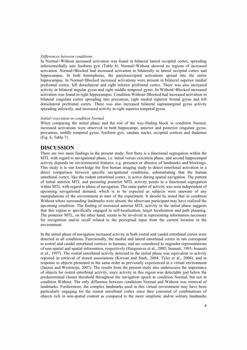
8
Differences between conditions In Normal>Without increased activation was found in bilateral lateral occipital cortex, spreading inferiormedially into fusiform gyri (Table 6). Normal<Without showed no regions of increased activation. Normal>Blocked had increased activation in bilaterally in lateral occipital cortex and hippocampus. In both hemispheres, the parietooccipital activations spread into the entire hippocampus. In Normal<Blocked increased activations were present in bilateral superior medial prefrontal cortex, left dorsolateral and right inferior prefrontal cortex. There was also increased activity in bilateral angular gyrus and right middle temporal gyrus. In Without>Blocked increased activation was found in right hippocampus. Condition Without<Blocked had increased activation in bilateral cingulate cortex spreading into precuneus, right medial superior frontal gyrus and left dorsolateral prefrontal cortex. There was also increased bilateral supramarginal gyrus activity spreading inferiorly, and increased activity in right superior temporal gyrus. Initial>execution in condition Normal When comparing the initial phase and the rest of the way-finding block in condition Normal, increased activations were observed in both hippocampi, anterior and posterior cingulate gyrus, precuneus, middle temporal gyrus, fusiform gyri, caudate nuclei, occipital cortices and thalamus (Fig. 6; Table 7). DISCUSSION There are two main findings in the present study: first there is a functional segregation within the MTL with regard to navigational phase, i.e. initial versus execution phase, and second hippocampal activity depends on environmental features, e.g. presence or absence of landmarks and blockings. This study is to our knowledge the first human imaging study to detect entorhinal activation in a direct comparison between specific navigational conditions, substantiating that the human entorhinal cortex, like the rodent entorhinal cortex, is active during spatial navigation. The pattern of initial anterior MTL and persisting posterior MTL activity points to a functional segregation within MTL with regard to phase of navigation. The same patter of activity was seen independent of upcoming navigational demand, which is to be expected as subjects were unaware of any manipulations of the environment at start of the experiment. It should be noted that in condition Without where surrounding landmarks were absent, the observant participant may have realized the upcoming condition. The finding of increased anterior MTL activity in the initial phase suggests that this region is specifically engaged in self-localization, target localization and path planning. The posterior MTL, on the other hand, seems to be involved in representing information necessary for recognition and/or recall related to the perceptual input from the current location in the environment. In the initial phase of navigation increased activity in both rostral and caudal entorhinal cortex were detected in all conditions. Functionally, the medial and lateral entorhinal cortex in rats correspond to rostral and caudal entorhinal cortices in humans, and are considered to engender representations of non-spatial and spatial information, respectively (Hargreaves et al., 2005; Insausti, 1993; Insausti et al., 1997). The rostral entorhinal activity detected in the initial phase was equivalent to activity reported in retrieval of stored associations (Kirwan and Stark, 2004; Tyler et al., 2004), and in response to objects presented in the same order as previously experienced in a virtual environment (Janzen and Weststeijn, 2007). The results from the present study also underscores the importance of objects for rostral entorhinal activity, since activity in this region was detectable just below the predetermined cluster threshold throughout the navigation epoch in condition Normal, but not in condition Without. The only difference between conditions Normal and Without was removal of landmarks. Furthermore, the complex landmarks used in this virtual environment may have been particularly engaging for the rostral entorhinal cortex since they consisted of combinations of objects rich in non-spatial content as compared to the more simplistic and/or solitary landmarks

9
used in most other virtual navigation studies (Antonova et al., 2008; Doeller et al., 2008; Ekstrom and Bookheimer, 2007; Iaria et al., 2007; Jordan et al., 2004; Parslow et al., 2005; Peigneux et al., 2004; Rauchs et al., 2008; Shipman and Astur, 2008). The initial increase in activity in caudal entorhinal cortex can also be connected to the presence of landmarks, but reflecting their spatial arrangement. Recognition and retrieval of object locations, as well as spatial ordering of objects have consistently been reported to engage caudal entorhinal cortex n in human neuroimaging studies (Adcock et al., 2006; Johnsrude et al., 1999; Owen et al., 1996). In monkeys, visually responsive cells in caudal entorhinal cortex respond to particular objects or places (Suzuki et al., 1997). In addition to engendering object-place associations, the caudal entorhinal cortex is considered to provide a representation of self-localization within an environment together with the hippocampal place cells (Moser et al., 2008). In rats, grid and border cells have been located to the medial entorhinal cortex, which is equivalent to the posterior caudal entorhinal cortex in humans (Fyhn et al., 2004; Insausti, 1993; Insausti et al., 1997; Solstad et al., 2008). The activity of grid and border cells in humans during different phases of navigation is unknown. The virtual environment used in the present experiment was large compared to those used in most neuroimaging studies of navigation (Antonova et al., 2008; Doeller et al., 2008; Ekstrom and Bookheimer, 2007; Grön et al., 2000; Iaria et al., 2007; Iaria et al., 2008; Jordan et al., 2004; Ohnishi et al., 2006; Parslow et al., 2005; Peigneux et al., 2004; Rauchs et al., 2008; Shipman and Astur, 2008). It is possible that the size of the virtual environment to be mentally represented was an important determinant for increasing the caudal entorhinal activity above level of detection. In support of this is the positive correlation between increasing distance to target and caudal entorhinal activity using London as the environment (Spiers and Maguire, 2007). It seems probable that the subjects will engender a representation of the entire virtual environment in order to self-localize anew in the initial phase, as well as determine target location and choose the appropriate path. When the navigational plan is executed, a limited representation of the virtual environment may suffice, possibly leading to a decline in caudal entorhinal activity. Indeed, spatial reference memory has been shown not to be updated during locomotion in humans (Mou et al., 2004), and one human lesion study demonstrates that entorhinal cortex is not necessary for path integration (Shrager et al., 2008). Lack of entorhinal activation in many neuroimaging navigation studies could be due to susceptibility artifacts in T2* weighted, blood-oxygen-level-dependent gradient echo echo planar imaging (i.e. BOLD fMRI) scans. These artifacts are most pronounced in the entorhinal cortex (Ojemann et al., 1997). The slice orientation in the present study reduces this problem (Chen et al., 2003). The presence of such susceptibility artifacts in BOLD fMRI may explain why PET studies have detected entorhinal activity in mental navigation (Ghaem et al., 1997; Mellet et al., 2000), which has not been reproduced in comparable fMRI studies (Avila et al., 2006; Kumaran and Maguire, 2005). It should be noted that brain activity detected using fMRI only depicts differences in activity in one condition relative to another. Persistent entorhinal cortex activity across all conditions can therefore not be visualized. Still, our results clearly demonstrate a dynamic role for the entorhinal cortex, with increased engagement in the initial phase. Mental navigation can be considered a type of self-projection or prospection; i.e. looking into the future. Interestingly, similar right entorhinal activity has been reported in one study of self-projection using construction of future episodes (Addis et al., 2008). Furthermore, bilateral anterior hippocampal activity is reported in studies of self-projection (Addis et al., 2008; Szpunar et al., 2007), and the location of this activity is similar to the bilateral anterior hippocampal activity detected in the initial phase in the present navigation study. Equivalent anterior hippocampal activity has been reported in studies of mental navigation (Ghaem et al., 1997; Mellet et al., 2000), bird’s eye view navigation (Jordan et al., 2004) and during way-finding conditions (Spiers and

10
Maguire, 2006). Mental and bird’s eye view navigation entail entering an imaginary, non-actual view of one’s position and actions in space. Changing one’s point of view from first to third person perspective also correlates with bilateral anterior hippocampal activity (Schmidt et al., 2007). The bilateral anterior hippocampal activity in the initial phase of navigation thus seems to draw on similar regions in the MTL as prospection and third person’s point of view (Addis et al., 2008; Szpunar et al., 2007). Thus it seems that self-localization, target localization and path planning involve construction of a mental representation of the environment based on a meta-perspective of the layout. Indeed, behavioral data show that individuals use mental imagery as strategy for navigational planning (Spiers and Maguire, 2008). Furthermore, also in rats are the hippocampi involved in planning of future actions, i.e. vicarious trial and error (Hu and Amsel, 1995), which is considered the rodent equivalent of prospection or self-projection. Moreover, right anterior hippocampal activity similar to that in the initial phase of navigation in the present study has been reported in target localization (Doeller et al., 2008; Schmidt et al., 2007; Shipman and Astur, 2008), especially when there is increasing demands on memory-based navigation (Shipman and Astur, 2008), navigational planning and re-planning (Spiers and Maguire, 2006), and in studies of object recognition and object-place associations in virtual environments (Bohbot et al., 2004; Janzen and Weststeijn, 2007). The finding that navigational success in condition Normal correlated with activation in the right anterior hippocampus for the initial phase and whole block of navigation further substantiates the claim that the anterior hippocampus is essential for accurate navigation. Previously, path integration (Wolbers et al., 2007) and spatial performance (Schmidt et al., 2007) have been shown to correlate with activity in the right anterior hippocampus with similar coordinates as in the current study. Since the anterior hippocampus is considered to support relational processing (Kirwan and Stark, 2004; Schacter and Wagner, 1999), including flexible (re)combination of elements extracted from previous learned associations (Preston et al., 2004), it is ideally suited to build a mental model based on previous experiences in the virtual environment. This is corroborated by animal studies where the anterior hippocampus has been shown to support a unitary representation of the environment as a whole (Kjelstrup et al., 2008). In most neuroimaging studies of navigation, way-finding has been shown to evoke activation of the posterior hippocampus (Antonova et al., 2008; Peigneux et al., 2004; Rauchs et al., 2008). In the present study hippocampal activity throughout the entire navigational period was only found in condition Normal in both the whole brain and MTL ROI analyses. The center of gravity for this enduring activity was in the right posterior hippocampus similar to that observed in numerous fMRI studies of way-finding in familiar virtual towns or indoor environments (Antonova et al., 2008; Iaria et al., 2007; Peigneux et al., 2004; Rauchs et al., 2008). Using multivariate pattern analysis, a recent study demonstrated that accurate allocentric differentiation of position within a familiar environment is located to the body-posterior of the right hippocampus (Hassabis et al., 2009), with coordinates similar to those observed in condition Normal for the entire navigation block in this study. Hippocampal theories suggest that the posterior hippocampus represents environmental detail (Moser and Moser, 1998). Changes in the environment as in condition Without decreased the activity in posterior hippocampus thus underscoring the importance of a familiar, stable environment to produce such positional activity within the hippocampus. Also condition Blocked did not have persisting hippocampal activity during navigation. The execution phase of this condition resembled to some extent the Morris water maze (Morris, 1984) with visible platform, as targets were visible quite early in the navigation block. It has been shown in humans that conditions similar to the visible platform condition do not require hippocampal activity (Shipman and Astur, 2008). An alternative interpretation is suppression of MTL activity by the rostral medial prefrontal cortex (Anderson et al., 2004; Miller and Cohen, 2003; Spiers and Maguire, 2006). The medial rostral prefrontal cortex was significantly more active during condition Blocked. Like the hippocampus, the parahippocampal cortex displayed an anterior-posterior division of activity during the course of navigation. Very few neuroimaging studies of navigation have reported

11
anterior parahippocampal activity (Parslow et al., 2005; Spiers and Maguire, 2006). In the current study the anterior parahippocampal cortex was more active in the initial phase, whereas the posterior part was active throughout the navigational period in all conditions. Animal and human studies, as well as theoretical models, suggest that the anterior parahippocampal cortex generates a representation of the environmental layout (Bird and Burgess, 2008; Moscovitch et al., 2005). Spatial memory and associations, and retrieval of indirect spatial relationships all engage the anterior parahippocampal cortex (Ekstrom and Bookheimer, 2007; Epstein, 2008; Preston et al., 2004). Again our results from the initial phase substantiate the claim that self-localization, target localization and path planning require a mental representation of the virtual environment at large. Furthermore, this representation of the virtual environment in the anterior parahippocampal cortex correlated with navigational success. The posterior parahippocampus cortex was, however, active throughout navigation and in all conditions, underscoring the perceptual role of this region (Preston et al., 2004). Behavioral data on performance in virtual environments points to the importance of a close resemblance between this and real world environments (Lessels and Ruddle, 2005; Ruddle et al., 1997). But the experience of virtual environments can never truly reflect the natural setting; the virtual re-creation of real world features are not yet truly lifelike, and more importantly virtual navigation relies solely on visual input, since input from the vestibular system and sensory feedback from the body are absent (Lessels and Ruddle, 2005). Still, the participants in the present study felt submerged in the environment and knew the environment well as indicated by the post-learning test scores and performance during scanning. Even though success rate appeared to be low, particularly for conditions Without and Blocked, the subjects did not wander aimlessly around. From the individual maps of all navigational tasks it was apparent that the participants were close to the target, but did not reach it before the block terminated. This suggests that the differences in activation between conditions arose from differences in the navigational demands due to environmental constraints. The size of the environment and the large number of landmarks allowed the subjects to be presented with novel combinations of spawning and target positions during learning and scanning. This was done in an attempt to promote an allocentric approach to navigation. Based on the SSSQ, participants did indeed use a more allocentric strategy when navigating during fMRI. Together these features of the experimental design appeared to actively engage the MTL substructures. Thus enabling us to clearly separate activity in MTL subregions in response to different navigational demands. In summary, our results demonstrate that navigation requires dynamic recruitment of MTL subregions as navigation progresses from self-localization, target localization, and navigational planning to execution of the navigation plan. Furthermore, the current findings suggest a functional segregation between anterior and posterior MTL. The anterior MTL is specifically engaged in the initial phase of navigation, irrespective of upcoming navigational demands, whereas posterior MTL activation persists throughout navigation. However, increased hippocampal activity depended on the environment remaining unaltered. For the first time the entorhinal cortex was shown to be specifically engaged in the initial phase of navigation confirming previous animal studies that suggest a critical function for the entorhinal cortex in spatial navigation. Also the anterior parahippocampal cortex and the anterior hippocampus were specifically engaged in the initial phase of navigation, and the right anterior hippocampal activity was directly related to successful navigation in the subsequent execution phase. Right posterior hippocampal activity throughout navigation appeared to be connected to keeping track of the current location in a well-known environment. ACKNOWLEDGEMENT This study was funded by the Norwegian University of Science and Technology (NTNU), and Center of competence for fMRI, St. Olav’s hospital

12
REFERENCES Adcock R, Thangavel A, Whitfield-Gabrieli S, Knutson B, Gabrieli J. 2006. Reward-motivated
learning: mesolimbic activation precedes memory formation. Neuron 50(3):507-517. Addis D, Pan L, Vu M, Laiser N, Schacter D. 2008. Constructive episodic simulation of the future
and the past: Distinct subsystems of a core brain network mediate imagining and remembering. Neuropsychologia.
Anderson M, Ochsner K, Kuhl B, Cooper J, Robertson E, Gabrieli S, Glover G, Gabrieli J. 2004. Neural systems underlying the suppression of unwanted memories. American Association for the Advancement of Science. p 232-235.
Antonova E, Parslow D, Brammer M, Dawson G, Jackson S, Morris R. 2008. Age-related neural activity during allocentric spatial memory. Memory 99999(1):1-19.
Avila C, Barros-Loscertales A, Forn C, Mallo R, Parcet M, Belloch V, Campos S, Feliu-Tatay R, Gonzalez-Darder J. 2006. Memory lateralization with 2 functional MR imaging tasks in patients with lesions in the temporal lobe. Am Soc Neuroradiology. p 498-503.
Bird C, Burgess N. 2008. The hippocampus and memory: insights from spatial processing. Bohbot V, Iaria G, Petrides M. 2004. Hippocampal function and spatial memory: evidence from
functional neuroimaging in healthy participants and performance of patients with medial temporal lobe resections. NEUROPSYCHOLOGY-NEW YORK- 18(3):418-425.
Chen N, Dickey C, Yoo S, Guttmann C, Panych L. 2003. Selection of voxel size and slice orientation for fMRI in the presence of susceptibility field gradients: application to imaging of the amygdala. Neuroimage 19(3):817-825.
Doeller C, King J, Burgess N. 2008. Parallel striatal and hippocampal systems for landmarks and boundaries in spatial memory. Proceedings of the National Academy of Sciences 105(15):5915.
Ekstrom A, Bookheimer S. 2007. Spatial and temporal episodic memory retrieval recruit dissociable functional networks in the human brain. Learning & Memory 14(10):645.
Ekstrom A, Kahana M, Caplan J, Fields T, Isham E, Newman E, Fried I. 2003. Cellular networks underlying human spatial navigation. Nature 425(6954):184-188.
Epstein R. 2008. Parahippocampal and retrosplenial contributions to human spatial navigation. Trends in Cognitive Sciences 12(10):388-396.
Flitney D, Webster M, Patenaude B, Seidman L, Goldstein J, Tordesillas Gutierrez D, Eickhoff S, Amunts K, Zilles K, Lancaster J. Anatomical Brain Atlases and Their Application in the FSLView Visualisation Tool; 2007.
Fyhn M, Molden S, Witter M, Moser E, Moser M. 2004. Spatial representation in the entorhinal cortex. Science 305(5688):1258-1264.
Ghaem O, Mellet E, Crivello F, Tzourio N, Mazoyer B, Berthoz A, Denis M. 1997. Mental navigation along memorized routes activates the hippocampus, precuneus, and insula. Neuroreport 8(3):739.
Grön G, Wunderlich A, Spitzer M, Tomczak R, Riepe M. 2000. Brain activation during human navigation: gender-different neural networks as substrate of performance. Nature Neuroscience 3(4):404-408.
Hargreaves E, Rao G, Lee I, Knierim J. 2005. Major dissociation between medial and lateral entorhinal input to dorsal hippocampus. American Association for the Advancement of Science. p 1792-1794.
Hassabis D, Chu C, Rees G, Weiskopf N, Molyneux P, Maguire E. 2009. Decoding Neuronal Ensembles in the Human Hippocampus. Current Biology.
Hu D, Amsel A. 1995. A simple test of the vicarious trial-and-error hypothesis of hippocampal function. Proceedings of the National Academy of Sciences 92(12):5506-5509.
Iaria G, Chen J, Guariglia C, Ptito A, Petrides M. 2007. Retrosplenial and hippocampal brain regions in human navigation: complementary functional contributions to the formation and use of cognitive maps. European Journal of Neuroscience 25(3):890.

13
Iaria G, Fox C, Chen J, Petrides M, Barton J. 2008. Detection of unexpected events during spatial navigation in humans: bottom-up attentional system and neural mechanisms. European Journal of Neuroscience 27(4):1017.
Insausti R. 1993. Comparative anatomy of the entorhinal cortex and hippocampus in mammals. Hippocampus 3:19.
Insausti R, Herrero M, Witter M. 1997. Entorhinal cortex of the rat: cytoarchitectonic subdivisions and the origin and distribution of cortical efferents. Hippocampus 7(2).
Insausti R, Juottonen K, Soininen H, Insausti A, Partanen K, Vainio P, Laakso M, Pitkanen A. 1998. MR volumetric analysis of the human entorhinal, perirhinal, and temporopolar cortices. American Journal of Neuroradiology 19(4):659-671.
Janzen G, Weststeijn C. 2007. Neural representation of object location and route direction: an event-related fMRI study. Brain research 1165:116-125.
Jeffery K. 2007. Self-localization and the entorhinal–hippocampal system. Current Opinion in Neurobiology 17(6):684-691.
Johnsrude I, Owen A, Crane J, Milner B, Evans A. 1999. A cognitive activation study of memory for spatialrelationships. Neuropsychologia 37(7):829-841.
Jordan K, Schadow J, Wuestenberg T, Heinze H, Jäncke L. 2004. Different cortical activations for subjects using allocentric or egocentric strategies in a virtual navigation task. Neuroreport 15(1):135.
Kearns M, Warren W, Duchon A, Tarr M. 2002. Path integration from optic flow and body senses in a homing task. PERCEPTION-LONDON- 31(3):348-374.
Kirschen M, Kahana M, Sekuler R, Burack B. 2000. Optic flow helps humans learn to navigate through synthetic environments. PERCEPTION-LONDON- 29(7):801-818.
Kirwan C, Stark C. 2004. Medial temporal lobe activation during encoding and retrieval of novel face-name pairs. Hippocampus 14(7).
Kjelstrup K, Solstad T, Brun V, Hafting T, Leutgeb S, Witter M, Moser E, Moser M. 2008. Finite scale of spatial representation in the hippocampus. Science 321(5885):140.
Kumaran D, Maguire E. 2005. The human hippocampus: cognitive maps or relational memory? Journal of Neuroscience 25(31):7254-7259.
Lessels S, Ruddle R. 2005. Movement around real and virtual cluttered environments. Presence: Teleoperators & Virtual Environments 14(5):580-596.
Mellet E, Bricogne S, Tzourio-Mazoyer N, Ghaem O, Petit L, Zago L, Etard O, Berthoz A, Mazoyer B, Denis M. 2000. Neural correlates of topographic mental exploration: the impact of route versus survey perspective learning. Neuroimage 12(5):588-600.
Miller E, Cohen J. 2003. An integrative theory of prefrontal cortex function. Morris R. 1984. Developments of a water-maze procedure for studying spatial learning in the rat. J
Neurosci Methods 11(1):47-60. Moscovitch M, Rosenbaum R, Gilboa A, Addis D, Westmacott R, Grady C, McAndrews M, Levine
B, Black S, Winocur G. 2005. Functional neuroanatomy of remote episodic, semantic and spatial memory: a unified account based on multiple trace theory. Journal of Anatomy 207(1):35.
Moser E, Kropff E, Moser M. 2008. Place cells, grid cells, and the brain's spatial representation system.
Moser M, Moser E. 1998. Functional differentiation in the hippocampus. Hippocampus 8(6). Mou W, McNamara T, Valiquette C, Rump B. 2004. Allocentric and egocentric updating of spatial
memories. Journal of Experimental Psychology Learning Memory and Cognition 30(1):142-157.
O'keefe J, Dostrovsky J. 1971. The hippocampus as a spatial map. Preliminary evidence from unit activity in the freely-moving rat. Brain research 34(1):171.
Ohnishi T, Matsuda H, Hirakata M, Ugawa Y. 2006. Navigation ability dependent neural activation in the human brain: an fMRI study. Neuroscience research 55(4):361-369.

14
Ojemann J, Akbudak E, Snyder A, McKinstry R, Raichle M, Conturo T. 1997. Anatomic localization and quantitative analysis of gradient refocused echo-planar fMRI susceptibility artifacts. Neuroimage 6(3):156-167.
Owen A, Milner B, Petrides M, Evans A. 1996. A SPECIFIC ROLE FOR THE RIGHT PARAHIPPOCAMPAL GYRUS IN THE RETRIEVAL OF OBJECT-LOCATION: A POSITRON EMISSION TOMOGRAPHY STUD. Journal of Cognitive Neuroscience 8(6):588-602.
Parslow D, Morris R, Fleminger S, Rahman Q, Abrahams S, Recce M. 2005. Allocentric spatial memory in humans with hippocampal lesions. Acta psychologica 118(1-2):123-147.
Peigneux P, Laureys S, Fuchs S, Collette F, Perrin F, Reggers J, Phillips C, Degueldre C, Del Fiore G, Aerts J. 2004. Are spatial memories strengthened in the human hippocampus during slow wave sleep? Neuron 44(3):535-545.
Preston A, Shrager Y, Dudukovic N, Gabrieli J. 2004. Hippocampal contribution to the novel use of relational information in declarative memory. Hippocampus 14(2):148-152.
Rauchs G, Orban P, Balteau E, Schmidt C, Degueldre C, Luxen A, Maquet P, Peigneux P. 2008. Partially segregated neural networks for spatial and contextual memory in virtual navigation. Hippocampus 18(5).
Ruddle R, Payne S, Jones D. 1997. Navigating buildings in" desk-top" virtual environments: Experimental investigations using extended navigational experience. Journal of Experimental Psychology Applied 3:143-159.
Sargolini F, Fyhn M, Hafting T, McNaughton B, Witter M, Moser M, Moser E. 2006. Conjunctive representation of position, direction, and velocity in entorhinal cortex. American Association for the Advancement of Science. p 758-762.
Schacter D, Wagner A. 1999. Medial temporal lobe activations in fMRI and PET studies of episodic encoding and retrieval. Hippocampus 9(1).
Schmidt D, Krause B, Weiss P, Fink G, Shah N, Amorim M, Müller H, Berthoz A. 2007. Visuospatial working memory and changes of the point of view in 3D space. Neuroimage 36(3):955-968.
Shipman S, Astur R. 2008. Factors affecting the hippocampal BOLD response during spatial memory. Behavioural Brain Research 187(2):433-441.
Shrager Y, Kirwan C, Squire L. 2008. Neural basis of the cognitive map: Path integration does not require hippocampus or entorhinal cortex. Proceedings of the National Academy of Sciences 105(33):12034.
Slater M, Usoh M, Steed A. 1995. Taking steps: the influence of a walking technique on presence in virtual reality. ACM Transactions on Computer-Human Interaction (TOCHI) 2(3):201-219.
Solstad T, Boccara C, Kropff E, Moser M, Moser E. 2008. Representation of geometric borders in the entorhinal cortex. Science 322(5909):1865.
Spiers H, Maguire E. 2006. Thoughts, behaviour, and brain dynamics during navigation in the real world. Neuroimage 31(4):1826-1840.
Spiers H, Maguire E. 2007. A navigational guidance system in the human brain. Hippocampus 17(8):618.
Spiers H, Maguire E. 2008. The dynamic nature of cognition during wayfinding. Journal of environmental psychology 28(3):232-249.
Suzuki W, Miller E, Desimone R. 1997. Object and place memory in the macaque entorhinal cortex. Journal of Neurophysiology 78(2):1062-1081.
Szpunar K, Watson J, McDermott K. 2007. Neural substrates of envisioning the future. Proceedings of the National Academy of Sciences 104(2):642.
Takeuchi Y. 1992. Sense of direction and its relationship with geographical orientation, personality traits and mental ability. Japanese Journal of Educational Psychology 40:47–53.
Tyler L, Stamatakis E, Bright P, Acres K, Abdallah S, Rodd J, Moss H. 2004. Processing objects at different levels of specificity. Journal of Cognitive Neuroscience 16(3):351-362.

15
Witmer B, Singer M. 1998. Measuring presence in virtual environments: A presence questionnaire. Presence 7(3):225-240.
Wolbers T, Wiener J, Mallot H, Buchel C. 2007. Differential recruitment of the hippocampus, medial prefrontal cortex, and the human motion complex during path integration in humans. Journal of Neuroscience 27(35):9408.
TABLES Table 1. Regions within the medial temporal lobe (MTL) with increased activity in conditions Normal, Without and Blocked versus the base line condition line following. The analysis was carried out using a hippocampal-parahippocampal gyrus mask and voxel based thresholding, p=0.05 corrected for multiple comparisons. Voxel size is 1 mm3. Only clusters with a cluster size > 45 voxels were reported. R; right; L, left; MNI, Montreal Neurological Institute 152 brain template.
Coordinates of peak activation (MNI) MTL region
X Y Z
Cluster size (no. of voxels)
Z score
Normal > Line following R. Hippocampus 29 -35 -14 567 5.19 L. Hippocampus -23 -29 -8 58 4.73 R. Parahippocampal cortex 29 -35 -14 632 5.19 L. Parahippocampal cortex -21 -41 -13 209 6.00
Without > Line following R. Parahippocampal cortex 17 -34 -15 557 5.57 L. Parahippocampal cortex -21 -41 -13 69 4.66 Blocked > Line following R. Parahippocampal cortex 28 -36 -14 305 4.83 L. Parahippocampal cortex -21 -41 -13 53 4.94
Table 2. Peak activations in the medial temporal lobe (MTL) ROI when comparing the different wayfinding conditions. The analysis was carried out using a hippocampal-parahippocampal gyrus mask, i.e. a MTL ROI, and voxel based thresholding (p=0.05, corrected for multiple comparisons). R, right; L, left; MTL, medial temporal lobe; MNI, Montreal Neurological Institute 152 template.
Coordinates of peak activation (MNI)
MTL region
X Y Z
Cluster size (no. of voxels)
Z score
Normal – Without L. Hippocampus -21 -31 -7 102 4.25 Without – Normal (no significant increase in activation observed) Normal – Blocked R. Hippocampus 30
35 -39 -18
0 -21
102 68
5.33 4.25
L. Hippocampus -31 -38 -5 312 5.12 L. Parahippocampal cortex -27 -32 -21 83 4.45 Blocked– Normal (no significant increase in activation observed) Without – Blocked (no significant increase in activation observed) Blocked– Without (no significant increase in activation observed)

16
Table 3. Regions of increased activity within the medial temporal lobe (MTL) ROI when comparing the initial phase with the execution phase in conditions Normal, Without and Blocked. The analysis was carried out using a hippocampal-parahippocampal gyrus mask and voxel based thresholding, p=0.05 corrected for multiple comparisons. Voxel size is 1 mm3. Only clusters with a cluster size >45 voxels were reported. The cluster number is given in parenthesis for secondary peaks within the respective clusters. Numbers in the cluster size column represent the actual number of voxels within the anatomical region in the respective row. R, right; L, left; MNI, Montreal Neurological Institute 152 brain template.
Coordinates of peak activation (MNI)
MTL region
X Y Z
Cluster no.
Cluster size(no. of voxels)
Z score
Normal
R. Hippocampus 25 23 24 31
-20 -25 -32 -17
-18 -11 -8 -14
1 (1) (1) (1)
1223
4.80 4.37 3.89 3.77
L. Hippocampus -22 -20 -20
-23 -16 -30
-15 -15 -10
2 (2) (2)
1311
4.49 3.90 3.82
R. Entorhinal cortex 20 25
-2 -19
-23 -25
3 (1)
45
3.65 3.66
R. Parahippocampal cortex
28 -29 -23 4 116 3.73
L. Parahippocampal cortex
-15 -29
-32 -35
-10 -15
5 6
49 198
4.16 4.00
Without
R. Hippocampus 20 -31 -6 1 69 3.91 L. Hippocampus -20
-27 -24
-34 -17 -25
-5 -20 -11
2 (2) (2)
1042
4.37 4.12 3.94
R. Entorhinal cortex 27 -17 -28 3 56 3.46
Blocked
R. Hippocampus 26 26 23
-22 -12 -35
-12 -27 -3
(1) (1) (1)
5.00 4.83 4.53
L. Hippocampus -19 -22 -20
-34 -24 -17
-7 -16 -18
4 (4) (4)
2395
4.64 4.36 4.12
R. Entorhinal cortex 26 19
-13 -3
-29 -27
1 (1)
2930
5.00 4.13
L. Entorhinal cortex -17 -17 -27 (4) 4.10 L. Perhinal cortex -30 -7 -32 2 62 3.92 R. Parahippocampal cortex
21 26
-35 -24
-18 -25
3 (3)
759
4.51 4.47
L. Parahippocampal cortex
-15 -24 -24
-32 -40 -23
-10 -14 -25
4 (4) (4)
3535
5.01 4.73 4.24
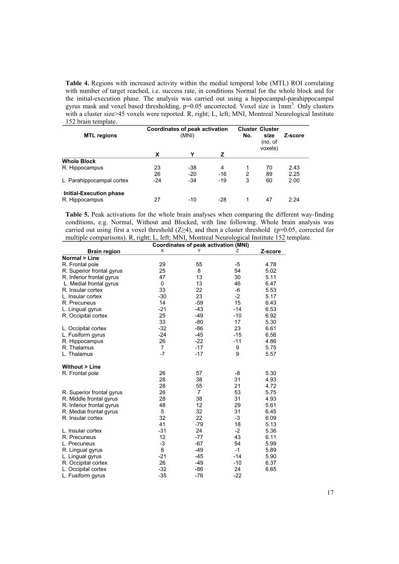
17
Table 4. Regions with increased activity within the medial temporal lobe (MTL) ROI correlating with number of target reached, i.e. success rate, in conditions Normal for the whole block and for the initial-execution phase. The analysis was carried out using a hippocampal-parahippocampal gyrus mask and voxel based thresholding, p=0.05 uncorrected. Voxel size is 1mm3. Only clusters with a cluster size>45 voxels were reported. R, right; L, left; MNI, Montreal Neurological Institute 152 brain template.
Coordinates of peak activation (MNI)
ClusterNo.
Cluster size
(no. of voxels)
Z-score
MTL regions
X Y Z
Whole Block
R. Hippocampus 23 26
-38 -20
4 -16
1 2
70 89
2.43 2.25
L. Parahippocampal cortex -24 -34 -19 3 60 2.00 Initial-Execution phase R. Hippocampus 27 -10 -28 1 47 2.24
Table 5. Peak activations for the whole brain analyses when comparing the different way-finding conditions, e.g. Normal, Without and Blocked, with line following. Whole brain analysis was carried out using first a voxel threshold (Z�4), and then a cluster threshold (p=0.05, corrected for multiple comparisons). R, right; L, left; MNI, Montreal Neurological Institute 152 template.
Coordinates of peak activation (MNI) Brain region X Y Z
Z-score
Normal > Line
R. Frontal pole 29 55 -5 4.78 R. Superior frontal gyrus 25 8 54 5.02 R. Inferior frontal gyrus 47 13 30 5.11 L. Medial frontal gyrus 0 13 46 6.47 R. Insular cortex 33 22 -6 5.53 L. Insular cortex -30 23 -2 5.17 R. Precuneus 14 -59 15 6.43 L. Lingual gyrus -21 -43 -14 6.53 R. Occipital cortex 25
33 -49 -80
-10 17
6.92 5.30
L. Occipital cortex -32 -86 23 6.61 L. Fusiform gyrus -24 -45 -15 6.56 R. Hippocampus 26 -22 -11 4.86 R. Thalamus 7 -17 9 5.75 L. Thalamus -7 -17 9 5.57 Without > Line R. Frontal pole 26
28 28
57 38 55
-8 31 21
5.30 4.93 4.72
R. Superior frontal gyrus 26 7 53 5.75 R. Middle frontal gyrus 28 38 31 4.93 R. Inferior frontal gyrus 48 12 29 5.61 R. Medial frontal gyrus 5 32 31 6.45 R. Insular cortex 32
41 22 -79
-3 18
6.09 5.13
L. Insular cortex -31 24 -2 5.36 R. Precuneus 12 -77 43 6.11 L. Precuneus -3 -67 54 5.99 R. Lingual gyrus 8 -49 -1 5.89 L. Lingual gyrus -21 -45 -14 5.90 R. Occipital cortex 26 -49 -10 6.37 L. Occipital cortex -32 -86 24 6.65 L. Fusiform gyrus -35 -76 -22
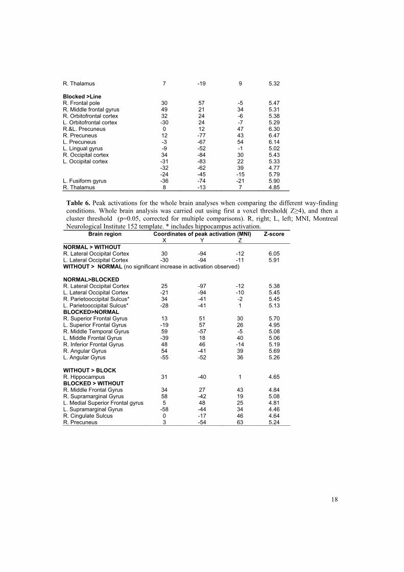
18
R. Thalamus 7 -19 9 5.32 Blocked >Line R. Frontal pole 30 57 -5 5.47 R. Middle frontal gyrus 49 21 34 5.31 R. Orbitofrontal cortex 32 24 -6 5.38 L. Orbitofrontal cortex -30 24 -7 5.29 R.&L. Precuneus 0 12 47 6.30 R. Precuneus 12 -77 43 6.47 L. Precuneus -3 -67 54 6.14 L. Lingual gyrus -9 -52 -1 5.02 R. Occipital cortex 34 -84 30 5.43 L. Occipital cortex -31
-32 -24
-83 -62 -45
22 39 -15
5.33 4.77 5.79
L. Fusiform gyrus -36 -74 -21 5.90 R. Thalamus 8 -13 7 4.85 Table 6. Peak activations for the whole brain analyses when comparing the different way-finding conditions. Whole brain analysis was carried out using first a voxel threshold( Z�4), and then a cluster threshold (p=0.05, corrected for multiple comparisons). R, right; L, left; MNI, Montreal Neurological Institute 152 template. * includes hippocampus activation.
Coordinates of peak activation (MNI) Brain region X Y Z
Z-score
NORMAL > WITHOUT R. Lateral Occipital Cortex 30 -94 -12 6.05 L. Lateral Occipital Cortex -30 -94 -11 5.91 WITHOUT > NORMAL (no significant increase in activation observed) NORMAL>BLOCKED R. Lateral Occipital Cortex 25 -97 -12 5.38 L. Lateral Occipital Cortex -21 -94 -10 5.45 R. Parietooccipital Sulcus* 34 -41 -2 5.45 L. Parietooccipital Sulcus* -28 -41 1 5.13 BLOCKED>NORMAL R. Superior Frontal Gyrus 13 51 30 5.70 L. Superior Frontal Gyrus -19 57 26 4.95 R. Middle Temporal Gyrus 59 -57 -5 5.08 L. Middle Frontal Gyrus -39 18 40 5.06 R. Inferior Frontal Gyrus 48 46 -14 5.19 R. Angular Gyrus 54 -41 39 5.69 L. Angular Gyrus -55 -52 36 5.26 WITHOUT > BLOCK R. Hippocampus 31 -40 1 4.65 BLOCKED > WITHOUT R. Middle Frontal Gyrus 34 27 43 4.84 R. Supramarginal Gyrus 58 -42 19 5.08 L. Medial Superior Frontal gyrus 5 48 25 4.81 L. Supramarginal Gyrus -58 -44 34 4.46 R. Cingulate Sulcus 0 -17 46 4.64 R. Precuneus 3 -54 63 5.24

19
Table 7. Brain activity patterns for the whole brain analyses when comparing the initial planning phase with the rest of the block for the normal way-finding condition. Whole brain analysis was carried out using first a voxel threshold( Z�4), and then a cluster threshold (p=0.05, corrected for multiple comparisons). R, right; L, left; MNI, Montreal Neurological Institute 152 template.
Coordinates of peak activation (MNI) Brain region X Y Z
Z-score
R. Cingulate gyrus, anterior part 2 -11 41 4.71 R. Cingulate gyrus, posterior part 1 -38 29 4.42 L. Cingulate gyrus, posterior part -12 -38 38 4.94 L. Postcentral gyrus -37 -36 57 5.19 R. Middle temporal gyrus 62
52 -52 -61
3 0
5.30 5.09
L. Precuneus -6 -50 52 4.49 L. Lateral Occipital cortex -51 -63 7 5.18 R. Cuneal cortex 2 -83 22 5.09 L. Primary visual cortex -7 -76 14 4.52 L. Lingual gyrus -17 -64 -4 5.27 R. Hippocampus 25 -20 -18 4.80 L. Caudate nucleus -11 10 0 4.64 L. Thalamus -4 -17 2 4.71 L. Cerebellum -36 -50 -26 5.37 FIGURES Figure 1. Overview of the virtual environment. (a) The initial view a participant was presented with at entering the virtual environment. The target landmark was shown as a small image at the bottom of the screen. (b) Movement patterns of all participants for one of the navigation tasks mapped onto a two dimensional map of the environment. (c) A two dimensional overview map of the virtual office landscape, each number indicates the location of one landmark.
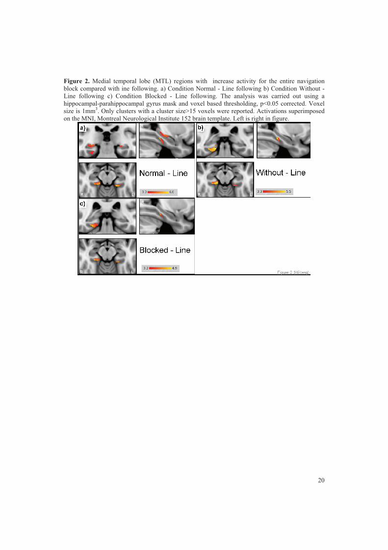
20
Figure 2. Medial temporal lobe (MTL) regions with increase activity for the entire navigation block compared with ine following. a) Condition Normal - Line following b) Condition Without -Line following c) Condition Blocked - Line following. The analysis was carried out using a hippocampal-parahippocampal gyrus mask and voxel based thresholding, p<0.05 corrected. Voxel size is 1mm3. Only clusters with a cluster size>15 voxels were reported. Activations superimposed on the MNI, Montreal Neurological Institute 152 brain template. Left is right in figure.

21
Figure 3. Medial temporal lobe (MTL) regions with increased activity in the initial phase compared to the execution phase of the navigation block. In a) condition Normal, b) condition Without, and c) condition Blocked. The analysis was carried out using a hippocampal-parahippocampal gyrus mask and voxel based thresholding, p<0.05 corrected for multiple comparisons. Voxel size is 1mm3. Only clusters with a cluster size>15 voxels were reported. Activations superimposed on the MNI, Montreal Neurological Institute 152 brain template. Left is right in figure.
Figure 4. Correlation between activation and the number of landmarks reached during condition Normal. (a) For Condition Normal > Line following (b), and for the contrast Initial>Execution in condition Normal. The analysis was carried out using a hippocampal-parahippocampal gyrus mask and voxel based thresholding, p<0.05 uncorrected. Voxel size is 1mm3. Only clusters with a cluster size>15 voxels were reported. Activations superimposed on the MNI, Montreal Neurological Institute 152 brain template. Left is right in figure.

22
Figure 5. Statistical parametric maps of increased brain activity for the different navigation conditions overlain on top of each other on the MNI, Montreal Neurological Institute, 152 template brain. Condition Normal - Line following in red, condition Without - line following in blue, and condition Blocked – Line following in yellow. Voxel based thresholding, p<0.05 corrected for multiple comparisons, was applied. Right hemisphere on the left in figure.
Figure 6. Comparison between the initial phase and the execution phase for the normal way-finding condition on the whole brain level presented on the MNI, Montreal Neurological Institute 152 template, brain. The thresholding was voxel based, p<0.05, corrected for multiple comparisons. Right hemisphere on the left in figure.

Paper V

Is not included due to copyright

LIST OF DISSERTATIONS


DISSERTATIONS AT THE FACULTY OF MEDICINE, NTNU 1977
1. Knut Joachim Berg: EFFECT OF ACETYLSALICYLIC ACID ON RENAL FUNCTION 2. Karl Erik Viken and Arne Ødegaard: STUDIES ON HUMAN MONOCYTES CULTURED IN VITRO
1978
3. Karel Bjørn Cyvin: CONGENITAL DISLOCATION OF THE HIP JOINT. 4. Alf O. Brubakk: METHODS FOR STUDYING FLOW DYNAMICS IN THE LEFT VENTRICLE AND THE
AORTA IN MAN. 1979
5. Geirmund Unsgaard: CYTOSTATIC AND IMMUNOREGULATORY ABILITIES OF HUMAN BLOOD MONOCYTES CULTURED IN VITRO
1980
6. Størker Jørstad: URAEMIC TOXINS 7. Arne Olav Jenssen: SOME RHEOLOGICAL, CHEMICAL AND STRUCTURAL PROPERTIES OF
MUCOID SPUTUM FROM PATIENTS WITH CHRONIC OBSTRUCTIVE BRONCHITIS 1981
8. Jens Hammerstrøm: CYTOSTATIC AND CYTOLYTIC ACTIVITY OF HUMAN MONOCYTES AND EFFUSION MACROPHAGES AGAINST TUMOR CELLS IN VITRO
1983
9. Tore Syversen: EFFECTS OF METHYLMERCURY ON RAT BRAIN PROTEIN. 10. Torbjørn Iversen: SQUAMOUS CELL CARCINOMA OF THE VULVA.
1984
11. Tor-Erik Widerøe: ASPECTS OF CONTINUOUS AMBULATORY PERITONEAL DIALYSIS. 12. Anton Hole: ALTERATIONS OF MONOCYTE AND LYMPHOCYTE FUNCTIONS IN REALTION TO
SURGERY UNDER EPIDURAL OR GENERAL ANAESTHESIA. 13. Terje Terjesen: FRACTURE HEALING AND STRESS-PROTECTION AFTER METAL PLATE FIXATION
AND EXTERNAL FIXATION. 14. Carsten Saunte: CLUSTER HEADACHE SYNDROME. 15. Inggard Lereim: TRAFFIC ACCIDENTS AND THEIR CONSEQUENCES. 16. Bjørn Magne Eggen: STUDIES IN CYTOTOXICITY IN HUMAN ADHERENT MONONUCLEAR BLOOD
CELLS. 17. Trond Haug: FACTORS REGULATING BEHAVIORAL EFFECTS OG DRUGS.
1985
18. Sven Erik Gisvold: RESUSCITATION AFTER COMPLETE GLOBAL BRAIN ISCHEMIA. 19. Terje Espevik: THE CYTOSKELETON OF HUMAN MONOCYTES. 20. Lars Bevanger: STUDIES OF THE Ibc (c) PROTEIN ANTIGENS OF GROUP B STREPTOCOCCI. 21. Ole-Jan Iversen: RETROVIRUS-LIKE PARTICLES IN THE PATHOGENESIS OF PSORIASIS. 22. Lasse Eriksen: EVALUATION AND TREATMENT OF ALCOHOL DEPENDENT BEHAVIOUR. 23. Per I. Lundmo: ANDROGEN METABOLISM IN THE PROSTATE.1986 24. Dagfinn Berntzen: ANALYSIS AND MANAGEMENT OF EXPERIMENTAL AND CLINICAL PAIN. 25. Odd Arnold Kildahl-Andersen: PRODUCTION AND CHARACTERIZATION OF MONOCYTE-DERIVED
CYTOTOXIN AND ITS ROLE IN MONOCYTE-MEDIATED CYTOTOXICITY. 26. Ola Dale: VOLATILE ANAESTHETICS.
1987
27. Per Martin Kleveland: STUDIES ON GASTRIN. 28. Audun N. Øksendal: THE CALCIUM PARADOX AND THE HEART. 29. Vilhjalmur R. Finsen: HIP FRACTURES
1988
30. Rigmor Austgulen: TUMOR NECROSIS FACTOR: A MONOCYTE-DERIVED REGULATOR OF CELLULAR GROWTH.
31. Tom-Harald Edna: HEAD INJURIES ADMITTED TO HOSPITAL. 32. Joseph D. Borsi: NEW ASPECTS OF THE CLINICAL PHARMACOKINETICS OF METHOTREXATE.

33. Olav F. M. Sellevold: GLUCOCORTICOIDS IN MYOCARDIAL PROTECTION. 34. Terje Skjærpe: NONINVASIVE QUANTITATION OF GLOBAL PARAMETERS ON LEFT VENTRICULAR
FUNCTION: THE SYSTOLIC PULMONARY ARTERY PRESSURE AND CARDIAC OUTPUT. 35. Eyvind Rødahl: STUDIES OF IMMUNE COMPLEXES AND RETROVIRUS-LIKE ANTIGENS IN
PATIENTS WITH ANKYLOSING SPONDYLITIS. 36. Ketil Thorstensen: STUDIES ON THE MECHANISMS OF CELLULAR UPTAKE OF IRON FROM
TRANSFERRIN. 37. Anna Midelfart: STUDIES OF THE MECHANISMS OF ION AND FLUID TRANSPORT IN THE BOVINE
CORNEA. 38. Eirik Helseth: GROWTH AND PLASMINOGEN ACTIVATOR ACTIVITY OF HUMAN GLIOMAS AND
BRAIN METASTASES - WITH SPECIAL REFERENCE TO TRANSFORMING GROWTH FACTOR BETA AND THE EPIDERMAL GROWTH FACTOR RECEPTOR.
39. Petter C. Borchgrevink: MAGNESIUM AND THE ISCHEMIC HEART. 40. Kjell-Arne Rein: THE EFFECT OF EXTRACORPOREAL CIRCULATION ON SUBCUTANEOUS
TRANSCAPILLARY FLUID BALANCE. 41. Arne Kristian Sandvik: RAT GASTRIC HISTAMINE. 42. Carl Bredo Dahl: ANIMAL MODELS IN PSYCHIATRY.
1989
43. Torbjørn A. Fredriksen: CERVICOGENIC HEADACHE. 44. Rolf A. Walstad: CEFTAZIDIME. 45. Rolf Salvesen: THE PUPIL IN CLUSTER HEADACHE. 46. Nils Petter Jørgensen: DRUG EXPOSURE IN EARLY PREGNANCY. 47. Johan C. Ræder: PREMEDICATION AND GENERAL ANAESTHESIA IN OUTPATIENT
GYNECOLOGICAL SURGERY. 48. M. R. Shalaby: IMMUNOREGULATORY PROPERTIES OF TNF-� AND THE RELATED CYTOKINES. 49. Anders Waage: THE COMPLEX PATTERN OF CYTOKINES IN SEPTIC SHOCK. 50. Bjarne Christian Eriksen: ELECTROSTIMULATION OF THE PELVIC FLOOR IN FEMALE URINARY
INCONTINENCE. 51. Tore B. Halvorsen: PROGNOSTIC FACTORS IN COLORECTAL CANCER.
1990
52. Asbjørn Nordby: CELLULAR TOXICITY OF ROENTGEN CONTRAST MEDIA. 53. Kåre E. Tvedt: X-RAY MICROANALYSIS OF BIOLOGICAL MATERIAL. 54. Tore C. Stiles: COGNITIVE VULNERABILITY FACTORS IN THE DEVELOPMENT AND
MAINTENANCE OF DEPRESSION. 55. Eva Hofsli: TUMOR NECROSIS FACTOR AND MULTIDRUG RESISTANCE. 56. Helge S. Haarstad: TROPHIC EFFECTS OF CHOLECYSTOKININ AND SECRETIN ON THE RAT
PANCREAS. 57. Lars Engebretsen: TREATMENT OF ACUTE ANTERIOR CRUCIATE LIGAMENT INJURIES. 58. Tarjei Rygnestad: DELIBERATE SELF-POISONING IN TRONDHEIM. 59. Arne Z. Henriksen: STUDIES ON CONSERVED ANTIGENIC DOMAINS ON MAJOR OUTER MEMBRANE
PROTEINS FROM ENTEROBACTERIA. 60. Steinar Westin: UNEMPLOYMENT AND HEALTH: Medical and social consequences of a factory closure in a
ten-year controlled follow-up study. 61. Ylva Sahlin: INJURY REGISTRATION, a tool for accident preventive work. 62. Helge Bjørnstad Pettersen: BIOSYNTHESIS OF COMPLEMENT BY HUMAN ALVEOLAR
MACROPHAGES WITH SPECIAL REFERENCE TO SARCOIDOSIS. 63. Berit Schei: TRAPPED IN PAINFUL LOVE. 64. Lars J. Vatten: PROSPECTIVE STUDIES OF THE RISK OF BREAST CANCER IN A COHORT OF
NORWEGIAN WOMAN. 1991
65. Kåre Bergh: APPLICATIONS OF ANTI-C5a SPECIFIC MONOCLONAL ANTIBODIES FOR THE ASSESSMENT OF COMPLEMENT ACTIVATION.
66. Svein Svenningsen: THE CLINICAL SIGNIFICANCE OF INCREASED FEMORAL ANTEVERSION. 67. Olbjørn Klepp: NONSEMINOMATOUS GERM CELL TESTIS CANCER: THERAPEUTIC OUTCOME AND
PROGNOSTIC FACTORS. 68. Trond Sand: THE EFFECTS OF CLICK POLARITY ON BRAINSTEM AUDITORY EVOKED POTENTIALS
AMPLITUDE, DISPERSION, AND LATENCY VARIABLES. 69. Kjetil B. Åsbakk: STUDIES OF A PROTEIN FROM PSORIATIC SCALE, PSO P27, WITH RESPECT TO ITS
POTENTIAL ROLE IN IMMUNE REACTIONS IN PSORIASIS. 70. Arnulf Hestnes: STUDIES ON DOWN´S SYNDROME.
71. Randi Nygaard: LONG-TERM SURVIVAL IN CHILDHOOD LEUKEMIA. 72. Bjørn Hagen: THIO-TEPA. 73. Svein Anda: EVALUATION OF THE HIP JOINT BY COMPUTED TOMOGRAMPHY AND
ULTRASONOGRAPHY. 1992
74. Martin Svartberg: AN INVESTIGATION OF PROCESS AND OUTCOME OF SHORT-TERM PSYCHODYNAMIC PSYCHOTHERAPY.
75. Stig Arild Slørdahl: AORTIC REGURGITATION. 76. Harold C Sexton: STUDIES RELATING TO THE TREATMENT OF SYMPTOMATIC NON-PSYCHOTIC
PATIENTS. 77. Maurice B. Vincent: VASOACTIVE PEPTIDES IN THE OCULAR/FOREHEAD AREA. 78. Terje Johannessen: CONTROLLED TRIALS IN SINGLE SUBJECTS. 79. Turid Nilsen: PYROPHOSPHATE IN HEPATOCYTE IRON METABOLISM. 80. Olav Haraldseth: NMR SPECTROSCOPY OF CEREBRAL ISCHEMIA AND REPERFUSION IN RAT. 81. Eiliv Brenna: REGULATION OF FUNCTION AND GROWTH OF THE OXYNTIC MUCOSA.
1993
82. Gunnar Bovim: CERVICOGENIC HEADACHE. 83. Jarl Arne Kahn: ASSISTED PROCREATION. 84. Bjørn Naume: IMMUNOREGULATORY EFFECTS OF CYTOKINES ON NK CELLS. 85. Rune Wiseth: AORTIC VALVE REPLACEMENT. 86. Jie Ming Shen: BLOOD FLOW VELOCITY AND RESPIRATORY STUDIES. 87. Piotr Kruszewski: SUNCT SYNDROME WITH SPECIAL REFERENCE TO THE AUTONOMIC NERVOUS
SYSTEM. 88. Mette Haase Moen: ENDOMETRIOSIS. 89. Anne Vik: VASCULAR GAS EMBOLISM DURING AIR INFUSION AND AFTER DECOMPRESSION IN
PIGS. 90. Lars Jacob Stovner: THE CHIARI TYPE I MALFORMATION. 91. Kjell Å. Salvesen: ROUTINE ULTRASONOGRAPHY IN UTERO AND DEVELOPMENT IN CHILDHOOD.
1994
92. Nina-Beate Liabakk: DEVELOPMENT OF IMMUNOASSAYS FOR TNF AND ITS SOLUBLE RECEPTORS. 93. Sverre Helge Torp: erbB ONCOGENES IN HUMAN GLIOMAS AND MENINGIOMAS. 94. Olav M. Linaker: MENTAL RETARDATION AND PSYCHIATRY. Past and present. 95. Per Oscar Feet: INCREASED ANTIDEPRESSANT AND ANTIPANIC EFFECT IN COMBINED
TREATMENT WITH DIXYRAZINE AND TRICYCLIC ANTIDEPRESSANTS. 96. Stein Olav Samstad: CROSS SECTIONAL FLOW VELOCITY PROFILES FROM TWO-DIMENSIONAL
DOPPLER ULTRASOUND: Studies on early mitral blood flow. 97. Bjørn Backe: STUDIES IN ANTENATAL CARE. 98. Gerd Inger Ringdal: QUALITY OF LIFE IN CANCER PATIENTS. 99. Torvid Kiserud: THE DUCTUS VENOSUS IN THE HUMAN FETUS. 100. Hans E. Fjøsne: HORMONAL REGULATION OF PROSTATIC METABOLISM. 101. Eylert Brodtkorb: CLINICAL ASPECTS OF EPILEPSY IN THE MENTALLY RETARDED. 102. Roar Juul: PEPTIDERGIC MECHANISMS IN HUMAN SUBARACHNOID HEMORRHAGE. 103. Unni Syversen: CHROMOGRANIN A. Phsysiological and Clinical Role.
1995
104. Odd Gunnar Brakstad: THERMOSTABLE NUCLEASE AND THE nuc GENE IN THE DIAGNOSIS OF Staphylococcus aureus INFECTIONS.
105. Terje Engan: NUCLEAR MAGNETIC RESONANCE (NMR) SPECTROSCOPY OF PLASMA IN MALIGNANT DISEASE.
106. Kirsten Rasmussen: VIOLENCE IN THE MENTALLY DISORDERED. 107. Finn Egil Skjeldestad: INDUCED ABORTION: Timetrends and Determinants. 108. Roar Stenseth: THORACIC EPIDURAL ANALGESIA IN AORTOCORONARY BYPASS SURGERY. 109. Arild Faxvaag: STUDIES OF IMMUNE CELL FUNCTION in mice infected with MURINE RETROVIRUS.
1996
110. Svend Aakhus: NONINVASIVE COMPUTERIZED ASSESSMENT OF LEFT VENTRICULAR FUNCTION AND SYSTEMIC ARTERIAL PROPERTIES. Methodology and some clinical applications.
111. Klaus-Dieter Bolz: INTRAVASCULAR ULTRASONOGRAPHY. 112. Petter Aadahl: CARDIOVASCULAR EFFECTS OF THORACIC AORTIC CROSS-CLAMPING. 113. Sigurd Steinshamn: CYTOKINE MEDIATORS DURING GRANULOCYTOPENIC INFECTIONS.
114. Hans Stifoss-Hanssen: SEEKING MEANING OR HAPPINESS? 115. Anne Kvikstad: LIFE CHANGE EVENTS AND MARITAL STATUS IN RELATION TO RISK AND
PROGNOSIS OF CANCER. 116. Torbjørn Grøntvedt: TREATMENT OF ACUTE AND CHRONIC ANTERIOR CRUCIATE LIGAMENT
INJURIES. A clinical and biomechanical study. 117. Sigrid Hørven Wigers: CLINICAL STUDIES OF FIBROMYALGIA WITH FOCUS ON ETIOLOGY,
TREATMENT AND OUTCOME. 118. Jan Schjøtt: MYOCARDIAL PROTECTION: Functional and Metabolic Characteristics of Two Endogenous
Protective Principles. 119. Marit Martinussen: STUDIES OF INTESTINAL BLOOD FLOW AND ITS RELATION TO TRANSITIONAL
CIRCULATORY ADAPATION IN NEWBORN INFANTS. 120. Tomm B. Müller: MAGNETIC RESONANCE IMAGING IN FOCAL CEREBRAL ISCHEMIA. 121. Rune Haaverstad: OEDEMA FORMATION OF THE LOWER EXTREMITIES. 122. Magne Børset: THE ROLE OF CYTOKINES IN MULTIPLE MYELOMA, WITH SPECIAL REFERENCE TO
HEPATOCYTE GROWTH FACTOR. 123. Geir Smedslund: A THEORETICAL AND EMPIRICAL INVESTIGATION OF SMOKING, STRESS AND
DISEASE: RESULTS FROM A POPULATION SURVEY. 1997
124. Torstein Vik: GROWTH, MORBIDITY, AND PSYCHOMOTOR DEVELOPMENT IN INFANTS WHO WERE GROWTH RETARDED IN UTERO.
125. Siri Forsmo: ASPECTS AND CONSEQUENCES OF OPPORTUNISTIC SCREENING FOR CERVICAL CANCER. Results based on data from three Norwegian counties.
126. Jon S. Skranes: CEREBRAL MRI AND NEURODEVELOPMENTAL OUTCOME IN VERY LOW BIRTH WEIGHT (VLBW) CHILDREN. A follow-up study of a geographically based year cohort of VLBW children at ages one and six years.
127. Knut Bjørnstad: COMPUTERIZED ECHOCARDIOGRAPHY FOR EVALUTION OF CORONARY ARTERY DISEASE.
128. Grethe Elisabeth Borchgrevink: DIAGNOSIS AND TREATMENT OF WHIPLASH/NECK SPRAIN INJURIES CAUSED BY CAR ACCIDENTS.
129. Tor Elsås: NEUROPEPTIDES AND NITRIC OXIDE SYNTHASE IN OCULAR AUTONOMIC AND SENSORY NERVES.
130. Rolf W. Gråwe: EPIDEMIOLOGICAL AND NEUROPSYCHOLOGICAL PERSPECTIVES ON SCHIZOPHRENIA.
131. Tonje Strømholm: CEREBRAL HAEMODYNAMICS DURING THORACIC AORTIC CROSSCLAMPING. An experimental study in pigs.
1998
132. Martinus Bråten: STUDIES ON SOME PROBLEMS REALTED TO INTRAMEDULLARY NAILING OF FEMORAL FRACTURES.
133. Ståle Nordgård: PROLIFERATIVE ACTIVITY AND DNA CONTENT AS PROGNOSTIC INDICATORS IN ADENOID CYSTIC CARCINOMA OF THE HEAD AND NECK.
134. Egil Lien: SOLUBLE RECEPTORS FOR TNF AND LPS: RELEASE PATTERN AND POSSIBLE SIGNIFICANCE IN DISEASE.
135. Marit Bjørgaas: HYPOGLYCAEMIA IN CHILDREN WITH DIABETES MELLITUS 136. Frank Skorpen: GENETIC AND FUNCTIONAL ANALYSES OF DNA REPAIR IN HUMAN CELLS. 137. Juan A. Pareja: SUNCT SYNDROME. ON THE CLINICAL PICTURE. ITS DISTINCTION FROM OTHER,
SIMILAR HEADACHES. 138. Anders Angelsen: NEUROENDOCRINE CELLS IN HUMAN PROSTATIC CARCINOMAS AND THE
PROSTATIC COMPLEX OF RAT, GUINEA PIG, CAT AND DOG. 139. Fabio Antonaci: CHRONIC PAROXYSMAL HEMICRANIA AND HEMICRANIA CONTINUA: TWO
DIFFERENT ENTITIES? 140. Sven M. Carlsen: ENDOCRINE AND METABOLIC EFFECTS OF METFORMIN WITH SPECIAL
EMPHASIS ON CARDIOVASCULAR RISK FACTORES. 1999
141. Terje A. Murberg: DEPRESSIVE SYMPTOMS AND COPING AMONG PATIENTS WITH CONGESTIVE HEART FAILURE.
142. Harm-Gerd Karl Blaas: THE EMBRYONIC EXAMINATION. Ultrasound studies on the development of the human embryo.
143. Noèmi Becser Andersen:THE CEPHALIC SENSORY NERVES IN UNILATERAL HEADACHES. Anatomical background and neurophysiological evaluation.
144. Eli-Janne Fiskerstrand: LASER TREATMENT OF PORT WINE STAINS. A study of the efficacy and limitations of the pulsed dye laser. Clinical and morfological analyses aimed at improving the therapeutic outcome.
145. Bård Kulseng: A STUDY OF ALGINATE CAPSULE PROPERTIES AND CYTOKINES IN RELATION TO INSULIN DEPENDENT DIABETES MELLITUS.
146. Terje Haug: STRUCTURE AND REGULATION OF THE HUMAN UNG GENE ENCODING URACIL-DNA GLYCOSYLASE.
147. Heidi Brurok: MANGANESE AND THE HEART. A Magic Metal with Diagnostic and Therapeutic Possibilites. 148. Agnes Kathrine Lie: DIAGNOSIS AND PREVALENCE OF HUMAN PAPILLOMAVIRUS INFECTION IN
CERVICAL INTRAEPITELIAL NEOPLASIA. Relationship to Cell Cycle Regulatory Proteins and HLA DQBI Genes.
149. Ronald Mårvik: PHARMACOLOGICAL, PHYSIOLOGICAL AND PATHOPHYSIOLOGICAL STUDIES ON ISOLATED STOMACS.
150. Ketil Jarl Holen: THE ROLE OF ULTRASONOGRAPHY IN THE DIAGNOSIS AND TREATMENT OF HIP DYSPLASIA IN NEWBORNS.
151. Irene Hetlevik: THE ROLE OF CLINICAL GUIDELINES IN CARDIOVASCULAR RISK INTERVENTION IN GENERAL PRACTICE.
152. Katarina Tunòn: ULTRASOUND AND PREDICTION OF GESTATIONAL AGE. 153. Johannes Soma: INTERACTION BETWEEN THE LEFT VENTRICLE AND THE SYSTEMIC ARTERIES. 154. Arild Aamodt: DEVELOPMENT AND PRE-CLINICAL EVALUATION OF A CUSTOM-MADE FEMORAL
STEM. 155. Agnar Tegnander: DIAGNOSIS AND FOLLOW-UP OF CHILDREN WITH SUSPECTED OR KNOWN HIP
DYSPLASIA. 156. Bent Indredavik: STROKE UNIT TREATMENT: SHORT AND LONG-TERM EFFECTS 157. Jolanta Vanagaite Vingen: PHOTOPHOBIA AND PHONOPHOBIA IN PRIMARY HEADACHES
2000
158. Ola Dalsegg Sæther: PATHOPHYSIOLOGY DURING PROXIMAL AORTIC CROSS-CLAMPING CLINICAL AND EXPERIMENTAL STUDIES
159. xxxxxxxxx (blind number) 160. Christina Vogt Isaksen: PRENATAL ULTRASOUND AND POSTMORTEM FINDINGS – A TEN YEAR
CORRELATIVE STUDY OF FETUSES AND INFANTS WITH DEVELOPMENTAL ANOMALIES. 161. Holger Seidel: HIGH-DOSE METHOTREXATE THERAPY IN CHILDREN WITH ACUTE LYMPHOCYTIC
LEUKEMIA: DOSE, CONCENTRATION, AND EFFECT CONSIDERATIONS. 162. Stein Hallan: IMPLEMENTATION OF MODERN MEDICAL DECISION ANALYSIS INTO CLINICAL
DIAGNOSIS AND TREATMENT. 163. Malcolm Sue-Chu: INVASIVE AND NON-INVASIVE STUDIES IN CROSS-COUNTRY SKIERS WITH
ASTHMA-LIKE SYMPTOMS. 164. Ole-Lars Brekke: EFFECTS OF ANTIOXIDANTS AND FATTY ACIDS ON TUMOR NECROSIS FACTOR-
INDUCED CYTOTOXICITY. 165. Jan Lundbom: AORTOCORONARY BYPASS SURGERY: CLINICAL ASPECTS, COST
CONSIDERATIONS AND WORKING ABILITY. 166. John-Anker Zwart: LUMBAR NERVE ROOT COMPRESSION, BIOCHEMICAL AND
NEUROPHYSIOLOGICAL ASPECTS. 167. Geir Falck: HYPEROSMOLALITY AND THE HEART. 168. Eirik Skogvoll: CARDIAC ARREST Incidence, Intervention and Outcome. 169. Dalius Bansevicius: SHOULDER-NECK REGION IN CERTAIN HEADACHES AND CHRONIC PAIN
SYNDROMES. 170. Bettina Kinge: REFRACTIVE ERRORS AND BIOMETRIC CHANGES AMONG UNIVERSITY STUDENTS
IN NORWAY. 171. Gunnar Qvigstad: CONSEQUENCES OF HYPERGASTRINEMIA IN MAN 172. Hanne Ellekjær: EPIDEMIOLOGICAL STUDIES OF STROKE IN A NORWEGIAN POPULATION.
INCIDENCE, RISK FACTORS AND PROGNOSIS 173. Hilde Grimstad: VIOLENCE AGAINST WOMEN AND PREGNANCY OUTCOME. 174. Astrid Hjelde: SURFACE TENSION AND COMPLEMENT ACTIVATION: Factors influencing bubble
formation and bubble effects after decompression. 175. Kjell A. Kvistad: MR IN BREAST CANCER – A CLINICAL STUDY. 176. Ivar Rossvoll: ELECTIVE ORTHOPAEDIC SURGERY IN A DEFINED POPULATION. Studies on demand,
waiting time for treatment and incapacity for work. 177. Carina Seidel: PROGNOSTIC VALUE AND BIOLOGICAL EFFECTS OF HEPATOCYTE GROWTH
FACTOR AND SYNDECAN-1 IN MULTIPLE MYELOMA.

2001 178. Alexander Wahba: THE INFLUENCE OF CARDIOPULMONARY BYPASS ON PLATELET FUNCTION
AND BLOOD COAGULATION – DETERMINANTS AND CLINICAL CONSEQUENSES 179. Marcus Schmitt-Egenolf: THE RELEVANCE OF THE MAJOR hISTOCOMPATIBILITY COMPLEX FOR
THE GENETICS OF PSORIASIS 180. Odrun Arna Gederaas: BIOLOGICAL MECHANISMS INVOLVED IN 5-AMINOLEVULINIC ACID BASED
PHOTODYNAMIC THERAPY 181. Pål Richard Romundstad: CANCER INCIDENCE AMONG NORWEGIAN ALUMINIUM WORKERS 182. Henrik Hjorth-Hansen: NOVEL CYTOKINES IN GROWTH CONTROL AND BONE DISEASE OF
MULTIPLE MYELOMA 183. Gunnar Morken: SEASONAL VARIATION OF HUMAN MOOD AND BEHAVIOUR 184. Bjørn Olav Haugen: MEASUREMENT OF CARDIAC OUTPUT AND STUDIES OF VELOCITY PROFILES
IN AORTIC AND MITRAL FLOW USING TWO- AND THREE-DIMENSIONAL COLOUR FLOW IMAGING
185. Geir Bråthen: THE CLASSIFICATION AND CLINICAL DIAGNOSIS OF ALCOHOL-RELATED SEIZURES 186. Knut Ivar Aasarød: RENAL INVOLVEMENT IN INFLAMMATORY RHEUMATIC DISEASE. A Study of
Renal Disease in Wegener’s Granulomatosis and in Primary Sjögren’s Syndrome 187. Trude Helen Flo: RESEPTORS INVOLVED IN CELL ACTIVATION BY DEFINED URONIC ACID
POLYMERS AND BACTERIAL COMPONENTS 188. Bodil Kavli: HUMAN URACIL-DNA GLYCOSYLASES FROM THE UNG GENE: STRUCTRUAL BASIS
FOR SUBSTRATE SPECIFICITY AND REPAIR 189. Liv Thommesen: MOLECULAR MECHANISMS INVOLVED IN TNF- AND GASTRIN-MEDIATED GENE
REGULATION 190. Turid Lingaas Holmen: SMOKING AND HEALTH IN ADOLESCENCE; THE NORD-TRØNDELAG
HEALTH STUDY, 1995-97 191. Øyvind Hjertner: MULTIPLE MYELOMA: INTERACTIONS BETWEEN MALIGNANT PLASMA CELLS
AND THE BONE MICROENVIRONMENT 192. Asbjørn Støylen: STRAIN RATE IMAGING OF THE LEFT VENTRICLE BY ULTRASOUND.
FEASIBILITY, CLINICAL VALIDATION AND PHYSIOLOGICAL ASPECTS 193. Kristian Midthjell: DIABETES IN ADULTS IN NORD-TRØNDELAG. PUBLIC HEALTH ASPECTS OF
DIABETES MELLITUS IN A LARGE, NON-SELECTED NORWEGIAN POPULATION. 194. Guanglin Cui: FUNCTIONAL ASPECTS OF THE ECL CELL IN RODENTS 195. Ulrik Wisløff: CARDIAC EFFECTS OF AEROBIC ENDURANCE TRAINING: HYPERTROPHY,
CONTRACTILITY AND CALCUIM HANDLING IN NORMAL AND FAILING HEART 196. Øyvind Halaas: MECHANISMS OF IMMUNOMODULATION AND CELL-MEDIATED CYTOTOXICITY
INDUCED BY BACTERIAL PRODUCTS 197. Tore Amundsen: PERFUSION MR IMAGING IN THE DIAGNOSIS OF PULMONARY EMBOLISM 198. Nanna Kurtze: THE SIGNIFICANCE OF ANXIETY AND DEPRESSION IN FATIQUE AND PATTERNS OF
PAIN AMONG INDIVIDUALS DIAGNOSED WITH FIBROMYALGIA: RELATIONS WITH QUALITY OF LIFE, FUNCTIONAL DISABILITY, LIFESTYLE, EMPLOYMENT STATUS, CO-MORBIDITY AND GENDER
199. Tom Ivar Lund Nilsen: PROSPECTIVE STUDIES OF CANCER RISK IN NORD-TRØNDELAG: THE HUNT STUDY. Associations with anthropometric, socioeconomic, and lifestyle risk factors
200. Asta Kristine Håberg: A NEW APPROACH TO THE STUDY OF MIDDLE CEREBRAL ARTERY OCCLUSION IN THE RAT USING MAGNETIC RESONANCE TECHNIQUES
2002
201. Knut Jørgen Arntzen: PREGNANCY AND CYTOKINES 202. Henrik Døllner: INFLAMMATORY MEDIATORS IN PERINATAL INFECTIONS 203. Asta Bye: LOW FAT, LOW LACTOSE DIET USED AS PROPHYLACTIC TREATMENT OF ACUTE
INTESTINAL REACTIONS DURING PELVIC RADIOTHERAPY. A PROSPECTIVE RANDOMISED STUDY.
204. Sylvester Moyo: STUDIES ON STREPTOCOCCUS AGALACTIAE (GROUP B STREPTOCOCCUS) SURFACE-ANCHORED MARKERS WITH EMPHASIS ON STRAINS AND HUMAN SERA FROM ZIMBABWE.
205. Knut Hagen: HEAD-HUNT: THE EPIDEMIOLOGY OF HEADACHE IN NORD-TRØNDELAG 206. Li Lixin: ON THE REGULATION AND ROLE OF UNCOUPLING PROTEIN-2 IN INSULIN PRODUCING
ß-CELLS 207. Anne Hildur Henriksen: SYMPTOMS OF ALLERGY AND ASTHMA VERSUS MARKERS OF LOWER
AIRWAY INFLAMMATION AMONG ADOLESCENTS 208. Egil Andreas Fors: NON-MALIGNANT PAIN IN RELATION TO PSYCHOLOGICAL AND
ENVIRONTENTAL FACTORS. EXPERIENTAL AND CLINICAL STUDES OF PAIN WITH FOCUS ON FIBROMYALGIA
209. Pål Klepstad: MORPHINE FOR CANCER PAIN 210. Ingunn Bakke: MECHANISMS AND CONSEQUENCES OF PEROXISOME PROLIFERATOR-INDUCED
HYPERFUNCTION OF THE RAT GASTRIN PRODUCING CELL 211. Ingrid Susann Gribbestad: MAGNETIC RESONANCE IMAGING AND SPECTROSCOPY OF BREAST
CANCER 212. Rønnaug Astri Ødegård: PREECLAMPSIA – MATERNAL RISK FACTORS AND FETAL GROWTH 213. Johan Haux: STUDIES ON CYTOTOXICITY INDUCED BY HUMAN NATURAL KILLER CELLS AND
DIGITOXIN 214. Turid Suzanne Berg-Nielsen: PARENTING PRACTICES AND MENTALLY DISORDERED ADOLESCENTS 215. Astrid Rydning: BLOOD FLOW AS A PROTECTIVE FACTOR FOR THE STOMACH MUCOSA. AN
EXPERIMENTAL STUDY ON THE ROLE OF MAST CELLS AND SENSORY AFFERENT NEURONS 2003
216. Jan Pål Loennechen: HEART FAILURE AFTER MYOCARDIAL INFARCTION. Regional Differences, Myocyte Function, Gene Expression, and Response to Cariporide, Losartan, and Exercise Training.
217. Elisabeth Qvigstad: EFFECTS OF FATTY ACIDS AND OVER-STIMULATION ON INSULIN SECRETION IN MAN
218. Arne Åsberg: EPIDEMIOLOGICAL STUDIES IN HEREDITARY HEMOCHROMATOSIS: PREVALENCE, MORBIDITY AND BENEFIT OF SCREENING.
219. Johan Fredrik Skomsvoll: REPRODUCTIVE OUTCOME IN WOMEN WITH RHEUMATIC DISEASE. A population registry based study of the effects of inflammatory rheumatic disease and connective tissue disease on reproductive outcome in Norwegian women in 1967-1995.
220. Siv Mørkved: URINARY INCONTINENCE DURING PREGNANCY AND AFTER DELIVERY: EFFECT OF PELVIC FLOOR MUSCLE TRAINING IN PREVENTION AND TREATMENT
221. Marit S. Jordhøy: THE IMPACT OF COMPREHENSIVE PALLIATIVE CARE 222. Tom Christian Martinsen: HYPERGASTRINEMIA AND HYPOACIDITY IN RODENTS – CAUSES AND
CONSEQUENCES 223. Solveig Tingulstad: CENTRALIZATION OF PRIMARY SURGERY FOR OVARAIN CANCER.
FEASIBILITY AND IMPACT ON SURVIVAL 224. Haytham Eloqayli: METABOLIC CHANGES IN THE BRAIN CAUSED BY EPILEPTIC SEIZURES 225. Torunn Bruland: STUDIES OF EARLY RETROVIRUS-HOST INTERACTIONS – VIRAL DETERMINANTS
FOR PATHOGENESIS AND THE INFLUENCE OF SEX ON THE SUSCEPTIBILITY TO FRIEND MURINE LEUKAEMIA VIRUS INFECTION
226. Torstein Hole: DOPPLER ECHOCARDIOGRAPHIC EVALUATION OF LEFT VENTRICULAR FUNCTION IN PATIENTS WITH ACUTE MYOCARDIAL INFARCTION
227. Vibeke Nossum: THE EFFECT OF VASCULAR BUBBLES ON ENDOTHELIAL FUNCTION 228. Sigurd Fasting: ROUTINE BASED RECORDING OF ADVERSE EVENTS DURING ANAESTHESIA –
APPLICATION IN QUALITY IMPROVEMENT AND SAFETY 229. Solfrid Romundstad: EPIDEMIOLOGICAL STUDIES OF MICROALBUMINURIA. THE NORD-
TRØNDELAG HEALTH STUDY 1995-97 (HUNT 2) 230. Geir Torheim: PROCESSING OF DYNAMIC DATA SETS IN MAGNETIC RESONANCE IMAGING 231. Catrine Ahlén: SKIN INFECTIONS IN OCCUPATIONAL SATURATION DIVERS IN THE NORTH SEA
AND THE IMPACT OF THE ENVIRONMENT 232. Arnulf Langhammer: RESPIRATORY SYMPTOMS, LUNG FUNCTION AND BONE MINERAL DENSITY
IN A COMPREHENSIVE POPULATION SURVEY. THE NORD-TRØNDELAG HEALTH STUDY 1995-97. THE BRONCHIAL OBSTRUCTION IN NORD-TRØNDELAG STUDY
233. Einar Kjelsås: EATING DISORDERS AND PHYSICAL ACTIVITY IN NON-CLINICAL SAMPLES 234. Arne Wibe: RECTAL CANCER TREATMENT IN NORWAY – STANDARDISATION OF SURGERY AND
QUALITY ASSURANCE 2004
235. Eivind Witsø: BONE GRAFT AS AN ANTIBIOTIC CARRIER 236. Anne Mari Sund: DEVELOPMENT OF DEPRESSIVE SYMPTOMS IN EARLY ADOLESCENCE 237. Hallvard Lærum: EVALUATION OF ELECTRONIC MEDICAL RECORDS – A CLINICAL TASK
PERSPECTIVE 238. Gustav Mikkelsen: ACCESSIBILITY OF INFORMATION IN ELECTRONIC PATIENT RECORDS; AN
EVALUATION OF THE ROLE OF DATA QUALITY 239. Steinar Krokstad: SOCIOECONOMIC INEQUALITIES IN HEALTH AND DISABILITY. SOCIAL
EPIDEMIOLOGY IN THE NORD-TRØNDELAG HEALTH STUDY (HUNT), NORWAY 240. Arne Kristian Myhre: NORMAL VARIATION IN ANOGENITAL ANATOMY AND MICROBIOLOGY IN
NON-ABUSED PRESCHOOL CHILDREN 241. Ingunn Dybedal: NEGATIVE REGULATORS OF HEMATOPOIETEC STEM AND PROGENITOR CELLS

242. Beate Sitter: TISSUE CHARACTERIZATION BY HIGH RESOLUTION MAGIC ANGLE SPINNING MR SPECTROSCOPY
243. Per Arne Aas: MACROMOLECULAR MAINTENANCE IN HUMAN CELLS – REPAIR OF URACIL IN DNA AND METHYLATIONS IN DNA AND RNA
244. Anna Bofin: FINE NEEDLE ASPIRATION CYTOLOGY IN THE PRIMARY INVESTIGATION OF BREAST TUMOURS AND IN THE DETERMINATION OF TREATMENT STRATEGIES
245. Jim Aage Nøttestad: DEINSTITUTIONALIZATION AND MENTAL HEALTH CHANGES AMONG PEOPLE WITH MENTAL RETARDATION
246. Reidar Fossmark: GASTRIC CANCER IN JAPANESE COTTON RATS 247. Wibeke Nordhøy: MANGANESE AND THE HEART, INTRACELLULAR MR RELAXATION AND
WATER EXCHANGE ACROSS THE CARDIAC CELL MEMBRANE 2005
248. Sturla Molden: QUANTITATIVE ANALYSES OF SINGLE UNITS RECORDED FROM THE HIPPOCAMPUS AND ENTORHINAL CORTEX OF BEHAVING RATS
249. Wenche Brenne Drøyvold: EPIDEMIOLOGICAL STUDIES ON WEIGHT CHANGE AND HEALTH IN A LARGE POPULATION. THE NORD-TRØNDELAG HEALTH STUDY (HUNT)
250. Ragnhild Støen: ENDOTHELIUM-DEPENDENT VASODILATION IN THE FEMORAL ARTERY OF DEVELOPING PIGLETS
251. Aslak Steinsbekk: HOMEOPATHY IN THE PREVENTION OF UPPER RESPIRATORY TRACT INFECTIONS IN CHILDREN
252. Hill-Aina Steffenach: MEMORY IN HIPPOCAMPAL AND CORTICO-HIPPOCAMPAL CIRCUITS 253. Eystein Stordal: ASPECTS OF THE EPIDEMIOLOGY OF DEPRESSIONS BASED ON SELF-RATING IN A
LARGE GENERAL HEALTH STUDY (THE HUNT-2 STUDY) 254. Viggo Pettersen: FROM MUSCLES TO SINGING: THE ACTIVITY OF ACCESSORY BREATHING
MUSCLES AND THORAX MOVEMENT IN CLASSICAL SINGING 255. Marianne Fyhn: SPATIAL MAPS IN THE HIPPOCAMPUS AND ENTORHINAL CORTEX 256. Robert Valderhaug: OBSESSIVE-COMPULSIVE DISORDER AMONG CHILDREN AND ADOLESCENTS:
CHARACTERISTICS AND PSYCHOLOGICAL MANAGEMENT OF PATIENTS IN OUTPATIENT PSYCHIATRIC CLINICS
257. Erik Skaaheim Haug: INFRARENAL ABDOMINAL AORTIC ANEURYSMS – COMORBIDITY AND RESULTS FOLLOWING OPEN SURGERY
258. Daniel Kondziella: GLIAL-NEURONAL INTERACTIONS IN EXPERIMENTAL BRAIN DISORDERS 259. Vegard Heimly Brun: ROUTES TO SPATIAL MEMORY IN HIPPOCAMPAL PLACE CELLS 260. Kenneth McMillan: PHYSIOLOGICAL ASSESSMENT AND TRAINING OF ENDURANCE AND
STRENGTH IN PROFESSIONAL YOUTH SOCCER PLAYERS 261. Marit Sæbø Indredavik: MENTAL HEALTH AND CEREBRAL MAGNETIC RESONANCE IMAGING IN
ADOLESCENTS WITH LOW BIRTH WEIGHT 262. Ole Johan Kemi: ON THE CELLULAR BASIS OF AEROBIC FITNESS, INTENSITY-DEPENDENCE AND
TIME-COURSE OF CARDIOMYOCYTE AND ENDOTHELIAL ADAPTATIONS TO EXERCISE TRAINING
263. Eszter Vanky: POLYCYSTIC OVARY SYNDROME – METFORMIN TREATMENT IN PREGNANCY 264. Hild Fjærtoft: EXTENDED STROKE UNIT SERVICE AND EARLY SUPPORTED DISCHARGE. SHORT
AND LONG-TERM EFFECTS 265. Grete Dyb: POSTTRAUMATIC STRESS REACTIONS IN CHILDREN AND ADOLESCENTS 266. Vidar Fykse: SOMATOSTATIN AND THE STOMACH 267. Kirsti Berg: OXIDATIVE STRESS AND THE ISCHEMIC HEART: A STUDY IN PATIENTS
UNDERGOING CORONARY REVASCULARIZATION 268. Björn Inge Gustafsson: THE SEROTONIN PRODUCING ENTEROCHROMAFFIN CELL, AND EFFECTS
OF HYPERSEROTONINEMIA ON HEART AND BONE 2006
269. Torstein Baade Rø: EFFECTS OF BONE MORPHOGENETIC PROTEINS, HEPATOCYTE GROWTH FACTOR AND INTERLEUKIN-21 IN MULTIPLE MYELOMA
270. May-Britt Tessem: METABOLIC EFFECTS OF ULTRAVIOLET RADIATION ON THE ANTERIOR PART OF THE EYE
271. Anne-Sofie Helvik: COPING AND EVERYDAY LIFE IN A POPULATION OF ADULTS WITH HEARING IMPAIRMENT
272. Therese Standal: MULTIPLE MYELOMA: THE INTERPLAY BETWEEN MALIGNANT PLASMA CELLS AND THE BONE MARROW MICROENVIRONMENT
273. Ingvild Saltvedt: TREATMENT OF ACUTELY SICK, FRAIL ELDERLY PATIENTS IN A GERIATRIC EVALUATION AND MANAGEMENT UNIT – RESULTS FROM A PROSPECTIVE RANDOMISED TRIAL

274. Birger Henning Endreseth: STRATEGIES IN RECTAL CANCER TREATMENT – FOCUS ON EARLY RECTAL CANCER AND THE INFLUENCE OF AGE ON PROGNOSIS
275. Anne Mari Aukan Rokstad: ALGINATE CAPSULES AS BIOREACTORS FOR CELL THERAPY 276. Mansour Akbari: HUMAN BASE EXCISION REPAIR FOR PRESERVATION OF GENOMIC STABILITY 277. Stein Sundstrøm: IMPROVING TREATMENT IN PATIENTS WITH LUNG CANCER – RESULTS FROM
TWO MULITCENTRE RANDOMISED STUDIES 278. Hilde Pleym: BLEEDING AFTER CORONARY ARTERY BYPASS SURGERY - STUDIES
ON HEMOSTATIC MECHANISMS, PROPHYLACTIC DRUG TREATMENT AND EFFECTS OF AUTOTRANSFUSION
279. Line Merethe Oldervoll: PHYSICAL ACTIVITY AND EXERCISE INTERVENTIONS IN CANCER PATIENTS
280. Boye Welde: THE SIGNIFICANCE OF ENDURANCE TRAINING, RESISTANCE TRAINING AND MOTIVATIONAL STYLES IN ATHLETIC PERFORMANCE AMONG ELITE JUNIOR CROSS-COUNTRY SKIERS
281. Per Olav Vandvik: IRRITABLE BOWEL SYNDROME IN NORWAY, STUDIES OF PREVALENCE, DIAGNOSIS AND CHARACTERISTICS IN GENERAL PRACTICE AND IN THE POPULATION
282. Idar Kirkeby-Garstad: CLINICAL PHYSIOLOGY OF EARLY MOBILIZATION AFTER CARDIAC SURGERY
283. Linn Getz: SUSTAINABLE AND RESPONSIBLE PREVENTIVE MEDICINE. CONCEPTUALISING ETHICAL DILEMMAS ARISING FROM CLINICAL IMPLEMENTATION OF ADVANCING MEDICAL TECHNOLOGY
284. Eva Tegnander: DETECTION OF CONGENITAL HEART DEFECTS IN A NON-SELECTED POPULATION OF 42,381 FETUSES
285. Kristin Gabestad Nørsett: GENE EXPRESSION STUDIES IN GASTROINTESTINAL PATHOPHYSIOLOGY AND NEOPLASIA
286. Per Magnus Haram: GENETIC VS. AQUIRED FITNESS: METABOLIC, VASCULAR AND CARDIOMYOCYTE ADAPTATIONS
287. Agneta Johansson: GENERAL RISK FACTORS FOR GAMBLING PROBLEMS AND THE PREVALENCE OF PATHOLOGICAL GAMBLING IN NORWAY
288. Svein Artur Jensen: THE PREVALENCE OF SYMPTOMATIC ARTERIAL DISEASE OF THE LOWER LIMB
289. Charlotte Björk Ingul: QUANITIFICATION OF REGIONAL MYOCARDIAL FUNCTION BY STRAIN RATE AND STRAIN FOR EVALUATION OF CORONARY ARTERY DISEASE. AUTOMATED VERSUS MANUAL ANALYSIS DURING ACUTE MYOCARDIAL INFARCTION AND DOBUTAMINE STRESS ECHOCARDIOGRAPHY
290. Jakob Nakling: RESULTS AND CONSEQUENCES OF ROUTINE ULTRASOUND SCREENING IN PREGNANCY – A GEOGRAPHIC BASED POPULATION STUDY
291. Anne Engum: DEPRESSION AND ANXIETY – THEIR RELATIONS TO THYROID DYSFUNCTION AND DIABETES IN A LARGE EPIDEMIOLOGICAL STUDY
292. Ottar Bjerkeset: ANXIETY AND DEPRESSION IN THE GENERAL POPULATION: RISK FACTORS, INTERVENTION AND OUTCOME – THE NORD-TRØNDELAG HEALTH STUDY (HUNT)
293. Jon Olav Drogset: RESULTS AFTER SURGICAL TREATMENT OF ANTERIOR CRUCIATE LIGAMENT INJURIES – A CLINICAL STUDY
294. Lars Fosse: MECHANICAL BEHAVIOUR OF COMPACTED MORSELLISED BONE – AN EXPERIMENTAL IN VITRO STUDY
295. Gunilla Klensmeden Fosse: MENTAL HEALTH OF PSYCHIATRIC OUTPATIENTS BULLIED IN CHILDHOOD
296. Paul Jarle Mork: MUSCLE ACTIVITY IN WORK AND LEISURE AND ITS ASSOCIATION TO MUSCULOSKELETAL PAIN
297. Björn Stenström: LESSONS FROM RODENTS: I: MECHANISMS OF OBESITY SURGERY – ROLE OF STOMACH. II: CARCINOGENIC EFFECTS OF HELICOBACTER PYLORI AND SNUS IN THE STOMACH
2007
298. Haakon R. Skogseth: INVASIVE PROPERTIES OF CANCER – A TREATMENT TARGET ? IN VITRO STUDIES IN HUMAN PROSTATE CANCER CELL LINES
299. Janniche Hammer: GLUTAMATE METABOLISM AND CYCLING IN MESIAL TEMPORAL LOBE EPILEPSY
300. May Britt Drugli: YOUNG CHILDREN TREATED BECAUSE OF ODD/CD: CONDUCT PROBLEMS AND SOCIAL COMPETENCIES IN DAY-CARE AND SCHOOL SETTINGS
301. Arne Skjold: MAGNETIC RESONANCE KINETICS OF MANGANESE DIPYRIDOXYL DIPHOSPHATE (MnDPDP) IN HUMAN MYOCARDIUM. STUDIES IN HEALTHY VOLUNTEERS AND IN PATIENTS WITH RECENT MYOCARDIAL INFARCTION

302. Siri Malm: LEFT VENTRICULAR SYSTOLIC FUNCTION AND MYOCARDIAL PERFUSION ASSESSED BY CONTRAST ECHOCARDIOGRAPHY
303. Valentina Maria do Rosario Cabral Iversen: MENTAL HEALTH AND PSYCHOLOGICAL ADAPTATION OF CLINICAL AND NON-CLINICAL MIGRANT GROUPS
304. Lasse Løvstakken: SIGNAL PROCESSING IN DIAGNOSTIC ULTRASOUND: ALGORITHMS FOR REAL-TIME ESTIMATION AND VISUALIZATION OF BLOOD FLOW VELOCITY
305. Elisabeth Olstad: GLUTAMATE AND GABA: MAJOR PLAYERS IN NEURONAL METABOLISM 306. Lilian Leistad: THE ROLE OF CYTOKINES AND PHOSPHOLIPASE A2s IN ARTICULAR CARTILAGE
CHONDROCYTES IN RHEUMATOID ARTHRITIS AND OSTEOARTHRITIS 307. Arne Vaaler: EFFECTS OF PSYCHIATRIC INTENSIVE CARE UNIT IN AN ACUTE PSYCIATHRIC
WARD 308. Mathias Toft: GENETIC STUDIES OF LRRK2 AND PINK1 IN PARKINSON’S DISEASE 309. Ingrid Løvold Mostad: IMPACT OF DIETARY FAT QUANTITY AND QUALITY IN TYPE 2 DIABETES
WITH EMPHASIS ON MARINE N-3 FATTY ACIDS 310. Torill Eidhammer Sjøbakk: MR DETERMINED BRAIN METABOLIC PATTERN IN PATIENTS WITH
BRAIN METASTASES AND ADOLESCENTS WITH LOW BIRTH WEIGHT 311. Vidar Beisvåg: PHYSIOLOGICAL GENOMICS OF HEART FAILURE: FROM TECHNOLOGY TO
PHYSIOLOGY 312. Olav Magnus Søndenå Fredheim: HEALTH RELATED QUALITY OF LIFE ASSESSMENT AND ASPECTS
OF THE CLINICAL PHARMACOLOGY OF METHADONE IN PATIENTS WITH CHRONIC NON-MALIGNANT PAIN
313. Anne Brantberg: FETAL AND PERINATAL IMPLICATIONS OF ANOMALIES IN THE GASTROINTESTINAL TRACT AND THE ABDOMINAL WALL
314. Erik Solligård: GUT LUMINAL MICRODIALYSIS 315. Elin Tollefsen: RESPIRATORY SYMPTOMS IN A COMPREHENSIVE POPULATION BASED STUDY
AMONG ADOLESCENTS 13-19 YEARS. YOUNG-HUNT 1995-97 AND 2000-01; THE NORD-TRØNDELAG HEALTH STUDIES (HUNT)
316. Anne-Tove Brenne: GROWTH REGULATION OF MYELOMA CELLS 317. Heidi Knobel: FATIGUE IN CANCER TREATMENT – ASSESSMENT, COURSE AND ETIOLOGY 318. Torbjørn Dahl: CAROTID ARTERY STENOSIS. DIAGNOSTIC AND THERAPEUTIC ASPECTS 319. Inge-Andre Rasmussen jr.: FUNCTIONAL AND DIFFUSION TENSOR MAGNETIC RESONANCE
IMAGING IN NEUROSURGICAL PATIENTS 320. Grete Helen Bratberg: PUBERTAL TIMING – ANTECEDENT TO RISK OR RESILIENCE ?
EPIDEMIOLOGICAL STUDIES ON GROWTH, MATURATION AND HEALTH RISK BEHAVIOURS; THE YOUNG HUNT STUDY, NORD-TRØNDELAG, NORWAY
321. Sveinung Sørhaug: THE PULMONARY NEUROENDOCRINE SYSTEM. PHYSIOLOGICAL, PATHOLOGICAL AND TUMOURIGENIC ASPECTS
322. Olav Sande Eftedal: ULTRASONIC DETECTION OF DECOMPRESSION INDUCED VASCULAR MICROBUBBLES
323. Rune Bang Leistad: PAIN, AUTONOMIC ACTIVATION AND MUSCULAR ACTIVITY RELATED TO EXPERIMENTALLY-INDUCED COGNITIVE STRESS IN HEADACHE PATIENTS
324. Svein Brekke: TECHNIQUES FOR ENHANCEMENT OF TEMPORAL RESOLUTION IN THREE-DIMENSIONAL ECHOCARDIOGRAPHY
325. Kristian Bernhard Nilsen: AUTONOMIC ACTIVATION AND MUSCLE ACTIVITY IN RELATION TO MUSCULOSKELETAL PAIN
326. Anne Irene Hagen: HEREDITARY BREAST CANCER IN NORWAY. DETECTION AND PROGNOSIS OF BREAST CANCER IN FAMILIES WITH BRCA1GENE MUTATION
327. Ingebjørg S. Juel : INTESTINAL INJURY AND RECOVERY AFTER ISCHEMIA. AN EXPERIMENTAL STUDY ON RESTITUTION OF THE SURFACE EPITHELIUM, INTESTINAL PERMEABILITY, AND RELEASE OF BIOMARKERS FROM THE MUCOSA
328. Runa Heimstad: POST-TERM PREGNANCY 329. Jan Egil Afset: ROLE OF ENTEROPATHOGENIC ESCHERICHIA COLI IN CHILDHOOD DIARRHOEA IN
NORWAY 330. Bent Håvard Hellum: IN VITRO INTERACTIONS BETWEEN MEDICINAL DRUGS AND HERBS ON
CYTOCHROME P-450 METABOLISM AND P-GLYCOPROTEIN TRANSPORT 331. Morten André Høydal: CARDIAC DYSFUNCTION AND MAXIMAL OXYGEN UPTAKE MYOCARDIAL
ADAPTATION TO ENDURANCE TRAINING 2008
332. Andreas Møllerløkken: REDUCTION OF VASCULAR BUBBLES: METHODS TO PREVENT THE ADVERSE EFFECTS OF DECOMPRESSION
333. Anne Hege Aamodt: COMORBIDITY OF HEADACHE AND MIGRAINE IN THE NORD-TRØNDELAG HEALTH STUDY 1995-97

334. Brage Høyem Amundsen: MYOCARDIAL FUNCTION QUANTIFIED BY SPECKLE TRACKING AND TISSUE DOPPLER ECHOCARDIOGRAPHY – VALIDATION AND APPLICATION IN EXERCISE TESTING AND TRAINING
335. Inger Anne Næss: INCIDENCE, MORTALITY AND RISK FACTORS OF FIRST VENOUS THROMBOSIS IN A GENERAL POPULATION. RESULTS FROM THE SECOND NORD-TRØNDELAG HEALTH STUDY (HUNT2)
336. Vegard Bugten: EFFECTS OF POSTOPERATIVE MEASURES AFTER FUNCTIONAL ENDOSCOPIC SINUS SURGERY
337. Morten Bruvold: MANGANESE AND WATER IN CARDIAC MAGNETIC RESONANCE IMAGING 338. Miroslav Fris: THE EFFECT OF SINGLE AND REPEATED ULTRAVIOLET RADIATION ON THE
ANTERIOR SEGMENT OF THE RABBIT EYE 339. Svein Arne Aase: METHODS FOR IMPROVING QUALITY AND EFFICIENCY IN QUANTITATIVE
ECHOCARDIOGRAPHY – ASPECTS OF USING HIGH FRAME RATE 340. Roger Almvik: ASSESSING THE RISK OF VIOLENCE: DEVELOPMENT AND VALIDATION OF THE
BRØSET VIOLENCE CHECKLIST 341. Ottar Sundheim: STRUCTURE-FUNCTION ANALYSIS OF HUMAN ENZYMES INITIATING
NUCLEOBASE REPAIR IN DNA AND RNA 342. Anne Mari Undheim: SHORT AND LONG-TERM OUTCOME OF EMOTIONAL AND BEHAVIOURAL
PROBLEMS IN YOUNG ADOLESCENTS WITH AND WITHOUT READING DIFFICULTIES 343. Helge Garåsen: THE TRONDHEIM MODEL. IMPROVING THE PROFESSIONAL COMMUNICATION
BETWEEN THE VARIOUS LEVELS OF HEALTH CARE SERVICES AND IMPLEMENTATION OF INTERMEDIATE CARE AT A COMMUNITY HOSPITAL COULD PROVIDE BETTER CARE FOR OLDER PATIENTS. SHORT AND LONG TERM EFFECTS
344. Olav A. Foss: “THE ROTATION RATIOS METHOD”. A METHOD TO DESCRIBE ALTERED SPATIAL ORIENTATION IN SEQUENTIAL RADIOGRAPHS FROM ONE PELVIS
345. Bjørn Olav Åsvold: THYROID FUNCTION AND CARDIOVASCULAR HEALTH 346. Torun Margareta Melø: NEURONAL GLIAL INTERACTIONS IN EPILEPSY 347. Irina Poliakova Eide: FETAL GROWTH RESTRICTION AND PRE-ECLAMPSIA: SOME
CHARACTERISTICS OF FETO-MATERNAL INTERACTIONS IN DECIDUA BASALIS 348. Torunn Askim: RECOVERY AFTER STROKE. ASSESSMENT AND TREATMENT; WITH FOCUS ON
MOTOR FUNCTION 349. Ann Elisabeth Åsberg: NEUTROPHIL ACTIVATION IN A ROLLER PUMP MODEL OF
CARDIOPULMONARY BYPASS. INFLUENCE ON BIOMATERIAL, PLATELETS AND COMPLEMENT 350. Lars Hagen: REGULATION OF DNA BASE EXCISION REPAIR BY PROTEIN INTERACTIONS AND
POST TRANSLATIONAL MODIFICATIONS 351. Sigrun Beate Kjøtrød: POLYCYSTIC OVARY SYNDROME – METFORMIN TREATMENT IN ASSISTED
REPRODUCTION 352. Steven Keita Nishiyama: PERSPECTIVES ON LIMB-VASCULAR HETEROGENEITY: IMPLICATIONS
FOR HUMAN AGING, SEX, AND EXERCISE 353. Sven Peter Näsholm: ULTRASOUND BEAMS FOR ENHANCED IMAGE QUALITY 354. Jon Ståle Ritland: PRIMARY OPEN-ANGLE GLAUCOMA & EXFOLIATIVE GLAUCOMA. SURVIVAL,
COMORBIDITY AND GENETICS 355. Sigrid Botne Sando: ALZHEIMER’S DISEASE IN CENTRAL NORWAY. GENETIC AND EDUCATIONAL
ASPECTS 356. Parvinder Kaur: CELLULAR AND MOLECULAR MECHANISMS BEHIND METHYLMERCURY-
INDUCED NEUROTOXICITY 357. Ismail Cüneyt Güzey: DOPAMINE AND SEROTONIN RECEPTOR AND TRANSPORTER GENE
POLYMORPHISMS AND EXTRAPYRAMIDAL SYMPTOMS. STUDIES IN PARKINSON’S DISEASE AND IN PATIENTS TREATED WITH ANTIPSYCHOTIC OR ANTIDEPRESSANT DRUGS
358. Brit Dybdahl: EXTRA-CELLULAR INDUCIBLE HEAT-SHOCK PROTEIN 70 (Hsp70) – A ROLE IN THE INFLAMMATORY RESPONSE ?
359. Kristoffer Haugarvoll: IDENTIFYING GENETIC CAUSES OF PARKINSON’S DISEASE IN NORWAY 360. Nadra Nilsen: TOLL-LIKE RECEPTOR 2 –EXPRESSION, REGULATION AND SIGNALING 361. Johan Håkon Bjørngaard: PATIENT SATISFACTION WITH OUTPATIENT MENTAL HEALTH SERVICES
– THE INFLUENCE OF ORGANIZATIONAL FACTORS. 362. Kjetil Høydal : EFFECTS OF HIGH INTENSITY AEROBIC TRAINING IN HEALTHY SUBJECTS AND
CORONARY ARTERY DISEASE PATIENTS; THE IMPORTANCE OF INTENSITY,, DURATION AND FREQUENCY OF TRAINING.
363. Trine Karlsen: TRAINING IS MEDICINE: ENDURANCE AND STRENGTH TRAINING IN CORONARY ARTERY DISEASE AND HEALTH.
364. Marte Thuen: MANGANASE-ENHANCED AND DIFFUSION TENSOR MR IMAGING OF THE NORMAL, INJURED AND REGENERATING RAT VISUAL PATHWAY

365. Cathrine Broberg Vågbø: DIRECT REPAIR OF ALKYLATION DAMAGE IN DNA AND RNA BY 2-OXOGLUTARATE- AND IRON-DEPENDENT DIOXYGENASES
366. Arnt Erik Tjønna: AEROBIC EXERCISE AND CARDIOVASCULAR RISK FACTORS IN OVERWEIGHT AND OBESE ADOLESCENTS AND ADULTS
367. Marianne W. Furnes: FEEDING BEHAVIOR AND BODY WEIGHT DEVELOPMENT: LESSONS FROM RATS
368. Lene N. Johannessen: FUNGAL PRODUCTS AND INFLAMMATORY RESPONSES IN HUMAN MONOCYTES AND EPITHELIAL CELLS
369. Anja Bye: GENE EXPRESSION PROFILING OF INHERITED AND ACQUIRED MAXIMAL OXYGEN UPTAKE – RELATIONS TO THE METABOLIC SYNDROME.
370. Oluf Dimitri Røe: MALIGNANT MESOTHELIOMA: VIRUS, BIOMARKERS AND GENES. A TRANSLATIONAL APPROACH
371. Ane Cecilie Dale: DIABETES MELLITUS AND FATAL ISCHEMIC HEART DISEASE. ANALYSES FROM THE HUNT1 AND 2 STUDIES
372. Jacob Christian Hølen: PAIN ASSESSMENT IN PALLIATIVE CARE: VALIDATION OF METHODS FOR SELF-REPORT AND BEHAVIOURAL ASSESSMENT
373. Erming Tian: THE GENETIC IMPACTS IN THE ONCOGENESIS OF MULTIPLE MYELOMA 374. Ole Bosnes: KLINISK UTPRØVING AV NORSKE VERSJONER AV NOEN SENTRALE TESTER PÅ
KOGNITIV FUNKSJON 375. Ola M. Rygh: 3D ULTRASOUND BASED NEURONAVIGATION IN NEUROSURGERY. A CLINICAL
EVALUATION 376. Astrid Kamilla Stunes: ADIPOKINES, PEROXISOME PROFILERATOR ACTIVATED RECEPTOR (PPAR)
AGONISTS AND SEROTONIN. COMMON REGULATORS OF BONE AND FAT METABOLISM 377. Silje Engdal: HERBAL REMEDIES USED BY NORWEGIAN CANCER PATIENTS AND THEIR ROLE IN
HERB-DRUG INTERACTIONS 378. Kristin Offerdal: IMPROVED ULTRASOUND IMAGING OF THE FETUS AND ITS CONSEQUENCES
FOR SEVERE AND LESS SEVERE ANOMALIES 379. Øivind Rognmo: HIGH-INTENSITY AEROBIC EXERCISE AND CARDIOVASCULAR HEALTH 380. Jo-Åsmund Lund: RADIOTHERAPY IN ANAL CARCINOMA AND PROSTATE CANCER 2009 381. Tore Grüner Bjåstad: HIGH FRAME RATE ULTRASOUND IMAGING USING PARALLEL
BEAMFORMING 382. Erik Søndenaa: INTELLECTUAL DISABILITIES IN THE CRIMINAL JUSTICE SYSTEM 383. Berit Rostad: SOCIAL INEQUALITIES IN WOMEN’S HEALTH, HUNT 1984-86 AND 1995-97, THE
NORD-TRØNDELAG HEALTH STUDY (HUNT) 384. Jonas Crosby: ULTRASOUND-BASED QUANTIFICATION OF MYOCARDIAL DEFORMATION AND
ROTATION 385. Erling Tronvik: MIGRAINE, BLOOD PRESSURE AND THE RENIN-ANGIOTENSIN SYSTEM 386. Tom Christensen: BRINGING THE GP TO THE FOREFRONT OF EPR DEVELOPMENT 387. Håkon Bergseng: ASPECTS OF GROUP B STREPTOCOCCUS (GBS) DISEASE IN THE NEWBORN.
EPIDEMIOLOGY, CHARACTERISATION OF INVASIVE STRAINS AND EVALUATION OF INTRAPARTUM SCREENING
388. Ronny Myhre: GENETIC STUDIES OF CANDIDATE TENE3S IN PARKINSON’S DISEASE 389. Torbjørn Moe Eggebø: ULTRASOUND AND LABOUR 390. Eivind Wang: TRAINING IS MEDICINE FOR PATIENTS WITH PERIPHERAL ARTERIAL DISEASE 391. Thea Kristin Våtsveen: GENETIC ABERRATIONS IN MYELOMA CELLS 392. Thomas Jozefiak: QUALITY OF LIFE AND MENTAL HEALTH IN CHILDREN AND ADOLESCENTS:
CHILD AND PARENT PERSPECTIVES 393. Jens Erik Slagsvold: N-3 POLYUNSATURATED FATTY ACIDS IN HEALTH AND DISEASE – CLINICAL
AND MOLECULAR ASPECTS 394. Kristine Misund: A STUDY OF THE TRANSCRIPTIONAL REPRESSOR ICER. REGULATORY
NETWORKS IN GASTRIN-INDUCED GENE EXPRESSION 395. Franco M. Impellizzeri: HIGH-INTENSITY TRAINING IN FOOTBALL PLAYERS. EFFECTS ON
PHYSICAL AND TECHNICAL PERFORMANCE 396. Kari Hanne Gjeilo: HEALTH-RELATED QUALITY OF LIFE AND CHRONIC PAIN IN PATIENTS
UNDERGOING CARDIAC SURGERY 397. Øyvind Hauso: NEUROENDOCRINE ASPECTS OF PHYSIOLOGY AND DISEASE 398. Ingvild Bjellmo Johnsen: INTRACELLULAR SIGNALING MECHANISMS IN THE INNATE IMMUNE
RESPONSE TO VIRAL INFECTIONS 399. Linda Tømmerdal Roten: GENETIC PREDISPOSITION FOR DEVELOPMENT OF PREEMCLAMPSIA –
CANDIDATE GENE STUDIES IN THE HUNT (NORD-TRØNDELAG HEALTH STUDY) POPULATION 400. Trude Teoline Nausthaug Rakvåg: PHARMACOGENETICS OF MORPHINE IN CANCER PAIN

401. Hanne Lehn: MEMORY FUNCTIONS OF THE HUMAN MEDIAL TEMPORAL LOBE STUDIED WITH fMRI
402. Randi Utne Holt: ADHESION AND MIGRATION OF MYELOMA CELLS – IN VITRO STUDIES – 403. Trygve Solstad: NEURAL REPRESENTATIONS OF EUCLIDEAN SPACE 404. Unn-Merete Fagerli: MULTIPLE MYELOMA CELLS AND CYTOKINES FROM THE BONE MARROW
ENVIRONMENT; ASPECTS OF GROWTH REGULATION AND MIGRATION 405. Sigrid Bjørnelv: EATING– AND WEIGHT PROBLEMS IN ADOLESCENTS, THE YOUNG HUNT-STUDY 406. Mari Hoff: CORTICAL HAND BONE LOSS IN RHEUMATOID ARTHRITIS. EVALUATING DIGITAL X-
RAY RADIOGRAMMETRY AS OUTCOME MEASURE OF DISEASE ACTIVITY, RESPONSE VARIABLE TO TREATMENT AND PREDICTOR OF BONE DAMAGE
407. Siri Bjørgen: AEROBIC HIGH INTENSITY INTERVAL TRAINING IS AN EFFECTIVE TREATMENT FOR PATIENTS WITH CHRONIC OBSTRUCTIVE PULMONARY DISEASE
408. Susanne Lindqvist: VISION AND BRAIN IN ADOLESCENTS WITH LOW BIRTH WEIGHT 409. Torbjørn Hergum: 3D ULTRASOUND FOR QUANTITATIVE ECHOCARDIOGRAPHY 410. Jørgen Urnes: PATIENT EDUCATION IN GASTRO-OESOPHAGEAL REFLUX DISEASE. VALIDATION
OF A DIGESTIVE SYMPTOMS AND IMPACT QUESTIONNAIRE AND A RANDOMISED CONTROLLED TRIAL OF PATIENT EDUCATION
411. Elvar Eyjolfsson: 13C NMRS OF ANIMAL MODELS OF SCHIZOPHRENIA 412. Marius Steiro Fimland: CHRONIC AND ACUTE NEURAL ADAPTATIONS TO STRENGTH TRAINING 413. Øyvind Støren: RUNNING AND CYCLING ECONOMY IN ATHLETES; DETERMINING FACTORS,
TRAINING INTERVENTIONS AND TESTING 414. Håkon Hov: HEPATOCYTE GROWTH FACTOR AND ITS RECEPTOR C-MET. AUTOCRINE GROWTH
AND SIGNALING IN MULTIPLE MYELOMA CELLS 415. Maria Radtke: ROLE OF AUTOIMMUNITY AND OVERSTIMULATION FOR BETA-CELL DEFICIENCY.
EPIDEMIOLOGICAL AND THERAPEUTIC PERSPECTIVES 416. Liv Bente Romundstad: ASSISTED FERTILIZATION IN NORWAY: SAFETY OF THE REPRODUCTIVE
TECHNOLOGY 417. Erik Magnus Berntsen: PREOPERATIV PLANNING AND FUNCTIONAL NEURONAVIGATION – WITH
FUNCTIONAL MRI AND DIFFUSION TENSOR TRACTOGRAPHY IN PATIENTS WITH BRAIN LESIONS
418. Tonje Strømmen Steigedal: MOLECULAR MECHANISMS OF THE PROLIFERATIVE RESPONSE TO THE HORMONE GASTRIN
419. Vidar Rao: EXTRACORPOREAL PHOTOCHEMOTHERAPY IN PATIENTS WITH CUTANEOUS T CELL LYMPHOMA OR GRAFT-vs-HOST DISEASE
420. Torkild Visnes: DNA EXCISION REPAIR OF URACIL AND 5-FLUOROURACIL IN HUMAN CANCER CELL LINES
2010 421. John Munkhaugen: BLOOD PRESSURE, BODY WEIGHT, AND KIDNEY FUNCTION IN THE NEAR-
NORMAL RANGE: NORMALITY, RISK FACTOR OR MORBIDITY ? 422. Ingrid Castberg: PHARMACOKINETICS, DRUG INTERACTIONS AND ADHERENCE TO TREATMENT
WITH ANTIPSYCHOTICS: STUDIES IN A NATURALISTIC SETTING 423. Jian Xu: BLOOD-OXYGEN-LEVEL-DEPENDENT-FUNCTIONAL MAGNETIC RESONANCE IMAGING
AND DIFFUSION TENSOR IMAGING IN TRAUMATIC BRAIN INJURY RESEARCH


![rumarubber.com · NBR 403-65 NBR 404-70 Hardheid [IRHD] 65 ± 5 70 ± 5 Nominale maten [mm] 100 - 250 100 - 250 Details van de ringen zijn opgenomen in de tekeningenlijst die onderdeel](https://img.pdfslide.us/doc/110x75/5c4a19a693f3c3245e25b93d/-nbr-403-65-nbr-404-70-hardheid-irhd-65-5-70-5-nominale-maten-mm-100.jpg)