Embed Size (px)
Citation preview

Blood-Biomaterial Interactions and Coagulation I
BNG 331 – Cell-Tissue Material Interactions

Course update
• Homework 1 due Wednesday • Paper distributed today for LBL this Friday
– Goodman SL. J Biomed Mater Res 1999

Blood-biomaterial interactions
• Blood is the first “tissue” that any surface of most implants will contact
• Implanting a device causes injury which results in bleeding thus starting the would healing cascade – Injury -- Coagulation -- Inflammation -- Repair and
Remodeling
• Goal for today: describe the roles of blood-borne cells and chemicals in the process of coagulation

Blood cell source
• Blood is a mixture of: – Plasma
• The non-cell containing component of blood – water, salts and proteins
– Numerous cell types • Platelets (P), luekocytes (L),
erythrocytes (red blood cells; E)
• Blood cells originate from the bone marrow by a process known as hematopoiesis
E
P P
L
Bars = 5 μm http://ncifrederick.cancer.gov/

Bone marrow
• At birth, marrow is red and produces blood cells – As one ages, much of
the marrow changes to yellow (adipose) tissue and stops creating new blood cells
• In the marrow there is stroma, sinuses, and sinusoidal capillaries
http://www.lab.anhb.uwa.edu.au/mb140/corepages/bone/images/bon02he.jpg
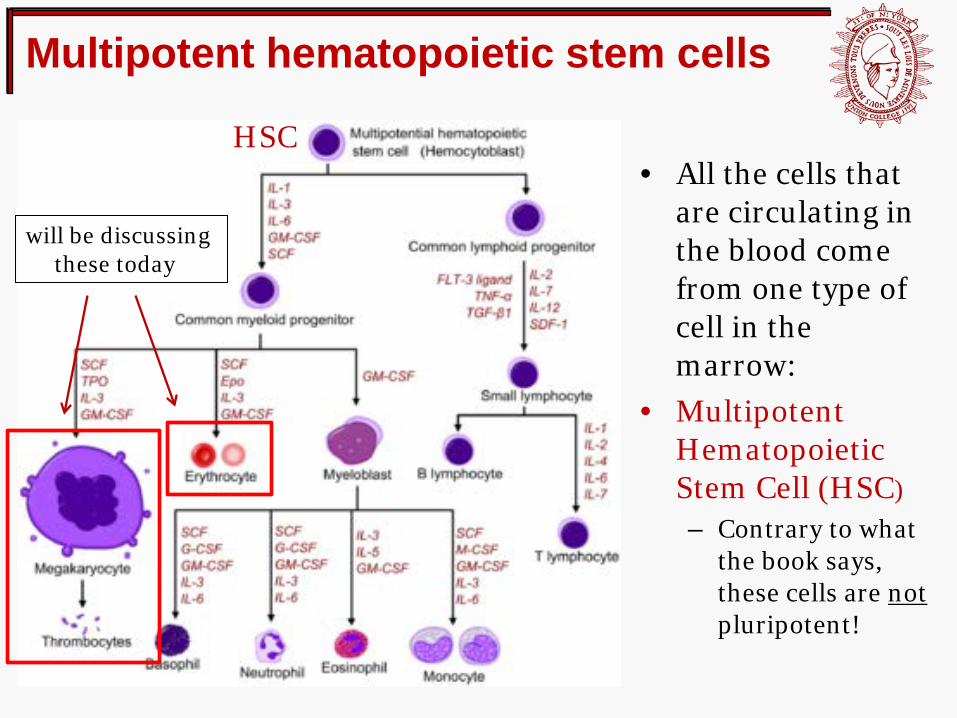
Multipotent hematopoietic stem cells
• All the cells that are circulating in the blood come from one type of cell in the marrow:
• Multipotent Hematopoietic Stem Cell (HSC) – Contrary to what
the book says, these cells are not pluripotent!
HSC
will be discussing these today

Pluripotent versus multipotent stem cells
http://protein.bio.msu.ru/biokhimiya/contents/v73/full/73131438Fig1.gif
ES cells are pluripotent
all stem cells on this level will be multipotent

Erythrocytes (red blood cells, RBCs)
HSC

Erythrocytes
• Play minimal role in wound healing and blood-biomaterial interactions
• Have no nucleus or cytoplasmic organelles needed for protein synthesis
• Do not proliferate • Mature RBCs do not
synthesize hemoglobin • Purely function to
transport oxygen and carbon dioxide
Fun fact: there are probably about 25,000,000,000,000 (25 * 1012) RBCs in your body right now! Average volume of one RBC: ~90 femtoliters (90 * 10-15 L) Total volume of RBCs? Calculate it!
How do RBCs get their hemoglobin if they don’t have a nucleus, and thus, don’t have any DNA to code for it?

Erythrocytes: Red Blood Cells
• RBC progenitors produce hemoglobin during the differentiation process from proerythroblast to reticulocyte – i.e., while they still have
nuclei • Only after nuclear
extrusion does the reticulocyte become an RBC and stop producing hemoglobin
• (will see the structure of hemoglobin later in the lecture)
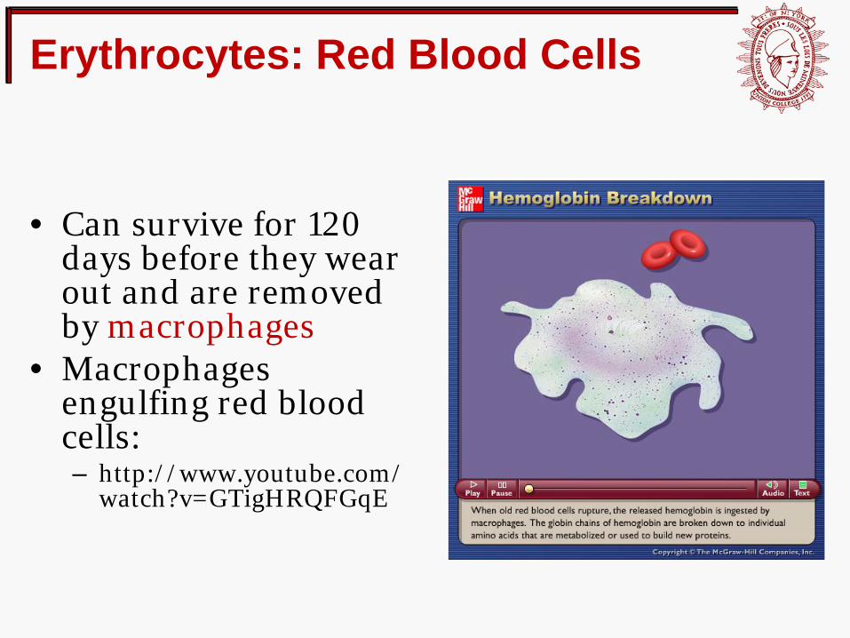
Erythrocytes: Red Blood Cells
• Can survive for 120 days before they wear out and are removed by macrophages
• Macrophages engulfing red blood cells: – http://www.youtube.com/
watch?v=GTigHRQFGqE

Erythrocytes: Red Blood Cells • Biconcave and ~7 μm in diameter (some
capillaries are 5 – 7 μm) • Pure blood plasma behaves like a Newtonian
Fluid (e.g., water) – Constant viscosity independent of the shear stress or shear
rate (shear: force applied along the surface of a material) • Whole blood behaves like a Non-Newtonian
fluid due to the flow behavior of RBCs – Instantaneous viscosity dependent on the shear stress or
shear rate – http://www.youtube.com/watch?v=aY7xiGQ-7iw (1:10)
• RBCs are somewhat similar; at very low flow (shear rates), RBCs aggregate into masses that act like solids (very high viscosity)
• Hence, whole blood is “shear thinning” – lower viscosity with higher shear stress – as opposed to “shear thickening”
Let’s examine this a bit closer!
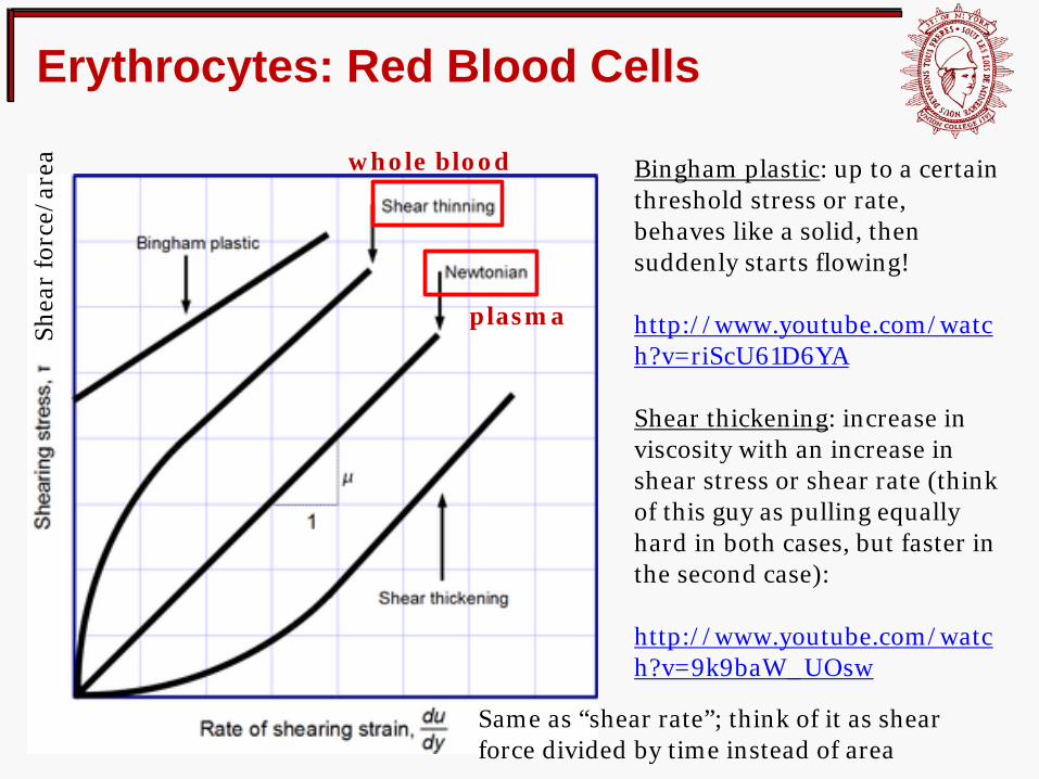
Erythrocytes: Red Blood Cells
Bingham plastic: up to a certain threshold stress or rate, behaves like a solid, then suddenly starts flowing! http://www.youtube.com/watch?v=riScU61D6YA Shear thickening: increase in viscosity with an increase in shear stress or shear rate (think of this guy as pulling equally hard in both cases, but faster in the second case): http://www.youtube.com/watch?v=9k9baW_UOsw
plasma
whole blood
Same as “shear rate”; think of it as shear force divided by time instead of area
Shea
r fo
rce/
area

Erythrocytes: Red Blood Cells
• Have no nucleus, and so are extremely deformable (due to the flow properties we just saw) – Membrane effects in a red
blood cell: http://www.youtube.com/watch?v=Ym1rvwP-po4
• The ability to deform is crucial to the function of RBCs
• Hypoxia -- oxygen shortage
Let’s look at sickle-cell anemia as a “case study” for
compromised RBC deformability
red blood cells inside a capillary

Sickle-cell anemia
Before discussing what is happening pathologically, what are the symptoms?
http://www.medexpressrx.com/blog/wp-content/uploads/2011/06/Symptoms_of_anemia.png

Sickle-cell anemia
• …is an inherited disorder affecting hemoglobin (right)
• Caused by a single AA mutation in the beta chain – GAA (glutamic acid)
replaced by GUA (valine) • Causes polymerization of
hemoglobin into elongated, inflexible crystals under low oxygen conditions
http://www.proprofs.com/flashcards/upload/q8683668.jpg
Hemoglobin (each “chain” is a different subunit)
heme groups with bound Fe atoms for oxygenation

Sickle-cell anemia (cont).
• In addition to blood vesssel blockage (right), the hemoglobin crystals can rupture the RBC membrane
• Many successive rupture events leads to elevated systemic hemoglobin
http://www.nhlbi.nih.gov/health/health-topics/topics/sca/

Platelets (thrombocytes)
HSC

Platelets
• Originate in marrow and are fragments of larger cells derived from megakaryoblasts
• Megakaryoblasts can range from 15 – 50 µm in diameter
• Differentiate to multiply the amount of DNA in the nucleus by ~30 times before becoming a megakaryocyte (35 – 150 µm in diameter)
http://student.nu.ac.th/wuth_web/pic.htm

Megakaryocyte
http://student.nu.ac.th/wuth_web/pic.htm

Megakaryocyte-Platelet

Platelets • Also called “thrombocytes” • ~2 – 4 µm in diameter • Have no nucleus and cannot
proliferate • Half-life of 8-10 days, get
cleared out of the spleen by macrophages
• Most prominent features are granules containing a variety of chemicals necessary for coagulation
• Contain actin, myosin, and thrombosthenin contractile proteins – The actin cytoskeleton differentially
regulates granule secretion (Flaumenhaft R, et al. Blood 2004)
• Platelets are active, while RBCs are passive
microtubules granules
micrograph of a platelet
Blair P, Flaumenhaft R. Blood Rev 2009

Platelets
• Platelets interact with the specific short peptide sequence arginine-glycine-aspartic acid (RGD) – Remember our discussion of
fibronectin last week
• RGD is part of the sequence of collagen, located in the basement membrane that is normally covered by healthy endothelial cells (right)
http://www.rci.rutgers.edu/~uzwiak/AnatPhys/Blood_Vessels.html
How does platelet adhesiveness to RGD (fibronectin) mediate clotting upon injury?

Aggregation and coagulation • Upon injury, platelets adhere to
a surface contractile proteins (actin and myosin) tighten platelet flattens and forms pseudopodia (“legs”) – Typically platelets adhere to the
connective tissue exposed when the endothelium of a blood vessel is ruptured (right)
– However, they also adhere to the surface of many man-made biomaterials
• Contraction causes platelets to degranulate, releasing ADP and thromboxane A2 – These are potent activators of
platelets, recruiting more locally – glycoprotein IIb/IIIa is also
upregulated for each platelet
“two-pronged approach”: more platelets, plus a higher affinity per platelet
Endothelial cell layer (inner blood vessel wall)
injury
platelet degranulation, recruitment,
and activation
platelet plug
Collagen in subendothelium

Aggregation and coagulation (cont.)
• The mass of aggregated platelets further contract actin/myosin, drawing the edges of the injury together
• An end product of coagulation is production of sticky threads of the protein fibrin – Fibrin threads attach to and help
consolidate the platelet plug – Also trap nearby erythrocytes
• At this point the platelet plus is a blood clot
Colorized scanning electron microscope (SEM) of a whole blood clot. Fibrin fibers are blue, platelet aggregates purple, and red blood cells red (Source: Yuri Veklich and John W. Weisel, University of Pennsylvania School of Medicine)

Platelet activation

Platelet activation

Biomaterials, devices, and thrombosis
• Normal, healthy endothelium does not induce coagulation!
• Such is the goal of implanatable blood-contacting biomaterials – Think of deliterious effects of an implant as in two categories:
Harmful to the implant or its function • Adsorption of blood components
(e.g., proteins) onto the biomaterial • Adsorption of blood cells onto the
biomaterial • Tissue growth around the
biomaterial
Harmful to the patient • Processes of coagulation and
fibrinolysis • Formation of clots on the surface of
the material • These can migrate and clot vessels
elsewhere! • Injury to blood cells, causing low
levels of blood-borne cells and clinical problems


![Abnormalities of Blood Coagulation[1]](https://img.pdfslide.us/doc/110x75/577cce2b1a28ab9e788d80ee/abnormalities-of-blood-coagulation1.jpg)