Embed Size (px)
Citation preview

Bleeding disorders:hemophilia, von Willebrand
disease
Dr. Ildikó IstenesSemmelweis University, I. Dept. of Medicine
16th March, 2016

2
Roles of the hemostatic system:
• Blood remains liquid within the blood vessel
• Formation of thrombus at the site of injury (no further blood loss) and reconstruction of the vessel wall

• Vascular constriction: bleedingdiminishes (seconds)
• Primary hemostasis:
– Platelet adhesion- activation-aggregation (3-5 minutes)
• Secondary hemostasis
– Clot formation- fibrin formation
(10-30 minutes)
– fibrinolysis and reconstruction of vessel wall (days, weeks)
Vessel wall damage-exposure of the subendothelium

Thrombocyte adhesion and
aggregation
Fibronectin, collagenei

Secondary hemostasis

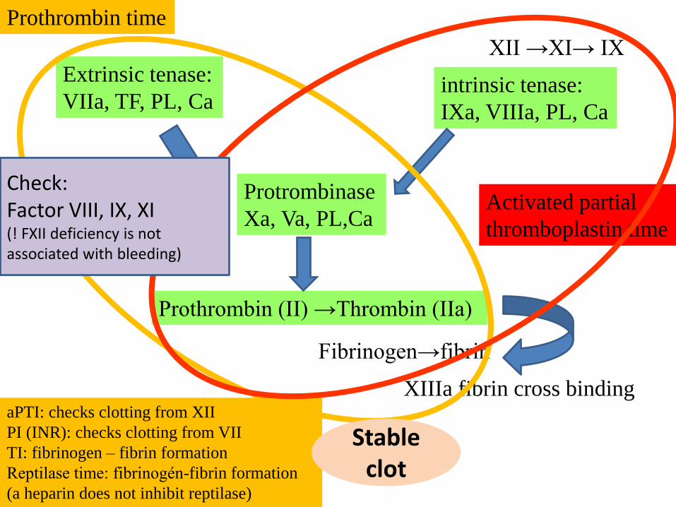
Protrombinase
Xa, Va, PL,Ca
Prothrombin (II) →Thrombin (IIa)
Extrinsic tenase:
VIIa, TF, PL, Caintrinsic tenase:
IXa, VIIIa, PL, Ca
Fibrinogen→fibrin
XIIIa :fibrin cross binding
plasmin
FDP: D-dimer
XII →XI→ IX
Injury Activated partial
thromboplastin time
Prothrombin time
aPTI: checks clotting from XII
PI (INR): checks clotting from VII
TI: fibrinogen – fibrin formation
Reptilase time: fibrinogén-fibrin formation
(a heparin does not inhibit reptilase)
clot
Initiation Enhancement
and propagation
stabilization
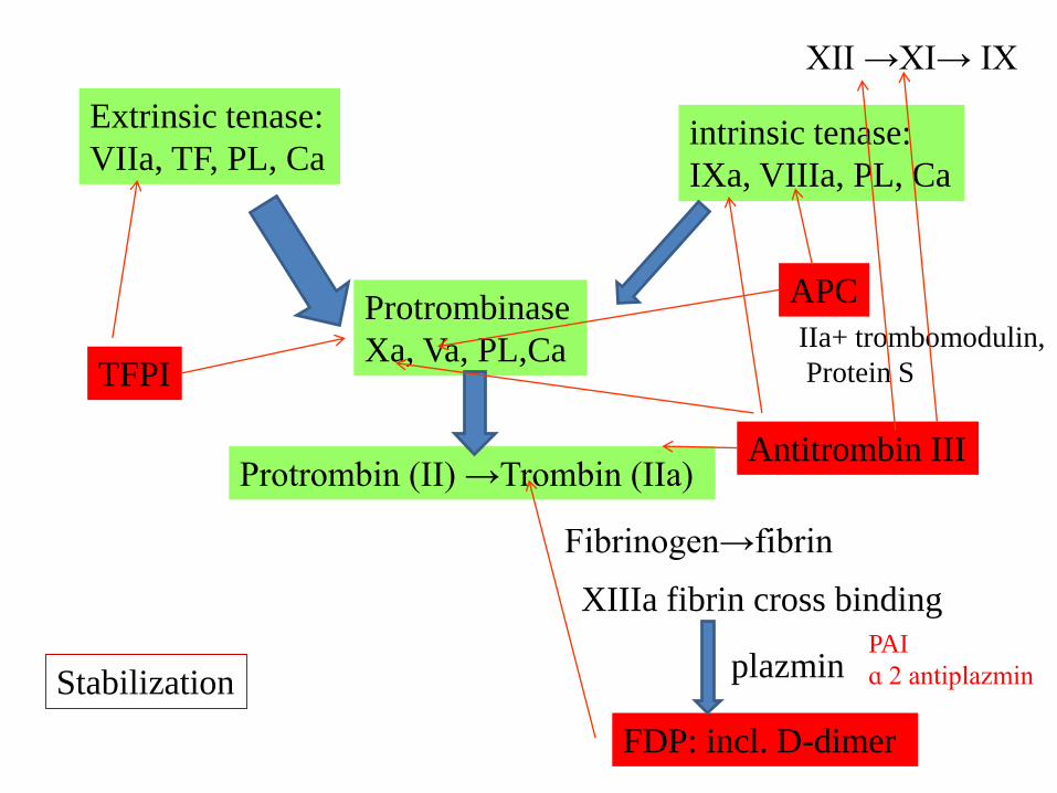
Protrombinase
Xa, Va, PL,Ca
Protrombin (II) →Trombin (IIa)
Extrinsic tenase:
VIIa, TF, PL, Caintrinsic tenase:
IXa, VIIIa, PL, Ca
Fibrinogen→fibrin
XIIIa fibrin cross binding
plazmin
FDP: incl. D-dimer
TFPI
APC
IIa+ trombomodulin,
Protein S
Antitrombin III
XII →XI→ IX
PAI
ɑ 2 antiplazminStabilization

Fibrin formation and fibrinolysis

Assessment of primary hemostasis
• Bleeding time: an overall assessment of primary hemostasis
– e.g. Ivy method
– PFA 100 – standard method
• Platelet count, function tests
• Von Willebrand factor
– Antigene
– Activity
– Multimer analysis

Ivy bleeding time
• A standard-sized incision is
made around 10 mm long and
1 mm deep.
• The time from when the
incision is made until all
bleeding has stopped is
measured and is called the
bleeding time.
• Every 30 seconds, filter paper
or a paper towel is used to
draw off the blood.
• The test is finished when
bleeding has stopped
completely.
• Normal values: between 3 – 10 minutes
depending on the method used.
• Causes of prolonged bleeding time:
•Disorders of the primary hemostasis
• technical error: e.g. depth of the
incision

PFA 100- Platelet Function Analyser Method:
It aspirates blood in vitro from a blood specimen into disposable test cartridges through a microscopic aperture cut into a biologically active membrane at the end of a capillary. The membrane of the cartridges are coated with collagen and adenosin diphosphate (ADP) or collagen and epinephrine inducing a platelet plug to form which closes the aperture.
The PFA test result is dependent on platelet function, plasma von Willebrand Factor level, platelet number, and (to some extent) the hematocrit.

Assessment of secondary hemostasis
Coagulation
• Screening tests (+ mixing studies when necessary)
– aPTT: checks clotting from XII
– PT (INR): checks clotting from VII
– TI: fibrinogen – fibrin formation
• Fibrinogen
• Factor levels
• Inhibitor levels
– Antithrombin, Protein C, Protein S
• Activated Protein C resitance
• Lupus anticoagulant
Fibrinolysis
• FDP: fibrinogen degradation products
• D-dimer
• Plasminogen
• Antiplasmin
• Plasminogen activator
• Plasminogen activator inhibitor

Bleeding disorders
– Disorders of primary hemostasis
• Vasculopathies, vasculitides
• Thrombocytopeniasand thrombocytopathies
– Disorders of secondary hemostasis
• Hemophilias
• Acquired coagulopathies
•Von Willebrand disease
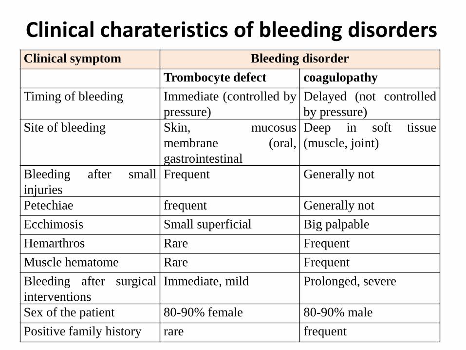
Clinical charateristics of bleeding disordersClinical symptom Bleeding disorder
Trombocyte defect coagulopathy
Timing of bleeding Immediate (controlled by
pressure)
Delayed (not controlled
by pressure)
Site of bleeding Skin, mucosus
membrane (oral,
gastrointestinal
Deep in soft tissue
(muscle, joint)
Bleeding after small
injuries
Frequent Generally not
Petechiae frequent Generally not
Ecchimosis Small superficial Big palpable
Hemarthros Rare Frequent
Muscle hematome Rare Frequent
Bleeding after surgical
interventions
Immediate, mild Prolonged, severe
Sex of the patient 80-90% female 80-90% male
Positive family history rare frequent

Clinically important bleeding types
Hemophiliatype
Hemarthros, hematuria, hematoma,intramuscular bleeding
Willebrandtype
Epistaxis, postextraction bleeding, menorrhagia, metrorrhagia, GI bleeding, mucosal bleeding
Thrombocyto-penia type
Purpuras and hematomas together, mucosal bleeding, no hemarthros
Vascular type Small skin bleedings, epistaxis,menorrhagia hematoma
Intramuscular bleeding
hemarthros
Mucosal bleedings, wet purpura Petechia, purpura

Vascular disorders
• Vasculopathies (no blood vessel inflammation)– Hereditary:
• hemorrhagic teleangiectasia (Osler-Weber-Rendu disease), Haemangioma, teleangiectasia (Kassabach-Merritt, von HippelLindau, ataxia teleangiectatica…)
– Acquired:• skorbut, purpura senile, psichogen purpura, diabetes, uraemia,
liver disease associated, drug induced
• Vasculitides– Large vessels: arteritis temporalis, Takayasu– Medium vessels: polyarteritis nodosa, Kawasaki– Small vessels: Wegener, Churg Strauss, Schönlein Henoch,
microscopic polyangitis

Thrombocytopenia
• Platelet count below 150G/l
• Risk of bleeding:
– <50G/l spontaneous bleeding which requires treatment
– < 10G/l spontaneous life threatening bleeding

Etiology of thrombocytopenia
1. Pseudothrombocytopenia:
- EDTA induced aggretation/agglutination
- Satellite formation between leukocytes and thrombocytes
2. Real thrombocytopenia
- Decreased production
- Increased destruction
- Increase storage in spleen

I. Decreased formation and maturation
Bone marrow: Megakariocytenumber is low orabsent
1.Hereditary
Pl. Fanconi anaemia, Wiskott-Aldrich sy, Bernard-Soulier
sy,
2.Acquired
Bone marrow damage:
-drugs (kemoth),
-Chemicals,
-radiation,
- infections
Bone marrow infiltration:
-leukaemia, lymphoma,
-carcinoma
Myelodysplasia, myelofibrosis
Vitamin deficiency (disturbed maturation)
B12 deficiency, folic acid deficiency
Etiology of real thrombocytopenias

II. Inreased damage
Bone marros:
Megakariocyte
number is normal
or elevated
1. Immunthrombocytopenias
Autoimmun Primary:
-Acute and
-Adult chronic ITP (Idiopathic/immun
thrombocytopenic purpura)
secondary:
-drugs (co-trimoxazol, chinidin, HIT),
- infection (HIV, H.pylori),
-autoimmune disease(SLE),
-Malignancy (lymphoma)
-Antiphospholipid syndrome
Alloimmun Posttransfusional purpura
2.Microangiopathic
- Disseminated intravascular coagulation
-Thrombotikus thrombocytopenic purpura
-Hemolytic uraemic syndrome
3. Other:
Valve implantation (mechanic),
Extracorporal circulation (surface)
Etiology of real thrombocytopenias
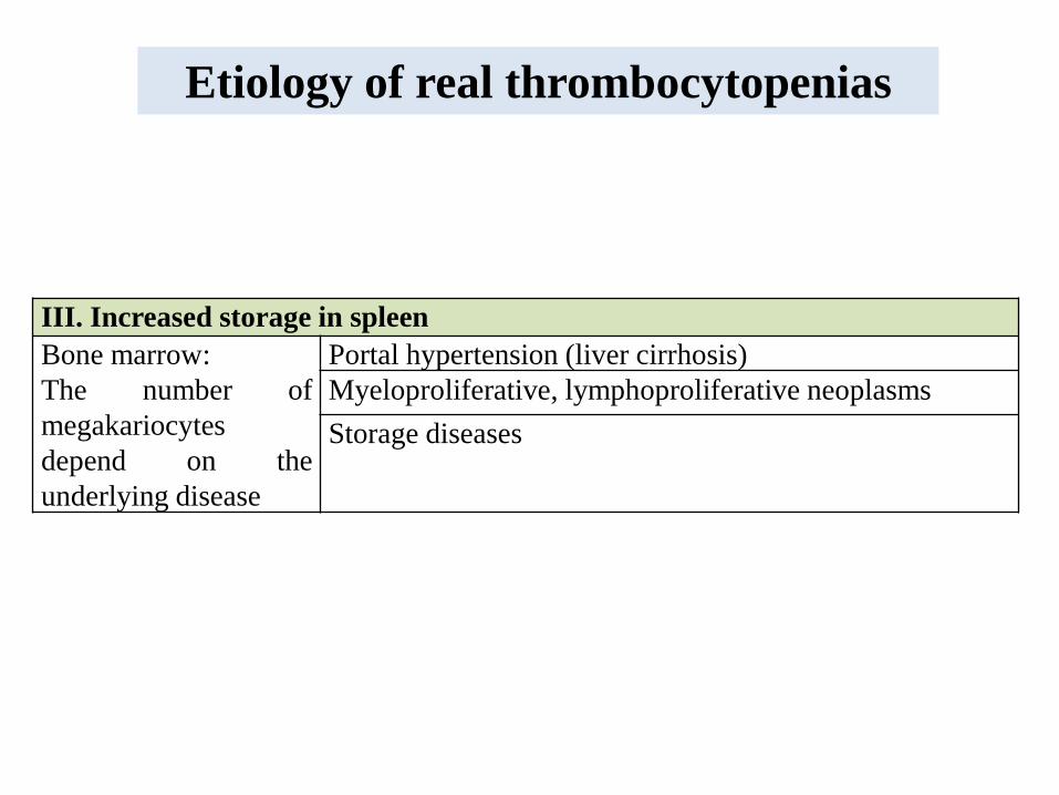
III. Increased storage in spleen
Bone marrow:
The number of
megakariocytes
depend on the
underlying disease
Portal hypertension (liver cirrhosis)
Myeloproliferative, lymphoproliferative neoplasms
Storage diseases
Etiology of real thrombocytopenias

Platelet count Risk of bleeding
(x 109/L)
>100 asymptomatic
50-100 invasive intervention
10-50 Purpura, haematoma
<10 Spontaneous severe bleeding
Platelet count and risk of bleeding

•Therapeutic supportation:
• Bleeding due to thrombopenia or thrombopathia („wet
purpuras”, internal beleeding)
•Preventive supportation:
•By critical platelet count (no bleeding)
Indication of platelet transfusion
Preventive supportation Plt count
Stable patient without fever: < 5-10G/l
Accompanying fever, sepsis, DIC, severe anaemia,
extreme leukocytosis, progressive thrombopenia
< 20G/l
Lumbalpunction, intrathecalis chemoth < 30G/l
Surgera, invasive diagnostic intervention (except for
sternum punction, crista biopsy
< 50G/l
Neurosurgery, eye surgery, polytraumatised patient < 100G/l
VON WILLEBRAND DISEASE
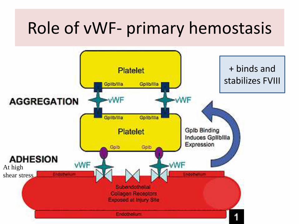
Role of vWF- primary hemostasis
+ binds and stabilizes FVIII
At high
shear stress

von Willebrand factor
• Produced/stored/
secreted by
– Endothel cells
– Megakaryocytes
• Structure:
• High molecule weight multimer glycoprotein (HMWM) (dimers-multimers)
• ADAMTS13 regulates the size of the multimer
• Binding sites: • FVIII, heparin, GPIb, GPIIb/IIIa,
collagen
• Function:
– primary hemostasis• Key role in the adhesion of
platelets to the injured wall)
• Aggregation
– Binds and stabilizes FVIII
• Its level is increased by
– Stress, pregnancy, oral contraceptives, DIC, chronic liver disease, hepatocellular cc., hyperthyreosis (decreased in hypothyreosis), blood group AB

Moake J. N Engl J Med 2002;347:589-600In von Willebrand disease:
The vWF is either:
-not enough
-not functioning properly (e.g. there are no large multimers)
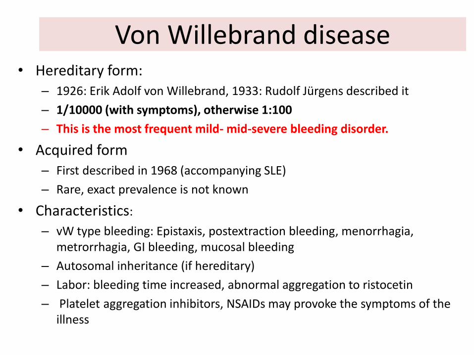
Von Willebrand disease• Hereditary form:
– 1926: Erik Adolf von Willebrand, 1933: Rudolf Jürgens described it
– 1/10000 (with symptoms), otherwise 1:100
– This is the most frequent mild- mid-severe bleeding disorder.
• Acquired form– First described in 1968 (accompanying SLE)
– Rare, exact prevalence is not known
• Characteristics:
– vW type bleeding: Epistaxis, postextraction bleeding, menorrhagia, metrorrhagia, GI bleeding, mucosal bleeding
– Autosomal inheritance (if hereditary)
– Labor: bleeding time increased, abnormal aggregation to ristocetin
– Platelet aggregation inhibitors, NSAIDs may provoke the symptoms of the illness

In 1924, a 5-year-old girl who lived on the Aland Islands was brought to Deaconess
Hospital in Helsinki, Finland, where she was seen by Dr. Erik von Willebrand. It turned out
that four out of 12 children of her family died of uncontrollable bleeding. This made him
curious, he went to the island to make further studies. He ultimately assessed 66 members
of her family and reported in 1926 that this was a previously undescribed bleeding disorder
that differed from hemophilia. He recognized that:
-The autosomal inheritance pattern
-Mucocutaneous bleeding,
-Normal clotting time
-Prolonged bleeding time
He subsequently found that blood transfusions were useful not only to correct the anemia
but also to control bleeding.
Variant forms of vWF disease were recognized in the 1970s, and we now recognize that
these variations are the result of synthesis of an abnormal protein.

Inheritance

Types of inherited vWDType Characteristics Clinical symptoms
1 Partial absence of vWF, quantitative problem
Mucosal bleedings: -epistaxis, -Postextractional bleeding-Gastrointestinal bleeding-Menorrhagia, metrorrhagia
(hematuria is rare, purpura isnot found)
2 qualitative problem
2A Decreased vWF dependentplatelet adhesion (GPIb), withselective absence of HMWM
2B Increased affinity of vWF to theGPIb receptor of the platelet
2M Decreased vWF dependentplatelet adhesion (GPIb), withoutselective absence of HMWM
2N Markedly decreased FVIII binding , no absence of HMWM
The above mentioned ones plus: - Articular (coagulopathic) bleeding can occur3 Complete absence of vWF
Platelet type Thrombocytopathia- the GPIb of the platelet is dysfunctioning-enhanced adhesion
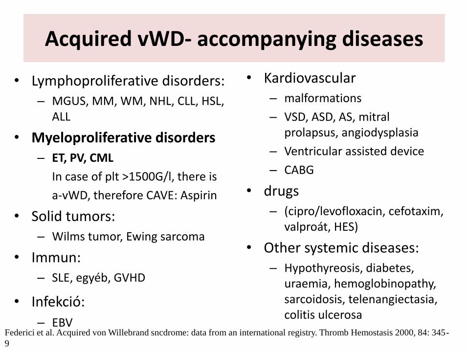
Acquired vWD- accompanying diseases
• Lymphoproliferative disorders:– MGUS, MM, WM, NHL, CLL, HSL,
ALL
• Myeloproliferative disorders – ET, PV, CML
In case of plt >1500G/l, there is
a-vWD, therefore CAVE: Aspirin
• Solid tumors: – Wilms tumor, Ewing sarcoma
• Immun: – SLE, egyéb, GVHD
• Infekció:
– EBV
• Kardiovascular– malformations
– VSD, ASD, AS, mitral prolapsus, angiodysplasia
– Ventricular assisted device
– CABG
• drugs– (cipro/levofloxacin, cefotaxim,
valproát, HES)
• Other systemic diseases: – Hypothyreosis, diabetes,
uraemia, hemoglobinopathy, sarcoidosis, telenangiectasia, colitis ulcerosa
Federici et al. Acquired von Willebrand sncdrome: data from an international registry. Thromb Hemostasis 2000, 84: 345-
9

(vWD honlapról letöltve)
inactivated by antibody Absorbed on
Malignant cells
High shear stress

vWD- diagnose
• Symptoms: – mild- to moderate mucosa type bleeding, starting at any
age
• Lab tests:– Screening: aPTI, plt count, PFA100, (bleeding time)
– Specific tests vWF:Ag, vWF:Rco, vWF:CB, VIIIF, RIPA, plasma vWF multimer analysis, vWF FVIII binding ability
• DD:– Thrombocytopathies
– mild hemophilia
– Aspirin treatment
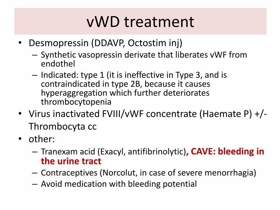
vWD treatment• Desmopressin (DDAVP, Octostim inj)
– Synthetic vasopressin derivate that liberates vWF fromendothel
– Indicated: type 1 (it is ineffective in Type 3, and is contraindicated in type 2B, because it causeshyperaggregation which further deterioratesthrombocytopenia
• Virus inactivated FVIII/vWF concentrate (Haemate P) +/-Thrombocyta cc
• other: – Tranexam acid (Exacyl, antifibrinolytic), CAVE: bleeding in
the urine tract– Contraceptives (Norcolut, in case of severe menorrhagia)– Avoid medication with bleeding potential

COAGULOPATHIES

History of hemophilia• 2nd century AD.: Rabbi Juda writes that those boys who had two brothers who
previously died of circumcision, don’t have to be circumcised.
• 12th century: Maimonides (1135-1204) (jewish doctor and philosopher) ads to this, that in this case the boy doesn’t have to circumcised even if the deceised brothers are from different fathers.
• 10th century : Abucalsis (moron surgeon writes )down the main characteristics of hemophilia: he described families whose males died of bleeding after only minor traumas
• In 1803, Dr. John Conrad Otto, a Philadelphian physician, wrote an account about "a hemorrhagic disposition existing in certain families" in which he called the affected males "bleeders".[He recognised that the disorder was hereditary and that it affected mostly males and was passed down by healthy females.
• In 1937, Patek and Taylor, two doctors from Harvard, discovered anti-haemophilic globulin.[
• In 1947, Pavlosky, a doctor from Buenos Aires, found haemophilia A and haemophilia B to be separate diseases by doing a lab test. This test was done by transferring the blood of one haemophiliac to another haemophiliac. The fact that this corrected the clotting problem showed that there was more than one form of haemophilia.

Hemophila B- a Royal disease
Alice Leopold Beatrice
Alexandra
Alexej Tsarevich
Empress Victoria
Affected man
Carrier woman

Epidemiology
Frequent hemophilias
• Hemophilia A: Factor VIII deficiency
– 1 in 5000 male births
• Hemophila B: Factor IX deficiency
– 1 in 30000 male birth
Rare hemophilias:
• Deficiency of factors other than FVIII-IX
– E.g. hemophilia C: Factor XI deficiency
• common in Jews of Ashkenazi, otherwise rare)

Genetics- hemophilia A,B• X-linked
• Mutation: – Point mutation:
• Non-sense: severe hemophilia
• Missense: mild- moderately severe hemophila
– Deletion: • Severe hemophilia
• Hemophilia A: – Intron 22 inversion is common among severe
hemophila A
• Hemophila B: – There is no characteristic mutation type
– One third is new mutation (with no family history)

Haemophilia type bleedings
– 90% of bleeding is musculoskeletal bleeding: 30% muscle hematoma, 70% hemarthros
hematoma
Intramuscular bleeding
hemarthros
Pseudotumor

Laboratory results
• Bleeding time, platelet function: normal
• coagulation tests: prolonged
– protrombin time: VII, X, II, V, fibrinogen
– thrombin time: fibrin
– Activated partial thromboplastin time: XII, XI, IX, X, VIII, V, II
• Mixing studies
• Tests for vWD
(Reptilase test
is normal)
Diff dg:
- vWD Type 3, 2N
- Acquired
coagulopathies:
(vitamin K deficiency
- Inhibitory hemophilia

Severity
Grade Factor level Symptoms
Severe <1% Spontaneous bleedings: muscle, joint
Moderate 1-5% Sponaneous bleedings are rare, severe bleedingsto trauma and surgery
mild 5-40% Severe bleeding after big trauma and surgery
• Factor levels remain stable througout life and in the family
• Carrier females can have symptoms as well (due to lyonisation- inactivation of X chromosome)
Hemophilia A, B
Other factor deficiencies
• Severe bleeding is less frequent (compared to A, B)
• It manifests usually after surgery, trauma, childbirth
• Factor level and the severity of bleeding do not correlate to each other

Complications
Bleeding related• Arthropathy-disability
– Synovitis, target joints withrecurrent bleeding, contractures
• Compartment syndrome: – Volkmann contracture, n.
femoralis lesion, quadricepsatrophy, n. peroneus atrophy
• Haemophiliac pseudotumor: – bone and soft tissue destruction
• Airway obstruction:– retropharyngeal, sublingual
bleeding
• Intracranial hemorrhage• Iron deficiency anaemia
Treatment related
• Side effects of factor suppl.– Urticaria, fever, sweating,
palpitation
• Transfusion transmitted infection: – HCV, HIV
• Alloantibodies (inhibitors) against factor supplementation

Quadriceps atrophy
Treatment options
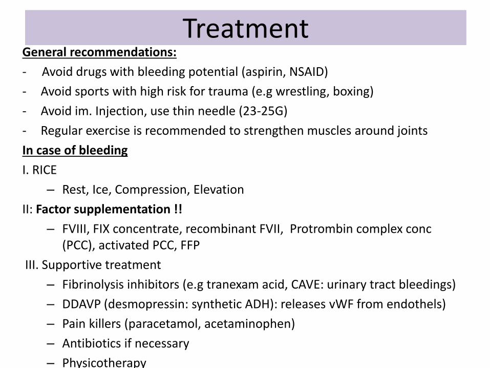
TreatmentGeneral recommendations:
- Avoid drugs with bleeding potential (aspirin, NSAID)
- Avoid sports with high risk for trauma (e.g wrestling, boxing)
- Avoid im. Injection, use thin needle (23-25G)
- Regular exercise is recommended to strengthen muscles around joints
In case of bleeding
I. RICE
– Rest, Ice, Compression, Elevation
II: Factor supplementation !!
– FVIII, FIX concentrate, recombinant FVII, Protrombin complex conc (PCC), activated PCC, FFP
III. Supportive treatment
– Fibrinolysis inhibitors (e.g tranexam acid, CAVE: urinary tract bleedings)
– DDAVP (desmopressin: synthetic ADH): releases vWF from endothels)
– Pain killers (paracetamol, acetaminophen)
– Antibiotics if necessary
– Physicotherapy

Factor supplementation
• On-demand – start within 2 hours!– Target factor level:
• Hemarthros: 25%• Soft tissue, GI bleeding: 50-60%• CNS bleeding, surgery: 100%
• Prophylactic – it has proven to be cost effective– factor level should not fall below 2 %– 2-3 times per week– Types:
• Primary: after the first bleeding under 2 years, or without bleedingunder 2 years
• Secondary: recurrent, frequent bleeding above 2 years• Terciary: in case of arthropathy• Permanent-transient
• Home treatment is possible (in Hungary since 1999)
Factorfirst!

Alloantibodies against factor
• 30% (!) of hemophilia A, and 3-8% in hemophilia B
• Usually within 50 exposition day
• Risks: Severe hemophilia, intensive, high dose substitution with FVIII
• Signs: Bleeding pattern changes– (large sc, im. Hematomas, GI bleedings, hematuria- similar to acquired
hemophilia)
• Diagnose: – Mixing studies (Bethesda Unit: 1 BU inactivates the 50% of normal plasma
factor)
– Recovery is not enough (it has to be measured regularly)
• treatment

Treatment of inhibitory hemophilia
• Stop bleeding with:
– Recombinant FVII or
– FEIBA: Factor Eight Inhibitor Bypassing Activity (=activated prothrombin complex concentrate, contains: IIa, Xa, VII, IX, Protein C, S)
• Immuntolerance induction:
– High dose of factor
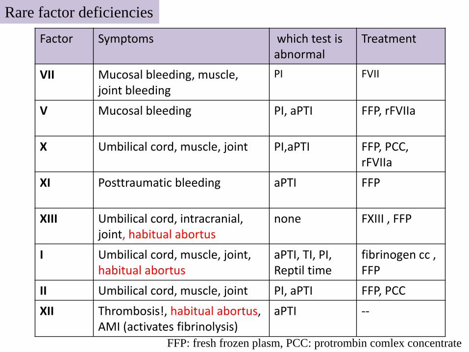
Factor Symptoms which test is abnormal
Treatment
VII Mucosal bleeding, muscle,joint bleeding
PI FVII
V Mucosal bleeding PI, aPTI FFP, rFVIIa
X Umbilical cord, muscle, joint PI,aPTI FFP, PCC, rFVIIa
XI Posttraumatic bleeding aPTI FFP
XIII Umbilical cord, intracranial, joint, habitual abortus
none FXIII , FFP
I Umbilical cord, muscle, joint, habitual abortus
aPTI, TI, PI, Reptil time
fibrinogen cc , FFP
II Umbilical cord, muscle, joint PI, aPTI FFP, PCC
XII Thrombosis!, habitual abortus, AMI (activates fibrinolysis)
aPTI --
FFP: fresh frozen plasm, PCC: protrombin comlex concentrate
Rare factor deficiencies

Acquired coagulopathies
• Deficiency of vitamin K dependent factors (II, VII, IX, X, protein C, S)– Neonatal hemorrhagic disease, biliar obstruction, insufficient vitamin
K absorption, vitamin K antagonist treatment, liver disease
• Nephrotic syndrome: – FIX, XIII, At III loss through proteinuria
• Acquired hemophilia
• Massive transfusion syndrome
• Disseminated intravascular coagulation

Acquired inhibitory hemophilia
• 0,2-1,5/1000000: between 60-80 ys (smaller peak: 20-30 ys, female, after delivery)
• Usually autoantibodies against FVIII
• 50% associated with disease (malignant, autoimmune, infection HBV, HCV, drugs, pregnancy), 50% idiopathic
• Dg: aPTT prolonged + mixing studies
• Clinical features: – Bleeding with negative family history
– Suffusions, big hematomas, mucosal bleedings(no joint bleeds!)
• Treatment: – Bleeding: bypassing agent, (plasmapheresis, immunadsoroption)
– Steroid- cyclofosfamid- Factor VIII

Case 1-• A 1-year-old boy is brought to your clinic. Developmentally
normal, his parents had noticed him to be bruising easily.Family history is negative.Coagulation tests show:
• What is the first step?: – Repeat test to confirm abnormality and exclude lab error
– Mixing studies to exclue/verify the presence of inhibitors
• Mixing with normal plasma corrects abnormal aPTT
• Which factors would you measure?
Test Patient Reference range
PT 13s 11-14s
aPTT 105s 23-35s
Fibrinogen 2,7g/l 1,5-4g/l
Trombin time 13s 10-13s
Protrombinase
Xa, Va, PL,Ca
Prothrombin (II) →Thrombin (IIa)
Extrinsic tenase:
VIIa, TF, PL, Caintrinsic tenase:
IXa, VIIIa, PL, Ca
Fibrinogen→fibrin
XIIIa fibrin cross binding
XII →XI→ IX
Activated partial
thromboplastin time
Prothrombin time
aPTI: checks clotting from XII
PI (INR): checks clotting from VII
TI: fibrinogen – fibrin formation
Reptilase time: fibrinogén-fibrin formation
(a heparin does not inhibit reptilase)
Stableclot
Check:Factor VIII, IX, XI(! FXII deficiency is notassociated with bleeding)

The tests are repeatedly abnormal
• his FIX and FXI assays are normal but the FVIII assay is <1 IU/dl.
• Diagnose: severe Haemophilia A
• Treatment: start prophylaxis e.g after the first intraarticular bleeding

If this patient is a girl (with FVIII def)?• Differential diagnose?
– vWD: Severe Type 1 or Type 3, or Type 2N• Bleeding time/PFA 100 should be abnormal
• vWF assays should be abnormal
• If vWF assays are normal, how could a girl have haemophilia A?
– Carrier mother and hemophiliac father• Family history should be positive
– There is only one X chromosome• Turner syndrome: X0 or
• Androgen insensitivity: patient is XY, but fenotypically female
Kariotype should be performed
– Extreme lyonisation of the X chromosome• inactivation of one X chromosome due to the mutation of Xist gene,
which is responsible for the process

Case 2
• A 45-year-old man presents with an extensive above knee DVT extending into the iliac veins. He is otherwise well with no past medical history of note.
Test Patient Reference range
PT 14s 11-14s
aPTT >120s 23-35s
Fibrinogen 3,2g/l 1,5-4g/l
Trombin time 13s 10-13s

Possible explanations for prolonged aPTT?
• Hereditary bleeding disorder?
– Probably not: age, no previous health problems, no bleeding (!but it is not impossible for a hemofiliac to have DVT!)
• APL?
– NO: the prolonged APTT corrects in a mix with normal plasma.
• XII deficiency?
– YES: Factor XII:C <1 %
– ! Factor XII can activate fibrinolysis and a deficiency may, therefore, lead
to defective fibrinolysis and potentially an increased risk of thrombosis. There is, great debate about the significance of hypofibrinolysis as a risk factor for thrombosis.

Case 3
• A 10-day-old baby, previously well, breast fed and born at home is found by his parents unconscious and bleeding from mouth and gums. The only history of note is that the mother had had a major post-partum haemorrhage and had required emergency admission to hospital.
Test Patient Reference range
PT 102s 11-14s
aPTT >120s 23-35s
Fibrinogen 1,9g/l 1,5-4g/l
Trombin time 13s 10-13s

• Acquired or inherited? – the absence of any birth complications, such as umbilical stump
haemorrhage, an acquired disorder is more likely.
• What could be the problem? – This is Vitamin K deficiency. In the 'drama' of the mothers sudden
admission to hospital vitamin K was not administered to the child and it was then forgotten.
• What could confirm that it is vitamin K deficiency and not liver insufficiency? – Factor V assay would be useful as it is synthesised by the liver but not
Vitamin K-dependent.

Take home messages
• Bleeding patterns differ in primary and secondary hemostasis defects
• Threshold for prophylactic platelet transfusion
• Role of vWF
• vWD is a frequent mild bleeding disorder
• Importance of factor supplementation, prophylaxis and home treatment in hemophilia

THANK YOU!


Inhibitors

• The levels of vitamin K dependent factors are physiologically low at birth. Factors that contribute to this deficiency include:– Low vitamin K stores at birth
– Poor placental transfer of vitamin K
– Low levels of vitamin K in breast milk [but not in cows milk]
– Sterility of the fetal gut• FINE PRINT
Other possibilities include:1. A deficiency of factors V, X, or II.2. Combined factor V + VIII deficiency. However, the PT and APTT are not as prolonged as this results shown above as the levels of FV and FVIII are not completely absent.3. An inherited deficiency of one of the enzymes involved in the gamma carboxylation of the vitamin K dependent clotting factors. These deficiencies prevent formation of active forms of the vitamin K dependent factors (a similar effect to warfarin) and present very early in life, usually with intracerebral haemorrhage This is rare but frequent in countries where consanguineous marriages are common.
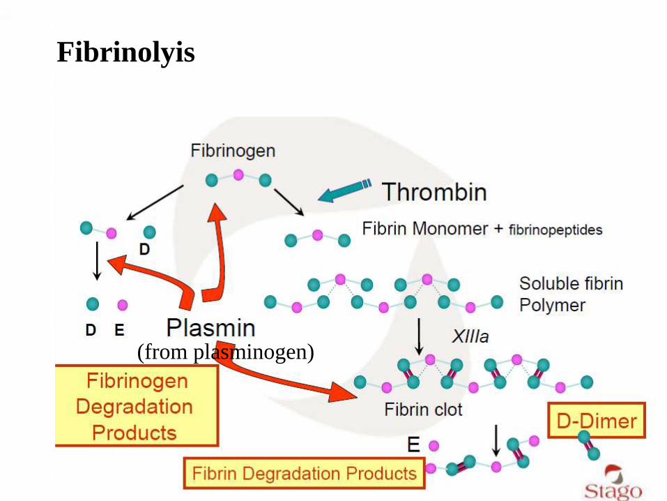
Fibrinolyis
(from plasminogen)

Condition prothrombintime
aPTI Bleeding time Platelet count
Vitamin K deficiency orwarfarin
Prolonged Normal ormildlyProlonged
unaffected unaffected
Disseminatedintravascularcoagulation
Prolonged Prolonged Prolonged decreased
Von Willebranddisease
unaffected Prolonged orunaffected
Prolonged Unaffected (ordecreased)
hemophilia unaffected Prolonged unaffected unaffected
aspirin unaffected unaffected Prolonged unaffected
thrombocytopenia unaffected unaffected Prolonged decreased
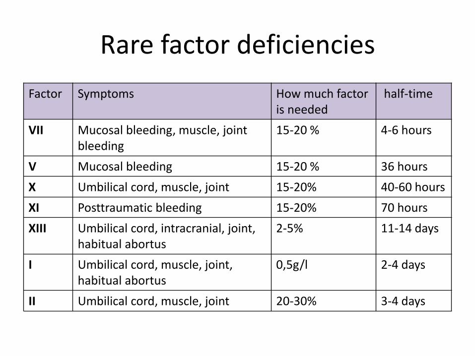
Rare factor deficiencies
Factor Symptoms How much factoris needed
half-time
VII Mucosal bleeding, muscle, jointbleeding
15-20 % 4-6 hours
V Mucosal bleeding 15-20 % 36 hours
X Umbilical cord, muscle, joint 15-20% 40-60 hours
XI Posttraumatic bleeding 15-20% 70 hours
XIII Umbilical cord, intracranial, joint, habitual abortus
2-5% 11-14 days
I Umbilical cord, muscle, joint, habitual abortus
0,5g/l 2-4 days
II Umbilical cord, muscle, joint 20-30% 3-4 days