Embed Size (px)
Citation preview
_________________________________________________________
Journal of Experimental Biology and Agricultural Sciences
http://www.jebas.org
KEYWORDS
Biotechnology
Immunoassay
Equine
Infectious disease
ABSTRACT
Rapid diagnosis of infectious diseases and appropriate treatment with in time are important steps that
promote optimal clinical outcomes and general public health. Today there is large number of new
technologies such as nanotechnology, biosensors, and microarray techniques, are being developed and
used as diagnostic tools for equine infectious diseases. Nucleic acid based techniques such as
polymerase chain reaction (PCR) have become conventional tools in veterinary research and plays an
important role in specific typing determinations as well as for rapid screening of ample numbers of
samples at the time of equine disease outbreaks. Other biotechnological techniques are populous to be
used in the coming times as they can enhance diagnostic efficacy in less time and cost as compared to
conventional techniques. This review focuses on biotechnological tools available for equine diseases
diagnosis and its applications hold great promise for improving the speed and accuracy of diagnostics
for equine infectious diseases.
Minakshi Prasad1,*
, Basanti Brar1, Ikbal
1, Koushlesh Ranjan
2, Upendra Lalmbe
1, J. Manimegalai
1,
Bhavya Vashisht1, Sandip Kumar Khurana
4 and
Gaya Prasad
3
1Department of Animal Biotechnology, LLR University of Veterinary and Animal Sciences, Hisar, Haryana, India, 125004
2Department of Veterinary Physiology and Biochemistry, Sardar Vallabhbhai Patel University of Agriculture and Technology, Meerut, India, 250110
3Sardar Vallabhbhai Patel University of Agriculture and Technology, Meerut, Uttar Pradesh, India, 250110
4NRCE, Hisar, Haryana, India, 125001
Received – November 05, 2016; Revision – November 20, 2016; Accepted – December 04, 2016
Available Online – December 04, 2016
DOI: http://dx.doi.org/10.18006/2016.4(Spl-4-EHIDZ).S161.S181
BIOTECHNOLOGICAL TOOLS FOR DIAGNOSIS OF EQUINE INFECTIOUS
DISEASES
E-mail: [email protected] (Minakshi Prasad)
Peer review under responsibility of Journal of Experimental Biology and
Agricultural Sciences.
* Corresponding author
Journal of Experimental Biology and Agricultural Sciences, December - 2016; Volume – 4(Spl-4-EHIDZ)
Journal of Experimental Biology and Agricultural Sciences
http://www.jebas.org
ISSN No. 2320 – 8694
Production and Hosting by Horizon Publisher India [HPI]
(http://www.horizonpublisherindia.in/).
All rights reserved.
All the article published by Journal of Experimental
Biology and Agricultural Sciences is licensed under a
Creative Commons Attribution-NonCommercial 4.0
International License Based on a work at www.jebas.org.
_________________________________________________________
Journal of Experimental Biology and Agricultural Sciences
http://www.jebas.org
1 Introduction
Modern molecular biology provides us newer technology to
diagnose and control several equine diseases. It is also used for
the development of novel diagnostic tools for equine infectious
disease control (Pusterla et al., 2006; Amaya, 2014). Several
diagnostic tools such as nucleic acid probes, monoclonal
antibodies, restriction fragment length polymorphisms, real
time PCR, proteomics, biosensors and nanotechnology have
increased the livestock productivity. These methods have been
commonly used for equine disease diagnosis and control (Yeh
et al., 2010; Johnson et al., 2010; Rakhshandehroo et al.,
2014). Several viral and bacterial pathogens such as Japanese
encephalitis virus, West Nile virus, Hendra virus, borna virus,
equine rabies, Rhodococcus equi, Bacillus anthracis etc., are
causes several serious diseases in equines and induce economic
to human population and these are zoonotic in nature (Yeh et
al., 2010; Booth et al., 2010; Priestnall et al., 2011; Khurana,
2015).
On various occasion equines are used for various purposes
such as ceremonies, riding, sports, draught racing, transport
and antitoxin/antibody production, throughout the world
(Burnouf et al., 2004). There is possibility of disease
transmission and spread at the time of equines movement from
one country to another. Therefore, OIE (World Organisation
for Animal Health) has enlisted several diagnostic tests for
international movement of equines (Table 1) (OIE, 2016).
Biotechnology may play an important role in prevention of
disease caused by these pathogens.
The correct knowledge of molecular biology of infectious
agents and their hosts is very important for controlling the
disease (Tavares et al., 2011). Biotechnological and protein
based assays can play a main role in equine disease control due
to its everlasting developments with the use of developed anti
pathogenic drugs and diagnostic chemicals. Even though
conventional techniques are still used commonly, recent
biotechnological assays have widened the scope of equine
diseases detection and give us powerful new techniques for
quick and specific identification of equine diseases. This
manuscript reviews the current and potential uses of
biotechnology tools for equine infectious disease diagnostics.
2 Serological assays
Protein based assays are based on antibody and antigen
interaction. These types of several assays such as enzyme
linked immunosorbent assay (ELISA), falcon assay screening
test –ELISA, indirect or direct immunofluroscencent antibody
tests, immunoblotting dot-ELISA, peptide based-ELISA,
complement fixation test, agar gel immunodiffusion and
neutralization test are used for equine infectious disease
diagnosis. These serological assays are highly sensitive and
specific than other techniques like microscopy and it allow
clearance of post-therapeutic pathogen.
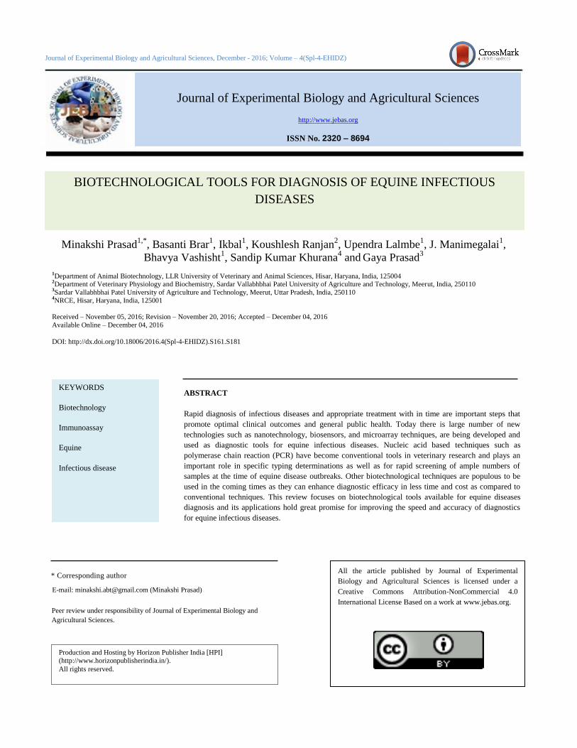
2.1 Enzyme-linked immunosorbant assay (ELISA)
Components of immune system used for detection of immune
response against infection in ELISA test. For detection of
specific immune response, ELISA assay involves antigen,
antibody and enzymes. The antigens are adhered to surface of
microtitre plate and antibody specific to the antigen is applied
over the surface for binding. It was followed by the
conjugation of antibody with an enzyme-Horseradish
peroxidise. Further, substrate was added to the plate for
producing visible colour change in a reaction mixture. Based
on use or not of a secondary antibody, the ELISA test may be
either direct or indirect (Figure 1). This test is successfully
used for diagnosis of various diseases in equines. Singha et al.
(2014) have reported an indirect ELISA using truncated TssB
protein for serodiagnosis of glanders.
A sensitive antigen capture ELISA was developed for the
detection of secreted NS1 from infected equines with West
Nile virus (Macdonald et al., 2005; Chung & Diamond, 2008).
Similarly, ELISA has been developed for the detection of
EHV-1, EHV-4 (Yasunaga et al., 2000), equine rhinitis virus A
(ERAV) (Kriegshauser et al., 2009) and equine rhinitis virus B
(ERBV) (Kriegshauser et al., 2008). In the recent studies,
ELISA targeting antibodies to the spike (S) of equine corona
virus was developed and validated to detect antibodies to
EqCoV in infected horses (Kooijman et al., 2016).
Table 1 Prescribed test for equine diseases according to OIE, 2016.
Disease name OIE prescribed tests
African horse sickness
CF, ELISA
Contagious equine metritis
Agent identification.
Dourine
CF
Equine infectious anaemia
AGID
Equine piroplasmosis
ELISA, IFA
Equine viral arteritis
Agent identification (semen only), Virus Neutralization
Glanders
Complement Fixation
S162 Prasad et al
_________________________________________________________
Journal of Experimental Biology and Agricultural Sciences
http://www.jebas.org
Figure 1 Principle of ELISA test a) Direct ELISA b) Indirect ELISA
2.2 Dot-ELISA
Dot-ELISA works on the basis of attachment of small amount
of antigen on to a nitrocellulose membrane. Specific antibody
is incubated with antigen containing dotted membrane
followed by adding of enzyme conjugated anti-antibody. A
substrate is added in the last which causes precipitation of a
detectable coloured dot on the membrane (Svobodova et al.,
2013). It was reported that the dot-ELISA is simple, quick,
specific, sensitive, low cost field test that detects minute levels
of antibodies much faster than complement fixation test and
indirect hemagglutination antibody test (Verma & Misra, 1989;
Verma et al., 1990). Dot-ELISA has been used for the sero-
diagnosis of glanders (John et al., 2010). By the use of
nitrocellulose membrane in this test makes it applicable in the
field. This assay is quick and specific in detection of various
diseases. It gives us low background as compared to ELISA
assay that can easily differentiate between the positive and
negative samples.
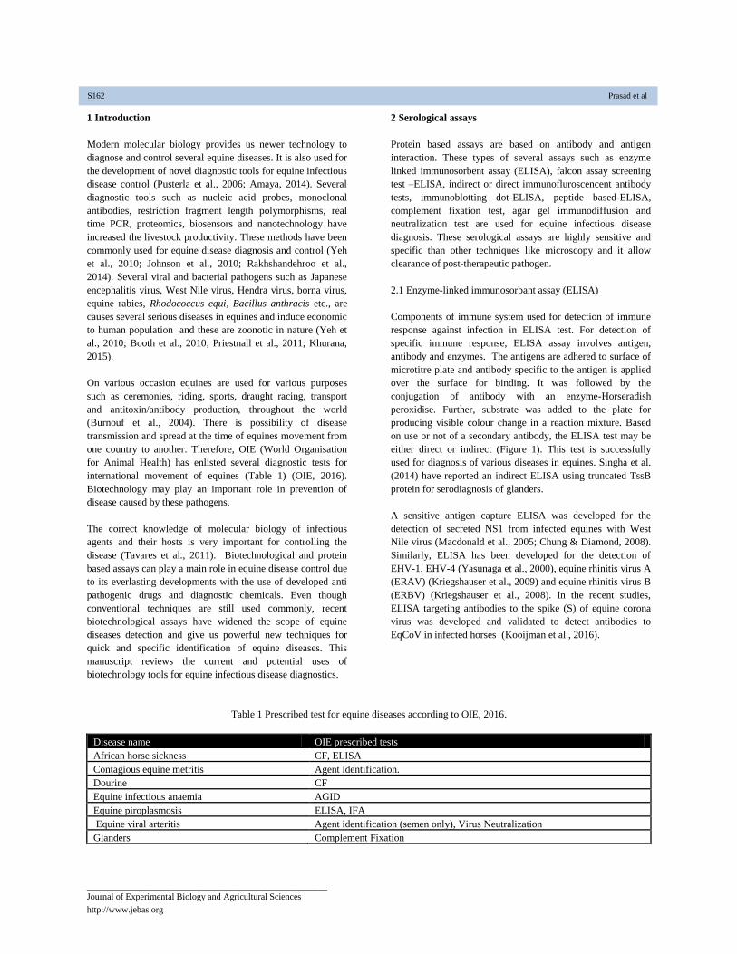
2.3 Fluorescent Antibody Test (FAT)
In Fat assay, antibody is labelled with fluorescent dye, is used
in visualization of antigen in a clinical specimens. The
antibody conjugated with fluorescent dye and antigen-antibody
complex gives a visible glow sign when examined under a
fluorescent microscope. The fluorescent dye can be tagged
directly with primary antibody which is known as direct
fluorescent antibody test or with a secondary anti-antibody
known as Indirect Fluorescent Antibody Test (Figure 2). The
FAT is used in diagnosis of several equine diseases. This assay
was recently investigated for diagnosis of equine leptospiral
abortion in mare (Erol et al., 2015). The sarcocystis neurona
causes a dreadful disease, equine protozoal myeloencephalitis
(EPM) in equines. The IFAT was successfully validated for
CSF testing for confirmation of EPM in equines (Duarte et al.,
2006; Johnson et al., 2013).
The overall specificity and accuracy of IFAT was shown to be
better than that of the western blot and modified western blot,
which showed its potential to use as a diagnostic assay for
detection of EPM caused by Sarcocystis neurona (Duarte et al.,
2003). The FAT and immune-histo-chemistry (IHC) assay
confirmed the presence of Australian bat lyssa virus (ABLV)
antigen in horse brain tissues (Shinwari et al., 2014). A FAT
assay has been used for the direct identification of bacterial
Helicobacter on the equine gastric mucosa (Perkins et al.,
2012). This technique has been used to describe the spatial
distribution of Helicobacter species in the stomach of healthy
horses to demonstrate the microbiota of normal appearing
squamous and glandular mucosa (Burton et al., 2007).
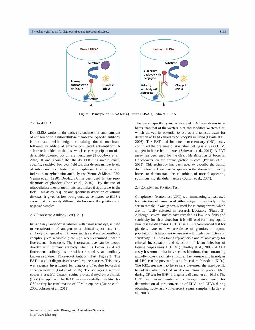
2.4 Complement Fixation Test
Complement fixation test (CFT) is an immunological test used
for detection of presence of either antigen or antibody in the
serum sample. It was generally used for microorganisms which
are not easily cultured in research laboratory (Figure 3).
Although, several studies have revealed its low specificity and
sensitivity for virus detection, it is still used for many equine
viral disease diagnoses. CFT is the OIE recommended test for
glanders. Due to low prevalence of glanders in equine
population it is important to use test with high specificity and
sensitivity. CFT was found reproducible and reliable assay for
clinical investigation and detection of latent infection of
Equine herpes virus 1 (EHV1) (Hartley et al., 2005). A CFT
assay has some limitations such as laborious, time consuming
and often cross reactivity in nature. The non-specific hemolysis
of RBC can be prevented using Potassium Periodate (KIO4).
The KIO4 treatment to horse sera prevented the non-specific
hemolysis which helped in determination of precise titers
during CF test for EHV-1 diagnosis (Bannai et al., 2013). The
CFT and virus neutralisation assays were used for
determination of sero-conversion of EHV1 and EHV4 during
obtaining acute and convalescent serum samples (Hartley et
al., 2005).
Biotechnological tools for diagnosis of equine infectious diseases. S163
_________________________________________________________
Journal of Experimental Biology and Agricultural Sciences
http://www.jebas.org
Figure 2 Principle of Direct Fluorescent antibody test (FAT).
Figure 3 Complement fixation test.
S164 Prasad et al
_________________________________________________________
Journal of Experimental Biology and Agricultural Sciences
http://www.jebas.org
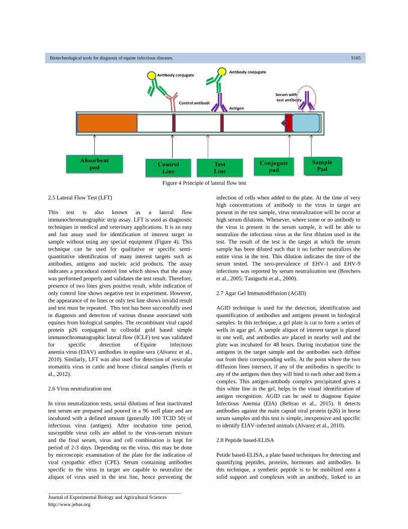
Figure 4 Principle of lateral flow test
2.5 Lateral Flow Test (LFT)
This test is also known as a lateral flow
immunochromatographic strip assay. LFT is used as diagnostic
techniques in medical and veterinary applications. It is an easy
and fast assay used for identification of interest target in
sample without using any special equipment (Figure 4). This
technique can be used for qualitative or specific semi-
quantitative identification of many interest targets such as
antibodies, antigens and nucleic acid products. The assay
indicates a procedural control line which shows that the assay
was performed properly and validates the test result. Therefore,
presence of two lines gives positive result, while indication of
only control line shows negative test in experiment. However,
the appearance of no lines or only test line shows invalid result
and test must be repeated. This test has been successfully used
in diagnosis and detection of various disease associated with
equines from biological samples. The recombinant viral capsid
protein p26 conjugated to colloidal gold based simple
immunochromatographic lateral flow (ICLF) test was validated
for specific detection of Equine infectious
anemia virus (EIAV) antibodies in equine sera (Alvarez et al.,
2010). Similarly, LFT was also used for detection of vesicular
stomatitis virus in cattle and horse clinical samples (Ferris et
al., 2012).
2.6 Virus neutralization test
In virus neutralization tests, serial dilutions of heat inactivated
test serum are prepared and poured in a 96 well plate and are
incubated with a defined amount (generally 100 TCID 50) of
infectious virus (antigen). After incubation time period,
susceptible virus cells are added to the virus-serum mixture
and the final serum, virus and cell combination is kept for
period of 2-3 days. Depending on the virus, this may be done
by microscopic examination of the plate for the indication of
viral cytopathic effect (CPE). Serum containing antibodies
specific to the virus in target are capable to neutralize the
aliquot of virus used in the test line, hence preventing the
infection of cells when added to the plate. At the time of very
high concentrations of antibody to the virus in target are
present in the test sample, virus neutralization will be occur at
high serum dilutions. Whenever, where some or no antibody to
the virus is present in the serum sample, it will be able to
neutralize the infectious virus at the first dilution used in the
test. The result of the test is the target at which the serum
sample has been diluted such that it no further neutralizes the
entire virus in the test. This dilution indicates the titre of the
serum tested. The sero-prevalence of EHV-1 and EHV-9
infections was reported by serum neutralization test (Borchers
et al., 2005; Taniguchi et al., 2000).
2.7 Agar Gel Immunodiffusion (AGID)
AGID technique is used for the detection, identification and
quantification of antibodies and antigens present in biological
samples. In this technique, a gel plate is cut to form a series of
wells in agar gel. A sample aliquot of interest target is placed
in one well, and antibodies are placed in nearby well and the
plate was incubated for 48 hours. During incubation time the
antigens in the target sample and the antibodies each diffuse
out from their corresponding wells. At the point where the two
diffusion lines intersect, if any of the antibodies is specific to
any of the antigens then they will bind to each other and form a
complex. This antigen-antibody complex precipitated gives a
thin white line in the gel, helps in the visual identification of
antigen recognition. AGID can be used to diagnose Equine
Infectious Anemia (EIA) (Beltrao et al., 2015). It detects
antibodies against the main capsid viral protein (p26) in horse
serum samples and this test is simple, inexpensive and specific
to identify EIAV-infected animals (Alvarez et al., 2010).
2.8 Peptide based-ELISA
Petide based-ELISA, a plate based techniques for detecting and
quantifying peptides, proteins, hormones and antibodies. In
this technique, a synthetic peptide is to be mobilized onto a
solid support and complexes with an antibody, linked to an
Biotechnological tools for diagnosis of equine infectious diseases. S165

_________________________________________________________
Journal of Experimental Biology and Agricultural Sciences
http://www.jebas.org
enzyme. Detection is done by assessing the conjugated enzyme
activity after incubation with a substrate to produce a
measurable end product (Figure 5). The very important step for
the detection strategy is a specific antigen-antibody interaction.
Peptide-ELISAs are performed in a 96-well microtiter plate
with synthetic peptide in carbonate buffer followed by
incubating the plate overnight at 4°C. Now, block the plate
with blocking buffer for 1 hour at 37°C followed by addition
of freshly prepared diluted primary antibody into each wells
and incubate the plate at 37°C for 1 hour. Subsequently, anti-
mouse IgG, diluted in 100 μl/well antibody dilution buffer is
added with the incubation at 37°C for 30 minutes. In last
enhancement solution is added in the plate and incubated at
37°C for 15 minutes. The plate was washed in between each
step at least five times with 1X PBST. Read the absorbance at
appropriate wavelength with an appropriate time resolved plate
reader.
Soutullo et al. (2001) evaluated the performance of an equine
infectious an Aemia-ELISA designed with synthetic peptides.
This assay could be important to prove for large throughput
screening and early detection of equine infectious anaemia
(EIA), when the results of the traditional Coggins test are still
negative. Recently, a sensitive and specific peptide-based
ELISA was developed to determine the sero-prevalance of
EHV-1 and EHV-9 (Abdelgawad et al., 2015). For
discrimination between serological responses to EHV 1 and
EHV4 immunoglobulins-IgG based type specific ELISA was
developed (Ma et al., 2013). This technique was also used to
discriminate between EHV-1 and EHV-4 glycoprotein E
peptides for EHV-1 and glycoprotein G (gG) for EHV-4 (Lang
et al., 2013; Yasunaga et al., 1998). Recently, a glycoprotein G
based peptide ELISA was developed for detection of equine
herpesvirus type 4 (Bannai et al., 2016).
Figure 5 Principle of peptide- ELISA
S166 Prasad et al
_________________________________________________________
Journal of Experimental Biology and Agricultural Sciences
http://www.jebas.org
2.9 Nucleic acid diagnostics
Nucleic acid-based detection methods including polymerase
chain reaction, reverse transcriptase-PCR, nested-PCR,
restriction fragment length polymorphism, amplified fragment
length polymorphism, random amplified polymorphic DNA,
loop-mediated isothermal amplification, microarray, real-time
PCR are used for identification of several equine diseases
(Pusterla et al., 2007; Monego et al., 2009; Yeh et al., 2012;
Quereda et al., 2000; Larrasa et al., 2002; Eischeid, 2011).
2.10 Polymerase Chain Reaction (PCR)
PCR uses the enzyme DNA polymerase that amplifies a small
length of targeted DNA using primers which are specific to the
target. It will amplify the selected target sequence from a
mixture of genome. PCR acts as an important tool for the
identification of parasites due to the insufficient amount of
availability of antigen and antigen products by using
conventional assays (Gasser, 2006). PCR can utilize almost all
kind of biological samples such as meat, blood, urine, skin
scrapings and faeces for parasitic infection study. When
compared to the conventional method the detection limit of
PCR is higher.
Therefore, it is useful for detecting low amount of antigen in
suspected samples (Varrasso et al., 2001; Nugent et al., 2006).
It can also be used for detection of many equine diseases
(Oldfield et al., 2004; Ocampo-Sosa et al., 2007; Pusterla et al.,
2007; Letek et al., 2008; Monego et al., 2009). Polymerase
chain reaction (PCR)–based diagnostic tests can allow rapid
and sensitive detection of equine infectious pathogen (Paxson,
2008). Yeh et al. (2010) developed a duplex reverse
transcriptse PCR which is sensitive, specific and very rapid
and is useful in both humans and as well as in horses for the
simultaneous and differential diagnosis of West Nile and
Japanese encephalitis viruses.
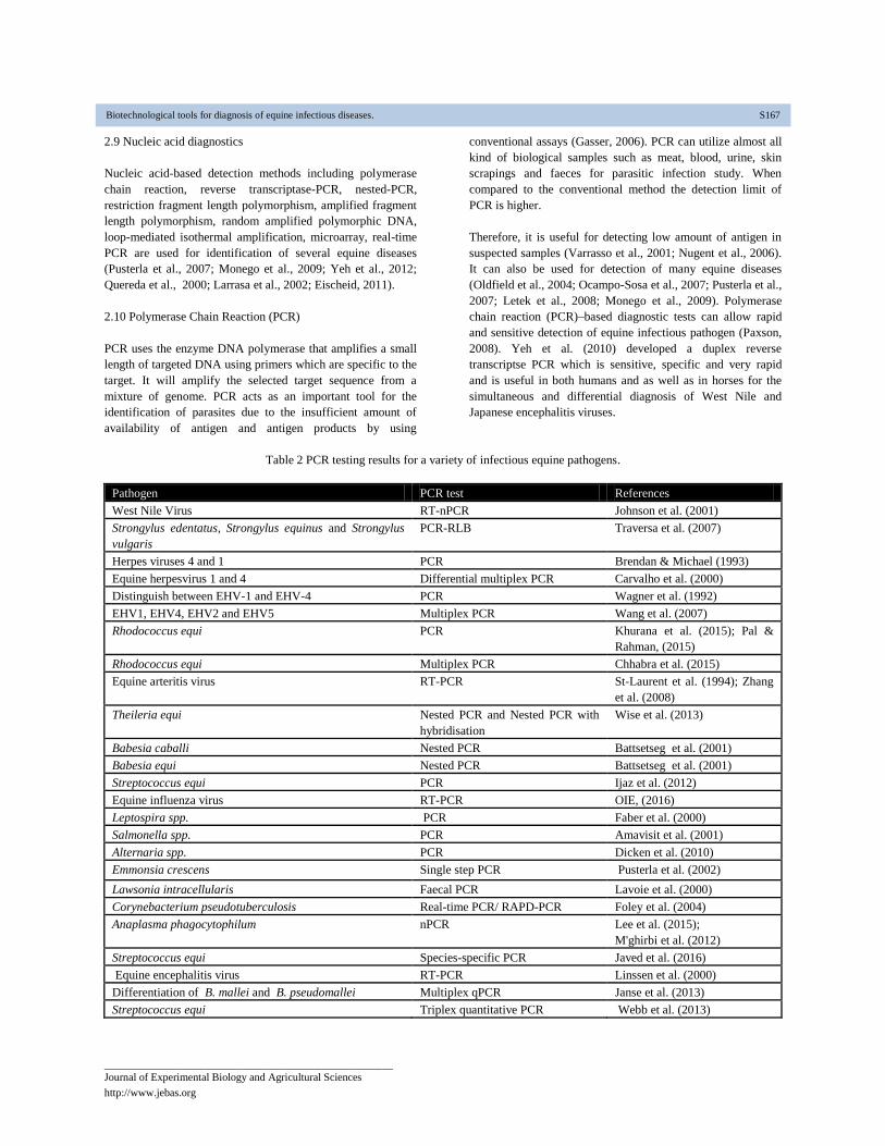
Table 2 PCR testing results for a variety of infectious equine pathogens.
Pathogen PCR test References
West Nile Virus RT-nPCR Johnson et al. (2001)
Strongylus edentatus, Strongylus equinus and Strongylus
vulgaris
PCR-RLB Traversa et al. (2007)
Herpes viruses 4 and 1 PCR Brendan & Michael (1993)
Equine herpesvirus 1 and 4 Differential multiplex PCR Carvalho et al. (2000)
Distinguish between EHV-1 and EHV-4 PCR Wagner et al. (1992)
EHV1, EHV4, EHV2 and EHV5 Multiplex PCR Wang et al. (2007)
Rhodococcus equi PCR Khurana et al. (2015); Pal &
Rahman, (2015)
Rhodococcus equi Multiplex PCR Chhabra et al. (2015)
Equine arteritis virus RT-PCR
St-Laurent et al. (1994); Zhang
et al. (2008)
Theileria equi Nested PCR and Nested PCR with
hybridisation
Wise et al. (2013)
Babesia caballi Nested PCR Battsetseg et al. (2001)
Babesia equi Nested PCR Battsetseg et al. (2001)
Streptococcus equi PCR Ijaz et al. (2012)
Equine influenza virus RT-PCR OIE, (2016)
Leptospira spp. PCR Faber et al. (2000)
Salmonella spp. PCR Amavisit et al. (2001)
Alternaria spp. PCR Dicken et al. (2010)
Emmonsia crescens Single step PCR Pusterla et al. (2002)
Lawsonia intracellularis Faecal PCR Lavoie et al. (2000)
Corynebacterium pseudotuberculosis Real-time PCR/ RAPD-PCR Foley et al. (2004)
Anaplasma phagocytophilum nPCR Lee et al. (2015);
M'ghirbi et al. (2012)
Streptococcus equi Species-specific PCR Javed et al. (2016)
Equine encephalitis virus RT-PCR Linssen et al. (2000)
Differentiation of B. mallei and B. pseudomallei Multiplex qPCR Janse et al. (2013)
Streptococcus equi Triplex quantitative PCR Webb et al. (2013)
Biotechnological tools for diagnosis of equine infectious diseases. S167
_________________________________________________________
Journal of Experimental Biology and Agricultural Sciences
http://www.jebas.org
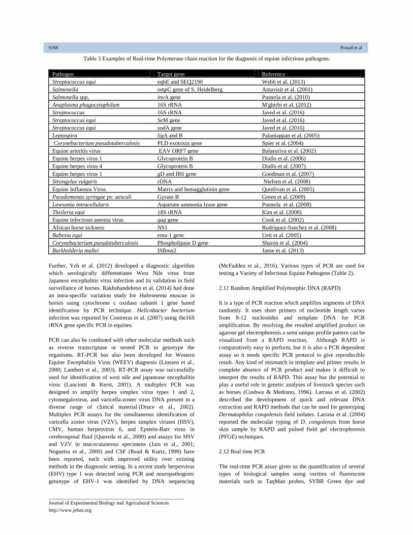
Table 3 Examples of Real-time Polymerase chain reaction for the diagnosis of equine infectious pathogens.
Pathogen
Target gene Reference
Streptococcus equi eqbE and SEQ2190 Webb et al. (2013)
Salmonella ompC gene of S. Heidelberg Amavisit et al. (2001)
Salmonella spp, invA gene Pusterla et al. (2010)
Anaplasma phagocytophilum 16S rRNA M'ghirbi et al. (2012)
Streptococcus 16S rRNA Javed et al. (2016)
Streptococcus equi SeM gene Javed et al. (2016)
Streptococcus equi sodA gene Javed et al. (2016)
Leptospira ligA and B Palaniappan et al. (2005)
Corynebacterium pseudotuberculosis PLD exotoxin gene Spier et al. (2004)
Equine arteritis virus EAV ORF7 gene Balasuriya et al. (2002)
Equine herpes virus 1 Glycoprotein B Diallo et al. (2006)
Equine herpes virus 4 Glycoprotein B Diallo et al. (2007)
Equine herpes virus 1 gD and IR6 gene Goodman et al. (2007)
Strongylus vulgaris rDNA Nielsen et al. (2008)
Equine Influenza Virus Matrix and hemagglutinin gene Quinlivan et al. (2005)
Pseudomonas syringae pv. aesculi Gyrase B Green et al. (2009)
Lawsonia intracellularis Aspartate ammonia lyase gene Pusterla et al. (2008)
Theileria equi 18S rRNA Kim et al. (2008)
Equine infectious anemia virus gag gene Cook et al. (2002)
African horse sickness NS1 Rodriguez-Sanchez et al. (2008)
Babesia equi ema-1 gene Ueti et al. (2005)
Corynebacterium pseudotuberculosis Phospholipase D gene Sharon et al. (2004)
Burkholderia mallei ISBma2 Janse et al. (2013)
Further, Yeh et al. (2012) developed a diagnostic algorithm
which serologically differentiates West Nile virus from
Japanese encephalitis virus infection and its validation in field
surveillance of horses. Rakhshandehroo et al. (2014) had done
an intra-specific variation study for Habronema muscae in
horses using cytochrome c oxidase subunit 1 gene based
identification by PCR technique. Helicobacter bacterium
infection was reported by Contreras et al. (2007) using the16S
rRNA gene specific PCR in equines.
PCR can also be combined with other molecular methods such
as reverse transcriptase or nested PCR to genotype the
organisms. RT-PCR has also been developed for Western
Equine Encephalitis Virus (WEEV) diagnosis (Linssen et al.,
2000; Lambert et al., 2003). RT-PCR assay was successfully
used for identification of west nile and japanease encephalitis
virus (Lanciotti & Kerst, 2001). A multiplex PCR was
designed to amplify herpes simplex virus types 1 and 2,
cytomegalovirus, and varicella-zoster virus DNA present in a
diverse range of clinical material (Druce et al., 2002).
Multiplex PCR assays for the simultaneous identification of
varicella zoster virus (VZV), herpes simplex viruses (HSV),
CMV, human herpesvirus 6, and Epstein-Barr virus in
cerebrospinal fluid (Quereda et al., 2000) and assays for HSV
and VZV in mucocutaneous specimens (Jain et al., 2001;
Nogueira et al., 2000) and CSF (Read & Kurtz, 1999) have
been reported, each with improved utility over existing
methods in the diagnostic setting. In a recent study herpesvirus
(EHV) type 1 was detected using PCR and neuropathogenic
genotype of EHV-1 was identified by DNA sequencing
(McFadden et al., 2016). Various types of PCR are used for
testing a Variety of Infectious Equine Pathogens (Table 2).
2.11 Random Amplified Polymorphic DNA (RAPD)
It is a type of PCR reaction which amplifies segments of DNA
randomly. It uses short primers of nucleotide length varies
from 8-12 nucleotides and template DNA for PCR
amplification. By resolving the resulted amplified product on
agarose gel electrophoresis a semi unique profile pattern can be
visualized from a RAPD reaction. Although RAPD is
comparatively easy to perform, but it is also a PCR dependent
assay so it needs specific PCR protocol to give reproducible
result. Any kind of mismatch in template and primer results in
complete absence of PCR product and makes it difficult to
interpret the results of RAPD. This assay has the potential to
play a useful role in genetic analyses of livestock species such
as horses (Cushwa & Medrano, 1996). Larrasa et al. (2002)
described the development of quick and relevant DNA
extraction and RAPD methods that can be used for genotyping
Dermatophilus congolensis field isolates. Larrasa et al. (2004)
reported the molecular typing of D. congolensis from horse
skin sample by RAPD and pulsed field gel electrophoresis
(PFGE) techniques.
2.12 Real time PCR
The real-time PCR assay gives us the quantification of several
types of biological samples using verities of fluorescent
materials such as TaqMan probes, SYBR Green dye and
S168 Prasad et al

_________________________________________________________
Journal of Experimental Biology and Agricultural Sciences
http://www.jebas.org
Scorpion primers (Nado, 2009). The pathogenic nucleic acids
from various biological and environmental samples can be
quantified to give the information about the extent of infection.
The SYBR Green dye based Real-time PCR assays have been
validated for many equine diseases from several decades.
Table 3 presents an overview of real-time PCR routinely used
for the detection of equine pathogens such as bacterial, viral
and parasitic pathogens. Although Real-time PCR is excellent
in showing sensitive and specific results but it is still
uncommon in routine laboratory diagnosis especially in rural
endemic areas due to its sophistication. In the Real-time
amplification protocols, other procedures such as DNA
extraction, choice of primers may cause heterogeneity in
results and causes difficulty in standardization of assay
(Bretagne & Costa, 2006).
A SYBR Green based assay was developed that could detect
100% of the different WNV target region variants in their
study, whereas a TaqMan assay failed to detect 47% of
possible single nucleotide variations in the probe-binding site
(Papin et al., 2004). Johnson et al. (2010) designed a pan-
flavivirus RT-PCR using degenerate primers for the NS5 gene
to allow the detection of a range of flaviviruses including
WNV. This SYBR Green based RT-PCR was able to detect the
WNV however the sensitivity was much lower compared to
WNV-specific TaqMan RT-PCR assays (Johnson et al., 2010).
SYBR Green has been shown to inhibit the PCR reaction to
some extent and melt curve analysis is troublesome by dye
redistribution during melting. Eischeid analyzed and reported
about the behaviour of other DNA dyes in Real-time PCR and
showed that EvaGreen and SYTO dyes out performed SYBR
Green in real-time PCR (Eischeid, 2011).
2.13 Probe Hybridization
Fragments of DNA or RNA usually around 100-1000 bases
length used to detect the presence of nucleotide sequences that
are complementary to the probe sequence called hybridization
probe and this probe hybridizes to single-stranded nucleic acid
sequence (Wetmur, 1991). Due to the nucleotide base
complementarily between the target and probe, the nucleotide
sequence of probe allows pairing of probe and the target
(Figure 6). The labelled probe is then hybridized to the target
RNA (Northern blotting) or ssDNA (Southern blotting)
immobilized on a membrane or in situ. The probe is tagged
with a molecular marker of either radioactive (P32, I125 etc.)
molecules or non-radioactive fluorescent molecules to detect
the hybridization (Digoxigenin).The probe hybridization based
assays have been used for diagnosis of equine infections such
as equine arteritis virus (Balasuriya et al., 2002; Westcott et al.,
2003). The probe hybridization assay is relatively easy to
perform. EHV-1virus strain was reported by means of
Southern blot and dot-blot hybridization (Morris & Field,
1988). The probe hybridization assay was confirmed and the
sensitivity was inferior to classical techniques such as virus
isolation (Morris & Field, 1988).
Figure 6 Principle of probe hybridization
Biotechnological tools for diagnosis of equine infectious diseases. S169
_________________________________________________________
Journal of Experimental Biology and Agricultural Sciences
http://www.jebas.org
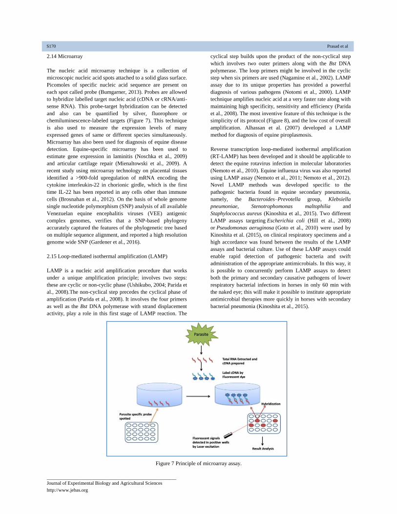
2.14 Microarray
The nucleic acid microarray technique is a collection of
microscopic nucleic acid spots attached to a solid glass surface.
Picomoles of specific nucleic acid sequence are present on
each spot called probe (Bumgarner, 2013). Probes are allowed
to hybridize labelled target nucleic acid (cDNA or cRNA/anti-
sense RNA). This probe-target hybridization can be detected
and also can be quantified by silver, fluorophore or
chemiluminescence-labeled targets (Figure 7). This technique
is also used to measure the expression levels of many
expressed genes of same or different species simultaneously.
Microarray has also been used for diagnosis of equine disease
detection. Equine-specific microarray has been used to
estimate gene expression in laminitis (Noschka et al., 2009)
and articular cartilage repair (Mienaltowski et al., 2009). A
recent study using microarray technology on placental tissues
identified a >900-fold upregulation of mRNA encoding the
cytokine interleukin-22 in chorionic girdle, which is the first
time IL-22 has been reported in any cells other than immune
cells (Brosnahan et al., 2012). On the basis of whole genome
single nucleotide polymorphism (SNP) analysis of all available
Venezuelan equine encephalitis viruses (VEE) antigenic
complex genomes, verifies that a SNP-based phylogeny
accurately captured the features of the phylogenetic tree based
on multiple sequence alignment, and reported a high resolution
genome wide SNP (Gardener et al., 2016).
2.15 Loop-mediated isothermal amplification (LAMP)
LAMP is a nucleic acid amplification procedure that works
under a unique amplification principle; involves two steps:
these are cyclic or non-cyclic phase (Ushikubo, 2004; Parida et
al., 2008).The non-cyclical step precedes the cyclical phase of
amplification (Parida et al., 2008). It involves the four primers
as well as the Bst DNA polymerase with strand displacement
activity, play a role in this first stage of LAMP reaction. The
cyclical step builds upon the product of the non-cyclical step
which involves two outer primers along with the Bst DNA
polymerase. The loop primers might be involved in the cyclic
step when six primers are used (Nagamine et al., 2002). LAMP
assay due to its unique properties has provided a powerful
diagnosis of various pathogens (Notomi et al., 2000). LAMP
technique amplifies nucleic acid at a very faster rate along with
maintaining high specificity, sensitivity and efficiency (Parida
et al., 2008). The most inventive feature of this technique is the
simplicity of its protocol (Figure 8), and the low cost of overall
amplification. Alhassan et al. (2007) developed a LAMP
method for diagnosis of equine piroplasmosis.
Reverse transcription loop-mediated isothermal amplification
(RT-LAMP) has been developed and it should be applicable to
detect the equine rotavirus infection in molecular laboratories
(Nemoto et al., 2010). Equine influenza virus was also reported
using LAMP assay (Nemoto et al., 2011; Nemoto et al., 2012).
Novel LAMP methods was developed specific to the
pathogenic bacteria found in equine secondary pneumonia,
namely, the Bacteroides–Prevotella group, Klebsiella
pneumoniae, Stenotrophomonas maltophilia and
Staphylococcus aureus (Kinoshita et al., 2015). Two different
LAMP assays targeting Escherichia coli (Hill et al., 2008)
or Pseudomonas aeruginosa (Goto et al., 2010) were used by
Kinoshita et al. (2015), on clinical respiratory specimens and a
high accordance was found between the results of the LAMP
assays and bacterial culture. Use of these LAMP assays could
enable rapid detection of pathogenic bacteria and swift
administration of the appropriate antimicrobials. In this way, it
is possible to concurrently perform LAMP assays to detect
both the primary and secondary causative pathogens of lower
respiratory bacterial infections in horses in only 60 min with
the naked eye; this will make it possible to institute appropriate
antimicrobial therapies more quickly in horses with secondary
bacterial pneumonia (Kinoshita et al., 2015).
Figure 7 Principle of microarray assay.
S170 Prasad et al

_________________________________________________________
Journal of Experimental Biology and Agricultural Sciences
http://www.jebas.org
Figure 8 Principle of LAMP.
2.16 Sequence analysis study
Whole genome sequencing is a process that gives the
complete DNA sequence of an organism's genome at a single
time. High-throughput genome sequencing technologies have
largely been used as a research tool and are currently being
introduced in the clinics (Van et al., 2013; Gilissen, 2014;
Nones et al., 2014). Genome sequencing of the domestic horse
and subsequent advancements in the field of equine genomics
have led to an explosion in the development of tools for
mapping traits and diseases and evaluating gene expression
(Finno & Bannasch, 2014). In 2011, whole-genome
sequencing of an individual American quarter horse mare was
performed using massively parallel paired-end sequencing
(Doan et al., 2012). Several single-gene disorders in quarter
horses, such as polysaccharide storage myopathy (McCue et
al., 2008; Tryon et al., 2009), hyperkalemic periodic paralysis,
glycogen branching enzyme deficiency (Rudolph et al.,1992),
and hereditary equine regional dermal asthenia (Ward et al.,
2004; Finno et al., 2009) has been reported due to whole-
genome sequencing of an individual American quarter horse
mare. A high-quality draft assembly was constructed and
additional sequence were provided by the inclusion of bacterial
artificial chromosome end sequences from a related male
thorough bred horse (Leeb et al., 2006). Kinoshita et al.,
(2014) reported the genera Bacteroides and Prevotella
especially B. fragilis and P. heparinolytica are dominant
anaerobes in lower respiratory tract infection in horses.
3 Biosensors
Biosensor is an advanced technique for the detection of either
the antigen or antibodies. This assay involves the use of a
receptor (mostly an antibody), a disease specific antibody and
a transducer that converts a biological interaction into a
measurable signal (Cruz et al., 2002). These biosensors are
frequently coupled to sophisticated instrumentation to produce
highly-specific analytical tools, most of which are still in use
only for the research and development purpose due to the high
cost of instrumentation, high cost of individual sample
analysis, and the need for highly trained persons to oversee the
testing. Fibre optic biosensors have the potential to do multi-
analyte analyses in an automated format. Portable fibre optic
biosensors, has been reported to detect four different analytes
in one coupon (King et al., 2000). Biosenors can be used as
self-contained field devices for the detection of foreign animal
disease agents. West nile virus was detected using biosensors
and microfluidic systems, a linear, 15 amino acid fragment of
domain III of WNV was successfully used as an antigen on an
amperometric immunosensor (Ionescu et al., 2007). Neng et al.
(2010) reported that, a surface enhanced Raman scattering
immunoassay was shown to be highly sensitive for the
Biotechnological tools for diagnosis of equine infectious diseases. S171

_________________________________________________________
Journal of Experimental Biology and Agricultural Sciences
http://www.jebas.org
detection of anti-WNV immunoglobulin. Hu et al. (2004)
developed a genetically biotinylated single chain fragment
variable antibody (scFv) against Venezuelan equine
encephalitis virus (VEE). Patrick et al. (2014) studied the
evolution of equine influenza and the origin of canine
influenza with the help of biosensor.
4 Nanotechnology
The systems or devices which are related to the features of
nanometre scale are broadly defined as nanotechnology. This
scale of technology as it applies to diagnostics would include
the detection of molecular interactions. The tiny dimensions of
this technology led a basement to the use of nanoarrays and
nanochips as test platforms (Jain, 2003). The potential use of
this technology is to analyse a sample for an array of infectious
agents on a single chip. Many research groups are considering
the use of chip assays that detect several agroerrorism agents in
each sample. Small, portable platforms are being designed to
allow pen-side testing of animals for diseases of concern.
The use of nanoparticles to label antibodies is another facet of
nanotechnology. These labelled antibodies can be used in
various assays to identify specific pathogens, structures or
molecules. The use of gold nanoparticles, nanobarcodes,
quantum dots and nanoparticle probes are the examples of
nanotechnology (Yguerabide & Yguerabide, 2001).
Nanopores, nanosensors, resonance light scattering and
cantilever arraysare some of the additional nanotechnologies
and it is anticipated that many of the specific nanotechnologies
will eventually be applied to the diagnosis of endemic
veterinary diseases in the future. Klier et al. (2012) reported
about an aerosol formulation of biodegradable, biocompatible
and nontoxic gelatine nanoparticle-bound CpG-ODN2216, to
treat equine recurrent airway obstruction in a clinical study.
5 Proteomics
Proteomics is the new emerging field to isolate and
characterize the protein produced by various etiological agents.
Different bacterial, viral as well as parasitic proteins can be
targeted with the help of this technology. Hence, proteomics
has potential applications in veterinary diagnostics. The usual
approach of proteomic involves separation of the proteins with
the help of two dimension gel electrophoresis and staining
them with appropriate protein marker. The protein ‘pattern’ is
different in different species; hence it can be recognized as a
fingerprint. It is then analyzed by performing image analysis
(Krah & Jungblut, 2004). Proteins that are up- or down-
regulated due to disease are compared and find by using
proteome maps. A protein of interest can be cut and taken out
from the gel and purified. This purified protein can be further
fully characterized using peptide-mass fingerprinting and/ or
mass spectrometry methods. Veterinary diagnostics may make
use of proteomics to identify or look for known disease
markers or patterns with biochip technology and
instrumentation that combines mass spectrometry with other
separation chromatography or molecular techniques in the
future. These instrumentations are designed to specifically
select, separate by molecular mass, and identify the complex
mixture of proteins in a sample, which can be compared to
known samples for diagnostic purposes.
In equine medicine, proteomics is been used in the diagnosis of
different metabolic as well as orthopedic diseases which show
some of the alteration in the expression levels of marker
proteins (Amaya, 2014). In the proteomic marker analysis
conducted in biopsy samples of horse muscles, it was found
that three significantly increased proteins: alpha actin,
tropomyosin alpha chain and creatine kinase M chain (CKM).
CKM was represented by multiple spots probably due to
posttranslational modification, one of which appeared to be
unique for tying-up suggesting that altered energy distribution
within muscle cells is part of the disease etiology (Freek et al.,
2010a). In another study they have identified, 20 differential
spots representing 16 different proteins. Evaluation of those
proteins complies with adaptation of the skeletal muscle after
normal training involving structural changes towards a higher
oxidative capacity, an increased capacity to take up long-chain
fatty acids, and to store energy in the form of glycogen.
Intensified training leads to additional changed spots. Alpha-1-
antitrypsin was found increased after intensified training but
not after normal training. This protein may thus be considered
as a marker for overtraining in horses and also linked to
overtraining in human athletes (Freek et al., 2010b). In an
another study, which was conducted on the proteomics, study
of cerebrospinal fluid, a total of 320 proteins were confidently
identified across six healthy horses, and these proteins were
further characterized by gene ontology terms mapped in
UniProt, and normalized spectral abundance factors were
calculated as a measure of relative abundance and these results
provide an optimized protocol for analysis of equine CSF and
laid the basement for future studies involving the CSF study of
equines in the context of pathogenic disease states (Carolyn et
al., 2014). The analysis of osteoarthritis and osteochondrosis
conducted by Elisabetta et al. (2012) has identified some
putative protein markers which can be further tried for the
definitive early diagnosis of osteoarthritis in the horses. A
highly sensitive proteomic comparison together with insightful
data mining enabled us to identify proteins and pathways
involved in early OA which could aid the development of early
OA diagnostic markers and therapeutics (Peffers et al., 2012).
In case of a very unpredictable disease of equines, laminitis
identification and measurement of novel protein biomarkers
present in blood that predict the onset and resolution of
laminitis would both aid clinical management of at-risk equine
patients and shed light on underlying mechanisms with the
intent of developing novel preventive strategies and therapeutic
approaches (Joseph et al., 2008).
Conclusion
A profound change has been occurred in recent years in
veterinary diagnostics with the introduction of new
biotechnological assays which completely changed the
S172 Prasad et al
_________________________________________________________
Journal of Experimental Biology and Agricultural Sciences
http://www.jebas.org
scenario of time-tested, traditional diagnostic techniques of
veterinary disease diagnosis. These new biotechnological
methods, includes the production of more specific antigens by
the use of recombination, expression vectors and synthetic
peptides. When coupled with the use of monoclonal antibodies,
the sensitivity and specificity of a number of traditional
diagnostic assays have been significantly improved. Various
forms of PCR have become a routine diagnostic tool in
veterinary laboratories for rapid screening of large number of
samples during disease outbreaks to develop prevention and
control measures and also to make specific typing
determinations for research purpose.
Other technologies are likely to be widely adopted in the future
as they demonstrate the ability to improve the diagnostic
capabilities while reducing the time and, perhaps cost
associated with more conventional technologies. Proteomics
has the potential to look at the broader picture of protein
expression for a pathogen of interest or by infected animals
and it may lead to a special niche of veterinary diagnostics.
Nanotechnologies hold the promise of screening numerous
pathogens in a single assay, while not yet implemented in
veterinary laboratories. Nanotechnology has become the choice
for mobile and pen-side testing of animal diseases due to its
small size and easy handling. Biotechnology and its
applications hold the great promise for improving the speed
and accuracy of diagnostic tests for veterinary pathogens.
Much developmental work will be required to realise the
potential with well-characterised, validated assay systems that
provide improved diagnostic capabilities to safeguard animal
health.
Traditionally, pathogens were detected by microscopic and
other conventional methods of various biological samples.
Later on several molecular and serological assays have been
employed for diagnostic purpose. These assays are shown
highly effective and sensitive results for the detection of
parasites regardless of the type of infection and sample.
Among the various available techniques, some are used for
treatment monitoring along with the diagnosis of parasites.
Thus they became a useful tool in the clinical decision making
process. The molecular and serological methods are also useful
in vast epidemiological studies, because they are also involved
in the geographical distribution study of parasites, genetic
diversity of populations, susceptibility of infections and
mutations in parasites. Detailed knowledge about the genetic
characteristics, morphology and behaviour of parasitic disease
in the affected population is provided by the molecular tools.
Although, the cost of molecular diagnosis is higher than the
conventional methods, they are highly used in veterinary
clinical diagnosis, epidemiological studies and treatment
monitoring of animals. The suitable molecular tests showing
rapid, sensitive, accurate and reliable result and which can
detect all or most targeted pathogens in a multiplex
amplification system should be developed. Moreover, for faster
surveillance strategies and monitoring of parasitic
epidemiology automated technology should be developed to
process the large number of serum samples for antibody
detection. Recently, advanced software tools and the
computing power for bioinformatics analysis of parasitic large
genome size data is a need of modern molecular diagnosis. The
major challenge regarding development of new technologies is
to optimize and evaluate the tools for control and eradication
programs of parasitic diseases. So it will help in the
development of newer technologies to a level of analytical
sensitivity which will be appropriate for testing of clinical
samples directly without previous processing.
Conflict of interest
Authors would hereby like to declare that there is no conflict of
interests that could possibly arise.
References
Abdelgawad A, Hermes R, Damiani A, Lamglait B, Czirják
GA, East M (2015) Comprehensive Serology Based on a
Peptide ELISA to Assess the Prevalence of Closely Related
Equine Herpesviruses in Zoo and Wild Animals. PLoS ONE
10 : e0138370. DOI:10.1371/journal.pone.0138370.
Alhassan A, Thekisoea OM, Yokoyamaa N, Inouea N,
Motloangb MY, Mbati PA (2007) Development of loop-
mediated isothermal amplification (LAMP) method for
diagnosis of equine piroplasmosis. Veterinary Parasitology
143:155-60. DOI: 10.1016/j.vetpar.2006.08.014
Alvarez I, Gutierrez G, Barrandeguy M, Trono K (2010)
Immunochromatographic lateral flow test for detection of
antibodies to Equine infectious anemia virus. Journal of
Virological Methods 167 :152-157. DOI:
10.1016/j.jviromet.2010.03.026
Amavisit GF, Browning D, Lightfoot S, Church GA, Anderson
KG, Whithear PF, Markham P (2001) Rapid PCR detection of
Salmonella in horse faecal samples. Veterinary Microbiology
79: 63-74.
Amaya M (2014) Proteomic strategies for the discovery of
novel diagnostic and therapeutic targets for infectious diseases.
Pathogen Disease 71:177-188. DOI: 10.1111/2049-
632X.12150
Balasuriya UB, Leutenegger CM, Topol JB (2002) Detection
of equine arteritis virus by real-time TaqMan reverse
transcription-PCR assay. Journal of Virological Methods
101:21-28.
Bannai H, Manabu N, Koji T, Takashi Y, Ken M, Takashi K
(2016) Improvement of an enzyme-linked immunosorbent
assay for by using a synthetic-peptide 24-mer repeat sequence
of glycoprotein G as an antigen. Journal of Veterinary Medical
Science 78 : 309–311.
Bannai H, Nemoto M, Tsujimura K, Yamanaka T, Kondo T,
Matsumura T (2013) Improving a Complement-fixation Test
Biotechnological tools for diagnosis of equine infectious diseases. S173

_________________________________________________________
Journal of Experimental Biology and Agricultural Sciences
http://www.jebas.org
for Equine Herpesvirus Type-1 by Pretreating Sera with
Potassium Periodate to Reduce Non-specific Hemolysis.
Journal of Equine veterinary Science 24 :71-74.
Battsetseg B, Xuan X, Ikadai H, Bautista JL, Byambaa B,
Boldbaatar D, Battur B, Battsetseg G, Batsukh Z, Igarashi I,
Nagasawa H, Mikami T, Fujisaki K (2001) Detection of
Babesia caballi and Babesia equi in dermacentornuttalli adult
ticks. International Journal of Parasitology 31:384–386.
Beltrao MROC, Alencar CAS, Leite AS, Freitas LT, Gonzalez
JC, Santana VLA, MansoFilho HC (2015) Comparison of Two
Protocols of Agar Gel Immunodiffusion (AGID) Used to
Diagnose of Equine Infectious Anemia (EIA). Open Journal of
Veterinary Medicine 5: 169-174. doi:
10.4236/ojvm.2015.57023.
Booth MG, Hood J, Brooks TJ, Hart A (2010) Health
protection Scotland anthrax clinical network. Anthrax infection
in drug users. The Lancet 375: 1345-1346. DOI:
http://dx.doi.org/10.1016/S0140-6736(10)60573-9
Borchers K, Wiik H, Frolich K, Ludwig H, East ML (2005)
Antibodies against equine herpesviruses and equine arteritis
virus in Burchell's zebras (Equus burchelli) from the Serengeti
ecosystem. Journal of Wildlife Diseases 41:80–6. DOI:
10.7589/0090-3558-41.1.80
Brendan SC, Michael J (1993) Epitopes of glycoprotein g of
equine herpesviruses 4 and 1 located near the c termini elicit
type-specific antibody responses in the natural host. Journal of
Virology 67 : 6332-6338.
Bretagne S, Costa JM (2006) Towards a nucleic acid based
diagnosis in clinical parasitology and mycology. Clinica
Chimica Acta 363 : 221-228.
Brosnahan MM, Miller DC, Adams M, Antczak DF. (2012)
IL-22 is expressed by the invasive trophoblast of the equine
(Equus caballus) chorionic girdle. Journal of Immunology
188:4181–4187.
Bumgarner R (2013) Overview of DNA microarrays: types,
applications, and their future. Current Protocols in Molecular
Biology Chapter 22:22.1.
Burnouf T, Griffiths E, Padilla A, Seddik S, Stephanoe MA,
Gutierrez JM (2004) Assessment of the viral safety of
antivenoms fractionated from equine plasma. Biologicals 32 :
115-128. DOI: 10.1016/j.biologicals.2004.07.001
Burton AB, Perkins GA, Parker J, Rosenthal R, Baumgart M,
Simpson KW (2007) The gastric mucosa of horses harbours an
abundant and diverse bacterial flora. Proceedings of American
College of Veterinary Internal Medicine, Annual meeting,
Seattle, WA.
Carolyn J, Broccardo, Gisela SH, Lutz G, Paul L, Jessica EP
(2014) Proteomic Characterization of Equine Cerebrospinal
Fluid. Journal of Equine Veterinary Science 34 : 451–458.
http://dx.doi.org/10.1016/j.jevs.2013.07.013
Carvalho R, Passos LMF, Martins AS (2000). Development of
a Differential Multiplex PCR Assay for Equine Herpesvirus 1
and 4 as a Diagnostic Tool. Zoonoses and Public Health 47 :
351–359.
Chhabra S, Khurana SK, Kapoor PK, Singha H, Singh Y,
Khirbat R (2015) Characterization of Rhodococcus equi
isolates from foals with respiratory problems using a multiplex
PCR for the vap genes. Advances in Animal and Veterinary
Sciences 3 : 28-32. |
http://dx.doi.org/10.14737/journal.aavs/2015/3.1s.28.32
Chung KM, Diamond MS (2008) Defining the levels of
secreted non-structural protein NS1 after West Nile virus
infection in cell culture and mice. Journal of Medical Virology
80:547–556.
Contreras M, Morales A, Garcia-Amado MA, De Vera M,
Bermudez V, Gueneau P (2007) Detection of Helicobacter-like
DNA in the gastric mucosa of Thoroughbred horses. Letters in
Applied Microbiology 45: 553-557. DOI: 10.1111/j.1472-
765X.2007.02227.x
Cook RF, Cook SJ, Li F, Montelaro RC, Issel CJ (2002)
Development of a multiplex real-time reverse transcriptase-
polymerase chain reaction for equine infectious anemia virus
(EIAV). Journal of Virological Methods 105: 171–179.
Cruz H, Rosa C, Oliva A (2002) Immunosensors for diagnostic
applications. Parasitology Research 88: 4-7. DOI:
10.1007/s00436-001-0559-2
Cushwa WT, Medrano JF (1996) Applications of the random
amplified polymorphic DNA (RAPD) assay for genetic
analysis of livestock species. Animal Biotechnology 7 : 11-31.
http://dx.doi.org/10.1080/10495399609525845
Diallo IS, Glen H, Lucia W, Barry JR, Bruce GC (2006)
Detection of equine herpesvirus type 1 using a real-time
polymerase chain reaction. Journal of Virological Methods
131: 92–98. DOI: 10.1016/j.jviromet.2005.07.010
Diallo IS, Glen H, Lucia W, Mark AK, Barry JR, Bruce GC
(2007) Multiplex real-time PCR for the detection and
differentiation of equid herpesvirus 1 (EHV-1) and equid
herpesvirus 4 (EHV-4). Veterinary Microbiology 123: 93–103.
Dicken M, Munday JS, Archer RM, Mayhew IG, Pandey SK.
(2010) Cutaneous fungal granulomas due to Alternaria spp.
infection in a horse in New Zealand. New Zealand Veterinary
Journal 58 : 319-320.
S174 Prasad et al

_________________________________________________________
Journal of Experimental Biology and Agricultural Sciences
http://www.jebas.org
Doan R, Cohen ND, Sawyer J, Ghaffari N, Johnson CD, Scott
VD (2012) Whole Genome sequencing and genetic variant
analysis of a quarter Horse mare. BMC Genomics 13: 78. DOI:
10.1186/1471-2164-13-78.
Druce, Mike C, Doris C, Kirsty M, David T, Renata K, Bill M,
Wendy LS, Marie G, Chris B, (2002) Utility of a Multiplex
PCR Assay for Detecting Herpesvirus DNA in Clinical
Samples. Journal of Clinical Microbiology 1728–1732.
Duarte PC, Daft BM, Conrad PA, Packham AE, Gardner IA
(2003) Comparison of a serum indirect fluorescent antibody
test with two Western blot tests for the diagnosis of equine
protozoal myeloencephalitis. Journal of Veterinary Diagnostic
Investigation 15 : 8-13.
Duarte PC, Ebel ED, Traub-Dargatz J, Wilson WD, Conrad
PA, Gardner IA (2006) Indirect fluorescent antibody testing of
cerebrospinal fluid for diagnosis of equine protozoal
myeloencephalitis. American Journal of Veterinary Reserach
67 : 869-876. DOI: 10.2460/ajvr.67.5.869
Eischeid AC (2011) SYTO dyes and EvaGreen outperform
SYBR Green in real-time PCR. BMC Research Notes 4:263.
DOI: 10.1186/1756-0500-4-263. DOI: 10.1186/1756-0500-4-
263
Elisabetta C, Marco P, Micaela T, Fausto S, Chiara D,
Giovanni R, Luca A, Franco M, Alberto G, Andrea B,
Francesca B, Andrea S (2012) Gambling on putative
biomarkers of osteoarthritis and osteochondrosis by equine
synovial fluid proteomics. Journal of Proteomics Special Issue:
Farm Animal Proteomics 75 : 4478–4493. DOI:
10.1016/j.jprot.2012.02.008
Erol E, Jackson CB, Steinman M, Meares K, Donahoe J, Kelly
N, Locke S, Smith JL, Carter CN (2015) A diagnostic
evaluation of real-time PCR, fluorescent antibody and
microscopic agglutination tests in cases of equine leptospiral
abortion. Equine Veterinary Journal 47 :171-174. DOI:
10.1111/evj.12281
Faber NA, Melissa C, Rance B, Lefebvre, Nedim C,
Buyukmihci, John E, Madigan, Neil HW (2000) Detection of
Leptospira spp. in the Aqueous Humor of Horses with
Naturally Acquired Recurrent Uveitis. Journal of Clinical
Microbiology 38: 2731–2733.
Ferris NP, Clavijo A, Yang M, Velazquez-Salinas L,
Nordengrahn A, Hutchings GH, Kristersson T, Merza M
(2012) Development and laboratory evaluation of two lateral
flow devices for the detection of vesicular stomatitis virus in
clinical samples. Journal of Virological Methods 180 : 96-100.
DOI: 10.1016/j.jviromet.2011.12.010
Finno CJ, Spier SJ, Valberg SJ (2009) Equine diseases caused
by known genetic mutations. Veterinary Journal 179 : 336–
347. http://dx.doi.org/10.1016/j.tvjl.2008.03.016
Finno CJ, Bannasch DL (2014) Applied equine genetics
Equine. Veterinary Journal 46 : 538–544. DOI:
10.1111/evj.12294
Foley JE, Sharon JS, Judy M, Niki BS, Drazenovich MS,
Christian ML (2004) Molecular epidemiologic features of
Corynebacterium pseudotuberculosis isolated from horses.
American Journal of Veterinary Research 65: 1734-1737.
Freek G, Bouwmana, Mireille ME, van Ginnekenb, Jean-Paul
N, Erik R, Ellen de Graaf-R, Inge DW, Johannes H, van der
K, Edwin CM, Eric van B (2010a) Differential expression of
equine muscle biopsy proteins during normal training and
intensified training in young standard bred horses using
proteomics technology. Comparative Biochemistry and
Physiology Part D: Genomics and Proteomics 5 : 55–64. DOI:
10.1016/j.cbd.2009.11.001
Freek G, Bouwmana, Mireille ME, van Ginnekenc, Johannes
H, van der Kolkc, Eric van B, Edwin CM, Mariman (2010b)
Novel markers for tying-up in horses by proteomics analysis of
equine muscle biopsies. Comparative Biochemistry and
Physiology Part D: Genomics and Proteomics 178–183. DOI:
10.1016/j.cbd.2010.03.009
Gardner SN, McLoughlin K, Be NA, Allen J, Weaver SC,
Forrester N (2016) Characterization of Genetic Variability of
Venezuelan Equine Encephalitis Viruses. PLoS ONE 11 :
e0152604. DOI:10.1371/journal.pone.0152604. DOI:
10.1371/journal.pone.0152604
Gasser RB (2006) Molecular tools- advances, opportunities
and prospects. Veterinary Parasitology 136 : 69-89. DOI:
10.1016/j.vetpar.2005.12.002
Gilissen (2014) Genome sequencing identifies major causes of
severe intellectual disability. Nature 511 : 344. DOI:
10.1016/j.vetpar.2005.12.002
Goodman LB, Loregian A, Perkins GA, Nugent J, Buckles EL,
Mercorelli B, Kydd JH, Palu G, Smith KC, Osterrieder N,
Davis-Poynter N (2007) A point mutation in a herpesvirus
polymerase determines neuropathogenicity. PLoS Pathogen 3:
e160. DOI: 10.1371/journal.ppat.0030160
Goto M, Shimada K, Sato A, Takahashi E, Fukasawa T,
Takahashi T, Ohka S, Taniguchi T, Honda E, Nomoto A,
Ogura A, Kirikae T, Hanaki K (2010) Rapid detection of
Pseudomonas aeruginosa in mouse feces by colorimetric loop-
mediated isothermal amplification. Journal of Microbiological
Methods 81:247–52. DOI: 10.1016/j.mimet.2010.03.008
Green S, Laue B, Fossdal CG, A Hara SW, Cottr JE (2009)
Infection of horse chestnut (Aesculus hippocastanum) by
Pseudomonas syringe pv. aesculi and its detection by
quantitative real-time PCR. Plant Pathology 58 : 731–744.
DOI: 10.1111/j.1365-3059.2009.02065.x
Biotechnological tools for diagnosis of equine infectious diseases. S175

_________________________________________________________
Journal of Experimental Biology and Agricultural Sciences
http://www.jebas.org
Hartley CA, Wilks CR, Studdert MJ, Gilkerson JR (2005)
Comparison of antibody detection assays for the diagnosis of
equine herpesvirus 1 and 4 infections in horses. American
Journal of Veterinary Research 66 : 921-928.
Hill J, Beriwal S, Chandra I, Paul VK, Kapil A, Singh T,
Wadowsky RM, Singh V, Goyal A, Jahnukainen T, Johnson
JR, Tarr PI, Vats A (2008) Loop-mediated isothermal
amplification assay for rapid detection of common strains of
Escherichia coli. Journal of Clinical Microbiology 46: 2800–4.
Hu WG, Thompson HG, Alvi AZ, Nagata LP, Suresh MR,
Fulton RE (2004) Development of immunofiltration assay by
light addressable potentiometric sensor with genetically
biotinylated recombinant antibody for rapid identification of
Venezuelan equine encephalitis virus. Journal of
Immunological Methods 289 : 27-35. DOI:
10.1016/j.jim.2004.03.007
Ijaz M, Khan MS, Khan MA, Maqbool A, Ali MM, Rehman
A, Mehmood K, Saleem MH, Shabbir MZ (2012) Comparative
study of culture and pcr for detection of carriers of
streptococcus equi in naturally infected mules in Pakistan.
Journal of Animal and Plant Sciences 22 : 256-259.
Ionescu RE, Cosnier S, Herrmann S, Marks RS (2007)
Amperometric immunosensor for the detection of anti-West
Nile virus IgG. Analytical Chemistry 79 : 8662–8668.
Jain K (2003) Nanodiagnostics: application of nanotechnology
in molecular diagnostics. Expert Review of Molecular
Diagnostics 3 : 153-161. DOI: 10.1586/14737159.3.2.153
Jain S, Wyatt D, McCaughey C, O'Neill HJ, Coyle PV (2001)
Nested multiplex polymerase chain reaction for the diagnosis
of cutaneous herpes simplex and herpes zoster infections and a
comparison with electron microscopy. Journal of Medical
Virology 63:52-56. DOI: 10.1002/1096-
9071(200101)63:1<52::AID-JMV1007>3.0.CO;2-H
Janse I, Raditijo AH, Amber CA, Hendriks, Bart JR (2013)
Multiplex qPCR for reliable detection and differentiation of
Burkholderia mallei and Burkholderia pseudomallei. BMC
Infectious Diseases 13:86. DOI: 10.1186/1471-2334-13-86
Javed R, Taku AK, Gangil R, Sharma RK (2016) Molecular
characterization of virulence genes of Streptococcus equi
subsp. equi and Streptococcus equi subsp. zooepidemicus in
equines. Veterinary World 9 : 875-881.
John JM, Kapoor PK, Malik PK (2010) Serodiagnosis of
glanders by dot-ELISA using various antigens. Indian Journal
of Animal Sciences 80 : 1084–86.
Johnson AL, Morrow JK, Sweeney RW (2013) Indirect
fluorescent antibody test and surface antigen ELISAs for
antemortem diagnosis of equine protozoal myeloencephalitis.
Journal of Veterinary Internal Medicine 27 :596-599. DOI:
10.1111/jvim.12061
Johnson DJ, Ostlund EN, Pedersen DD, Schmitt BJ (2001)
Detection of North American West Nile virus in animal tissue
by a reverse transcription-nested polymerase chain reaction
assay. Emerging Infectious Disease 7:739-41. DOI:
10.3201/eid0704.010425
Johnson N, Wakeley PR, Mansfield KL, McCracken F, Haxton
B, Phipps LP, Fooks AR (2010) Assessment of a novel real-
time pan-flavivirus RT-polymerase chain reaction. Vector
Borne Zoonotic Diseases 10:665–671. DOI:
10.1089/vbz.2009.0210
Joseph L, Mankowski, David R, Graham (2008) Potential
Proteomic-Based Strategies for Understanding Laminitis:
Predictions and Pathogenesis. Journal of Equine Veterinary
Science 28 : 484–487.
http://dx.doi.org/10.1016/j.jevs.2008.07.011
Khurana SK (2015) Current understanding of Rhodococcus
equi infection and its zoonotic implications. Advances in
Animal and Veterinary Sciences 3 : 1-10. DOI :
10.14737/journal.aavs/2015/3.1.1.10
Khurana SK, Singha H, Malik P, Gulati BR, Singh RK (2015)
Molecular characterization of clinical isolates of Rhodococcus
equi with PCR assay based on virulence plasmid marker.
Indian Journal of Animal Sciences 85 : 1063–1066.
Kim C, Lidia BCB, Andy A, Hiroshi I, Naoaki Y, Xuenan X,
Ikuo I (2008) Diagnostic real-time PCR assay for the
quantitative detection of Theileria equi from equine blood
samples. Veterinary Parasitology 151: 158–163. DOI:
10.1016/j.vetpar.2007.10.023
King K, Vanniere J, Leblanc J, Bullock K, Anderson G (2000)
Automated fiber optic biosensor for multiplexed
immunoassays. Environment Science and Technology 34:
2845-2850. DOI: 10.1021/es9913535
Kinoshita Y, Niwa H, Katayama Y (2015) Use of loop-
mediated isothermal amplification to detect six groups of
pathogens causing secondary lower respiratory bacterial
infection in horses. Microbiology and Immunology 59, 365–
370. DOI: 10.1111/1348-0421.12257
Kinoshita Y, Niwa H, Katayama Y, Hariu K (2014) Dominant
obligate anaerobes revealed in lower respiratory tract infection
in horses by 16S rRNA gene sequencing. Journal of Veterinary
Medicine and Science 76:587-591.
Klier J, Fuchs S, May A, Schillinger U, Plank C, Winter G,
Coester C, Gehlen H (2012) A nebulized gelatin nanoparticle-
based CpG formulation is effective in immunotherapy of
allergic horses. Pharmaceutical Research 29 :1650-7. DOI:
10.1007/s11095-012-0686-8
S176 Prasad et al

_________________________________________________________
Journal of Experimental Biology and Agricultural Sciences
http://www.jebas.org
Kooijman LJ, Mapes SM, Pusterla N (2016) Development of
an equine coronavirus-specific enzyme-linked immunosorbent
assay to determine serologic responses in naturally infected
horses. Journal of Veterinary Diagnostic Investigation 28 :
414-8. DOI: 10.1177/1040638716649643
Krah A, Jungblut P (2004) Immunoproteonomics. In Decker J,
Reischl U (Eds.) Molecular diagnosis of infectious diseases.
Humana Press Inc Totowa 19-32.
Kriegshauser G, Cullinane A, Kuechler E (2008) Denatured
virion protein 1 of equine rhinitis B virus 1 contains authentic
B-cell epitopes recognised in an enzymelinked immunosorbent
assay-short communication. Acta Veterinaria Hungarica
56:265-270. DOI: 10.1556/AVet.56.2008.2.14
Kriegshauser G, Kuechler E, Skern T (2009) Aggregation-
associated loss of antigenicity observed for denatured virion
protein 1 of Equine rhinitis A virus in an enzyme-linked
immunosorbent assay. Virus Research 143:130-133. DOI:
10.1016/j.virusres.2009.03.003
Lambert AJ, Martin DA, Lanciotti RS (2003) Detection of
North American and western equine encephalitis viruses by
nucleic acid amplification assays. Journal of Clinical
Microbiology 41: 379-385.
Lanciotti RS, Kerst AJ (2001) Nucleic acid sequence-based
amplification assays for rapid detection of West Nile and St.
Louis encephalitis viruses. Journal of Clinical Microbiology
39:4506–4513.
Lang A, de Vries M, Feineis S, Muller E, Osterrieder N,
Damiani AM (2013) Development of a peptide ELISA for
discrimination between serological responses to equine
herpesvirus type 1 and 4. Journal of virological methods 193 :
667-673. DOI: 10.1016/j.jviromet.2013.07.044
Larrasa J, Garcia A, Ambrose NC, Alonso JM, Parra A, de
Mendoza MH, Salazar J, Rey J, de Mendoza JH (2002) A
simple random amplified polymorphic DNA Genotyping
method for field isolates of Dermatophilus congolensis.
Journal of Veterinary Medicine B, Infectious Diseases and
Veterinary Public Health 49 :135-41.
Larrasa J, Garcia-Sanchez A, Ambrose NC, Parra A, Alonso
JM, Rey JM, Hermoso-de-Mendoza M, Hermoso-de-Mendoza
J (2004) Evaluation of randomly amplified polymorphic DNA
and pulsed field gel electrophoresis techniques for molecular
typing of Dermatophilus congolensis. FEMS Microbiology
Letters 240 : 87-97. DOI: 10.1016/j.femsle.2004.09.016
Lavoie JP, Drolet R, Parsons D, Leguillette R, Sauvageau R,
Shapiro J, Houle L, Halle G, Gebhart CJ (2000) Equine
proliferative enteropathy: a cause of weight loss, colic,
diarrhoea and hypoproteinaemia in foals on three breeding
farms in Canada. Equine veterinary Journal 32 : 418-425.
Lee SH, Kim KT, Yun SH, Choi E, Lee GH, Park YS, Cho
KH, Yi S, Kwon OD, Kim TH, Kwak D (2015) Serological
and molecular detection of Anaplasma phagocytophilum in
horses reared in Korea. Veterinarni Medicina 60 : 533–538.
doi: 10.17221/8491-VETMED
Leeb T, Vogl C, Zhu B, de Jong PJ, Binns MM, Chowdhary
BP, Scharfe M, Jarek M, Nordsiek G, Schrader F, Blocker H
(2006) A human-horse comparative map based on equine BAC
end sequences. Genomics 87 : 772-6. DOI:
10.1016/j.ygeno.2006.03.002
Letek M, Ocampo-Sosa AA, Sanders M, Fogarty U, Buckley
T, Leadon DP, Gonzalez P, Scortti M, Meijer WG, Parkhill J,
Bentley S, Vazquez-Boland JA (2008) Evolution of the
Rhodococcus equi vap pathogenicity island seen through
comparison of host-associated vapA and vapB virulence
plasmids. Journal of Bacteriology 190: 5797-5805. doi:
10.1128/JB.00468-08
Linssen B, Kinney RM, Aguilar P, Russel KL, Watts DM,
Kaaden OR, Pfeffer M (2000) Development of reverse
transcription-PCR assays specific for detection of equine
encephalitis viruses. Journal of Clinical Microbiology 38:
1527-1535.
M’ghirbi Y, Hela Y, Abderazek G, Ali B (2012) Anaplasma
phagocytophilum in horses and ticks in Tunisia. Parasites and
Vectors 5:180.
Ma G, Azab W, Osterrieder N (2013) Equine herpesviruses
type 1 (EHV-1) and 4 (EHV-4) masters of coevolution and a
constant threat to equids and beyond. Veterinary Microbiology
167 : 123–34. DOI: 10.1016/j.vetmic.2013.06.018
Macdonald J, Tonry J, Hall RA, Williams B, Palacios G,
Ashok MS, Jabado O, Clark D, Tesh RB, Briese T, Lipkin WI
(2005) NS1 protein secretion during the acute phase of West
Nile virus infection. Journal of Virology 79:13924–13933.
DOI: 10.1128/JVI.79.22.13924-13933.2005
McCue ME, Valberg SJ, Miller MB, Wade C, DiMauro S,
Akman HO, Mickelson JR (2008) Glycogen synthase (GYS1)
mutation causes a novel skeletal muscle glycogenosis.
Genomics 91 : 458–466. DOI: 10.1016/j.ygeno.2008.01.011
McFadden AM, Hanlon D, McKenzie RK, Gibson I, Bueno
IM, Pulford DJ, Orr D, Dunowska M, Stanislawek WL, Spence
RP, McDonald WL,Munro G, Mayhew IG (2016) The first
reported outbreak of equine herpesvirus myeloencephalopathy
in New Zealand. New Zealand Veterinary Journal 64 :125-34.
DOI: 10.1080/00480169.2015.1096853
Mienaltowski MJ, Huang L, Frisbie DD, McIlwraith CW,
Stromberg AJ, Bathke AC, Macleod JN (2009) Transcriptional
profiling differences for particular cartilage and repair tissue in
Biotechnological tools for diagnosis of equine infectious diseases. S177
_________________________________________________________
Journal of Experimental Biology and Agricultural Sciences
http://www.jebas.org
equine joint surface lesions. BMC Medicine Genomics 2:60.
DOI: 10.1186/1755-8794-2-60
Monego F, Maboni F, Krewer C, Vargas A, Costa M, Loreto E
(2009) Molecular characterization of Rhodococcus equi from
horse-breeding farms by means of multiplex PCR for the vap
gene family. Current Microbiology 58 : 399-403. DOI:
10.1007/s00284-009-9370-6
Morris C, Field H (1988) Application of cloned fragments of
equine herpesvirus type-1 DNA for detection of virus-specific
DNA in equine tissues. Equine Veterinary Journal 20 : 335-
340.
Nagamine K, Hase T, Notomi T (2002) Accelerated reaction
by loop-mediated isothermal amplification using loop primers.
Molecular and Cellular Probes 16:223–9.
Ndao M (2009) Diagnosis of parasitic diseases: old and new
approaches. Interdisciplinary Perspection of Infectious
Diseases 2009:1-15. http://dx.doi.org/10.1155/2009/278246
Nemoto M, Imagawa H, Tsujimura K, Yamanaka T, Kondo T,
Matsumura T (2010) Detection of equine rotavirus by reverse
transcription loop-mediated isothermal amplification (RT-
LAMP). Journal of Veterinary Medical Science 72 :823-6.
Nemoto M, Takashi Y, Hiroshi B, Koji T, Takashi K,
Tomiomatsumura (2011) Development and evaluation of a
reverse transcription loop-mediated isothermal amplification
assay for H3N8 equine influenza virus. Journal of virological
methods 178: 239–242. DOI: 10.1016/j.jviromet.2011.07.015
Nemoto M, Takashi Y, Hiroshi B, Koji T, Takashi K,
Tomiomatsumura (2012) Development and evaluation of a
reverse transcription loop-mediated isothermal amplification
assay for H7N7 equine influenza virus. Journal of veterinary
medical science.74: 929-931.
Neng J, Harpster MH, Zhang H, Mecham JO, Wilson WC,
Johnson PA (2010) A versatile SERS-based immunoassay for
immunoglobulin detection using antigen-coated gold
nanoparticles and malachite green-conjugated protein A/G.
Biosensors and Bioelectronics 26 : 1009–1015. DOI:
10.1016/j.bios.2010.08.015
Nielsen MK, David S, Peterson, Jesper M, Stig M, Thamsborg,
Susanne N, Olsen, Ray MK (2008) Detection and semi-
quantification of Strongylus vulgaris DNA in equine faeces by
real-time quantitative PCR. International Journal for
Parasitology 38: 443–453.
http://dx.doi.org/10.1016/j.ijpara.2007.07.014
Nogueira ML, Amorim JB, Oliveira JG, Bonjardim CA,
Ferreira PC, Kroon EG (2000) Comparison of virus isolation
and various polymerase chain reaction methods in the
diagnosis of mucocutaneous herpesvirus infection. Acta
Virologica 44:61-65.
Nones K, Waddell N, Wayte N, Patch AM, Bailey P, Newell F,
Holmes O, Fink JL, Quinn MC, Tang YH, Lampe G, Quek K,
Loffler KA, Manning S, Idrisoglu S, Miller D, Xu Q, Waddell
N, Wilson PJ, Bruxner TJ, Christ AN, Harliwong I, Nourse C,
Nourbakhsh E, Anderson M, Kazakoff S, Leonard C, Wood S,
Simpson PT, Reid LE, Krause L, Hussey DJ, Watson DI, Lord
RV, Nancarrow D, Phillips WA, Gotley D, Smithers BM,
Whiteman DC, Hayward NK, Campbell PJ, Pearson JV,
Grimmond SM, Barbour AP (2014) Genomic catastrophes
frequently arise in esophageal adenocarcinoma and drive
tumorigenesis. Nature Communications 5: 5224.
DOI:10.1038/ncomms6224
Noschka E, Vandenplas ML, Hurley DJ, Moore JN (2009)
Temporal aspects of laminar gene expression during the
developmental stages of equine laminitis. Veterinary
Immunology and Immunopathology 129:242–253. DOI:
10.1016/j.vetimm.2008.11.002
Notomi T, Okayama H, Masubuchi H, Yonekawa T, Watanabe
K, Amino N, Hase T (2000) Loop-mediated isothermal
amplification of DNA. Nucleic Acids Research 28: e63.
Nugent J, Birch-Machin I, Smith KC, Mumford JA, Swann Z,
Newton JR, Bowden RJ, Allen GP, Davis-Poynter N (2006)
Analysis of equine herpesvirus type 1 strain variation reveals a
point mutation of the DNA polymerase strongly associated
with neuropathogenic versus non-neuropathogenic disease
outbreaks. Journal of Virology 80: 4047–4060.
Ocampo-Sosa AA, Lewis DA, Navas J, Quigley F, Callejo R,
Scortti M, Leadon DP, Fogarty U, Vazquez-Boland JA (2007)
Molecular epidemiology of Rhodococcus equi based on traA,
vapA, and vapB virulence plasmid markers. Journal of
Infectious Diseases 196 : 763-769. DOI: 10.1086/519688
OIE (2016) OIE Terrestrial Manual 2016.
Oldfield C, Bonella H, Renwick L, Dodson HI, Alderson G,
Goodfellow M (2004) Rapid determination of vapA/vapB
genotype in Rhodococcus equi using a differential polymerase
chain reaction method. Antonie Van Leeuwenhoek 85 : 317-
326. DOI: 10.1023/B:ANTO.0000020383.66622.4d
Pal M, Rahman MT (2015) Rhodococcusequi: An emerging
zoonotic pathogen. Annals of Veterinary and Animal Science
2 : 3-10.
Palaniappan RU, Chang YF, Chang CF, Pan MJ, Yang CW,
Harpending P, McDonough SP, Dubovi E, Divers T, Qu J, Roe
B (2005) Evaluation of lig-based conventional and real time
PCR for the detection of pathogenic leptospires. Molecular and
Cellular Probes 19: 111–117. DOI: 10.1016/j.mcp.2004.10.002
Papin JF, Vahrson W, Dittmer DP (2004) SYBR green-based
real-time quantitative PCR assay for detection of West Nile
Virus circumvents false-negative results due to strain
variability. Journal of Clinical Microbiology 42:1511–1518.
S178 Prasad et al

_________________________________________________________
Journal of Experimental Biology and Agricultural Sciences
http://www.jebas.org
Parida M, Sannarangaiah S, Dash PK, Rao PVL, Morita K
(2008) Loop Mediated Isothermal Amplification (LAMP): A
new generation of innovative gene amplification technique;
per¬spectives in clinical diagnosis of infectious diseases.
Reviews in Medical Virology 18:407–21. DOI:
10.1002/rmv.593
Patrick J, Collinsa B, Sebastien G, Vachieria, Lesley F, Haireb,
Roksana W, Ogrodowiczb, Stephen R, Martinc, Philip A,
Walkerb, Xiaoli X, Steven J, Gamblinb J, Skehela J (2014)
Recent evolution of equine influenza and the origin of canine
influenza. PNAS 111 : 11175–11180. DOI:
10.1073/pnas.1406606111
Paxson J (2008) Evaluating Polymerase Chain Reaction–Based
Tests for Infectious Pathogens. Equine compendium 308-320.
Peffers MJ, Beynon RJ, Thornton D, Clegg PD (2012)
Proteomic characterisation and quantification of an in-vitro
early equine osteoarthritis model. Osteoarthritis and Cartilage
20 : 261–262. DOI:
http://dx.doi.org/10.1016/j.joca.2012.02.439
Perkins GA, den Bakker HC, Burton AJ (2012) Equine
stomachs harbor an abundant and diverse mucosal microbiota.
Applied Environmental Microbiology 78: 2522–2532. DOI:
10.1128/AEM.06252-11
Priestnall SL, Schoniger S, Ivens PA, Eickmann M,
Brachthauser L, Kehr K, Tupper C, Piercy RJ, Menzies-Gow
NJ, Herden C (2011) Borna Disease virus infection of a horse
in Great Britain. Veterinary Record 168 : 380b.
doi:10.1136/vr.c6405
Pusterla N, Barbara A, Byrne, Emir H, Samantha M, Spencer
S, Jang K, Gary M (2010) Use of quantitative real-time PCR
for the detection of Salmonella spp. in fecal samples from
horses at a veterinary teaching hospital. The Veterinary Journal
186: 252–255. DOI:
http://dx.doi.org/10.1016/j.tvjl.2009.08.022
Pusterla N, Madigan JE, Leutenegger CM (2006) Real-Time
Polymerase Chain Reaction: A Novel Molecular Diagnostic
Tool for Equine Infectious Diseases. Journal of Veterinary
Internal Medicine 20: 3–12.
Pusterla N, Pesavento PA, Leutenegger CM, Hay J,
Lowenstine LJ, Durando MM, Magdesian KG (2002)
Disseminated pulmonary adiaspiromycosis caused by
Emmonsiacrescens in a horse. Equine Veterinary Journal 34 :
749-752.
Pusterla N, Samantha M, Daniel R, Connie G (2008) Detection
of Lawsonia intracellularis by Real-time PCR in the Feces of
Free-living Animals from Equine Farms with Documented
Occurrence of Equine Proliferative Enteropathy. Journal of
Wildlife Diseases 44 : 992-998. DOI: 10.7589/0090-3558-
44.4.992
Pusterla N, Wilson WD, Mapes S, Leutenegger CM (2007)
Diagnostic evaluation of real-time PCR in the detection of
Rhodococcus equi in faeces and nasopharyngeal swabs from
foals with pneumonia.Veterinary Records 161 : 272-275. DOI:
10.1136/vr.161.8.272
Quereda A, Corrall I, Laguna F, Valencia ME, Tenorio A.
Echeverria JE, Navas E, Martin-Davila P, Moreno A, Moreno
V, Gonzalez-Lahoz JM, Arribas JR, Guerrero A (2000)
Diagnostic utility of a multiplex herpesvirus PCR assay
performed with cerebrospinal fluid from human
immunodeficiency virus-infected patients with neurological
disorders. Journal of Clinical Microbiology 38:3061-3067.
Quinlivan M, Eugene D, Fergus R, Sean A, Ann C (2005)
Real-Time Reverse Transcription PCR for Detection and
Quantitative Analysis of Equine Influenza Virus. Journal of
Clinical Microbiology 43 : 5055–5057.
Rakhshandehroo E, Sharifiyazdi H, Shayegh H, Ahmadi A
(2014) Molecular and morphological comparison of two
different types of Habronema muscae (Nematoda:
Habronematidae) in horse. Parasitological Research 113 :
4439-4445. DOI: 10.1007/s00436-014-4123-2
Read SJ, Kurtz JB (1999) Laboratory diagnosis of common
viral infections of the central nervous system by using a single
multiplex PCR screening assay. Journal of Clinical
Microbiology 37:1352-1355.
Rodriguez-Sanchez B, Jovita FP, Corinne S, Stephan Z,
Sandor B, Marisa A, Jose M, Sanchez-Vizcainoa (2008) Novel
gel-based and real-time PCR assays for the improved detection
of African horse sickness virus. Journal of Virological
Methods 151:87. DOI: 10.1016/j.jviromet.2008.03.029
Rudolph JA, Spier SJ, Byrns G, Rojas CV, Bernoco D,
Hoffman EP (1992) Periodic paralysis in quarter horses: a
sodium channel mutation disseminated by selective breeding.
Nature Genetics 2 :144–147. DOI: 10.1038/ng1092-144
Sharon J, Spier DVM, Christian M, Leutenegger, Scott PC,
Jenella EL, Jeannine BP, Tim EC, Judy E, Mihalyi BS, John
EM (2004) Use of a real-time polymerase chain reaction-based
fluorogenic 5' nuclease assay to evaluate insect vectors of
Corynebacterium pseudotuberculosis infections in horses.
American Journal of Veterinary Research 65: 829–834.
Shinwari MW, Annand EJ, Driver L, Warrilow D, Harrower B,
Allcock RJ, Pukallus D, Harper J, Bingham J, Kung N, Diallo
IS (2014) Australian bat lyssavirus infection in two horses.
Veterinary Microbiology 173 : 224-231. DOI:
10.1016/j.vetmic.2014.07.029
Singha H, Malik P, Goyal SK, Khurana SK, Mukhopadyaya C,
Eshwara VK, Singh RK (2014) Optimization and validation of
indirect ELISA using truncated TssB protein for serodiagnosis
Biotechnological tools for diagnosis of equine infectious diseases. S179

_________________________________________________________
Journal of Experimental Biology and Agricultural Sciences
http://www.jebas.org
of glanders amongst equines. Scientific World Journal 2014:
469407. DOI: http://dx.doi.org/10.1155/2014/469407
Soutullo A, Verwimp V, Riveros M, Pauli R, Tonarelli G
(2001) Design and validation of an ELISA for equine
infectious anemia (EIA) diagnosis using synthetic peptides.
Veterinary Microbiology 79 : 111-21.
Spier SJ, Leutenegger CM, Carroll SP, Loye JE, Pusterla JB,
Carpenter TE, Mihalyi JE, Madigan JE (2004) Use of a real-
time polymerase chain reaction-based fluorogenic 5' nuclease
assay to evaluate insect vectors of Corynebacterium
pseudotuberculosis infections in horses. American Journal of
Veterinary Research 65 : 829-34.
St-Laurent G, Morin G, Archambault D (1994) Detection of
equine arteritis virus following amplification of structural and
nonstructural viral genes by reversetranscription-PCR. Journal
of Clinical Microbiology 32 : 658-665.
Svobodova Z, Jankovicova B, Horak D, Bilkova Z (2013) Dot-
ELISA Affinity Test: An Easy, Low-Cost Method to Estimate
Binding Activity of Monoclonal Antibodies. Journal of
Analytical Bioanalytical Techniques 4:3.
Taniguchi A, Fukushi H, Matsumura T, Yanai T, Masegi T,
Hirai K (2000) Pathogenicity of a new neurotropic equine
herpesvirus 9 (gazelle herpesvirus 1) in horses. The Journal of
Veterinary Medical Science / the Japanese Society of
Veterinary Science 62 : 215–218.
Tavares RG, Staggemeier R, Borges ALP, Rodrigues MT,
Castelan LA, Vasconcelos J, Anschau ME, Spalding SM
(2011) Molecular techniques for the study and diagnosis of
parasite infection. Journal of Venomous Animals and Toxins
including Tropical Diseases. 17 : 239-248.
Traversa D, RaffaellaIorio, Thomas RK, Vitaliy A,
Kharchenko, Jakub G, Otranto D, Olivier AE, Sparagano
(2007) New Method for Simultaneous Species-Specific
Identification of Equine Strongyles (Nematoda, Strongylida)
by Reverse Line Blot Hybridization. Journal of Clinical
Microbiology 45 : 2937–2942. DOI:10.1128/JCM.00714-07
Tryon RC, Penedo MC, McCue ME, Valberg SJ, Mickelson
JR, Famula TR, Wagner ML, Jackson M, Hamilton MJ,
Nooteboom S, Bannasch DL (2009) Evaluation of allele
frequencies of inherited disease genes in subgroups of
American Quarter Horses. Journal of the American Veterinary
Medical Association 234 :120–125. DOI:
10.2460/javma.234.1.120
Ueti MW, Guy HP, Lowell SK, Mary S, Glen AS, Donald PK
(2005) Ability of the Vector Tick Boophilus microplus To
Acquire and Transmit Babesia equi following Feeding on
Chronically Infected Horses with Low-Level Parasitemia.
Journal of Clinical Microbiology 43 : 3755–3759. DOI:
10.1128/JCM.43.8.3755-3759.2005
Ushikubo H (2004) Principle of LAMP method - A simple and
rapid gene amplification method. Virus 54 : 107–12.
Van El, Cornel CG, Borry MC, Hastings P, Fellmann RJ,
Hodgson F, Howard SV, Cambon-Thomsen HC, Knoppers A,
Meijers-Heijboer BM, Scheffer H, Tranebjaerg H, Dondorp L,
de Wert W (2013) Whole-genome sequencing in health care.
Recommendations of the European Society of Human
Genetics. European Journal of Human Genetics 21 : 1–5
DOI:10.1038/ejhg.2013.46. DOI: 10.1038/ejhg.2013.46
Varrasso A, Dynon K, Ficorilli N, Hartley CA, Studdert MJ,
Drummer HE (2001) Identification of equine herpes viruses 1
and 4 by polymerase chain reaction. Australian Veterinary
Journal 8:563-569.
Verma RD, Misra VC (1989) Research on epizootic, diagnosis
and control of glanders with a view to eradicate the disease
from India. Project Report. pp. 31–42. Indian Council of
Agricultural Research, New Delhi.
Verma RD, Sharma JK, Venkateswaran KS, Batra HV (1990)
Development of an avidin-biotin dot enzyme-linked
immunosorbent assay and its comparison with other
serological tests for diagnosis of glanders in equines.
Veterinary Microbiology 25: 77–85.
Wagner WN, Jaret B, Haines D, Townsend HGG, Misra V
(1992) Detection of equine herpesvirus and differentiation of
equine herpesvirus type 1 from type 4 by the polymerase chain
reaction. Canadian Journal of Microbiology 38 : 1193-1196.
Wang L, Raidal SL, Pizzirani A, Wilcox GE (2007) Detection
of respiratory herpesviruses in foals and adult horses
determined by nested multiplex PCR. Veterinary Microbiology
121: 18-28. DOI: 10.1016/j.vetmic.2006.11.009
Ward TL, Valberg SJ, Adelson DL, Abbey CA, Binns MM,
Mickelson JR (2004) Glycogen branching enzyme (GBE1)
mutation causing equine glycogen storage disease IV.
Mammalian genome: Official Journal of the International
Mammalian Genome Society 15 : 570–577. DOI:
10.1007/s00335-004-2369-1
Webb K, Colin B, Tihana H, Zoe H, Karen FS, Carl R, Richard
JN, Andrew SW (2013) Detection of Streptococcus equi
subspecies equi using a triplex qPCR assay. The Veterinary
Journal 195: 300–304.
Westcott DG, King DP, Drew TW (2003) Use of an internal
standard in a closed one-tube RT-PCR for the detection of
equine arteritis virus RNA with fluorescent probes. Veterinary
Research 34:165-176. DOI: 10.1051/vetres:2002063
Wetmur JG (1991) DNA probes: applications of the principles
of nucleic acid hybridization. Critical Reviews in Biochemistry
and Molecular Biology 26 : 227-259.
S180 Prasad et al

_________________________________________________________
Journal of Experimental Biology and Agricultural Sciences
http://www.jebas.org
Wise LN, Kappmeyer LS, Mealey RH, Knowles DP (2013)
Review of equine piroplasmosis. Internatinal Journal of
Veterinary Medicne 27:1334–1346. DOI: 10.1111/jvim.12168
Yasunaga S, Maeda K, Matsumura T (2000) Application of a
type-specific enzyme-linked immunosorbent assay for equine
herpesvirus types 1 and 4 (EHV-1 and -4) to horse populations
inoculated with inactivated EHV-1 vaccine. Journal of
Veterinary Medical Science 62:687-691.
Yasunaga S, Maeda K, Matsumura T, Kai K, Iwata H, Inoue T
(1998) Diagnosis and sero-epizootiology of equine herpesvirus
type 1 and type 4 infections in Japan using a type-specific
ELISA. The Journal of veterinary medical science/the Japanese
Society of Veterinary Science 60 :1133–7.
Yeh J, Lee J, Park J, Seo H, Moon J, Cho I, Kim H, Yang Y,
Ahn K, Kyung S, Choi I, Lee J (2012) A diagnostic algorithm
to serologically differentiate west Nile virus from Japanese
encephalitis virus infections and its validation in field
surveillance of poultry and horse. Vector-Borne Zoonotic
Diseases 12 : 372-379. DOI: 10.1089/vbz.2011.0709
Yeh J, Lee J, Park J, Seo H, Park J, Moon J, Cho I, Lee J, Park
S, Song C, Choi I (2010) Fast duplex one-step reverse
transcriptase PCR for rapid deferential detection of west Nile
and Japanese encephalitis. Journal of Clinical Microbiology 48
: 4010-4014. DOI: 10.1128/JCM.00582-10
Yguerabide J, Yguerabide E (2001) Resonance light scattering
particles as ultrasensitive labels for detection of analytes in a
wide range of applications. Journal of Cellular Biochemistry
37 (Suppl.) 71-81.
Zhang J, Timoney PJ, MacLachlan NJ, McCollum WH,
Balasuriya UBR (2008) Persistent equine arteritis virus
infection in HeLa cells. Virology Journal 82 : 8456–8464.
DOI: 10.1128/JVI.01249-08
Biotechnological tools for diagnosis of equine infectious diseases. S181