Embed Size (px)
Citation preview
Biosynthesis of novel Sophorolipids using Candida bombicola ATCC 22214: Characterization and applications
A THESIS SUBMITTED BY
REETIKA GUPTA
FOR THE DEGREE OF
DOCTOR OF PHILOSOPHY
IN BIOTECHNOLOGY
SUBMITTED TO
THE UNIVERSITY OF PUNE
UNDER THE GUIDANCE OF
Dr. ASMITA PRABHUNE
DIVISION OF BIOCHEMICAL SCIENCES NATIONAL CHEMICAL LABORATORY
PUNE, INDIA
JUNE 2012

………Dedicated to my beloved parents
CERTIFICATE This is to certify that the work incorporated in the thesis entitled:
"Biosynthesis of novel Sophorolipids using Candida bombicola ATCC 22214: Characterization and applications", submitted by Reetika Gupta, for
the Degree of Doctor of Philosophy, was carried out by the candidate under
my supervision at Division of Biochemical Sciences, National Chemical
Laboratory, Pune 411008, India. Materials that have been obtained from other
sources are duly acknowledged in the thesis.
Asmita Prabhune (Research Guide)
DECLARATION BY RESEARCH SCHOLAR
I hereby declare that the thesis entitled "Biosynthesis of novel Sophorolipids using Candida bombicola ATCC 22214: Characterization and applications", submitted by me for the Degree of Doctor of Philosophy
to the University of Pune, has been carried out by me at Division of
Biochemical Sciences, National Chemical Laboratory, Pune, India, under the
guidance of Dr. Asmita Prabhune. The work is original and has not formed the
basis for the award of any other degree, diploma, associate ship, fellowship
and titles, in this or any other University or other institution of higher learning.
I further declare that the materials obtained from other sources have
been duly acknowledged in the thesis.
Reetika Gupta (Research Scholar)
C O N T E N T S
Page
No. Acknowledgement vi
List of abbreviations viii
Abstract x
Chapter 1: General Introduction 1-30
1.1 Introduction 2
1.2 Sophorolipids 3
1.2.1 Sophorolipid structure 4
1.2.2 Properties of sophorolipids 7
1.2.3 Biosynthesis of Sophorolipids 9
1.2.4 Effect of carbon sources on sophorolipid synthesis 14
1.2.4.1 Effect of hydrophilic substrates on SL production 14
1.2.4.2 Effect of lipophilic substrates on SL production 16
1.2.5 Physiological role of sophorolipids 19
1.2.6 Applications of sophorolipids 19
1.2.6.1 Cosmetic industry 19
1.2.6.2 Cleaning industry 20
1.2.6.3 Petroleum industry 21
1.2.6.4 Sophorolipids in bioremediation process 21
1.2.6.5 Food Industry 23
1.2.6.6 Sophorolipids as therapeutic agents 23
1.2.6.7 Sophorolipids as a source of specialty chemicals 25
1.2.6.8 Sophorolipids role in Nanotechnology 26
1.2.6.9 Sophorolipids as an inducer in enzyme synthesis 27
1.2.6.10 Sophorolipids in self assembly and polymer
formation
27
1.3 Objectives of the present work 29
1.3.1 Alpha Linolenic acid (α-Linolenic acid) 29
Chapter 2: Optimization of Fermentation parameters for production of linolenic acid derived Sophorolipids from Candida bombicola ATCC 22214
31-57
2.1 Summary 32
2.2 Introduction 32
2.3 Materials and methods 34
2.3.1 Materials 34
2.3.2 Microorganism and maintenance 35
2.3.3 Media optimization 35
2.3.4 Fermentative procedures 37
2.3.4.1 Inoculum development 37
2.3.4.2 Fermentative production 37
2.3.4.3 Sophorolipid estimation 37
2.3.4.4 Study of media pH optimization on LNNSL production
by C. bombicola
38
2.3.4.5 Temperature optimization study on LNNSL
production by C. bombicola
39
2.3.4.6 Optimization of inoculum size for LNNSL production
by C. bombicola
39
2.3.4.7 Optimization study of inoculum age for LNNSL
production by C. bombicola
40
2.3.4.8 Optimization study of glucose concentration for
LNNSL production by C. bombicola
41
2.3.4.9 Optimization study of fatty acid concentration for
LNNSL production by C. bombicola
42
2.3.4.10 Optimization study of yeast extract concentration for
LNNSL production by C. bombicola
43
2.4 Results and discussion 44
2.4.1 Effect of media pH on LNNSL production by C.
bombicola
45
2.4.2 Effect of temperature on LNNSL production by C.
bombicola
46
ii
2.4.3 Effect of inoculum size on LNNSL production by C.
bombicola
48
2.4.4 Effect of inoculum age on LNNSL production by C.
bombicola
49
2.4.5 Effect of glucose concentration for LNNSL production
by C. bombicola
50
2.4.6 Effect of fatty acid concentration on LNNSL production
by C. bombicola
50
2.4.7 Effect of yeast extract concentration on LNNSL
production by C. bombicola
52
2.4.8 Time kinetics on LNNSL/Biomass production 54
2.5 Conclusions 57
Chapter 3: Production and Purification of Linolenic acid derived Sophorolipids
58-71
3.1 Summary 59
3.2 Introduction 59
3.3 Materials and methods 61
3.3.1 Materials 61
3.3.2 Growth conditions and microorganism maintenance 61
3.3.3 Inoculum development 62
3.3.4 Production of sophorolipids 62
3.3.5 Isolation of sophorolipids 62
3.3.6 Microscopic analysis of SL mixture 63
3.3.7 Qualitative analysis of SL mixture by thin layer
chromatography (TLC)
63
3.3.8 Purification of sophorolipids 64
3.3.8.1 High performance liquid chromatography (HPLC) 64
3.3.8.2 Purification by chemical esterification of SL mixture 64
3.4 Results and discussion 66
3.4.1 SL yield 66
3.4.2 Microscopic analysis of SL 66
3.4.3 Analysis of SL mixture by thin layer chromatography 67
3.4.4 High performance liquid chromatography 69
iii
3.4.5 Chemical esterification of LNNSL mixture 70
3.5 Conclusions 71
Chapter 4: Structural Determination and physical properties of Linolenic acid derived Sophorolipid and its methyl ester form
72-98
4.1 Summary 73
4.2 Introduction 73
4.3 Materials and methods 74
4.3.1 Chemicals 74
4.3.2 ESI and CID-MS analysis of LNNSL fractions 74
4.3.3 ESI and NMR (nuclear magnetic resonance) analysis
of LNNSLME
75
4.3.4 Surface tension and critical micelle concentration
determination
75
4.3.4.1 Wilhelmy plate method 76
4.4 Results and discussion 77
4.4.1 Mass spectrometric analysis of LNNSL fractions 78
4.4.1.1 Mass spectra of first fraction obtained at retention
time 6.8 min
78
4.4.1.2 Mass spectra of second fraction obtained at retention
time 14.8 min
82
4.4.1.3 Mass spectra of third fraction obtained at retention
time 27.6 min
84
4.4.2 Mass spectrometric (Electro-spray ionization) analysis
of SL methyl ester
86
4.4.3 NMR analysis of LNNSLME 87
4.4.4 Surface tension and critical micelle concentration
determination
90
4.5 Conclusions 97
Chapter 5: Antibacterial properties of Linolenic acid derived Sophorolipid and its methyl ester form
99-128
5.1 Summary 100
5A Antibacterial activity 101 5A.1 Summary 101
iv
5A.2 Introduction 101
5A.3 Materials and methods 102
5A.3.1 Bacterial cultures and growth conditions 102
5A.3.2 Bacteria 103
5A.3.3 Sample preparation 104
5A.3.4 Minimun inhibitory concentration 105
5A.4 Results and discussion 105
5A.5 Conclusions 113
5B Insight into the antibacterial action using Atomic force microscopy
114
5B.1 Summary 114
5B.2 Introduction 114
5B.3 Materials and methods 116
5B.3.1 AFM analysis 116
5B.3.2 Sample preparation for AFM study 118
5B.4 Results and discussion 119
5B.4.1 AFM images of untreated E. coli 119
5B.4.2 AFM images of E. coli bacteria treated with LNNSL 120
5B.4.3 AFM images of E. coli bacteria treated with
LNNSLME
120
5B.4.4 AFM images of untreated P. aeruginosa 122
5B.4.5 AFM images of P. aeruginosa bacteria treated with
LNNSL
123
5B.4.6 AFM images of P. aeruginosa bacteria treated with
LNNSLME
123
5B.4.7 AFM images of untreated B. subtilis 125
5B.4.8 AFM images of B. subtilis bacteria treated with
LNNSL
125
5B.4.9 AFM images of B. subtilis bacteria treated with
LNNSLME
125
5B.5 Conclusions 128
Summary and conclusion 129
References 132
Publications 157
v
A C K N OW L E D G E M E N T I have been accompanied and supported by many peoples. It is a
pleasant aspect that I have now the opportunity to express my gratitude for
all of them.
I take this opportunity to gratefully acknowledge my supervisor, Dr.
Asmita Prabhune, for her guidance, keen interest and encouragement during
the course of research work. I am very thankful to her for providing me the
intellectual freedom in the work undertaken. She has given me the freedom to
think and work.
Thanks to the members of my progress committee Dr. Archana Pundle,
Division of Biochemical Sciences, NCL for her help whenever required, and Dr.
J. K. Pal, Professor, Department of Biotechnology, Pune University for
assessing my work during my Ph.D. registration and fellowship extensions.
Thanks to them for valuable suggestions.
I am thankful to all scientific and non‐scientific staff members of the
Biochemical Sciences Division, who have directly or indirectly helped during
the study.
I would like to extend my thanks to Pooja from Centre of Materials
Chracterization (CMC), NCL for helping me in obtaining AFM images; Institute
of Chemical Technology, Mumbai for help in surface tension studies.
I thank my past and present labmates Dr. Sachin, Dr. Sulabha, Dr. Atul,
Dr. Sridevi, Asmita, Nupur, Bhushan, Uma, Jayshree, Kasturi, Avinash, Umesh,
Siddharth, Aparna and Vrushali for their cooperation.
Special thanks are to my colleagues in NCL who have made my stay in
NCL enjoyable. Gauri, Roshna, Vijaydas, Tanpreet and Arvind. I am also
thankful to other colleagues for their charming company. Bhuban, Anand,
Poorva, Nishant, Manas.
vi
I find no words for my parents who have been a constant inspiration
and support for me. I am also thankful to my sisters Shikha and Roli for their
love and faith in me.
I am very thankful to most important person in my life, my husband Dr.
Ambrish Rathore who has been a moral support to me, standing by me and
boosting my morale in times of hurdles and stress. Who was strong enough to
support me when I was weak and brave enough to be there when I was afraid.
He stood by my side through ups and downs. He is my best friend, my great
companion.
I also thank all those who directly or indirectly helped in their own way.
Finally, I am thankful to the Head of Biochemical Sciences Division for
providing divisional facilities and grateful to the Dr. S. Sivaram, former
Director; Dr. Sourav Pal, Director and Dr. B. D. Kulkarni, Deputy Director for
allowing me to carry out research in this institute and making all the facilities
available for my research work at NCL; and permitting me to submit the
present work in the form of thesis. My thanks are duly acknowledged to
Department of Biotechnology (DBT), New Delhi for their financial support in
the form of Junior and Senior Research Fellowships.
…………..Reetika Gupta
vii
LIST OF ABBREVIATIONS
°C : degree centigrade
Å : Angstrom
µg : microgram
µl/µL : microlitre
µm : micro meter
a.m.u. : Atomic mass unit
ALA : α-linolenic acid
ATCC : American Type Culture Collection
CID-MS : Collision induced dissociation mass spectrmetry
Conc. : Concentrations
Da : Dalton
ESI-MS : Electrospray ionization mass spectrometry
g : gram
h : hour
kV : kilo volts
kDa : kilo dalton
l/L : liter
LB : Luria-Bertani
LCMS : Liquid chromatography mass spectrometry
LNNSL : Linolenic acid derived sophorolipid
LNNSLME : Sophorolipid methyl ester of Linolenic acid
m : metre
m/z : mass to charged ratio
mg : milli gram
MGYP : Malt extract-glucose-yeast extract-peptone media
min : Minute
mm : milli meter
ml/mL : milli liter
mN : milli Newton
MS : Mass spectrometry
NCIM : National Collection of Industrial Microorganisms.
viii
ix
NMR : Nuclear magnetic resonance
nm : Nano metre
OD : Optical density/Absorbance
Pt : Platinum
rpm : revolution per minute
SL : Sophorolipid
TOF MS : Time of flight mass spectrometry
TLC : Thin layer chromatography
TMS : Tetramethyl silane
Abstract
Preparation of new sophorolipid (SL) analogues with different
functionalities has widespread use in pharmaceutical and industrial applications.
SL composition can be modified by using both in vivo and in vitro methods.
Different lipophilic substrates have been used by researchers for SL production
such as oleic acid, stearic acid, palmitic acid and different vegetable oils. To our
knowledge, there is no such report on the analysis of individual SL molecule
produced using pure α-linolenic acid (ALA) as a lipophilic substrate. SL
production using α-linolenic acid as the lipophilic substrate may become a
valuable product of interest. In order to biosynthesize novel SLs using Candida
bombicola, Linolenic acid was used as the lipophilic and glucose as a hydrophilic
source in the fermentation medium. The present study is expected to provide
information on the production under optimized fermentation conditions,
purification , characterization and application of the SL derived using α-linolenic
acid (ALA) as the lipophilic source.
The thesis will be divided in to the following chapters:
Chapter 1: General Introduction
The first chapter is the general introduction of the thesis and it gives
detailed literature survey, significance of the sophorolipids (SLs) and objectives of
the study. It also describes the biosynthesis and various industrial applications of
different sophorolipids derived using various lipophilic substrates. Chapter
specific introduction will be discussed in respective chapters.
x
Chapter 2: Optimization of Fermentation parameters for production of
linolenic acid derived Sophorolipids from Candida bombicola ATCC 22214
Sophorolipid production is regulated by various parameters of culture
condition and media components. This chapter describes the fermentation
parameters of Candida bombicola (ATCC 22214) for the production of linolenic
acid derived Sophorolipids. These parameters included testing of different
medium, medium pH, temperature, inoculum size and age, glucose, fatty acid
and yeast extract concentration etc. as well as time kinetics of SL and biomass
production.
Chapter 3: Production and Purification of Linolenic acid derived
Sophorolipids
The sophorolipid was produced using optimum fermentation condition in
shake flasks. The SL from Candida bombicola is secreted in the culture broth.
After fermentation, culture broth was centrifuged and the supernatant was
extracted twice with equal volumes of ethyl acetate, the organic layer was dried
over anhydrous sodium sulphate and the solvent was removed by rotary-
evaporation. The brownish semi-crystalline product (SL mixture) was washed
twice with n-hexane to remove un-reacted fatty-acid and was stored at 4°C.
Sophorolipid mixture (LNNSL) was analyzed by reverse phase high performance
liquid chromatography (HPLC) with a Waters 2487 separation module (Waters Co.
Milford, Massachusetts) using 250 × 4.6 mm2 analytical symmetry C18, 5 µm
column. The gradient solvent elution profile used was as follows: water/
acetonitrile (95:5, v/v) holding for 10 min; to a final composition of
water/acetonitrile (5:95, v/v) with a linear gradient over 50 min and holding for 10
xi
min. The flow rate was 0.5 ml min-1. The peaks were detected at 220 nm
wavelength by absorbance detector. Fractions from different peaks were collected
and pooled separately in many runs. Further, for the ease of purification, chemical
esterification reaction from the literature was followed in order to get single
homogenous product (Bisht et al., 1999).
Chapter 4: Structural Determination and physical properties of Linolenic
acid derived Sophorolipid and its methyl ester form
Positive and negative ESI and CID (collision induced dissociation) mass
spectra were obtained with an API QSTAR PULSAR hybrid MS/MS quadrupole
TOF system (Applied biosystems). Samples from different fractions (collected from
HPLC) were injected separately into mass spectrometer for analysis. ESI and CID
mass spectral analysis confirmed that the Candida bombicola when grown on
glucose and α-linolenic acid produces a mixture of glycolipids consisting of free
acid, lactone and diacetylated lactone forms of C18:3 (linolenic acid) SLs as well
as diacetylated lactone form of C18:1 SL. The composition of the fermentation
product (SL mixture) was 7.5 % free acid, 80 % lactone and 4.5 % diacetylated
lactone of C18:3 molecules and 8 % of diacetylated lactone of C18:1 SL
molecules. This composition was determined from the initial crude SL loaded on
the column. Further, for the ease of purification, chemical esterification reaction
from the literature was followed in order to get single homogenous product (Bisht
et al. 1999). SL mixture was converted into the sophorolipid methyl ester and ESI-
MS analysis confirmed the presence of C18:3 moieties in the fatty acid chain of
this product and further the structure of this SL methyl ester (LNNSLME) was
confirmed by 1H and 13C nuclear magnetic resonance spectroscopy which showed
the presence of ester group in LNNSLME. Physical properties such as surface
xii
tension and critical micelle concentration were determined for SL mixture
containing 80 % Lactone and its purified sophorolipid methyl ester form and it was
found that both the compounds are good surface active agents.
Chapter 5: Antibacterial properties of Linolenic acid derived Sophorolipid
and its methyl ester form
Antibacterial properties of Sophorolipid mixture containing 80% Lactone of
C18:3 fatty acid (linolenic acid) and its chemically derived Sophorolipid methyl
ester form were checked against Gram-positive (B. subtilis) and Gram-negative
(E. coli and P. aeruginosa) bacteria. Antibacterial tests of SL mixture (LNNSL) and
sophorolipid methyl ester were performed using standard dilution micromethod.
SL mixture and its methyl ester form were diluted to 5, 10, 20 µg ml-1 with sterile
millipore water. Bacterial suspensions at a concentration of 106 CFU ml-1 were
added into each of these dilutions of SL mixture and SL methyl ester separately
and incubated for 6 h at their respective temperatures. 100 μl aliquots were taken
out from the respective suspensions at 2 h intervals and plated on LB agar plates
followed by incubation at their respective temperatures. Colonies were visualized
after 24 h and digital images of the plates were captured. The effectiveness of
compounds was analyzed by plotting percentage cell survival versus incubation
time. It was observed that bacterial colonies of both the Gram-positive and Gram-
negative bacteria decreased with increasing amount of compounds as well as
increasing incubation time. SL mixture (LNNSL) containing 80 % Lactone of C18:3
molecules were found more effective as compared to Sophorolipid methyl ester
against both Gram-positive and Gram-negative bacteria.
xiii
xiv
The antibacterial action of LNNSL containing 80 % Lactone of C18:3 fatty
acid (linolenic acid) and its methyl ester derivative on Gram-positive and Gram-
negative bacteria is investigated with the help of atomic force microscopy (AFM).
Mode of action of SL mixture containing 80 % Lactone and Sophorolipid methyl
ester was in agreement with other biosurfactants that act on the integrity of cell
membrane (Baek et al. 2003) which involves bactericidal action as confirmed from
AFM study. Further, The peptidoglycan cell wall of bacteria are mainly open
networks of macromolecules and generally do not offer significant permeability
barriers to compounds of molecular mass less than 50 kDa. So it was
hypothesized that sophorolipids due to their amphiphilic nature and molecular
mass in the range of 600-800 Da can make entry into the bacterial cell through
both the lipid bilayer as well as through porin channels. At some concentrations,
sophorolipid becomes toxic to the bacterial cell and show bactericidal action.
Sophorolipid mixture (LNNSL) containing 80 % Lactone and Sophorolipid methyl
ester (LNNSLME) both showed the similar mode of bactericidal action.
Summary and Conclusions
This study successfully demonstrated the analysis of chemically distinct
forms in the SL mixture produced by Candida bombicola when grown on glucose
and α-linolenic acid as well as its conversion into the single homogenous product
by chemical esterification reaction. These novel Sophorolipids were good surface
active and antibacterial agents. They have potential for various applications and
offer the advantages of further modifications and can produce functionalized SLs
according to their applications. Future aspects of these novel SLs will be
discussed in this section of the thesis.
Chapter 1
General Introduction
Chapter 1 2
1.1 Introduction
Sophorolipids (SLs) are extracellular surface-active agents produced
by various yeasts, such as Torulopsis bombicola (Cooper and Paddock, 1984;
Gobbert et al., 1984), Torulopsis apicola (Tulloch et al., 1967), Torulopsis
petrophilum (Cooper and Paddock, 1983) and Candida bombicola (Asmer at
al., 1988; Davila et al., 1992 and Albrecht et al., 1996) etc. SLs are one of the
types of glycolipid biosurfactants that are gaining importance for commercial
purposes due to their biodegradability, low eco-toxicity and production based
on renewable resources (Muller-Hurtig et al., 1993; Mulligan, 2005; Van
Bogaert et al., 2007 and 2011). These glycolipids are low molecular weight
biosurfactants and have been considered as secondary metabolites (Stodola
et al., 1967; Bentley and Campbell, 1968 and Rosenberg and Ron, 1999) and
are produced in the late exponential and stationary phases of the producer
organism (Hommel et al., 1987). Growing environmental awareness has
attracted the attention towards the production of glycolipid biosurfactants from
renewable resources through fermentation processes (Makkar et al., 2011).
The production of glycolipid biosurfactants through fermentation has replaced
the first generation glycolipids (e.g., alkylpolyglucosides, APGs) as well as
other synthetic surfactants produced by chemical means (Desai and Banat,
1997). The presence of a carbohydrate and a lipid moiety within the same
molecule of glycolipid are responsible for its amphiphilic nature and is one of
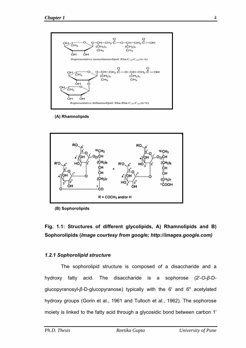
the important characteristic of a biosurfactant. The well studied and effective
glycolipid biosurfactants from the point of view of surface-active properties are
Rhamnolipid and sophorolipids (Fig. 1.1 A and B).
Ph.D. Thesis Reetika Gupta University of Pune
Chapter 1 3
Production of rhamnose-containing glycolipids was first described in
Pseudomonas aeruginosa by Jarvis and Johnson (1949) and are named as
rhamnolipids. Rhamnolipids are composed of one or two molecules of
rhamnose, linked to one or two molecules of β-hydroxy decanoic acid. L-
rhamnosyl-L-rhamnosyl-β-hydroxydecanoyl-β-hydroxydecanoate and L-
rhamnosyl-β-hydroxydecanoyl-β-hydroxydecanoate, referred to as
rhamnolipid 1 and 2, respectively, are the principal glycolipids produced by P.
aeruginosa (Fig. 1.1 A). The major drawback towards rhamnolipids production
is that the productive strains are pathogenic bacteria such as Pseudomonas
aeruginosa. In contrast to Rhamnolipids, sophorolipids’ productive strains are
non-pathogenic yeasts and also the high production yield of sophorolipids
makes it favorable for commercial production and use. The focus of the
present work is on sophorolipids so it will be discussed in detail.
1.2 Sophorolipids
SLs were first described in the early sixties by Gorin et al. (1961) as
extracellular glycolipids synthesized by the yeast Torulopsis magnoliae. Later,
Tulloch and Spencer in 1968 reported that the producing strain was actually
Torulopsis apicola, currently known as Candida apicola. Another SL
producing yeast discovered was Candida bombicola (Spencer et al., 1970).
Candida bombicola is osmophilic yeast isolated from the honey of bumble-
bees (Barnett et al., 1983 and Rosa and Lachance, 1998). Candida bombicola
ATCC 22214 is known to produce high yields (400 g l-1) of sophorolipids (Van
Bogaert et al., 2011). In the present work, Candida bombicola ATCC 22214 is
used for SL production because of its high production yields and non
pathogenic nature.
Ph.D. Thesis Reetika Gupta University of Pune
Chapter 1 4
(A) Rhamnolipids
(B) Sophorolipids
Fig. 1.1: Structures of different glycolipids, A) Rhamnolipids and B) Sophorolipids (Image courtesy from google; http://images.google.com)
1.2.1 Sophorolipid structure
The sophorolipid structure is composed of a disaccharide and a
hydroxy fatty acid. The disaccharide is a sophorose (2'-O-β-D-
glucopyranosyl-β-D-glucopyranose) typically with the 6' and 6" acetylated
hydroxy groups (Gorin et al., 1961 and Tulloch et al., 1962). The sophorose
moiety is linked to the fatty acid through a glycosidic bond between carbon 1'
Ph.D. Thesis Reetika Gupta University of Pune
Chapter 1
Ph.D. Thesis Reetika Gupta University of Pune
5
and the terminal (ω) or sub-terminal (ω-1) carbon of a long chain fatty acid
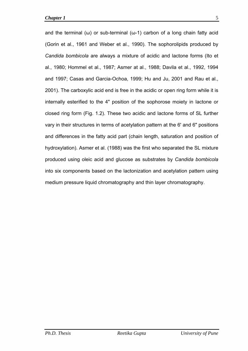
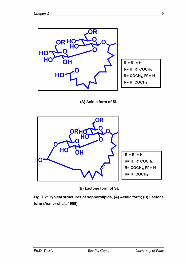
(Gorin et al., 1961 and Weber et al., 1990). The sophorolipids produced by
Candida bombicola are always a mixture of acidic and lactone forms (Ito et
al., 1980; Hommel et al., 1987; Asmer at al., 1988; Davila et al., 1992, 1994
and 1997; Casas and Garcia-Ochoa, 1999; Hu and Ju, 2001 and Rau et al.,
2001). The carboxylic acid end is free in the acidic or open ring form while it is
internally esterified to the 4" position of the sophorose moiety in lactone or
closed ring form (Fig. 1.2). These two acidic and lactone forms of SL further
vary in their structures in terms of acetylation pattern at the 6' and 6" positions
and differences in the fatty acid part (chain length, saturation and position of
hydroxylation). Asmer et al. (1988) was the first who separated the SL mixture
produced using oleic acid and glucose as substrates by Candida bombicola
into six components based on the lactonization and acetylation pattern using
medium pressure liquid chromatography and thin layer chromatography.
Chapter 1 6
R = R’ = H R= H, R’ COCH3
R= COCH3, R’ = H
R= R’ COCH3
(A) Acidic form of SL
R= R’ COCH3
R = R’ = H
R= H, R’ COCH3
R= COCH3, R’ = H
(B) Lactone form of SL
Fig. 1.2: Typical structures of sophorolipids, (A) Acidic form; (B) Lactone form (Asmer et al., 1988)
Ph.D. Thesis Reetika Gupta University of Pune
Chapter 1 7
1.2.2 Properties of sophorolipids
Being a biosurfactant sophorolipids possess all the properties of a
surface active molecule. The criteria for evaluating the biosurfactant activity
are surface tension and critical micelle concentration (Rosen, 1978). SLs are
reported to reduce the surface tension of water from 72.8 m N m-1 down to 40
to 30 m N m-1, with a critical micelle concentration (CMC) of 11 to 250 mg/l
(Develter and Lauryssen, 2010). The surface tension lowering and CMC
values of SLs are comparable to those of commercially available surfactants
such as surfactin (Arima et al., 1968). As stated above, that SLs are produced
as a mixture of acidic and lactone forms. The properties and applications of
SLs depend mainly on the abundance of certain structural form in the SL
mixture such as lactone and acidic form (Inoue, 1988 and Klekner and
Kosaric, 1993). Ratio of the acidic and lactone forms in the SL mixture varies
with the growth conditions and differentiated supply of lipophilic substrates
(Cavalero and Cooper, 2003). SL mixtures are typically viscous brown oils
and are denser than water (Gorin et al., 1961; Tulloch et al. 1962 and
Cavalero and Cooper, 2003). When the lactone form is present in the high
concentration in SL mixture, it results in the formation of crystals instead of
the more common viscous oil. For example, Cavalero and Cooper (2003)
reported the formation of crystalline SLs, when alkanes such as hexadecane
and heptadecane were used as substrates.
The French company Soliance (2004) reported that SLs are unstable at
pH values higher than 7.0-7.5 for long term storage and beyond this point
irreversible hydrolysis of the acetyl groups and ester bonds is observed.
Soliance (2004) also reported the effect of pH on the solubility of SLs. They
Ph.D. Thesis Reetika Gupta University of Pune
Chapter 1 8
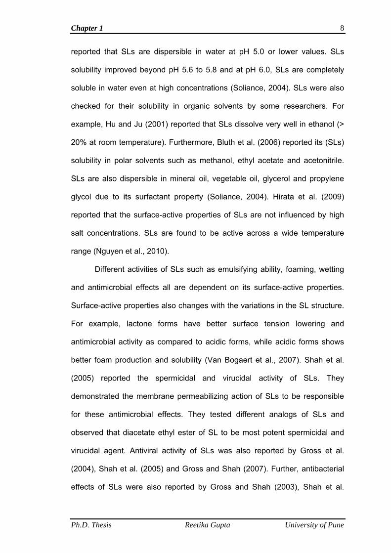
reported that SLs are dispersible in water at pH 5.0 or lower values. SLs
solubility improved beyond pH 5.6 to 5.8 and at pH 6.0, SLs are completely
soluble in water even at high concentrations (Soliance, 2004). SLs were also
checked for their solubility in organic solvents by some researchers. For
example, Hu and Ju (2001) reported that SLs dissolve very well in ethanol (>
20% at room temperature). Furthermore, Bluth et al. (2006) reported its (SLs)
solubility in polar solvents such as methanol, ethyl acetate and acetonitrile.
SLs are also dispersible in mineral oil, vegetable oil, glycerol and propylene
glycol due to its surfactant property (Soliance, 2004). Hirata et al. (2009)
reported that the surface-active properties of SLs are not influenced by high
salt concentrations. SLs are found to be active across a wide temperature
range (Nguyen et al., 2010).
Different activities of SLs such as emulsifying ability, foaming, wetting
and antimicrobial effects all are dependent on its surface-active properties.
Surface-active properties also changes with the variations in the SL structure.
For example, lactone forms have better surface tension lowering and
antimicrobial activity as compared to acidic forms, while acidic forms shows
better foam production and solubility (Van Bogaert et al., 2007). Shah et al.
(2005) reported the spermicidal and virucidal activity of SLs. They
demonstrated the membrane permeabilizing action of SLs to be responsible
for these antimicrobial effects. They tested different analogs of SLs and
observed that diacetate ethyl ester of SL to be most potent spermicidal and
virucidal agent. Antiviral activity of SLs was also reported by Gross et al.
(2004), Shah et al. (2005) and Gross and Shah (2007). Further, antibacterial
effects of SLs were also reported by Gross and Shah (2003), Shah et al.
Ph.D. Thesis Reetika Gupta University of Pune
Chapter 1 9
(2007) and Sleiman et al. (2009). SLs are also reported for their antifungal
(Gross and Shah, 2004 and 2009) and anti-cancerous activities (Chen et al.,
2006a and 2006b).
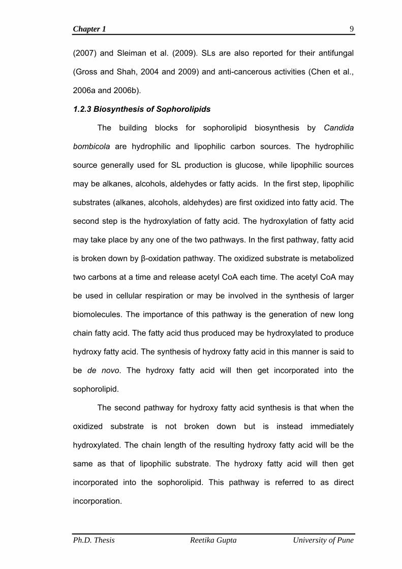
1.2.3 Biosynthesis of Sophorolipids
The building blocks for sophorolipid biosynthesis by Candida
bombicola are hydrophilic and lipophilic carbon sources. The hydrophilic
source generally used for SL production is glucose, while lipophilic sources
may be alkanes, alcohols, aldehydes or fatty acids. In the first step, lipophilic
substrates (alkanes, alcohols, aldehydes) are first oxidized into fatty acid. The
second step is the hydroxylation of fatty acid. The hydroxylation of fatty acid
may take place by any one of the two pathways. In the first pathway, fatty acid
is broken down by β-oxidation pathway. The oxidized substrate is metabolized
two carbons at a time and release acetyl CoA each time. The acetyl CoA may
be used in cellular respiration or may be involved in the synthesis of larger
biomolecules. The importance of this pathway is the generation of new long
chain fatty acid. The fatty acid thus produced may be hydroxylated to produce
hydroxy fatty acid. The synthesis of hydroxy fatty acid in this manner is said to
be de novo. The hydroxy fatty acid will then get incorporated into the
sophorolipid.
The second pathway for hydroxy fatty acid synthesis is that when the
oxidized substrate is not broken down but is instead immediately
hydroxylated. The chain length of the resulting hydroxy fatty acid will be the
same as that of lipophilic substrate. The hydroxy fatty acid will then get
incorporated into the sophorolipid. This pathway is referred to as direct
incorporation.
Ph.D. Thesis Reetika Gupta University of Pune
Chapter 1 10
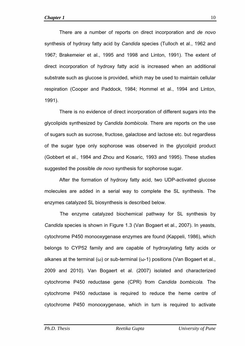
There are a number of reports on direct incorporation and de novo
synthesis of hydroxy fatty acid by Candida species (Tulloch et al., 1962 and
1967; Brakemeier et al., 1995 and 1998 and Linton, 1991). The extent of
direct incorporation of hydroxy fatty acid is increased when an additional
substrate such as glucose is provided, which may be used to maintain cellular
respiration (Cooper and Paddock, 1984; Hommel et al., 1994 and Linton,
1991).
There is no evidence of direct incorporation of different sugars into the
glycolipids synthesized by Candida bombicola. There are reports on the use
of sugars such as sucrose, fructose, galactose and lactose etc. but regardless
of the sugar type only sophorose was observed in the glycolipid product
(Gobbert et al., 1984 and Zhou and Kosaric, 1993 and 1995). These studies
suggested the possible de novo synthesis for sophorose sugar.
After the formation of hydroxy fatty acid, two UDP-activated glucose
molecules are added in a serial way to complete the SL synthesis. The
enzymes catalyzed SL biosynthesis is described below.
The enzyme catalyzed biochemical pathway for SL synthesis by
Candida species is shown in Figure 1.3 (Van Bogaert et al., 2007). In yeasts,
cytochrome P450 monooxygenase enzymes are found (Kappeli, 1986), which
belongs to CYP52 family and are capable of hydroxylating fatty acids or
alkanes at the terminal (ω) or sub-terminal (ω-1) positions (Van Bogaert et al.,
2009 and 2010). Van Bogaert et al. (2007) isolated and characterized
cytochrome P450 reductase gene (CPR) from Candida bombicola. The
cytochrome P450 reductase is required to reduce the heme centre of
cytochrome P450 monooxygenase, which in turn is required to activate
Ph.D. Thesis Reetika Gupta University of Pune
Chapter 1 11
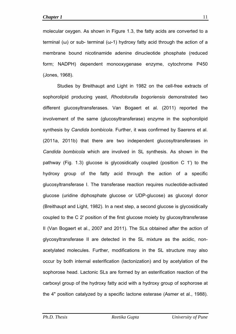
molecular oxygen. As shown in Figure 1.3, the fatty acids are converted to a
terminal (ω) or sub- terminal (ω-1) hydroxy fatty acid through the action of a
membrane bound nicotinamide adenine dinucleotide phosphate (reduced
form; NADPH) dependent monooxygenase enzyme, cytochrome P450
(Jones, 1968).
Studies by Breithaupt and Light in 1982 on the cell-free extracts of
sophorolipid producing yeast, Rhodotorulla bogoriensis demonstrated two
different glucosyltransferases. Van Bogaert et al. (2011) reported the
involvement of the same (glucosyltransferase) enzyme in the sophorolipid
synthesis by Candida bombicola. Further, it was confirmed by Saerens et al.
(2011a, 2011b) that there are two independent glucosyltransferases in
Candida bombicola which are involved in SL synthesis. As shown in the
pathway (Fig. 1.3) glucose is glycosidically coupled (position C 1') to the
hydroxy group of the fatty acid through the action of a specific
glucosyltransferase I. The transferase reaction requires nucleotide-activated
glucose (uridine diphosphate glucose or UDP-glucose) as glucosyl donor
(Breithaupt and Light, 1982). In a next step, a second glucose is glycosidically
coupled to the C 2' position of the first glucose moiety by glucosyltransferase
II (Van Bogaert et al., 2007 and 2011). The SLs obtained after the action of
glycosyltransferase II are detected in the SL mixture as the acidic, non-
acetylated molecules. Further, modifications in the SL structure may also
occur by both internal esterification (lactonization) and by acetylation of the
sophorose head. Lactonic SLs are formed by an esterification reaction of the
carboxyl group of the hydroxy fatty acid with a hydroxy group of sophorose at
the 4" position catalyzed by a specific lactone esterase (Asmer et al., 1988).
Ph.D. Thesis Reetika Gupta University of Pune
Chapter 1 12
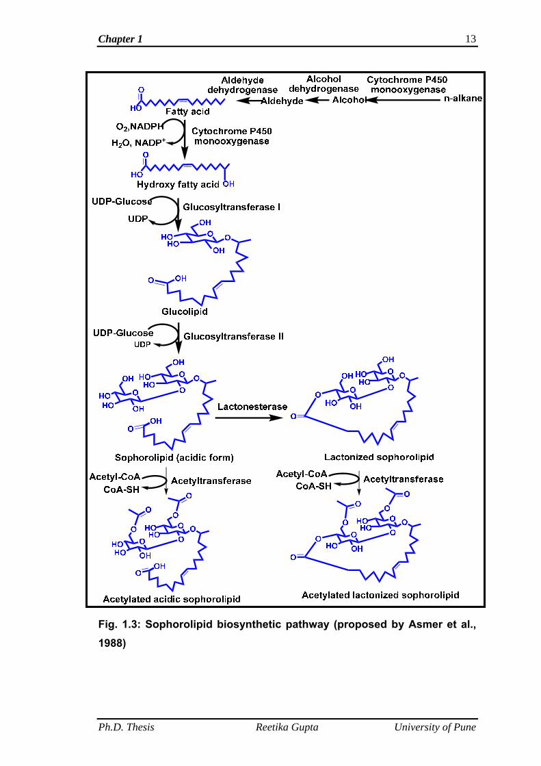
The acetylation at the 6'- and/ or 6"- position is carried out by an acetyl-
coenzyme A (CoA) dependent acetyl transferase (Esders and Light, 1972 and
Bucholtz and Light, 1976). The pathway shows that SL synthesized by the
yeast (C. bombicola) is a mixture of acidic, lactonic and their acetylated forms.
Figure 1.3 gives a schematic overview of the biochemical pathways involved
in sophorolipid synthesis.
Ph.D. Thesis Reetika Gupta University of Pune
Chapter 1 13
Fig. 1.3: Sophorolipid biosynthetic pathway (proposed by Asmer et al., 1988)
Ph.D. Thesis Reetika Gupta University of Pune
Chapter 1 14
1.2.4 Effect of carbon sources on sophorolipid synthesis
Candida bombicola can synthesize sophorolipids even when no carbon
source is provided but to a very lesser extent. It was reported that when no
lipophilic substrate is provided in the medium, there will be de novo synthesis
of fatty acid from the acetyl CoA derived from glycolysis (Van Bogaert et al.,
2007). Studies carried out by many researchers have reported that SL
synthesis increases when both the primary (hydrophilic substrate) and
secondary (lipophilic substrate) carbon sources are provided in the medium
(Cooper and Paddock, 1984; Asmer et al., 1988; Rau et al., 1996 and Davila
et al., 1997). Hommel and Huse (1993) reported that the yield of SL is much
larger, when both sugars (hydrophilic carbon source) and n-alkanes
(hydrophobic carbon source) are used than when only sugar was used as the
sole carbon source. According to Linton (1991) when sugar is supplied along
with the lipophilic substrate, then the large amount of sugar will be available
both for energy supply as well as a direct precursor of sophorose, which will
be energetically favourable than using sugar alone for both the energy supply
and as lipid precursor.
1.2.4.1 Effect of hydrophilic substrates on SL production
Influence of hydrophilic substrates such as different sugars was
investigated by many researchers. For example, Klekner et al. (1991) used a
disaccharide, sucrose in place of glucose as a hydrophilic substrate, but the
obtained SL yield was lower and no effect on the structure of sugar part of SL
was observed. Further, Zhou and Kosaric (1993 and 1995) investigated the
effect of galactose and lactose on SL synthesis. They reported that there was
no growth when only lactose was provided in the medium but when
Ph.D. Thesis Reetika Gupta University of Pune
Chapter 1 15
hydrophobic substrates such as canola, olive or safflower oil were
supplemented both the growth and SL formation were observed. For reducing
the substrate costs, Daniel et al. (1998a) used deproteinized whey
concentrate along with the rapeseed oil for SL production. They observed that
the lactose (main sugar component of whey) was not consumed during the
fermentation and the high yield of SL obtained was due to consumption of
rapeseed oil only. In the same year, Daniel et al. (1998 b) reported a method
to use deproteinized whey concentrate as a hydrophilic substrate by a two
stage cultivation process. In the first stage, Daniel et al. (1998b) cultivated the
oleaginous yeast Cryptococcus curvatus on the whey. The yeast cells
accumulated a high level of single cell oil. These cells were then harvested
and disrupted and served as a lipophilic substrate for the Candida bombicola
cells. Solaiman et al. (2004 and 2007) reported the use of low cost soy-
molasses, which is a co-product of soybean oil processing in place of glucose.
Soy molasses contains about 30 % (w/v) carbohydrates. The main
components of carbohydrates of soy molasses are raffinose, stachyose and
disaccharide sucrose other minor amount of monosacharrides. Lower yields
of SL were observed. Daverey and Pakshirajan (2009) reported sugarcane
molasses as a substitute of glucose and soybean oil as lipophilic substrate for
SL production. Sugarcane molasses contains about 62 % sugar content, of
which 35 % (w/v) is sucrose and remaining content is of glucose and fructose.
The yield of SL was less as compared to the experiment where only glucose
was used as a substrate. Perkin et al. (2005) used honey as a hydrophilic
substrate but at the end of fermentation process when the initially added
glucose was consumed. Gobbert et al. (1984) used different mono-, di- and
Ph.D. Thesis Reetika Gupta University of Pune
Chapter 1 16
trisaccharides such as glucose, fructose, mannose, maltose and raffinose, but
the yield was lower as compared to SL yield obtained from the experiment
using glucose as a substrate. Another observation was that there is no
influence on the sugar part of SL. It means that all the sugars are metabolized
into two glucose units in every case and incorporated as a sophorose moiety
in the SL structure. In all the cases of use of different sugars and cheap low
cost substrates in place of glucose, the SL yields were lower. It means that
glucose is the preferred hydrophilic substrate of choice.
1.2.4.2 Effect of lipophilic substrates on SL production
There are several evidences on influence of lipophilic carbon sources
on SL structure and yield. The lipophilic portion of the SL may vary in its
structure in terms of chain length and presence of unsaturations. Different
lipophilic carbon sources such as alkanes, fatty acids, fatty acid esters and
vegetable oils have been used for SL production. Cavalero and Cooper
(2003) reported that when alkanes were used as substrate, as the chain
length is increased from C12 to C18 the percentage of hydroxy fatty acids
having the same length as the substrate is increased and get directly
incorporated into the SL structure. Hexadecane and octadecance gave the
best yields of SLs. The same trend for incorporation of fatty acids or their
esters into the SL molecule was found. Free fatty acids and their
corresponding methyl or ethyl esters can be used as hydrophobic carbon
sources (Davila et al., 1994). Among different fatty acids used till now, oleic
acid was found to be best for production yield of SLs (Asmer et al., 1988).
Different vegetable oils such as sunflower, corn, soybean, safflower and
rapeseed oil and animal fat has been used as a lipophilic substrate for SL
Ph.D. Thesis Reetika Gupta University of Pune
Chapter 1
Ph.D. Thesis Reetika Gupta University of Pune
17
production (Cooper and Paddock, 1984; Deshpande and Daniels, 1995; Kim
et al., 1997; Casas, 1996; Casas and Garcia-Ochoa, 1999; Daniel et al.,
1998a and 1998b; Rau et al., 1999, Rau et al., 2001 and Kim et al., 2005).
Cheap lipophilic substrates such as restaurant waste oil and waste frying oil
were also used for SL production (Shah et al., 2007 and Fleurackers, 2006)
but the yield was low as compared to the fermentation where oleic acid was
used as lipophilic substrate. Different lipophilic carbon sources used by
various researchers for SL production are shown in the Table 1.1.
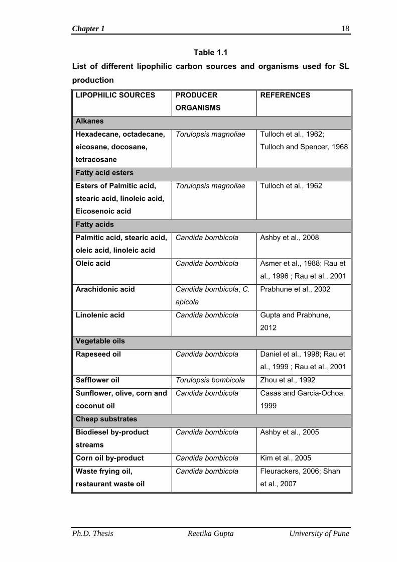
Chapter 1 18
Table 1.1
List of different lipophilic carbon sources and organisms used for SL production
LIPOPHILIC SOURCES PRODUCER ORGANISMS
REFERENCES
Alkanes
Hexadecane, octadecane, eicosane, docosane, tetracosane
Torulopsis magnoliae Tulloch et al., 1962;
Tulloch and Spencer, 1968
Fatty acid esters
Esters of Palmitic acid, stearic acid, linoleic acid, Eicosenoic acid
Torulopsis magnoliae Tulloch et al., 1962
Fatty acids
Palmitic acid, stearic acid, oleic acid, linoleic acid
Candida bombicola Ashby et al., 2008
Oleic acid Candida bombicola Asmer et al., 1988; Rau et
al., 1996 ; Rau et al., 2001
Arachidonic acid Candida bombicola, C.
apicola
Prabhune et al., 2002
Linolenic acid Candida bombicola Gupta and Prabhune,
2012
Vegetable oils
Rapeseed oil Candida bombicola Daniel et al., 1998; Rau et
al., 1999 ; Rau et al., 2001
Safflower oil Torulopsis bombicola Zhou et al., 1992
Sunflower, olive, corn and coconut oil
Candida bombicola Casas and Garcia-Ochoa,
1999
Cheap substrates
Biodiesel by-product streams
Candida bombicola Ashby et al., 2005
Corn oil by-product Candida bombicola Kim et al., 2005
Waste frying oil, restaurant waste oil
Candida bombicola Fleurackers, 2006; Shah
et al., 2007
Ph.D. Thesis Reetika Gupta University of Pune
Chapter 1 19
1.2.5 Physiological role of sophorolipids
It was reported by authors that microbial extracellular surface-active
products or biosurfactants are produced for the assimilation of hydrophobic or
water insoluble substrates by microorganisms. Biosurfactants emulsify those
substrates in water phase and thus make them available for the
microorganisms (Ito et al., 1980; Inoue and Itoh, 1982 and Hommel and
Ratledge, 1993). It has been suggested that the sophorolipid synthesis by
Candida bombicola is related to uptake of carbon sources (Ito et al., 1980 and
Otto et al., 1999).
1.2.6 Applications of sophorolipids
Sophorolipids offer an environmental friendly alternative for the
chemically derived surfactants because of their biodegradability, low eco-
toxicity and the production based on renewable-resource substrates. SLs
applications in various fields can be summarized in following points below.
1.2.6.1 Cosmetic industry
SLs exhibit moisturizing, antibacterial, antioxidant and other properties
such wetting, foaming and emulsifying, which make them useful component
for various cosmetic formulations (Klekner and Kosaric, 1993; Shete et al.,
2006 and Lourith and Kanlayavattanakul, 2009). For example, SLs exhibited
lower cytotoxicity than surfactin, which is a commercialized cosmetic
ingredient (Yoshihiko et al., 2009). SL products are FDA (Food and drug
administration) approved and are available on commercial level, for example
the French company Soliance (http://www.groupesoliance.com) produces SL
based cosmetics for body and skin. SL acts as an emulsifying agent as well
as a bactericidal agent in the treatment of acne, dandruff and body odors
Ph.D. Thesis Reetika Gupta University of Pune
Chapter 1 20
(Magar et al., 1987). Magar et al. (1987) also reported the use of SL lactone
as a component of cosmetic formulations. SLs are also effective ingredient of
pharmaco-dermatological products. They stimulate the dermal fibroblast
metabolism and collagen neosynthesis, inhibit free radical and elastase
activity, possess macrophage-activating and fibrinolytic properties, and act as
desquamating and depigmenting agents (Hillion et al., 1998; Borzeix, 1999;
Maingault, 1997 and1999 and Concaix, 2003). There are a number of patents
on the use of SL esters in cosmetics (Inoue et al., 1980; Abe et al., 1981 and
Kawano et al., 1981a, b).
1.2.6.2 Cleaning industry
Sophorolipids are fermentation products of yeasts and their production
is based on renewable raw materials, hence they are biodegradable after use
in various industrial applications. The biodegradability and surface-active
properties make them favorable for household cleaning and detergent
formulations. For example, The Japanese Company Saraya
(http://www.saraya.com) has commercialized sophoron, a dish washer
containing SLs as cleaning agent (Futura et al., 2002). SLs can also be
applied in laundry detergents (Hall et al., 1996). Detergent compositions
comprise of at least two surfactants of different characteristics and at least
one of which must be a glycolipid biosurfactant (Hall et al., 1995 and 1996).
The glycolipid biosurfactants such as sophorolipids, rhamnolipids, trehalose
lipids etc. are active ingredient in the detergent compositions (Hall et al.,
1996). Free acid and lactone form of SL or the mixtures of these two forms
are utilized as components in detergent formulations. The weight ratio of
sophorolipids to additional surfactant used in detergent formulations is
Ph.D. Thesis Reetika Gupta University of Pune
Chapter 1 21
generally 4:1 to 3:2 (Hall et al., 1996). SLs preserve their surface lowering
properties despite high salt concentrations.
1.2.6.3 Petroleum industry
Surface active and emulsifying properties of SLs can be exploited in
the petroleum industry. The use of biosurfactants in petroleum industry is for
enhanced oil recovery. The presence of biosurfactant lowers the surface and
interfacial tensions of oil in the reservoir, which facilitates oil flow and
penetration through pores in the reservoir during water, steam or fire flooding
operations in enhanced oil recovery (Brown et al., 1986; Banat, 1995 and
Marchal et al., 1999). Furthermore, SLs are useful in removing hydrocarbons
from drill material and in the regeneration of hydrocarbons from dregs and
muds (Baviere et al., 1994; Marchal et al., 1999 and Pesce, 2002). In the oil
drilling process, the cuttings are pulled out by the tool are taken up to the
surface by the up flow of drilling fluid injected through the channel of the drill
string. Discharge of such hydrocarbon impregnated drill cuttings into sea is
environmentally not safe. The patent filed by Baviere et al. 1994, reported the
use of cleaning solution containing sophorolipids to clean these drill cuttings
impregnated with polluted fluid comprising hydrocarbons before their
discharge into sea. Baviere et al. 1994 reported that the cleaning solution for
polluted cuttings comprises sophorolipids at concentrations ranging between
0.1 and 30 g l-1. The properties of SLs which allows their use in such
applications are biodegradability and surface-active nature.
1.2.6.4 Sophorolipids in bioremediation process
According to Head (1998), bioremediation is a process which involves
the contaminant specific treatment in order to reduce the concentration of
Ph.D. Thesis Reetika Gupta University of Pune
Chapter 1 22
individual or mixed environmental contaminants. There are several reports on
the applications of biosurfactants in the treatment of hydrocarbon polluted
soils (Bartha, 1986; Van Dyke et al., 1993; Zhang and Miller, 1994 and 1995;
Banat, 1995; Deziel, 1996; Volkering et al., 1997; Bruheim et al., 1997 and
Whang et al., 2008). The contamination of soils and groundwater tables by
hydrocarbons may occur by leakage of petroleum tanks or pipes or may be by
accidental spillage at ground level. The risks associated with contaminated
soils are related to health and environment which may be caused by the
vaporization of hydrocarbons in the environment as well as by the presence of
aromatic hydrocarbons such as benzene, toluene, xylene etc. in the
underground water.
SLs are reported for their solubilising action on poorly soluble aromatic
compounds present in soil and water for enhanced degradation by
microorganisms (Schippers et al., 2000). SLs are also reported for controlling
the harmful algal blooms in water bodies by their antialgal action (Sun et al.,
2004).
Ducreux et al. (1997) reported the use of SLs for decontaminating the
hydrocarbon polluted soils and groundwater tables. They demonstrated that
SLs can remove hydrocarbons from soil by forming hydrocarbon emulsions in
water as well as by enhancing the biodegradation of hydrocarbons in soil by
bacteria.
Miller (1995) reported that the addition of biosurfactant may promote
desorption of heavy metals from soils in two ways. The first is through
complexation of the free form of the metal residing in solution which
decreases the solution-phase activity of the metal and therefore promotes
Ph.D. Thesis Reetika Gupta University of Pune
Chapter 1 23
desorption. The second occurs under conditions of reduced interfacial
tension; the biosurfactant accumulate at the solid- solution interface, which
may allow direct contact between the biosurfactant and sorbed metal. SLs can
also be used in the removal of heavy metals from sediments (Mulligan et al.,
2001).
1.2.6.5 Food Industry
Sophorolipids are also used in food industry as a food formulation
ingredient. In food formulations, apart from their obvious role as agents that
decrease surface and interfacial tensions, thus promoting the formation and
stabilization of emulsions, biosurfactants can have some other functions too.
For example, to control the agglomeration of fat- globules, stabilize aerated
systems, improve texture and shelf- life of starch- containing products, modify
rheological properties of wheat dough and improve consistency and texture of
fat based products (Kachholz and Schlingmann, 1987 and Nitschke and
Coast, 2007). SLs are reported for their use in the food industry to improve
the quality of wheat flour products (Akari and Akari, 1987) and in the cold
storage transportation in air conditioning systems for the prevention of ice
particle formation (Masaru et al., 2001). Furthermore, chemically modified
derivatives of SLs such as sophorolipid alkyl esters (Allingham, 1971) are
reported to enhance the characteristics of prepared food products (bakery and
oily emulsions).
1.2.6.6 Sophorolipids as therapeutic agents
There are so many reports on the antimicrobial activities of
biosurfactants such as lipopeptides and glycolipids (Scholz et al., 1998;
Bernheimer et al., 1970; Itoh et al., 1971; Marahiel, 1993 and Tsuge et al.,
Ph.D. Thesis Reetika Gupta University of Pune
Chapter 1 24
1996). The antimicrobial properties of biosurfactants have direct connection
with their amphiphilic properties. Most of the biosurfactants, such as
rhamnolipids produced by Pseudomonas aeruginosa (Itoh et al., 1971) and
surfactin produced by Bacillus subtilis (Bernheimer and Avigad, 1970),
function as antibiotics by solubilizing the major components of cell
membranes. SLs are also reported for the same kind of solubilizing action on
cell membranes (Gi, 2004 and Shah et al., 2005). SLs are reported to inhibit
harmful algal blooms (Gi, 2004) and possess the antihuman
immunodeficiency virus and spermicidal activities (Shah et al., 2005). SLs are
used as a component in germicidal mixtures suitable for cleaning fruits and
vegetables, skin and hair by lysing microbes attached to the object surface
(Pierce and Heilman, 1998; Solaiman, 2005 and Yuan et al., 2011). They also
act as antifungal agents against plant pathogenic fungi such as Phytophthora
sp. and Pythium sp. (Yoo et al., 2005).
SLs are also finding importance in medicine because of beneficial
effects such as their ability to induce cell differentiation instead of cell
proliferation and the inhibition of protein kinase C activity of the human
promyelocytic leukemia cell line HL 60 (Isoda et al., 1997). The anti-
cancerous action of SL is attributed to a specific interaction with the plasma
membrane (Isoda et al., 1997). Joshi-Navare et al. (2011) also reported the
cell differentiation ability of SLs against the human tumorigenic glioma cell
lines (LN-229). These cell lines were derived from grade IV glioblastoma,
which is one of the frequent tumors of central nervous system and are
resistant to conventional chemotherapy. Cell differentiation inducing ability of
SLs in such brain tumors may become one of the preventive measures. Chen
Ph.D. Thesis Reetika Gupta University of Pune
Chapter 1 25
et al. (2006a) reported the anti-cancerous activity of di-acetylated lactone form
of SL against several human cancer cell lines. It was found by Chen et al.
(2006b) that the cytotoxic effect of SL on the human liver cancer cells H7402
was due to its ability to induce apoptosis. SLs are also reported to decrease
the mortality caused by septic shock in rat model (Bluth et al., 2006;
Napolitano, 2006).
1.2.6.7 Sophorolipids as a source of specialty chemicals
SLs are a source of rare and expensive components ω and ω-1
hydroxy fatty acids (Rau et al., 2001). These fatty acids are non-toxic,
biodegradable and can be produced in large amounts and are important for
industrial use (Ashby et al., 2005). Inoue and Miyamoto (1980) reported the
use of these fatty acids in polymerization reactions and their lactonization into
macrocyclic esters, which find application in the perfume and fragrance
industry. Prabhune et al. (2002) reported the synthesis of bioactive molecules,
such as 19-Hydroxyeicosatetraenoic acid (19-HETE) and 20-
Hydroxyeicosatetraenoic acid (20-HETE) by acidic hydrolysis of SLs produced
using arachidonic acid. 19-HETE and 20-HETE are penultimate and terminal
hydroxylated fatty acids of arachidonic acid and have pharmacological
applications. The pharmacological applications involves the role of 19- and
20-HETE in stimulation of renal Na+/K+ - ATPase and role of 20-HETE as a
secondary messenger in cellular processes such as autoregulation of renal
blood flow, tubuloglomerular feedback and effects on Na+ transport etc. (Oliw
et al., 1981; Escalante et al., 1988; Alonso-Gaicia et al., 1999 and Lasker et
al., 2000).
Ph.D. Thesis Reetika Gupta University of Pune
Chapter 1 26
1.2.6.8 Sophorolipids role in Nanotechnology
SLs have been reported to have various applications in
nanotechnology as well. Metal nanoparticles find applications in various fields
such as mechano- and electrical applications, catalysis and biomedical use.
These nanoparticles are stabilized with a capping agent to prevent
aggregation and allow dispersion in organic solvents or water. For biomedical
applications, the capped nanoparticles need to be dispersible in aqueous
solutions. When acidic de-acetylated SLs are used to shield cobalt or silver
particles, their fatty acid tail will interact with the metal particle either through
the terminal –COOH or through the double bond. As the sugar moiety is
exposed to the solvent, the nanoparticles acquire hydrophilic properties and
are readily dispersible. SLs also act as a reducing agent, eliminating the
necessity for an exogenous reducing agent (Kasture et al., 2007 and 2008).
These SL capped nanoparticles can act as carriers for various bioactive
molecules and have medicinal and diagnostic applications (Singh et al.,
2010). For example, Singh et al. (2009) demonstrated the antibacterial activity
of SL coated silver nanoparticles as such against both Gram positive and
Gram negative bacteria. Britto et al. (2011) reported the synthesis of silver
ions studded polymer scaffolds. In the preparation of polymer scaffold they
incorporated sophorolipid on the polymer surface through covalent bonding.
Then the prepared scaffold was studded with silver ions. The purpose of the
use of SL on scaffold was to use it as a reducing and a capping agent for
silver ions, and help in liberating the silver nanoparticles. These nanoparticle
then act as a antibacterial agent for treating the medical implants such as
catheters, surgical equipments, which are susceptible to bacterial infections.
Ph.D. Thesis Reetika Gupta University of Pune
Chapter 1 27
1.2.6.9 Sophorolipids as an inducer in enzyme synthesis
In the literature, there are some reports about the role of SL as an
inducer of several enzymes. For example, Lo and Ju (2009) reported that SLs
act as inducer in cellulase production in Hypocrea jecorina. They
demonstrated the degradation of SLs into the inducer sophorose by the
fungus. They also reported that Cellulase synthesis could be further increased
when C. bombicola was co-cultured with H. jecorina (Lo and Ju, 2009). SLs
are also reported to induce amylase production in Bacillus subtilis and laccase
and manganese peroxidase production in Pleurotus ostreatus (Gross and
Shah, 2007).
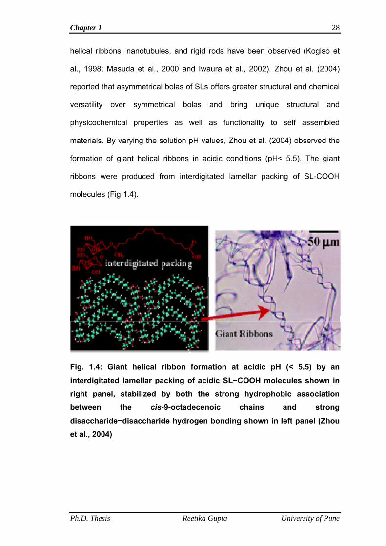
1.2.6.10 Sophorolipids in self assembly and polymer formation
Zhou et al. (2004) reported the formation of supramolecular structures
of self assembled aggregates of SL molecules under different pH conditions.
They reported that acidic form of SL molecules represent a novel type of
asymmetrical bolaamphiphiles because of the presence of disaccharide and
carboxylic acid group as polar end groups, a kinked hydrophobic core (cis-9-
octadecenoic chain), and a non-amide polar-nonpolar linkage.
Bolaamphiphiles are the molecules which bears the polar groups at both the
ends of a hydrophobic moiety. These bolaamphiphiles are the mimics of
natural trans-membrane lipids and are important for the stability of membrane
proteins in order to study their biochemical and structural features (Li et al.,
2009). Lots of study has been carried out on the synthesis of symmetrical
bolaamphiphiles bearing amino acids, monosaccharides, nucleotides as head
groups and linear methylene chains or diacetylene chains as the hydrophobic
core and supramolecular structures of monolayer vesicles, helical fibers,
Ph.D. Thesis Reetika Gupta University of Pune
Chapter 1 28
helical ribbons, nanotubules, and rigid rods have been observed (Kogiso et
al., 1998; Masuda et al., 2000 and Iwaura et al., 2002). Zhou et al. (2004)
reported that asymmetrical bolas of SLs offers greater structural and chemical
versatility over symmetrical bolas and bring unique structural and
physicochemical properties as well as functionality to self assembled
materials. By varying the solution pH values, Zhou et al. (2004) observed the
formation of giant helical ribbons in acidic conditions (pH< 5.5). The giant
ribbons were produced from interdigitated lamellar packing of SL-COOH
molecules (Fig 1.4).
Fig. 1.4: Giant helical ribbon formation at acidic pH (< 5.5) by an interdigitated lamellar packing of acidic SL−COOH molecules shown in right panel, stabilized by both the strong hydrophobic association between the cis-9-octadecenoic chains and strong disaccharide−disaccharide hydrogen bonding shown in left panel (Zhou et al., 2004)
Ph.D. Thesis Reetika Gupta University of Pune
Chapter 1 29
1.3 Objectives of the present work
The modification of SL structural composition can be done by using
different lipophilic substrates, which in turn bring unique physicochemical
properties and functionality to SL molecules (Davila et al., 1994; Nunez et al.,
2001; Glenns and Cooper, 2006). Lipophilic substrates such as fatty acids
and vegetable oils have been used for SL production as already discussed
before. There are several reports on the analysis of sophorolipids produced
using oleic acid and different vegetable oils with the help of analytical
methods such as FAB-MS, APCI-MS and ESI-MS techniques (Asmer et al.,
1988; Koster et al., 1995; Nunez et al., 2001 and Nunez et al., 2004). There is
no such report on the analysis of individual SL molecule produced using pure
α-linolenic acid (ALA) as a lipophilic substrate. SL production using α- linolenic
acid as the lipophilic substrate may become a valuable product of interest with
many enhanced beneficial properties. The objective of the present work was
to synthesize novel SLs using Candida bombicola. In this study, effect of α-
linolenic acid on the composition of SL mixture using different optimization
parameters in fermentation, purification and characterization of individual SL
molecule present in the SL mixture was done. Further, Physical and
antibacterial properties were also checked in order to evaluate the potency of
these SLs. Candida bombicola as a producer organism was used because of
its non-pathogenic character and high production yields (Van Bogaert et al.,
2007).
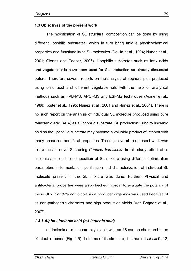
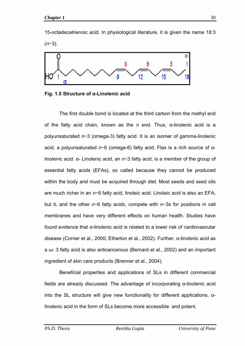
1.3.1 Alpha Linolenic acid (α-Linolenic acid)
α-Linolenic acid is a carboxylic acid with an 18-carbon chain and three
cis double bonds (Fig. 1.5). In terms of its structure, it is named all-cis-9, 12,
Ph.D. Thesis Reetika Gupta University of Pune
Chapter 1 30
15-octadecatrienoic acid. In physiological literature, it is given the name 18:3
(n−3).
Fig. 1.5 Structure of α-Linolenic acid
The first double bond is located at the third carbon from the methyl end
of the fatty acid chain, known as the n end. Thus, α-linolenic acid is a
polyunsaturated n−3 (omega-3) fatty acid. It is an isomer of gamma-linolenic
acid, a polyunsaturated n−6 (omega-6) fatty acid. Flax is a rich source of α-
linolenic acid. α- Linolenic acid, an n−3 fatty acid, is a member of the group of
essential fatty acids (EFAs), so called because they cannot be produced
within the body and must be acquired through diet. Most seeds and seed oils
are much richer in an n−6 fatty acid, linoleic acid. Linoleic acid is also an EFA,
but it, and the other n−6 fatty acids, compete with n−3s for positions in cell
membranes and have very different effects on human health. Studies have
found evidence that α-linolenic acid is related to a lower risk of cardiovascular
disease (Corner et al., 2000; Etherton et al., 2002). Further, α-linolenic acid as
a ω- 3 fatty acid is also anticancerous (Bernard et al., 2002) and an important
ingredient of skin care products (Brenner et al., 2004).
Beneficial properties and applications of SLs in different commercial
fields are already discussed. The advantage of incorporating α-linolenic acid
into the SL structure will give new functionality for different applications. α-
linolenic acid in the form of SLs become more accessible and potent.
Ph.D. Thesis Reetika Gupta University of Pune
Chapter 2
Optimization of Fermentation parameters for production of linolenic acid derived Sophorolipids from Candida bombicola
ATCC 22214
Chapter 2 32
2.1 Summary
This chapter deals with the optimization of different process
parameters to produce linolenic acid derived sophorolipids (designated as
LNNSL). These process parameters include testing of different medium used
by researchers for production of SLs using other lipophilic substrates, medium
pH, effect of temperature, inoculum size and age as well as the optimum
concentrations of glucose, fatty acid and yeast extract concentration in the
medium. Results showed that out of six tested medium (A, B, C, D, E and F),
medium A was chosen for LNNSL production and was used for optimization of
different parameters (pH, temperature, inoculum size and age, optimum
concentrations of glucose, fatty acid and yeast extract) which influenced SL
production.
2.2 Introduction
Sophorolipid, a glycolipid biosurfactant is proving its importance in
various industrial and pharmaceutical fields and are thus commercially
beneficial (Baviere et al., 1994; Schippers et al., 2000; Masaru et al., 2001;
Futura et al., 2002; Shah et al., 2005; Lourith and Kanlayavattanakul, 2009
and Britto et al., 2011). Some companies are using it on commercial scale
because of its eco-friendly nature and surface-active properties in place of
chemically synthesized surfactants as discussed in Introduction chapter
(Soliance, 2004). Lot of research work is going on to increase the functionality
of SLs in order to increase its usefulness in almost most of the industrial and
pharmaceutical sectors according to need. For example, Shah et al. (2005)
reported about the role of natural SLs obtained from fermentation in crude
form and its chemical derivatives such as, methyl, ethyl and hexyl esters and
Ph.D. Thesis Reetika Gupta University of Pune
Chapter 2 33
monoacetate and diacetate ethyl esters of SL as potent spermicidal and
virucidal agents. In 2006, Bluth et al. reported some new chemo-enzymatically
modified forms of SLs, which are effective septic shock antagonists. Further,
some authors reported amino acid conjugated SLs (Azim et al., 2006), but
these were having low microbicidal activity as compared to those reported by
Shah et al. in 2005. Efforts were made not only in chemical and enzymatic
modifications of SLs but also in modifications generated by introducing
different hydrophilic and lipophilic carbon sources in the fermentation media in
order to generate new structural analogues differing in their compositions
(Tulloch et al., 1962; Gobbert et al., 1984; Cooper and Paddock, 1984;
Klekner et al., 1991; Davila et al., 1992; Zhou and Kosaric, 1993 and 1995;
Casas, 1996; Kim et al., 1997; Daniel et al., 1998a and 1998b; Casas and
Garcia-Ochoa, 1999; Prabhune et al., 2002 and Daverey and Pakshirajan,
2009 and 2010). In the Introduction chapter it was described about the reports
on the use of different hydrophilic substrates such as different mono-, di- and
trisaccharides as well as waste cheap substrates of soy and sugarcane
molasses and whey but the composition of the sugar part of SL was not
influenced and it was always sophorose (Gobbert et al., 1984; Klekner et al.,
1991; Davila et al., 1992, 1993; Daniel et al., 1998a and 1998b; Solaiman et
al., 2004 and Daverey and Pakshirajan, 2009 and 2010). Then, the possible
mechanism was put forward that sugars are metabolized into two glucose
units, to form sophorose (Gobbert et al., 1984). It was also observed by the
researchers that the yield of SL was lower as compared to yield obtained by
using glucose as a substrate and thus glucose was the preferred substrate of
choice (Gobbert et al., 1984).
Ph.D. Thesis Reetika Gupta University of Pune
Chapter 2 34
Work has been done using several lipophilic substrates also. It was
reported by the researchers that lipophilic substrates has profound effect on
the composition of SL molecules (Asmer et al., 1988; Cavalero and Cooper,
2003 and Davila et al., 1994).
In the present work, the fermentative production of α-linolenic acid
derived SL (designated as LNNSL) molecules is presented. In order to
produce these novel SL molecules fermentation parameters must be
standardized. In this chapter, the optimization of different parameters such as
testing of different medium, medium pH, temperature, effect of concentrations
of glucose, α-linolenic acid and yeast extract concentration etc. is presented.
The biosynthesis was carried out using Candida bombicola ATCC
22214 as a producer organism. In the fermentation media glucose was used
as a hydrophilic, primary carbon source and α-linolenic acid was used as a
lipophilic, secondary carbon source. This is the first report on the synthesis of
α-linolenic acid derived SLs. Candida bombicola is used for SL production
because of its non-pathogenicity (Asmer et al., 1988; Solaiman et al., 2004
and Van Bogaert et al., 2007).
2.3 Materials and methods
2.3.1 Materials
Malt extract, glucose, yeast extract and peptone used in this study
were purchased from Hi-media, India; α-Linolenic acid was purchased from
Sigma-Aldrich (USA). Sodium sulphate was purchased from Merck, India.
Organic solvents such as ethyl acetate and n-hexane used were of analytical
grade and were purchased from Rankem India.
Ph.D. Thesis Reetika Gupta University of Pune

Chapter 2 35
2.3.2 Microorganism and maintenance
The yeast used for sophorolipid (SL) production was Candida
bombicola (ATCC 22214). It was procured from American type culture
collection, USA. Candida bombicola was grown for 48 h at 28 °C incubation
on agar slants containing: malt extract, 0.3 %; glucose, 5.0 %; yeast extract,
0.3 %; peptone, 0.5 % and agar, 2.0 %. The microorganism was sub-cultured
monthly and maintained at 4 °C in a refrigerator.
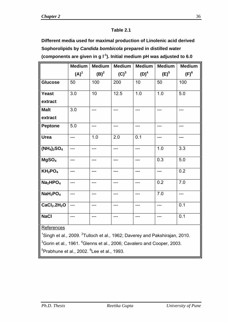
2.3.3 Media optimization
Six different media varying in composition were tested for optimal
production of linolenic acid derived SLs (LNNSL). These six media tested
were used by various researchers for SL production using different lipophilic
substrates. These six media screened for LNNSL production are summarized
in Table 2.1.
Ph.D. Thesis Reetika Gupta University of Pune
Chapter 2 36
Table 2.1
Different media used for maximal production of Linolenic acid derived Sophorolipids by Candida bombicola prepared in distilled water (components are given in g l-1). Initial medium pH was adjusted to 6.0
Medium (A)1
Medium (B)2
Medium (C)3
Medium (D)4
Medium (E)5
Medium (F)6
Glucose 50 100 200 10 50 100
Yeast extract
3.0 10 12.5 1.0 1.0 5.0
Malt extract
3.0 --- --- --- --- ---
Peptone 5.0 --- --- --- --- ---
Urea --- 1.0 2.0 0.1 --- ---
(NH4)2SO4 --- --- --- --- 1.0 3.3
MgSO4 --- --- --- --- 0.3 5.0
KH2PO4 --- --- --- --- --- 0.2
Na2HPO4 --- --- --- --- 0.2 7.0
NaH2PO4 --- --- --- --- 7.0 ---
CaCl2.2H2O --- --- --- --- --- 0.1
NaCl --- --- --- --- --- 0.1
References 1Singh et al., 2009. 2Tulloch et al., 1962; Daverey and Pakshirajan, 2010. 3Gorin et al., 1961. 4Glenns et al., 2006; Cavalero and Cooper, 2003. 5Prabhune et al., 2002. 6Lee et al., 1993.
Ph.D. Thesis Reetika Gupta University of Pune
Chapter 2 37
2.3.4 Fermentative procedures
2.3.4.1 Inoculum development
Inoculum was developed by transferring a loopful of C. bombicola cells
from slants in MGYP (malt extract, 0.3%; glucose, 5.0%; yeast extract, 0.3%;
peptone, 0.5%) medium and incubated for 24 h at 28 °C with 180 rpm orbital
shaking.
2.3.4.2 Fermentative production
All the optimization experiments were performed in 500 ml Erlenmeyer
flasks containing 100 ml media by varying the medium composition (Table
2.1). The fermentative production of SLs was initiated by transferring the
inoculum (10%, v/v) into 100 ml of respective medium (A, B, C, D, E and F)
followed by incubation at 28 °C with 180 rpm orbital shaking. Each medium
(A, B, C, D, E and F) was supplemented with 1 ml of α-linolenic acid
dispersed in 1 ml of ethanol.
The fermented broths were examined for the estimation of SLs at
regular intervals (24, 48, 72, 96, and 120….. up to192 h). For this, 100 mL
culture broths of each tested media (A, B, C, D, E and F) were examined at
regular intervals (24, 48, 72, 96, and 120….. up to192 h).
2.3.4.3 Sophorolipid estimation
100 ml culture broths of each tested media (A, B, C, D, E and F) at
regular time intervals (24, 48, 72, 96, and 120….. up to192 h) were
centrifuged at 5000 rpm for 20 min at 25 °C and supernatant was separated.
The supernatant (aqueous phase) collected were extracted twice with ethyl
acetate in separating funnel. The ethyl acetate layer (organic phase layer)
was collected and rotary-evaporated at 40 °C to remove solvent. The residue
Ph.D. Thesis Reetika Gupta University of Pune
Chapter 2 38
obtained was washed with n-hexane two times to remove the un-reacted fatty
acid. After the washing with n-hexane the partially purified SL residue was
dried under vacuum and weighed to estimate the SL content.
2.3.4.4 Study of media pH optimization on LNNSL production by C.
bombicola
To study the effect of media pH on SL formation, 500 ml Erlenmeyer
flasks were taken and each containing 100 ml of medium A were autoclaved
at 121 °C for 15 min. The composition of medium A as already mentioned
above was: glucose, 5.0 %; malt extract, 0.3 %; yeast extract, 0.3 %; peptone,
0.5 % and this composition was kept constant in all the 500 ml flasks
containing 100 ml media while varying the pH. The pH tested was 4.5, 5.0,
5.5, 6.0, 6.5 and 7.0 respectively. The experimental media flasks were
supplied with 1 ml of α-linolenic acid dispersed in 1 ml of ethanol. All the
flasks were inoculated with 10 % (v/v) inoculum and kept for SL formation in
incubator shaker at 180 rpm for 168 h at 28 °C.
After incubation period, all the flasks were taken out and 100 ml culture
broth of each flask were centrifuged separately to remove cells and
supernatant was separated. The supernatants collected from each of the
flasks were extracted twice with ethyl acetate separately. The ethyl acetate
layer (organic layer) of each media flask was dried over anhydrous sodium
sulphate and the solvent was removed by rotary-evaporation separately at 40
°C. The residues obtained were washed separately with n-hexane to remove
unused fatty acid. The partially purified SL residues thus obtained from each
flask were measured gravimetrically to estimate SL production. The
experiment was performed in triplicates.
Ph.D. Thesis Reetika Gupta University of Pune
Chapter 2 39
2.3.4.5 Temperature optimization study on LNNSL production by C.
bombicola
After the optimization of medium pH, the experiment for temperature
optimization was performed by varying the temperature of incubator shaker
for the estimation of maximum SL production. Composition of media A
(glucose, 5.0 %; malt extract, 0.3 %; yeast extract, 0.3 %; peptone, 0.5 %),
media pH at 6.0, supply of fatty acid (1 ml of α-linolenic acid dispersed in 1 ml
of ethanol) and inoculum size and age (10 %, v/v; 24 h grown inoculum) to
each of the experimental flasks (500 ml flasks containing 100 ml of media A)
were kept constant. Separate runs of the experiment at different incubator
temperatures were performed. The temperatures used for the study were 26,
28, 30, 32, 34 and 36 °C. The experiment was carried out at 180 rpm for 168
h.
After the incubation period of 168 h, all the experimental flasks were
taken out of the incubator shaker. 100 ml culture broths of each experimental
flask were centrifuged to remove cells from supernatant. The supernatants of
each experimental flask were extracted twice with ethyl acetate separately
and dried over anhydrous sodium sulphate and the solvent was rotary-
evaporated at 40 °C. The SL residue obtained was washed twice with n-
hexane to remove unused fatty acid. The SL was dried under vacuum and
weighed gravimetrically. The experiment was performed in triplicates.
2.3.4.6 Optimization of inoculum size for LNNSL production by C.
bombicola
Effect of inoculum size was performed by varying the volume of 24 h
grown inoculum while keeping rest of the parameters such as composition of
Ph.D. Thesis Reetika Gupta University of Pune
Chapter 2 40
media A (glucose, 5.0 %; malt extract, 0.3 %; yeast extract, 0.3 %; peptone,
0.5 %), media pH (6.0), supply of fatty acid substrate (1 ml of α-linolenic acid
dispersed in 1 ml of ethanol) and incubation period (168 h) constant.
Experimental flasks (500 ml Erlenmeyer flasks) containing 100 ml of medium
A was inoculated separately with different inoculum sizes such as 5.0 %, 10
% and 20 % (v/v). All the experimental flasks were kept in incubator shaker at
180 rpm for 168 h at 28 °C.
After 168 hours of incubation period, flasks were taken out. Cells were
removed by centrifugation and the supernatants were separated.
Supernatants (100 ml) obtained from each experimental flask were extracted
twice with equal volumes of ethyl acetate and solvents were removed by
rotary-evaporation at 40 °C. Residues were washed twice with n-hexane and
dried under vacuum and weighed gravimetrically. Three sets of parallel
experiments were performed.
2.3.4.7 Optimization study of inoculum age for LNNSL production by C.
bombicola
The age of inoculum was also checked for its influence on SL
production, while keeping other parameters such as composition of media A
(glucose, 5.0 %; malt extract, 0.3 %; yeast extract, 0.3 %; peptone, 0.5 %),
media pH (6.0), supply of fatty acid substrate (1 ml of α-linolenic acid
dispersed in 1 ml of ethanol), inoculum size (10 %, v/v), incubation period
(168 h) and temperature of incubator shaker (28 °C) constant. Experimental
flasks (500 ml Erlenmeyer flasks) containing 100 ml media A were inoculated
separately with 10 % (v/v) inoculum of 24, 48, 72 and 96 h grown cultures of
Ph.D. Thesis Reetika Gupta University of Pune
Chapter 2 41
C. bombicola. All the flasks were kept for incubation at 180 rpm for 168 h at
28 °C.
The steps involving separation and isolation of SL from each
experimental flask were same as described above in each optimization
parameters. The SL was measured gravimetrically. Experiment was
performed in triplicates.
2.3.4.8 Optimization study of glucose concentration for LNNSL
production by C. bombicola
After the optimization for medium pH, temperature, inoculum size and
age, the effect of glucose concentration on SL production was checked by
using different concentrations of glucose in medium A such as, 5.0 %, 7.5 %,
10 %, 12.5 % and 15 % (w/v) while rest of the media components such as
malt extract, 0.3 %; yeast extract, 0.3 %; peptone, 0.5 % were kept constant.
The other parameters such as media pH (6.0), supply of fatty acid substrate
(1 ml of α-linolenic acid dispersed in 1 ml of ethanol), inoculum size and age
(10 %, v/v; 72 h grown culture), incubation period (168 h) and temperature of
incubator shaker (28 °C) were kept constant. Experimental flasks (500 ml
Erlenmeyer flasks) were supplied with 100 ml of media A containing varying
concentrations of glucose (5.0 %, 7.5 %, 10 %, 12.5 % and 15 %, w/v)
separately and were autoclaved at 121 °C for 15 min. All the experimental
flasks were supplied with 1 ml of α-linolenic acid dispersed in 1 ml of ethanol
and inoculated with 10 % (v/v), 72 h grown inoculum separately. The flasks
were incubated at 28 °C for 168 h at 180 rpm.
The flasks were taken out after the incubation period of 168 h. 100 ml
culture broths of each flask were centrifuged separately to remove cells from
Ph.D. Thesis Reetika Gupta University of Pune
Chapter 2 42
supernatants. The supernatants were collected separately and extracted twice
with equal volumes of ethyl acetate. The ethyl acetate layer containing SLs
and unused fatty acids were dried over anhydrous sodium sulphate and were
rotary-evaporated to remove solvent. The obtained SL residues of each
experimental flask were washed twice with n-hexane to remove unused fatty
acid and dried under vacuum. The SL residues obtained were measured
gravimetrically. Experiment was performed in triplicates.
2.3.4.9 Optimization study of fatty acid concentration for LNNSL
production by C. bombicola
The fatty acid concentration (secondary carbon source) was also
checked for the maximal SL production. For this purpose, different
concentrations of fatty acid were used such as 0.1 %, 0.2 %, 0.3 %, 0.4 %,
0.5 % and 0.6 % (v/v) while keeping the other parameters such as
composition of media A (malt extract, 0.3 %; yeast extract, 0.3 %; peptone,
0.5 % and optimal glucose conc. 10 %, w/v), media pH (6.0), incubation
temperature (28 °C), inoculum size and age (10 %, v/v; 72 h grown culture)
constant. 500 ml Erlenmeyer flasks containing 100 ml of media A were
supplied with varying amounts of α-linolenic acid such as 0.1 %, 0.2 %, 0.3 %,
0.4 %, 0.5 % and 0.6 % (v/v) dispersed in equal volumes of ethanol and
inoculated separately with 10 % (v/v), 72 h grown inoculum of C. bombicola.
The flasks were kept for incubation at 180 rpm for 168 h at 28 °C.
The experimental flasks were taken out after the incubation period (168
h) and 100 ml culture broths of each flask were centrifuged to remove cells
from supernatants. The supernatants were collected separately and extracted
twice with equal volumes of ethyl acetate. The ethyl acetate layers collected
Ph.D. Thesis Reetika Gupta University of Pune
Chapter 2 43
were dried over anhydrous sodium sulphate and were rotary-evaporated
separately to remove solvent. The SL residues obtained were washed twice
with n-hexane to remove un-used fatty acid. After washing the SL residues
were dried under vacuum and weighed gravimetrically. Experiment was
performed in triplicates.
2.3.4.10 Optimization study of yeast extract concentration for LNNSL
production by C. bombicola
Yeast extract concentration was also optimized for maximal SL
production. Different yeast extract concentrations used for optimization study
were 0.1 %, 0.2 %, 0.3 %, 0.4 % and 0.5 % (w/v) and all the other parameters
such as composition of media A except yeast extract concentration (malt
extract, 0.3 %; peptone, 0.5 % and optimal glucose conc. 10 %, w/v), media
pH at 6.0, incubator temperature (28 °C), inoculum size and age (10 %, v/v,
72 h grown culture), α-linolenic acid (0.4 %, v/v) supply and incubation time
(168 h) were kept constant. 500 ml Erlenmeyer flasks containing 100 ml of
media A were supplied with 0.4 % (v/v) α-linolenic acid dispersed in equal
volume of ethanol and inoculated with 10 % (v/v), 72 h grown culture of C.
bombicola separately. All the flasks were kept for incubation at 28 °C, 180
rpm for 168 h for SL production.
After completion of incubation period of 168 h, all the flasks were taken
out and the culture broths of each flask were centrifuged to remove cells from
supernatant. The supernatants were collected separately and extracted twice
with equal volumes of ethyl acetate. The ethyl acetate layers from each
experiment were dried over anhydrous sodium sulphate and rotary-
evaporated to remove solvent. The residues were washed with n-hexane to
Ph.D. Thesis Reetika Gupta University of Pune
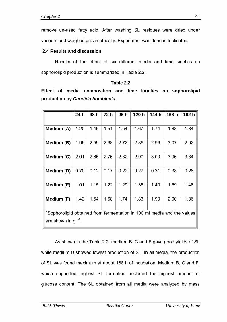
Chapter 2 44
remove un-used fatty acid. After washing SL residues were dried under
vacuum and weighed gravimetrically. Experiment was done in triplicates.
2.4 Results and discussion
Results of the effect of six different media and time kinetics on
sophorolipid production is summarized in Table 2.2.
Table 2.2 Effect of media composition and time kinetics on sophorolipid production by Candida bombicola
24 h 48 h 72 h 96 h 120 h 144 h 168 h 192 h
Medium (A) 1.20 1.46 1.51 1.54 1.67 1.74 1.88 1.84
Medium (B) 1.96 2.59 2.68 2.72 2.86 2.96 3.07 2.92
Medium (C) 2.01 2.65 2.76 2.82 2.90 3.00 3.96 3.84
Medium (D) 0.70 0.12 0.17 0.22 0.27 0.31 0.38 0.28
Medium (E) 1.01 1.15 1.22 1.29 1.35 1.40 1.59 1.48
Medium (F) 1.42 1.54 1.68 1.74 1.83 1.90 2.00 1.86
*Sophorolipid obtained from fermentation in 100 ml media and the values
are shown in g l-1.
As shown in the Table 2.2, medium B, C and F gave good yields of SL
while medium D showed lowest production of SL. In all media, the production
of SL was found maximum at about 168 h of incubation. Medium B, C and F,
which supported highest SL formation, included the highest amount of
glucose content. The SL obtained from all media were analyzed by mass
Ph.D. Thesis Reetika Gupta University of Pune
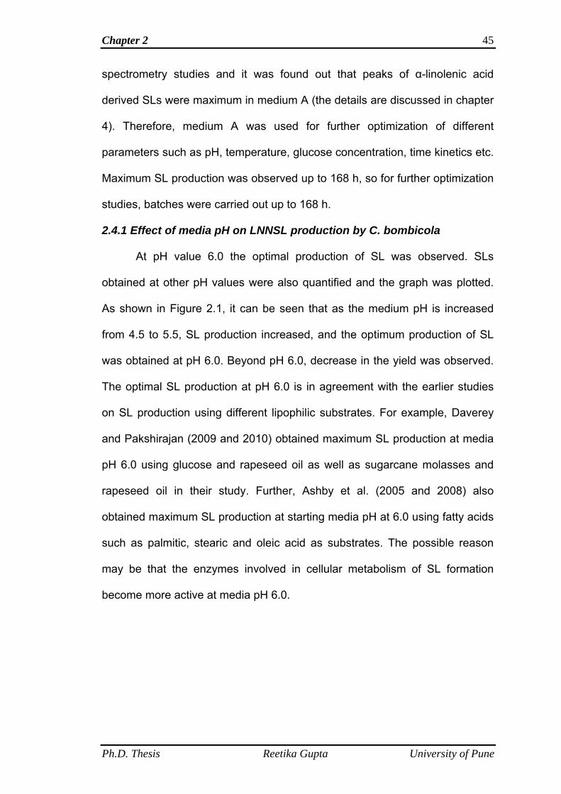
Chapter 2 45
spectrometry studies and it was found out that peaks of α-linolenic acid
derived SLs were maximum in medium A (the details are discussed in chapter
4). Therefore, medium A was used for further optimization of different
parameters such as pH, temperature, glucose concentration, time kinetics etc.
Maximum SL production was observed up to 168 h, so for further optimization
studies, batches were carried out up to 168 h.
2.4.1 Effect of media pH on LNNSL production by C. bombicola
At pH value 6.0 the optimal production of SL was observed. SLs
obtained at other pH values were also quantified and the graph was plotted.
As shown in Figure 2.1, it can be seen that as the medium pH is increased
from 4.5 to 5.5, SL production increased, and the optimum production of SL
was obtained at pH 6.0. Beyond pH 6.0, decrease in the yield was observed.
The optimal SL production at pH 6.0 is in agreement with the earlier studies
on SL production using different lipophilic substrates. For example, Daverey
and Pakshirajan (2009 and 2010) obtained maximum SL production at media
pH 6.0 using glucose and rapeseed oil as well as sugarcane molasses and
rapeseed oil in their study. Further, Ashby et al. (2005 and 2008) also
obtained maximum SL production at starting media pH at 6.0 using fatty acids
such as palmitic, stearic and oleic acid as substrates. The possible reason
may be that the enzymes involved in cellular metabolism of SL formation
become more active at media pH 6.0.
Ph.D. Thesis Reetika Gupta University of Pune
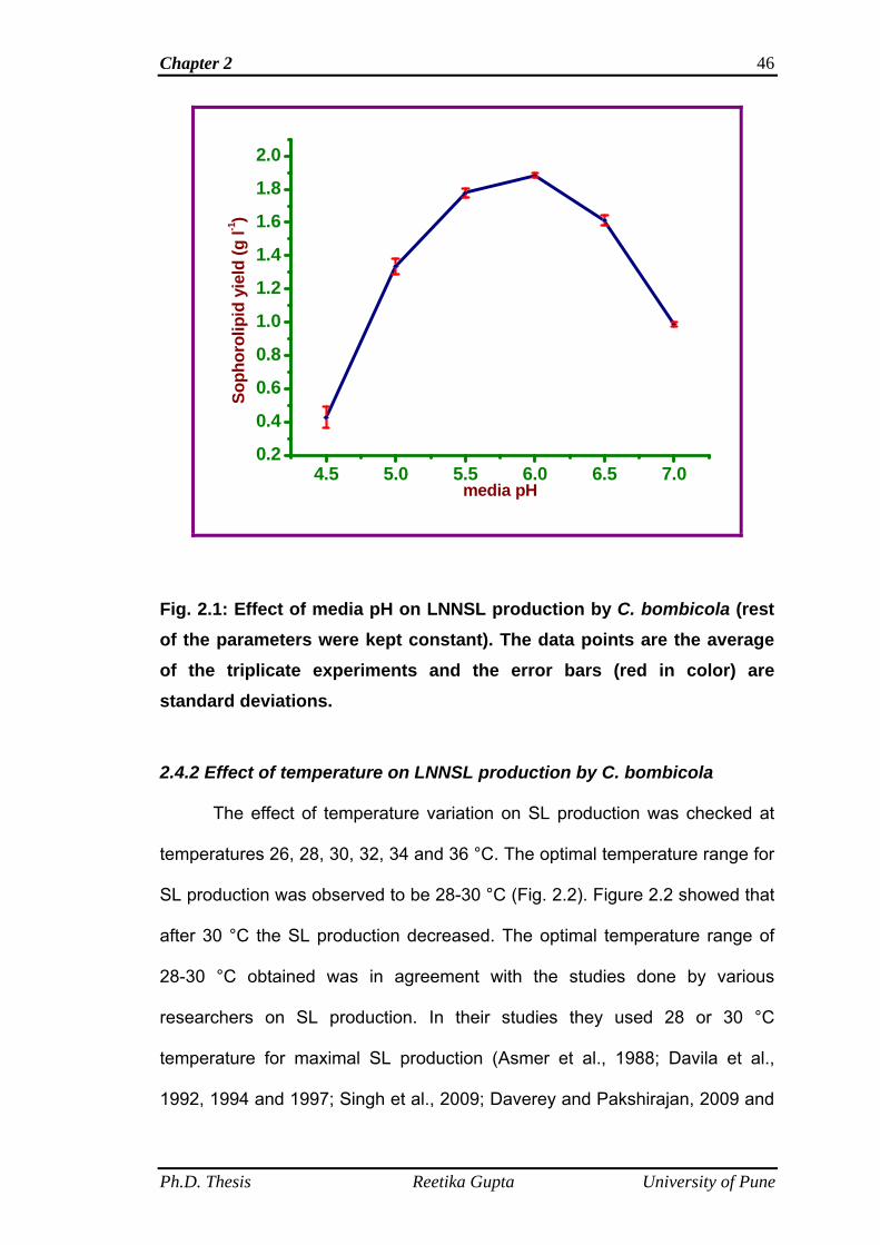
Chapter 2 46
4.5 5.0 5.5 6.0 6.5 7.00.2
0.4
0.6
0.8
1.0
1.2
1.4
1.6
1.8
2.0
Soph
orol
ipid
yie
ld (g
l-1)
media pH
Fig. 2.1: Effect of media pH on LNNSL production by C. bombicola (rest of the parameters were kept constant). The data points are the average of the triplicate experiments and the error bars (red in color) are standard deviations.
2.4.2 Effect of temperature on LNNSL production by C. bombicola
The effect of temperature variation on SL production was checked at
temperatures 26, 28, 30, 32, 34 and 36 °C. The optimal temperature range for
SL production was observed to be 28-30 °C (Fig. 2.2). Figure 2.2 showed that
after 30 °C the SL production decreased. The optimal temperature range of
28-30 °C obtained was in agreement with the studies done by various
researchers on SL production. In their studies they used 28 or 30 °C
temperature for maximal SL production (Asmer et al., 1988; Davila et al.,
1992, 1994 and 1997; Singh et al., 2009; Daverey and Pakshirajan, 2009 and
Ph.D. Thesis Reetika Gupta University of Pune
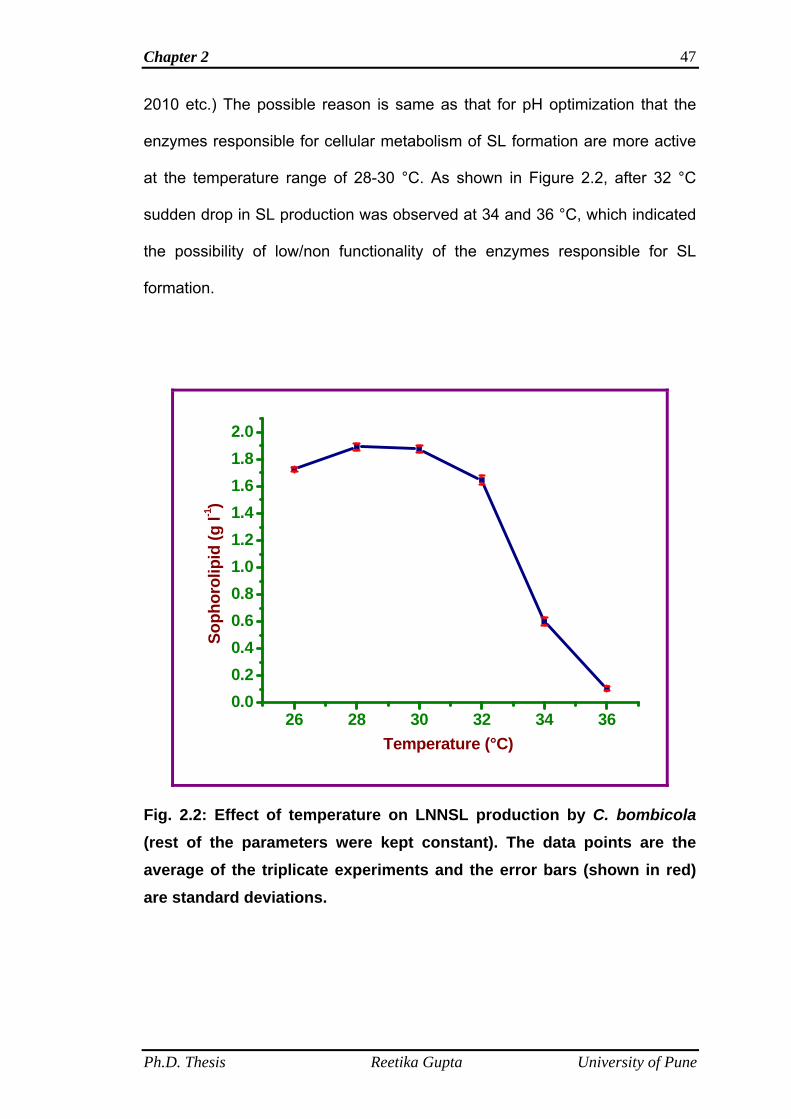
Chapter 2 47
2010 etc.) The possible reason is same as that for pH optimization that the
enzymes responsible for cellular metabolism of SL formation are more active
at the temperature range of 28-30 °C. As shown in Figure 2.2, after 32 °C
sudden drop in SL production was observed at 34 and 36 °C, which indicated
the possibility of low/non functionality of the enzymes responsible for SL
formation.
26 28 30 32 34 360.00.20.40.60.81.01.21.41.61.82.0
Soph
orol
ipid
(g l-1
)
Temperature (°C)
Fig. 2.2: Effect of temperature on LNNSL production by C. bombicola (rest of the parameters were kept constant). The data points are the average of the triplicate experiments and the error bars (shown in red) are standard deviations.
Ph.D. Thesis Reetika Gupta University of Pune
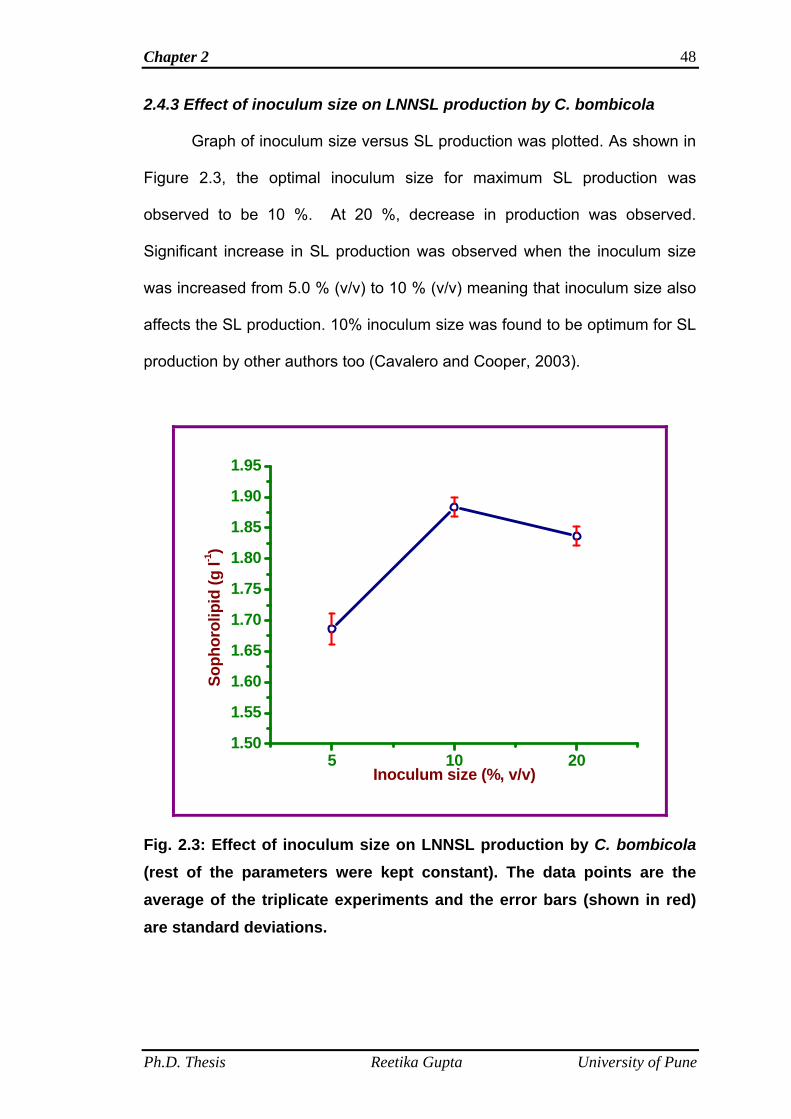
Chapter 2 48
2.4.3 Effect of inoculum size on LNNSL production by C. bombicola
Graph of inoculum size versus SL production was plotted. As shown in
Figure 2.3, the optimal inoculum size for maximum SL production was
observed to be 10 %. At 20 %, decrease in production was observed.
Significant increase in SL production was observed when the inoculum size
was increased from 5.0 % (v/v) to 10 % (v/v) meaning that inoculum size also
affects the SL production. 10% inoculum size was found to be optimum for SL
production by other authors too (Cavalero and Cooper, 2003).
5 10 201.50
1.55
1.60
1.65
1.70
1.75
1.80
1.85
1.90
1.95
Soph
orol
ipid
(g l-1
)
Inoculum size (%, v/v)
Fig. 2.3: Effect of inoculum size on LNNSL production by C. bombicola (rest of the parameters were kept constant). The data points are the average of the triplicate experiments and the error bars (shown in red) are standard deviations.
Ph.D. Thesis Reetika Gupta University of Pune
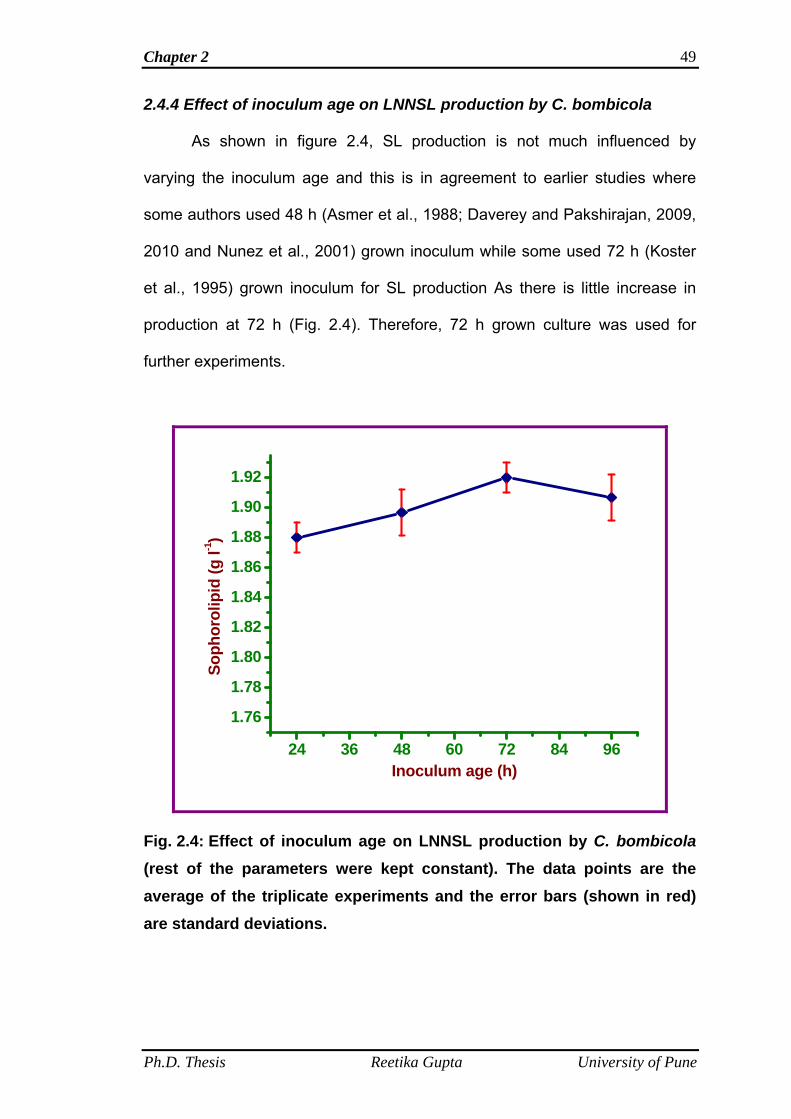
Chapter 2 49
2.4.4 Effect of inoculum age on LNNSL production by C. bombicola
As shown in figure 2.4, SL production is not much influenced by
varying the inoculum age and this is in agreement to earlier studies where
some authors used 48 h (Asmer et al., 1988; Daverey and Pakshirajan, 2009,
2010 and Nunez et al., 2001) grown inoculum while some used 72 h (Koster
et al., 1995) grown inoculum for SL production As there is little increase in
production at 72 h (Fig. 2.4). Therefore, 72 h grown culture was used for
further experiments.
24 36 48 60 72 84 96
1.76
1.78
1.80
1.82
1.84
1.86
1.88
1.90
1.92
Soph
orol
ipid
(g l-1
)
Inoculum age (h)
Fig. 2.4: Effect of inoculum age on LNNSL production by C. bombicola (rest of the parameters were kept constant). The data points are the average of the triplicate experiments and the error bars (shown in red) are standard deviations.
Ph.D. Thesis Reetika Gupta University of Pune
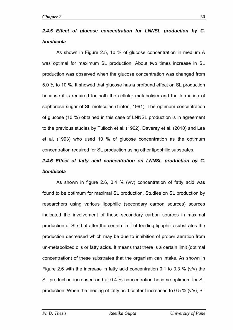
Chapter 2 50
2.4.5 Effect of glucose concentration for LNNSL production by C.
bombicola
As shown in Figure 2.5, 10 % of glucose concentration in medium A
was optimal for maximum SL production. About two times increase in SL
production was observed when the glucose concentration was changed from
5.0 % to 10 %. It showed that glucose has a profound effect on SL production
because it is required for both the cellular metabolism and the formation of
sophorose sugar of SL molecules (Linton, 1991). The optimum concentration
of glucose (10 %) obtained in this case of LNNSL production is in agreement
to the previous studies by Tulloch et al. (1962), Daverey et al. (2010) and Lee
et al. (1993) who used 10 % of glucose concentration as the optimum
concentration required for SL production using other lipophilic substrates.
2.4.6 Effect of fatty acid concentration on LNNSL production by C.
bombicola
As shown in figure 2.6, 0.4 % (v/v) concentration of fatty acid was
found to be optimum for maximal SL production. Studies on SL production by
researchers using various lipophilic (secondary carbon sources) sources
indicated the involvement of these secondary carbon sources in maximal
production of SLs but after the certain limit of feeding lipophilic substrates the
production decreased which may be due to inhibition of proper aeration from
un-metabolized oils or fatty acids. It means that there is a certain limit (optimal
concentration) of these substrates that the organism can intake. As shown in
Figure 2.6 with the increase in fatty acid concentration 0.1 to 0.3 % (v/v) the
SL production increased and at 0.4 % concentration become optimum for SL
production. When the feeding of fatty acid content increased to 0.5 % (v/v), SL
Ph.D. Thesis Reetika Gupta University of Pune
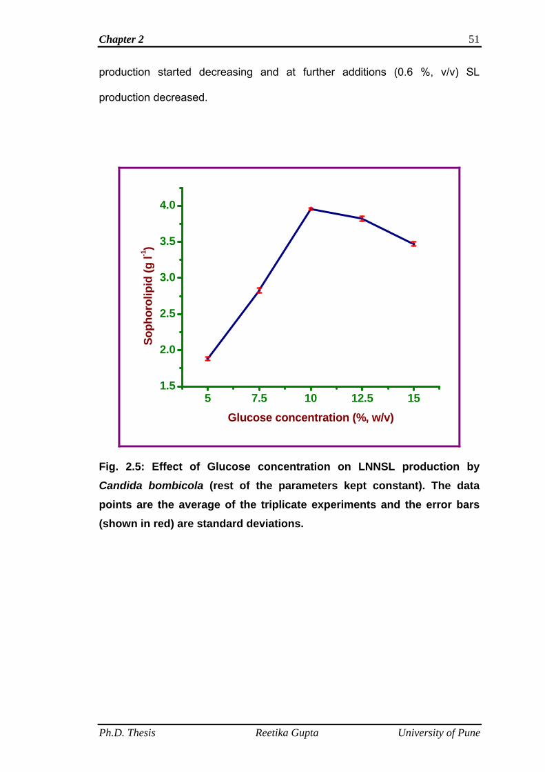
Chapter 2 51
production started decreasing and at further additions (0.6 %, v/v) SL
production decreased.
5 7.5 10 12.5 151.5
2.0
2.5
3.0
3.5
4.0
Soph
orol
ipid
(g l-1
)
Glucose concentration (%, w/v)
Fig. 2.5: Effect of Glucose concentration on LNNSL production by Candida bombicola (rest of the parameters kept constant). The data points are the average of the triplicate experiments and the error bars (shown in red) are standard deviations.
Ph.D. Thesis Reetika Gupta University of Pune
Chapter 2 52
0.1 0.2 0.3 0.4 0.5 0.6
2.0
2.5
3.0
3.5
4.0So
phor
olip
id (g
l-1)
Fatty acid concentration (%,v/v)
Fig. 2.6: Effect of fatty acid concentration on LNNSL production by Candida bombicola (rest of the parameters kept constant). The data points are the average of the triplicate experiments and the error bars (shown in red) are standard deviations.
2.4.7 Effect of yeast extract concentration on LNNSL production by C.
bombicola
Yeast extract as a source of nitrogen is useful for yeast growth and SL
production. Therefore, yeast extract concentrations were varied in medium A
to know the optimal yeast concentration. As shown in Figure 2.7, SL
production was low at 0.1 % yeast extract concentration and as the
concentration increased to 0.2 %, SL production also increased and at 0.3 %
yeast extract concentration optimal production of SL was observed. Beyond
0.3 % yeast extract concentration, the yield of SL decreased slightly, it may be
Ph.D. Thesis Reetika Gupta University of Pune
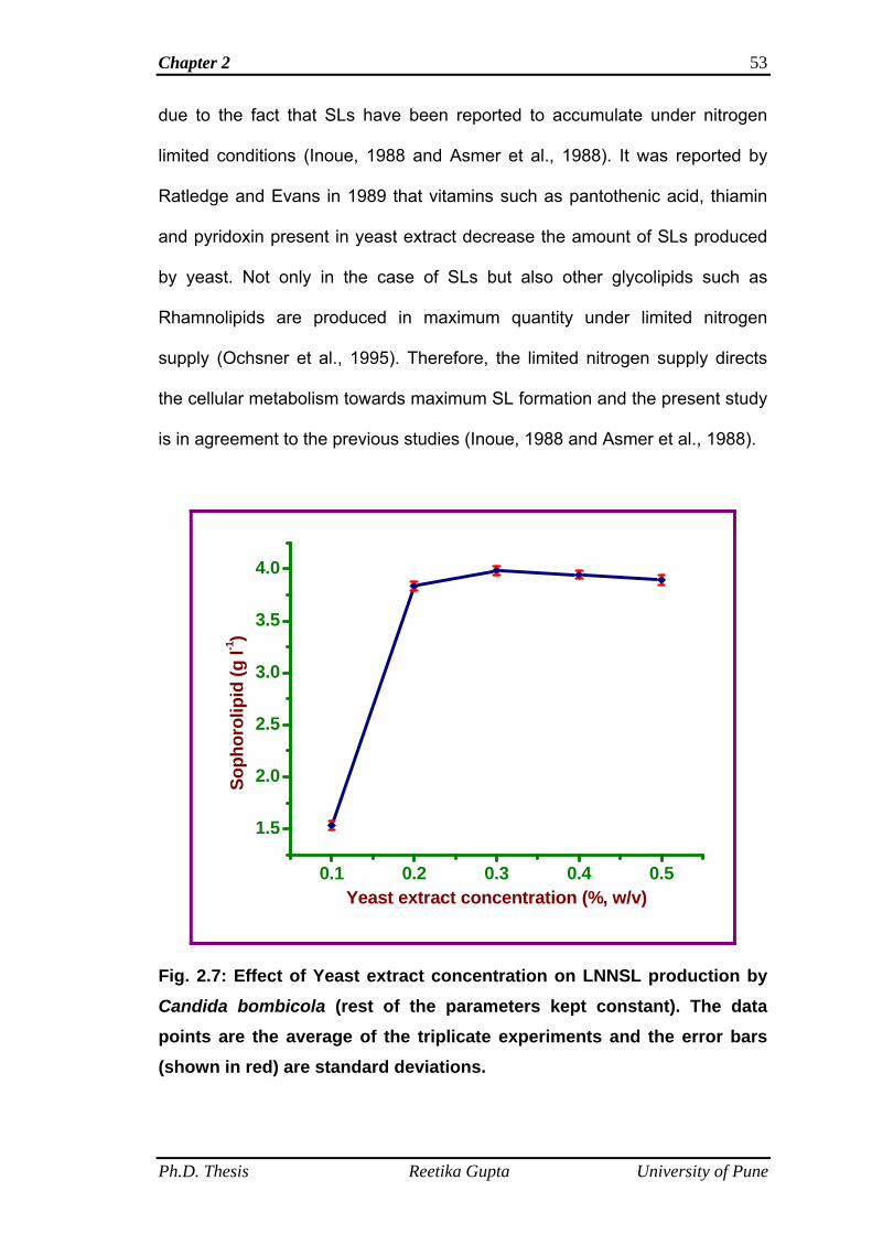
Chapter 2 53
due to the fact that SLs have been reported to accumulate under nitrogen
limited conditions (Inoue, 1988 and Asmer et al., 1988). It was reported by
Ratledge and Evans in 1989 that vitamins such as pantothenic acid, thiamin
and pyridoxin present in yeast extract decrease the amount of SLs produced
by yeast. Not only in the case of SLs but also other glycolipids such as
Rhamnolipids are produced in maximum quantity under limited nitrogen
supply (Ochsner et al., 1995). Therefore, the limited nitrogen supply directs
the cellular metabolism towards maximum SL formation and the present study
is in agreement to the previous studies (Inoue, 1988 and Asmer et al., 1988).
0.1 0.2 0.3 0.4 0.5
1.5
2.0
2.5
3.0
3.5
4.0
Soph
orol
ipid
(g l-1
)
Yeast extract concentration (%, w/v)
Fig. 2.7: Effect of Yeast extract concentration on LNNSL production by Candida bombicola (rest of the parameters kept constant). The data points are the average of the triplicate experiments and the error bars (shown in red) are standard deviations.
Ph.D. Thesis Reetika Gupta University of Pune
Chapter 2 54
The optimum levels of process parameters were decided and
summarized in the Table 2.3.
Table 2.3 Optimum levels of the process parameters chosen for LNNSL production
Process parameter Optimum level
Medium pH 6.0
Temperature 28 °C
Inoculum size 10 %
Inoculum age 72 h
Glucose concentration 10 % (w/v)
Fatty acid concentration 0.4 % (v/v)
Yeast extract concentration 0.3 % (w/v)
Fermentation period 168 h
2.4.8 Time kinetics on LNNSL/Biomass production
Under the optimized fermentation parameters, time-kinetics on LNNSL
and biomass production was performed at regular time intervals (24, 48, 72,
96, 120, 144, 168 and 192 h). The production of LNNSL and biomass in g l-1 is
shown in Table 2.4 and a graphical representation between time (h) and
LNNSL/biomass production (g l-1) is shown in Figure 2.8.
Ph.D. Thesis Reetika Gupta University of Pune
Chapter 2 55
Table 2.4 Time kinetics on Sophorolipid and biomass production under optimized conditions. The values are the average of the triplicate experiments with their standard deviation
Time (hours) SL (g l-1) Biomass* (g l-1)
24 1.89 ± 0.037 2.01 ± 0.061
48 1.91 ± 0.047 2.17 ± 0.035
72 2.20 ± 0.021 2.55 ± 0.035
96 2.53 ± 0.071 2.84 ± 0.050
120 2.98 ± 0.078 3.11 ± 0.040
144 3.43 ± 0.060 3.11 ± 0.031
168 3.98 ± 0.045 3.11 ± 0.035
192 3.84 ± 0.061 2.83 ± 0.045
* After the separation of supernatant, cells were washed two times with ethyl acetate to remove the glycolipids and oil. Then the cell biomass was washed with distilled water. The cells were centrifuged twice and collected and then dried in an oven at 70 °C for 48 h and weighed to measure the biomass.
Time-kinetics performed on LNNSL/Biomass production showed that
LNNSL production started after 48 hours but increase in SL production was
observed from late exponential phase (after 96 h). Maximum accumulation of
LNNSL was observed in stationary and late stationary phases and reached its
maximum level at 168 h after which SL production decreased. Biomass
growth started after 24 hours and continued till 120 hours. No increase in
growth was observed after 120 hours and the biomass remained constant.
Biomass decreased after 168 h.
Ph.D. Thesis Reetika Gupta University of Pune
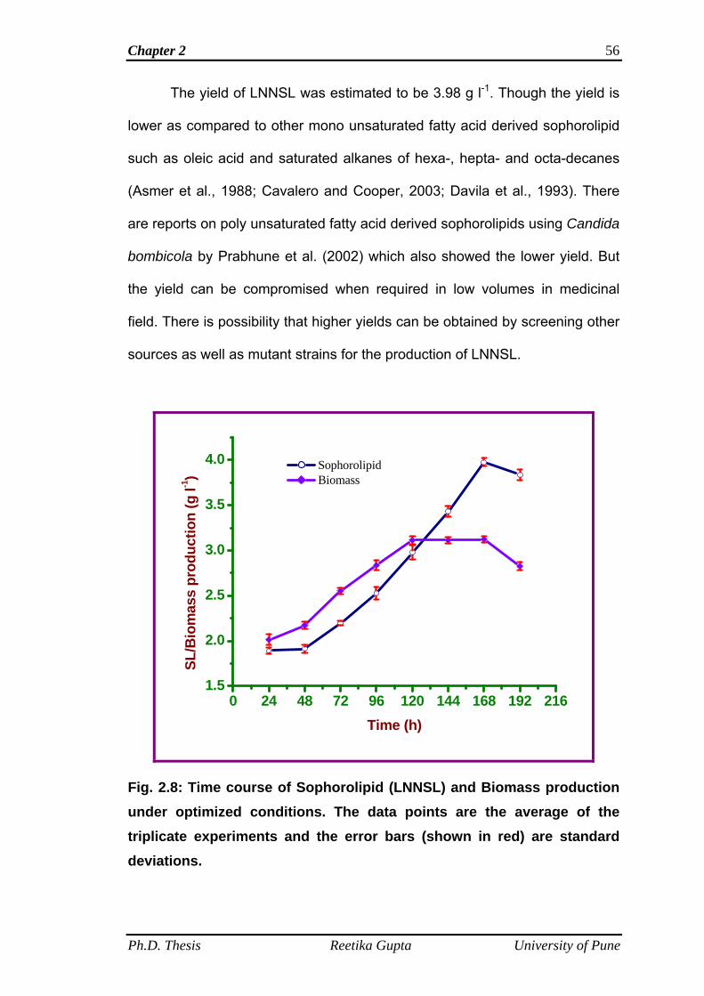
Chapter 2 56
The yield of LNNSL was estimated to be 3.98 g l-1. Though the yield is
lower as compared to other mono unsaturated fatty acid derived sophorolipid
such as oleic acid and saturated alkanes of hexa-, hepta- and octa-decanes
(Asmer et al., 1988; Cavalero and Cooper, 2003; Davila et al., 1993). There
are reports on poly unsaturated fatty acid derived sophorolipids using Candida
bombicola by Prabhune et al. (2002) which also showed the lower yield. But
the yield can be compromised when required in low volumes in medicinal
field. There is possibility that higher yields can be obtained by screening other
sources as well as mutant strains for the production of LNNSL.
0 24 48 72 96 120 144 168 192 2161.5
2.0
2.5
3.0
3.5
4.0 Sophorolipid Biomass
SL/B
iom
ass
prod
uctio
n (g
l-1)
Time (h)
Fig. 2.8: Time course of Sophorolipid (LNNSL) and Biomass production under optimized conditions. The data points are the average of the triplicate experiments and the error bars (shown in red) are standard deviations.
Ph.D. Thesis Reetika Gupta University of Pune
Chapter 2
Ph.D. Thesis Reetika Gupta University of Pune
57
2.5 Conclusions
The study of different process parameters showed that about 7 days
(168 h) are required for maximum production of LNNSL. The optimum
physiological pH and temperature for production was found to be 6.0 and 28
°C respectively. Inoclulum size of 10 % (v/v) was found optimum, but
inoculum age did not have much influence on LNNSL production. Glucose at
10 % (w/v) gave maximum yield and fatty acid supply of 0.4 % (v/v) was found
to be optimum. Yeast extract at 0.3 % (w/v) was required for optimum
production. The maximum accumulation of SLs in stationary and late
stationary phases is in agreement with the previous reports on SL production
using fatty acids (Hommel et al., 1987) other than the linolenic acid and also
in agreement with the other glycolipid biosurfactants (Stodola et al., 1967;
Bentley and Campbell, 1968 and Rosenberg and Ron, 1999).
Chapter 3
Production and Purification of Linolenic acid derived Sophorolipids
Chapter 3 59
3.1 Summary
In chapter 2, the optimization of different process parameters such as
pH, temperature, effect of glucose, fatty acid and yeast extract concentrations
on SL production using glucose and α-linolenic acid as carbon substrates by
Candida bombicola was demonstrated and the optimum medium for SL
production was decided. This chapter deals with the study on production of
LNNSL using 1 liter batches under optimized conditions to see the effect on
yield of SL. Further, in this chapter purification of LNNSL is described using
high performance liquid chromatography and by chemical modification of SL
mixture following esterification reaction. Results showed that there is no
change in the yield of SL when the production is carried out for 1 liter batch.
The results from HPLC showed the presence of three prominent peaks which
must be characterized to identify different SL molecules present in LNNSL
mixture. The characterization of these peaks is described in the chapter 4.
The esterification reaction of LNNSL mixture produce single product as
confirmed from thin layer chromatography. The complete characterization of
this product is also described in the chapter 4.
3.2 Introduction
It was reported earlier by Tulloch et al. (1962) that the composition of
fatty acid portion of SL varies according to the substrate added into the
medium. They worked on fatty acid esters of palmitic, stearic, oleic and
linoleic acid and found that the maximum incorporation was observed for
stearic and oleic acid into the lipophilic portion of the SL molecule by means
of acid methanolysis and gas liquid chromatography. Asmer et al. (1988)
reported the production (fermentor level) and structural studies of SLs
Ph.D. Thesis Reetika Gupta University of Pune
Chapter 3 60
produced by Candida bombicola using glucose and oleic acid and oleic acid
alone in the medium. They reported that the yield of SL in the medium where
only oleic acid was used as a carbon source was maximum (77 g l-1) as
compared to the medium where both the glucose and oleic acid were used as
carbon sources (38 g l-1) and the structure elucidation was done with the help
of medium pressure liquid chromatography and thin layer chromatography.
This study is in contrast to other studies which showed that both the
hydrophilic (sugars) and lipophilic (fatty acids) carbon sources are required for
maximum production of SLs (Cavalero and Cooper, 2003). Davila et al. (1992)
reported that the production of SL using rapeseed oil fatty acid (main
component of oil is oleic acid) esters and glucose generated the major
product to be the diacetylated lactone form of SL having oleic acid in the
lipophilic portion and they showed that both the carbon sources glucose and
fatty acid are required for maximum yield of SLs. Studies done by several
researchers showed that both the carbon sources are necessary for maximum
yield of SLs (Gobbert et al.,1984; Linton, 1991 ; Zhou et al., 1992; Davila et
al., 1994; and Hommel et al., 1994). These authors suggested that the
hydrophilic carbon source such as glucose is used primarily for cellular
metabolism and the formation of sophorose moiety of SL, while the lipophilic
carbon source is used for the synthesis of hydroxy-carboxylic acid moiety.
Different analytical techniques were used for purification of SL. For example,
Asmer et al. (1988) used thin layer chromatography and medium pressure
liquid chromatography. Nunez et al. (2001, 2004) and Davila et al. (1994)
used high performance liquid chromatography and purification by silica gel
chromatography using dialysis tubing method was used by Shah and
Ph.D. Thesis Reetika Gupta University of Pune
Chapter 3 61
Prabhune (2007) in place of tedious and time taking silica gel
chromatography.
The structural analysis and applications of the SLs produced using
glucose and α-linolenic acid could be done only when the sufficient amount of
the product is available. Therefore, the production was done using one liter
batches under optimized fermentation conditions. Further, the purification of
LNNSL was done by two methods (1) High performance liquid
chromatography of LNNSL mixture. (2) Chemical esterification of LNNSL
mixture in order to produce single homogenous product following chemical
esterification reaction given in literature (Bisht et al., 1999).
3.3 Materials and methods
3.3.1 Materials
The yeast Candida bombicola ATCC 22214 was used for the
production of α-linolenic acid derived sophorolipids (designated as LNNSL).
This yeast was procured from American type culture collection. Glucose,
yeast extract, malt extract and peptone were purchased from Hi-media, India
and sodium sulphate and acetonitrile was procured from Merck, India. α-
linolenic acid (≥99%, purity, C18:3) was purchased from Sigma-Aldrich.
Solvents such as chloroform and methanol were purchased from Rankem,
India.
3.3.2 Growth conditions and microorganism maintenance
Candida bombicola ATCC 22214 was grown for 48 h at 28 °C on agar
slants containing (g l-1): malt extract, 3.0; glucose, 50; yeast extract, 3.0;
peptone, 5.0 and agar, 20. The microorganism was sub-cultured every month
and maintained at 4 °C.
Ph.D. Thesis Reetika Gupta University of Pune
Chapter 3 62
3.3.3 Inoculum development
Inoculum was developed by growing Candida bombicola (ATCC
22214) cells at 28 °C in 100 ml MGYP medium containing (g l-1): malt extract,
3.0; glucose, 100; yeast extract, 3.0; peptone, 5.0; pH 6.0 and shaken at 180
rpm for 72 h.
3.3.4 Production of sophorolipids
LNNSL production was carried out in 1 liter Erlenmeyer flasks
containing 250 ml of optimized media of composition (g l-1): malt extract, 3.0;
glucose, 100; yeast extract, 3.0; peptone, 5.0. Media pH was adjusted to 6.0.
All the flasks containing 250 ml of media were supplied with 1 ml of α-linolenic
acid dispersed in 1 ml of ethanol separately. Total 4 ml of fatty acid was used
for 1 liter batch. The flasks were then inoculated separately with 10 % (v/v, 25
ml inoculum to each 250 ml media) 72 h grown inoculum and kept for SL
production in incubator shaker at 180 rpm for 168 h at 28 °C.
3.3.5 Isolation of sophorolipids
After incubation of 168 h it was observed that brownish oily viscous
layer of SL settled down at the bottom of the culture broth. Culture medium
was centrifuged at 5000 rpm for 20 min. The supernatant was separated and
extracted twice with equal volumes of ethyl acetate. To ensure the complete
isolation of SLs, the cells were also washed with ethyl acetate so as to
remove the glycolipids and fatty acid adhered to cells. Then ethyl acetate
layer obtained from both the supernatant extraction and washing of cells were
pooled and was dried over anhydrous sodium sulphate. The solvent was
removed by rotary-evaporation at 40 °C. The brownish viscous product was
obtained, which was washed twice with n-hexane to remove the un-reacted
Ph.D. Thesis Reetika Gupta University of Pune
Chapter 3 63
fatty acid and was dried under vacuum. SL yield was calculated
gravimetrically. The product was stored at 4 °C till further use.
3.3.6 Microscopic analysis of SL mixture
A small amount of SL mixture obtained above was dissolved in water
and drop casted on a glass slide and covered with a cover slip. It was
observed under different optical zooms and digital pictures were taken.
3.3.7 Qualitative analysis of SL mixture by thin layer chromatography
(TLC)
Thin layer chromatography (TLC) is the simplest, quickest and
cheapest of the various analytical techniques that have been used extensively
for the qualitative analysis of compounds. Preferred adsorbent used for TLC is
silica gel. Commercially available standard silica gel coated on aluminium
plates (Kiesel-gel 60F254, Merck, Darmstadt, Germany, silica layer thickness 2
mm) are used for the analysis of SL mixture. Glass micro-capillary is used for
the spot application of samples. Many solvent systems have been used for
the separation of compounds. Ultra-visible absorbing compounds can be
visualized under detection at 254 nm of UV light. The plates are coated with
silica impregnated with the fluorescent indicator F254 nm. The other methods
used for detection are charring with anisaldehyde solution and staining with
iodine vapours. The simplest, staining with iodine vapours works best for
compounds with double bonds. The ethyl acetate extract of SL mixture was
used for the determination of number of components present in the mixture by
means of thin layer chromatography. Developing system used was
CHCl3:CH3OH:H2O in the ratio of 95:4:1, v/v/v. Spots were visualized by either
of the two detection techniques: exposure to iodine vapour and the second
Ph.D. Thesis Reetika Gupta University of Pune
Chapter 3 64
one is charring with anisaldehyde solution (anisaldehyde:sulphuric acid:acetic
acid = 0.5:1:50, v/v/v (Asmer et al., 1988).
3.3.8 Purification of sophorolipids
The brownish viscous product obtained in step 3.3.5 was used for the
purification of sophorolipids by two methods:
High performance liquid chromatography
Purification by chemical esterification of SL mixture
3.3.8.1 High performance liquid chromatography (HPLC)
Sophorolipid mixture obtained from fermentative production was
dissolved in acetonitrile and analyzed by reversed-phase high performance
liquid chromatography (HPLC) with a Waters 2487 separation module (Waters
Co. Milford, Massachusetts) using analytical symmetry C18, 5 µm column
(250 × 4.6 mm2). The gradient solvent elution profile used was as follows:
water:acetonitrile (95:5, v/v) holding for 10 min; to a final composition of
water:acetonitrile (5:95, v/v) with a linear gradient over 50 min and holding for
10 min. The flow rate was 0.5 ml min-1. The peaks were detected at 220 nm
wavelength by absorbance detector. Fractions from different peaks were
collected and pooled separately in many runs.
3.3.8.2 Purification by chemical esterification of SL mixture
Synthesis of Sophorolipid methyl ester
Synthesis of SL methyl ester was done by following the methodology
for chemical esterification reaction given by Bisht et al. in 1999 (for conversion
of oleic acid derived SLs into single homogenous esterified product). Steps
involved in the reaction are shown in the flow sheet form in Figure 3.1.
Chemical esterfication reaction is shown in Figure 3.2.
Ph.D. Thesis Reetika Gupta University of Pune
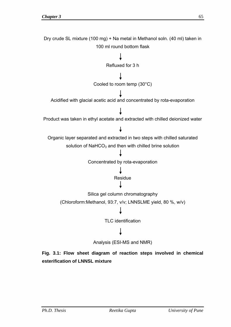
Chapter 3 65
Dry crude SL mixture (100 mg) + Na metal in Methanol soln. (40 ml) taken in
100 ml round bottom flask
Refluxed for 3 h
Cooled to room temp (30°C)
Acidified with glacial acetic acid and concentrated by rota-evaporation
Product was taken in ethyl acetate and extracted with chilled deionized water
Organic layer separated and extracted in two steps with chilled saturated
solution of NaHCO3 and then with chilled brine solution
Concentrated by rota-evaporation
Residue
Silica gel column chromatography
(Chloroform:Methanol, 93:7, v/v; LNNSLME yield, 80 %, w/v)
TLC identification
Analysis (ESI-MS and NMR)
Fig. 3.1: Flow sheet diagram of reaction steps involved in chemical esterification of LNNSL mixture
Ph.D. Thesis Reetika Gupta University of Pune
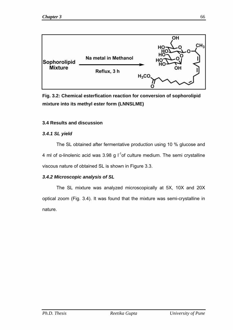
Chapter 3 66
Fig. 3.2: Chemical esterfication reaction for conversion of sophorolipid mixture into its methyl ester form (LNNSLME)
3.4 Results and discussion
3.4.1 SL yield
The SL obtained after fermentative production using 10 % glucose and
4 ml of α-linolenic acid was 3.98 g l-1of culture medium. The semi crystalline
viscous nature of obtained SL is shown in Figure 3.3.
3.4.2 Microscopic analysis of SL
The SL mixture was analyzed microscopically at 5X, 10X and 20X
optical zoom (Fig. 3.4). It was found that the mixture was semi-crystalline in
nature.
Ph.D. Thesis Reetika Gupta University of Pune
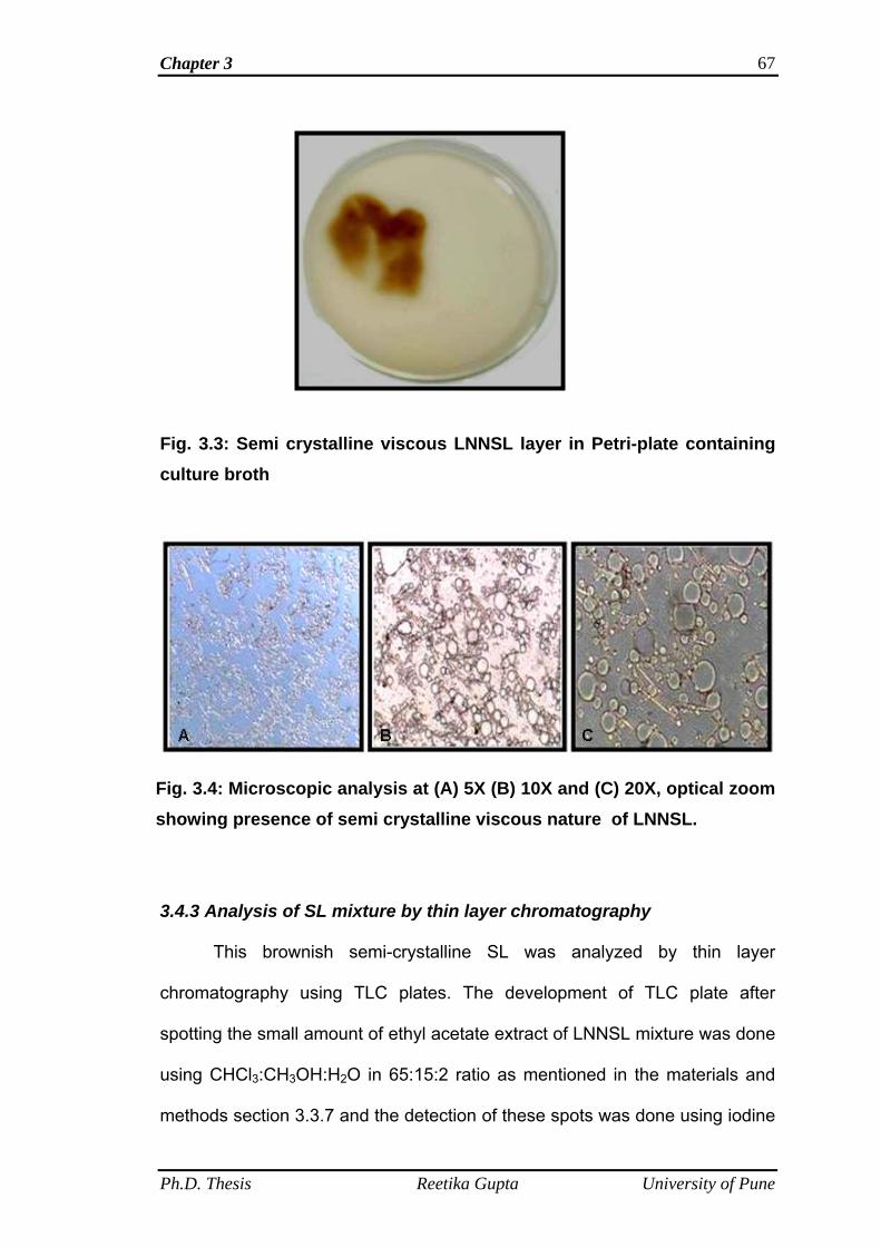
Chapter 3 67
Fig. 3.3: Semi crystalline viscous LNNSL layer in Petri-plate containing culture broth
Fig. 3.4: Microscopic analysis at (A) 5X (B) 10X and (C) 20X, optical zoom showing presence of semi crystalline viscous nature of LNNSL. 3.4.3 Analysis of SL mixture by thin layer chromatography
This brownish semi-crystalline SL was analyzed by thin layer
chromatography using TLC plates. The development of TLC plate after
spotting the small amount of ethyl acetate extract of LNNSL mixture was done
using CHCl3:CH3OH:H2O in 65:15:2 ratio as mentioned in the materials and
methods section 3.3.7 and the detection of these spots was done using iodine
Ph.D. Thesis Reetika Gupta University of Pune
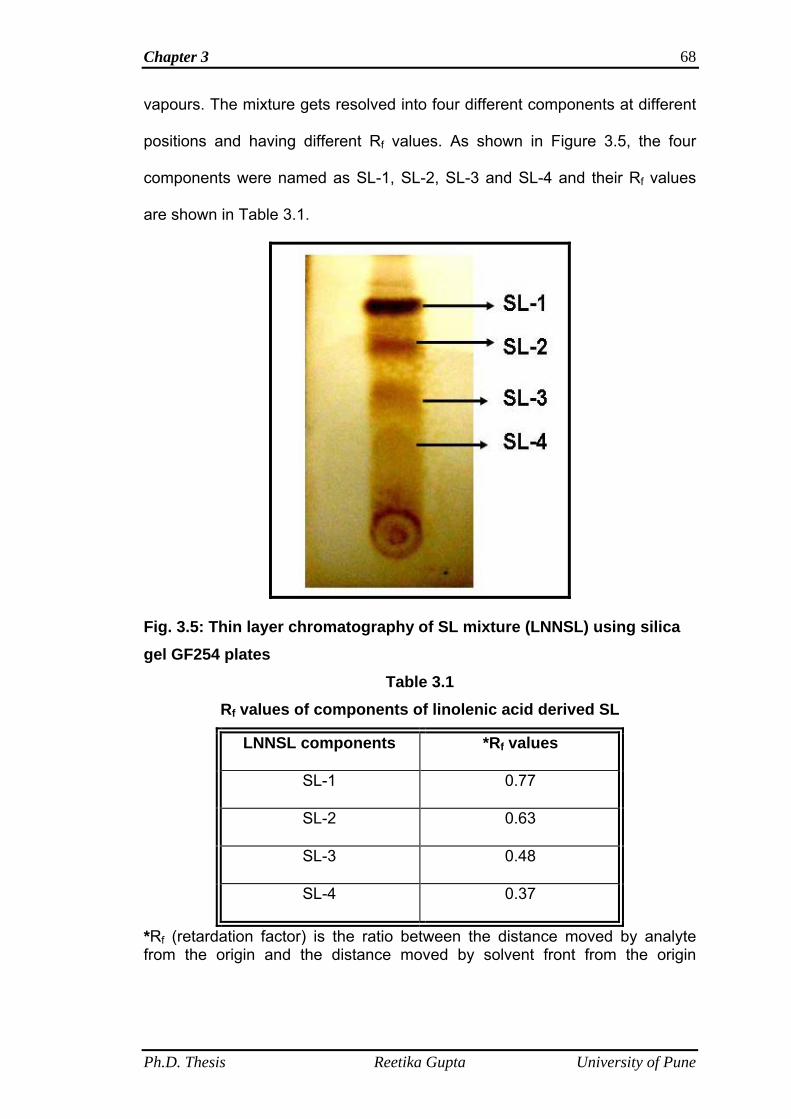
Chapter 3 68
vapours. The mixture gets resolved into four different components at different
positions and having different Rf values. As shown in Figure 3.5, the four
components were named as SL-1, SL-2, SL-3 and SL-4 and their Rf values
are shown in Table 3.1.
Fig. 3.5: Thin layer chromatography of SL mixture (LNNSL) using silica gel GF254 plates
Table 3.1 Rf values of components of linolenic acid derived SL
LNNSL components *Rf values
SL-1 0.77
SL-2 0.63
SL-3 0.48
SL-4 0.37
*Rf (retardation factor) is the ratio between the distance moved by analyte from the origin and the distance moved by solvent front from the origin
Ph.D. Thesis Reetika Gupta University of Pune
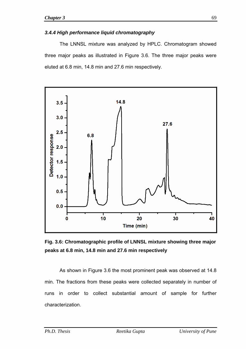
Chapter 3 69
3.4.4 High performance liquid chromatography
The LNNSL mixture was analyzed by HPLC. Chromatogram showed
three major peaks as illustrated in Figure 3.6. The three major peaks were
eluted at 6.8 min, 14.8 min and 27.6 min respectively.
Fig. 3.6: Chromatographic profile of LNNSL mixture showing three major peaks at 6.8 min, 14.8 min and 27.6 min respectively
As shown in Figure 3.6 the most prominent peak was observed at 14.8
min. The fractions from these peaks were collected separately in number of
runs in order to collect substantial amount of sample for further
characterization.
Ph.D. Thesis Reetika Gupta University of Pune
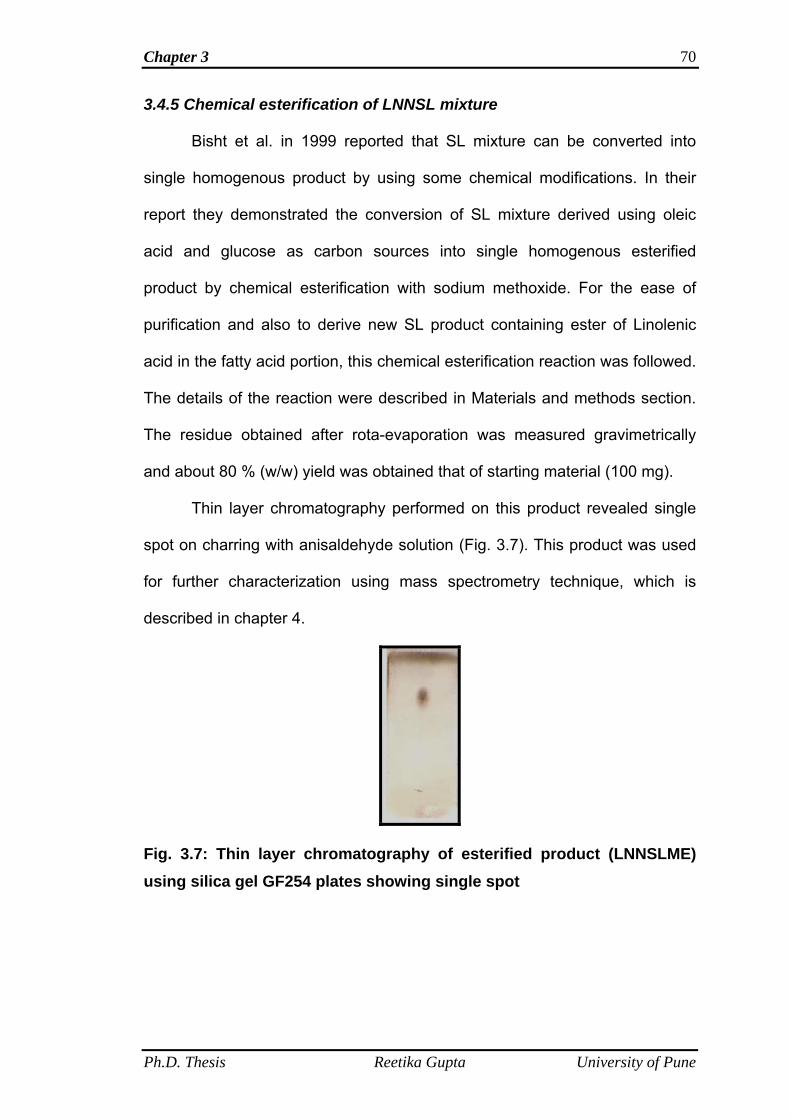
Chapter 3 70
3.4.5 Chemical esterification of LNNSL mixture
Bisht et al. in 1999 reported that SL mixture can be converted into
single homogenous product by using some chemical modifications. In their
report they demonstrated the conversion of SL mixture derived using oleic
acid and glucose as carbon sources into single homogenous esterified
product by chemical esterification with sodium methoxide. For the ease of
purification and also to derive new SL product containing ester of Linolenic
acid in the fatty acid portion, this chemical esterification reaction was followed.
The details of the reaction were described in Materials and methods section.
The residue obtained after rota-evaporation was measured gravimetrically
and about 80 % (w/w) yield was obtained that of starting material (100 mg).
Thin layer chromatography performed on this product revealed single
spot on charring with anisaldehyde solution (Fig. 3.7). This product was used
for further characterization using mass spectrometry technique, which is
described in chapter 4.
Fig. 3.7: Thin layer chromatography of esterified product (LNNSLME) using silica gel GF254 plates showing single spot
Ph.D. Thesis Reetika Gupta University of Pune
Chapter 3 71
3.5 Conclusions
The conclusions drawn from the above study indicated that the yield of
LNNSL was not affected when the production was carried out using one liter
batches. The yield of LNNSL from one liter batch was 3.98 g l-1. The LNNSL
mixture obtained from fermentation was subjected to microscopic analysis
which showed that it is semi crystalline viscous in nature. The semi crystalline
viscous nature of LNNSL mixture is indicative of the presence of both the
lactones and acidic forms in the mixture as lactones are known to exhibit
crystalline property while acidic forms exhibit viscous nature (Cavalero and
Cooper, 2003). Further, analysis of LNNSL mixture by thin layer
chromatography showed that it consists of four different SLs (SL-1, SL-2, SL-
3, SL-4) having Rf values 0.77, 0.63, 0.48, 0.37 respectively. The component
adjacent to solvent front or migrating first (SL-1) was most non-polar or
hydrophobic while that migrated in last (SL-4) was most polar or hydrophilic in
nature. This analysis indicated that both lactones (hydrophobic) and acidic
(hydrophilic) SLs were present in LNNSL mixture. Analysis by HPLC had
shown three major peaks at 6.8, 14.8 and 27.6 min. Peak detected at 14.8
min was more prominent. Fractions from these three different peaks were
collected and pooled separately in number of runs for further characterization
studies which are described in chapter 4.
For the ease of purification LNNSL mixture was subjected to chemical
esterification reaction. The yield of methyl ester of linolenic acid SL
(designated as LNNSLME) was 80 % (w/w). Thin layer chromatography
revealed a single spot. The characterization of this product is described in
chapter 4.
Ph.D. Thesis Reetika Gupta University of Pune
Chapter 4
Structural Determination and physical properties of Linolenic acid derived Sophorolipid and its methyl ester form
Chapter 4 73
4.1 Summary
This chapter deals with the analysis of chemically distinct forms
present in the heterogeneous mixture of LNNSL as well as the analysis of its
methyl ester form (LNNSLME). The structural characterization of individual
form present in the LNNSL mixture was done with the help of mass
spectrometry technique. The mass spectrometry technique used for the
structural determination was Electro-spray ionization (ESI) and further the
structures were confirmed using fragmentation patterns obtained from tandem
mass spectra involving collision induced dissociation mass spectrometry.
Analysis of LNNSLME was done using ESI-MS and further structural
confirmation was done by NMR studies.
Physical properties such as surface tension and critical micelle
concentration of LNNSL and its methyl ester, LNNSLME were also
determined using Wilhelmy plate method.
4.2 Introduction
In the literature, there are several reports on analysis and structural
determination of individual components in SL mixture produced using different
lipophilic substrates. Different analytical techniques have been used by
researchers to identify chemically distinct forms of SLs. For example, Asmer
et al. (1988), Koster et al. (1995) and Nunez et al. (2001 and 2004) identified
SL structures derived using oleic acid as lipophilic substrate with the help of
fast atom bombardment mass spectrometry (FAB-MS), atmospheric pressure
chemical ionization mass spectrometry (APCI-MS) and electro-spray
ionization mass spectrometry (ESI-MS) and NMR respectively. Cavalero and
cooper (2003) used gas chromatography mass spectrometry for the
Ph.D. Thesis Reetika Gupta University of Pune
Chapter 4 74
identification of SLs generated using different alkanes, palmitoleic and oleic
fatty acids and sunflower oil. They observed the maximum direct incorporation
(95 %) for C18:1 fatty acid moiety into the SL structure. Mass spectrometry
techniques have vast application in the field of structural determination of
biomolecules (Tomer et al., 1983 and Adams, 1990).
SL offers great structural diversity and hence different physicochemical
and biological properties of the distinct functional forms (Glenns and Cooper,
2006). For the exploitation of individual functional form in the industrial,
pharmaceutical and other sectors, these forms must be fully characterized.
Therefore, in this chapter structural characterization of the three major
fractions collected from three prominent peaks observed in HPLC and the
esterified product obtained by following chemical esterification reaction (Bisht
et al., 1999) was also characterized with the help of mass spectrometry is
presented. Further, the surface tension and critical micelle concentration,
which are the most widely used criteria for evaluating biosurfactant activity
were determined for LNNSL and its methyl ester, LNNSLME with the help of
Wilhelmy plate method. This is the first report on the characterization of
linolenic acid derived SLs.
4.3 Materials and methods
4.3.1 Chemicals
Acetonitrile used was of HPLC grade and was procured from Merck,
India. CD3OD (Deuterated methanol) was purchased from Sigma.
4.3.2 ESI and CID-MS analysis of LNNSL fractions
Positive and negative ESI and CID mass spectra were obtained with an API
QSTAR PULSAR hybrid MS/MS quadrupole TOF system (Applied
Ph.D. Thesis Reetika Gupta University of Pune
Chapter 4 75
biosystems). ESI mass spectra were obtained with time-of-flight mass
spectrometry (TOF MS). Ion source was set at 3.8 kV. Tandem mass spectra
(MS/MS) were acquired by selecting the desired precursor ion with Q1, which
fragments ions in a collision cell (Q2). N2 gas was used in the collision cell as
collisionally activated dissociation gas. These fragment ions were detected in
the time of flight section. Preliminary identifications of different SL forms
present in LNNSL mixture was done using ESI-MS analysis and further
structural confirmation of these forms were done from fragmentation patterns
obtained from CID-MS analysis.1 µl samples from different fractions of LNNSL
(collected from HPLC) were injected separately into mass spectrometer for
analysis using both ESI and CID-MS technique.
4.3.3 ESI and NMR (nuclear magnetic resonance) analysis of LNNSLME
LNNSLME was analyzed by ESI-MS analysis and for this 1 µl of
LNNSLME (stock solution of 2-3 mg dissolved in acetonitrile) was injected into
the mass spectrometer for analysis.
Further structural confirmation of LNNSLME was also done using NMR
technique. 1H and 13C NMR spectra were recorded using Avance Bruker-200
MHz spectrometer. Chemical shifts are given in parts per million downfield
from 0.00 ppm using tetramethylsilane (TMS) as the internal reference. The
1H NMR spectra of LNNSLME was taken in CD3OD (deuterated methanol).
The following abbreviations are used to present the spectral data: s = singlet,
m = multiplet, d = doublet.
4.3.4 Surface tension and critical micelle concentration determination
SL solutions (100 ml) of different concentrations were prepared using
distilled water and kept for two hours for equilibration. The surface tension
Ph.D. Thesis Reetika Gupta University of Pune
Chapter 4 76
(S.T.) measurements of aqueous solutions of sophorolipids against air were
done using the Wilhelmy plate method on Kruss K-11 tensiometer (automated
tensiometer) at room temperature (25 °C). Different concentrations ranging
from 0 to 200 mg l-1 of LNNSL and its methyl ester (LNNSLME) were used for
the measurement of surface tension. Before measuring surface tension of SL
solutions, the surface tension of water was determined.
4.3.4.1 Wilhelmy plate method
In this method, thin platinum (Pt) plate was used to measure
equilibrium surface tension at the air-liquid interface. The plate is cleaned
thoroughly and attached to a balance via a thin metal wire. During the
experiment, the plate is lowered to the surface of sophorolipid solutions until a
meniscus is formed, and then raised so that the bottom edge of the plate lies
on the plane of the undisturbed surface. This method determines surface
tension by measuring the force bringing the plate downward via a counter
balance (Fig. 4.1) and used to calculate the surface-tension (σ) using the
Wilhelmy equation:
σ = F / (cosθ * L)
where,
σ = surface tension
F = force
θ = contact angle of the sample against the Pt plate
L = wetted perimeter (2w+2d) of the Wilhelmy plate
For almost all liquids against Pt, θ=0 and so the equation simplifies to:
σ = F / L
Ph.D. Thesis Reetika Gupta University of Pune
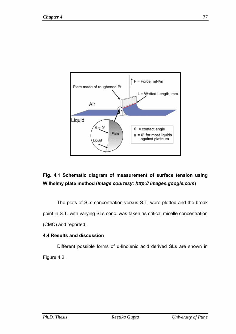
Chapter 4 77
Fig. 4.1 Schematic diagram of measurement of surface tension using Wilhelmy plate method (Image courtesy: http:// images.google.com)
The plots of SLs concentration versus S.T. were plotted and the break
point in S.T. with varying SLs conc. was taken as critical micelle concentration
(CMC) and reported.
4.4 Results and discussion
Different possible forms of α-linolenic acid derived SLs are shown in
Figure 4.2.
Ph.D. Thesis Reetika Gupta University of Pune
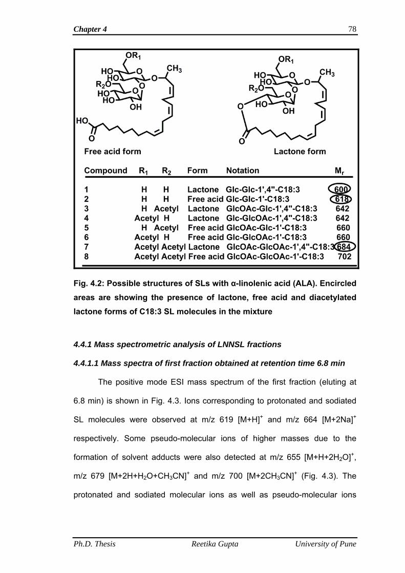
Chapter 4 78
Fig. 4.2: Possible structures of SLs with α-linolenic acid (ALA). Encircled areas are showing the presence of lactone, free acid and diacetylated lactone forms of C18:3 SL molecules in the mixture
4.4.1 Mass spectrometric analysis of LNNSL fractions
4.4.1.1 Mass spectra of first fraction obtained at retention time 6.8 min
The positive mode ESI mass spectrum of the first fraction (eluting at
6.8 min) is shown in Fig. 4.3. Ions corresponding to protonated and sodiated
SL molecules were observed at m/z 619 [M+H]+ and m/z 664 [M+2Na]+
respectively. Some pseudo-molecular ions of higher masses due to the
formation of solvent adducts were also detected at m/z 655 [M+H+2H2O]+,
m/z 679 [M+2H+H2O+CH3CN]+ and m/z 700 [M+2CH3CN]+ (Fig. 4.3). The
protonated and sodiated molecular ions as well as pseudo-molecular ions
Ph.D. Thesis Reetika Gupta University of Pune
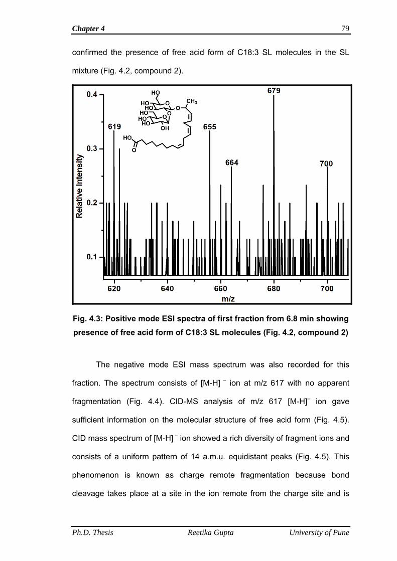
Chapter 4 79
confirmed the presence of free acid form of C18:3 SL molecules in the SL
mixture (Fig. 4.2, compound 2).
Fig. 4.3: Positive mode ESI spectra of first fraction from 6.8 min showing presence of free acid form of C18:3 SL molecules (Fig. 4.2, compound 2)
The negative mode ESI mass spectrum was also recorded for this
fraction. The spectrum consists of [M-H] – ion at m/z 617 with no apparent
fragmentation (Fig. 4.4). CID-MS analysis of m/z 617 [M-H]– ion gave
sufficient information on the molecular structure of free acid form (Fig. 4.5).
CID mass spectrum of [M-H] – ion showed a rich diversity of fragment ions and
consists of a uniform pattern of 14 a.m.u. equidistant peaks (Fig. 4.5). This
phenomenon is known as charge remote fragmentation because bond
cleavage takes place at a site in the ion remote from the charge site and is
Ph.D. Thesis Reetika Gupta University of Pune
Chapter 4 80
well recognized in the CID spectra of fatty acid carboxylate anions (Adams,
1990). Characteristic charge-remote fragmentation of ions containing a
saturated hydrocarbon chain involve losses of the elements of CnH2n+2 from
the alkyl terminus resulting in a regular series of peaks with a spacing of 14
mass units. The presence of a double bond and its position are established by
the absence of specific CnH2n+2 losses, namely those which would arise as a
result of vinylic hydrogen transfer or double bond cleavages (Tomer et al.,
1983). As shown in Fig. 4.5 the charge remote fragment ion series results
from collisionally induced losses of CnH2n+1COO- from m/z 617, yielding peaks
at m/z 558, 544, 530, and so on. The series is interrupted between m/z; 488
and 434, 434 and 394, 394 and 355, indicative of the presence of un-
saturation between C9 and C10, C12 and C13, C15 and C16 of the fatty acid
alkyl chain. The low abundances of peaks at interruption sites are most likely
caused by the relatively high energy requirement for vinylic hydrogen
transfers and double bond cleavages.
Ph.D. Thesis Reetika Gupta University of Pune
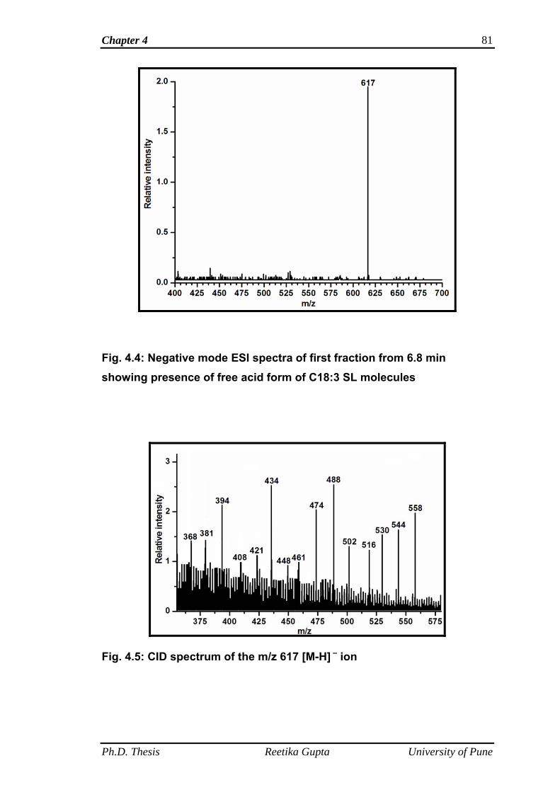
Chapter 4 81
Fig. 4.4: Negative mode ESI spectra of first fraction from 6.8 min showing presence of free acid form of C18:3 SL molecules
Fig. 4.5: CID spectrum of the m/z 617 [M-H] – ion
Ph.D. Thesis Reetika Gupta University of Pune
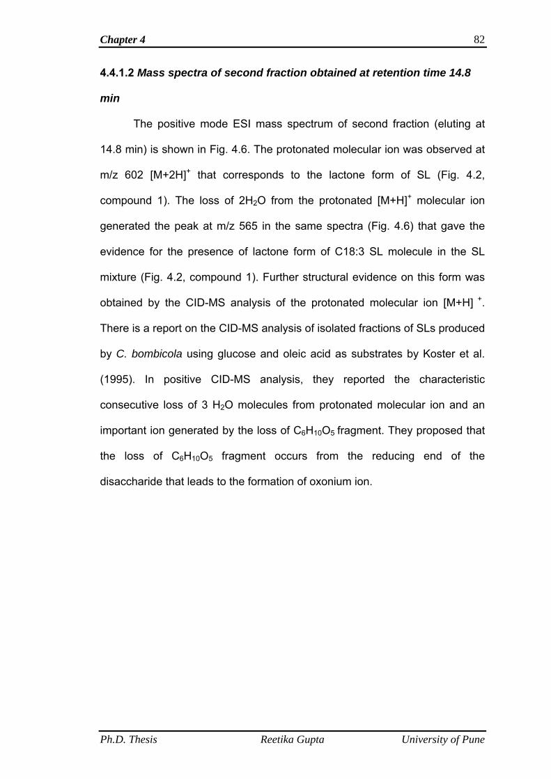
Chapter 4 82
4.4.1.2 Mass spectra of second fraction obtained at retention time 14.8
min
The positive mode ESI mass spectrum of second fraction (eluting at
14.8 min) is shown in Fig. 4.6. The protonated molecular ion was observed at
m/z 602 [M+2H]+ that corresponds to the lactone form of SL (Fig. 4.2,
compound 1). The loss of 2H2O from the protonated [M+H]+ molecular ion
generated the peak at m/z 565 in the same spectra (Fig. 4.6) that gave the
evidence for the presence of lactone form of C18:3 SL molecule in the SL
mixture (Fig. 4.2, compound 1). Further structural evidence on this form was
obtained by the CID-MS analysis of the protonated molecular ion [M+H] +.
There is a report on the CID-MS analysis of isolated fractions of SLs produced
by C. bombicola using glucose and oleic acid as substrates by Koster et al.
(1995). In positive CID-MS analysis, they reported the characteristic
consecutive loss of 3 H2O molecules from protonated molecular ion and an
important ion generated by the loss of C6H10O5 fragment. They proposed that
the loss of C6H10O5 fragment occurs from the reducing end of the
disaccharide that leads to the formation of oxonium ion.
Ph.D. Thesis Reetika Gupta University of Pune
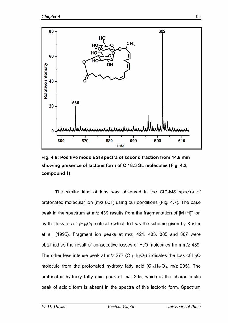
Chapter 4 83
Fig. 4.6: Positive mode ESI spectra of second fraction from 14.8 min showing presence of lactone form of C 18:3 SL molecules (Fig. 4.2, compound 1)
The similar kind of ions was observed in the CID-MS spectra of
protonated molecular ion (m/z 601) using our conditions (Fig. 4.7). The base
peak in the spectrum at m/z 439 results from the fragmentation of [M+H]+ ion
by the loss of a C6H10O5 molecule which follows the scheme given by Koster
et al. (1995). Fragment ion peaks at m/z, 421, 403, 385 and 367 were
obtained as the result of consecutive losses of H2O molecules from m/z 439.
The other less intense peak at m/z 277 (C18H29O2) indicates the loss of H2O
molecule from the protonated hydroxy fatty acid (C18H31O3, m/z 295). The
protonated hydroxy fatty acid peak at m/z 295, which is the characteristic
peak of acidic form is absent in the spectra of this lactonic form. Spectrum
Ph.D. Thesis Reetika Gupta University of Pune
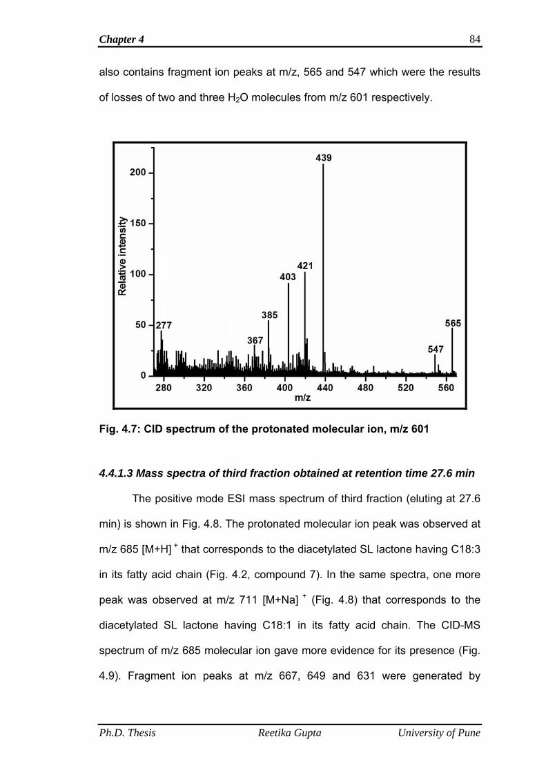
Chapter 4 84
also contains fragment ion peaks at m/z, 565 and 547 which were the results
of losses of two and three H2O molecules from m/z 601 respectively.
Fig. 4.7: CID spectrum of the protonated molecular ion, m/z 601
4.4.1.3 Mass spectra of third fraction obtained at retention time 27.6 min
The positive mode ESI mass spectrum of third fraction (eluting at 27.6
min) is shown in Fig. 4.8. The protonated molecular ion peak was observed at
m/z 685 [M+H] + that corresponds to the diacetylated SL lactone having C18:3
in its fatty acid chain (Fig. 4.2, compound 7). In the same spectra, one more
peak was observed at m/z 711 [M+Na] + (Fig. 4.8) that corresponds to the
diacetylated SL lactone having C18:1 in its fatty acid chain. The CID-MS
spectrum of m/z 685 molecular ion gave more evidence for its presence (Fig.
4.9). Fragment ion peaks at m/z 667, 649 and 631 were generated by
Ph.D. Thesis Reetika Gupta University of Pune
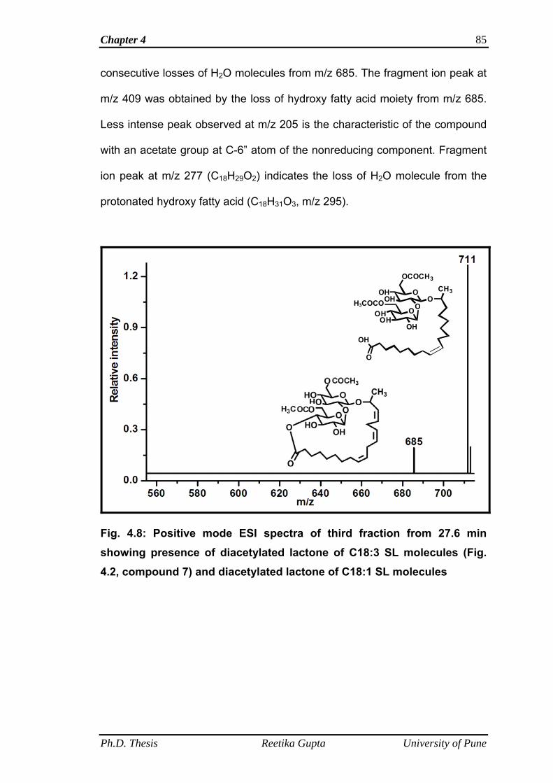
Chapter 4 85
consecutive losses of H2O molecules from m/z 685. The fragment ion peak at
m/z 409 was obtained by the loss of hydroxy fatty acid moiety from m/z 685.
Less intense peak observed at m/z 205 is the characteristic of the compound
with an acetate group at C-6” atom of the nonreducing component. Fragment
ion peak at m/z 277 (C18H29O2) indicates the loss of H2O molecule from the
protonated hydroxy fatty acid (C18H31O3, m/z 295).
OOH
OH
OOH
OH
OCOCH3
OCH3
O
OH
OH
O
H3COCO
Fig. 4.8: Positive mode ESI spectra of third fraction from 27.6 min showing presence of diacetylated lactone of C18:3 SL molecules (Fig. 4.2, compound 7) and diacetylated lactone of C18:1 SL molecules
Ph.D. Thesis Reetika Gupta University of Pune
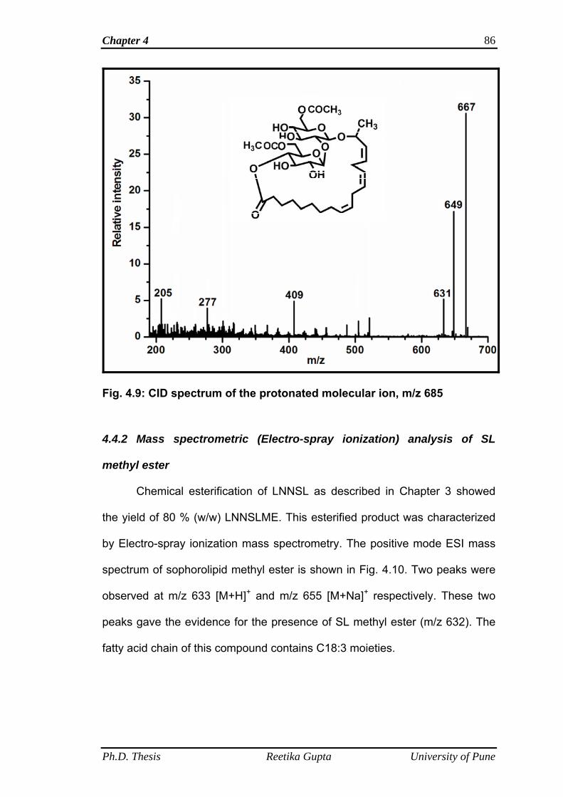
Chapter 4 86
Fig. 4.9: CID spectrum of the protonated molecular ion, m/z 685
4.4.2 Mass spectrometric (Electro-spray ionization) analysis of SL
methyl ester
Chemical esterification of LNNSL as described in Chapter 3 showed
the yield of 80 % (w/w) LNNSLME. This esterified product was characterized
by Electro-spray ionization mass spectrometry. The positive mode ESI mass
spectrum of sophorolipid methyl ester is shown in Fig. 4.10. Two peaks were
observed at m/z 633 [M+H]+ and m/z 655 [M+Na]+ respectively. These two
peaks gave the evidence for the presence of SL methyl ester (m/z 632). The
fatty acid chain of this compound contains C18:3 moieties.
Ph.D. Thesis Reetika Gupta University of Pune
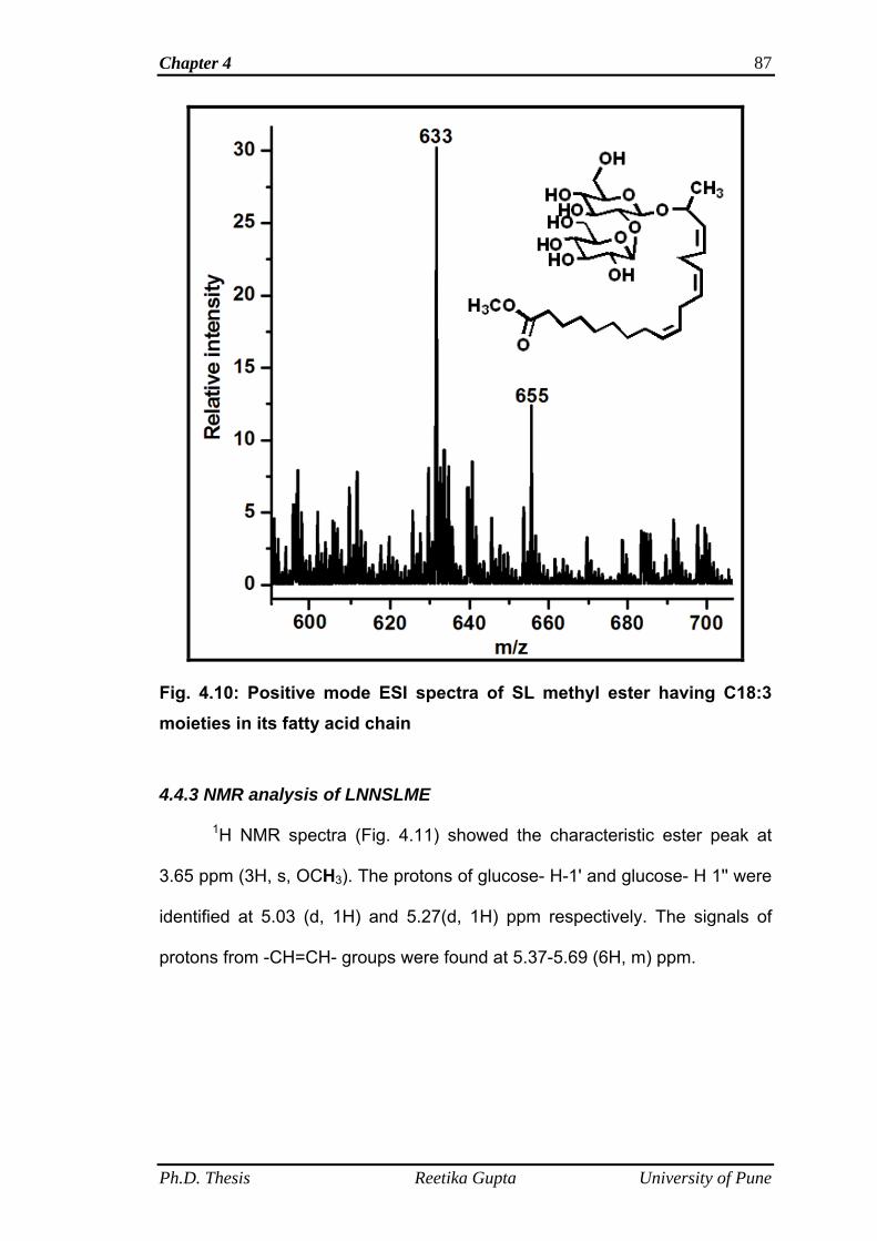
Chapter 4 87
Fig. 4.10: Positive mode ESI spectra of SL methyl ester having C18:3 moieties in its fatty acid chain
4.4.3 NMR analysis of LNNSLME
1H NMR spectra (Fig. 4.11) showed the characteristic ester peak at
3.65 ppm (3H, s, OCH3). The protons of glucose- H-1' and glucose- H 1'' were
identified at 5.03 (d, 1H) and 5.27(d, 1H) ppm respectively. The signals of
protons from -CH=CH- groups were found at 5.37-5.69 (6H, m) ppm.
Ph.D. Thesis Reetika Gupta University of Pune
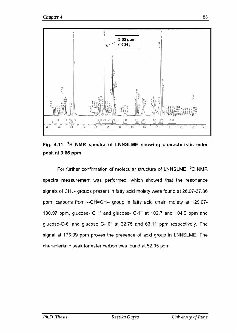
Chapter 4 88
3.65 ppm OCH3
Fig. 4.11: 1H NMR spectra of LNNSLME showing characteristic ester peak at 3.65 ppm
For further confirmation of molecular structure of LNNSLME 13C NMR
spectra measurement was performed, which showed that the resonance
signals of CH2 - groups present in fatty acid moiety were found at 26.07-37.86
ppm, carbons from --CH=CH-- group in fatty acid chain moiety at 129.07-
130.97 ppm, glucose- C 1' and glucose- C-1'' at 102.7 and 104.9 ppm and
glucose-C-6' and glucose C- 6'' at 62.75 and 63.11 ppm respectively. The
signal at 176.09 ppm proves the presence of acid group in LNNSLME. The
characteristic peak for ester carbon was found at 52.05 ppm.
Ph.D. Thesis Reetika Gupta University of Pune
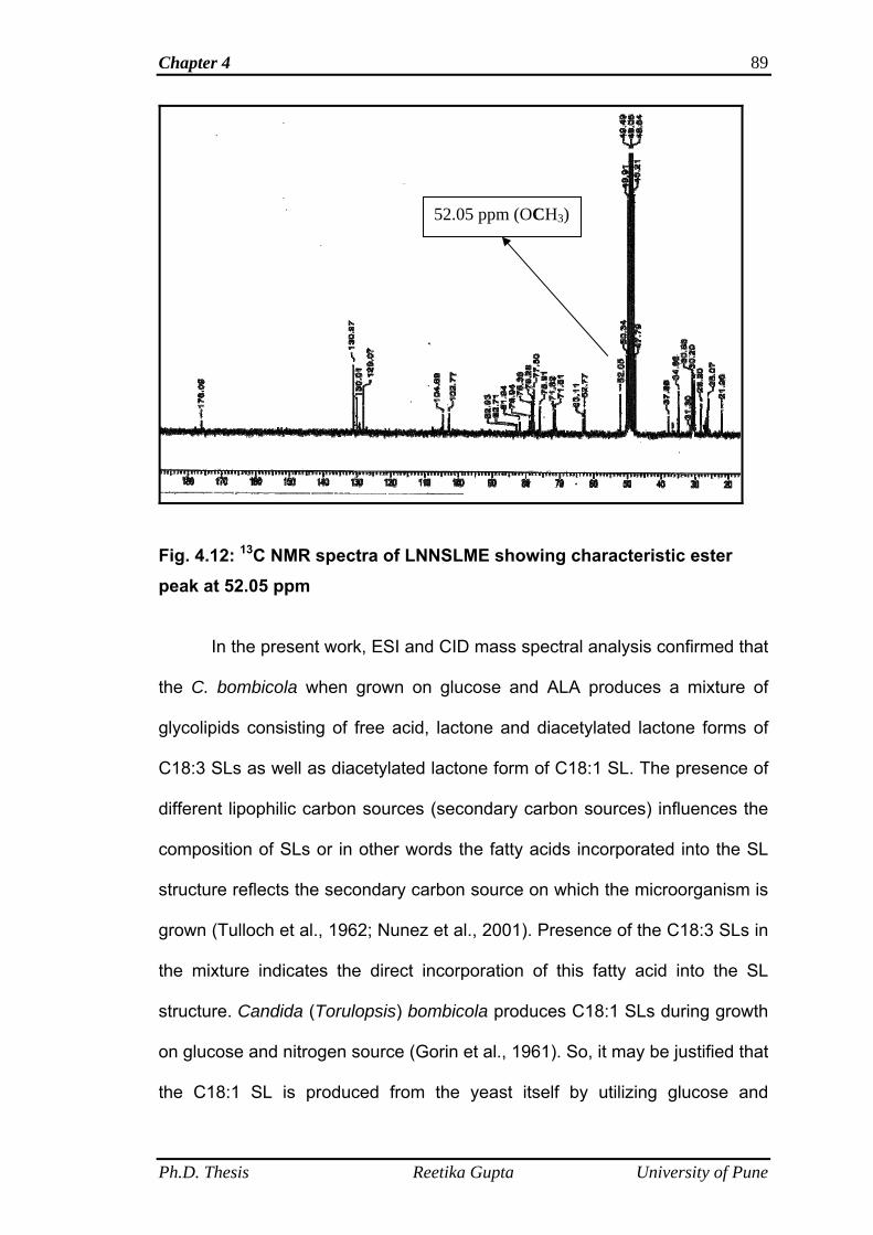
Chapter 4 89
52.05 ppm (OCH3)
Fig. 4.12: 13C NMR spectra of LNNSLME showing characteristic ester peak at 52.05 ppm
In the present work, ESI and CID mass spectral analysis confirmed that
the C. bombicola when grown on glucose and ALA produces a mixture of
glycolipids consisting of free acid, lactone and diacetylated lactone forms of
C18:3 SLs as well as diacetylated lactone form of C18:1 SL. The presence of
different lipophilic carbon sources (secondary carbon sources) influences the
composition of SLs or in other words the fatty acids incorporated into the SL
structure reflects the secondary carbon source on which the microorganism is
grown (Tulloch et al., 1962; Nunez et al., 2001). Presence of the C18:3 SLs in
the mixture indicates the direct incorporation of this fatty acid into the SL
structure. Candida (Torulopsis) bombicola produces C18:1 SLs during growth
on glucose and nitrogen source (Gorin et al., 1961). So, it may be justified that
the C18:1 SL is produced from the yeast itself by utilizing glucose and
Ph.D. Thesis Reetika Gupta University of Pune
Chapter 4 90
nitrogen sources from the medium. The approximate composition of the
fermentation product (SL mixture) was 7.5 % free acid, 80 % lactone and 4.5
% diacetylated lactone of C18:3 molecules and 8 % of diacetylated lactone of
C18:1 SL molecules. This composition was determined from the initial crude
SL loaded on the column. Further, for the ease of purification, we followed the
chemical esterification reaction from the literature in order to get a single
homogenous product (Bisht et al., 1999). We observed the conversion of SL
mixture into the SL methyl ester and ESI-MS analysis confirmed the presence
of C18:3 moieties in the fatty acid chain of this product and 1H and 13C NMR
study of LNNSLME also confirmed the formation of esterified product.
4.4.4 Surface tension and critical micelle concentration determination
The criteria for evaluating biosurfactants activity are surface tension
and critical micelle concentration (CMC). The surface tension correlates with
the concentration of the surface-active compound until the critical micelle
concentration (CMC) is reached. The CMC is defined as the minimum
surfactant concentration required for reaching the lowest surface tension
values. Efficient surfactants have a low critical micelle concentration (i.e. less
surfactant is necessary to decrease the surface tension or less surfactant is
required to saturate interfaces between air/liquid or liquid/liquid interface). At
concentrations above the CMC, amphiphilic molecules associate readily to
form supra-molecular structures such as micelles, bilayers and vesicles. The
surface tension and critical micelle concentration of linolenic acid derived SL
(LNNSL) and its methyl ester form designated as LNNSLME were determined
using Wilhelmy plate method. It was observed that with the increase in
concentration of LNNSL, there is a decrease in the surface tension value and
Ph.D. Thesis Reetika Gupta University of Pune
Chapter 4 91
as the conc. reached to 140 mg l-1, the minimum surface tension (32.384, mN
m-1) was achieved and beyond this point the constant value of surface tension
was obtained (Table 4.1, Fig. 4.13). The inflexion point between the minimum
surface tension value (32.384, mN m-1) and slope of surface tension is
reported as the critical micelle concentration which is 140 mg l-1 (Fig. 4.13)
As shown in Table 4.2, the same phenomena of decrease in surface
tension values were observed with the increase in the concentration of
LNNSLME and a point is reached where the minimum value of surface
tension (34.37, mN m-1) was obtained after which the constant values of
surface tension was observed and the inflexion point between the minimum
surface tension and value (34.37, mN m-1) and slope of surface tension was
reported as the critical micelle concentration which is 100 mg l-1 (Fig. 4.14).
Ph.D. Thesis Reetika Gupta University of Pune
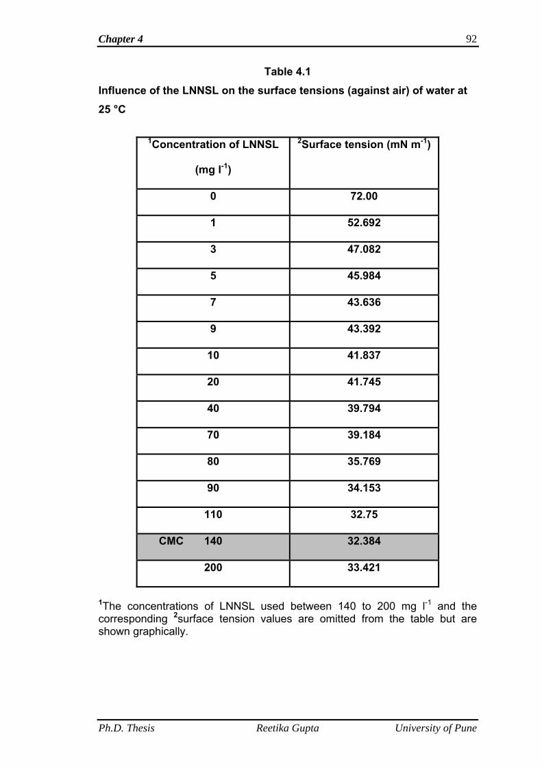
Chapter 4 92
Table 4.1 Influence of the LNNSL on the surface tensions (against air) of water at 25 °C
1Concentration of LNNSL
(mg l-1)
2Surface tension (mN m-1)
0 72.00
1 52.692
3 47.082
5 45.984
7 43.636
9 43.392
10 41.837
20 41.745
40 39.794
70 39.184
80 35.769
90 34.153
110 32.75
CMC 140 32.384
200 33.421
1The concentrations of LNNSL used between 140 to 200 mg l-1 and the corresponding 2surface tension values are omitted from the table but are shown graphically.
Ph.D. Thesis Reetika Gupta University of Pune
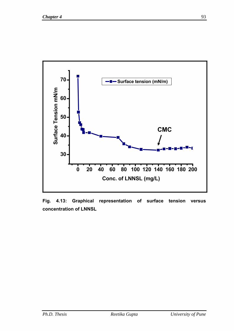
Chapter 4 93
0 20 40 60 80 100 120 140 160 180 200
30
40
50
60
70
Surf
ace
Tens
ion
mN
/m
Conc. of LNNSL (mg/L)
Surface tension (mN/m)
CMC
Fig. 4.13: Graphical representation of surface tension versus concentration of LNNSL
Ph.D. Thesis Reetika Gupta University of Pune
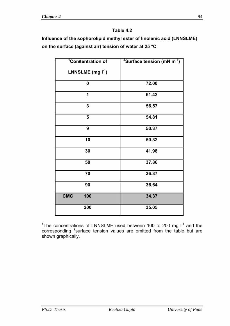
Chapter 4 94
Table 4.2 Influence of the sophorolipid methyl ester of linolenic acid (LNNSLME) on the surface (against air) tension of water at 25 °C
1Concentration of
LNNSLME (mg l-1)
2Surface tension (mN m-1)
0 72.00
1 61.42
3 56.57
5 54.81
9 50.37
10 50.32
30 41.98
50 37.86
70 36.37
90 36.64
CMC 100 34.37
200 35.05
1The concentrations of LNNSLME used between 100 to 200 mg l-1 and the corresponding 2surface tension values are omitted from the table but are shown graphically.
Ph.D. Thesis Reetika Gupta University of Pune
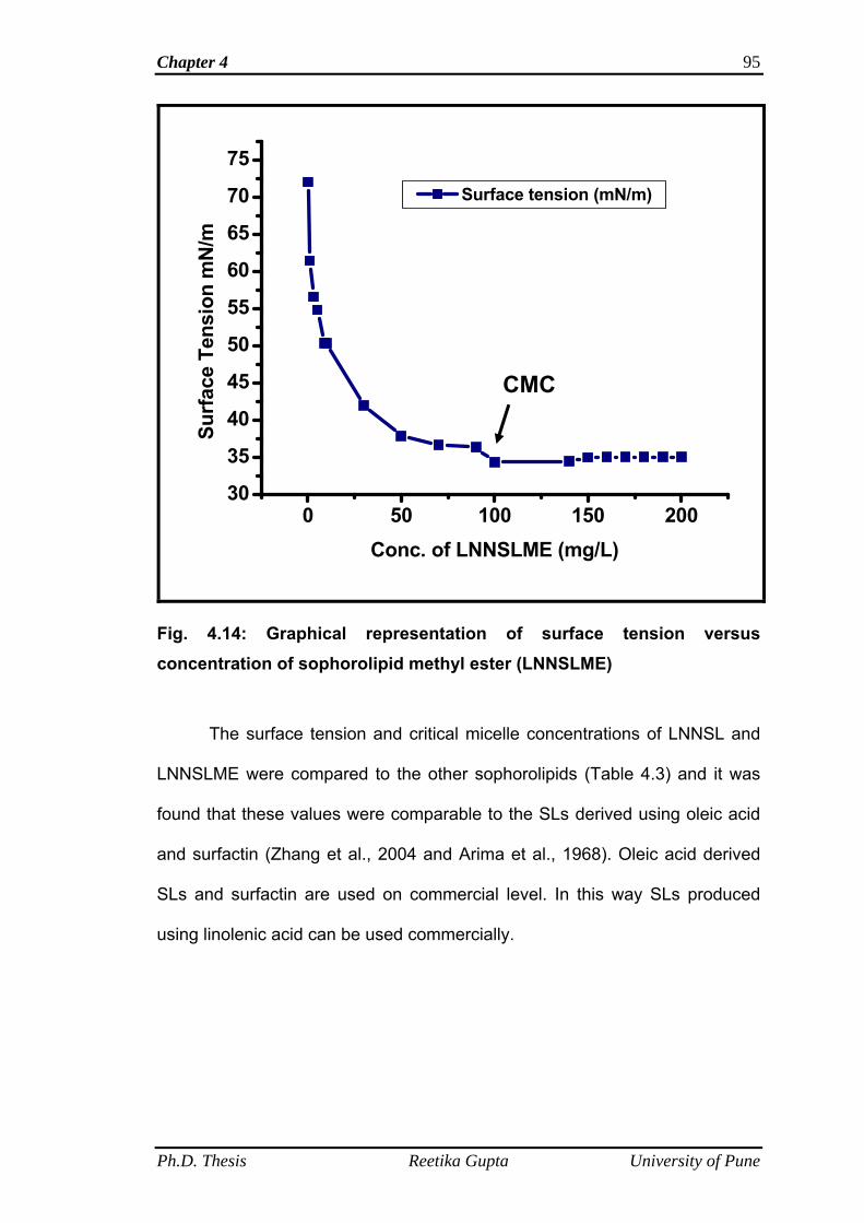
Chapter 4 95
0 50 100 150 20030
35
40
45
50
55
60
65
70
75
Ph.D. Thesis Reetika Gupta University of Pune
Surface tension (mN/m)Su
rfac
e Te
nsio
n m
N/m
Conc. of LNNSLME (mg/L)
CMC
Fig. 4.14: Graphical representation of surface tension versus concentration of sophorolipid methyl ester (LNNSLME)
The surface tension and critical micelle concentrations of LNNSL and
LNNSLME were compared to the other sophorolipids (Table 4.3) and it was
found that these values were comparable to the SLs derived using oleic acid
and surfactin (Zhang et al., 2004 and Arima et al., 1968). Oleic acid derived
SLs and surfactin are used on commercial level. In this way SLs produced
using linolenic acid can be used commercially.
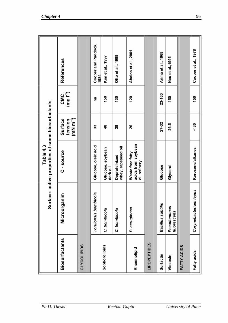
Chapter 4 96
Ref
eren
ces
Coo
per a
nd P
addo
ck,
1984
Kim
et a
l., 1
997
Otto
et a
l., 1
999
Aba
los
et a
l., 2
001
Arim
a et
al.,
196
8
Neu
et a
l.,19
96
Coo
per e
t al.,
197
8
CM
C
(mg
l-1)
na
150
130
120
23-1
60
150
150
Surf
ace
tens
ion
(mN
m-1
)
33
48
39
26
27-3
2
26.5
< 30
C -
sour
ce
Glu
cose
, ole
ic a
cid
Glu
cose
, soy
bean
da
rk o
il
Dep
rote
iniz
ed
whe
y, ra
pese
ed o
il
Was
te fr
ee fa
tty
acid
s fr
om s
oybe
an
oil r
efin
ery
Glu
cose
Gly
cero
l
Ker
osen
e/al
kane
s
Mic
roor
gani
m
Toru
lops
is b
ombi
cola
C. b
ombi
cola
C. b
ombi
cola
P. a
erug
inos
a
Bac
illus
sub
tilis
Pseu
dom
onas
flu
ores
cens
Cor
yneb
acte
rium
lepu
s
Tabl
e 4.
3 Su
rfac
e- a
ctiv
e pr
oper
ties
of s
ome
bios
urfa
ctan
ts
Bio
surf
acta
nts
GLY
CO
LIPI
DS
Soph
orol
ipid
s
Rha
mno
lipid
LIPO
PEPT
IDES
Surf
actin
Visc
osin
FATT
Y A
CID
S
Fatty
aci
ds
Ph.D. Thesis Reetika Gupta University of Pune
Chapter 4 97
4.5 Conclusions
The largest portion (about 80 %) of the LNNSL mixture was observed
in the lactone form of C18:3 (linolenic acid) SL molecules. This is in
agreement with the studies done by Tulloch et al. (1967), Asmer et al. (1988)
who showed in their work using different lipophilic substrates that the lactones
represent the largest fraction of the SL mixture. Weber et al. (1992) reported
that yeasts belonging to Candida species are capable of directly incorporating
fatty acids into the SL product and also Linton (1991) showed that direct
incorporation of fatty acids supplied as a carbon source is more efficient over
de novo synthesis of fatty acids. The present work showed the direct
incorporation of C18:3 fatty acid moieties into the SL structure which is in
agreement to the studies done by Linton (1991) and Weber et al. (1992) using
other fatty acids. Chemical esterification reaction reported for the conversion
of SL mixture into single homogenous product by Bisht et al. (1999) also
proved efficient in conversion of LNNSL mixture into esterified product of
linolenic acid SL (LNNSLME) and also good yield (80 %) of SL methyl ester
(LNNSLME) was obtained. The ESI-MS analysis confirmed the presence of
linolenic acid methyl ester in the fatty acid portion of SL molecules.
SLs when solved in water lower the surface tension from 72 mN m-1 to
40 to 30 mN m-1 (Van Bogaert et al., 2007) with a critical micelle concentration
(CMC) of 11 to 250 mg l-1 (Develter and Lauryssen, 2010). Results showed
that LNNSL and LNNSLME reduced the surface tension (ST) of water from72
mN m-1 to 32 and 34 mN m-1 respectively. The CMC values of LNNSL and
LNNSLME required is 140 and 100 mg l-1 respectively. Thus the obtained
values of surface tension and critical micelle concentration are comparable to
Ph.D. Thesis Reetika Gupta University of Pune
Chapter 4
Ph.D. Thesis Reetika Gupta University of Pune
98
the reported values for different types of SLs discussed above in the Table 4.3
of results and discussion section. Thus, the linolenic acid derived SL as well
as its methyl ester form both has the potential for industrial applications
(foaming for food processing, emulsifying and phase dispersion activity for
cosmetics, solubilization of agrochemicals, emulsification processes etc.) and
can replace chemically synthesized surfactants because of their
biodegradability and environment friendly nature.
Chapter 5
Antibacterial properties of Linolenic acid derived Sophorolipid and its methyl ester form
Chapter 5 100
5.1 Summary
The objective of this study is to introduce new antimicrobial agents with
high efficiency. This chapter is divided into two parts 5A and 5B. First part
deals with the antibacterial activity of LNNSL and LNNSLME including the
determination of minimum inhibitory concentrations of both the compounds
against Gram negative and Gram positive bacteria and second part deals with
the investigation of antibacterial action of these two compounds on Gram
negative and Gram positive bacteria with the help of Atomic force microscopy.
5A Antibacterial activity
5B Insight into the antibacterial action using Atomic force
microscopy
Ph.D. Thesis Reetika Gupta University of Pune
Chapter 5 101
5A Antibacterial activity
5A.1 Summary
Sophorolipids (SLs), due to their surface-active properties,
biocompatibility and various biological applications, have become a vital topic
of research. SLs have been used extensively, especially for antimicrobial
activity as most of the pathogenic microbes are gaining resistance to present
day antibiotics. In this chapter, the antibacterial activity of α-linolenic acid
derived sophorolipid, LNNSL containing 80 % lactone and its methyl ester
(LNNSLME) against both Gram negative (E. coli and P. aeruginosa) and
Gram positive (B. subtilis) bacteria is presented.
5A.2 Introduction
Sophorolipids (SLs) possess unique biological properties. Their
antibacterial, antifungal, antiviral and anti-cancerous activities make them
relevant molecules for applications in combating many diseases and as
therapeutic agents (Van Bogaert et al., 2007). There are many reports on the
use of SLs as antimicrobial agents in the literature. Shah et al. (2005) first
reported the spermicidal, anti-HIV and cytotoxic activities of SL mixture and its
chemical derivatives which are similar to commercially available nonoxynol-9
(nonionic spermicide). Further, Bluth et al. (2006) showed that SL mixture is
effective septic shock antagonists. There are a number of patents on
antibacterial, antiviral and antifungal activities of SL mixture and its alkyl ester
forms (Gross and Shah, 2003, 2004, 2007 and 2009 and Gross et al., 2004).
SLs have also been demonstrated to be effective anticancer agents against
cancerous cell lines (Chen et al., 2006a, 2006b and Joshi-Navare et al.,
Ph.D. Thesis Reetika Gupta University of Pune
Chapter 5 102
2011). Biosurfactant’s use as an antimicrobial agent is still limited on
commercial scale due to low cost of production of chemically synthesized
antibiotics (Bonomo, 2000; Shah et al., 2005; Azim et al., 2006; Napolitano,
2006; Shah et al., 2007 and Singh et al., 2009). However, the occurrence of
antibiotic-resistant pathogens increased the attention on other types of
antimicrobial agents (Bonomo, 2000 and Rodrigues et al., 2006). SL products
are FDA (Food and drug administration) approved and are available on
commercial level, for example, the French company Soliance produces SL
based cosmetics for body and skin (Van Bogaert et al., 2007 and Kaelynn,
2009). SLs act as emulsifier as well as a bactericidal agent in the treatment of
acne, dandruff, and body odours (Magar et al., 1987). SLs are also known for
their role as anti-adhesive agents against several pathogens which indicate
their utility as suitable anti-adhesive coating agents for medical insertional
materials leading to a reduction in a large number of hospital infections
without the use of synthetic drugs and chemicals (Rodrigues et al., 2006). In
this part of the chapter, antibacterial activity of LNNSL and LNNSLME against
Gram positive and Gram negative bacteria is presented.
5A.3 Materials and methods
5A.3.1 Bacterial cultures and growth conditions
Bacterial cultures used were obtained from National Collection of
Industrial Microorganisms (NCIM), National Chemical Laboratory, Pune, India.
Bacillus subtilis (NCIM 2063) was used as a representative of Gram positive
bacteria and Escherichia coli (NCIM 2931) and Pseudomonas aeruginosa
(NCIM 5029) were used as representatives of Gram negative bacteria. Luria
Ph.D. Thesis Reetika Gupta University of Pune
Chapter 5 103
bertini (LB) medium was used for the growth of both Gram positive and Gram
negative bacteria. The composition of LB medium used was in g l-1: tryptone,
10.0; yeast extract, 5.0; NaCl, 10.0; agar, 20.0; pH, 7.0. E. coli was grown at
37 °C while B. subtilis and P. aeruginosa were grown at 30 °C for overnight.
After overnight growth, at their respective temperatures, bacterial cultures
were centrifuged at 5000 rpm for 4 min and the wet pellets were re-
suspended in normal saline and were re-centrifuged at 5000 rpm for 4 min to
remove the growth medium. The final pellets were again re-suspended in
saline.
5A.3.2 Bacteria
Escherichia coli is a Gram negative, rod shaped bacterium
about 2.0 µm long and 0.5 µm in diameter with peritrichous flagella and are
found in the lower intestine of warm blooded animals (Eckburg et al., 2005).
Most of the E. coli strains are harmless but some strains may cause serious
food poisoning in humans (Vogt and Dippold, 2005).
Pseudomonas aeruginosa is a Gram negative and rod shaped
opportunistic pathogen. As a nosocomial pathogen it causes the highest
mortality rate of any other bacterium. Bacteremia, pneumonia, osteomyelitis,
and endocarditis due to P. aeruginosa have therapeutic failure and mortality
rates are notably higher than those for other bacterial pathogens.
Bacillus subtilis is a Gram positive bacterium. They are commonly
found in soil. They are non pathogenic to humans but sometimes may cause
food contamination. Its spores can survive the extreme heating used to cook
Ph.D. Thesis Reetika Gupta University of Pune
Chapter 5 104
food, and it is responsible for causing ropiness, caused by the bacterial
production of long chain polysaccharides.
5A.3.3 Sample preparation
Antibacterial tests of LNNSL, containing 80 % lactone and LNNSLME
were performed using standard dilution method and spread plate method.
Bacterial suspensions at a concentration of 106 CFU/ml were taken in a test
tube and LNNSL and LNNSLME to the final concentrations of 5, 10 and 20 µg
ml-1 diluted in sterile Millipore water were added separately. Test tubes
containing only respective cells without SL served as controls. All the test
tubes were incubated for 6 h at their respective temperatures. 100 μl aliquots
were taken out from the respective suspensions at every 2 h intervals and
plated on LB agar plates followed by incubation at their respective
temperatures (37 °C for E. coli and 30 °C for B. subtilis and P. aeruginosa).
Colonies were visualized after 24 h and digital images of the plates were
captured. Colonies were counted and percentage cell survival was calculated
using the following formula:
100(%) ×=platecontroloncoloniesofNumber
platetestoncoloniesofNumbersurvivalcellPercentage
Ph.D. Thesis Reetika Gupta University of Pune
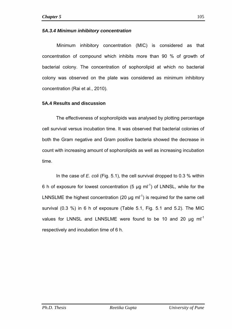
Chapter 5 105
5A.3.4 Minimun inhibitory concentration
Minimum inhibitory concentration (MIC) is considered as that
concentration of compound which inhibits more than 90 % of growth of
bacterial colony. The concentration of sophorolipid at which no bacterial
colony was observed on the plate was considered as minimum inhibitory
concentration (Rai et al., 2010).
5A.4 Results and discussion
The effectiveness of sophorolipids was analysed by plotting percentage
cell survival versus incubation time. It was observed that bacterial colonies of
both the Gram negative and Gram positive bacteria showed the decrease in
count with increasing amount of sophorolipids as well as increasing incubation
time.
In the case of E. coli (Fig. 5.1), the cell survival dropped to 0.3 % within
6 h of exposure for lowest concentration (5 µg ml-1) of LNNSL, while for the
LNNSLME the highest concentration (20 µg ml-1) is required for the same cell
survival (0.3 %) in 6 h of exposure (Table 5.1, Fig. 5.1 and 5.2). The MIC
values for LNNSL and LNNSLME were found to be 10 and 20 µg ml-1
respectively and incubation time of 6 h.
Ph.D. Thesis Reetika Gupta University of Pune
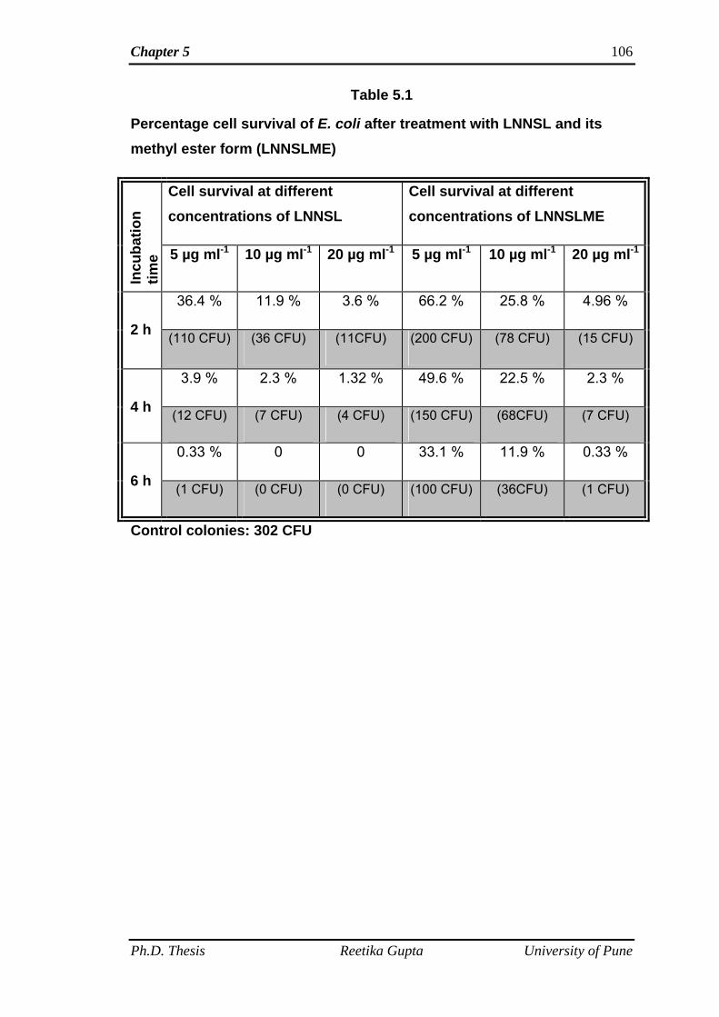
Chapter 5 106
Table 5.1
Percentage cell survival of E. coli after treatment with LNNSL and its methyl ester form (LNNSLME)
Cell survival at different concentrations of LNNSL
Cell survival at different concentrations of LNNSLME
Incu
batio
n tim
e 5 µg ml-1 10 µg ml-1 20 µg ml-1 5 µg ml-1 10 µg ml-1 20 µg ml-1
36.4 % 11.9 % 3.6 % 66.2 % 25.8 % 4.96 %
2 h (110 CFU) (36 CFU) (11CFU) (200 CFU) (78 CFU) (15 CFU)
3.9 % 2.3 % 1.32 % 49.6 % 22.5 % 2.3 %
4 h (12 CFU) (7 CFU) (4 CFU) (150 CFU) (68CFU) (7 CFU)
0.33 % 0 0 33.1 % 11.9 % 0.33 %
6 h (1 CFU) (0 CFU) (0 CFU) (100 CFU) (36CFU) (1 CFU)
Control colonies: 302 CFU
Ph.D. Thesis Reetika Gupta University of Pune
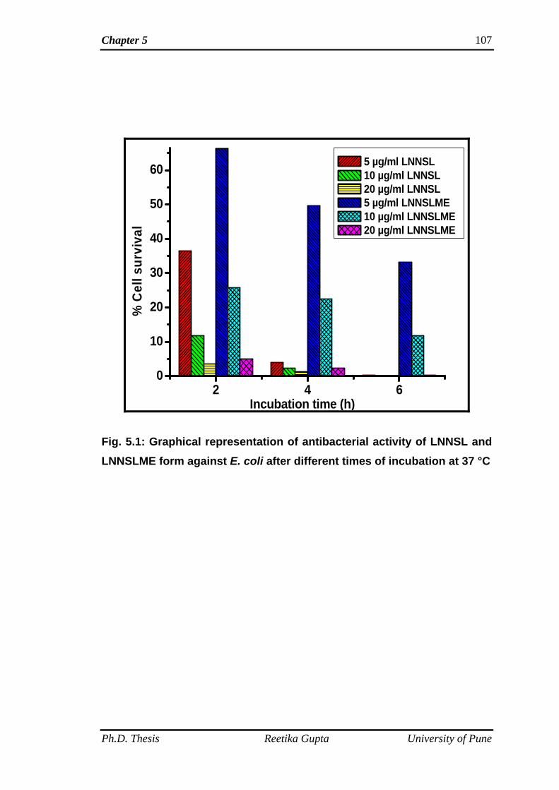
Chapter 5 107
2 4 60
10
20
30
40
50
60
% C
ell s
urvi
val
Incubation time (h)
5 µg/ml LNNSL 10 µg/ml LNNSL 20 µg/ml LNNSL 5 µg/ml LNNSLME 10 µg/ml LNNSLME 20 µg/ml LNNSLME
Fig. 5.1: Graphical representation of antibacterial activity of LNNSL and LNNSLME form against E. coli after different times of incubation at 37 °C
Ph.D. Thesis Reetika Gupta University of Pune
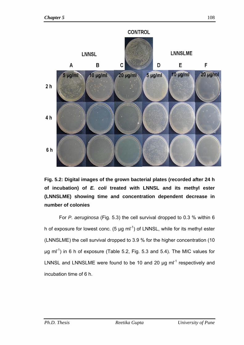
Chapter 5 108
Fig. 5.2: Digital images of the grown bacterial plates (recorded after 24 h of incubation) of E. coli treated with LNNSL and its methyl ester (LNNSLME) showing time and concentration dependent decrease in number of colonies
For P. aeruginosa (Fig. 5.3) the cell survival dropped to 0.3 % within 6
h of exposure for lowest conc. (5 µg ml-1) of LNNSL, while for its methyl ester
(LNNSLME) the cell survival dropped to 3.9 % for the higher concentration (10
µg ml-1) in 6 h of exposure (Table 5.2, Fig. 5.3 and 5.4). The MIC values for
LNNSL and LNNSLME were found to be 10 and 20 µg ml-1 respectively and
incubation time of 6 h.
Ph.D. Thesis Reetika Gupta University of Pune
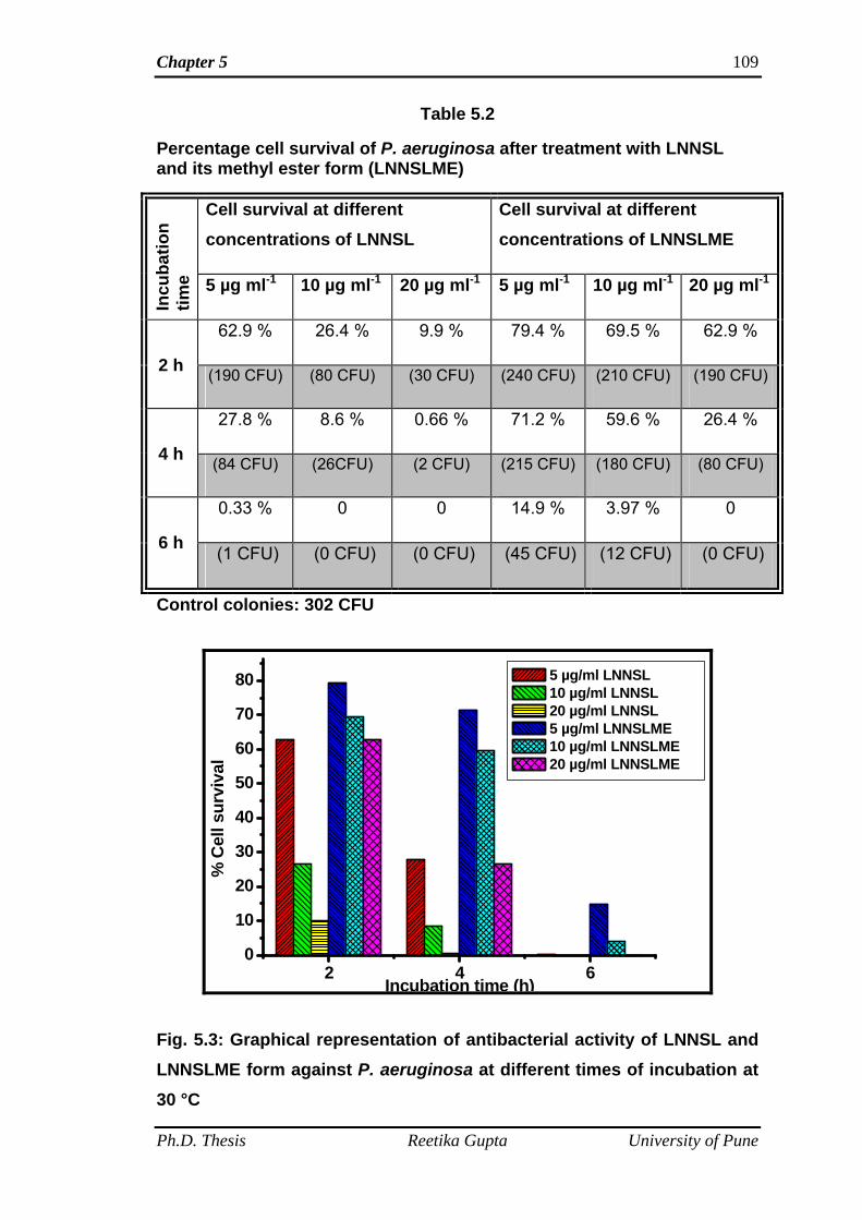
Chapter 5 109
Table 5.2
Percentage cell survival of P. aeruginosa after treatment with LNNSL and its methyl ester form (LNNSLME)
Cell survival at different concentrations of LNNSL
Cell survival at different concentrations of LNNSLME
Incu
batio
n tim
e 5 µg ml-1 10 µg ml-1 20 µg ml-1 5 µg ml-1 10 µg ml-1 20 µg ml-1
62.9 % 26.4 % 9.9 % 79.4 % 69.5 % 62.9 %
2 h (190 CFU) (80 CFU) (30 CFU) (240 CFU) (210 CFU) (190 CFU)
27.8 % 8.6 % 0.66 % 71.2 % 59.6 % 26.4 %
4 h (84 CFU) (26CFU) (2 CFU) (215 CFU) (180 CFU) (80 CFU)
0.33 % 0 0 14.9 % 3.97 % 0
6 h (1 CFU) (0 CFU) (0 CFU) (45 CFU) (12 CFU) (0 CFU)
Control colonies: 302 CFU
2 4 60
10
20
30
40
50
60
70
80
% C
ell s
urvi
val
Incubation time (h)
5 µg/ml LNNSL 10 µg/ml LNNSL 20 µg/ml LNNSL 5 µg/ml LNNSLME 10 µg/ml LNNSLME 20 µg/ml LNNSLME
Fig. 5.3: Graphical representation of antibacterial activity of LNNSL and LNNSLME form against P. aeruginosa at different times of incubation at 30 °C
Ph.D. Thesis Reetika Gupta University of Pune
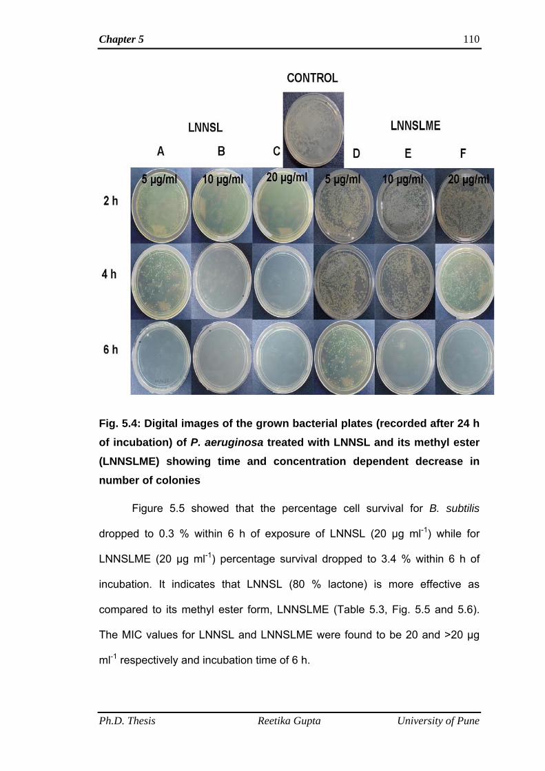
Chapter 5 110
Fig. 5.4: Digital images of the grown bacterial plates (recorded after 24 h of incubation) of P. aeruginosa treated with LNNSL and its methyl ester (LNNSLME) showing time and concentration dependent decrease in number of colonies
Figure 5.5 showed that the percentage cell survival for B. subtilis
dropped to 0.3 % within 6 h of exposure of LNNSL (20 µg ml-1) while for
LNNSLME (20 µg ml-1) percentage survival dropped to 3.4 % within 6 h of
incubation. It indicates that LNNSL (80 % lactone) is more effective as
compared to its methyl ester form, LNNSLME (Table 5.3, Fig. 5.5 and 5.6).
The MIC values for LNNSL and LNNSLME were found to be 20 and >20 µg
ml-1 respectively and incubation time of 6 h.
Ph.D. Thesis Reetika Gupta University of Pune
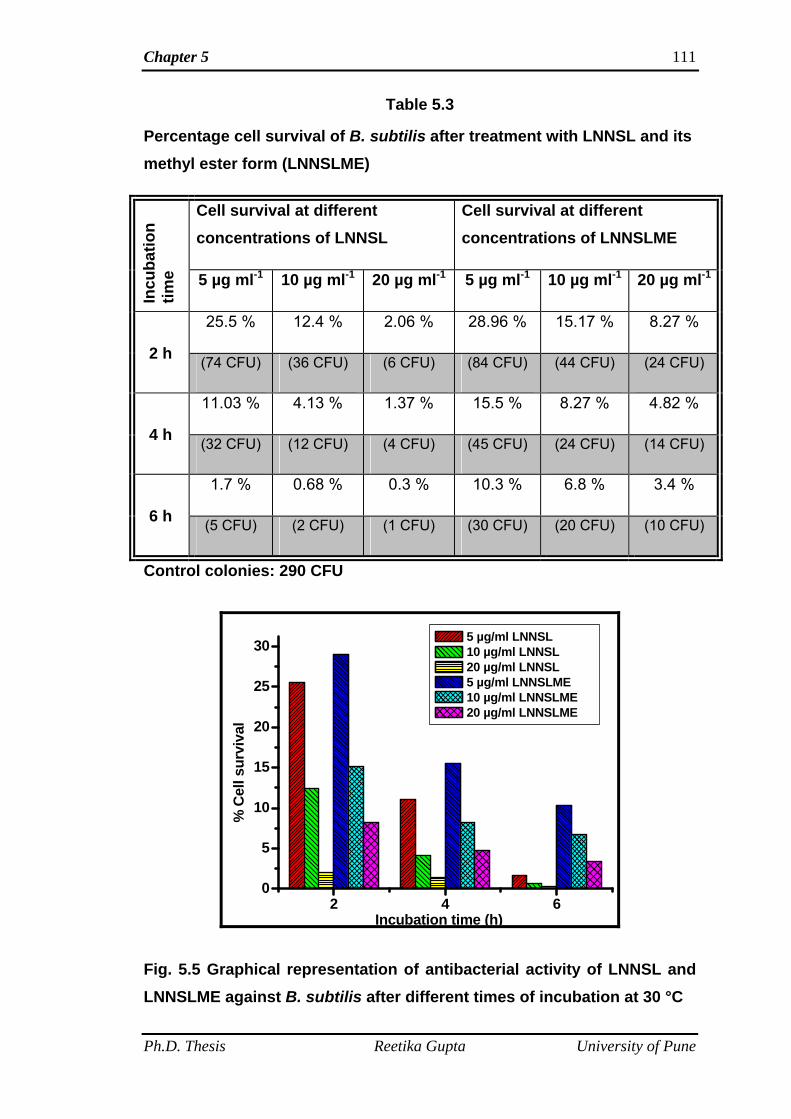
Chapter 5 111
Table 5.3
Percentage cell survival of B. subtilis after treatment with LNNSL and its methyl ester form (LNNSLME)
Cell survival at different concentrations of LNNSL
Cell survival at different concentrations of LNNSLME
Incu
batio
n tim
e 5 µg ml-1 10 µg ml-1 20 µg ml-1 5 µg ml-1 10 µg ml-1 20 µg ml-1
25.5 % 12.4 % 2.06 % 28.96 % 15.17 % 8.27 %
2 h (74 CFU) (36 CFU) (6 CFU) (84 CFU) (44 CFU) (24 CFU)
11.03 % 4.13 % 1.37 % 15.5 % 8.27 % 4.82 %
4 h (32 CFU) (12 CFU) (4 CFU) (45 CFU) (24 CFU) (14 CFU)
1.7 % 0.68 % 0.3 % 10.3 % 6.8 % 3.4 %
6 h (5 CFU) (2 CFU) (1 CFU) (30 CFU) (20 CFU) (10 CFU)
Control colonies: 290 CFU
2 4 60
5
10
15
20
25
30
% C
ell s
urvi
val
Incubation time (h)
5 µg/ml LNNSL 10 µg/ml LNNSL 20 µg/ml LNNSL 5 µg/ml LNNSLME 10 µg/ml LNNSLME 20 µg/ml LNNSLME
Fig. 5.5 Graphical representation of antibacterial activity of LNNSL and LNNSLME against B. subtilis after different times of incubation at 30 °C
Ph.D. Thesis Reetika Gupta University of Pune
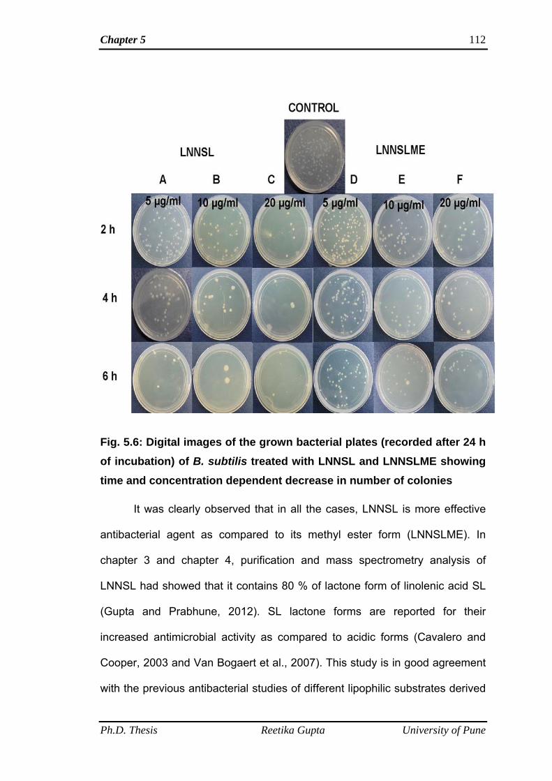
Chapter 5 112
Fig. 5.6: Digital images of the grown bacterial plates (recorded after 24 h of incubation) of B. subtilis treated with LNNSL and LNNSLME showing time and concentration dependent decrease in number of colonies
It was clearly observed that in all the cases, LNNSL is more effective
antibacterial agent as compared to its methyl ester form (LNNSLME). In
chapter 3 and chapter 4, purification and mass spectrometry analysis of
LNNSL had showed that it contains 80 % of lactone form of linolenic acid SL
(Gupta and Prabhune, 2012). SL lactone forms are reported for their
increased antimicrobial activity as compared to acidic forms (Cavalero and
Cooper, 2003 and Van Bogaert et al., 2007). This study is in good agreement
with the previous antibacterial studies of different lipophilic substrates derived
Ph.D. Thesis Reetika Gupta University of Pune
Chapter 5
Ph.D. Thesis Reetika Gupta University of Pune
113
SL by various researchers (Cavalero and Cooper, 2003 and Van Bogaert et
al., 2007). As methyl ester form had also showed its antibacterial activity
against both Gram negative and Gram positive bacteria, it can also be used
as antibacterial agent. The other observation made was that LNNSL and
LNNSLME both inhibited Gram negative bacteria at lower concentrations as
compared to Gram positive bacteria. So, these SLs of linolenic acid were
found to be more potent against Gram negative bacteria as compared to other
SLs reported in the literature which was more effective against Gram positive
bacteria (Gross and Shah 2003). The more antibacterial effect towards Gram
negative bacteria may be attributed to the less resistance offered by thin
peptidoglycan layer adjacent to the inner cytoplasmic membrane as compared
to the thick peptidoglycan layer of Gram positive bacteria which offer more
resistance (Singh et al. 2009).
5A.5 Conclusions
The minimum inhibitory concentrations (MIC) of LNNSL, containing 80
% lactone against B. subtilis, E. coli and P. aeruginosa were found to be 20,
10 and 10 µg ml-1 respectively. The MIC values of methyl ester form
(LNNSLME) against B. subtilis, E. coli and P. aeruginosa were determined to
be >20, 20 and 20 µg ml-1 respectively. The results suggests that Linolenic SL
mixture (containing 80% lactone) as compared to its methyl ester derivative
showed good antibacterial activity towards both the Gram positive and Gram
negative bacteria and was found to be more potent against Gram negative
bacteria.
Chapter 5 114
5B Insight into the antibacterial action using Atomic force microscopy
5B.1 Summary
This part of the chapter deals with the investigation of antibacterial
action of LNNSL and LNNSLME with the help of atomic force microscopy
(AFM). Nanometer scale images of both the control (without SL treatment)
and treated bacterial cells showed the membranolytic effects of LNNSL and
LNNSLME on the cell membrane of both the Gram negative and Gram
positive bacteria. This is the first report on the investigation of antibacterial
action of novel linolenic acid derived SL and its methyl ester (LNNSLME)
using AFM study.
5B.2 Introduction
There are some reports on the antibacterial activity of sophorolipids as
well as other glycolipids [Gross and Shah, 2003, 2004, 2007 and 2009 and
Gross et al., 2004]. It is already reported that biosurfactants act on the
integrity of cell membranes, which leads to cell lysis, (Cameotra and Makkar
2004). However, the ways in which the biosurfactants affect the membrane
integrity differ. Rhamnolipids are thought to act on the lipid part of cell
membranes or outer proteins causing structural fluctuations in the membrane
(Rodrigues et al., 2006 and Sanchez et al., 2010). There are many evidences
on the actions of antibiotics by means of the direct observation of
morphological alterations by optical microscopy, scanning probe microscopy
and atomic force microscopy (Klainer and Perkins, 1971; Nishino and
Nakazawa, 1973; Nakao et al., 1981; Goi et al., 1985 and Carlo and Ricci,
1998). The great advantage of AFM compared to TEM or SEM is that it is
Ph.D. Thesis Reetika Gupta University of Pune
Chapter 5 115
simple to operate in almost any environment such as aqueous solutions, but
also other solvents, in air, vacuum, or other gases. It also provides the details
of surface morphology with very high resolution without the need for
complicated sample preparation (Binnig et al., 1986). AFM is an advanced
non-destructive imaging technique which can be used to understand the
nanoscale interactions between bacteria and different molecules. Since its
introduction in 1986 (Binnig et al., 1986), AFM has been used extensively in
the field of life sciences (Braga and Ricci, 1998 and Bonomo, 2000). AFM
images can reveal the surface ultra structure of living microbial cells with
unprecedented resolution (Braga and Ricci, 1998 and Dufrene et al., 1999).
AFM is a kind of scanning probe microscope and is based on the
concept of near field microscopy which overcomes the problem of the limited
diffraction related resolution inherent in conventional microscopes because in
AFM the probe is located in the immediate vicinity of the sample itself (usually
within a few nanometers), records the intensity and not the interference
signal, and this greatly improves the final resolution. The principle of AFM is to
scan the sample surface with the sharp probing tip (radius of few nanometers)
attached to a cantilever. The three dimensional imaging is performed by
measuring the displacement in x, y and z axis with the help of piezoelectric
ceramic. When the probe tip is brought into the proximity of the sample
surface, forces between the tip and the sample lead to a deflection of the
cantilever according to the Hook’s law. The forces between the probe tip and
samples are detected through the deflection of the cantilever. The deflection
of the cantilever is sensed using a laser beam reflected on the rear side of the
cantilever and for this a position sensitive detector monitors the position of the
Ph.D. Thesis Reetika Gupta University of Pune
Chapter 5 116
reflected beam. Forces that are measured in AFM include mechanical contact
force, vander Waals forces, capillary forces, chemical bonding, electrostatic
forces, magnetic forces and salvation forces etc. AFM is an advanced
technique of scanning probe microscopes that can be used for observation of
nonconductive surfaces and has thus opened the possibility for the surface
analysis of various samples.
The aim of this study was to investigate this new tool (AFM) for
analyzing surface and morphological alterations of Gram negative and Gram
positive bacteria by examining the samples of bacteria exposed to linolenic
acid derived sophorolipid (LNNSL) and its methyl ester form (LNNSLME). In
the first part of this chapter (5A), the minimum inhibitory concentrations of
LNNSL and LNNSLME were decided for both the Gram negative (Escherichia
coli and Pseudomonas aeruginosa) and Gram positive (Bacillus subtilis)
bacteria. In this part, the effect of these concentrations (MICs) of LNNSL and
LNNSLME on the morphology of both the Gram negative (E. coli and P.
aeruginosa) and Gram positive (B. subtilis) bacteria were investigated with the
help of AFM and also this is the first study on the visualization of
morphological changes in bacteria treated with SLs using atomic force
microscopy.
5B.3 Materials and methods
5B.3.1 AFM analysis
The atomic force microscopy measurements were performed using a
multimode scanning probe microscope equipped with a Nanoscope IV
controller (Veeco Instrument Inc., Santa Barbara, CA). Soft AFM cantilevers,
Ph.D. Thesis Reetika Gupta University of Pune
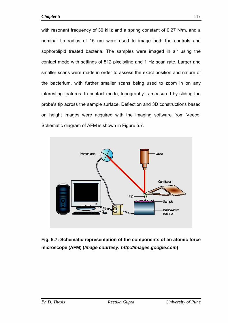
Chapter 5 117
with resonant frequency of 30 kHz and a spring constant of 0.27 N/m, and a
nominal tip radius of 15 nm were used to image both the controls and
sophorolipid treated bacteria. The samples were imaged in air using the
contact mode with settings of 512 pixels/line and 1 Hz scan rate. Larger and
smaller scans were made in order to assess the exact position and nature of
the bacterium, with further smaller scans being used to zoom in on any
interesting features. In contact mode, topography is measured by sliding the
probe’s tip across the sample surface. Deflection and 3D constructions based
on height images were acquired with the imaging software from Veeco.
Schematic diagram of AFM is shown in Figure 5.7.
Fig. 5.7: Schematic representation of the components of an atomic force microscope (AFM) (Image courtesy: http://images.google.com)
Ph.D. Thesis Reetika Gupta University of Pune
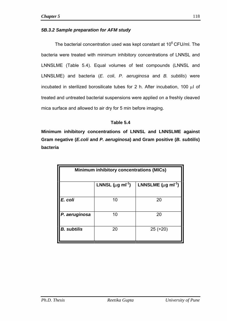
Chapter 5 118
5B.3.2 Sample preparation for AFM study
The bacterial concentration used was kept constant at 106 CFU/ml. The
bacteria were treated with minimum inhibitory concentrations of LNNSL and
LNNSLME (Table 5.4). Equal volumes of test compounds (LNNSL and
LNNSLME) and bacteria (E. coli, P. aeruginosa and B. subtilis) were
incubated in sterilized borosilicate tubes for 2 h. After incubation, 100 μl of
treated and untreated bacterial suspensions were applied on a freshly cleaved
mica surface and allowed to air dry for 5 min before imaging.
Table 5.4
Minimum inhibitory concentrations of LNNSL and LNNSLME against Gram negative (E.coli and P. aeruginosa) and Gram positive (B. subtilis) bacteria
Minimum inhibitory concentrations (MICs)
LNNSL (μg ml-1) LNNSLME (μg ml-1)
E. coli 10 20
P. aeruginosa 10 20
B. subtilis 20 25 (>20)
Ph.D. Thesis Reetika Gupta University of Pune
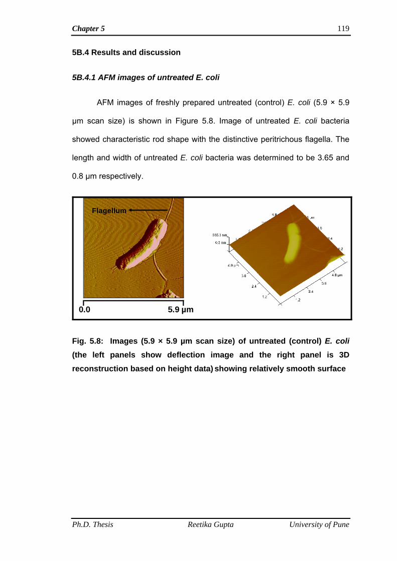
Chapter 5 119
5B.4 Results and discussion
5B.4.1 AFM images of untreated E. coli
AFM images of freshly prepared untreated (control) E. coli (5.9 × 5.9
µm scan size) is shown in Figure 5.8. Image of untreated E. coli bacteria
showed characteristic rod shape with the distinctive peritrichous flagella. The
length and width of untreated E. coli bacteria was determined to be 3.65 and
0.8 µm respectively.
5.9 µm0.0
Flagellum
Fig. 5.8: Images (5.9 × 5.9 µm scan size) of untreated (control) E. coli (the left panels show deflection image and the right panel is 3D reconstruction based on height data) showing relatively smooth surface
Ph.D. Thesis Reetika Gupta University of Pune
Chapter 5 120
5B.4.2 AFM images of E. coli bacteria treated with LNNSL
AFM images of E. coli bacteria treated with minimum inhibitory
concentration (10 µg ml-1) of LNNSL are shown in Figure 5.9 A, B and C.
Figure 5.9 A is the large scan size (50 × 50 µm) image and captured in
deflection and height mode. The images showed bacterial cells with copious
amount of exuded fluid. Images were further zoomed in a particular area and
smaller scan size images of (13.3 × 13.3 µm) and (4.0 × 4.0 µm) were taken
in deflection and height modes (Fig. 5.9 A, 5.9 B and 5.9 C). Bacterial cells
were severely damaged and the presence of copious amount of cytoplasmic
fluid around the bacteria indicates the permeabilization of inner membrane
due to the membranolytic action of LNNSL.
5 B.4.3 AFM images of E. coli bacteria treated with LNNSLME
AFM images of E. coli treated with minimum inhibitory concentration of
20 µg ml-1 of sophorolipid methyl ester, LNNSLME are shown in Figure 5.10 A
and B. Both larger (26.9 × 26.9 µm) and smaller (8.3 ×8.3 µm) scan size
images in deflection and height modes were taken. Figure 5.10 A and B
showed damaged or lysed bacterial cells with the exuded cytoplasmic fluid
from both the ends of the bacterial cell. It means that SL methyl ester exerted
the similar mode of action of LNNSL on bacterial cells.
Ph.D. Thesis Reetika Gupta University of Pune
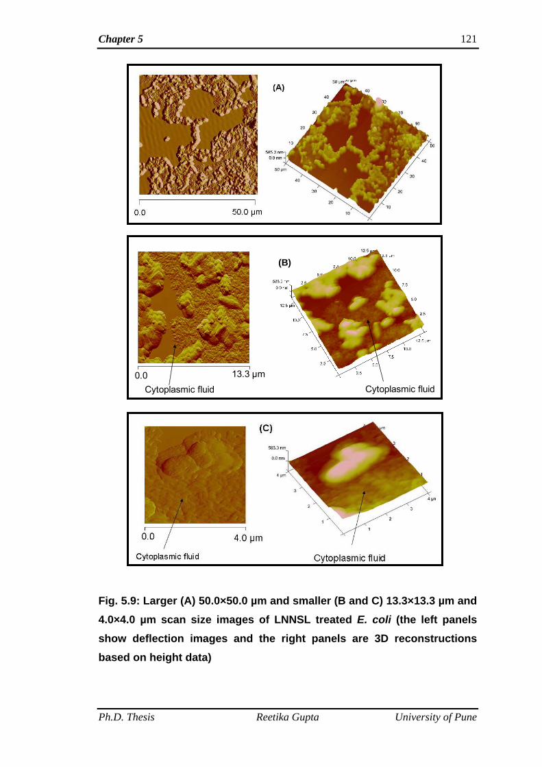
Chapter 5 121
Ph.D. Thesis Reetika Gupta University of Pune
0.0 13.3 µm
Cytoplasmic fluid Cytoplasmic f
(B)
luid
Fig. 5.9: Larger (A) 50.0×50.0 µm and smaller (B and C) 13.3×13.3 µm and 4.0×4.0 µm scan size images of LNNSL treated E. coli (the left panels show deflection images and the right panels are 3D reconstructions based on height data)
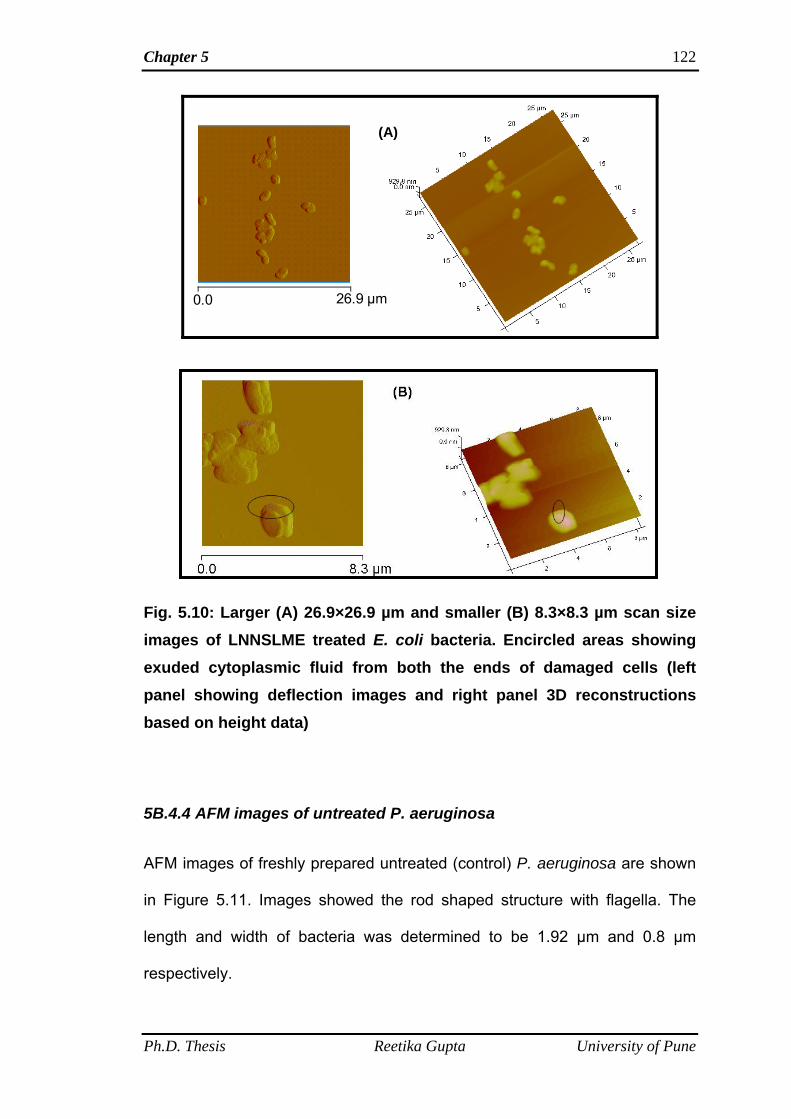
Chapter 5 122
0.0
(A)
26.9 µm
Fig. 5.10: Larger (A) 26.9×26.9 µm and smaller (B) 8.3×8.3 µm scan size images of LNNSLME treated E. coli bacteria. Encircled areas showing exuded cytoplasmic fluid from both the ends of damaged cells (left panel showing deflection images and right panel 3D reconstructions based on height data)
5B.4.4 AFM images of untreated P. aeruginosa
AFM images of freshly prepared untreated (control) P. aeruginosa are shown
in Figure 5.11. Images showed the rod shaped structure with flagella. The
length and width of bacteria was determined to be 1.92 µm and 0.8 µm
respectively.
Ph.D. Thesis Reetika Gupta University of Pune
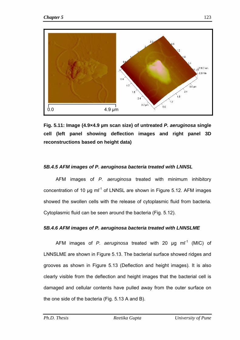
Chapter 5 123
0.0 4.9 µm
Fig. 5.11: Image (4.9×4.9 µm scan size) of untreated P. aeruginosa single cell (left panel showing deflection images and right panel 3D reconstructions based on height data)
5B.4.5 AFM images of P. aeruginosa bacteria treated with LNNSL
AFM images of P. aeruginosa treated with minimum inhibitory
concentration of 10 µg ml-1 of LNNSL are shown in Figure 5.12. AFM images
showed the swollen cells with the release of cytoplasmic fluid from bacteria.
Cytoplasmic fluid can be seen around the bacteria (Fig. 5.12).
5B.4.6 AFM images of P. aeruginosa bacteria treated with LNNSLME
AFM images of P. aeruginosa treated with 20 µg ml-1 (MIC) of
LNNSLME are shown in Figure 5.13. The bacterial surface showed ridges and
grooves as shown in Figure 5.13 (Deflection and height images). It is also
clearly visible from the deflection and height images that the bacterial cell is
damaged and cellular contents have pulled away from the outer surface on
the one side of the bacteria (Fig. 5.13 A and B).
Ph.D. Thesis Reetika Gupta University of Pune
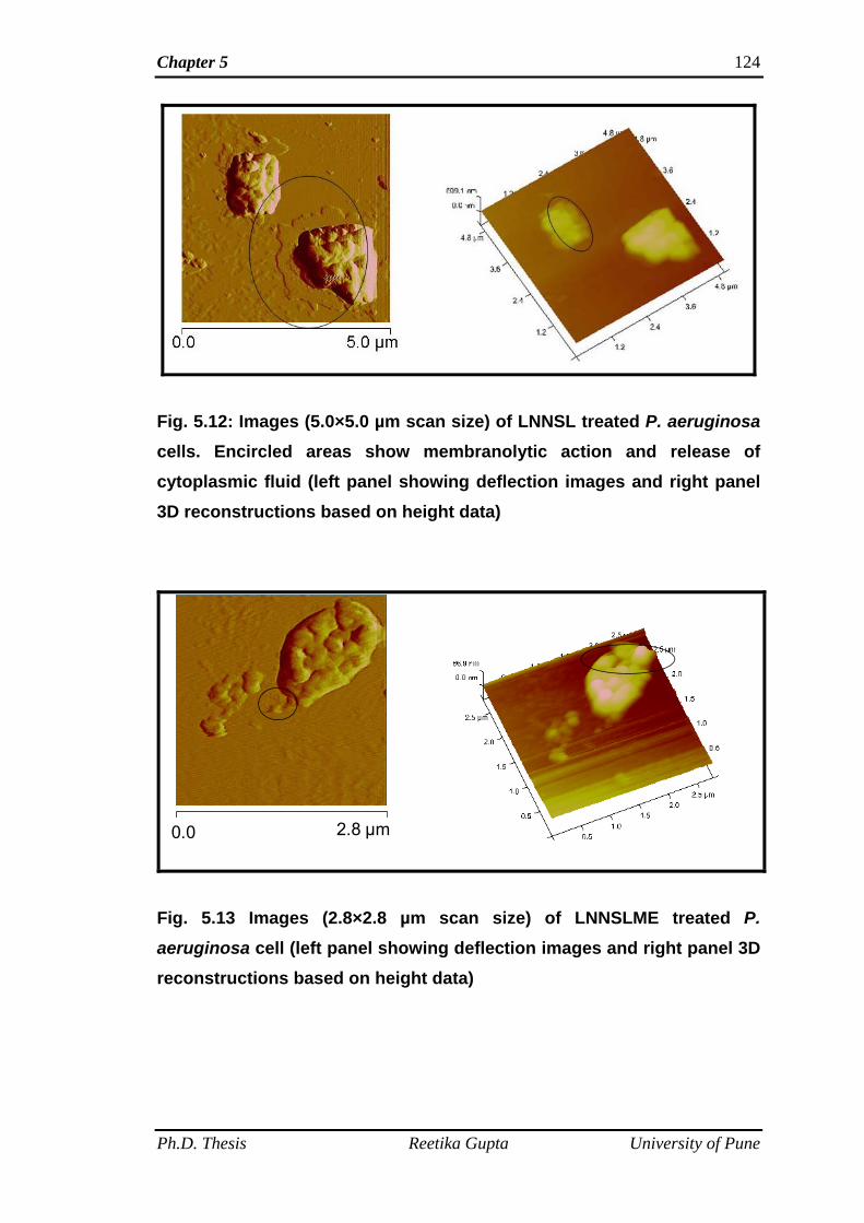
Chapter 5 124
Fig. 5.12: Images (5.0×5.0 µm scan size) of LNNSL treated P. aeruginosa cells. Encircled areas show membranolytic action and release of cytoplasmic fluid (left panel showing deflection images and right panel 3D reconstructions based on height data)
0.0 2.8 µm
Fig. 5.13 Images (2.8×2.8 µm scan size) of LNNSLME treated P. aeruginosa cell (left panel showing deflection images and right panel 3D reconstructions based on height data)
Ph.D. Thesis Reetika Gupta University of Pune
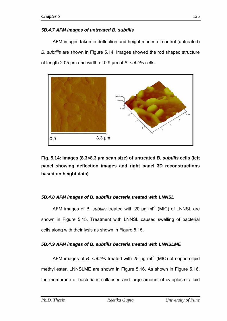
Chapter 5 125
5B.4.7 AFM images of untreated B. subtilis
AFM images taken in deflection and height modes of control (untreated)
B. subtilis are shown in Figure 5.14. Images showed the rod shaped structure
of length 2.05 µm and width of 0.9 µm of B. subtilis cells.
Fig. 5.14: Images (8.3×8.3 µm scan size) of untreated B. subtilis cells (left panel showing deflection images and right panel 3D reconstructions based on height data)
5B.4.8 AFM images of B. subtilis bacteria treated with LNNSL
AFM images of B. subtilis treated with 20 µg ml-1 (MIC) of LNNSL are
shown in Figure 5.15. Treatment with LNNSL caused swelling of bacterial
cells along with their lysis as shown in Figure 5.15.
5B.4.9 AFM images of B. subtilis bacteria treated with LNNSLME
AFM images of B. subtilis treated with 25 µg ml-1 (MIC) of sophorolipid
methyl ester, LNNSLME are shown in Figure 5.16. As shown in Figure 5.16,
the membrane of bacteria is collapsed and large amount of cytoplasmic fluid
Ph.D. Thesis Reetika Gupta University of Pune
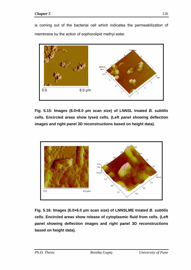
Chapter 5 126
is coming out of the bacterial cell which indicates the permeabilization of
membrane by the action of sophorolipid methyl ester.
Fig. 5.15: Images (8.0×8.0 µm scan size) of LNNSL treated B. subtilis
cells. Encircled areas show lysed cells. (Left panel showing deflection images and right panel 3D reconstructions based on height data).
Fig. 5.16: Images (6.0×6.0 µm scan size) of LNNSLME treated B. subtilis cells. Encircled areas show release of cytoplasmic fluid from cells. (Left panel showing deflection images and right panel 3D reconstructions based on height data).
Ph.D. Thesis Reetika Gupta University of Pune
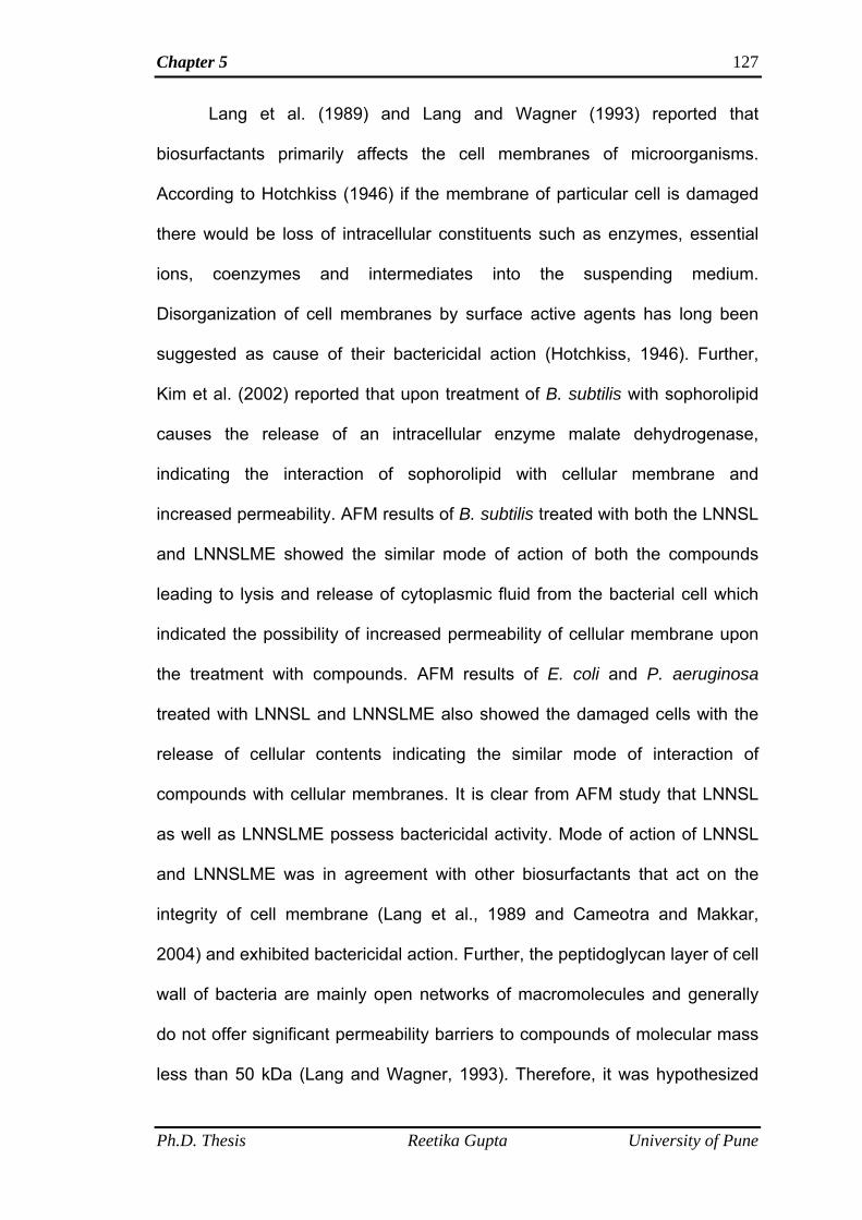
Chapter 5 127
Lang et al. (1989) and Lang and Wagner (1993) reported that
biosurfactants primarily affects the cell membranes of microorganisms.
According to Hotchkiss (1946) if the membrane of particular cell is damaged
there would be loss of intracellular constituents such as enzymes, essential
ions, coenzymes and intermediates into the suspending medium.
Disorganization of cell membranes by surface active agents has long been
suggested as cause of their bactericidal action (Hotchkiss, 1946). Further,
Kim et al. (2002) reported that upon treatment of B. subtilis with sophorolipid
causes the release of an intracellular enzyme malate dehydrogenase,
indicating the interaction of sophorolipid with cellular membrane and
increased permeability. AFM results of B. subtilis treated with both the LNNSL
and LNNSLME showed the similar mode of action of both the compounds
leading to lysis and release of cytoplasmic fluid from the bacterial cell which
indicated the possibility of increased permeability of cellular membrane upon
the treatment with compounds. AFM results of E. coli and P. aeruginosa
treated with LNNSL and LNNSLME also showed the damaged cells with the
release of cellular contents indicating the similar mode of interaction of
compounds with cellular membranes. It is clear from AFM study that LNNSL
as well as LNNSLME possess bactericidal activity. Mode of action of LNNSL
and LNNSLME was in agreement with other biosurfactants that act on the
integrity of cell membrane (Lang et al., 1989 and Cameotra and Makkar,
2004) and exhibited bactericidal action. Further, the peptidoglycan layer of cell
wall of bacteria are mainly open networks of macromolecules and generally
do not offer significant permeability barriers to compounds of molecular mass
less than 50 kDa (Lang and Wagner, 1993). Therefore, it was hypothesized
Ph.D. Thesis Reetika Gupta University of Pune
Chapter 5
Ph.D. Thesis Reetika Gupta University of Pune
128
that sophorolipids due to their amphiphilic nature and molecular mass in the
range of 600-800 Da can make entry into the bacterial cell through both the
lipid bilayer as well as porin channels and at the particular concentration
become bactericidal in nature. In our case minimum inhibitory concentrations
of LNNSL and LNNSLME were found to be bactericidal against both Gram
negative and Gram positive bacteria.
5B.4 Conclusions
There is no clear report on the AFM study of antimicrobial action of
other glycolipids. This is the first report on the AFM study of action of Linolenic
acid derived sophorolipid, LNNSL and its methyl ester form, LNNSLME.
These data confirmed the membranolytic action on cell membranes of Gram
negative and Gram positive bacteria. These compounds showed their
potential as a bactericidal agent and thus can be used as a potential
candidate against bacterial infections.
Summary and Conclusions
130
The work presented in this thesis mainly focuses on the biological
synthesis of novel sophorolipids using Candida bombicola as a producer
organism. Preparation of new sophorolipid (SL) analogues with different
functionalities has widespread use in pharmaceutical and industrial applications.
SL composition can be modified by using both in vivo and in vitro methods.
Different lipophilic substrates have been used by researchers for SL production
such as oleic acid, stearic acid, palmitic acid and different vegetable oils. SL
production using α-linolenic acid as the lipophilic substrate may become a
valuable product of interest of enhanced functionality. This is the first report on
the linolenic acid derived sophorolipids (LNNSL).
The production of LNNSL was carried out under shake flask conditions.
Physiological parameters like effect of pH, temperature, glucose, fatty acid and
yeast extract concentrations were studied and optimized. As these parameters
play an important role in the cellular metabolism and in turn affect the
biosynthesis of sophorolipid. The production of LNNSL is dependent on optimum
supply of hydrophilic and lipophilic substrate (glucose and α-linolenic acid). It was
found out that Candida bombicola is capable of directly incorporating the
Linolenic acid into the lipophilic portion of sophorolipid structure. The yield of
LNNSL from optimized conditions was found to be 3.98 g l-1 and is comparable to
the yields reported for polyunsaturated fatty acid derived sophorolipid. High
production cost can be tolerated for biosurfactants used in low volumes specialty
markets such cosmetics and health care. Therefore the use of pure α-linolenic
acid for production of LNNSL can be beneficial for such application fields.
This study successfully demonstrated the analysis of chemically distinct
forms in the SL mixture produced by Candida bombicola when grown on glucose
Ph.D. Thesis Reetika Gupta University of Pune
Ph.D. Thesis Reetika Gupta University of Pune
131
and α-linolenic acid as well as its conversion into the single homogenous product
by chemical esterification reaction. These novel Sophorolipids were good surface
active and antibacterial agents. They have potential for various applications and
offer the advantages of further modifications and can produce functionalized SLs
according to their applications.
LNNSL and its methyl ester form, LNNSLME showed potential against
both Gram positive and Gram negative bacteria and are more effective against
Gram negative bacteria which is an advantageous feature as compared to other
SLs reported till now which are more effective against Gram positive bacteria.
AFM has proved efficient for investigating the mode of action of LNNSL and
LNNSLME. Their surface tension and critical micelle concentrations are
comparable to the values reported for other commercially available surfactants
and biosurfactants.
Future aspects
This study opened the opportunity for investigating the potential of these
novel SLs as an antifungal and antiviral agent. LNNSL, containing three double
bonds (polyunsaturated) can served as good candidate for self assembly and
polymer formation. The other important feature of LNNSL and its methyl ester
form is that it can serve as a mediator for providing linolenic acid in accessible
form to applications such as cosmetics and food applications etc.
LNNSL and LNNSLME are also the sources of speciality chemicals such
as ω and ω-1 fatty acids which possess beneficial properties and are important
for polymer, perfumes and fragrance industries etc. Linolenic acid derived SLs
may also checked for cell differentiation ability in cancerous cells.
References
References 133
Abalos A, Pinazo A, Infante M R, Casals M, Garcia F and Manresa A (2001)
Physicochemical and antimicrobial properties of new rhamnolipids
produced by Pseudomonas aeruginosa AT10 from soybean oil refinery
wastes. Langmuir 17: 1367-1371
Abe Y, Inoue S and Ishida A (1981) Cosmetic composition of skin and hair
treatment. US patent 427340
Adams J (1990) Charge-remote fragmentations: Analytical applications and
fundamental studies. Mass Spectrom Rev 9:141-186
Akari S and Akari Y (1987) Method of modifying quality of wheat flour product.
Japanese patent 61205449
Albrecht A, Rau U and Wagner F (1996) Initial steps of sophoroselipid
biosynthesis by Candida bombicola ATCC 22214 grown on glucose. Appl
Microbiol Biotechnol 46: 67-73
Allingham R P (1971) Sophoroside esters in prepared food products. US
patent 3622344
Alonso-Galicia M, Falck J R, Reddy K M and Roman R (1999) 20-HETE
agonist and antagonist in the renal circulation. Am J Physiol 277: 790-796
Arima K, Kakinuma A and Tamura G (1968) Surfactin, a crystalline peptide
lipid surfactant produced by Bacillus subtilis: isolation, characterization and
its inhibition of fibrin clot formation. Biochem Biophys Res Commun 31:
488- 494
Ashby R D, Nunez A, Solaiman D K Y and Foglia T A (2005) Sophorolipid
biosynthesis from a biodiesel co-product stream. J Am Oil Chem Soc 82:
625-630
Ph.D. Thesis Reetika Gupta University of Pune
References 134
Ashby R D, Solaiman D K Y and Foglia T A (2008) Property control of
sophorolipids: influence of fatty acid substrate and blending. Biotechnol
Lett 30: 1093-1100
Asmer H J, Lang S, Wagner F and Wray V (1988) Microbial-production,
structure elucidation and bioconversion of sophorose lipids. J Am Oil
Chem Soc 65:1460-1466
Azim A, Shah V, Doncel G F, Peterson N, Gao W and Gross R (2006) Amino
acid conjugated sophorolipids: a new family of biological active
functionalized glycolipids. Bioconjug Chem 17: 1523-1529
Banat I M (1995) Biosurfactants production and possible uses in microbial
enhanced oil recovery and oil pollution remediation: A review. Bioresour
Technol 51: 1-12
Banat I M (1995b) Characterization of biosurfactants and their use in pollution
removal- state of art. Acta Biotechnol 15: 251-267
Barnett J A, Payne R W and Yarrow D (1983) Yeasts: Characteristics and
identification. Cambridge University Press
Bartha R (1986) Biotechnology of petroleum polluted biodegradation.
Microbiol Ecol 12: 155-172
Baviere M, Degouy D and Lecourtier J (1994) Process for washing solid
particles comprising a sophoroside solution. US patent 5326407
Bentley R and Campbell I M (1968) Secondary metabolism of fungi. Comp
Biochem Physiol 20: 415-489
Bernard-Gallon D J, Vissac-Sabatier C, Antoine-Vincent D, Rio P G, Maurizis J
C, Fustier P, Bignon Y J (2002) Differential effects of n-3 and n-6
Ph.D. Thesis Reetika Gupta University of Pune
References 135
polyunsaturated fatty acids on BRCA1 and BRCA2 gene expression in
breast cell lines. Br J Nutr 87(4) 281-289
Bernheimer A W and Avigad L S (1970) Nature and properties of a cytolytic
agent produced by Bacillus subtilis. J Gen Microbiol 61: 361–369
Binnig G, Quate C F and Gerber C (1986) Atomic force microscope. Physical
review Letters 56: 930-933
Bisht K S, Gross R A and Kaplan D L (1999) Enzyme-mediated regioselective
acylations of sophorolipids. J Org Chem 64: 780-789
Bluth M H, Kandil E, Mueller C M, Shah V, Lin Y Y, Zhang H, Drersner L,
Lempert L, Nowakowski M, Gross R, Schulze R and Zenilman M E (2006)
Sophorolipids block lethal effects of septic shock in rats in a cecal ligation
and puncture model of experimental sepsis. Crit Care Med 34: 188-195
Bonomo R A (2000) multiple antibiotic-resistant bacteria in long-term care
facilities: an emerging problem in the practice of infectious diseases. Clin
Infect Dis 31: 1414-1422
Borzeix CF (1999) Use of sophorolipids comprising diacetyl lactones as agent
for stimulating skin fibroblast metabolism. World patent 99/62479
Braga P C and Ricci D (1998) Atomic force microscopy: Application to
investigation of Escherichia coli Morphology before and after the exposure
to Cefodizime. Antimicrob Agents Chemother 42 (1): 18-22
Brakemeier A, Lang S, Wullbrandt D, Merschel L, Benninghoven A,
Buschmann N and Wagner F (1995) Novel sophorose lipids from microbial
conversion of 2-alkanols. Biotechnol Lett 17: 1183-1188
Ph.D. Thesis Reetika Gupta University of Pune
References 136
Brakemeier A, Wullbrandt D, and Lang S (1998) Candida bombicola:
production of novel alkyl glycosides based on glucose/2-dodecanol. Appl
Microbiol Biotechnol 50: 161-166
Breithaupt T B and Light R J (1982) Affinity-chromatography and further
characterization of the glucosyltransferases involved in hydroxy-
docosanoic acid sophoroside production in Candida bogoriensis. J Biol
Chem 257: 9622- 9628
Brenner J Cosmetics & Toiletries, March 2004, pp. 75-79
Britto D V, Kapse H, Babrekar H, Prabhune A A, Bhoraskar S V, Premnath V
and Prasad B L V (2011) Silver nanoparticle studded porous polyethylene
scaffolds: bacteria struggle to grow on them while mammalian cells thrive.
Nanoscale 3: 2957-2963
Brown M J, Robinson J P and Springham D G (1986) Microbial enhanced oil
recovery: progress and prospects. CRC Crit Rev in Biotechnol 3 (2):
159–197
Bruheim P, Bredholt H and Eimhjellen K (1997) Bacterial degradation of
emulsified crude oil and the effect of various surfactants. Can J Microbiol
43 (1): 17-22
Bucholtz M L and Light R J (1976) Acetylation of 13-sophorosyloxydocosanoic
acid by an acetytransferase purified from Candida bogoriensis. J Biol
Chem 251: 424-430
Cameotra S S and Makkar R S (2004) Recent applications of biosurfactants
as biological and immunological molecules. Curr Opi Microbiol 7: 262-
266
Ph.D. Thesis Reetika Gupta University of Pune
References 137
Carlo B P and Ricci D (1998) Atomic force microscopy: Application to
investigation of Escherichia coli morphology before and after exposure to
cefodizime. Antimicrob Agents Chemother 42 (1): 18-22
Casas J A (1996) Production of sophorolipids by Candida bombicola. Ph. D.
thesis. Madrid, Spain: Universidad Complutense
Casas J A and Garcia-Ochoa F (1999) Sophorolipid production by Candida
bombicola: medium composition and culture methods. J Biosci Bioeng 88:
488- 494
Cavalero D A and Cooper D G (2003) The effect of medium composition on the
structure and physical state of sophorolipids produced by Candida
bombicola ATCC 22214. J Biotechnol 103: 31-41
Chen J, Song X, Zhang H, Qu Y B and Miao J Y (2006a) Production, structure
elucidation and anticancer properties of sophorolipid from Wickerhamiella
domercqiae. Enzyme Microb Technol 39: 501-506
Chen J, Song X, Zhang H, Qu Y B and Miao J Y (2006b) Sophorolipid
produced from the new yeast strain Wickerhamiella domercqiae induces
apoptosis in H 7402 human liver cancer cells. Appl Microbiol Biotechnol
72: 52-59
Concaix B (2003) Use of Sophorolipids comprising diacetyl lactones as agent
for stimulating skin fibroblast metabolism. U S Patent 6596265
Connor W E (2000) Importance of n−3 fatty acids in health and disease. Am J
Clin Nutr 71: 171–175
Cooper D G and Paddock D A (1983) Torulopsis petrophilum and surface
activity. Appl Environ Microbiol 46: 1426- 1429
Ph.D. Thesis Reetika Gupta University of Pune
References 138
Cooper D G and Paddock D A (1984) Production of a biosurfactant from
Torulopsis bombicola. Appl Environ Microbiol 47: 173-176
Cooper D G, Zajic J E and Gerson D F (1978) Production of surface active
lipids by Corynebacterium lepus. Appl Environ Microbiol 37: 4-10
Daniel H J, Otto R T, Reuss M and Syldatk C (1998a) Sophorolipid production
with high yields on whey concentrate and rapessed oil without consumption
of lactose. Biotechnol Lett 20: 805-807
Daniel H J, Reuss M and Syldatk C (1998b) Production of sophorolipids in high
concentration from deproteinized whey and rapeseed oil in a two stage fed
batch process using Candida bombicola ATCC 22214 and Cryptococcus
curvatus ATCC 20509. Biotechnol Lett 20: 1153-1156
Daverey A and Pakshirajan K (2009) Production, Characterization, and
Properties of Sophorolipids from the Yeast Candida bombicola using a
Low-cost Fermentative Medium. Appl Biochem Biotechnol 158: 663-674
Daverey A and Pakshirajan K (2010) Kinetics of Growth and Enhanced
Sophorolipids Production by Candida bombicola using a Low-Cost
Fermentative Medium Appl Biochem Biotechnol 160: 2090-2101
Daverey A and Pakshirajan K (2010) Sophorolipids from Candida bombicola
using mixed hydrophilic substrates: Production, purification and
characterization Colloid Surf B: Biointerfaces 79: 246–253
Davila A M, Marchal R and Vandecasteele J P (1992) Kinetics and balance of
fermentation free from product inhibition: sophorose lipid production by
Candida bombicola. Appl Microbiol Biotechnol 38: 6-11
Ph.D. Thesis Reetika Gupta University of Pune
References 139
Davila A M, Marchal R and Vandecasteele J P (1994) Sophorose lipid
production from lipidic precursors: Predictive evaluation of industrial
substrates. J Ind Microbiol 13: 249-257
Davila A M, Marchal R and Vandecasteele J P (1997) Sophorose lipid
fermentation with differentiated substrate supply for growth and production
phases. Appl Microbiol Biotechnol 47: 496-501
Davila A M, Marchal R, Monin N and Vandecasteele J P (1993) Identification
and determination of individual sophorolipids in fermentation products by
gradient elution high-performance liquid chromatography with evaporative
light-scattering detection. J Chromatogr 648: 139-149
Desai J D and Banat I M (1997) Microbial production of surfactants and their
commercial production. Microbiol Mol Biol Rev 61: 47-64
Deshpande M and Daniels L (1995) Evaluation of sophorolipid biosurfactant
production by Candida bombicola using animal fat. Biores Technol 54:
143-150
Develter D and Lauryssen L M L (2010) Properties and industrial applications
of sophorolipids. Eur J Lipid Sci Technol 112: 628- 638
Deziel E, Paquette G, Villemur R, Lepine F and Bisaillon J G (1996)
Biosurfactant production by a soil Pseudomonas strain growing on
polycyclic aromatic hydrocarbons. Appl Environ Microbiol 62: 1908-1912
Ducreux J, Ballerini D, Baviere M, Bocard C and Monin N (1997) Composition
containing a surface active compound and glycolipids and decontamination
process for a porous medium polluted by hydrocarbons. US patent
5654192
Ph.D. Thesis Reetika Gupta University of Pune
References 140
Dufrene Y F, Boonaert C J P, Berin P A and Asther M (1999) J Bacteriol 181:
5350
Eckburg P B, Bik E M, Bernstein C N, Purdom E and Dethlefsen L et al. (2005)
Diversity of the human intestinal microbial flora. Science 308 (5728), 1635-
1638
Escalante B, Falck J R, Yadgiri P, Sun L and Laniado-Schwartzman M (1988)
19(S) Hydroxyeicosatetraenoic acid is a potent stimulator of renal Na+/K+
ATPase. Biochem Biophys Res Commun 152: 1269-1274
Esders T W and Light R J (1972) Glucosyl- and acetyltransferases involved in
the biosynthesis of glycolipids from Candida bogoriensis. J Biol Chem 247:
1375-1386
Fleurackers S J J (2006) On the use of waste frying oil in the synthesis of
sophorolipids. Eur J Lipid Sci Technol 108: 5-12
Futura T, Igarashi K and Hirata Y (2002) Low-foaming detergent compositions.
World patent 03/002700
Gi K E (2004) Red tide inhibiting agent comprising yellow earth and
biodegradable sophorolipid. Korean patent 20040083614
Glenns R N and Cooper D G (2006) Effect of substrate on sophorolipids
properties. J Am Oil Chem Soc 83(2):137-145
Gobbert U, Lang S and Wagner F (1984) Sophorose lipid formation by resting
cells of Torulopsis bombicola. Biotechnol Lett 6: 225-230
Goi H, Watanabe T, Miyauchi K, Kazuno Y and Inouye S (1985) Comparative
bactericidal and morphological effects of five cephamycins on cells of three
Gram-negative bacilli at decreasing drug concentrations. Drugs Exp Clin
Res 11: 771-780
Ph.D. Thesis Reetika Gupta University of Pune
References 141
Gorin A P J, Spencer J F T and Tulloch AP (1961) Hydroxy fatty acid
glycosides of sophorose from Torulopsis magnoliae. Can J Chem 39: 846-
855
Gross R and Shah V (2007) Sophorolipids as protein inducers and inhibitors in
fermentation medium. World patent WO2007073371
Gross R and Shah V (2003) Antimicrobial properties of various forms of
sophorolipids. U. S. Patent Application PCT/US2003/035871
Gross R and Shah V (2004) Antifungal activity of various forms of
sophorolipids. U. S. Patent Application 11/020683
Gross R and Shah V (2007) Anti-herpes virus properties of various forms of
sophorolipids. U. S. Patent Application PCT/US 2007/063701
Gross R and Shah V (2009) Method for neutralizing fungi using sophorolipids
and antifungal sophorolipids for use therein. U. S. Patent Application US
2009/0186836 A1
Gross R, Shah V and Gustavo D (2004) Spermicidal and virucidal properties
of various forms of sophorolipids. U. S. Patent Application 10/804778
Gupta R and Prabhune A A (2012) Structural determination and chemical
esterification of the sophorolipids produced by Candida bombicola grown
on glucose and α-linolenic acid. Biotechnol Lett 34 (4): 701-707
Hall P J, Haverkamp J, Van Kralingen C G and Schmidt M (1996) Laundry
detergent composition containing synergistic combination of sophorose lipid
and nonionic surfactant. US patent 5520839
Hall P J, Haverkamp J, van Kralingen C J and Schmidt M (1995) Synergistic
dual surfactant detergent composition containing sophoroselipid. U.S.
Patent 5,417,879
Ph.D. Thesis Reetika Gupta University of Pune
References 142
Head I M (1998) Bioremediation: towards a credible technology. Microbiol
144: 599-608
Hillion G, Marchal R, Stoltz C and Borzeix CF (1998) Use of a sophorolipid to
provide free radical formation inhibiting activity or elastase inhibiting activity.
US patent 5756471
Hirata Y, Ryu M, Oda Y, Igarashi K, Nagatsuka A, Futura T, et al. (2009) Novel
characteristics of sophorolipids, yeast glycolipid biosurfactants, as
biodegradable low- foaming surfactants. J Biosci Bioeng 108: 142-146
Hommel R K and Huse K (1993) Regulation of sophorose lipid production by
Candida apicola. Biotechnol Lett 15 (8): 853-858
Hommel R K and Ratledge, C. (1993) Biosynthetic mechanisms to low
molecular weight surfactants and their precursor molecules, Biosurfactants:
Production-Properties-Applications, pp. 3-63, Marcel Dekker, New York.
Hommel R K, Weber L, Weiss A, Himmelreich U, Rilke O, Kleber H P (1994)
Production of sophorose lipid by Candida (Torulopsis) apicola grown on
glucose. J Biotechnol 33: 147-155
Hommel R, Stuwer O, Stuber W, Haferburg D and Kleber H P (1987)
Production of water-soluble surface-active exolipids by Torulopsis apicola.
Appl Microbiol Biotechnol 26: 199–205
Hotchkiss R D (1946) The nature of the bactericidal action of surface-active
agents. In: Surface Active Agents (Ed. by R.W. Miner & E. I. Valko), pp.
479-492. Annals of the New york academy of sciences, New York
Hu Y and Ju L K (2001) Sophorolipid production from different lipid precursors
observed with LC-MS. Enzyme Microb Technol 29: 593-601
Ph.D. Thesis Reetika Gupta University of Pune
References 143
Inoue S (1988) Biosurfactants in cosmetic applications. In: Proceedings. World
Conference on Biotechnology for fats and Oil industry, T. H., Applewhite,
ed., pp 206-209, Am Oil Chem Soc, Champaign, III, USA
Inoue S and Itoh S (1982) Sophorolipids from Torulopsis bombicola as
microbial surfactants in alkane fermentation. Biotechnol Lett 4: 308-312
Inoue S and Miyamoto N (1980) Process for producing a hydroxy fatty acid
ester. US patent 4201844
Inoue S, Kimura Y and Kinta M (1980) Process for producing a glycolipid ester.
U.S. Patent 4,215,213
Isoda H, Kitamoto D, Shinmoto H, Matsumura M and Nakahara T (1997)
Microbial extracellular glycolipid induction of differentiation and inhibition of
the protein kinase C activity of human promyelocytic leukemia cell line
HL60. Biosci Biotechnol Biochem 61: 609-614
Ito S, Kinta M and Inoue S (1980) Growth of yeasts on alkanes: inhibition by a
lactonic sophorolipid produced by Torulopsis bombicola. Agric Biol Chem
44: 2221-2223
Itoh A, Honda H, Tomita F and Suzuki T (1971) Rhamnolipids produced by
Pseudomonas aeruginosa grown on n-paraffin. J Antibiot 24: 855–859
Iwaura R, Yoshida K, Masuda M, Yase K, Shimizu T (2002) Spontaneous Fiber
Formation and Hydrogelation of Nucleotide Bolaamphiphiles. Chem Mater
14, 3047-3053
Jarvis F G and Johnson M J (1949) A glycolipid produced by Pseudomonas
aeruginosa. J Am Chem Soc 71: 4124- 4126
Jones D F (1968) Microbiological oxidation of long-chain aliphatic compounds.
Part V. Mechanism of hydroxylation. J Chem Soc (C): 2827- 2833
Ph.D. Thesis Reetika Gupta University of Pune
References 144
Joshi-Navare K, Shiras A and Prabhune A (2011) Differentiation-inducing
ability of sophorolipids of oleic and linoleic acids using a glioma cell line.
Biotechnol J 6(5):509-512
Kachholtz T and Schlingmann M (1987) Possible food and agricultural
applications of microbial surfactants: an assessment. In Biosurfactants
and Biotechnology, eds N Kosaric , W L Cairns and N C C Gary. Marcel
dekker, New York, 183-210
Kaelynn W (2009) Biosurfactants for cosmetic application: Overcoming
production challenges. MMG 445 Basic Biotechnol 5: 78-83
Kappeli O (1986) Cytochromes P-450 of yeasts. Microbiol Rev 50: 244-258
Kasture M B, Patel P, Prabhune A A, Ramana C V, Kulkarni A A, Prasad B L V
(2008) Synthesis of silver nanoparticles by sophorolipids: effect of
temperature and sophorolipid structure on the size of particles. J Chem Sci
120: 515- 520
Kasture M B, Singh S, Patel P, Joy P A, Prabhune A A, Ramana C V, et al.
(2007) Multiutility sophorolipids as nanoparticle capping agents: synthesis
of stable and water dispersible Co nanoparticles. Langmuir 23: 11409-
11412
Kawano J, Suzuki T, Inoue S, Hayashi S (1981a) Powdered compressed
cosmetic material. US patent 4305931
Kawano J, Suzuki T, Inoue S, Hayashi S (1981b) Stick-shaped cosmetic
material. US patent 4305929
Kim H S, Kim Y B, Lee B S and Kim E K (2005) Sophorolipid production by
Candida bombicola ATCC 22214 from a corn-oil processing byproduct. J
Microbiol Biotechnol 15: 55- 58
Ph.D. Thesis Reetika Gupta University of Pune
References 145
Kim S Y, Oh D K, Lee K H and Kim J H (1997) Effect of soybean oil and
glucose on sophorose lipid fermentation by Torulopsis bombicola in
continuous culture. Appl Microbiol Biotechnol 48: 23–26
Kim, Kapjung, Yoo D, Kim Y, Lee B, Shin D, Kim EK (2002) Characteristics of
sophorolipid as an antimicrobial agent. J Microbiol Biotechnol 12 (2): 235-
241
Klainer A S and Perkins P L (1971) Surface-effects of cell-wall active
antimicrobial agents. Scanning Electron Microsc (Part 1) 1971: 329-336
Klekner V and Kosaric N (1993) Biosurfactant for cosmetic. In: Kosaric N,
editor. Biosurfactant: production, properties, applications. New York:Marcel
Dekker, p. 65-99
Klekner V, Kosaric N and Zhou Q H (1991) Sophorose lipids produced from
sucrose. Biotechnol Lett 13: 345-348
Kogiso M, Ohnishi S, Yase K, Masuda M, Shimizu T (1998) Dicarboxylic
Oligopeptide Bolaamphiphiles: Proton-Triggered Self-Assembly of
Microtubes with Loose Solid Surfaces. Langmuir 14: 4978-4986
Koster C G, Heerma W, Pepermans H A M, Groenevewegen A, Peters H and
Haverkamp J (1995) Tandem mass spectrometry and nuclear magnetic
resonance spectroscopy studies of Candida bombicola, sophorolipids and
product formed on hydrolysis by cutinase. Anal Biochem 230: 135-148
Kris-Etherton P M, Harris W S and Lawrence J (2002) Appel , for the Nutrition
Committee "Fish consumption, fish oil, omega-3 fatty acids, and
cardiovascular disease". Circulation 106 (21): 2747-2757
Ph.D. Thesis Reetika Gupta University of Pune
References 146
Lang S and Wagner F (1993) Biological activities of biosurfactants. In:
Kosaric, N. (Ed.), Biosurfactants: Production, Properties, Application.
Marcel dekker, New York, pp. 251-268
Lang S, Katsiwela E and Wagner F (1989) Antimicrobial effects of
biosurfactants. Fett Wiss Technol Fat Sci Technol 91: 363-366
Lasker J M, Chen W B, Wolf I, Bloswick B P, Wilson P D and Powell P K
(2000) Formation of 20-hydroxyeicosatetraenoic acid, a vasoactive and
natriuretic eicosanoid, in human kidney. J Biol Chem 275: 4118-4126
Lee K H and Kim J H (1993) Distribution of substrates carbon in sophorose
lipid production by Torulopsis bombicola. Biotechnol Lett 15 (3): 263-266
Li Q, Mittal R, Huang L, Travis B and Sanders C R (2009) Bolaamphiphile
class surfactants can stabilize and support the function of solubilized
integral membrane proteins. Biochemistry 48: 11606–11608
Linton J D (1991) Metabolite production and growth efficiency. Ant van Leeuw
60: 3-4
Lo C M and Ju L K (2009) Sophorolipids-induced cellulose production in
cocultures of Hypocrea jecorina Rut C 30 and Candida bombicola. Enzyme
Microb Technol 44: 107- 111
Lourith N and Kanlayavattanakul M (2009) Natural surfactants used in
cosmetics: glycolipids. Int J Cosmet Sci 31: 225- 261
Magar H, Rothlisberger R and Wzgner F (1987) Use of sophorose-lipid
lactone for the treatment of dandruffs and body odor. European patent
0209783
Maingault M (1997) Pharmaceutical and cosmetic compositions containing
sophorolipids. Canadian Patent CAN 126,242874
Ph.D. Thesis Reetika Gupta University of Pune
References 147
Maingault M (1999) Utilization of sophorolipids as therapeutically active
substances or cosmetic products, in particular for the treatment of the skin.
US patent 5981497
Makkar R S, Cameotra S S and Banat I M (2011) Advances in utilization of
renewable substrates for biosurfactant production. AMB Express 1 (5): 1-
19
Marahiel M A, Nakano M M and Zuber P (1993) Regulation of peptide
antibiotic production in Bacillus. Mol Microbiol 7: 631–636
Marchal R, Lemal J, Sulzer C and Davila A M (1999) Production of
sophorolipid acetate acids from oils or esters. US patent 5900366
Masaru K, Takashi N, Yoji A, Kazuo N, Tatsu N, Sumiko T and Jotaro N (2001)
Composition for high-density cold storage transportation. Japanese patent
2001131538
Masuda M, Hanada T, Okada Y, Yase K, Shimizu T (2000) Polymerization in
Nanometer-Sized Fibers: Molecular Packing Order and Polymerizability
Macromolecules 33: 9233-9238
Miller R M (1995) Biosurfactant-facilitated remediation of metal contaminated
soils. Environ Health Perspect 103: 59-62
Muller-Hurtig R, Blaszczyk R, Wagner F and Kosaric N (1993) Biosurfactants
for environmental control. In Biosurfactants, ed. N Kosaric. Marcel
Dekker, New York, 447–469
Mulligan C N (2005) Environmental applications for biosurfactants. Environ
Pollut 133: 183- 198
Mulligan C N, Yong R N and Gibbs B F (2001) Heavy metal removal from
sediments by biosurfactants. J Hazard Mater 85: 111-125
Ph.D. Thesis Reetika Gupta University of Pune
References 148
Nakao M, Kondo M and Tsuchiya K (1981) Light and electron microscopy
response of Escherichia coli and Serratia marcescens to cefmenoxime
(Sce-1365), a new broad spectrum cephalosporin. J Antibiot 34: 1046-
1054
Napolitano L M (2006) Sophorolipids in sepsis: Antiinflammatory or
antibacterial? Crit Care Med 34: 258- 259
Neu T R (1996) Significance of bacterial surface-active compounds in
interaction of bacteria with interfaces. Microbiol Rev 60: 151–166
Nguyen T T L, Edelen A, Neighbors B and Sabatini D A (2010) Biocompatible
lecithin- based microemulsions with rhamnolipid and sophorolipid
biosurfactants: formulation and potential applications. J Colloid Interface
Sci 348: 498-504
Nishino T and Nakazawa S (1973) Cephalexin induced morphological
alterations in the surface structures of Staphylococcus aureus and
Escherichia coli demonstrated by scanning electron microscopy. Jpn J
Microbiol 17: 383-392
Nitschke M and Coast S G (2007) Biosurfactants in food industry. Trends
Food Sci Technol 18: 252-259
Nunez A, Ashby R, Foglia T A and Solaiman D K Y (2001) Analysis and
characterization of sophorolipids by liquid chromatography with
atmospheric pressure chemical ionization. Chromatographia 53: 673-677
Nunez A, Ashby R, Foglia T A and Solaiman D K Y (2004) LC/MS Analysis and
Lipase modification of the Sophorolipids produced by Rhodotorula
bogoriensis. Ibid. 26: 1087-1093
Ph.D. Thesis Reetika Gupta University of Pune

References 149
Ochsner U A, Hembach T and Fiechter A (1995) Production of rhamnolipid
biosurfactants. Adv Biochem Engin/Biotechnol 53, 89-118
Oliw E H, Lawson J H, Brash A R and Oates J A (1981) Arachidonic acid
metabolism in rabbit renal cortex. J Biol Chem 256: 9924-9931
Otto R T, Daniel H J, Pekin G, Muller-Decker K, Furstenberger G, Reuss M,
and Syldatk C (1999) Production of sophorolipids from whey. II. Product
composition, surface active properties, cytotoxicity and stability against
hydrolases by enzymatic treatment. Appl Microbiol Biotechnol 52: 495-
501
Perkin G, Vardar-Sukan F and Kosaric N (2005) Production of sophorolipids
from Candida bombicola ATCC 22214 using Turkish corn oil and honey.
Eng Life Sci 5: 357-362
Pesce L (2002) A biotechnological method for the regeneration of
hydrocarbons from dregs and muds, on the base of biosurfactants. World
patent 02/062495
Pierce D and Heilman T J (1998) Germicidal composition. World patent
98/16192
Prabhune A, Fox S R and Ratledge C (2002) Transformation of arachidonic
acid to 19- hydroxy and 20-hydroxy eicosatetraenoic acid using Candida
bombicola. Biotechnol Lett 24: 1041-1044
Rai A, Prabhune A and Perry C C (2010) Antibiotic mediated synthesis of gold
nanoparticles with potent antimicrobial activity and their application in
antimicrobial coatings. J Mater Chem 20: 6789-6798
Ph.D. Thesis Reetika Gupta University of Pune
References 150
Ratledge C and Evans C T (1989) In Yeasts: Metabolism and Physiology of
Yeasts, Vol. 3, edited by A. H. Rose, and J. S. Harrison Academic Press,
London, England, pp. 367-455
Rau U, Hammen S, Heckmann R, Wray V and Lang S (2001) Sophorolipids: a
source for novel compounds. Ind Crops Prod 13: 85-92 (plastics and
flavours)
Rau U, Heckmann R, Wray V and Lang S (1999) Enzymatic conversion of a
sophorolipid into a glucose lipid. Biotechnol Lett 21: 973-977
Rau U, Manzke C and Wagner F (1996) Influence of substrate supply on the
production of sophorose lipids by Candida bombicola ATCC 22214.
Biotechnol Lett 18: 149-154
Rau U, Manzke C and Wagner F (1996) Influence of substrate supply on the
production of sophorose lipids by Candida bombicola ATCC 22214.
Biotechnol Lett 18 (2): 149- 154
Rodrigues L, Banat I M, Teixeira J, Oliveira R (2006) Biosurfactants: potential
applications in medicine. J Antimicrob Chemother 57: 609-618
Rosa C A and Lachance M A (1998) The yeast genus Starmerella gen. no. and
Starmerella bombicola sp. nov., the teleomorph of Candida bombicola
(Spencer, Gorin & Tulloch) Meyer Yarrow. Int J Syst Bacteriol 48: 1413-
1471
Rosen M J (1978) Surfactants and interfacial phenomena. John Wiley and
Sons, New York
Rosenberg E and Ron E Z (1999) High and low molecular mass microbial
surfactants. Appl Microbiol Biotechnol 52: 154-162
Ph.D. Thesis Reetika Gupta University of Pune
References 151
Saerens K M J, Roelants S L K W, Van Bogaert I N A and Soetaert W (2011a)
Identification of the UDP-glucosyltransferase gene UGTA1, responsible for
the first glucosylation step in the sophorolipids biosynthetic pathway of
Candida bombicola ATCC 22214. FEMS Yeast Res 11: 123-132
Saerens K M J, Zhang J, Saey L, Van Bogaert I N A and Soetaert W (2011b)
Cloning and functional characterization of the UDP-glucosyltransferase
UGT B1 involved in sophorolipid production by Candida bombicola and
creation of a glucolipid producing yeast strain. Yeast, DOI
10.1002/yea.1838
Sanchez M, Aranda F J, Teruel J A, Espuny M J, Marques A, Manresa A, Ortiz
A (2010) Permeabilization of biological and artificial membranes by a
bacterial dirhamnolipid produced by Pseudomonas aeruginosa. J Colloid
Interface Sci 341: 240–247
Schippers C, Gebner K, Muller T, Scheper T (2000) Microbial degradation of
phenanthrene by addition of sophorolipid mixture. J Biotechnol 83, 189-
198
Scholz C, Mehta S, Bisht K, Guilmanov V, Kaplan D, Nicolosi R, Gross R
(1998) Bioactivity of extracellular glycolpids: Investigation of potential anti-
cancer activity of sophorolipids and sophorolipid derivatives. Proc Am
Chem Soc Polym Preprints 39, 168-169
Shah S and Prabhune A (2007) Purification by silica gel chromatography using
dialysis tubing and characterization of sophorolipids produced from
Candida bombicola grown on glucose and arachidonic acid Biotechnol
Lett 29: 267-272
Ph.D. Thesis Reetika Gupta University of Pune
References 152
Shah V, Badia D and Ratsep P (2007) Sophorolipids having enhanced
antibacterial activity. Antimicrob Agents Chemother 51 (1): 397-400
Shah V, Doncel G F, Seyoum T, Eaton K M, Zalenskaya I, Hagver R, Azim A
and Gross R (2005) Sophorolipids, microbial glycolipids with anti-human
immunodeficiency virus and sperm-immobilizing activities. Antimicrob
Agents Chemother 49: 4093-4100
Shah V, Jurjevic M and Badia D (2007) Utilization of restaurant waste oil as a
precursor for sophorolipid production. Biotechnol Prog 23, 512-515
Shete A M, Wadhawa G, Banat I M and Chopade B A (2006) Mapping of
patents on bioemulsifier and biosurfactant: A review. J Sci Ind Res (India)
65: 91-115
Singh S, D’ Britto V, Prabhune A A, Ramana C V, Dhawan A, Prasad B L V
(2010) Cytotoxic and genotoxic assessment of glycolipid- reduced and -
capped gold and silver nanoparticles. New J Chem 34: 294- 301
Singh S, Patel P, Jaiswal S, Prabhune A A, Ramana C V and Prasad B L V
(2009) A direct method for the preparation of glycolipid-metal nanoparticle
conjugates: sophorolipids as reducing and capping agents for the synthesis
of water re-dispersible silver nanoparticles and their antibacterial activity.
New J Chem 33: 646- 652
Sleiman J N, Kohlhoff S A, Roblin P M, Wallner S, Gross R, Hammerschlag M
R, Zenilman M E, Bluth M H (2009) Sophorolipids as antibacterial agents.
Ann Clin Lab Sci 39 (1): 60-63
Solaiman D K Y (2005) Applications of microbial biosurfactants. Inform 16,
408-410
Ph.D. Thesis Reetika Gupta University of Pune
References 153
Solaiman D K Y, Ashby R D, Nunez A and Foglia T A (2004) Production of
sophorolipids by Candida bombicola grown on soy molasses as substrate.
Biotechnol Lett 26: 1241-1245
Solaiman D K Y, Ashby R D, Zerkowski J A and Foglia T A (2007) Simplified
soy molasses-based medium for reduced-cost production of sophorolipids
by Candida bombicola. Biotechnol Lett 29: 1341-1347
Soliance (2004) Product guide-sopholiance: sophorolipids specific antibacterial
biological agent
Spencer J F T, Gorin P A J and Tulloch A P (1970) Torulopsis bombicola sp.
n. Antonie Van Leeuwenhoek 36: 129–133
Stodola F H, Deinema M H and Spencer J F T (1967) Extracellular lipids of
yeasts. Bacteriol Rev 31:194-213
Sun X X, Choi J K and Kim E K (2004) A preliminary study on the mechanism
of harmful algal bloom mitigation by use of sophorolipid treatment. J Exp
Mar Biol Ecol 304: 35-49
Tomer K B, Crow F W and Gross M L (1983) Location of double bond position
in unsaturated fatty acids by negative ion MS/MS. J Am Chem Soc 105:
5487-5488
Tsuge K, Ano T and Shoda M (1996) Isolation of a gene essential for
biosynthesis of the lipopeptide antibiotics plipastatin B1 and surfactin in
Bacillus subtilis YB8. Arch Microbiol 165: 243–251
Tulloch A P and Spencer J F T (1968) Fermentation of long chain compounds
by Torulopsis apicola. IV. Products from esters and hydrocarbons with 14
and15 carbon atoms and from methyl palmitoleate. Can J Chem 46: 1523-
1528
Ph.D. Thesis Reetika Gupta University of Pune
References 154
Tulloch A P, Hill A and Spencer J F T (1967) A new type of macrocyclic
lactone from Torulopsis apicola. J Chem Soc Chem Commun 7: 584-586
Tulloch A P, Hill A and Spencer J F T (1968) Structure and reactions of
lactonic and acidic sophorosides of 17-hydroxyoctadecanoic acid. Can J
Chem 46: 3337–3351
Tulloch A P, Spencer J F T and Gorin P A J (1962) The fermentation of long
chain compounds by Torulopsis magnoliae. Can J Chem 40: 1326 -1338
Van Bogaert I N A, Demey M, Develter D, Soetaert W and Vandamme E J
(2009) Importance of the cytochrome P450 monooxygenase CYP52 family
for the sophorolipid producing yeast Candida bombicola. FEMS Yeast Res
9: 87-94
Van Bogaert I N A, Groeneboer S, Saerens K, Soetaert W (2010) The role of
cytochrome P450 monooxygenases in microbial fatty acid metabolism.
FEBS J 278: 206-221
Van Bogaert I N A, Saerens K, De Muynck C, Develter D, Soetaert W and
Vandamme E J (2007) Microbial production and application of
sophorolipids. Appl Microbiol Biotechnol 76: 23-34
Van Bogaert I N A, Zhang J and Soetaert W (2011) Microbial synthesis of
sophorolipids. Process Biochem 46: 821-833
Van Bogaert I, Fleurackers S, Van Kerrebroeck S, Develter D, Soetaert W
(2011) Production of new-to-nature sophorolipids by cultivating the yeast
Candida bombicola on unconventional hydrophobic substrates. Biotechnol
Bioeng 108 (4): 734-741
Ph.D. Thesis Reetika Gupta University of Pune
References 155
Vogt R L and Dippold L (2005) Escherichia coli O157:H7 outbreak associated
with consumption of ground beef, June–July 2002. Public Health Rep 120
(2), 174–178
Volkering F, Breure A and Rulkens W (1997) Microbiological aspects of
surfactant use for biological soil remediation. Biodegradation 8: 401- 417
Weber L, Doge C, Haufe G (1992) Oxygenation of hexadecane in the
biosynthesis of cyclic glycolipids in Torulopsis apicola. Biocatalysis 5(4):
267-272
Weber L, Stach J, Haufe G, Hommel R and Kleber H P (1990) Elucidation of
the structure of an unusual cyclic glycolipid from Torulopsis apicola.
Carbohydr Res 206: 13-19
Whang L, Liu P G, Ma C, Cheng S (2008) Applications of biosurfactants,
rhamnolipid and surfactin, for enhanced biodegradation of diesel
contaminated water and soil. J Hazardous Materials 151: 155-163
Yoo D S, Lee B S and Kim E K (2005) Characteristics of microbial
biosurfactant as an antifungal agent against plant pathogenic fungus. J
Microbiol Biotechnol 15:1164–1169
Yoshihiko H, Mizuyuki R, Yuko O, Keisuke I, Asami N, Taro F and Masaki S
(2009) Novel characteristics of sophorolipids, yeast glycolipid biosurfactants
as biodegradable low- foaming surfactants. J Biosci Bioeng 108: 142-146
Yuan B, Yang S, Chen J (2011) Antimicrobial activity of sophorolipids on
pathogenic fungi isolated from fruits. Chin J Appl Environ Biol 17 (3):
330-333
Ph.D. Thesis Reetika Gupta University of Pune
References
Ph.D. Thesis Reetika Gupta University of Pune
156
Zhang L, Somasundaran P, Singh S K, Felse A P, Gross R (2004) Synthesis
and interfacial properties of sophorolipid derivatives. Colloids and
Surfaces A: Physicochem Eng Aspects 240: 75–82
Zhang Y and Miller R M (1994) Effect of a Pseudomonas rhamnolipid
biosurfactant on cell hydrophobicity and biodegradation of octadecane.
Appl Environ Microbiol 60: 2101–2106
Zhang Y and Miller R M (1995) Effect of rhamnolipid (biosurfactant) structure
on solubilization and biodegradation of n-alkanes. Appl Environ Microbiol
61: 2247–2251
Zhou Q H and Kosaric N (1993) Effect of lactose and olive oil on intracellular
and extracellular lipids of Torulopsis bombicola. Biotechnol Lett 15: 477-
482
Zhou Q H and Kosaric N (1995) Utilization of canola oil and lactose to produce
biosurfactants with Candida bombicola. J Am Oil Chem Soc 72: 67- 71
Zhou Q H, Kleckner N and Kosaric N (1992) Production of sophorose lipids by
Torulopsis bombicola from safflower oil and glucose. J Am Oil Chem Soc
69: 89-91
Zhou S, Xu C, Wang J, Gao W, Akhverdiyeva R, Shah V and Gross R (2004)
Supramolecular assemblies of a naturally derived sophorolipid. Langmuir
20: 7926-7932
157
PUBLICATIONS
Structural determination and chemical esterification of the sophorolipids
produced by Candida bombicola grown on glucose and α-linolenic acid.
Reetika Gupta and Asmita A. Prabhune. Biotechnol Lett (2012) 34: 701–
707
Antibacterial properties of linolenic sophorolipid and its chemically
esterified methyl ester form. Reetika Gupta, Uma S. Kumar and Asmita A.
Prabhune (Communicated)
Insight into the antibacterial action of Linolenic acid derived sophorolipid
and its methyl ester form with the help of Atomic Force Microscopy.
Reetika Gupta and Asmita A. Prabhune (Communicated)
Optimiztion of fermentation parameters for production of Linolenic acid
derived sophorolipids using Candida bombicola ATCC 22214. Reetika
Gupta and Asmita A. Prabhune (Manuscript under preparation)
Posters presented
Synthesis of Linolenic sophorolipid using Candida bombicola:
chemical esterification, characterization. Oral presentation in Young
research Conference, Institute of Chemical Technology, Mumbai, 14-16
Jan. 2010.
Antimicrobial property of newly synthesized sophorolipid methyl
ester (17-[2’ –O-β- D-glucopyranosyl- β- D- glucopyranosyl]- oxy)-
cis,cis,cis-9,12,15-octadecatrienoate). Poster presentation in Young
Ph.D. Thesis Reetika Gupta University of Pune
Ph.D. Thesis Reetika Gupta University of Pune
158
research Conference, Institute of Chemical Technology, Mumbai, 14-16
Jan. 2010.
Synthesis of Linolenic sophorolipid using Candida bombicola:
chemical esterification, characterization and antimicrobial activity.
Poster presentation in "The Society of Biological Chemists (India),
Bangalore". December 13-15, 2010.
Antimicrobial property of newly synthesized sophorolipid methyl
ester (17-([2’-O-β-D-glucopyranosyl-β-D-glucopyranosyl]-oxy)-
cis,cis,cis-9,12,15-octadecatrienoate). Poster presentation in Science
Day at National Chemical Laboratory, India (March, 2010)
![PARIPEX - INDIAN JOURNAL OF RESEARCH | Volume-8 | …...The less commonly identified species are Candida tropcalis, Candida glabrata, Candida parapsilosis, and Candida krusei [5].Identification](https://img.pdfslide.us/doc/110x75/60d53496ab798671291c20a1/paripex-indian-journal-of-research-volume-8-the-less-commonly-identified.jpg)