Embed Size (px)
Citation preview

i
BIOSYNTHESIS AND FUNCTIONAL ANALYSIS OF THE COR-LIKE
METABOLITES PRODUCED BY THE COMMON SCAB PATHOGEN
STREPTOMYCES SCABIES
by
© Yuting Li
A thesis submitted to the School of Graduate Studies
in partial fulfillment of the requirements for the degree of
Master of Science
Department of Biology
Memorial University of Newfoundland
May, 2015
St. John’s Newfoundland and Labrador
ii
Abstract
Streptomyces scabies is a Gram-positive soil bacterium that causes common scab
disease, which is identified by the round corky lesions that form on the surface of root
and tuber crops such as potatoes. Virulence factors that contribute to the plant pathogenic
phenotype of S. scabies include the phytotoxic secondary metabolite thaxtomin A and the
secreted necrogenic protein Nec1. In addition, S. scabies produces a family of secondary
metabolites called the COR-like metabolites, which are structurally similar to the COR
(coronatine) phytotoxin produced by the bacterial plant pathogen Pseudomonas syringae.
The goal of this thesis research was to characterize the biosynthesis and function of the S.
scabies COR-like metabolites. In the first research chapter, the role of three genes,
scab79711, cfa8 and scab79691, in metabolite biosynthesis was elucidated by
constructing gene deletion mutants in S. scabies and assessing the effect of each deletion
on the production of the COR-like metabolites. In the second research chapter, the
bioactivity of the COR-like metabolites was investigated by testing S. scabies culture
supernatants or extracts containing the metabolites in various plant bioassays. In addition,
the bioactivity of the primary COR-like metabolite, coronafacoyl-L-isoleucine, was tested
alongside equimolar amounts of COR in order to compare the relative toxicity of the two
metabolites. The results of this study provide important insight into the biosynthetic
pathway responsible for COR-like metabolite production in S. scabies as well as the role
of the metabolites in S. scabies plant pathogenicity. Future directions for this research
were discussed.

iii
Acknowledgements
I would like to acknowledge my supervisor Dr. Dawn Bignell for her support and
guidance. I would also like to thank my supervisory committee members, Dr. Kapil
Tahlan and Dr. Andrei Igamberdiev, for giving me their time and suggestions for my
research. I would also like to acknowledge the Bignell and Tahlan lab members for their
advices and academic support: Dr. Joanna Fyans, Zhenlong Cheng, Mead Altowairish,
Luke Brown, Marcus Moore, Kelcey King, Nicole Ferguson and Milka Podder.

iv
Table of Contents
Abstract……………………………………………………………………………………ii
Acknowledgements…………………………………………………………………..…...iii
Table of Contents………………………………………………………………………....iv
List of Figures…………………………………………………………………………...viii
List of Tables……………………………………………………………………………..xi
List of Symbols, Nomenclature and Abbreviations………………………………….…xii
CHAPTER 1: Introduction and Overview……………………...........................................1
1.1 General features of Streptomyces ……….……………………...……………..1
1.2 The Streptomyces life cycle…………………....………………………………1
1.3 Secondary metabolism in the genus Streptomyces…………………..………..2
1.4 Plant pathogenicity in the genus Streptomyces…………………………….….4
1.5 Control strategies for potato scab disease…………………………………......5
1.6 Virulence factors produced by scab-causing Streptomyces species………...…6
1.7 The S. scabies COR-like metabolites…………….…...…………………….....8
1.8 Biosynthesis of COR and COR-like molecules in P. syringae………………10
1.9 Biosynthesis of COR-like metabolites in S. scabies …………………….…..12
1.10 Biological activities of COR and COR-like molecules………………….…14
1.11 Thesis objectives……………………………………………………..……..15
1.12 References………………………………………………………………......16
Co-Authorship Statement………………………………………………………………...35
CHAPTER 2: Characterizing the Role of scab79691, scab79711 and cfa8 in the
Biosynthesis of the Streptomyces scabies COR-like Metabolites…………………….…36

v
2.1 Introduction…………………………………..………………………………36
2.2 Materials and methods….…………………………………………………….38
2.2.1 Bacterial strains, culturing conditions and maintenance………..…38
2.2.1.1 Escherichia coli strains………………………………..…38
2.2.1.2 Streptomyces scabies strains……………………………..39
2.2.2 DNA manipulations…………………………………….………..…41
2.2.3 PCR………………………………………………………………...42
2.2.3.1 Generation of the hyg + oriT extended resistance
cassettes…………………………………………………………..42
2.2.3.2 Verification of constructed mutant cosmids and strains…43
2.2.3.3 Amplification of the scab79691 and cfa8 genes…………44
2.2.4 Construction of the S. scabies gene deletion mutants…………...…44
2.2.5 Chemical extraction of the COR-like metabolites…………………45
2.2.5.1 Small-scale extractions for HPLC analysis………………45
2.2.5.2 Large-scale extractions for potato bioassays………..……46
2.2.6 HPLC analysis of COR-like metabolite production……..…………46
2.2.7 LC-MS analysis…………..………………………………………...47
2.2.8 Potato tuber slice bioassay…………………………………………48
2.2.9 Complementation of the cfa8 and scab79691 mutants………….49
2.2.10 Bioinformatics analyses………………………………..…………49
2.3 Results……………………………………………………………………..…50
2.3.1 Bioinformatics analysis of Scab79711, Cfa8 and Scab79691……..50

vi
2.3.1.1 Scab79711 and Cfa8 homologues in the database…….…50
2.3.1.2 Scab79691 homologues in the database………………….51
2.3.2 Construction of the S. scabies gene deletion mutants………….….52
2.3.3 The S. scabies deletion mutants differ in their ability to produce the
CFA-L-Ile COR-like metabolite………………………………………….53
2.3.4 Culture extracts from the scab79691 mutant display reduced
bioactivity on potato tuber tissue………………………………………...54
2.3.5 Genetic complementation of the Δcfa8 and Δscab79691 mutants…54
2.4 Discussion……………………………………………………………………56
2.4.1 scab79711 and cfa8 are dispensable for COR-like metabolite
biosynthesis………………………………………………………………56
2.4.2 scab79691 is required for COR-like metabolite biosynthesis…….59
2.4.3 Proposed role of Scab79691 in COR-like metabolite biosynthesis..61
2.5 References……………………………………………………………………62
CHAPTER 3: Characterizing the Biological Activity of the Streptomyces scabies COR-
like Metabolites………………………………………………………………………....100
3.1 Introduction……………………………………………………………....…100
3.2 Materials and methods………………………………………………………103
3.2.1 Bacterial strains, culturing conditions and maintenance………….103
3.2.2 Chemical extraction of the COR-like metabolites………………..103
3.2.3 Plant bioassays……………………………………………………103
3.2.3.1 Leaf infiltration bioassay using culture supernatants…..103
vii
3.2.3.2 Radish seedling bioassay using culture supernatants or
organic culture extracts…………………………………………104
3.2.3.3 Radish seedling bioassay using pure CFA-L-Ile………..106
3.2.3.4 Analysis of anthocyanin production in radish seedlings..106
3.2.3.5 Potato tuber slice bioassay using pure CFA-L-Ile………107
3.3 Results………………………………………………………………………107
3.3.1 The S. scabies COR-like metabolites are associated with necrosis of
N. benthamiana leaf tissue……………………………………………...107
3.3.2 The S. scabies COR-like metabolites can cause stunting of radish
seedlings……………………………………………………………...…108
3.3.3 Analysis of anthocyanin production in response to the COR-like
metabolites……………………………………………………………...110
3.3.4 The S. scabies CFA-L-Ile COR-like metabolite causes hypertrophy of
potato tuber tissue………………………………………………….……111
3.4 Discussion…………………………………………………………………..111
3.5 References…………………………………………………………………..115
CHAPTER 4: Concluding Remarks ……………………………………………………129
4.1 Summary and future directions…………………………………..….……....129
4.2 References……………………...………………………………………...….131

viii
List of Figures
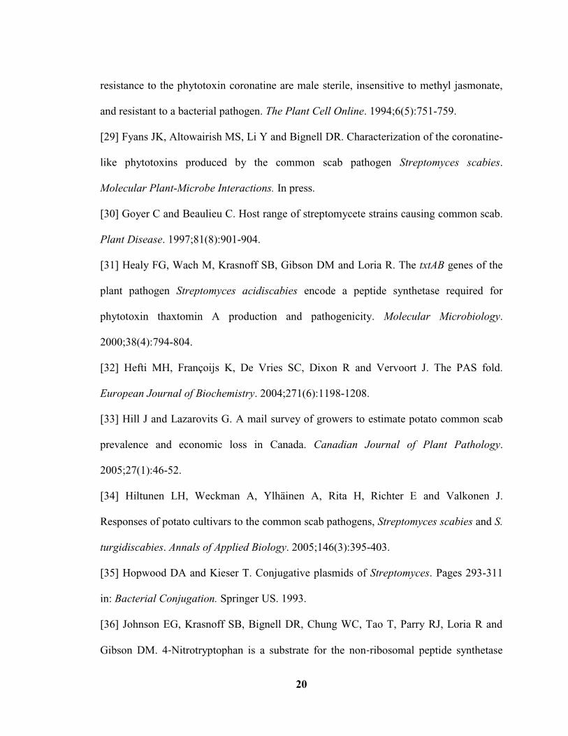
Figure 1.1: The life cycle of Streptomyces spp………………………………………..…29
Figure 1.2: Potato tuber showing the characteristic erumpent (raised) lesions that form as
a result of infection by scab-causing Streptomyces species………………………….….30
Figure 1.3: Genetic organization of the CFA-like biosynthetic gene cluster from S.
scabies and the CFA biosynthetic gene cluster from Pseudomonas syringae.
…………………………...…………………………………………………………....….31
Figure 1.4: Structures of coronafacoyl compounds produced by P.
syringae.…………………..………………………………………………………......….32
Figure 1.5: The hypothetical biosynthetic pathway for COR production in P.
syringae…………………………………………………………………………………..33
Figure 2.1: Metabolic pathways leading to the formation of ethylmalonyl-CoA in
bacteria……………………………………………………………………………...........73
Figure 2.2: Phylogenetic relationships of hydroxybutyryl-CoA dehydrogenase
homologues from Streptomyces spp…………………………………….………………..75
Figure 2.3: Phylogenetic relationships of CCR homologues in the database…………...77
Figure 2.4: Phylogenetic relationships of CYP450 monooxygenase homologues in the
database…………………………………………………………………………….…….79
Figure 2.5: Partial amino acid alignment of CYP450 monooxygenase homologues in the
database…………………………………………………………………………………..81
Figure 2.6: PCR verification of the Δscab79711 mutant cosmid DNA…………………83
Figure 2.7: PCR verification of the Δcfa8 mutant cosmid DNA……………………......84
Figure 2.8: PCR verification of the Δscab79691 mutant cosmid DNA…………………85
ix
Figure 2.9: PCR verification of the six Δscab79711 mutant isolates……………….…..86
Figure 2.10: PCR verification of six Δcfa8 mutant isolates……………………….…....87
Figure 2.11: PCR verification of the six Δscab79691 mutant isolates……………….....88
Figure 2.12: HPLC analysis of acidic culture extract from the S. scabies ΔtxtA/pRLDB51-
1 strain and the Δscab79711 (isolate N.2) and Δcfa8 (isolate N.18)
mutants…………………………………………………………………………………...89
Figure 2.13: Relative production levels of the CFA-L-Ile COR-like metabolite in the
ΔtxtA/pRLDB51-1 strain and the Δscab79711 mutant isolates…………….……………90
Figure 2.14: Relative production levels of the CFA-L-Ile COR-like metabolite in the
ΔtxtA/pRLDB51-1 strain and the Δcfa8 mutant isolates………………………………..91
Figure 2.15: HPLC analysis of the acidic culture extracts from the S. scabies
ΔtxtA/pRLDB51-1 strain and the Δscab79691 mutant (isolate N.2 and N.4)……….….92
Figure 2.16: LC-MS analysis of the S. scabies the ∆txtA/pRLDB51-1 and Δscab79691
(isolate N.2) strains………………………………………………………………………93
Figure 2.17: Bioactivity of the S. scabies Δscab79711 (isolate N.2), Δcfa8 (isolate N.18)
and Δscab79691 (isolate N.2) organic acidic culture extracts on potato tuber tissue…..94
Figure 2.18: Strategy for genetic complementation of the S. scabies ∆scab79691 and
∆cfa8 deletion mutants…………………………………………………………………..95
Figure 2.19: Verification of gene scab79691 and cfa8 complementation plasmids by
restriction digestion………………………………………………………………………96
Figure 2.20: HPLC analysis of the acidic culture extracts from the S. scabies
ΔtxtA/pRLDB51-1 strain, Δscab79691 mutant (isolate N.2) strains, complemented

x
Δscab79691 N.2 + pMSAK13/scab79691 strains and its negative control Δscab79691
N.2 + pMSAK13 strain…………………………………………………………………..97
Figure 2.21: Proposed biosynthetic pathway for the CFA-L-Ile COR-like metabolite in S.
scabies………………………………………………………………………………..…..99
Figure 3.1: Phytotoxin COR (A) produced by P. syringae is structurally similar to the
plant defense-related signal JA-Ile (B)………………………………………………….121
Figure 3.2: Infiltration bioassay showing the effect of the S. scabies COR-like
metabolites on leaf plant tissue…………………………………………………………122
Figure 3.3: Radish seedling bioassay showing the effect of the S. scabies COR-like
metabolites on seedling size……………………………………………………………123
Figure 3.4: Quantification of radish seedling stunting by the S. scabies COR-like
metabolites………………………………………………………..…………………….124
Figure 3.5: Radish seedling bioassay showing the effect of different amounts (0.9, 9 and
90 nmol) of pure COR (dissolved in MeOH) and CFA-L-Ile (dissolved in DMSO) on
seeding root and shoot length…………………………………………………………...125
Figure 3.6: Effect of culture supernatant of the COR-like metabolite overproduction
strain (∆txtA/pRLDB51-1) on radish anthocyanin production.
………………………………………………………...………………………………...126
Figure 3.7: Effect of CFA-L-Ile on radish anthocyanin production………………….. 127
Figure 3.8: Potato tuber slice bioassay showing the induction of tissue hypertrophy by
equimolar amounts (16 nmol) of COR (dissolved in MeOH) and CFA-L-Ile (dissolved in
DMSO)………………………………………………………………………………….128

xi
List of Tables
Table 2.1: Bacterial strains, plasmids and cosmids used in this study………………….67
Table 2.2: Oligonucleotide primers used in this study…………………………………..69
Table 2.3: Closest homologue, predicted protein domains, and predicted function of the
Scab79711, Cfa8 and Scab79691 proteins encoded in the S. scabies CFA-like gene
cluster…………………………………………………………………………………….71
Table 2.4: Closest homologues of the Scab79711 and Cfa8 proteins encoded on the S.
scabies 87-22 chromosome………………………………………………………………72

xii
List of Symbols, Nomenclature and Abbreviations
ACP: Acyl carrier protein
ampr: Ampicillin resistant
aprar: Apramycin resistant
AT: Acyltransferase
BLAST: Basic Local Alignment Search Tool
bp: Base pair
camr: Chloramphenicol resistant
CCR: Crotonyl-CoA carboxylase/reductase
CFA: Coronafacic acid
CMA: Coronamic acid
CoA: Coenzyme A
COI1: Coronatine insensitive1
COR: Coronatine
CPC: 2-carboxy-2-cyclopentenone
CPE: 2-[1-oxo-2-cyclopenten-2-ylmethyl] butanoic acid
CYP450: Cytochrome P450
DH: Dehydratase
dNTP: Deoxyribonucleoside triphosphate
DMSO: Dimethylsulfoxide
ER: Enoylreductase
GFP: Green fluorescent protein
HPLC: High-performance liquid chromatography
hygr: Hygromycin B resistant
Ile: Isoleucine
JA: Jasmonic acid
JAZ: Jasmonate zim domain
kanr: Kanamycin resistant
KR: Ketoreductase
KS: Ketosynthase
LB: Luria-Bertani medium
LC-MS: Liquid chromatography–mass spectrometry
Mb: Megabase

xiii
MEGA: Molecular Evolutionary Genetic Analysis
MeOH: Methanol
NA: Nutrient agar
NAD: Nicotinamide adenine dinucleotide
NADPH: Reduced nicotinamide adenine dinucleotide phosphate
NEB: New England Biolabs
NorVal: Norvaline
NRPS: Non-ribosome peptide synthetase
nt: Nucleotide
OBA: Oat bran agar
oriT: Origin of transfer from RK2
Oxr: Oxidoreductase
PAMP: Pathogen-associated molecular pattern
PAI: Pathogenicity island
PAS: Period circadian, aryl hydrocarbon receptor nuclear translocator and single-minded
protein
PCR: Polymerase chain reaction
Pfam: protein families
PKS: Polyketide synthase
PTFE: Polytetrafluoroethylene
pv: Pathovar
rpm: Revolutions per minute
RT-PCR: Reverse transcription polymerase chain reaction
SA: Salicylic acid
SCF: Skp1-Cullin1-F-box
Sdr: Short chain dehydrogenase/reductase
SDS: Sodium dodecyl sulfate
SFMA: Soy flour mannitol agar
SFMB: Soy flour mannitol broth
SOB: Super optimal broth
SOC: Super optimal broth with catabolite repression
TBE: Tris-Borate-EDTA
tetr: Tetracycline resistant
thior: Thiostrepton resistant
TSB: Trypticase Soy Broth

1
CHAPTER 1: Introduction and Overview
1.1 General features of Streptomyces
Streptomyces spp. are Gram-positive filamentous Actinobacteria that are abundant
in heterogeneous terrestrial soil environments and can also be found in aquatic marine
environments (Garrity et al. 2007; Ward and Bora 2006). Members of this genus are
primarily saprophytic and can decompose complex organic compounds like starch,
lignocellulose and chitin in soil, and as such they play a critical role in carbon recycling
in the environment (Strap and Crawford 2006). Streptomyces spp. have a single, large
linear chromosome that is generally 8 – 10 Mb (Megabase) in size and has a high G+C
content ( 70%). Many species also harbour large linear and/or circular plasmids
(Ventura et al. 2007). The genomes of several Streptomyces spp. have been sequenced
(http://strepdb.streptomyces.org.uk), and among the features shared by the different
genomes are the abundance of genes involved in regulation, secretion, morphological
differentiation and secondary metabolism (Zhou et al. 2012). Furthermore, Streptomyces
genomes are rich in gene duplication and lateral gene transfer events, which most likely
have contributed to genomic diversification within the genus (Zhou et al. 2012). It is
thought that the flexible genetic strategy of the Streptomyces has allowed for a more
complex life cycle and for the ability to adapt to complex and variable soil environments
(Bentley et al. 2002; Chen et al. 2002; Hopwood and Kieser 1993).
1.2 The Streptomyces life cycle

2
A distinguishing characteristic of the Streptomyces is the ability to undergo
morphological differentiation as part of their life cycle. At the beginning of the life cycle
(Fig. 1.1), germination of a single spore takes place under favorable environmental
conditions, and the resulting filamentous cells grow by apical extension and branching to
form a network of hyphae called the substrate mycelium (Elliot et al. 2008; McCormick
and Flärdh 2012). When facing nutrient limitation or environmental stress, the organism
begins to form structures called aerial hyphae that grow away from the colony surface,
and this process is fueled by nutrients released from the autolysis of the substrate
mycelium (Elliot et al. 2008). Intriguingly, the formation of aerial hyphae coincides with
the production of secondary metabolites such as antibiotics, which may protect the lysing
colony from invading foreign microorganisms (McCormick and Flärdh 2012). The
developmental process then continues with the septation of the aerial hyphae and the
formation of chains of unigenomic spores (Chater 1993), which accumulate a gray
polyketide pigment that turns the aerial mycelium from white to gray (Davis and Chater
1992; Kelemen et al. 1998). The mature spores are resistant to environmental stresses
such as desiccation conditions, and also are responsible for the dispersal of the non-
mobile Streptomyces bacteria (McCormick and Flärdh 2012).
1.3 Secondary metabolism in the genus Streptomyces
The ability to produce a great number of secondary metabolites is the best known
feature of the Streptomyces. Secondary metabolites are chemically diverse compounds
that are usually small (MW<3000Da) and exhibit a wide range of biological activities
(Berdy 2005). In contrast to primary metabolism, which is indispensable for microbial

3
growth, secondary metabolism is thought to have evolved to provide a selective
advantage to the producing organism (Berdy 2005; O’Brien and Wright 2011). The roles
proposed for Streptomyces secondary metabolites in nature include warfare agents for
competing with other microorganisms in nutrient-poor environments, signalling
molecules for intra- and inter-generic communication with other microorganisms, and
regulators of symbiotic relationships between Streptomyces spp. and eukaryotic hosts
such as plants and animals (Berdy 2005; O’Brien and Wright 2011).
To date Streptomyces spp. produce ~8000 bioactive secondary metabolites that
have been widely used in human and/or veterinary medicine as anti-bacterial, anti-fungal,
anti-parasitic, anti-viral, anti-tumor, and immuno-suppressive compounds, and also in
agriculture as herbicides, insecticides, and biofertilizers for promoting plant growth
(Berdy 2005; Korn-Wendisch et al. 1992; Sadeghi et al. 2012). Streptomyces spp. use
multimodular enzymatic assembly lines to generate important families of secondary
metabolites including polyketides, nonribosomal peptides and hybrid PKS/NRPS
(Polyketide synthase/Non-ribosome peptide synthetase)-derived compounds (Walsh
2004; Wenzel and Müller 2005). Remarkably, the genes encoding secondary metabolite
systhesis are mostly located at the unstable terminal region of the chromosome (Pang et
al. 2004), where abundant transposable elements reside (Chen et al. 2002; Leblond et al.
1996). This dynamic feature may be related to the spread of antibiotic resistance among
microbes since resistance genes are usually found clustered together with the
corresponding secondary metabolite biosynthetic genes (Chen et al. 2002; Mazel and
Davies 1999).

4
1.4 Plant pathogenicity in the genus Streptomyces
Over 580 species of Streptomyces have been identified so far (Garrity et al. 2007),
of which only a very small number have the ability to infect living plant tissue and cause
plant diseases (Bignell et al. 2010a). Three of the best studied plant-pathogenic species
are Streptomyces scabies, Streptomyces acidiscabies and Streptomyces turgidiscabies
(Loria et al. 2006), which cause scab disease of potato (Fig. 1.2). The main symptom
associated with this disease is the formation of round-shaped corky-like lesions on the
tuber surface. The lesions can be superficial, erumpent (raised) or they can extend deep
into the tuber tissue (Dees and Wanner 2012). The oldest and most widely distributed
pathogen, S. scabies, is ubiquitous in well-drained soils where root and tuber crops are
typically grown, and it exhibits optimum growth at 30◦C and a pH of 5.2 - 7, conditions
that are associated with increased scab severity in the field (Loria et al. 1997). Studies
have shown that S. scabies primarily penetrates the potato tuber at the immature lenticels,
and rapid expansion of the tuber is required for pathogen infection and lesion expansion
(Loria et al. 1997, 2006, 2008). As S. scabies is neither tissue nor host specific, it can also
cause scab disease symptoms on other economically important root crops such as radish,
carrot, beet and turnip (Dees and Wanner 2012). It has been also reported that S. scabies
causes “pod wart” on peanuts in South Africa (De Klerk et al. 1997). Even seedlings of
model plants such as Arabidopsis thaliana and Nicotiana tabacum (tobacco) can be
infected by S. scabies, though such infections result in root stunting, swelling, necrosis
and seedling death rather than scab lesion formation (Loria et al. 2006). Potato scab
disease is the most important disease caused by S. scabies and is a worldwide problem. In
Canada, the disease was estimated to cause losses of $15.3-17.3 million dollars to potato
5
growers in 2002 (Hill and Lazarovits 2005). In the USA, potato scab has been rated
among the top five diseases affecting production of seed potatoes (Slack 1991), and in
Tasmania, Australia, the disease has been reported to cause losses of up to 4% of the total
industry value (Wilson 2004). The scab lesions affect the quality and market value of
potato crops, and there is also evidence that infection by scab-causing pathogens can
decrease the total crop yield and increase the proportion of smaller tubers in the yield
(Hiltunen et al. 2005).
1.5 Control strategies for potato scab disease
Traditional ways to manage scab disease include soil irrigation during tuber
growth since high moisture levels have been shown to decrease the severity of disease
symptoms (Lapwood and Hering 1970). However, this strategy often fails (Dees and
Wanner 2012), most likely because high soil moisture levels need to be maintained for
extended periods of time, and this is impractical for many growers (Loria et al. 1997).
Furthermore, maintaining high soil moisture levels can also promote the development of
other undesired potato diseases (Loria et al. 1997). Reduction of soil pH ( 5.2) is another
strategy that has been commonly used since S. scabies does not grow well under acidic
conditions. This strategy also has limited success since low pH soils are unfavorable for
the growth of many crops (Loria et al. 1997), and emerging pathogenic species such as S.
acidiscabies and S. turgidiscabies are able to tolerate lower pH conditions than S. scabies
(Lambert and Loria 1989a; Lindholm et al. 1997). Other chemical control methods such
as soil fumigation and foliar sprays are costly and are not environmentally friendly, and

6
they can affect tuber size and weight (Dees and Wanner 2012). Crop rotation generally
produces inconsistent results, most likely because plant pathogenic Streptomyces spp. are
able to survive in soils as saprophytes, and they can also infect many different types of
crops (Loria et al. 2006). Biological control of scab disease is considered a promising
alternative to the traditional methods. Microorganisms such as non-pathogenic
Streptomyces spp., Pseudomonas spp., Bacillus spp. and different fungal species have
been reported to inhibit pathogenic Streptomyces spp. under controlled conditions
(Beauséjour et al. 2003; Liu et al. 1995; Lorang et al. 1995; St-Onge et al. 2011; Tagawa
et al. 2010). Also, the use of bacteriophages as biocontrol agents for scab disease has
been studied (Goyer 2005; McKenna et al. 2001). However, more research needs to be
done to determine the effectiveness of biological control in the field (Dees and Wanner
2012). The use of disease-resistant potato cultivars is considered the most desirable and
reliable strategy for controlling scab disease; however, the genetic and physiological
mechanisms of resistance and susceptibility are poorly understood, and true scab-resistant
varieties of potato have yet been found (Dees and Wanner 2012). Overall, the lack of
understanding of both the pathogen and host resistance mechanisms has hindered the
development of effective control strategies for scab disease.
1.6 Virulence factors produced by scab-causing Streptomyces species
The successful infection of a plant host is an intricate process that requires the
pathogen to detect the presence of a susceptible host, to penetrate and grow within the
host tissues, and to avoid the host defense mechanisms (Chisholm et al. 2006). Plant
pathogenic Streptomyces species are distinguished from their non-pathogenic relatives in

7
the ability to produce virulence determinants that participate in one or more steps in the
infection process. Modern genetic tools have provided the opportunity to further
characterize a number of known or predicted virulence factors in order to better
understand their role in Streptomyces plant pathogenicity.
The primary virulence factor produced by S. scabies and other scab-causing
pathogens is a family of phytotoxic secondary metabolites called the thaxtomins, of
which thaxtomin A is the predominant member produced by these organisms (King and
Calhoun 2009). Thaxtomin A is a nitrated 5, 2-diketopiperazine non-ribosomal peptide
synthesized from L-phenylalanine and 4-nitro-L-tryptophan (Healy et al. 2000; Johnson et
al. 2009; King and Calhoun 2009). Thaxtomin A primarily functions as a cellulose
biosynthesis inhibitor. In A. thaliana, thaxtomin A has been shown to affect the
expression of cell wall synthesis genes, it reduces the number of cellulose synthase
complexes in the plant cell plasma membrane, and it causes ectopic lignification
(Bischoff et al. 2009). Other physiological effects of thaxtomin A have been reported. For
example in Arabidopsis, the influx of Ca2+
and efflux of H+ ions has been shown to be
induced by thaxtomin A, thus eliciting an early defence response (Tegg et al. 2005;
Errakhi et al. 2008; Bischoff et al. 2009).
Another virulence determinant that has been described is the Nec1 protein, which
is produced by many, though not all scab-causing Streptomyces species (Bukhalid et al.
1998; Wanner 2006, 2009). Nec1 is a secreted protein that causes necrosis of potato
tuber tissue (Loria and Bukhalid 1997) and is required for the colonization of radish
seedling roots (Joshi et al. 2007). However unlike thaxtomin A, it is not essential for the
pathogenic phenotype of Streptomyces spp. The nec1 gene has a much lower GC content
8
(54%) than the average GC content of a Streptomyces genome, which suggests that it was
acquired by horizontal gene transfer from another organism (Loria and Bukhalid 1997).
In S. turgidiscabies, nec1, along with the thaxtomin biosynthetic genes, is present on a
large mobilizable PAI (Pathogenicity island), and the transfer of this island is believed to
facilitate the spread of plant pathogenicity among Streptomyces spp. in the environment
(Kers et al. 2005; Bukhalid et al. 1998). Since there are no close homologues of Nec1 in
database, and no characterized motifs are present in the protein sequence, the function of
Nec1 remains elusive (Joshi et al. 2007).
The S. scabies genome encodes other putative virulence factors that may
contribute to the plant pathogenic phenotype of this organism (Bignell et al. 2010a). For
example, the tomA gene encodes a tomatinase enzyme that hydrolyzes -tomatine, a
phytoanticipin is an antimicrobial compound produced by tomato plants (Seikpe and
Loria 2008). tomA is conserved in S. scabies, S. turgidiscabies and S. acidiscabies and is
located together with nec1 and the thaxtomin biosynthetic genes on the PAI in S.
turgidiscabies (Kers et al. 2005; Wanner 2006, 2009). Although a tomA deletion mutant
of S. scabies was not affected in virulence, it is possible that tomA contributes to the
ability of S. scabies to suppress plant defense responses during infection as reported for
other tomatinase – producing plant pathogens (Bouarb et al. 2002; Ito et al. 2004).
1.7 The S. scabies COR-like metabolites
The focus of this thesis is a new virulence-associated locus that was discovered in
the genome sequence of S. scabies 87-22 and is called CFA (Coronafacic acid)-like

9
biosynthetic gene cluster (Bignell et al. 2010b). The CFA-like biosynthetic gene cluster is
composed of at least 15 genes, of which nine are homologous to genes from the CFA
biosynthetic gene cluster found in the Gram-negative plant pathogenic bacterium
Pseudomonas syringae (Fig. 1.3). In P. syringae, CFA (Fig. 1.4A) is a polyketide
secondary metabolite that is linked to CMA (Coronamic acid) to form COR (Fig. 1.4B),
which is a nonhost-specific phytotoxin that contributes to the plant pathogenic phenotype
of the organism (Bender et al. 1999a, b). Although COR is the predominant coronafacoyl
compound produced by P. syringae, other minor COR-like metabolites can also be made
in which CFA is linked to amino acids such as L-Ile (Isoleucine) (Fig. 1.4c), L-allo-Ile
(Fig. 1.4d), L-Val (Fig. 1.4e) and L-norVal (Norvaline) (Fig. 1.4f) (Bender et al. 1999a,b).
It has been predicted that S. scabies also produces COR-like metabolites since the CFA-
like biosynthetic gene cluster contains all of the genes needed for CFA production
whereas the CMA biosynthetic genes are absent (Bignell et al. 2010a, b, 2014). Recent
studies conducted in the Bignell laboratory have confirmed that at least three different
COR-like metabolites are produced by S. scabies, the predominant of which is CFA-L-Ile
(Fyans et al. 2014). Promoter reporter studies using GFP (Green fluorescent protein) have
shown that the S. scabies CFA-like biosynthetic cluster is expressed when the pathogen is
colonizing the seedling roots of both N. tabacum and A. thaliana (Bignell et al. 2010a, b),
and deletion of cfa6 from the gene cluster provided further evidence that the COR-like
metabolites contribute to seedling root symptom development in N. tabacum (Bignell et
al. 2010a, b). It is predicted that the metabolites may also be important for potato scab
disease development; however, as other scab-causing Streptomyces spp. do not appear to
produce the metabolites (Bignell et al. 2010b), it is likely that they are not required for the

10
disease to occur.
1.8 Biosynthesis of COR and COR- like molecules in P. syringae
The predicted COR biosynthetic pathway in P. syringae is demonstrated in Figure
1.5. The biosynthesis of CFA is thought to begin with the decarboxylation of α-
ketoglutarate followed by the formation of succinic semialdehylde-CoA (Coenzyme A).
This may involve either of the ligase-encoding genes cfl or cfa5 found within the CFA
biosynthetic gene cluster, or it may involve other genes located outside of the cluster
(Rangaswamy et al. 1998a, b). Succinic semialdehylde-CoA then may serve as the starter
unit for type II polyketide synthesis involving Cfa1 (ACP: acyl carrier protein), Cfa3
(KS) and Cfa2 (DH: dehydratase). Malonyl-CoA is predicted to serve as the extender unit
that is linked to the –SH group of Cfa1, and chain elongation by Cfa3 may be followed by
ring formation by Cfa4, a predicted cyclase, to produce the enzyme-bound intermediate 2-
carboxy-3-hydroxycyclopentanone (Rangaswamy et al. 1998b). Cfa2 may then catalyze
the dehydration of 2-carboxy-3-hydroxycyclopentanone to produce enzyme-bound CPC
(2-carboxy-2-cyclopentenone), which may in turn serve as a starter unit for type I
polyketide synthesis by the modular PKS encoded by cfa6 and cfa7. The CoA ester of
CPC is predicted to be loaded onto Cfa6, which possesses a loading module AT0
(Acyltransferase)-ACP0 and an extension module KS1 (Ketosynthase)-AT1-DH1-ER1
(Enoylreductase)-KR1 (Ketoreductase)-ACP1. The Cfa6 extension module would allow
for CPC to be extended by a butyrate unit followed by complete reduction of the -keto
ester to give enzyme-bound CPE (2-[1-oxo-2-cyclopenten-2-ylmethyl] butanoic acid)

11
(Fig. 1.5). Then, CPE is predicted to be directly transferred to Cfa7, which possesses the
second extension module (KS2-AT2-DH2-KR2-ACP2) that would allow for extension of
CPE by malonate followed by reduction and dehydration of the -keto ester to give CFA.
Cfa7 also possesses a TE domain that presumably allows for release of CFA from the
PKS. Finally, the cfl gene encodes the coronafacate ligase that is predicted to link the free
CFA to CMA via amide bond formation to produce COR (Fig. 1.5; Bender et al. 1993;
Liyanage et al. 1995). CMA is an ethylcyclopropyl amino acid derived from L-Ile, and the
genes involved in its biosynthesis (cmaABCDELT) form a cluster that is separate from the
CFA biosynthetic gene cluster (Brooks et al. 2005; Mitchell et al. 1994; Rangaswamy et
al. 1998). Though CMA is the preferred substrate for ligation to CFA, the Cfl enzyme is
believed to be able to utilize other amino acid substrates in order to form the minor COR-
like molecules CFA-L-Ile, CFA-L-allo-Ile, CFA-L-Val, CFA-L-norVal (Fig. 1.4), CFA-L-
Ser and CFA-L-Thr (Mitchell et al. 1986; Mitchell and Ford 1998; Mitchell and Frey
1986; Mitchell and Young 1985).
It has previously been noted that the production of COR in P. syringae is
regulated by temperature since maximum metabolite production occurred when the
organism was cultured at 18C, while very little production occurred at 28-30C (Palmer
and Bender 1993; Ullrich et al. 1995). A chromosomal locus controlling CFA and CMA
production has been identified and consists of three genes, designated corP, corS and
corR, encoding a modified two-component regulatory system. CorP and CorR show
significant similarity to response regulatory proteins, and CorS is related to sensor
histidine protein kinases (Ullrich et al. 1995). CorR contains a helix-turn-helix DNA
12
binding domain and has been shown to function as a positive activator of cfa and cma
gene expression by binding to promoter regions within the CFA and CMA biosynthetic
gene clusters (Penaloza-Vazquez and Bender 1998; Sreedharan et al. 2006; Wang et al.
1999). The DNA binding activity of CorR is regulated by CorS, which has been shown to
phosphorylate CorR in vitro (Rangaswamy and Bender 2000). Although CorP does not
harbour any typical DNA binding motifs, it does contain a highly conserved phosphate
receiving domain (Ullrich et al. 1995), and it has been proposed that CorP may function
to modulate CorR and/or CorS activity (Smirnova et al. 2002). Furthermore, all three
regulatory proteins are believed to be responsible for the thermoregulation of COR
(Ullrich et al. 1995).
1.9 Biosynthesis of the COR- like metabolites in S. scabies
In S. scabies, the CFA-like biosynthetic gene cluster contains homologues of the
cfa1-7 genes (Fig. 1.3), which as discussed in section 1.8, are thought to be involved in
synthesis of the CFA backbone in P. syringae. In addition, a homologue of the cfl gene,
which in P. syringae encodes the coronafacate ligase enzyme required for ligation of CFA
to CMA or other amino acids, is also present in the CFA-like gene cluster (Fig. 1.3).
Interestingly, the Cfa7 extension module in S. scabies has been predicted to contain an
ER domain that is absent from the Cfa7 homologue in P. syringae (Bignell et al. 2010b).
This domain was predicted to be active based on the presence of the conserved NADPH
(Reduced Nicotinamide Adenine Dinucleotide Phosphate) binding motif
[LXHX(G/A)XGGVG] that is characteristic of ER domains (Donadio and Katz 1992),
and it was hypothesized that the C=C double bond that is present in the CFA backbone in

13
P. syringae would be reduced in the metabolite produced by S. scabies (Bignell et al.
2010b). In addition, the S. scabies CFA-like biosynthetic gene cluster contains six genes
that have no homologues in the P. syringae CFA biosynthetic gene cluster (Fig. 1.3), and
four of these genes are predicted to encode enzymes that may play a role in metabolite
biosynthesis. Together, these observations led to the proposal that S. scabies may produce
novel COR-like metabolites (Bignell et al. 2010b). However, as mentioned in section 1.7,
it is now known that the primary S. scabies COR-like metabolite is CFA-L-Ile (Fyans et
al. 2014), a metabolite that is also produced by P. syringae in minor amounts.
Recent research in the Bignell laboratory has demonstrated that at least two of the
novel genes in the CFA-like gene cluster are involved COR-like metabolite biosynthesis
in S. scabies. Deletion of scab79681 (oxr: encoding oxidoreductase) and scab79721 (sdr:
encoding short chain dehydrogenase/reductase), which encode a predicted oxidoreductase
and a short chain dehydrogenase, respectively, resulted in a significant decrease in
production of CFA-L-Ile. Production in each mutant was restored by genetic
complementation with the corresponding gene (Altowairish 2014). Based on these results,
a hypothetical biosynthetic pathway for CFA-L-Ile biosynthesis was proposed in which
production of CFA in S. scabies requires not only the cfa1-7 genes, but also the oxr and
sdr genes (Altowairish 2014). In addition, it was proposed that CFA biosynthesis might
also involve the scab79691 gene, which encodes a predicted CYP450 (Cytochrome P450)
monooxygenase (Altowairish 2014; Bignell et al. 2010b) and is the focus of Chapter 2 in
this thesis. scab79711 is another gene that is present in the S. scabies CFA-like gene
cluster but not in the P. syringae CFA gene cluster (Fig. 1.3). Preliminary bioinformatics
analysis of the gene product suggested that it may work together with the cfa8 gene
14
product to produce the ethylmalonyl-CoA extender unit that is required for CFA
biosynthesis (Bignell et al. 2010b). Gene cfa8 is conserved in both S. scabies and P.
syringae and encodes a predicted CCR (Crotonyl-CoA carboxylase/reductase) enzyme. A
more thorough discussion of scab79711 and cfa8 is provided in Chapter 2.
Gene scab79591/cfaR (Fig. 1.3) encodes a member of the PAS (Period circadian,
aryl hydrocarbon receptor nuclear translocator and single-minded protein) - LuxR family
of transcriptional regulators that are found only in actinomycetes (Bignell et al. 2014a).
The C-terminal LuxR domain is thought to function as a DNA binding domain for
transcription activation, while the N-terminal PAS motif may control the DNA binding
activity of the protein in response to environmental stimuli (Hefti 2004; Taylor and
Zhulin 1999; Subramoni 2009). RT-PCR (Reverse transcription polymerase chain
reaction) analysis of scab79591/cfaR deletion and overexpression strains indicated that
the expression of the CFA-like biosynthetic genes is positively activated by
Scab79591/CfaR (Bignell et al. 2010b). Furthermore, scab79591/cfaR was shown to be
co-transcribed with scab79581, which encodes a ThiF-family protein of unknown
function (Bignell et al. 2010b).
1.10 Biological activities of COR and COR-like molecules
COR has been shown to function as an important virulence determinant in
different pathovars (pv) of P. syringae (Bender et al. 1999a, b). It allows the pathogen to
penetrate and colonize the plant host, it facilitates the suppression of plant defense
responses, and it contributes to disease symptom development (Xin and He 2013). As
discussed in Chapter 3, the primary symptom induced by COR is leaf chlorosis on diverse
15
species of plants, although other biological effects have also been attributed to this
phytotoxin (Durbin 1991; Ferguson and Mitchell 1985; Kenyon and Tuner, 1992; Bent et
al. 1992; Zare et al. 2013; Lee et al. 2013). It has been shown that COR can function as a
molecular mimic of the L-Ile conjugate of the plant defense and wound response signaling
molecule JA (Jasmonic acid) (Katsir et al. 2008a, b; Melotto et al. 2008), and as such it
plays an important role in allowing P. syringae to overcome plant defense responses
during host colonization (Feys et al. 1994; Thilmony et al. 2006; Uppalapati et al. 2005;
Zhao et al. 2003). There is the evidence that COR-like metabolites such as CFA-L-Val
exhibit similar biological activities as COR, though they are not as toxic in their activity
(Bender et al. 1999a; Mitchell 1991; Uppalapati et al. 2005). Work from the Bignell lab
has shown that the S. scabies COR-like metabolites are able to induce potato tissue
hypertrophy in a similar manner as COR (Altowairish 2014; Fyans et al. 2014). However,
a thorough examination of other potential biological activities of the S. scabies COR-like
metabolites, and in particular CFA-L-Ile, has not been performed.
1.11 Thesis objectives
This study has two main objectives and is divided into two separate chapters. The
first objective was to characterize the role of three genes, scab79711, cfa8 and scab79691
(Fig. 1.3), in the biosynthesis of the CFA-L-Ile COR-like metabolite produced by S.
scabies. As discussed in Chapter 2, deletion mutants were constructed for each gene in S.
scabies, and the effect of each mutation on CFA-L-Ile production was assessed using
HPLC (High-performance liquid chromatography) and bioassays. The second objective of
this study was to characterize the biological activities of the S. scabies COR-like

16
metabolites using different plant hosts. As described in Chapter 3, culture supernatants
and extracts from COR-like metabolite producing and nonproducing strains of S. scabies
were used in different plant bioassays in order to determine whether the metabolites
exhibit the same biological activities described for COR. Furthermore, the relative
toxicity of pure COR and CFA-L-Ile were compared in two different bioassays. Together,
these chapters provide important insights into the biosynthesis and function of the
virulence-associated COR-like metabolites, which are produced by the most important
and widely-distributed scab-causing pathogen.
1.12 References
[1] Altowairish MS. Characterizing the role of Cfl, Oxr and Sdr in the biosynthesis of the
Streptomyces scabies COR-like metabolites. Master of Science Thesis, Memorial
University of Newfoundland. 2014.
[2] Beauséjour J, Clermont N and Beaulieu C. Effect of Streptomyces
melanosporofaciens strain EF-76 and of chitosan on common scab of potato. Plant and
Soil. 2003;256(2):463-468.
[3] Bender CL, Liyanage H, Palmer D, Ullrich M, Young S and Mitchell R.
Characterization of the genes controlling the biosynthesis of the polyketide phytotoxin
coronatine including conjugation between coronafacic and coronamic acid. Gene.
1993;133(1):31-38.
[4] Bender CL, Rangaswamy V and Loper J. Polyketide production by plant-associated
Pseudomonads. Annual Review of Phytopathology. 1999a;37(1):175-196.
[5] Bender CL, Alarcon-Chaidez F and Gross DC. Pseudomonas syringae phytotoxins:

17
Mode of action, regulation, and biosynthesis by peptide and polyketide synthetases.
Microbiology and Molecular Biology Review. 1999b;63(2):266-292.
[6] Bent AF, Innes RW, Ecker JR and Staskawicz BJ. Disease development in ethylene-
insensitive Arabidopsis thaliana infected with virulent and avirulent Pseudomonas and
Xanthomonas pathogens. Molecular Plant-Microbe Interactions. 1992;5:372-372.
[7] Bentley S, Chater K, Cerdeño-Tárraga A, Challis G, Thomson N, James K, Harris D,
Quail M, Kieser H, Harper D, Bateman A, Brown S, Chandra G, Chen C, Collins M,
Cronin A, Fraser A, Goble A, Hidalgo J, Hornsby T, Howarth S, Huang C, Kieser T,
Larke L, Murphy L, Oliver K, O'Neil S, Rabbinowitsch E, Rajandream M, Rutherford K,
Rutter S, Seeger K, Saunders D, Sharp S, Squares R, Squares S, Taylor K, Warren T,
Wietzorrek A, Woodward J, Barrell B, Parkhill J and Hopwood D. Complete genome
sequence of the model actinomycete Streptomyces coelicolor A3 (2). Nature.
2002;417(6885):141-147.
[8] Berdy J. Bioactive microbial metabolites. The Journal of Antibiotics. 2005;58(1):1-26.
[9] Bignell DR, Fyans JK and Cheng Z. Phytotoxins produced by plant pathogenic
Streptomyces species. Journal of Applied Microbiology. 2014;116(2):223-235.
[10] Bignell DR, Huguet-Tapia JC, Joshi MV, Pettis GS and Loria R. What does it take to
be a plant pathogen: Genomic insights from Streptomyces species. Antonie Van
Leeuwenhoek. 2010a;98(2):179-194.
[11] Bignell DR, Seipke RF, Huguet-Tapia JC, Chambers AH, Parry RJ and Loria R.
Streptomyces scabies 87-22 contains a coronafacic acid-like biosynthetic cluster that
contributes to plant-microbe interactions. Molecular Plant-Microbe Interactions.
2010b;23(2):161-175.

18
[12] Bouarab K, Melton R, Peart J, Baulcombe D and Osbourn A. A saponin-detoxifying
enzyme mediates suppression of plant defences. Nature. 2002;418(6900):889-892.
[13] Bischoff V, Cookson SJ, Wu S and Scheible WR. Thaxtomin A affects CESA-
complex density, expression of cell wall genes, cell wall composition, and causes ectopic
lignification in Arabidopsis thaliana seedlings. Journal of Experimental Botany.
2009;60(3): 955-965.
[14] Brooks DM, Bender CL and Kunkel BN. The Pseudomonas syringae phytotoxin
coronatine promotes virulence by overcoming salicylic acid‐dependent defences in
Arabidopsis thaliana. Molecular Plant Pathology. 2005;6(6):629-639.
[15] Bukhalid RA, Chung SY and Loria R. nec1, a gene conferring a necrogenic
phenotype, is conserved in plant-pathogenic Streptomyces spp. and linked to a
transposase pseudogene. Molecular Plant-Microbe Interactions. 1998;11(10):960-967.
[16] Chater KF. Genetics of differentiation in Streptomyces. Annual Reviews in
Microbiology. 1993;47(1):685-711.
[17] Chen CW, Huang C, Lee H, Tsai H and Kirby R. Once the circle has been broken:
Dynamics and evolution of Streptomyces chromosomes. Trends in Genetics.
2002;18(10):522-529.
[18] Chisholm ST, Coaker G, Day B and Staskawicz BJ. Host-microbe interactions:
Shaping the evolution of the plant immune response. Cell. 2006;124(4):803-814.
[19] Davis N and Chater K. The Streptomyces coelicolor whiB gene encodes a small
transcription factor-like protein dispensable for growth but essential for sporulation.
Molecular and General Genetics. 1992;232(3):351-358.
[20] Dees MW, Somervuo P, Llyoee E, Aittamaa M and Valkonen J. Species'

19
identification and microarray‐based comparative genome analysis of Streptomyces
species isolated from potato scab lesions in Norway. Molecular Plant Pathology.
2012;13(2):174-186.
[21] Dees MW and Wanner LA. In search of better management of potato common scab.
Potato Research. 2012;55(3-4):249-268.
[22] De Klerk A, McLeod A, Faurie R and Van Wyk P. Net blotch and necrotic warts
caused by Streptomyces scabies on pods of peanut (Arachis hypogaea). Plant Disease.
1997;81(8):958-958.
[23] Donadio S and Katz L. Organization of the enzymatic domains in the multifunctional
polyketide synthase involved in erythromycin formation in Saccharopolyspora erythraea.
Gene. 1992;111(1):51-60.
[24] Durbin R. Bacterial phytotoxins: Mechanisms of action. Experientia.
1991;47(8):776-783.
[25] Elliot MA, Buttner MJ and Nodwell JR. Multicellular development in Streptomyces.
Pages 419-438 in Myxobacteria: Multicellularity and Differentiation. D. E. Whitworth,
ed. ASM Press, Washington, DC. 2008.
[26] Errakhi R, Dauphin A, Meimoun P, Lehner A, Reboutier D, Vatsa P, Briand J,
Madiona K, Rona JP, Barakate M, Wendehenne D, Beaulieu C and Bouteau F. An early
Ca2+
influx is a prerequisite to thaxtomin A-induced cell death in Arabidopsis thaliana
cells. Journal of Experimental Botony. 2008;59(15):4259-4270.
[27] Ferguson IB and Mitchell RE. Stimulation of ethylene production in bean leaf discs
by the Pseudomonad phytotoxin coronatine. Plant Physiology. 1985;77(4):969-973.
[28] Feys BJ, Benedetti CE, Penfold CN and Turner JG. Arabidopsis mutants selected for
20
resistance to the phytotoxin coronatine are male sterile, insensitive to methyl jasmonate,
and resistant to a bacterial pathogen. The Plant Cell Online. 1994;6(5):751-759.
[29] Fyans JK, Altowairish MS, Li Y and Bignell DR. Characterization of the coronatine-
like phytotoxins produced by the common scab pathogen Streptomyces scabies.
Molecular Plant-Microbe Interactions. In press.
[30] Goyer C and Beaulieu C. Host range of streptomycete strains causing common scab.
Plant Disease. 1997;81(8):901-904.
[31] Healy FG, Wach M, Krasnoff SB, Gibson DM and Loria R. The txtAB genes of the
plant pathogen Streptomyces acidiscabies encode a peptide synthetase required for
phytotoxin thaxtomin A production and pathogenicity. Molecular Microbiology.
2000;38(4):794-804.
[32] Hefti MH, Françoijs K, De Vries SC, Dixon R and Vervoort J. The PAS fold.
European Journal of Biochemistry. 2004;271(6):1198-1208.
[33] Hill J and Lazarovits G. A mail survey of growers to estimate potato common scab
prevalence and economic loss in Canada. Canadian Journal of Plant Pathology.
2005;27(1):46-52.
[34] Hiltunen LH, Weckman A, Ylhäinen A, Rita H, Richter E and Valkonen J.
Responses of potato cultivars to the common scab pathogens, Streptomyces scabies and S.
turgidiscabies. Annals of Applied Biology. 2005;146(3):395-403.
[35] Hopwood DA and Kieser T. Conjugative plasmids of Streptomyces. Pages 293-311
in: Bacterial Conjugation. Springer US. 1993.
[36] Johnson EG, Krasnoff SB, Bignell DR, Chung WC, Tao T, Parry RJ, Loria R and
Gibson DM. 4‐Nitrotryptophan is a substrate for the non‐ribosomal peptide synthetase

21
TxtB in the thaxtomin A biosynthetic pathway. Molecular Microbiology. 2009;73(3):409-
418.
[37] Joshi MV, Bignell DR, Johnson EG, Sparks JP, Gibson DM and Loria R. The
AraC/XylS regulator TxtR modulates thaxtomin biosynthesis and virulence in
Streptomyces scabies. Molecular Microbiology. 2007;66(3):633-642.
[38] Katsir L, Chung HS, Koo AJ and Howe GA. Jasmonate signaling: A conserved
mechanism of hormone sensing. Current Opinion in Plant Biology. 2008a;11(4):428-435.
[39] Katsir L, Schilmiller AL, Staswick PE, He SY and Howe GA. COI1 (Coronatine
insensitive1) is a critical component of a receptor for jasmonate and the bacterial
virulence factor coronatine. Proceedings of the National Academy of Sciences USA.
2008b;105(19):7100-7105.
[40] Kenyon JS and Turner JG. The stimulation of ethylene synthesis in Nicotiana
tabacum leaves by the phytotoxin coronatine. Plant Physiology. 1992;100(1):219-224.
[41] Kelemen GH, Brian P, Flardh K, Chamberlin L, Chater KF and Buttner MJ.
Developmental regulation of transcription of whiE, a locus specifying the polyketide
spore pigment in Streptomyces coelicolor A3 (2). Journal of Bacteriology.
1998;180(9):2515-2521.
[42] Kers JA, Cameron KD, Joshi MV, Bukhalid RA, Morello JE, Wach MJ, Gibson DM
and Loria R. A large, mobile pathogenicity island confers plant pathogenicity on
Streptomyces species. Molecular Microbiology. 2005;55(4):1025-1033.
[43] King RR and Calhoun LA. The thaxtomin phytotoxins: Sources, synthesis,
biosynthesis, biotransformation and biological activity. Phytochemistry. 2009;70(7):833-
841.

22
[44] Korn-Wendisch F, Kutzner H, Balows A, Truper HG, Dworkin M, Harder W and
Schleifer KH. The family Streptomycetaceae. Pages 921-995 in The Prokaryotes. Ed. 2.
1992.
[45] Lambert D and Loria R. Streptomyces scabies sp. nov., nom. rev. International
Journal of Systematic Bacteriology. 1989;39(4):387-392.
[46] Lapwood D and Hering T. Soil moisture and the infection of young potato tubers by
Streptomyces scabies (common scab). Potato Research. 1970;13(4):296-304.
[47] Leblond P, Fischer G, Francou F, Berger F, Guérineau M and Decaris B. The
unstable region of Streptomyces ambofaciens includes 210 kb terminal inverted repeats
flanking the extremities of the linear chromosomal DNA. Molecular Microbiology.
1996;19(2):261-271.
[48] Lee S, Ishiga Y, Clermont K and Mysore KS. Coronatine inhibits stomatal closure
and delays hypersensitive response cell death induced by nonhost bacterial pathogens.
PeerJ. 2013;1:e34.
[49] Lindholm P, Kortemaa H, Kokkola M, Haahtela K, Salkinoja-Salonen M and
Valkonen JP. Streptomyces spp. isolated from potato scab lesions under nordic conditions
in Finland. Plant Disease. 1997;81(11):1317-1322.
[50] Liu D, Anderson NA and Kinkel LL. Biological control of potato scab in the field
with antagonistic Streptomyces scabies. Phytopathology. 1995;85(7):827-831.
[51] Liyanage H, Palmer DA, Ullrich M and Bender CL. Characterization and
transcriptional analysis of the gene cluster for coronafacic acid, the polyketide component
of the phytotoxin coronatine. Applied and Environmental Microbiology.
1995;61(11):3843-3848.
23
[52] Lorang J, Liu D, Anderson N and Schottel J. Identification of potato scab inducing
and suppressive species of Streptomyces. Phytopathology. 1995;85(3):261-268.
[53] Loria R, Bignell DR, Moll S, Huguet-Tapia JC, Joshi MV, Johnson EG, Seipke RF
and Gibson DM . Thaxtomin biosynthesis: The path to plant pathogenicity in the genus
Streptomyces. Antonie Van Leeuwenhoek. 2008;94(1):3-10.
[54] Loria R, Bukhalid RA, Fry BA and King RR. Plant pathogenicity in the genus
Streptomyces. Plant Disease. 1997;81(8):836-846.
[55] Loria R, Kers J and Joshi M. Evolution of plant pathogenicity in Streptomyces.
Annual Review of Phytopathology. 2006;44:469-487.
[56] Mazel D and Davies J. Antibiotic resistance in microbes. Cellular and Molecular
Life Sciences. 1999;56(9-10):742-754.
[57] McCormick JR and Flärdh K. Signals and regulators that govern Streptomyces
development. Federation of European Microbiological Societies Microbiology Review.
2012;36(1):206-231.
[58] McKenna F, El‐Tarabily K, Hardy GS and Dell B. Novel in vivo use of a polyvalent
Streptomyces phage to disinfest Streptomyces scabies‐infected seed potatoes. Plant
Pathology. 2001;50(6):666-675.
[59] Melotto M, Mecey C, Niu Y, Chung HS, Katsir L, Yao J, Zeng W, Thines B,
Staswick P, Browse J, Howe GA and He SY. A critical role of two positively charged
amino acids in the jas motif of Arabidopsis JAZ proteins in mediating coronatine‐and
jasmonoyl isoleucine‐dependent interactions with the COI1 F‐box protein. The Plant
Journal. 2008;55(6):979-988.

24
[60] Mitchell R. Implications of toxins in the ecology and evolution of plant pathogenic
microorganisms: Bacteria. Experientia. 1991;47(8):791-803.
[61] Mitchell RE and Ford KL. Chlorosis-inducing products from Pseudomonas syringae
pathovars: New-coronafacoyl compounds. Phytochemistry. 1998;49(6):1579-1583.
[62] Mitchell RE and Frey EJ. Production of N-coronafacoyl-L-amino acid analogues of
coronatine by Pseudomonas syringae pv. atropurpurea in liquid cultures supplemented
with L-amino acids. Journal of General Microbiology. 1986;132(6):1503-1507.
[63] Mitchell RE and Young H. N-coronafacoyl-L-isoleucine and N-coronafacoyl-L-
alloisoleucine, potential biosynthetic intermediates of the phytotoxin coronatine.
Phytochemistry. 1985;24(11):2716-2717.
[64] Mitchell RE, Young SA and Bender CL. Coronamic acid, an intermediate in
coronatine biosynthesis by Pseudomonas syringae. Phytochemistry. 1994;35(2):343-348.
[65] O’Brien J and Wright GD. An ecological perspective of microbial secondary
metabolism. Current Opinion in Biotechnology. 2011;22(4):552-558.
[66] Palmer DA and Bender CL. Ultrastructure of tomato leaf tissue treated with the
Pseudomonad phytotoxin coronatine and comparison with methyl jasmonate. Molecular
Plant-Microbe Interactions. 1995;8(5):683-692.
[67] Penaloza-Vazquez A and Bender CL. Characterization of CorR, a transcriptional
activator which is required for biosynthesis of the phytotoxin coronatine. Journal of
Bacteriology. 1998;180(23):6252-6259.
[68] Rangaswamy V and Bender CL. Phosphorylation of CorS and CorR, regulatory
proteins that modulate production of the phytotoxin coronatine in Pseudomonas syringae.
Federation of European Microbiological Societies Microbiology Letters. 2000;193(1):13-

25
18.
[69] Rangaswamy V, Mitchell R, Ullrich M and Bender C. Analysis of genes involved in
biosynthesis of coronafacic acid, the polyketide component of the phytotoxin coronatine.
Journal of Bacteriology. 1998a;180(13):3330-3338.
[70] Rangaswamy V, Jiralerspong S, Parry R and Bender CL. Biosynthesis of the
Pseudomonas polyketide coronafacic acid requires monofunctional and multifunctional
polyketide synthase proteins. Proceedings of National Academy of Science USA.
1998b;95(26):15469-15474.
[71] Sadeghi A, Karimi E, Dahaji PA, Javid MG, Dalvand Y and Askari H. Plant growth
promoting activity of an auxin and siderophore producing isolate of Streptomyces under
saline soil conditions. World Journal of Microbiology and Biotechnology.
2012;28(4):1503-1509.
[72] Seipke RF and Loria R. Streptomyces scabies 87-22 possesses a functional
tomatinase. Journal of Bacteriology. 2008;190(23):7684-7692.
[73] Slack S. A look at potato leaf-roll virus and potato virus Y: past, present and future.
Badger Common ‘Tater. 1991;43:16-21.
[74] Strap JL and Crawford DL. Ecology of Streptomyces in soil and rhizosphere. Page
166 in Molecular Approaches to Soil, Rhizosphere and Plant Microorganism Analysis.
Commonwealth Agricultural Bureaux International. UK. 2006.
[75] Smirnova AV, Wang L, Rohde B, Budde I, Weingart H and Ullrich MS. Control of
temperature-responsive synthesis of the phytotoxin coronatine in Pseudomonas syringae
by the unconventional two-component system CorRPS. Journal of Molecular
Microbiology and Biotechnology. 2002;4(3):191-196.

26
[76] Sreedharan A, Penaloza-Vazquez A, Kunkel BN and Bender CL. CorR regulates
multiple components of virulence in Pseudomonas syringae pv. tomato DC3000.
Molecular Plant-Microbe Interactions. 2006;19(7):768-779.
[77] St-Onge R, Goyer C, Coffin R and Filion M. Genetic diversity of Streptomyces spp.
causing common scab of potato in eastern Canada. Systematic Applied Microbiology.
2008;31(6):474-484.
[78] Subramoni S and Venturi V. LuxR-family 'solos': Bachelor sensors/regulators of
signalling molecules. Microbiology. 2009;155(5):1377-1385.
[79] Tagawa M, Tamaki H, Manome A, Koyama O and Kamagata Y. Isolation and
characterization of antagonistic fungi against potato scab pathogens from potato field
soils. Federation of European Microbiological Societies Microbiology Review.
2010;305(2):136-142.
[80] Taylor BL and Zhulin IB. PAS domains: Internal sensors of oxygen, redox potential,
and light. Microbiology and Molecular Biology Reviews. 1999;63(2):479-506.
[81] Tegg RS, Melian L, Wilson CR and Shabala S. Plant cell growth and ion flux
responses to the streptomycete phytotoxin thaxtomin A: Calcium and hydrogen flux
patterns revealed by the non-invasive MIFE technique. Plant Cell Physiology.
2005;46(4):638-648.
[82] Thilmony R, Underwood W and He SY. Genome‐wide transcriptional analysis of the
Arabidopsis thaliana interaction with the plant pathogen Pseudomonas syringae pv.
tomato DC3000 and the human pathogen Escherichia coli O157: H7. The Plant Journal.
2006;46(1):34-53.
[83] Ullrich M, Penaloza-Vazquez A, Bailey AM and Bender CL. A modified two-

27
component regulatory system is involved in temperature-dependent biosynthesis of the
Pseudomonas syringae phytotoxin coronatine. Journal of Bacteriology.
1995;177(21):6160-6169.
[84] Uppalapati SR, Ayoubi P, Weng H, Palmer DA, Mitchell RE, Jones W and Bender
CL. The phytotoxin coronatine and methyl jasmonate impact multiple phytohormone
pathways in tomato. The Plant Journal. 2005;42(2):201-217.
[85] Ventura M, Canchaya C, Tauch A, Chandra G, Fitzgerald GF, Chater KF and
Sinderen D. Genomics of Actinobacteria: Tracing the evolutionary history of an ancient
phylum. Microbiology and Molecular Biology Reviews. 2007;71(3):495-548.
[86] Walsh CT. Polyketide and nonribosomal peptide antibiotics: Modularity and
versatility. Science. 2004;303(5665):1805-1810.
[87] Wang L, Bender CL and Ullrich M. The transcriptional activator CorR is involved in
biosynthesis of the phytotoxin coronatine and binds to the cmaABT promoter region in a
temperature-dependent manner. Molecular and General Genetics.1999;262(2):250-260.
[88] Wanner LA. A survey of genetic variation in Streptomyces isolates causing potato
common scab in the United States. Phytopathology. 2006;96(12):1363-1371.
[89] Wanner LA. A patchwork of Streptomyces species isolated from potato common
scab lesions in North America. American Journal of Potato Research. 2009;86(4):247-
264.
[90] Ward AC and Bora N. Diversity and biogeography of marine Actinobacteria.
Current Opinion in Microbiology. 2006;9(3):279-286.
[91] Wenzel SC and Müller R. Formation of novel secondary metabolites by bacterial
multimodular assembly lines: Deviations from textbook biosynthetic logic. Current
28
Opinion in Chemical Biology. 2005;9(5):447-458.
[92] Wilson C. A summary of common scab disease of potato research from Australia.
Proceedings of the International Potato Scab Symposium. 2004.
[93] Xin X and He SY. Pseudomonas syringae pv. tomato DC3000: A model pathogen
for probing disease susceptibility and hormone signaling in plants. Annual Review of
Phytopathology. 2013;51:473-498.
[94] Zare Dehabadi S, Shoushtari A and Asrar Z. Modulation of arsenic toxicity-induced
oxidative damage by coronatine pretreatment in sweet basil (Ocimum basilicum)
seedlings. Botany. 2013;91(7):442-448.
[95] Zhao Y, Thilmony R, Bender CL, Schaller A, He SY and Howe GA. Virulence
systems of Pseudomonas syringae pv. tomato promote bacterial speck disease in tomato
by targeting the jasmonate signaling pathway. The Plant Journal. 2003;36(4):485-499.
[96] Zhou Z, Gu J, Li Y and Wang Y. Genome plasticity and systems evolution in
Streptomyces. BioMed Central Bioinformatics. 2012;13(Suppl 10):S8.
29
Figure 1.1: The life cycle of Streptomyces spp. Adapted from Elliot et al. (2008) and
Flärdh and Buttner (2009).

30
Figure 1.2: Potato tuber showing the characteristic erumpent (raised) lesions that form as
a result of infection by scab-causing Streptomyces spp. Image courtesy of J. Fyans.

31
Figure 1.3: Genetic organization of the CFA-like biosynthetic gene cluster from S.
scabies and the CFA biosynthetic gene cluster from Pseudomonas syringae. White arrows
indicate genes encoding homologous proteins in S. scabies 87-22 (A) and P. syringae pv
tomato DC3000 (B). Gray arrows indicate genes that are unique to the S. scabies cluster,
and black arrows indicate genes that are unique to the P. syringae cluster. The S. scabies
genes that are the focus of Chapter 2 are outlined with dash lines.

32
Figure 1.4: Structures of coronafacoyl compounds produced by P. syringae. A: core
structure of the coronafacoyl compounds, B: structure of coronafacic acid (CFA),
coronatine (COR), coronafacoyl-L-Ile, coronafacoyl-L-allo-Ile, coronafacoyl-L-Val and
coronafacoyl-L-norVal.

33
34
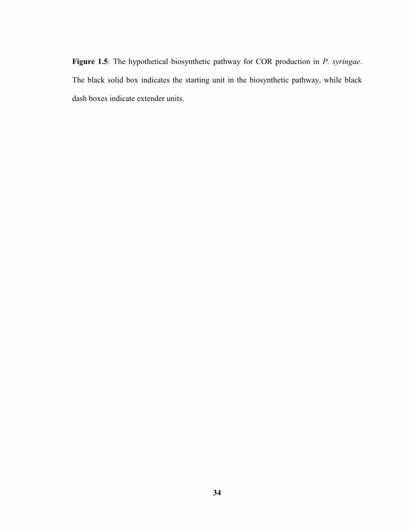
Figure 1.5: The hypothetical biosynthetic pathway for COR production in P. syringae.
The black solid box indicates the starting unit in the biosynthetic pathway, while black
dash boxes indicate extender units.
35
Co-Authorship Statement
Chapter 2 is a draft of a manuscript that will be submitted for publication. The
basic concept of the study was designed by D. Bignell, and the experimental methodology
was designed by D. Bignell and Y. Li. Y. Li performed all of the described experimental
work except for the HPLC and LC-MS analyses, which were conducted by J. Fyans. The
manuscript was written by Y. Li with editorial assistance provided by D. Bignell.
Parts of Chapter 3 were included in a manuscript that was recently accepted for
publication [Fyans JK, Altowairish MS, Li Y and Bignell DR. Characterization of the
coronatine-like phytotoxins produced by the common scab pathogen Streptomyces
scabies. Molecular Plant-Microbe Interactions. In Press]. Y. Li designed and conducted
the potato tuber disk assays using pure COR and CFA-L-Ile as well as the radish seedling
bioassays. Y. Li also wrote the associated parts of the manuscript with editorial assistance
provided by D. Bignell.
The remainder of Chapter 3 is a draft of a manuscript that will be submitted for
publication in the future. The basic concept of the project was designed by D. Bignell,
and the experimental methodology was designed by D. Bignell and Y. Li. Y. Li
performed all of the described experimental work, and the manuscript was written by Y.
Li with editorial assistance provided by D. Bignell.

36
CHAPTER 2: Characterizing the Role of scab79691, scab79711 and cfa8 in the
Biosynthesis of the Streptomyces scabies COR-like Metabolites
2.1 Introduction
The CFA-like biosynthetic gene cluster from S. scabies 87-22 contains
homologues of the cfl and cfa1-8 genes that are present in the CFA gene cluster from P.
syringae (Fig. 1.3). In P. syringae, the cfa1-7 and cfl genes are proposed to be directly
involved in the biosynthesis of COR and COR-like molecules, while the cfa8 gene
encodes a protein with significant similarity to CCR enzymes involved in ethylmalonyl-
CoA biosynthesis (Bender et al. 1999). CCR enzymes are well conserved in bacteria and
are believed to act as both reductases and carboxylases (Erb et al. 2007). In primary
metabolism, CCRs have been proposed to reduce crotonyl-CoA to butyryl-CoA (Fig.
2.1), which serves as a starter unit for fatty acid biosynthesis (Wilson and Moore 2012).
As carboxylases, CCR enzymes commence the reaction by taking the hydride from
NADPH and then reducing crotonyl-CoA to ethylmalonyl-CoA in the presence of CO2
(Fig. 2.1). The resulting ethylmalonyl-CoA subsequently enters central metabolism or is
passed to polyketide synthases involved in secondary metabolism (Erb et al. 2007;
Wilson and Moore 2012). Although there are several pathways known for synthesizing
ethylmalonyl-CoA (Fig. 2.1), CCR-dependent pathways seem to be the main supplier of
this extender unit for polyketide synthesis (Wilson and Moore 2012). This was
demonstrated in P. syringae as deletion of cfa8 resulted in complete loss of CFA and
COR production (Rangaswamy et al. 1998).
In S. scabies, the CFA-like biosynthetic gene cluster also contains six genes that

37
have no homologues in the P. syringae CFA gene cluster. Among these genes is
scab79711, which was previously proposed to be involved in the biosynthesis of
ethylmalonyl-CoA together with the cfa8 gene (Bignell et al. 2010). The product of the
scab79711 gene shows similarity to 3-hydroxybutyryl-CoA dehydrogenases that catalyze
the reduction of acetoacetyl-CoA to 3-hydroxybutyryl-CoA, a precursor of crotonyl-CoA
(Fig. 2.1). Crotonyl-CoA, in turn, can be reduced to ethylmalonyl-CoA by the action of
CCR (e.g. Cfa8) as described above. Genes that are homologous to scab79711 and cfa8
can be found in other polyketide biosynthetic gene clusters, including those for
concanamycin A, elaiophylin and indanomycin (Chan et al. 2009; Li et al. 2009). It is
likely that such genes are required to ensure a sufficient supply of ethylmalonyl-CoA for
polyketide biosynthesis during secondary metabolism.
Another unique gene within the CFA-like biosynthetic gene cluster, scab79691,
was previously predicted to encode a putative CYP450 monooxygenase that may function
in the oxidative modification of the COR-like metabolite backbone at or near the end of
the biosynthetic pathway (Bignell et al. 2010). CYP450s belong to a superfamily of
heme-containing proteins that are characterized by an absorption maximum wavelength
of 450nm (O’Keefe and Harder 1991). They catalyze the monooxygenation of a broad
range of substrates including cholesterol, lipids, steroid hormones, xenobiotics drugs and
toxic chemicals among all five kingdoms of life (Hasemann et al. 1995). Bacterial
CYP450 superfamilies have been intensively studied and are designated CYP101 to
CYP184 (Nelson et al. 1996). For their monooxygenase activity, one atom from O2 is
reduced to water while the other oxygen atom is inserted into the substrate typically as a
hydroxyl group. NADPH and ferredoxin/ferredoxin reductase are usually the electron

38
donors needed to provide the reducing equivalents (Takemori et al. 1993). CYP450
monooxygenases have been found to be highly abundant in the genus Streptomyces where
some may be involved in detoxifying molecules from other living organism and decaying
organic material in soil, and some are associated with the biosynthesis of secondary
metabolites (Lamb et al. 2002). In polyketide biosynthetic pathways within Streptomyces
spp., CYP450s are typically involved in the post-PKS tailoring of the metabolite during
the later stages of the pathway (Zhao and Waterman 2007). In the case of antibiotic
secondary metabolites, CYP450s often enhance the bioactivity of the molecule through
the addition one or more hydroxyl groups (Lamb et al. 2006).
In this study, the role of the cfa8, scab79711 and scab79691 genes in the COR-
like metabolite biosynthetic pathway was elucidated. Deletion mutants of S. scabies were
constructed for each gene, and the effect of each mutation on CFA-L-Ile biosynthesis was
assessed using HPLC and bioassays. The results show that scab79711 and cfa8 are
dispensable for CFA-L-Ile biosynthesis in S. scabies, though cfa8 is required for optimum
production of the metabolite. On the other hand, scab79691 was found to be essential for
metabolite biosynthesis, and the implications of this finding are discussed.
2.2 Materials and Methods
2.2.1 Bacterial strains, culturing conditions and maintenance
2.2.1.1 Escherichia coli strains
E. coli strains used in this study are listed in Table 2.1. Strains were routinely
grown at 37°C unless otherwise indicated. Liquid cultures were grown with shaking (200

39
– 250 rpm: revolutions per minute) in 5 – 50 mL of DifcoTM
LB (Luria-Bertani medium)
Lennox broth (BP1427-2; Fisher Scientific, Waltham, MA), low salt LB broth (1% w/v
tryptone; 0.5% w/v yeast extract; 0.25% w/v NaCl), SOB (Super optimal broth)
(Sambrook and Russell 2001) or SOC (Super optimal broth with catabolite repression)
medium (B9020S; New England Biolabs, Whitby, ON), while solid cultures were grown
on LB Lennox (or low salt LB) medium containing 1.5% w/v agar (105791A; NEOGEN,
Michigan, US). When necessary, the solid or liquid growth media were supplemented
with ampicillin (100 µg/mL final concentration; 0339-25G; Amresco, Solon, OH),
kanamycin (50 µg/mL final concentration; 420311; Calbiochem, San Diego, CA),
hygromycin B (100 µg/mL final concentration; 400051; Calbiochem) or chloramphenicol
(25 µg/mL final concentration; AC227920250; Acros Organic, New Jersey, USA). E. coli
strains were maintained at 4C for short-term storage or at - 80C as glycerol stocks for
long-term storage. Glycerol stocks were prepared by growing the strains overnight in 2 –
5 mL of LB or low salt LB liquid medium (with or without antibiotics) and then pelleting
the cells by centrifugation (13,000g) for 5 min. The resulting cell pellets were
resuspended in 0.5 – 1 mL of 20% v/v glycerol and were frozen at - 80°C.
2.2.1.2 Streptomyces scabies strains
S. scabies strains used in this study are listed in Table 2.1. Strains were routinely
grown at 28°C unless otherwise indicated. Liquid cultures were typically grown with
shaking (200 rpm) in TSB (Trypticase Soy Broth) (DF0370173; BD Biosciences,
Mississauga, ON) medium in 50 or 125 mL flasks with stainless steel springs. Plate

40
cultures were grown on SFMA (Soy flour mannitol agar) (Kieser et al. 2000), OBA (Oat
bran agar) (Johnson et al. 2007) or Difco NA (Nutrient agar) (DF0003178, BD
Biosciences) containing 1.5% w/v agar. When necessary, the growth media were
supplemented with hygromycin B (50 µg/mL final concentration), apramycin (50 µg/mL
final concentration), nalidixic acid (50 µg/mL final concentration; BP908-25; Thermo
Fisher Scientific), kanamycin (50 µg/mL final concentration), or thiostrepton (25 µg/mL
final concentration; T8902-1G; Sigma-Aldrich, Oakville, ON). S. scabies strains were
maintained at 4C for short-term storage or at - 80°C as spore or mycelial stocks for long-
term storage. Spore stocks were prepared by scraping gray spores from a 7-10 day old
OBA plate and then transferring the spores to a sterile 1.5 mL microcentrifuge tube
containing 1 mL of 40% v/v glycerol. The contents were mixing thoroughly and the tubes
were placed into the - 80°C freezer. Mycelial stocks were prepared by inoculating spores
into 25 mL of TSB and then growing for 48 – 72 hrs or until the culture was dense. Next,
950 L of the TSB culture was transferred into sterile 1.5 mL microcentrifuge tubes
containing 50 L of 100% v/v DMSO. After mixing the contents, the tubes were frozen at
- 80°C. For production of the COR-like metabolites, a single DMSO stock tube for each
strain was thawed on ice, inoculated into 9 mL of TSB, and incubated for 24 – 48 hrs
until the culture was dense. Then, the seed culture was subcultured (1% v/v) into 5 or 50
mL of SFMB (Soy flour mannitol broth) (Kieser et al. 2000) in 6-well tissue culture
plates (353046; BD Falcon) or in 2 125 mL spring flasks, respectively, and were
incubated for 7 days at 25 or 28°C with shaking (125 rpm for 6-well plates, 200 rpm for
spring flasks).
41
2.2.2 DNA manipulations
Standard molecular biology procedures were implemented for all DNA
manipulations performed in this study (Sambrook and Russell 2001). Restriction
digestions were performed using enzymes purchased from New England Biolabs
according to the manufacturer’s instructions. DNA was routinely analyzed by agarose gel
electrophoresis using 1% w/v agarose gels in 1 TBE (Tris-Borate-EDTA) buffer, and the
size of the DNA bands was estimated using 100bp (base pair) and 1kb DNA ladders
(DM001-0500 and DM010-0500; FroggaBio Inc., Toronto, ON). The EZ-Vision In-Gel
stain (N391-0.5ML; Amresco LLC, Solon, OH) or ethidium bromide was used for
visualizing the DNA in the gel, and gel images were acquired using a GelDoc-It Imager
(UVP, Upland, CA). Gel extraction of DNA was routinely performed using the Wizard
SV Gel and PCR (Polymerase chain reaction) Clean-Up system (A9281; Promega
Corporation, Madison, WI) as per the manufacturer’s directions. Quantification of DNA
was performed using a P300 Nanophotometer (Implen Inc., Westlake Village, CA)
according to the instrument instructions. Cloning of PCR products was performed using
the pGEM-T Easy vector system (PR-A1360, Promega Corporation) as per the
manufacturer’s protocol. Prior to cloning, the PCR products were modified using the A-
tailing procedure described in the pGEM-T Easy vector system protocol and using Taq
DNA polymerase (M0273S, New England Biolabs). Cloning of other DNA fragments
was performed using T4 DNA ligase (M0202S; New England Biolabs) as per the
manufacturer’s instructions except that the reactions were incubated at room temperature

42
for 4hrs. A vector: insert ratio of 1: 3 was used for all ligation reactions, and reactions
were routinely transformed into NEB (New England Biolabs) 5-alpha high efficiency
competent E. coli cells (C2987H; New England Biolabs) as per the manufacturer’s
directions. Plasmid and cosmid DNA was routinely isolated from overnight cultures of E.
coli using the alkaline lysis with SDS (Sodium dodecyl sulfate) minipreparation method
(Sambrook and Russell 2001). When highly pure DNA was required, the EZ-10 Spin
Column Plasmid DNA kit (BS614-250Preps; Bio Basic Inc., Markham, ON) was used as
described by the manufacturer. All cosmids and plasmids that were used or constructed in
this study are listed in Table 2.1. Genomic DNA was prepared using the SpeedMill PLUS
Beat Beater (MBI lab equipment, Montreal, CA) and the QIAamp DNA Mini Kit (51304;
QIAGEN) or the One-Tube Bacterial Genomic DNA Extraction Kit (BS8413-100Preps;
Bio Basic Inc.). Sequencing of DNA was performed by The Centre for Applied Genomics
(Toronto, ON). Standard desalted oligonucleotide primers used for sequencing were
purchased from Integrated DNA Technologies (Coralville, IA) and are listed in Table 2.2.
2.2.3 PCR
2.2.3.1 Generation of the hyg + oriT (Origin of transfer from RK2) extended resistance
cassettes
PCR was performed in order to generate the hyg + oriT extended resistance
cassettes that were used for constructing the ∆scab79711, ∆cfa8, and ∆scab79691 mutant
cosmids (Table 2.1) as part of the Redirect PCR targeting system (Gust et al. 2003a,b).
Reactions (50µL) contained 50 – 100 ng of pIJ10700 plasmid template, 1 Taq reaction

43
buffer with KCl, 2 mM MgCl2, 200 µM dNTPs (Deoxyribonucleoside triphosphate), 50
pmol each of the long (58-59 nt: nucleotide) forward and reverse primers, 5% v/v DMSO
(Dimethylsulfoxide) and 2.5 U of Taq DNA polymerase (FEREP0402, Thermo Fisher
Scientific, Waltham, MA). The cycling conditions started with an initial denaturation
step of 95°C for 2 min followed by 10 cycles of denaturation at 95°C for 45 sec, primers
annealing at 50°C for 45 sec, and extension at 72°C for 90 sec. Then, an additional 15
cycles was carried out with denaturation at 95°C for 45 sec, annealing at 55°C for 45 sec
and extension at 72°C for 90 sec. At the end, a final extension step was performed at
72°C for 5 min. The PCR reactions described in this section were performed in 0.2 mL
thin walled PCR tubes (10011-776; VWR International, Mississauga, ON) using a
Mastercycler Pro thermal cycler (Eppendorf Canada, Mississauga, ON). Following PCR,
the extended resistance cassettes were analyzed by agarose gel electrophoresis and were
gel-purified as described in section 2.2.2.
2.2.3.2 Verification of constructed mutant cosmids and strains
PCR reactions were also performed in order to verify the constructed mutant
cosmids (1770/∆scab79711, 1770/∆cfa8 and 1770/∆scab79691) and the S. scabies mutant
strains (∆scab79711, ∆cfa8 and ∆scab79691). Reactions were performed in 25 L
volumes using cosmid or genomic DNA (2.5 L) as template and using gene-specific
oligonucleotide primers (25 pmol each). Reactions were performed using the same
enzyme and reaction conditions as described in section 2.2.3.1, and the cycling conditions
consisted of an initial denaturation step at 95°C for 1 min followed by 30 cycles of
44
denaturation at 95°C for 30 sec, primer annealing at 60°C for 30 sec and extension at
72°C for 1 min/kb. All reactions were performed using an Eppendorf Mastercycler Pro
thermal cycler, and the resulting products were analyzed by agarose gel electrophoresis
(section 2.2.2).
2.2.3.3 Amplification of the scab79691 and cfa8 genes
The scab79691 and cfa8 genes were PCR-amplified for subsequent cloning into
the pGEM®-T Easy vector (Table 2.1). Amplifications were conducted in 50 µL reaction
volumes with 25-50 ng of Cosmid 1770 DNA as a template, 1 high GC buffer, 10mM
dNTPs, 25 pmol of forward and reverse primer, 3% v/v DMSO, and 0.5U of Phusion
DNA polymerase (M0530S, New England Biolabs). The cycling conditions consisted of
an initial denaturation step at 98°C for 30 sec followed by 30 cycles of denaturation at
98°C for 10 sec, primer annealing at 60°C and 72°C for scab79691 and cfa8, respectively,
for 45 sec and extension at 72°C for 5 min. Then, a final extension step of 72°C for 5 min
was followed. The PCR reactions described in this section were all performed using a
C1000TM
Thermal Cycler (BIO-RAD, Mississauga, Ontario), and the resulting products
were analyzed by agarose gel electrophoresis prior to cloning into pGEM®-T Easy. All
oligonucleotide primers used for PCR are listed in Table 2.2.
2.2.4 Construction of the S. scabies gene deletion mutants
The Redirect PCR targeting system (Gust et al. 2003a,b) was used to construct
mutant cosmids harbouring a deletion of the S. scabies scab79711, cfa8 and scab79691

45
genes (Table 2.1). Each gene was replaced with an extended resistance cassette (which
was PCR amplified as described in section 2.2.3.1) harbouring the hyg gene (conferring
resistance to hygromycin B) and oriT, which allowed for conjugal transfer of the mutant
cosmids into S. scabies from E. coli. Intergeneric conjugations were performed as
described previously (Altowairish 2014), and S. scabies exoconjugants were selected
using hygromycin B and nalidixic acid. To confirm that gene replacement had occurred in
the resulting exconjugants due to a double-crossover event, the strains were cultured on
NA containing kanamycin in order to identify those strains that were hygromycin
resistant and kanamycin sensitive. A total of six hygromycin resistant and kanamycin
sensitive isolates for each mutant were selected and numbered as ∆scab79711 N.
5/10/11/12/14/18, ∆cfa8 N. 2/7/10/12/17/18 and ∆scab79691 N. 1/2/3/4/5/6. Each isolate
was streaked for single colonies, and spores from a single colony were then spread over
the surface of an entire OBA plate containing the appropriate antibiotics. Finally, spore
and mycelial stocks of each isolate were prepared as described in section 2.2.1.2.
2.2.5 Chemical extraction of the COR-like metabolites
2.2.5.1 Small-scale extractions for HPLC analysis
S. scabies SFMB cultures grown in 6 well plates (see section 2.2.1.2) were used
for small-scale extraction of the COR-like metabolites. Samples (1.5 mL) of the SFMB
cultures were centrifuged at 13,300 g for 5 min, and 1 mL of each culture supernatant was
transferred into a clean 2 mL microcentrifuge tube (87003-298, VWR). The pH of the
supernatants was adjusted to 10-11 with 1N NaOH, after which the supernatants were

46
extracted twice with 0.5 volumes of chloroform (319988-4L; Sigma Aldrich Canada Co.,
Oakville, ON) to remove non-acidic hydrophobic compounds. Next, the aqueous
supernatants were adjusted to pH 1-2 with 1N HCl and were then extracted three times
with 0.5 volumes of chloroform. The acidic organic extracts were pooled and dried down
in a fume hood, and the resulting material was redissolved in 100 µL of 100% v/v HPLC-
grade methanol (34860-4L-R; Sigma Aldrich Canada Co., Oakville, ON). The samples
were filtered using a 0.2 µm pore size syringe filter (28145-491; VWR) to remove any
undissolved particulates prior to HPLC analysis.
2.2.5.2 Large-scale extractions for potato bioassays
S. scabies SFMB cultures grown in 2 125 mL flasks (section 2.2.1.2) were used
for large-scale extraction of the COR-like metabolites. Culture supernatants were
harvested by centrifugation (4000 rpm for 5 min) and were extracted as described in
section 2.2.5.1 except that the dried extracts were redissolved in 200 µL of 100% v/v
HPLC-grade methanol.
2.2.6 HPLC analysis of COR-like metabolite production
Filtered culture extracts were transferred to flat bottom glass inserts (5181-3377;
Agilent Technologies Canada Inc., Mississauga, ON), which were then placed into 2 mL
glass screw cap vials (5182-0716; Agilent Technologies Canada Inc.) and were sealed
with a PTFE (Polytetrafluoroethylene)/silicone rubber septum (5182-0731; Agilent
Technologies Canada Inc.). Samples (10 µL) of each culture extract were analyzed using

47
an Agilent 1260 Infinity Quaternary LC system with a Poroshell 120 EC-C18 column (4.6
× 50 mm, 2.7 µm particle size; Agilent Technologies Inc.). The initial mobile phase
concentration consisted of acetonitrile and water (30:70) with 0.1% v/v formic acid. After
1.5 min, the concentration was increased linearly to 50:50 acetonitrile:water over a time
period of 2.5 min, and this concentration was held for 1 min. Next, the mobile phase
concentration was returned to the initial conditions (30:70 acetonitrile:water) in 1.5 min
using a linear gradient, and this concentration was held for 2 min prior to injection of the
next sample. The flow rate (1 mL/min), column temperature (40°C) and detection
wavelength (230 nm) were kept constant throughout the analysis. A synthetic standard of
CFA-L-Ile (provided by Carol Bender) was included to assist in the identification of the
primary COR-like metabolite. All data were collected and analyzed using the
ChemStation software (version B.04.03, Agilent Technologies Canada Inc.), and the
Student’s t-test was used to identify significant differences in metabolite production in the
mutants compared to the txtA/pLDRB51-1 strain.
2.2.7 LC-MS (Liquid chromatography–mass spectrometry) analysis
Culture extracts (10 L) were also analyzed by LC-MS using an Agilent 1100
series HPLC system (Agilent Technologies Inc.) interfaced to a Waters G1946A single
quadrupole mass spectrometer (Waters Corporation). Separation was achieved using the
Agilent Technologies ZORBAX SB-C18 column (4.6 × 150 mm, 5 µm particle size) and
flow rate and column temperature as described in 2.2.6 except that the solvent gradient
system was modified. The initial mobile phase concentration consisted of acetonitrile and

48
water (30:70) with 0.1% v/v formic acid. After 4.5 min, the concentration was increased
linearly to 50:50 acetonitrile: water over a time period of 12 min, and this concentration
was held for 3 min. Next, the mobile phase concentration was returned to the initial
conditions (30:70 acetonitrile: water) in 4.5 min using a linear gradient, and this
concentration was held for 6 min prior to injection of the next sample. Detection was by
ultraviolet radiation (230 nm) and by electrospray ionization MS in negative ion mode.
2.2.8 Potato tuber slice bioassay
This was performed as described previously (Bignell et al. 2010) with some
modifications. Potato tubers were peeled and the surface was sterilized by immersing in
15% v/v bleach (Chlorox) for 10 min with occasional stirring. After rinsing twice with
sterile HPLC - grade water, the tubers were cut into equivalent size pieces (approximately
2.5 × 1.5 × 1 cm) using a sterile knife, and the potato pieces were placed onto pre-wetted
Whatman filter paper in sterile Petri dishes (4-6 potato pieces per dish). Next, one 6
mm diameter sterile paper disk (2017-006; Whatman) was placed onto each potato piece,
and 25 µL of filter-sterilized acidic culture extract was pipetted onto each disk. Pure COR
(500 ng) (C8115-1MG; Sigma Aldrich Canada Co., Oakville, ON) was included as a
positive control in this experiment while 100% v/v methanol was used as a negative
control. The potato disks were incubated under high humidity for 5-7 days at ambient
temperatures in the dark, and then they were assessed for the presence of tissue
hypertrophy.

49
2.2.9 Complementation of the cfa8 and scab79691 mutants
The basic strategy that was used is outlined in Figure 2.18. The target genes (cfa8
and scab79691) were PCR-amplified as described in section 2.2.3.3 using primers
containing engineered NdeI and XhoI restriction sites (Table 2.2). After cloning the PCR
products into pGEM-T EASY, the resulting clones were verified by digestion with
EcoRI and by sequencing. Next, the pGEM clones were digested with NdeI and XhoI to
release the cloned inserts, which were then gel-purified. The Streptomyces expression
plasmid pMSAK13, which contains the strong constitutive Streptomyces promoter
ermEp* and integrates into the chromosome at the BT1 site (Table 2.1), was digested
with the same enzymes and was also gel-purified. Ligation reactions were set up using the
digested plasmid and the cfa8 or scab79691 DNA fragments. Recombinant plasmids
obtained were verified by PCR and sequencing, and a single positive clone was
introduced into the corresponding S. scabies deletion mutant strain (cfa8 or
scab79691) by conjugation with E. coli as described previously (Altowairish 2014). As
a control, the pMSAK13 plasmid without cloned insert was also introduced into each
strain. After this, all the resulting strains were tested for COR-like metabolite production
using HPLC as described above.
2.2.10 Bioinformatics analyses
S. scabies DNA and protein sequences were obtained from the StrepDB website
(http://strepdb.streptomyces.org.uk). Amino acid sequence alignments were performed
using the Geneious 6.1.2 software. Predicted protein functions were assigned based on

50
database similarity searches using the National Center for Biotechnology Information
Protein BLAST (Basic Local Alignment Search Tool)
(http://blast.ncbi.nlm.nih.gov/Blast.cgi) and the StrepDB BLAST program. Protein
domain analyses were performed using the Pfam (protein families) database
(http://pfam.sanger.ac.uk/) (Finn et al. 2008). Phylogenetic trees were constructed using
maximum likelihood method of MEGA (Molecular Evolutionary Genetic Analysis)
program version 5.10 (Tamura et al. 2011). The significance of the branch order was
tested using the bootstrapping method with 1000 repetitions.
2.3 Results
2.3.1 Bioinformatics analysis of Scab79711, Cfa8 and Scab79691
2.3.1.1 Scab79711 and Cfa8 homologues in the database
Protein BLAST analysis indicated that the most similar homologues of Scab79711
and Cfa8 are IdmE and IdmF, respectively, from Streptomyces antibioticus (Table 2.3).
IdmE is a predicted hydroxybutyryl-CoA (or hydroxyacyl-CoA) dehydrogenase and IdmF
is a predicted CCR, and both are responsible for supplying a sufficient pool of the
extender unit ethylmalony-CoA for indamycin biosynthesis (Li et al. 2009). Pfam
analysis identified two 3-hydroxyacyl-CoA dehydrogenase NAD (Nicotinamide adenine
dinucleotide) binding domains and two C-terminal 3-hydroxyacyl-CoA dehydrogenase
domains in Scab79711 (Table 2.3). This is consistent with the fact that the N-terminal
domain of hydroxybutyryl-CoA dehydrogenases is usually responsible for the binding of
NAD+ and the C-terminal domain is involved in binding acyl-CoA substrates (Barycki et

51
al. 1999; Kim et al. 2014). Pfam analysis of Cfa8 suggested the presence of a conserved
C-terminal NAD(P)H/NAD(P)+ binding domain that binds the electron donor NAD(P)H,
and an N-terminal alcohol dehydrogenase GroES-like domain that is responsible for
binding a zinc cofactor atom(s) (Table 2.3; Hedlund et al. 2010; Murzin 1996). CCR
enzymes are members of the zinc-dependent alcohol dehydrogenase-like medium chain
dehydrogenase/reductase family (Rosas et al. 2003), which prefer NADPH as the electron
donor and usually require zinc atom(s) as the cofactor (Hedlund et al. 2010).
Homologues of Scab79711 and Cfa8 were identified in several Streptomyces spp.
including the plant pathogenic species S. turgidiscabies, S. acidiscabies and S. ipomoeae
(Fig. 2.2 and 2.3). Based on the phylogenetic analysis performed, the Cfa8 protein from S.
scabies 87-22 is more closely related to CCRs from other Streptomyces spp. than to Cfa8
homologues from P. syringae pathovars (Fig. 2.3).
2.3.1.2 Scab79691 homologues in the database
Protein BLAST and phylogenetic analyses revealed that an unspecific
monooxygenase from S. ipomoeae is the closest relative of Scab79691 (Table 2.3 and Fig.
2.4). Several known or predicted CYP450 monooxygenases from other actinomycetes
were also found to be similar to Scab79691 (Fig. 2.4). In addition, Scab79691 contains a
cytochrome P450 domain according to Pfam analysis (Table 2.3). Thus, Scab79691 is
predicted to serve as a monooxygenase in the biosynthesis of CFA-L-Ile. CYP450
enzymes are known to catalyze a variety of different reactions including primary
oxidation reactions, reductions and some non-redox P450 reactions (Guengerich and
Munro 2013). Monooxygenation catalyzed by CYP450s requires the input of two protons

52
to insert one oxygen group into the substrate (De Mot and Parret 2002). Alignment of the
Scab79691 amino acid sequence with that of other known or predicted CYP450s
indicated three conserved features: (1) the consensus sequence FXXGXXXCXG, which
contains the invariant residue cysteine (C) and is found in the heme-binding loop adjacent
to the L-helix; (2) an EXXR motif, which stabilizes the core structure by building up an
extensive network of hydrogen bonds with other amino acids and is found in the K-helix
on the proximal side of heme; (3) a P450 signature consensus sequence A/GGXD/ETT,
which is involved in proton transfer and is found on the distal side of the heme group
(Fig. 2.5; Werck-Reichhart and Feyereisen 2000). This further supports the idea that
Scab79691 functions as a CYP450 monooxygenase enzyme that catalyzes a
hydroxylation reaction during COR-like metabolite biosynthesis.
2.3.2 Construction of the S. scabies gene deletion mutants
Using the Redirect PCR targeting system, Δscab79711, Δcfa8 and Δscab79691
mutant cosmids were constructed, and each was verified by PCR (Fig. 2.6 – 2.8). The
mutant cosmids were then transferred into the S. scabies txtA/pRLDB51-1 strain by
conjugation with E. coli. Strain txtA/pRLDB51-1 was used to study the biosynthesis of
the COR-like metabolites since it is able to produces high levels of the COR-like
metabolites due to overexpression of the scab79591 regulatory gene, and it is also unable
to produce the dominant virulence factor thaxtomin A (Bignell et al. 2010). Exconjugants
obtained from the conjugations were screened to confirm that they arose from a double
crossover event, and spore and mycelial stocks were prepared for six scab79711

53
isolates, six cfa8 isolates and six scab79691 isolates. All mutant isolates were
confirmed by PCR (Fig 2.9 – 2.11).
2.3.3 The S. scabies deletion mutants differ in their ability to produce the CFA-L-Ile
COR-like metabolite
To analyze the production of the COR-like metabolites by the Δscab79711, cfa8
and Δscab79691 mutants, the strains, together with the original txtA/pRLDB51-1 strain,
were grown in SFMB, a medium which is known to support the production of the S.
scabies COR-like metabolites (Fyans et al. 2014). The resulting culture supernatants were
then extracted with chloroform under basic and acidic conditions, and the acidic organic
extracts were assessed for the presence of the primary COR-like metabolite (CFA-L-Ile)
using HPLC. As shown in Fig. 2.12 and 2.13, the majority of the Δscab79711 isolates
were not significantly different in production of CFA-L-Ile from the txtA/pRLDB51-1
strain, indicating that scab79711 is not required for metabolite production. On the other
hand, all of the cfa8 mutant isolates produced significantly less CFA-L-Ile as compared
to the txtA/pRLDB51-1 strain, though all were still able to produce the metabolite (Fig.
2.12 and 2.14). Quantitative analysis revealed that the Δcfa8 isolates produced 45-64% of
the averaged metabolite level in the txtA/pRLDB51-1 cultures (Fig. 2.14). In contrast,
the Δscab79691 isolates were all found to be abolished in CFA-L-Ile production (Fig.
2.15; data not shown), and instead they accumulated a metabolite with a longer retention
time. LC-MS analysis of the extracts from Δscab79691 N.2 and N.4 revealed that the
accumulated metabolite has a mass of 307.2 (Fig. 2.16D). It is also noteworthy that the
54
basic extracts from the Δscab79691 isolates were also analyzed by HPLC, and no
accumulated metabolites were found in those extracts (data not shown). Thus, scab79691
appears to be essential for COR-like metabolite biosynthesis, whereas cfa8 contributes to
metabolite production but is not essential, and scab79711 is not required for metabolite
biosynthesis. Furthermore, the accumulated metabolite in the Δscab79691 mutant extracts
may represent an intermediate in the COR-like metabolite biosynthetic pathway.
2.3.4 Culture extracts from the scab79691 mutant display reduced bioactivity on potato
tuber tissue
The bioactivity of the mutant culture extracts was also assessed using a potato
tuber bioassay, which detects the tissue hypertrophy-inducing activity of COR and COR-
like molecules (Bignell et al. 2010; Bignell et al. 2014). As shown in Figure 2.17, the
Δscab79691 mutant culture extract was found to induce only a very small amount of
tissue hypertrophy, which is consistent with the inability of this mutant to produce CFA-
L-Ile. The fact that some hypertrophy was observed suggests that the accumulated
metabolite in the culture extract (Fig. 2.15) may exhibit some of the same bioactivity as
CFA-L-Ile. On the other hand, the Δscab79711 and Δcfa8 mutant extracts exhibited a
similar level of hypertrophy - inducing activity as the ΔtxtA/pRLDB51-1 culture extract, a
finding that is consistent with the HPLC results for these extracts.
2.3.5 Genetic complementation of the Δcfa8 and Δscab79691 mutants
Genetic complementation was next performed to confirm that the observed

55
decrease in CFA-L-Ile production in the Δcfa8 and Δscab79691 mutants is due to deletion
of the corresponding gene. Given that there is only one promoter that drives expression of
the entire CFA-like metabolite biosynthetic gene cluster and that this promoter is located
upstream of cfa1 (Bignell et al. 2010), it was necessary to clone scab79691 and cfa8 into
a plasmid that harbours a promoter that would allow for expression of the genes in the
corresponding mutant. As such, the scab79691 and cfa8 coding sequences were PCR-
amplified and cloned into the pMSAK13 plasmid, which integrates into the BT1 phage
site in Streptomyces chromosomes and harbours the ermEp* promoter, allowing for
strong constitutive expression of the cloned genes (Table 2.1; Altowairish 2014). This
particular promoter was chosen as it has been successfully used in complementation
studies involving other COR-like metabolite biosynthetic mutants (Altowairish 2014).
The complementation plasmids were each verified by restriction digestion using the same
enzymes that were used for cloning (Fig. 2.19), after which they were introduced into the
corresponding mutant by conjugation with E. coli. As a control, the pMSAK13 without
any cloned insert was also introduced into each mutant.
As shown in Figure 2.20, the ability of pMSAK13/scab79691 to restore CFA-L-Ile
production in the Δscab79691 (isolate N.2) mutant was variable since two of the
complementation strains did not produce CFA-L-Ile (Fig. 2.20D, E) while a third strain
did produce a small amount of the metabolite (Fig. 2.20F). When complementation was
attempted using a second Δscab79691 mutant strain (isolate N.4), the
pMSAK13/scab79691 plasmid again failed to restore CFA-L-Ile production in the mutant
(data not shown). Attempts to complement the Δcfa8 (isolate N.18) mutant were also

56
unsuccessful as introduction of the pMSAK13/cfa8 plasmid severely affected the growth
of the complemented strain, and production of CFA-L-Ile was determined to be much
lower in the complementation strain than in the original cfa8 mutant (data not shown).
2.4 Discussion
This study focused on the role of three genes in the biosynthesis of the CFA-L-Ile
COR-like metabolite in S. scabies. One gene, cfa8, is conserved in the P. syringae CFA
biosynthetic gene cluster and was previously proposed to encode a CCR enzyme involved
in the biosynthesis of the ethylmalonyl-CoA extender unit (Bignell et al. 2010). Mutation
of cfa8 in P. syringae resulted in loss of CFA and COR production, indicating that the
gene is required for COR biosynthesis in that organism (Rangaswamy et al. 1998). The
other two genes, scab79711 and scab79691, are unique to S. scabies and were previously
predicted to be involved in ethylmalonyl-CoA biosynthesis and tailoring of the COR-like
metabolite backbone, respectively (Bignell et al. 2010). It was predicted that all three
genes may be required for COR-like metabolite biosynthesis in S. scabies, and therefore
gene deletion mutants were constructed in order to test this hypothesis.
2.4.1 scab79711 and cfa8 are dispensable for COR-like metabolite biosynthesis
Previously, it was suggested that scab79711 encodes a 3-hydroxybutyryl-CoA
dehydrogenase that functions to reduce acetoacetyl-CoA to 3-hydroxybutyryl-CoA,
which is an intermediate in crotonyl-CoA biosynthesis (Fig. 2.1; Bignell et al. 2010). The
bioinformatics analyses performed in this study support this idea as Scab79711 was

57
shown to display significant homology to other predicted 3-hydroxybutyryl-CoA
dehydrogenases that are currently in the database (Table 2.3 and Fig. 2.2). Four functional
domains that are responsible for catalyzing the reduction reaction are conserved in
Scab79711 and in its closest homologue IdmE from S. antibioticus. Though it was
predicted that scab79711 may be required for supplying a sufficient amount of
ethylmalonyl-CoA for CFA and COR-like metabolite biosynthesis in S. scabies, deletion
of the gene caused only a slight reduction of CFA-L-Ile production as determined by
HPLC and bioassay, indicating that Scab79711 is dispensable for COR-like metabolite
biosynthesis.
Bioinformatics analysis revealed that Cfa8 is most similar to IdmF (Table 2.3),
which is a predicted CCR enzyme encoded along with IdmE within the indamycin
biosynthetic gene cluster in S. antibioticus (Li et al. 2009). Cfa8 was found to contain
predicted domains that are conserved in other CCR homologues and which bind NADPH
and zinc (Murzin 1996; Hedlund et al. 2010). CCR enzymes are well conserved in
Streptomyces spp. and catalyze both reduction and carboxylation reactions involved in
ethylmalonyl-CoA biosynthesis (Fig. 2.1; Erb et al. 2007; Wilson and Moore 2012).
Although the BLAST results indicated that Cfa8 is most similar to IdmF from S.
antibioticus, the phylogenetic analysis revealed that Cfa8 may form a distinct CCR
lineage as compared to IdmF and other Streptomyces homologues (Fig. 2.3). This is
probably because the amino acids conserved in Cfa8 are more distinguishable than other
Streptomyces homologues according to the protein alignment used for the tree. As with
scab79711, it was predicted that Cfa8 is required to supply a sufficient amount of
ethylmalonyl-CoA for CFA and COR-like metabolite biosynthesis in S. scabies. The

58
results of this study show that although deletion of cfa8 in S. scabies did significantly
reduce the CFA-L-Ile production levels, production could still occur in the absence of the
enzyme. This is in contrast to the P. syringae cfa8 mutant, which was abolished in CFA
and COR biosynthesis (Rangaswamy et al. 1998).
In Streptomyces tsukubaensis, which produces the macrolide secondary metabolite
FK520, it has been shown that the total disruption of metabolite biosynthesis requires the
deletion of two ccr homologues in the genome (Kosec et al. 2012). A search of the S.
scabies 87-22 genome sequence revealed the presence of genes encoding three possible
homologues of Scab79711 and one possible homologue of Cfa8 (Table 2.4). Scab17601,
a predicted oxidoreductase, was found to be most closely related to Scab79711, and the
same domain properties were found in this protein as in Scab79711. The homologue of
Cfa8, Scab17621, is encoded within the same vicinity of Scab17601 and is 88% identical
to Cfa8 at the amino acid level (Table 2.4). It is possible that the homologous proteins
allow S. scabies to produce ethylmalonyl-CoA for CFA-L-Ile biosynthesis in the absence
of Cfa8 or Scab79711, though Cfa8 is apparently needed for optimum metabolite
biosynthesis.
Alternatively, or in addition, it is possible that S. scabies can use alternative
pathways for ethylmalonyl-CoA production that are independent of CCR and/or 3-
hydroxybutyryl-CoA activity. For example, in P. syringae, there are no homologues of
Scab79711 encoded within the genome, and it was previously suggested that
ethylmalonyl-CoA production in this organism occurs via the conversion of acetoacetyl-
CoA to butyryl-CoA, a pathway that is dependent on CCR but not on 3-hydroxybutyryl-
CoA dehydrogenase (Fig. 2.1; Rangaswamy et al. 1998). In Streptomyces hygroscopicus

59
var. ascomyceticus, it has been reported that the storage compound polyhydroxybutyrate
can be metabolized by polyhydroxybutyrate depolymerase to produce hydroxybutyryl-
CoA, which can then be converted to ethylmalony-CoA by the action of CCR (Wu et al.
2000). Further studies showed that this organism can also generate ethylmalonyl-CoA
directly from butyryl-CoA through the β-oxidation pathway (Fig. 2.1; Wu et al. 2000).
Whether such alternative pathways play a role in ethylmalonyl-CoA production in S.
scabies is currently unknown and warrants further investigation.
Attempts to complement the Δcfa8 mutant phenotype were not successful due to
the fact that the mutant strains containing pMSAK13/cfa8 grew very poorly. This could
be because the ermEp* promoter used for cfa8 expression is a strong, constitutive
promoter, and perhaps the high level of cfa8 expression is toxic to the cells. The use of an
alternative promoter for cfa8 expression might therefore be the answer for successful
complementation of this mutant.
2.4.2 scab79691 is required for COR-like metabolite biosynthesis
Analysis of the scab79691 gene product revealed that it is most closely related to
CYP450 monooxygenase enzymes from Streptomyces spp. and other actinomycetes
(Table 2.3 and Fig. 2.4). The EXXR, FXXGXXXCXG and A/GGXD/ETT motifs that are
characteristic of CYP450s were found to be well conserved in Scab79691 (Fig. 2.5),
supporting the idea that Scab79691 is in fact a CYP450 enzyme. CYP450s are commonly
associated with the biosynthesis of secondary metabolites in Streptomyces spp. For
example, CYP450s are required for hydroxylation, epoxidation and/or carbonylation
reactions involved in the biosynthesis of pharmaceutically important secondary
60
metabolites such as pladienolide B, oleandomycin, FD-891, filipin, albaflavenone and
7,30,40-trihydroxyisoflavonein (Urlacher and Girhard 2012). In S. scabies and other
thaxtomin A - producing species, two CYP450s, TxtC and TxtE, are required for
hydroxylation and nitration of thaxtomin A, respectively (Barry et al. 2012). HPLC
analysis of the culture extracts from the ∆scab79691 mutant isolates showed no
detectable production of CFA-L-Ile, indicating that Scab79691 is essential for COR-like
metabolite biosynthesis. Interestingly, a possible biosynthetic intermediate with a
molecular weight of 307.2 was found to accumulate in the mutant culture extracts (Fig.
2.15 and 2.16). Given that the culture extracts were shown to exhibit some hypertrophy-
inducing activity in the potato tuber bioassay (Fig. 2.17), it appears as though the putative
intermediate displays some of the same bioactivity as CFA-L-Ile.
The genetic complementation of the Δscab79691 mutant was also unsuccessful
overall, although CFA-L-Ile production did occur at low levels in one of the mutant
isolates containing pMSAK13/scab79691 (Fig. 2.20). Given that the pMSAK13 plasmid
integrates into the S. scabies chromosome at a different location than the CFA-like
biosynthetic gene cluster, it is possible that the expression of the cloned scab79691 gene
is affected by the surrounding genetic context. Alternatively, there might be an unknown
problem with the pMSAK13/scab79691 expression plasmid itself that does not allow for
proper expression of the scab79691 gene. It also cannot be completely ruled out that the
deletion of scab79691 has polar effects on the expression of downstream genes in the
CFA-like biosynthetic gene cluster. Future work should therefore focus on ruling out any
potential polar effects of the scab79691 gene deletion by testing for expression of the
downstream genes or by making a marker-less deletion mutant. In addition, the
61
construction of a new scab79691 expression plasmid may also be useful in future
complementation studies with this mutant.
2.4.3 Proposed role of Scab79691 in COR-like metabolite biosynthesis
The results of this study as well as those from a previous study show that at least
three of the novel genes within the S. scabies CFA-like biosynthetic gene cluster are
necessary for normal production of the CFA-L-Ile COR-like metabolite. Altowairish
(2014) demonstrated that deletion of the scab79681/oxr and scab79721/sdr genes leads to
a drastic decrease in CFA-L-Ile production, and in the case of the scab79721/sdr mutant,
three new metabolites are known to accumulate in the mutant culture extracts. Based on
these findings, a hypothetical biosynthetic pathway for production of CFA-L-Ile in S.
scabies was proposed in which Scab79681/Oxr and Scab79721/Sdr are directly involved
in the biosynthesis of the CFA moiety (Fig. 2.21). Scab79681/Oxr is a putative F420-
dependent oxidoreductase, and it has been proposed to be the enzyme that introduces the
C=C double bond that is present in the CFA molecule (Fig. 2.21; Altowairish 2014).
Scab79721/Sdr is a predicted short chain dehydrogenase/reductase, and this enzyme has
been proposed to catalyze the formation of the carbonyl group on the CFA backbone by
reduction of a hydroxyl group. Support for the role of Scab79721/Sdr comes from the fact
that two of the accumulated metabolites in the mutant culture extract have a molecular
mass that is the same as the mass of (e), which is the proposed Scab79721/Sdr substrate
(c) linked to isoleucine (Fig. 2.21; Altowairish 2014). Based on the results of the current
study, it can now be said that the Scab79691 CYP450 is also directly involved in the

62
biosynthesis of the CFA moiety, and it may do so by serving as the enzyme that
introduces the hydroxyl group that is subsequently reduced by Scab79721/Sdr (Fig. 2.21).
The mass of the accumulated metabolite in the scab79691 mutant culture extract (307.2)
is consistent with the predicted Scab79691substrate (b) linked to isoleucine, which agrees
with the proposed role of Scab79691. The fact that (b) itself was not detected in the
mutant culture extracts suggests that the Cfl enzyme is able to utilize CFA biosynthetic
intermediates as substrates for ligation with isoleucine (Fig. 2.21), an idea that was also
proposed based on the scab79721/sdr mutant results (Altowairish 2014).
The involvement of Scab79691, together with Scab79681/Oxr and
Scab79721/Sdr, in the biosynthesis of the CFA moiety of the S. scabies COR-like
metabolites is a novel finding. Similar enzymes have not previously been implicated in
CFA biosynthesis in P. syringae, and a search of the P. syringae pv tomato DC3000
genome sequence (http://pseudomonas-syringae.org/) revealed that there are no
homologues of Scab79691 produced by this organism. It is possible that S. scabies uses a
novel biosynthetic pathway to produce the same family of phytotoxins that is made by P.
syringae.
2.5 References
[1] Altowairish MS. Characterizing the role of Cfl, Oxr and Sdr in the biosynthesis of the
Streptomyces scabies COR-like metabolites. Master of Science Thesis, Memorial
University of Newfoundland. 2014.
[2] Barry SM, Kers JA, Johnson EG, Song L, Aston PR, Patel B, Krasnoff SB, Crane BR,

63
Gibson DM, Loria R and Challis G. Cytochrome P450–catalyzed L-tryptophan nitration
in thaxtomin phytotoxin biosynthesis. Nature Chemical Biology. 2012;8(10): 814-816.
[3] Barycki JJ, O'Brien LK, Bratt JM, Zhang R, Sanishvili R, Strauss AW and Banaszak
LJ. Biochemical characterization and crystal structure determination of human heart short
chain L-3-hydroxyacyl-CoA dehydrogenase provide insights into catalytic mechanism.
Biochemistry. 1999;38(18):5786-5798.
[4] Bignell DR, Fyans JK and Cheng Z. Phytotoxins produced by plant pathogenic
Streptomyces species. Journal of Applied Microbiology. 2014;116(2):223-235.
[5] Bignell DR, Seipke RF, Huguet-Tapia JC, Chambers AH, Parry RJ and Loria R.
Streptomyces scabies 87-22 contains a coronafacic acid-like biosynthetic cluster that
contributes to plant-microbe interactions. Molecular Plant-Microbe Interactions.
2010;23(2):161-175.
[6] Chan YA, Podevels AM, Kevany BM and Thomas MG. Biosynthesis of polyketide
synthase extender units. Natural Product Reports. 2009;26(1):90-114.
[7] De Mot R and Parret AH. A novel class of self-sufficient cytochrome P450
monooxygenases in prokaryotes. Trends in Microbiology. 2002;10(11):502-508.
[8] Erb TJ, Berg IA, Brecht V, Muller M, Fuchs G and Alber BE. Synthesis of C5-
dicarboxylic acids from C2-units involving crotonyl-CoA carboxylase/reductase: The
ethylmalonyl-CoA pathway. Proceedings of National Academy of Sciences USA.
2007;104(25):10631-10636.
[9] Fyans JK, Altowairish MS, Li Y and Bignell DR. Characterization of the coronatine-
like phytotoxins produced by the common scab pathogen Streptomyces scabies.
Molecular Plant-Microbe Interactions. In press.

64
[10] Gandecha AR, Large SL and Cundliffe E. Analysis of four tylosin biosynthetic genes
from the tylLM region of the Streptomyces fradiae genome. Gene. 1997;184(2):197-203.
[11] Guengerich FP and Munro AW. Unusual cytochrome p450 enzymes and reactions.
Journal of Biological Chemistry. 2013;288(24):17065-17073.
[12] Gust B, Challis GL, Fowler K, Kieser T and Chater KF. PCR-targeted Streptomyces
gene replacement identifies a protein domain needed for biosynthesis of the sesquiterpene
soil odor geosmin. Proceedings of National Academy of Science USA.
2003a;100(4):1541-1546.
[13] Gust B, O'Rourke S, Bird N, Kieser T and Chater KF. Recombineering in
Streptomyces coelicolor. The John Innes Foundation, Norwich, UK. 2003b.
[14] Hasemann CA, Kurumbail RG, Boddupalli SS, Peterson JA and Deisenhofer J.
Structure and function of cytochromes P450: A comparative analysis of three crystal
structures. Structure. 1995;3(1):41-62.
[15] Hedlund J, Jornvall H and Persson B. Subdivision of the MDR superfamily of
medium-chain dehydrogenases/reductases through iterative hidden markov model
refinement. BioMed Central Bioinformatics. 2010;11:534-2105-11-534.
[16] Kieser T, Bibb M, Buttner M, Chater K and Hopwood D. Practical Streptomyces
genetics. The John Innes Foundation: Norwich, UK. 2000.
[17] Kim J, Chang JH, Kim E and Kim K. Crystal structure of (R)-3-hydroxybutyryl-CoA
dehydrogenase PhaB from Ralstonia eutropha. Biochemical and Biophysical Research
Communications. 2014;443(3):783-788.
[18] Kosec G, Goranovič D, Mrak P, Fujs S, Kuščer E, Horvat J, Kopitar G and Petković
H. Novel chemobiosynthetic approach for exclusive production of FK506. Metabolic
65
Engineering. 2012;14(1):39-46.
[19] Lamb DC, Guengerich FP, Kelly SL and Waterman MR. Exploiting Streptomyces
coelicolor A3 (2) P450s as a model for application in drug discovery. Expert Opinion on
Drug Metabolism & Toxicology. 2006;2(1): 27-40.
[20] Lamb DC, Skaug T, Song HL, Jackson CJ, Podust LM, Waterman MR, Kell DB,
Kelly DE and Kelly SL. The cytochrome P450 complement (CYPome) of Streptomyces
coelicolor A3(2). Journal of Biological Chemistry. 2002;277(27):24000-24005.
[21] Li C, Roege KE and Kelly WL. Analysis of the indanomycin biosynthetic gene
cluster from Streptomyces antibioticus NRRL 8167. ChemBioChem. 2009;10(6):1064-
1072.
[22] MacNeil DJ, Gewain KM, Ruby CL, Dezeny G, Gibbons PH and MacNeil T.
Analysis of Streptomyces avermitilis genes required for avermectin biosynthesis. Gene.
1992; 111(1): 61-68.
[23] Murzin AG. Structural classification of proteins: New superfamilies. Current
Opinion in Structual Biology. 1996;6(3):386-394.
[24] Nelson DR, Koymans L, Kamataki T, Stegeman JJ, Feyereisen R, Waxman DJ,
Waterman MR, Gotoh O, Coon MJ, Estabrook RW, Gunsalus IC and Nebert DW. P450
superfamily: Update on new sequences, gene mapping, accession numbers and
nomenclature. Pharmacogenetics and Genomics. 1996;6(1):1-42.
[25] O'Keefe D and Harder P. Occurrence and biological function of cytochrome P450
monooxygenases in the Actinomycetes. Molecular Microbiology. 1991;5(9):2099-2105.
[26] Rangaswamy V, Jiralerspong S, Parry R and Bender CL. Biosynthesis of the
Pseudomonas polyketide coronafacic acid requires monofunctional and multifunctional
66
polyketide synthase proteins. Proceedings of National Academy of Sciences USA.
1998;95(26):15469-15474.
[27] Sambrook J, Fritsch EF and Maniatis T. Molecular cloning: A laboratory manual.
Cold Spring Harbor Laboratory Press, Cold Spring Harbor, NY, U.S.A. 1989.
[28] Takemori S, Yamazaki T and Ikushiro S. Cytochrome P-450-linked electron
transport system in monooxygenase reaction. Plant Physiology. 1993;450:44-63.
[29] Tamura T, Cormier JH and Hebert DN. Characterization of early EDEM1 protein
maturation events and their functional implications. Journal of Biological Chemistry.
2011;286(28):24906-24915.
[30] Urlacher VB and Girhard M. Cytochrome P450 monooxygenases: An update on
perspectives for synthetic application. Trends in Biotechnology. 2012;30(1):26-36.
[31] Werck-Reichhart D and Feyereisen R. Cytochromes P450: A success story. Genome
Biology. 2000;1(6):3003.1-3003.9.
[32] Wilson MC and Moore BS. Beyond ethylmalonyl-CoA: The functional role of
crotonyl-CoA carboxylase/reductase homologs in expanding polyketide diversity. Natural
Product Reports. 2012;29(1):72-86.
[33] Wu K, Chung L, Revill WP, Katz L and Reeves CD. The FK520 gene cluster of
Streptomyces hygroscopicus var. ascomyceticus ATCC 14891 contains genes for
biosynthesis of unusual polyketide extender units. Gene. 2000;251(1):81-90.
[34] Zhao B and Waterman MR. Novel properties of P450s in Streptomyces coelicolor.
Drug Metabolism Reviews. 2007;39(2-3):343-352.

67
Table 2.1: Bacterial strains, plasmids and cosmids used in this study.
Strain or
plasmid/cosmid
Genotype or comments Antibiotic
resistance
Source or
reference
Escherichia coli strains
BW25113 Host for Redirect PCR
targeting system
N/A Gust et al.
2003a,b
DH5α General cloning host N/A Gibco-BRL
ET12567 Non-methylating host (dam-
dcm- hsdS
-)
Camr
(Chloramphenicol
resistant), Tetr
(Tetracycline
resistant)
MacNeil et al.
1992
NEB 5-alpha DH5α derivative; high
efficiency competent cells
used for transformation
N/A New England
Biolabs
Streptomyces scabies strains
ΔtxtA/
pRLDB51-1
S. scabies strain containing
a deletion of the txtA gene
and carrying the scab79591
overexpression plasmid
pRLDB51-1 integrated at
the C31 attB site
Thior, Apr
r
(Apramycin
resistant)
Bignell et al.
2010
Δscab79711 ΔtxtA/pRLDB51-1
derivative containing a
deletion of the
scab79711gene.
Thior
(Thiostrepton
resistant), Aprr,
Hygr
(Hygromycin B
resistant)
This study
Δcfa8 ΔtxtA/pRLDB51-1
derivative containing a
deletion of the cfa8 gene.
Thior, Apr
r, Hyg
r This study
Δscab79691 ΔtxtA/pRLDB51-1
derivative containing a
deletion of the scab79691
gene.
Thior, Apr
r, Hyg
r This study
Plasmids or cosmids
pIJ790 RED recombination
plasmid; expresses the
RED recombinase genes
(gam, bet and exo) from the
L-arabinose inducible
pBAD promoter
Camr Gust et al.
2003a,b
pUZ8002 Encodes the machinery for Kanr (Kanamycin Kieser et al.
68
the conjugal transfer of
DNA from E. coli into
Streptomyces
resistant) 2000
Cosmid 1770 SuperCos1 derivative
containing the S. scabies
CFA-like gene cluster
Aprr, Kan
r Bignell et al.
2010
1770/
scab79711
Cosmid 1770 derivative in
which the scab79711 gene
was replaced with the
[hygr+oriT] disruption
cassette
Ampr (Ampicillin
resistant), Kanr
Hygr
This study
1770/cfa8 Cosmid 1770 derivative in
which the cfa8 gene was
replaced with the
[hygr+oriT] disruption
cassette
Ampr, Kan
r Hyg
r This study
1770/
scab79691
Cosmid 1770 derivative in
which the scab79691 gene
was replaced with the
[hygr+oriT] disruption
cassette
Ampr, Kan
r Hyg
r This study
pMSAK13 pIJ10257 derivative
containing the neo gene and
promoter sequence cloned
into the EcoRV site;
integrates into the ɸBT1
attB site in Streptomyces
chromosomes and carries
the strong, constitutive
promoter
ermEp*
Hygr, Kan
r Altowairish
2014
pGEM® - T
Easy
Cloning vector for PCR
products
Ampr Promega
pMSAK13/cfa8 pMSAK13 derivative
containing the cfa8 gene
cloned into the NdeI and
XhoI sites
Hygr, Kan
r This study
pMSAK13/
scab79691
pMSAK13 derivative
containing the scab79691
gene cloned into the NdeI
and XhoI sites
Hygr, Kan
r This study
69
Table 2.2: Oligonucleotide primers used in this study.
Primer
Name Sequence* (5′3′) Use
YL1 ATGACACCGCACAAGCCCGTGGTCGGG
ATCGTCGGCCTCATTCCGGGGATCCGT
CGACC
Redirect primer for
scab79711
mutant cosmid
construction
YL2 TCACTGCGATCTCAGTCCCTTCCGGTTC
TTGCGTCCCAATGTAGGCTGGAGCTGC
TTC
Redirect primer for
scab79711
mutant cosmid
construction
YL4 TCATGACGAAGGTGTCCCCTTCTCCGG
CAGGGAAGCGGCTGTAGGCTGGAGCTG
CTTC
Redirect primer for cfa8
mutant cosmid
construction
YL5 CCGCGGGCGACCCGTCGGCGGCGCGGG
GAAACCCGATGAATTCCGGGGATCCGT
CGACC
Redirect primer for cfa8
mutant cosmid
construction
JS3 GTGGTGCTGGGCGCGGAGTTCGTGCGG
AATCCGCATGAGATTCCGGGGATCCGT
CGACC
Redirect primer for
scab79691
mutant cosmid
construction
YL7 CGCTGTGCCGTCCGGACGGGCAGGGAC
CGCAGCCCGTGGTGTAGGCTGGAGCTG
CTTC
Redirect primer for
scab79691
mutant cosmid
construction
YL12 GTGAGGCGTGGGAGGCCAAC PCR verification of
scab79711 mutant
cosmid and strain
(junction 1)
YL13 GCGTCAGCCGGGCAGGATAG PCR verification of
scab79711 mutant
cosmid and strain
(junction 1)
YL14 GACTTCGCCCGCGAACTGCT PCR verification of
scab79711 mutant
cosmid and strain
(junction 2)
YL15 TCCCCAGCTCCACCACGGAG PCR verification of
scab79711 mutant
cosmid and strain
(junction 2)
YL10 CCCCAGCATGCTCCACCACG PCR verification of cfa8

70
mutant
cosmid and strain
YL11 AGGCCGACGATCCCGACCA PCR verification of cfa8
mutant
cosmid and strain
YL8 GGGACAAGGAGTGCGGAGCC PCR verification of
scab79691 mutant
cosmid and strain
YL9 CATCGGGTTTCCCCGCGCC PCR verification of
scab79691 mutant
cosmid and strain
YL18 GCGCCATATGATGAGTTCCACAACGAG
TGA
PCR amplification of
scab79691
for construction of
complementation plasmid
YL19 GCGCCTCGAGACGATGTCCTTCATCGG
GTT
PCR amplification of
scab79691
for construction of
complementation plasmid
YL22 CCATGGTCAGGACGGTCATT Verification of scab79691
complementation plasmid
YL23 CGCGATGAACACGAACTCAC Verification of scab79691
complementation plasmid
YL20 GCGCCATATGATGACCGCGGGCGACC
CGTC
PCR amplification of cfa8
for construction of
complementation plasmid
YL21 GCGCCTCGAGAATCCGATCACCTCGTG
CCC
PCR amplification of cfa8
for construction of
complementation plasmid
YL24 GGGCGACAACATCCTCATCT Verification of cfa8
complementation plasmid
YL25 GGAGTACACCTTGGAGAGCG Verification of cfa8
complementation plasmid
ermEp*
For
GCGATGCTGTTGTGGGC Sequencing of pMSKA13
clones
* Non-homologous extensions are underlined, while engineered restriction sites are
indicated in bold. N/A, not applicable.

71
Table 2.3: Closest homologue, predicted protein domains, and predicted function of the
Scab79711, Cfa8 and Scab79691 proteins encoded in the S. scabies CFA-like gene
cluster.
Protein Closest homologue in
database
(% identity/ similarity)
Domains present
(E-value)
Predicted Function
Scab
79711
S. antibioticus
Hydroxybutyryl-CoA
dehydrogenase (69/78)
3-hydroxyacyl-CoA
dehydrogenase NAD
binding domain (1.8e-
58); 3-hydroxyacyl-
CoA dehydrogenase
NAD binding domain
(9.7 e-23);
3-hydroxyacyl-CoA
dehydrogenase, C
terminal domain (7.1e-
25);
3-hydroxyacyl-CoA
dehydrogenase, C
terminal domain (1.3e-
24)
Hydroxybutyryl-CoA
dehydrogenase
Cfa8 S. antibioticus crotonyl-
CoA
reductase/carboxylase
(85/93)
Zinc-binding
dehydrogenase family
(3.7e-41); Alcohol
dehydrogenase GroES-
like domain (6.6e-07)
Crotonyl-CoA
reductase/carboxylase
Scab
79691
S. ipomoeae unspecific
monooxygenase (70/81)
Cytochrome P450
domain (3.4e-22)
CYP450
monooxygenase

72
Table 2.4: Closest homologues of the Scab79711 and Cfa8 proteins encoded on the S.
scabies 87-22 chromosome.
Protein Predicted Function Homologues % Identity /Similarity
E-
value
Cfa8 Crotonyl-CoA
carboxylase/reductase Scab17621 88/95 0.0
Scab79711 Hydroxybutyryl-CoA
dehydrogenase Scab17601 45/59 6e-158
Scab74071 41/55 4e-57
Scab28641 37/55 6e-52

73
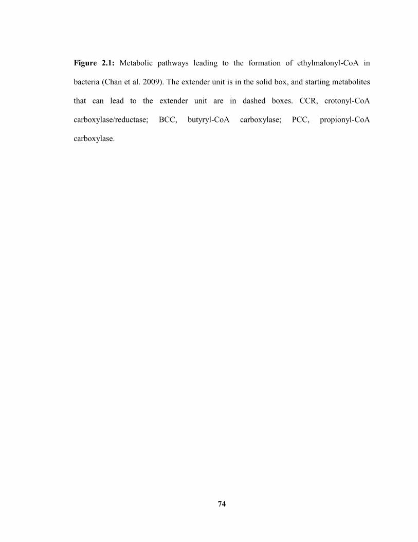
74
Figure 2.1: Metabolic pathways leading to the formation of ethylmalonyl-CoA in
bacteria (Chan et al. 2009). The extender unit is in the solid box, and starting metabolites
that can lead to the extender unit are in dashed boxes. CCR, crotonyl-CoA
carboxylase/reductase; BCC, butyryl-CoA carboxylase; PCC, propionyl-CoA
carboxylase.

75
Figure 2.2: Phylogenetic relationships of hydroxybutyryl-CoA dehydrogenase
homologues from Streptomyces spp. Bootstrap values are shown at the branch points. The
scale bar indicates the number of amino acid substitutions per site. Accession numbers for
each sequence are as follows: 3-hydroxybutyryl-CoA dehydrogenase Streptomyces
zinciresistens (WP_007498531.1); 3-hydroxybutyryl-CoA dehydrogenase Streptomyces
viridochromogenes (WP_003994089.1); 3-hydroxyacyl-CoA dehydrogenase
Streptomyces turgidiscabies (WP_006377336.1); 3-hydroxybutyryl-CoA dehydrogenase
Streptomyces hygroscopicus (WP_014675988.1); 3-hydroxybutyryl-CoA dehydrogenase
Streptomyces acidiscabies (WP_010355092.1); prevent host death family protein
Streptomyces ipomoeae (WP_009327483.1); Scab17601 Streptomyces scabies 87-22
(WP_012999615.1); 3-hydroxybutyryl-CoA dehydrogenase Streptomyces peucetius
(WP_031189204.1); 3-hydroxybutyryl-CoA dehydrogenase Streptomyces rimosus
(KEF19360.1); Scab28641 Streptomyces scabies 87-22 (WP_013000643.1); Scab74071
Streptomyces scabies 87-22 (WP_013004929.1); 3-hydroxybutyryl-CoA dehydrogenase

76
Streptomyces sp. NRRL B-1140 (AGZ94354.1); IdmE Streptomyces antibioticus
(ACN69981.1); Scab79711 Streptomyces scabies 87-22 (WP_013005381.1).

77
Figure 2.3 Phylogenetic relationships of CCR homologues in the database. Bootstrap
values are shown at the branch points. The scale bar indicates the number of amino acid
substitutions per site. Accession numbers for each sequence are as follows: Scab17621
Streptomyces scabies 87-22 (WP_012999617.1); crotonyl CoA reductase Streptomyces
bottropensis (WP_005482325.1); crotonyl CoA reductase Streptomyces collinus
(WP_020943170.1); crotonyl CoA reductase Streptomyces coelicoflavus ZG0656
(EHN79708.1); crotonyl CoA reductase Streptomyces gancidicus (EMF26267.1);
crotonyl CoA reductase Streptomyces ipomoeae (WP_009327493.1); crotonyl CoA
reductase Streptomyces turgidiscabies (WP_006377331.1); NADPH quinone reductase S.
acidiscabies (WP_010355090.1); Crotonyl CoA reductase Streptomyces afghaniensis
(WP_020277510.1); IdmF Streptomyces antibioticus (ACN69982.1); crotonyl CoA
reductase Streptomyces fradiae (CAA57474.2) ; NADPH quinone reductase
Streptomyces rimosus (WP_030642327.1) ; Cfa8 Streptomyces scabies 87-22
(WP_013005380.1); crotonyl CoA reductase Pseudomonas syringae pv. tomato str.

78
DC3000 (AAO58133.1); oxidoreductase Cfa8 Pseudomonas syringae pv. glycinea
(AAC38656.1).
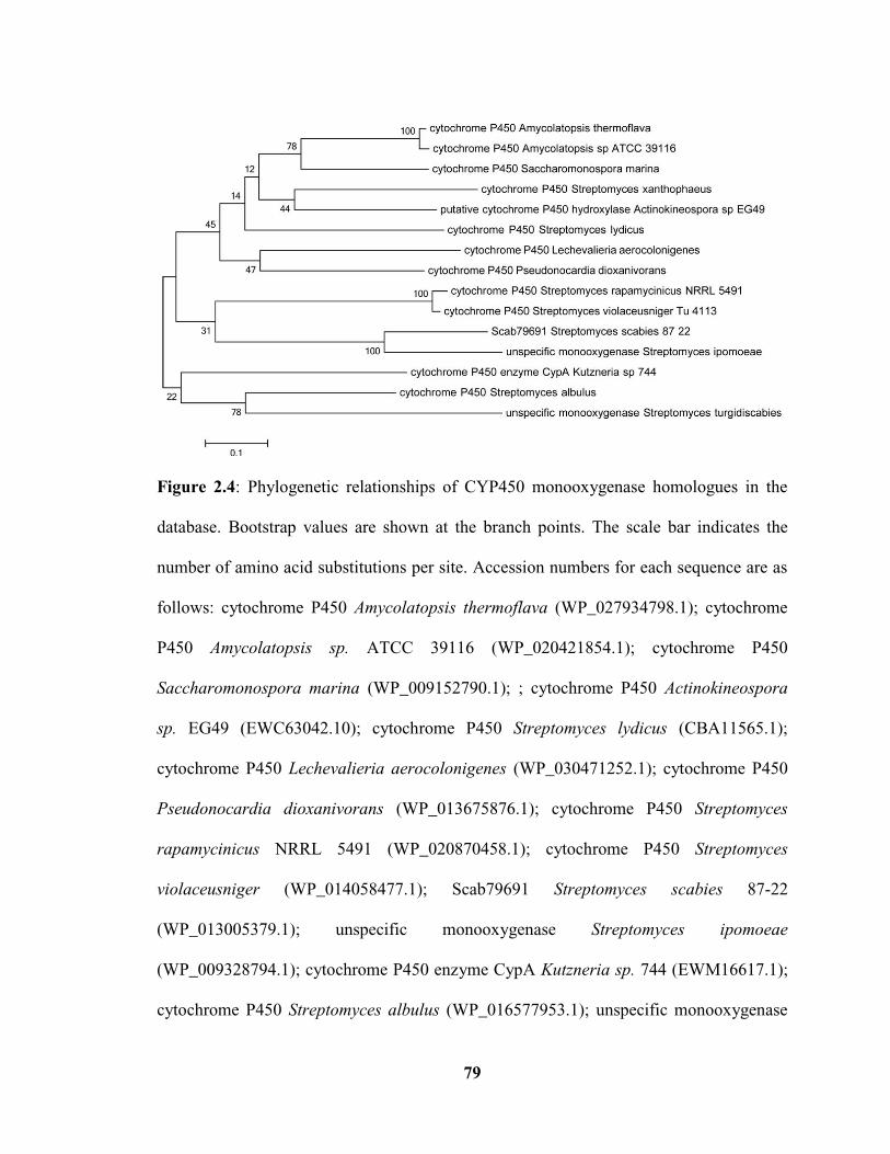
79
Figure 2.4: Phylogenetic relationships of CYP450 monooxygenase homologues in the
database. Bootstrap values are shown at the branch points. The scale bar indicates the
number of amino acid substitutions per site. Accession numbers for each sequence are as
follows: cytochrome P450 Amycolatopsis thermoflava (WP_027934798.1); cytochrome
P450 Amycolatopsis sp. ATCC 39116 (WP_020421854.1); cytochrome P450
Saccharomonospora marina (WP_009152790.1); ; cytochrome P450 Actinokineospora
sp. EG49 (EWC63042.10); cytochrome P450 Streptomyces lydicus (CBA11565.1);
cytochrome P450 Lechevalieria aerocolonigenes (WP_030471252.1); cytochrome P450
Pseudonocardia dioxanivorans (WP_013675876.1); cytochrome P450 Streptomyces
rapamycinicus NRRL 5491 (WP_020870458.1); cytochrome P450 Streptomyces
violaceusniger (WP_014058477.1); Scab79691 Streptomyces scabies 87-22
(WP_013005379.1); unspecific monooxygenase Streptomyces ipomoeae
(WP_009328794.1); cytochrome P450 enzyme CypA Kutzneria sp. 744 (EWM16617.1);
cytochrome P450 Streptomyces albulus (WP_016577953.1); unspecific monooxygenase

80
Streptomyces turgidiscabies (WP_006383617.1).

81
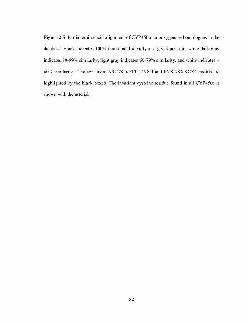
82
Figure 2.5: Partial amino acid alignment of CYP450 monooxygenase homologues in the
database. Black indicates 100% amino acid identity at a given position, while dark gray
indicates 80-99% similarity, light gray indicates 60-79% similarity, and white indicates
60% similarity. The conserved A/GGXD/ETT, EXXR and FXXGXXXCXG motifs are
highlighted by the black boxes. The invariant cysteine residue found in all CYP450s is
shown with the asterisk.
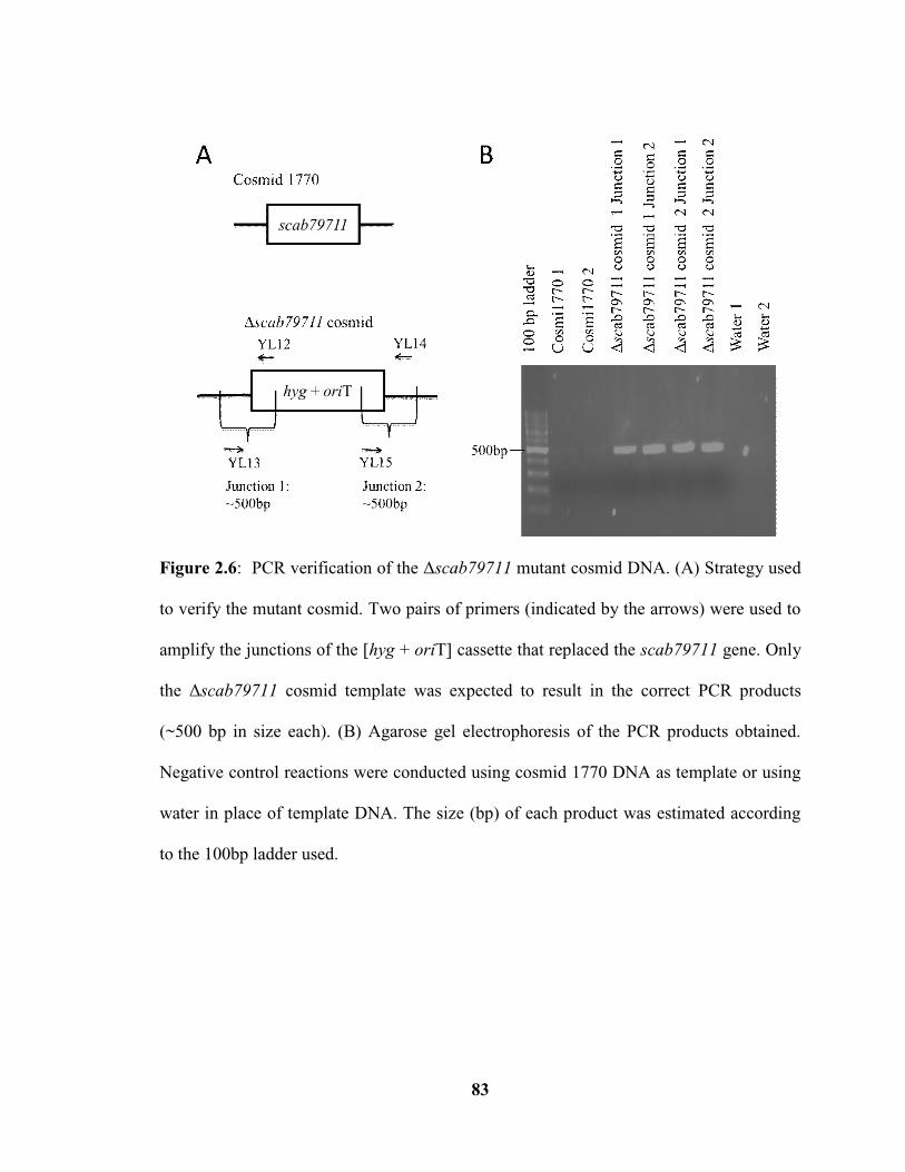
83
Figure 2.6: PCR verification of the Δscab79711 mutant cosmid DNA. (A) Strategy used
to verify the mutant cosmid. Two pairs of primers (indicated by the arrows) were used to
amplify the junctions of the [hyg + oriT] cassette that replaced the scab79711 gene. Only
the Δscab79711 cosmid template was expected to result in the correct PCR products
(~500 bp in size each). (B) Agarose gel electrophoresis of the PCR products obtained.
Negative control reactions were conducted using cosmid 1770 DNA as template or using
water in place of template DNA. The size (bp) of each product was estimated according
to the 100bp ladder used.

84
Figure 2.7: PCR verification of the Δcfa8 mutant cosmid DNA. (A) Strategy used to
verify the mutant cosmid. The primers used for amplification are indicated by the arrows.
The expected product sizes using cosmid 1770 and the Δcfa8 cosmid as template are
indicated. (B) Agarose gel electrophoresis of the resulting PCR products. Negative
control reactions were conducted using water in place of template DNA. The size (kb) of
each product was estimated according to the 1kb ladder used.
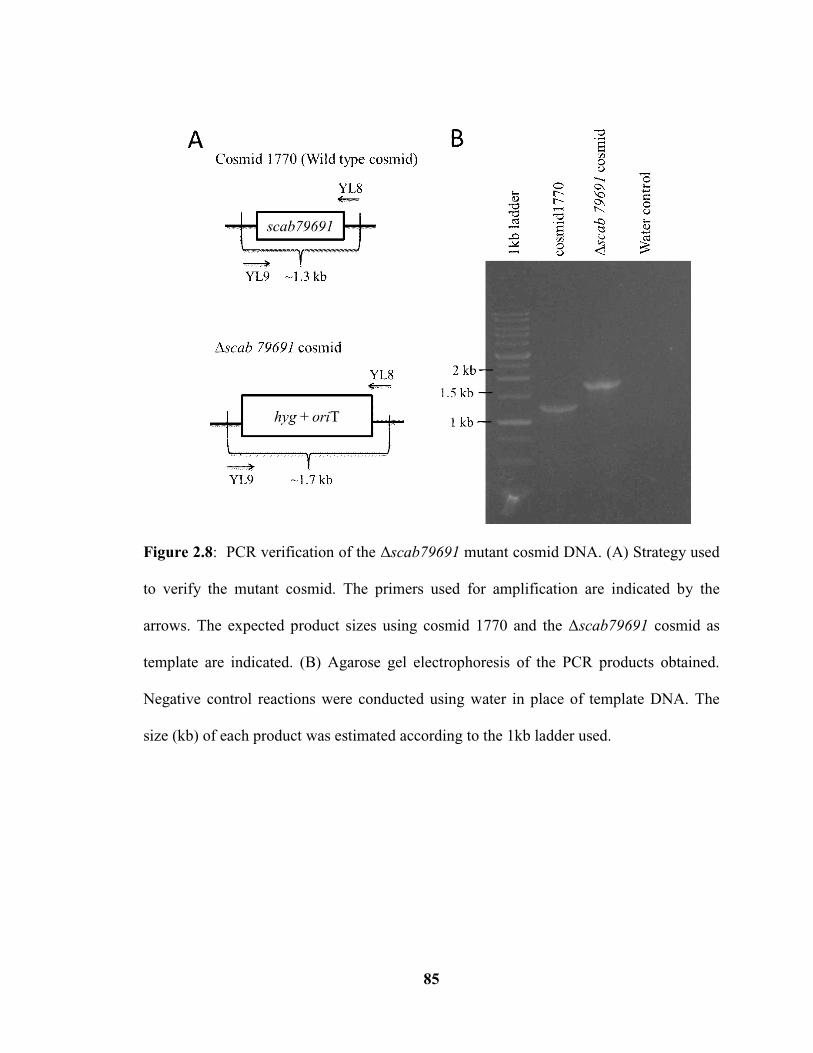
85
Figure 2.8: PCR verification of the Δscab79691 mutant cosmid DNA. (A) Strategy used
to verify the mutant cosmid. The primers used for amplification are indicated by the
arrows. The expected product sizes using cosmid 1770 and the Δscab79691 cosmid as
template are indicated. (B) Agarose gel electrophoresis of the PCR products obtained.
Negative control reactions were conducted using water in place of template DNA. The
size (kb) of each product was estimated according to the 1kb ladder used.

86
Figure 2.9: PCR verification of the six S. scabies Δscab79711 mutant isolates. Genomic
DNA was harvested from each isolate and was used as a template in the PCR reactions.
The primers used and the expected product sizes are described in Fig. 2.6. Negative
control reactions were conducted using genomic DNA from the S. scabies
ΔtxtA/pRLDB51-1 strain. The size (bp) of each product was estimated according to the
100bp marker ladder that was used.

87
Figure 2.10: PCR verification of the six S. scabies Δcfa8 mutant isolates. Genomic DNA
was harvested from each isolate and was used as a template in the PCR reactions. The
primers used and the expected product sizes are described in Fig. 2.7. Genomic DNA
from the S. scabies ΔtxtA/pRLDB51-1 strain was included as a control. The size (kb) of
each product was estimated according to the 1kb marker ladder that was used.
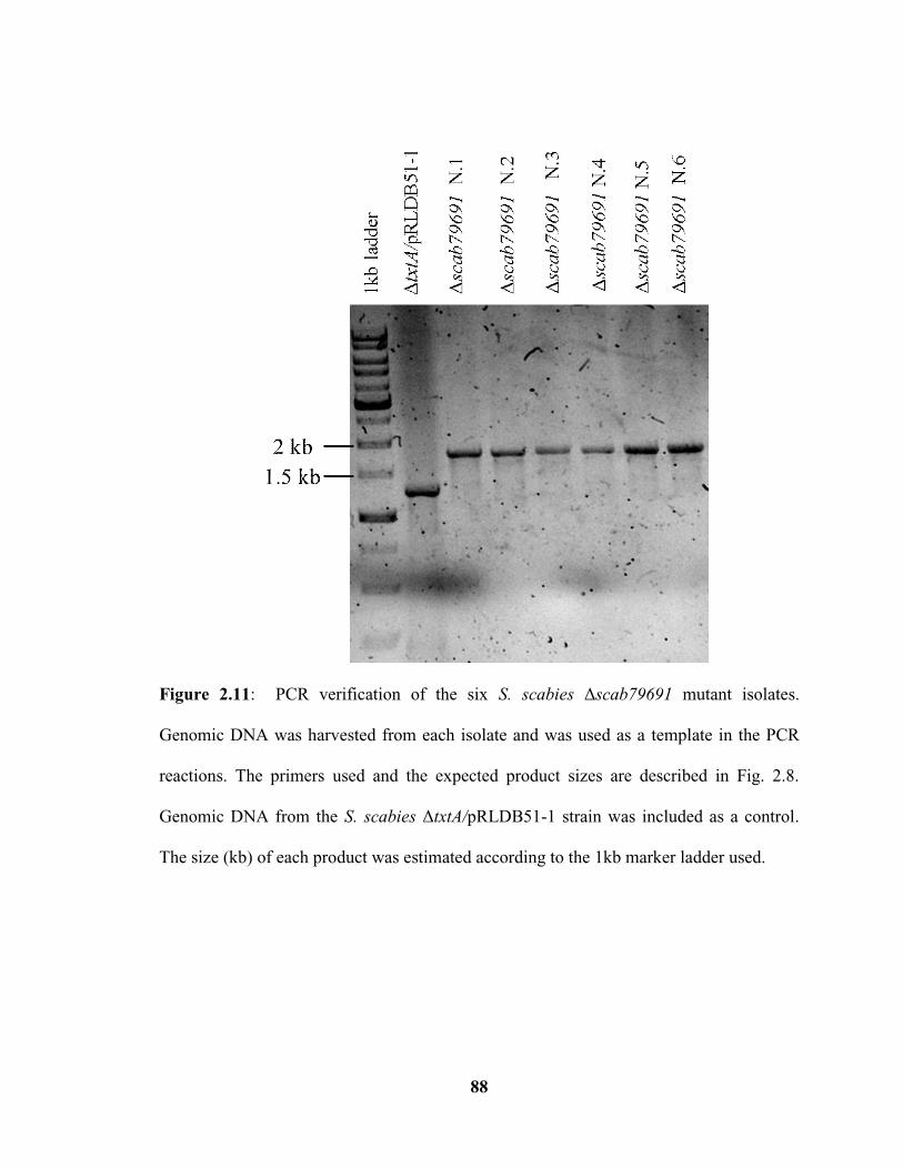
88
Figure 2.11: PCR verification of the six S. scabies Δscab79691 mutant isolates.
Genomic DNA was harvested from each isolate and was used as a template in the PCR
reactions. The primers used and the expected product sizes are described in Fig. 2.8.
Genomic DNA from the S. scabies ΔtxtA/pRLDB51-1 strain was included as a control.
The size (kb) of each product was estimated according to the 1kb marker ladder used.

89
Figure 2.12: HPLC analysis of acidic culture extract from the S. scabies
ΔtxtA/pRLDB51-1 strain and the Δscab79711 (isolate N.2) and Δcfa8 (isolate N.18)
mutants. * Indicates the peak corresponding to the primary COR-like metabolite (CFA-L-
Ile) produced by S. scabies. The HPLC chromatogram of an authentic CFA-L-Ile standard
(where ● indicates the CFA-L-Ile peak) is also shown.

90
Figure 2.13: Relative production levels of the CFA-L-Ile COR-like metabolite in the
ΔtxtA/pRLDB51-1 strain and the Δscab79711 mutant isolates. The bars indicate the
mean production level from triplicate cultures of each strain, and error bars indicate the
standard deviation from the mean. Mutant production levels that were determined to be
significantly different (* p 0.05) from that of the txtA/pRLDB51-1 strain are indicated.

91
Figure 2.14: Relative production levels of the CFA-L-Ile COR-like metabolite in the
ΔtxtA/pRLDB51-1 strain and the Δcfa8 mutant isolates. The bars indicate the mean
production level from triplicate cultures of each strain, and error bars indicate the
standard deviation from the mean. Mutant production levels that were determined to be
significantly different (* p 0.05; ** p 0.01) from that of the txtA/pRLDB51-1 strain
are indicate.

92
Figure 2.15: HPLC analysis of the acidic culture extracts from the S. scabies
ΔtxtA/pRLDB51-1 strain and the Δscab79691 mutant (isolate N.2 and N.4). The peak
representing the CFA-L-Ile is indicated with (*), while the putative intermediate
accumulating in the Δscab79691 mutant cultures (isolate N.2 and N.4) is indicates with
(). The molecular mass (in Da) of CFA-L-Ile and of the accumulated metabolite, as
determined by LC-MS, is indicated.

93
Figure 2.16: LC-MS analysis of the S. scabies the txtA/pRLDB51-1 and Δscab79691
(isolate N.2) strains. Total ion current chromatograms were obtained for the
txtA/pRLDB51-1 (A) and Δscab79691 (C) strains, with (*) indicating the CFA-L-Ile
peak in the txtA/pRLDB51-1 extract and () indicating the accumulated metabolite in
the Δscab79691 mutant extract. The extracted-ion chromatogram for the CFA-L-Ile peak
(B) and the accumulated metabolite peak (D) are also shown along with the
corresponding m/z in negative ion mode M-H-.

94
Figure 2.17: Bioactivity of the S. scabies Δscab79711 (isolate N.2), Δcfa8 (isolate N.18)
and Δscab79691 (isolate N.2) organic acidic culture extracts on potato tuber tissue. Pure
authentic COR and extract from the txtA/pRLDB51-1 strain served as positive controls,
whereas methanol and extract from a COR-like metabolite deficient strain txtA/cfa6
were included as negative controls.
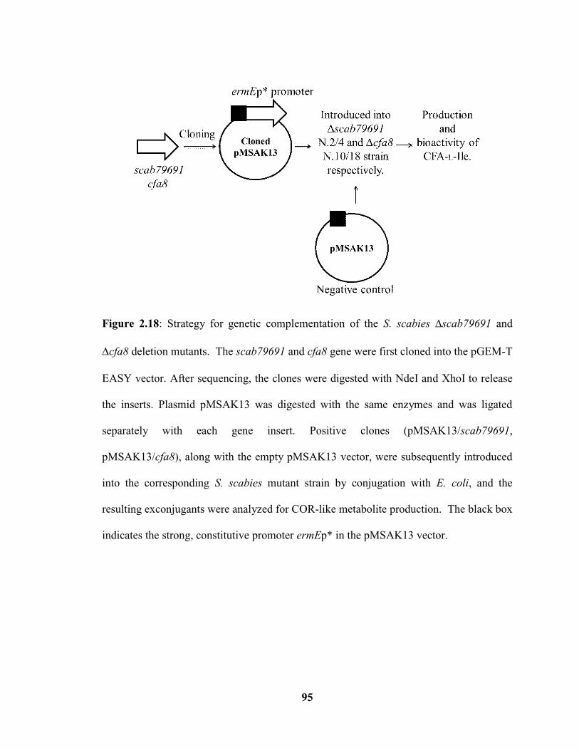
95
Figure 2.18: Strategy for genetic complementation of the S. scabies scab79691 and
cfa8 deletion mutants. The scab79691 and cfa8 gene were first cloned into the pGEM-T
EASY vector. After sequencing, the clones were digested with NdeI and XhoI to release
the inserts. Plasmid pMSAK13 was digested with the same enzymes and was ligated
separately with each gene insert. Positive clones (pMSAK13/scab79691,
pMSAK13/cfa8), along with the empty pMSAK13 vector, were subsequently introduced
into the corresponding S. scabies mutant strain by conjugation with E. coli, and the
resulting exconjugants were analyzed for COR-like metabolite production. The black box
indicates the strong, constitutive promoter ermEp* in the pMSAK13 vector.

96
Figure 2.19: Verification of gene scab79691 and cfa8 complementation plasmids by
restriction digestion. Plasmids pMSAK13/scab79691 (A), pMSAK13/cfa8 (B),
pMSAK13 (C) were digested with NdeI and XhoI to release the cloned inserts, and the
resulting products were analyzed by agarose gel electrophoresis. The expected size of
scab79691 insert is 1270bp and the cfa8 insert is 1513bp. The bands (bp) of the 1kb
marker are indicated in the first line.

97
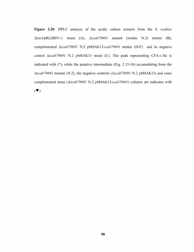
98
Figure 2.20: HPLC analysis of the acidic culture extracts from the S. scabies
ΔtxtA/pRLDB51-1 strain (A), Δscab79691 mutant (isolate N.2) strains (B),
complemented Δscab79691 N.2 pMSAK13/scab79691 strains (D-F) and its negative
control Δscab79691 N.2 pMSAK13 strain (C). The peak representing CFA-L-Ile is
indicated with (*), while the putative intermediate (Fig. 2.15-16) accumulating from the
Δscab79691 mutant (N.2), the negative controls (Δscab79691 N.2 pMSAK13) and some
complemented strain (Δscab79691 N.2 pMSAK13/scab79691) cultures are indicates with
().

99
Figure 2.21: Proposed biosynthetic pathway for the CFA-L-Ile COR-like metabolite in S.
scabies.

100
CHAPTER 3: Characterizing the biological activity of the Streptomyces scabies
COR-like metabolites
3.1 Introduction
COR is a non-host-specific polyketide phytotoxin produced by at least five
pathovars of the hemibiotrophic pathogen P. syringae, including atropurpurea, glycinea,
maculicola, morsprunorum, and tomato. Though not essential for P. syringae
pathogenicity, COR plays several important roles during the host infection process (Xin
and He 2013). Infection by P. syringae begins with the pathogen colonizing and
penetrating the plant surface. This process is facilitated by COR, which inhibits the
closure of stomata (Lee et al. 2013). Stomata are tiny pores found in the epidermis of
terrestrial plants, and the closure of such pores in response to PAMPs (Pathogen-
associated molecular pattern) is considered an important innate defense mechanism
against foliar pathogens (Melotto et al. 2006). It has been shown that COR-deficient
mutants of P. syringae inoculated on the surface of wild-type Arabidopsis thaliana leaves
are considerably reduced in their ability to cause infection, whereas such mutants can
readily infect A. thaliana mutants that are deficient in stomatal closure (Melotto et al.
2006; Zeng et al. 2011; Zeng and He 2010), thereby illustrating the importance of COR in
overcoming stomatal defenses. After successful invasion, COR also facilitates bacterial
multiplication and persistence within the plant intercellular spaces by overcoming the
apoplastic defenses (Zeng et al. 2011). Furthermore, COR contributes to disease
symptom development by P. syringae. The primary symptom caused by COR is leaf
chlorosis (yellowing), which can be observed in a variety of different plant species
101
(Bender et al. 1999a, b). Intense chlorosis in response to COR develops in soybean and
tobacco leaf tissues, and less intense chlorosis forms in potato, tomato, pepper, cabbage
and corn leaf tissues (Gnanamanickam et al. 1982; Uppalapati et al. 2005, 2007; Wangdi
et al., 2010). The chlorosis symptom has been attributed to the ability of COR to induce
the expression of chlorophyllase, which breaks down chlorophyll (Tsuchiya et al. 1999).
In Arabidopsis and tomato, COR induces the production of anthocyanins, which result in
the formation of a purple colour in the plant tissue (Feys et al., 1994; Uppalapati et al.
2005). Other symptoms attributed to COR include root stunting in Arabidopsis and
tomato (Feys et al., 1994; Uppalapati et al. 2005), hypertrophy of potato tuber tissue
(Völksch et al. 1989), ethylene emission (Ferguson and Mitchell 1985; Kenyon and
Tuner, 1992) and tendril coiling in Bryonia dioica (Blechert et al. 1999). Furthermore,
COR application to tomato leaves results in epidermal cell wall thickening, shrunken and
more intensely stained chloroplasts, and deposition of proteinase inhibitor particles in
plant cell vacuoles (Palmer and Bender 1995; Uppalapati et al. 2005).
It was previously noted by many that COR is structurally and functionally similar to
the plant hormone JA, and in particular, the bioactive derivative JA-Ile (Fig. 3.1). JA is a
plant growth regulator derived from the octadecanoid signaling pathway and is involved
in controlling defense responses to biological stresses (Feys et al. 1994; Sembdner and
Parthier 1993; Wasternack and Parthier 1997). When JA-Ile levels are low, JA-responsive
genes are repressed by JAZ (Jasmonate zim domain) repressor proteins. In response to
biological stresses, JA-Ile accumulates and binds to the COI1 F-box protein, which
determines the target specificity of the SCF (Skp1-Cullin1-F-box)COI1
E3 ubiquitin ligase.
Binding of JA-Ile to COI1 promotes the formation of a SCFCOI1
JA-IleJAZ ternary
102
complex and the subsequent ubiquitination and degradation of the JAZ proteins, leading
to activation of JA-responsive gene expression (Katsir et al. 2008a, b). As a molecular
mimic of JA-Ile, COR can also bind with high affinity to COI1 and promote the
degradation of JAZ proteins and the activation of JA-responsive genes (Katsir et al.
2008a, b; Melotto et al. 2008). This, in turn, leads to suppression of SA (Salicylic acid)-
dependent signalling pathways, which are critical for defense against P. syringae
infection (Zhao et al. 2003; Brooks et al. 2005; Cui et al. 2005; Laurie-Berry et al. 2006).
Remarkably, COR is more effective than JA-Ile in promoting SCFCOI1
-JAZ interaction in
vitro, although COR and JA-Ile are recognized by the same receptor (Katsir et al. 2008a).
There is also evidence that COR suppresses plant defense responses to PAMPs by callose
deposition inhibition in an SA signaling-independent manner (Geng et al. 2012).
S. scabies was recently shown to produce the COR-like metabolite CFA-L-Ile as a
major product as well as other minor related metabolites, including CFA-D-Val (Fyans et
al. 2014). CFA-L-Ile and other coronafacoyl-amino acid conjugates are produced in minor
amounts by P. syringae (Bender et al. 1999a), and production of such molecules has also
been reported for the plant pathogen Xanthomonas capmpestris pv phormiicola (Mitchell
and Frey 1986; Mitchell 1985). The COR-like molecules produced by P. syringae and X.
capmpestris are known to exhibit chlorosis-inducing activity (Bender et al. 1999a;
Mitchell and Ford 1998), and like COR, the CFA-L-Val produced by P. syringae can
cause changes in chloroplast ultrastructure, cell wall thickening, and accumulation of
proteinase inhibitors, though it is not as toxic as COR (Uppalapati et al. 2005). Given that
S. scabies culture extracts containing CFA-L-Ile and other minor COR-like metabolites
are able to induce potato tuber tissue hypertrophy in a similar manner as COR (Fig. 2.17;

103
Altowairish 2014; Bignell et al. 2014), it is likely that the metabolites exhibit some or all
of the other biological effects attributed to COR, though this has not been demonstrated.
As such, the objective of this study was to further investigate the biological activity of the
S. scabies COR-like metabolites using different plant bioassays.
3.2 Materials and methods
3.2.1 Bacterial strains, culturing conditions and maintenance
The S. scabies COR-like metabolite overproducing strain txtA/pRLDB51-1 and
the non-producing strains txtA/cfa6 and cfl (Table 2.1; Altowairish 2014) were used
in this study. Strains were maintained as spore and mycelial stocks at -80C as described
in section 2.2.1.2. Growth of the strains for COR-like metabolite production was as
described in section 2.2.1.2 except that the TSB seed cultures were sub-cultured into 25
ml of SFMB.
3.2.2 Chemical extraction of the COR-like metabolites
SFMB culture supernatants were extracted using chloroform as described
previously (section 2.2.5.1). Extraction of uninoculated SFMB medium was also
performed for use as a negative control in subsequent experiments. The resulting acidic
extracts were redissolved in 2 mL of 100% v/v of HPLC-grade MeOH (Methanol).
3.2.3 Plant bioassays
3.2.3.1 Leaf infiltration bioassay using culture supernatants

104
SFMB culture supernatants were filter-sterilized using a syringe filter (0.2 m pore
size; VWR International), and 50 L of each was infiltrated into leaves of 6-7 weeks old
Nicotiana benthamiana plants. The infiltration was conducted by making a small incision
on the upper side of a leaf using a syringe needle, and then placing the tip of a syringe
(without a needle) containing culture supernatant against the leaf surface at the point of
the incision. Next, the supernatant was injected while pressure was simultaneously
applied to the underside of the leaf. This, in turn, allowed the supernatant to enter the
intercellular space within the leaf in the vicinity of the point of injection. Each
supernatant sample was infiltrated into the same leaf, and the infiltration was repeated on
three leaves of the same plant and on three different plants for a total of 9 infiltrations per
sample. Supernatant from an uninoculated SFMB culture served as the negative control
while 3 nmol of COR (C8115; Sigma Aldrich Canada, Oakville, ON) served as the
positive control. The plants were kept at room temperature (~22±2°C) under a 16 hour
photoperiod for 7 days, after which the leaves were removed and photographed. The
bioassay was performed three times.
3.2.3.2 Radish seedling bioassay using culture supernatants or organic culture extracts
Radish seeds (cv Cherry Belle; Heritage Harvest Seed, Carman, MB) were surface
sterilized by treating with 40 mL of 70% v/v ethanol for 5 minutes with gentle agitation
followed by 40 mL of 1% v/v sodium hypochlorite (Chlorox) with 0.1% v/v polysorbate
20 (ICN10316890, MP Biomedicals) for 10 minutes with gentle agitation. Next, the
seeds were rinsed 8-10 times with sterile distilled water (40 mL). The surface sterilized

105
radish seeds were placed into a deep Petri dish (89107-632, VWR International)
containing Whatman #4 filter paper (90 mm) that had been wetted using 2 mL of sterile
water containing nystatin (N6261-25MU; Sigma Aldrich Canada) at 50 g/mL final
concentration. The dish was wrapped with Parafilm and was incubated for ~24 hours at
room temperature (~22±2°C) in the dark in order to allow seed germination to occur.
Filter-sterilized SFMB culture supernatants (5 mL) or organic culture extracts (0.5
mL of undiluted or 10-fold diluted extract) were pipetted into triplicate wells in 6-well
tissue culture plates (BD Falcon). When organic extracts were used, the plates were left
open in a biosafety cabinet to allow the solvent to evaporate completely, after which 5 mL
of sterile water was added to the each well. Then, germinated seeds with a uniform
hypocotyl and root length were selected, and four seeds were transferred to each well.
Negative control wells contained either water, uninoculated SFMB medium, or extract
from an uninoculated SFMB culture, while positive control wells containing 30 nmol of
COR in water. The plates were wrapped with Parafilm and were incubated with gentle
shaking (125 rpm) at room temperature (222C) for 4 days under a 16 hour
photoperiod, after which the total length of each plant (root + shoot) was measured. The
maximum and minimum measured seedling length in each treatment was omitted, and the
remaining values were used to calculate the average seedling length per treatment.
Statistically significant differences in seedling length were determined using the Student’s
t-test (tails: 2, type: 2) in Microsoft Excel. The bioassay was performed twice using
supernatants and three times using organic extracts.
106
3.2.3.3 Radish seedling bioassay using pure CFA-L-Ile
Radish seedlings were surface sterilized and germinated as described in section
3.2.3.2. CFA-L-Ile, purified from S. scabies culture supernatant and dissolved in DMSO
(provided by Dr. J. Fyans, Memorial University), was pipetted into triplicate wells in a 6-
well tissue culture plate. Similarly, pure COR dissolved in 100% MeOH was pipetted into
triplicate wells. Three different amounts of each metabolite (90, 9 and 0.9 nmol dissolved
in 30 L of solvent) were transferred to triplicate wells, and control wells contained 30
L of the corresponding solvent (MeOH or DMSO), after which 5 ml of sterile water was
added to the each well. Then, germinated seeds with uniform hypocotyl and root length
were selected and transferred to the wells (4 plants per well). Incubation and processing
of the plants was as described in section 3.2.3.2.
3.2.3.4 Analysis of anthocyanin production in radish seedlings
Anthocyanins were extracted and quantified from radish seedlings obtained in the
bioassays described in sections 3.2.3.1 and 3.2.3.2. The method used is similar to the one
described by Uppalapati et al. (2005) with some modifications. Three seedlings were
placed into a tube containing 1 mL of a solution of 3M HCl:H2O:MeOH (1:3:16 by
volume). The tubes were agitated and then incubated at 4°C in the dark overnight. Next,
the anthocyanin extracts (200 L) were transferred to wells in a 96-well plate, and the
absorbance at 530 and 653 nm (A530 and A653) was measured in each well using a
microplate Synergy H1 Hybrid Reader (BIOTEK, Winooski, Vermont, USA). Extracts
were prepared using all 12 seedlings from each treatment resulting in three extracts per
107
treatment, and every extract was analyzed three times by the microplate reader. The
anthocyanin levels were determined in each extract using the formula A530 – 0.24A653 and
were normalized using seedling wet weight. The average and standard deviation of the
normalized anthocyanin levels for each treatment were determined, and the Student’s t
test (tails: 2, type: 2) in Excel was used to identify statistically significant differences
between the treatments and the control. The bioassay was performed twice in total.
3.2.3.5 Potato tuber slice bioassay using pure CFA-L-Ile
This was performed as described previously in section 2.2.8 with some
modifications mentioned below. Purified CFA-L-Ile (8, 16 and 32 nmol) dissolved in
DMSO, and COR (8, 16 and 32 nmol) dissolved in MeOH, were applied to sterile paper
disks on potato tuber slices. After 24 hours incubation, 25 µL of sterile water was added
onto each disk, and the tuber slices were incubated for a further 4-6 days. Control slices
were treated with MeOH or DMSO. Following incubation, the tuber slices were dissected
and imaged. The bioassay was performed twice in total.
3.3 Results
3.3.1 The S. scabies COR-like metabolites are associated with necrosis of N. benthamiana
leaf tissue
The induction of chlorosis in a variety of plant hosts is the primary disease
symptom attributed to COR during P. syringae infections (Bender et al. 1999a, b). To
determine whether the COR-like metabolites produced by S. scabies can also cause
108
chlorosis, a leaf infiltration bioassay was performed using N. benthamiana plants. N.
benthamiana was chosen for this assay since COR is known to induce chlorosis in this
plant (Uppalapati et al. 2011). Furthermore, the large, broad leaves of this plant make it a
suitable host for infiltration assays. The assay was performed using filter-sterilized SFMB
culture supernatants of a S. scabies COR-like metabolite overproducer (txtA/pRLDB51-
1) and of two COR-like metabolite non-producing strains (txtA/cfa6 and cfl).
Supernatants, rather than organic culture extracts, were used in the assay in order to avoid
any toxic effects due to the infiltration of organic solvents.
As shown in Figure 3.2, the N. benthamiana leaf tissue infiltrated with the
txtA/pRLDB51-1 culture supernatant did not show any obvious chlorosis, but instead it
consistently showed brown, water-soaked necrosis. Leaf tissue infiltrated with the
txtA/cfa6 and cfl culture supernatants also showed water-soaked necrosis surrounded
by mild chlorosis in some instances; however, the effect of these supernatants was
variable among the different replicates and was consistently less severe than the
txtA/pRLDB51-1 culture supernatant (Fig. 3.2B, C). Although infiltration with 3 nmol
of COR did cause slight chlorosis in some replicates, the results were quite variable and
in many cases, there was no apparent chlorosis induced by the COR as expected (Fig.
3.2D). It is likely that the concentration of COR used in this assay is not enough to induce
chlorosis as 20 nmol of COR was reported to cause 5-10 mm chlorotic zone on tomato
leaves (Uppalapati et al. 2005).
3.3.2 The S. scabies COR-like metabolites can cause stunting of radish seedlings

109
A radish seedling bioassay was next performed in order to determine whether the
S. scabies COR-like metabolites can cause seedling stunting as previously described for
COR (Feys et al. 1994; Uppalapati et al. 2005). The assay was performed using SFMB
culture supernatants of the txtA/pRLDB51-1, txtA/cfa6 and cfl strains as well as two
different concentrations (undiluted and 10-fold diluted) of acidic organic extracts
prepared from the culture supernatants. The results using supernatants (data not shown)
and culture extracts (Figs. 3.3 and 3.4) were consistent and showed that the S. scabies
COR-like metabolites can indeed cause stunting of the radish seedlings. Treatment with
the txtA/pRLDB51-1 culture extract caused significant stunting as compared to the
mock (water) treated plants, with the undiluted extract being comparable to the COR
treatment (Figs. 3.3 and 3.4). Although significant stunting was also observed with the
undiluted txtA/cfa6 and cfl extracts, the effect was not as severe as with the undiluted
txtA/pRLDB51-1 culture extract (Figs. 3.3 and 3.4).
The stunting activity of the primary S. scabies COR-like metabolite, CFA-L-Ile,
was also investigated in order to directly compare the bioactivity of this metabolite to that
of COR. Equimolar amounts of each metabolite (90, 9 and 0.9 nmol) were used in the
radish seedling bioassay, and as shown in Fig. 3.5, the growth of seedlings treated with all
three concentrations of COR showed a statistically significant reduction when compared
to the solvent (methanol) control plants, whereas only seedlings treated with 90 nmol of
CFA-L-Ile showed a statistically significant reduction in growth when compared to the
solvent (DMSO) control. Furthermore, the treatment with 90 nmol of CFA-L-Ile did not
cause the same amount of seedling stunting as observed with any of the COR treatments
110
(Fig. 3.5). The results therefore demonstrate that CFA-L-Ile can cause stunting of radish
seedlings at nanomolar concentrations; however it is less toxic than COR in its
bioactivity.
3.3.3 Analysis of anthocyanin production in response to the COR-like metabolites
Next, the ability of the COR-like metabolites to induce anthocyanin production
was investigated in a radish seedling bioassay. Figure 3.6 shows that treatment with filter-
sterilized supernatant from cultures of the txtA/pRLDB51-1 strain resulted in a
significantly higher level of anthocyanins in radish seedlings compared to treatment with
the txtA/cfa6 and cfl supernatants as well as treatment with uninoculated SFMB
supernatant. It should be pointed out, however, that there was a high degree of variation
in the results from one experiment to the next, and it was very difficult to consistently
demonstrate a positive association between the presence of the COR-like metabolites and
an increase in anthocyanin production. The assay was also performed using culture
extracts (data not shown) as well as pure CFA-L-Ile (Fig. 3.7), and again, there was no
consistent correlation between anthocyanin production and the presence of the COR-like
metabolites. Treatment with 90 nmol of the pure CFA-L-Ile did cause an increase in
anthocyanin production as compared to the solvent (DMSO) control, although the results
were not deemed to be statistically significant. Though treatment with 0.9 nmol of COR
did cause a significant increase in anthocyanin production compared to the solvent
(MeOH) control (Fig. 3.7), it is noteworthy that the effect of COR on anthocyanin
production was also found to be highly variable from one experiment to the next in this

111
bioassay.
3.3.4 The S. scabies CFA-L-Ile COR-like metabolite causes hypertrophy of potato tuber
tissue
Results from this thesis (see Fig. 2.17) as well as from other studies in our lab
(Altowairish 2014; Bignell et al. 2014) have demonstrated that S. scabies culture extracts
containing the COR-like metabolites can induce hypertrophy of potato tuber tissue, an
effect that has been described for COR (Völksch et al. 1989). To further study the
hypertrophy-inducing activity of the CFA-L-Ile primary COR-like metabolite as
compared to COR, a potato tuber slice bioassay was performed using equimolar amounts
of COR and CFA-L-Ile. Figure 3.8 shows that treatment with 16 nmol of COR and CFA-
L-Ile led to the formation of hypertrophic outgrowths on the potato tissue; however, the
amount of hypertrophy observed was much less with the CFA-L-Ile treatment than with
the COR treatment. Similar results were observed using higher and lower (8 and 32 nmol)
amounts of each metabolite (data not shown). Therefore, though CFA-L-Ile exhibits the
same effect as COR on potato tuber tissue, it is not as toxic as COR in its bioactivity.
3.4 Discussion
The COR phytotoxin from P. syringae is known to cause a variety of different
effects against different plant hosts, including tissue hypertrophy, seedling stunting,
anthocyanin production and chlorosis (Bender et al. 1999a,b). The aim of this study was
to better characterize the bioactivity of the S. scabies COR-like metabolites in order to
determine whether the metabolites exhibit some or all of the same bioactivities described

112
for COR and whether the primary metabolite, CFA-L-Ile, exhibits the same level of
toxicity as COR. Previous studies showed that COR can cause stunting of tomato and
Arabidopsis seedlings (Feys et al., 1994; Uppalapati et al. 2005), and results from the
current study indicate that the S. scabies COR-like metabolites exhibit the similar effect
against radish seedlings. Treatment of radish plants with culture extracts containing the
COR-like metabolites caused comparable stunting as the COR control (Fig. 3.3 and 3.4).
However in this study, bioassays using equimolar amounts of COR and CFA-L-Ile
showed that the ability of CFA-L-Ile to cause seedling stunting is less than that of COR
(Fig. 3.5). Similar results were observed in the potato tuber slice bioassay where an
equimolar amount of CFA-L-Ile caused reduced tuber tissue hypertrophy compared to
COR (Fig. 3.8). The finding that CFA-L-Ile is not as toxic as COR is in agreement with
previous reports which showed that COR is the most toxic coronafacoyl compound
produced by P. syringae (Shiraishi et al. 1979; Uppalapati et al. 2005). The fact that the S.
scabies culture extracts containing the COR-like metabolites showed similar seedling
stunting and tissue hypertrophy – inducing activity as COR in the radish (Fig. 3.3 and 3.4)
and potato tuber (Fig. 2.17; Altowairish 2014; Bignell et al. 2014) bioassays, respectively,
may be due to the presence of very high amounts of CFA-L-Ile in the extracts, or perhaps
the other minor COR-like metabolites in the extracts are also required for optimal
bioactivity against plant tissues.
This study also addressed whether or not the S. scabies COR-like metabolites are
able to cause the production of anthocyanins, an effect that has been observed in tomato
and Arabidopsis plants in response to COR (Feys et al., 1994; Uppalapati et al. 2005).
Anthocyanins are natural pigments produced by plants as a hallmark of stress induced by
113
pathogens, herbivores and UV light (Shan et al. 2009). Although results obtained in this
study suggest that anthocyanin production does occur in response to the COR-like
metabolites in radish seedlings (Fig. 3.6 and 3.7), the results from replicate experiments
were too inconsistent to conclusively say this. It is worth pointing out, though, that
inconsistent results were also observed in the bioassays with the COR control, suggesting
that the assay used was not ideal for testing this bioactivity. Red skin radish (Raphanus
savitus L. var. “Cherry Belle”) used in the bioassay contains a high level of anthocyanins
naturally (Papetti et al. 2014), and this may be the reason why there was no obvious and
reproducible increase in anthocyanins in response to COR and CFA-L-Ile. Therefore, to
study the effect of the COR-like metabolites on anthocyanin production, tomato and
Arabidopsis plants, which are both naturally low in anthocyanins, could be used so that
any changes can be more readily observed.
The primary symptom induced by COR is chlorosis, the severity of which varies
depending on the plant host (Gnanamanickam et al. 1982; Uppalapati et al. 2005, 2007;
Wangdi et al., 2010). Chlorosis-inducing activity has also been reported for other
coronafacoyl compounds (Mitchell 1985), and it was anticipated that a similar effect
would be observed in response to the S. scabies COR-like metabolites. However, in N.
benthaminana infiltration bioassays performed in this study, no chlorosis was observed in
response to culture supernatants containing the COR-like metabolites, and instead the
liquid- infiltrated tissue became water-soaked and necrotic (Fig. 3.2), an effect that is
reminiscence of the hypersensitive response in plants (Heath 2000). A similar effect
could also be observed in tissues infiltrated with culture supernatants lacking the COR-
like metabolites, though the severity of tissue damage was always less than that observed

114
when the COR-like metabolites were present (Fig. 3.2). It is likely that the observed
tissue damage is in response to other known or predicted virulence factors such as TomA
and Nec1 that are likely present within the supernatants, and that the COR-like
metabolites enhance the severity of the tissue damage that is caused by these other
virulence factors. The chlorosis-inducing activity of the S. scabies COR-like metabolites,
therefore, will require further investigation, most likely by testing aqueous solutions of
the pure metabolites rather than complex culture supernatants.
COR is known to function as a molecular mimic of bioactive JA derivatives such
as JA-Ile, thereby stimulating JA-mediated signaling pathways in the plant host (Katsir et
al. 2008a, b). JA/ET-mediated signaling pathways play a critical role in controlling host
defense responses to necrotrophic pathogens, which kill host tissue early during infection
in order to obtain nutrients from the dead or dying host cells (Glazebrook 2005). In
contrast, SA-mediated signaling pathways are generally regarded as being important for
inducing defense responses to biotrophic pathogens, which feed on living plant tissue
without causing host cell death (Glazebrook 2005). SA- and JA/ET-mediated signaling
pathways exhibit antagonistic crosstalk such that activation of SA-mediated pathways
leads to suppression of JA/ET-mediated pathways, and vice versa (Derksen et al. 2013).
Previous reports have provided convincing evidence that SA-mediated defense response
pathways are important for defense against P. syringae (Brooks et al. 2005; Laurie-Berry
et al. 2006), and it has been proposed that COR is produced by P. syringae in order to
take advantage of the antagonistic interactions between JA/ET- and SA- mediated
signaling pathways in order to suppress the defense responses that are relevant to P.
syringae (Xin and He 2013). As P. syringae is considered a hemibiotrophic pathogen

115
since it exhibits a biotrophic phase during the early stages of infection and a necrotrophic
phase during the later stages (Xin and He 2013), it is likely that production of COR is
critical for overcoming host defenses early during the infection process.
The structural and phenotypic similarities shared by COR and the S. scabies COR-
like metabolites suggest that the COR-like metabolites may also have a role in
suppressing SA-mediated defense pathways during pathogen infection. Whether SA-
mediated signaling pathways play an important role in host defense against S. scabies
infection, however, is currently unknown, and warrants further investigation. It also
cannot be ruled out that the COR-like metabolites have additional roles during pathogen
infection, and therefore further investigations into the function of the metabolites during
host-pathogen interactions is needed.
3.5 References
[1] Annemart K and Pieterse CM. Cross talk in defense signaling. Plant Physiology.
2008; 146(3): 839-844.
[2] Bender C, Rangaswamy V and Loper J. Polyketide production by plant-associated
Pseudomonads. Annual Review of Phytopathology. 1999a;37(1):175-196.
[3] Bender CL, Alarcon-Chaidez F and Gross DC. Pseudomonas syringae phytotoxins:
Mode of action, regulation, and biosynthesis by peptide and polyketide synthetases.
Microbiology and Molecular Biology Reviews. 1999b;63(2):266-292.
[4] Blechert S, Bockelmann C, Füßlein M, Schrader TV, Stelmach B, Niesel U and
Weiler EW. Structure-activity analyses reveal the existence of two separate groups of
active octadecanoids in elicitation of the tendril-coiling response of Bryonia dioica Jacq.

116
Planta. 1999;207(3):470-479.
[5] Brooks DM, Bender CL and Kunkel BN. The Pseudomonas syringae phytotoxin
coronatine promotes virulence by overcoming salicylic acid‐dependent defences in
Arabidopsis thaliana. Molecular Plant Pathology. 2005;6(6):629-639.
[6] Cui J, Bahrami AK, Pringle EG, Hernandez-Guzman G, Bender CL, Pierce NE and
Ausubel FM. Pseudomonas syringae manipulates systemic plant defenses against
pathogens and herbivores. Proceedings of National Academy of Sciences USA.
2005;102(5):1791-1796.
[7] Derksen D, Rampitsch C and Daayf F. Signaling cross-talk in plant disease resistance.
Plant Science. 2013;207:79-87.
[8] Ferguson IB and Mitchell RE. Stimulation of ethylene production in bean leaf discs
by the Pseudomonad phytotoxin coronatine. Plant Physiology. 1985;77(4):969-973.
[9] Feys BJ, Benedetti CE, Penfold CN and Turner JG. Arabidopsis mutants selected for
resistance to the phytotoxin coronatine are male sterile, insensitive to methyl jasmonate,
and resistant to a bacterial pathogen. The Plant Cell. 1994;6(5):751-759.
[10] Fyans JK, Altowairish MS, Li Y and Bignell DR. Characterization of the coronatine-
like phytotoxins produced by the common scab pathogen Streptomyces scabies.
Molecular Plant-Microbe Interactions. In press.
[11] Geng X, Cheng J, Gangadharan A and Mackey D. The coronatine toxin of
Pseudomonas syringae is a multifunctional suppressor of Arabidopsis defense. The Plant
Cell. 2012;24(11):4763-4774.
[12] Glazebrook J. Contrasting mechanisms of defense against biotrophic and
necrotrophic pathogens. Annual Review in Phytopathology. 2005;43:205-227.

117
[13] Gnanamanickam S, Starratt A and Ward E. Coronatine production in vitro and in
vivo and its relation to symptom development in bacterial blight of soybean. Canadian
Journal of Botany. 1982;60(5):645-650.
[14] Heath MC. Hypersensitive response-related death. Programmed Cell Death in
Higher Plants. 2000;44(3):321-34.
[15] Katsir L, Chung HS, Koo AJ and Howe GA. Jasmonate signaling: A conserved
mechanism of hormone sensing. Current Opinion in Plant Biology. 2008a;11(4):428-435.
[16] Katsir L, Schilmiller AL, Staswick PE, He SY and Howe GA. COI1 is a critical
component of a receptor for jasmonate and the bacterial virulence factor coronatine.
Proceedings of the National Academy of Sciences USA. 2008b;105(19):7100-7105.
[17] Kenyon JS and Turner JG. The stimulation of ethylene synthesis in Nicotiana
tabacum leaves by the phytotoxin coronatine. Plant Physiology. 1992;100(1):219-224.
[18] Laurie-Berry N, Joardar V, Street IH and Kunkel BN. The Arabidopsis thaliana
JASMONATE INSENSITIVE 1 gene is required for suppression of salicylic acid-
dependent defenses during infection by Pseudomonas syringae. Molecular Plant-Microbe
Interactions. 2006;19(7):789-800.
[19] Lee S, Ishiga Y, Clermont K and Mysore KS. Coronatine inhibits stomatal closure
and delays hypersensitive response cell death induced by nonhost bacterial pathogens.
PeerJ. 2013;1:e34.
[20] Melotto M, Mecey C, Niu Y, Chung HS, Katsir L, Yao J, Zeng W, Thines B,
Staswick P, Browse J, Howe GA and He SY. A critical role of two positively charged
amino acids in the Jas motif of Arabidopsis JAZ proteins in mediating coronatine‐and
jasmonoyl isoleucine‐dependent interactions with the COI1 F‐box protein. The Plant

118
Journal. 2008;55(6):979-988.
[21] Melotto M, Underwood W, Koczan J, Nomura K and He SY. Plant stomata function
in innate immunity against bacterial invasion. Cell. 2006;126(5):969-980.
[22] Mitchell RE. Norcoronatine and N-coronafacoyl-L-valine, phytotoxic analogues of
coronatine produced by a strain of Pseudomonas syringae pv. glycinea. Phytochemistry.
1985;24(7):1485-1487.
[23] Mitchell RE and Ford KL. Chlorosis-inducing products from Pseudomonas syringae
pathovars: New N-coronafacoyl compounds. Phytochemistry. 1998;49(6):1579-1583.
[24] Mitchell RE and Frey EJ. Production of N-coronafacoyl-L-amino acid analogues of
coronatine by Pseudomonas syringae pv. atropurpurea in liquid cultures supplemented
with L-amino acids. Journal of General Microbiology. 1986;132(6):1503-1507.
[25] Palmer DA and Bender CL. Ultrastructure of tomato leaf tissue treated with the
Pseudomonad phytotoxin coronatine and comparison with methyl jasmonate. Molecular
Plant-Microbe Interactions. 1995;8(5):683-692.
[26] Papetti A, Chiara M, Chiara Z and Gabriella G. HPLC–DAD–ESI/MS
characterization of environmentally friendly polyphenolic extract from Raphanus sativus
L. var. “Cherry Belle” skin and stability of its red components. Food Research
International. 2014;04:46.
[27] Sembdner G and Parthier B. The biochemistry and the physiological and molecular
actions of jasmonates. Annual Review of Plant Biology. 1993;44(1):569-589.
[28] Shan X, Zhang Y, Peng W, Wang Z and Xie D. Molecular mechanism for
jasmonate-induction of anthocyanin accumulation in Arabidopsis. Journal of
Experimental Botony. 2009;60(13):3849-3860.
119
[29] Strauss SY, Irwin RE and Lambrix VM. Optimal defence theory and flower petal
colour predict variation in the secondary chemistry of wild radish. Journal of Ecology.
2004;92(1):132-141.
[30] Tsuchiya T, Ohta H, Okawa K, Iwamatsu A, Shimada H, Masuda T and Takamiya
K. Cloning of chlorophyllase, the key enzyme in chlorophyll degradation: Finding of a
lipase motif and the induction by methyl jasmonate. Proceedings of National Academy of
Sciences USA. 1999;96(26):15362-15367.
[31] Uppalapati SR, Ayoubi P, Weng H, Palmer DA, Mitchell RE, Jones W and Bender
CL. The phytotoxin coronatine and methyl jasmonate impact multiple phytohormone
pathways in tomato. The Plant Journal. 2005;42(2):201-217.
[32] Uppalapati SR, Ishiga Y, Wangdi T, Kunkel BN, Anand A, Mysore KS and Bender
CL. The phytotoxin coronatine contributes to pathogen fitness and is required for
suppression of salicylic acid accumulation in tomato inoculated with Pseudomonas
syringae pv. tomato DC3000. Molecular Plant-Microbe Interactions. 2007;20(8):955-
965.
[33] Uppalapati SR, Ishiga1 Y, Ryu CM, Ishiga T, Wang K, Noël RD, Parker JE and
Mysore KS. SGT1 contributes to coronatine signaling and Pseudomonas syringae pv.
tomato disease symptom development in tomato and Arabidopsis. New Phytologist. 2011;
189(1): 83-93.
[34] Völksch B, Bublitz F and Fritsche W. Coronatine production by Pseudomonas
syringae pathovars: Screening method and capacity of product formation. Journal of
Basic Microbiology. 1989;29(7):463-468.
[35] Wangdi T, Uppalapati SR, Nagaraj S, Ryu CM, Bender CL and Mysore KS. A role

120
for chloroplast-localized thylakoid formation 1 (THF1) in bacterial speck disease
development. Plant Signal Behavior. 2010;5(4):425-427.
[36] Wasternack C. Jasmonates: An update on biosynthesis, signal transduction and
action in plant stress response, growth and development. Annual Botany.
2007;100(4):681-697.
[37] Xin XF and He SY. Pseudomonas syringae pv. tomato DC3000: A model pathogen
for probing disease susceptibility and hormone signaling in plants. Annual Review of
Phytopathology. 2013;51:473-498.
[38] Zeng W, Brutus A, Kremer JM, Withers JC, Gao X, Jones AD and He SY. A genetic
screen reveals Arabidopsis stomatal and/or apoplastic defenses against Pseudomonas
syringae pv. tomato DC3000. PLoS Pathogens. 2011;7(10):e1002291.
[39] Zeng W, Melotto M and He SY. Plant stomata: A checkpoint of host immunity and
pathogen virulence. Current Opinion in Biotechnology. 2010;21(5):599-603.
[40] Zhao Y, Thilmony R, Bender CL, Schaller A, He SY and Howe GA. Virulence
systems of Pseudomonas syringae pv. tomato promote bacterial speck disease in tomato
by targeting the jasmonate signaling pathway. The Plant Journal. 2003;36(4):485-499.
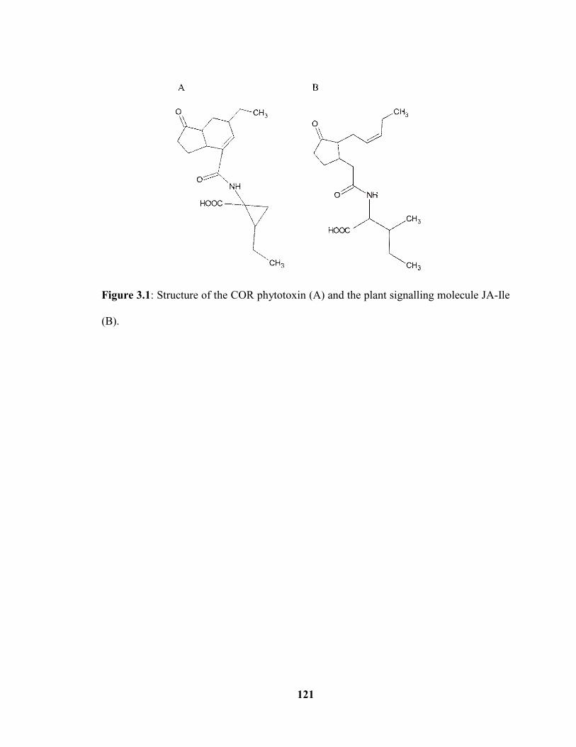
121
Figure 3.1: Structure of the COR phytotoxin (A) and the plant signalling molecule JA-Ile
(B).

122
Figure 3.2: Infiltration bioassay showing the effect of the S. scabies COR-like
metabolites on leaf plant tissue. Leaves of 6-7 week old Nicotiana benthamiana plants
were infiltrated with filter-sterilized culture supernatant of the COR-like metabolite
overproduction strain (A: txtA/pRLDB51-1) and of the COR-like metabolite non-
producing strains (B: txtA/cfa6 and C: cfl). Infiltration with 3 nmol of pure COR (D)
was performed as a positive control while infiltration with supernatant from an
uninoculated SFMB culture (E) was used as a negative control. The circled regions on the
leaf represent the area of liquid infiltration for each treatment.
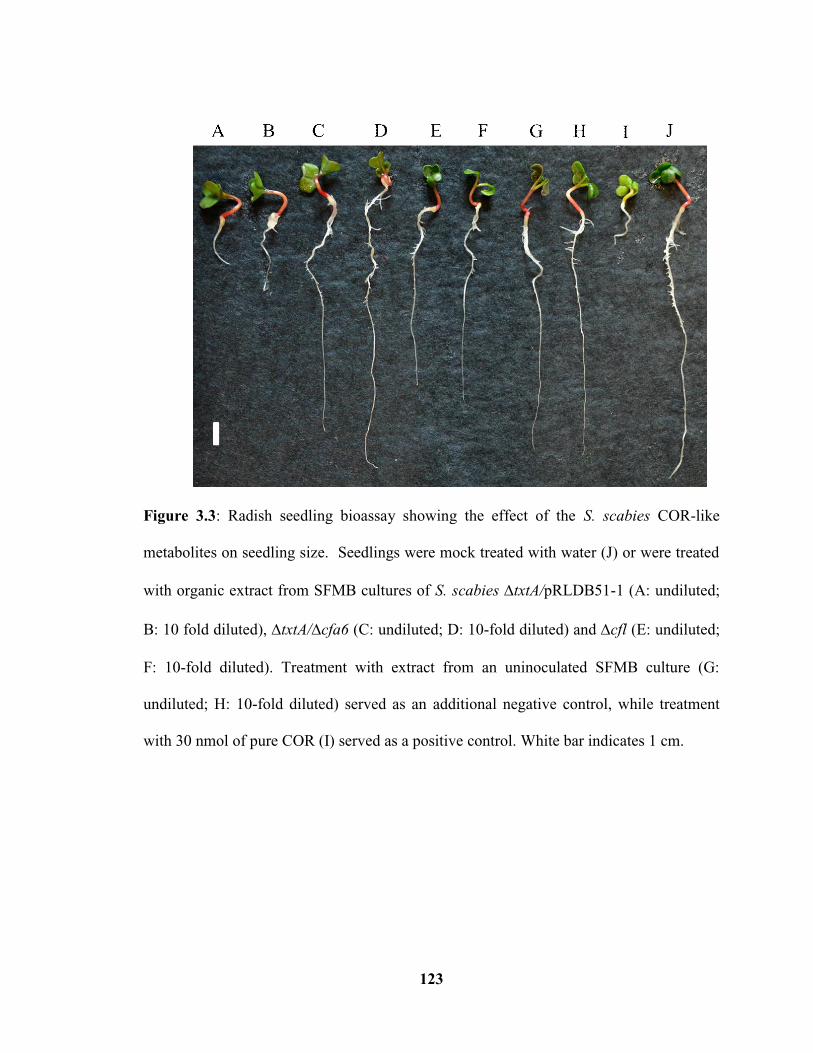
123
Figure 3.3: Radish seedling bioassay showing the effect of the S. scabies COR-like
metabolites on seedling size. Seedlings were mock treated with water (J) or were treated
with organic extract from SFMB cultures of S. scabies txtA/pRLDB51-1 (A: undiluted;
B: 10 fold diluted), txtA/cfa6 (C: undiluted; D: 10-fold diluted) and cfl (E: undiluted;
F: 10-fold diluted). Treatment with extract from an uninoculated SFMB culture (G:
undiluted; H: 10-fold diluted) served as an additional negative control, while treatment
with 30 nmol of pure COR (I) served as a positive control. White bar indicates 1 cm.

124
Figure 3.4: Quantification of radish seedling stunting by the S. scabies COR-like
metabolites. The mean root and shoot length for 10 radish seedlings per treatment is
shown, and the error bars represent the standard deviation from the mean. Seedlings were
mock treated with water (H2O) or were treated with organic extract from SFMB cultures
of S. scabies txtA/pRLDB51-1 (undiluted and 10 fold diluted), txtA/cfa6 (undiluted
and 10-fold diluted) and cfl (undiluted and 10-fold diluted). Treatment with extract from
an uninoculated SFMB culture (undiluted and 10-fold diluted) served as an additional
negative control, while treatment with 30 nmol of pure COR served as a positive control.
Treatments that produced a statistically significant result compared to the mock (H2O)
treatment are indicated by * (p 0.001) or ** (p 0.01).

125
Figure 3.5: Radish seedling bioassay showing the effect of different amounts (0.9, 9 and
90 nmol) of pure COR (dissolved in MeOH) and CFA-L-Ile (dissolved in DMSO) on
seeding root and shoot length. The mean root and shoot length for 10 radish seedlings per
treatment is shown, and the error bars represent the standard deviation from the mean.
Treatments that produced a statistically significant result compared to the relevant solvent
control treatments (MeOH and DMSO) are indicated by * (p 0.001).

126
Figure 3.6: Effect of culture supernatant of the COR-like metabolite overproduction
strain (txtA/pRLDB51-1) on radish anthocyanin production. Pure COR (30 nmol) was
used as a positive control while treatment with supernatant from COR-like metabolite
non-producing strains (txtA/cfa6 and cfl) and an uninoculated SFMB culture were
used as negative controls. Treatments that produced a statistically significant result
compared to the mock (SFMB culture supernatant) treatment are indicated by ** (p
0.05).

127
Figure 3.7: Effect of CFA-L-Ile on radish anthocyanin production. Radish seedlings
treated with different amounts (0.9, 9 and 90 nmol) of pure COR (dissolved in MeOH)
and CFA-L-Ile (dissolved in DMSO). MeOH and DMSO were used as a negative control
for the COR and CFA-L-Ile treatments, respectively. The Student’s t-test indicated a
significant difference between the treatment with 0.9 nmol of COR and the corresponding
negative control treatment (p 0.05).
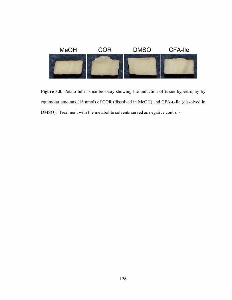
128
Figure 3.8: Potato tuber slice bioassay showing the induction of tissue hypertrophy by
equimolar amounts (16 nmol) of COR (dissolved in MeOH) and CFA-L-Ile (dissolved in
DMSO). Treatment with the metabolite solvents served as negative controls.

129
CHAPTER 4: Concluding Remarks
4.1 Summary and future directions
The research described in this thesis provides valuable insights into the
biosynthesis and function of the COR-like metabolites, which are important for the plant
pathogenic phenotype of S. scabies. In Chapter 2, it was demonstrated that the cfa8 gene,
which encodes a predicted CCR, is required for optimal production of CFA-L-Ile, and this
is likely because it is important for providing a sufficient supply of ethylmalonyl-CoA for
CFA biosynthesis. It was also shown that the scab79691 gene, which encodes a predicted
CYP450, is essential for CFA-L-Ile production, and it was proposed that this enzyme may
participate directly in CFA biosynthesis by the introduction of a hydroxyl group. This
result is of particular significance since a homologue of the Scab79691 protein does not
exist in P. syringae, and it suggests that P. syringae and S. scabies may use different
biosynthetic pathways to produce the same family of phytotoxins. In Chapter 3, it was
revealed that CFA-L-Ile from S. scabies displays some of the same toxic effects as COR
against different plant hosts, though COR is more toxic in its bioactivity. This suggests
that the S. scabies COR-like metabolites may have a similar role as COR in manipulating
plant defense responses during infection, thereby allowing the pathogen to colonize and
grow within the plant host.
A number of questions stemming from this research remain and can serve as the
basis for future studies into the S. scabies COR-like metabolites. For example, the identity
of the putative intermediate accumulating in the scab79691 mutant remains to be

130
determined, and such information is critical for validating the proposed role of Scab79691
in the biosynthetic pathway for production of CFA-L-Ile. The proposed function of
Scab79691 can also be validated by in vitro characterization of the enzyme activity
following protein overexpression and purification. In addition, the genetic
complementation of both the cfa8 and scab79691 mutants warrants further
investigation since this will confirm that the observed phenotype of the mutants is due to
the deletion of the target gene. The role of CFA-L-Ile in inducing anthocyanin production
and chlorosis also should be further investigated to determine whether this metabolite
shares all of the same bioactivities as COR. Given that COR promotes the formation of
COI1-JAZ complexes and the ubiquination and degradation of JAZ proteins in order to
upregulate JA-responsive genes (Katsir et al. 2008a, b), it would be interesting to
determine whether CFA-L-Ile has a similar function. This could be accomplished using
plant gene expression studies in order to identify the defense pathway(s) that is activated
in response to CFA-L-Ile, as well as in vitro protein interaction studies to examine the
effect of CFA-L-Ile on the formation of COI1-JAZ complexes (Katsir et al. 2008b). Of
particular interest would be a comparison of the relative activities of COR, CFA-L-Ile and
JA-Ile in promoting the formation of COI1-JAZ complexes since it has been reported
that COR is 1000 times more active than JA-Ile in this activity (Katsir et al. 2008b).
Knowledge of the biosynthesis and bioactivity of the S. scabies COR-like
metabolites contributes to the basic understanding of how S. scabies is able to infect a
plant host and cause disease. The ultimate goal of research on plant pathogenic
Streptomyces spp. is to use the knowledge gained towards the development of control

131
strategies that are more reliable and effective for scab disease management in agricultural
settings. In addition, such knowledge could have broader applications to the study of
other host-pathogen systems in agriculture and in human or animal health.
4.2 References
[1] Katsir L, Chung HS, Koo AJ, Howe GA. Jasmonate signaling: A conserved
mechanism of hormone sensing. Current Opinion in Plant Biology. 2008a;11(4):428-435.
[2] Katsir L, Schilmiller AL, Staswick PE, He SY, Howe GA. COI1 is a critical
component of a receptor for jasmonate and the bacterial virulence factor coronatine.
Proceedings of the National Academy of Sciences USA. 2008b;105(19):7100-7105.