Embed Size (px)
Citation preview

Biosafety and Cell Sorting: A Review of the New ISAC Standards
2014 New England Cytometry User Group Meeting
2 October 2014

ISAC (and other) History and Highlights
• 1981: T4 Bacteriophage method described (BD FACS II) • 1985-1988: Universal Precaution Documents issued • 1995: ISAC Biohazard Working Group formed • 1997: First ISAC Biosafety Guidelines published – Biosafety Guidelines for Sorting of Unfixed Samples (Cytometry
28:99-117, 1997) • 2001: Use of GloGerm beads as substitute for T4 – Novel Rapid Method for Visualization of Extent and Location of
Aerosol Contamination During High-Speed Sorting of Potentially Biohazardous Samples (Cytometry 43:217-222, 2001)
• 2001: Public workshop on safety issues pertaining to the clinical application of flow cytometry to human-derived cells (FDA)
• 2003: Biosafety Concerns for Shared Flow Cytometry Core facilities (Cytometry 56A:113-119, 2003) • 2003: GloGerm modification; Anderson air sampler - Measuring Containment of Viable Infectious Cell Sorting in a
High-Velocity Cell Sorter (Cytometry 52A:122-130, 2003) • 2007: Standard Safety Practices for Sorting of Unfixed Cells (Current Protocols in Cytometry (2007) 3.6.1-3.6.20 • 2007: Special Report: ISAC Biosafety Standard for Sorting of Unfixed Cells (Cytometry 71A:414-437, 2007) • 2009: NIH Cell Sorter Biosafety Task Force formed • 2011: Characterization of aerosols produced by cell sorters and evaluation of containment (Cytometry Part A 79A:
1000-1008, 2011) • 2012: NIH IBC adopts the Policy for Biosafety of Cell Sorters (approved in August, 2012 for all intramural NIH cell
sorters) • 2014: ISAC Cell Sorter Biosafety Standards (Cytometry Part A 85A: 434-453, 2014)
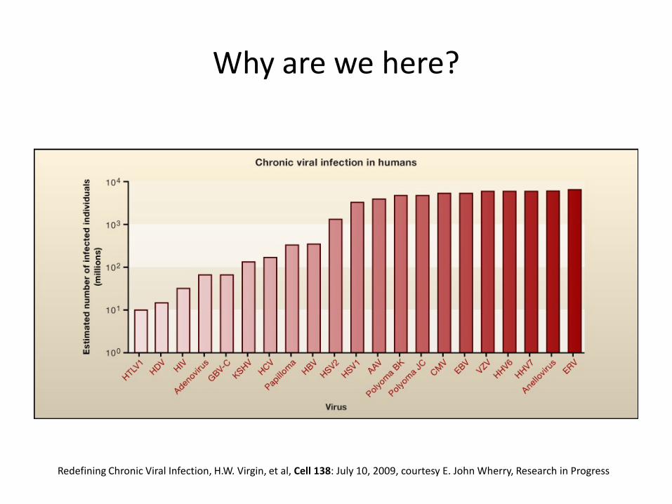
Why are we here?
Redefining Chronic Viral Infection, H.W. Virgin, et al, Cell 138: July 10, 2009, courtesy E. John Wherry, Research in Progress

Aerosols, Cell Sorters and Biosafety
• Cell sorters produce aerosols – ~40-200 µm plus smaller satellite droplets – ‘secondary aerosols of various and undefined
droplet sizes’ produced during failures (clogs) (ISAC biosafety guidelines)
Satellite droplets
From Safe Sorting: Principles and Practices, Kevin a. Holmes, Ph.D., NIAID, NIH

Size and Infectivity
• Lung Deposition – Size determines retention and deposition
Hatch, T. 1961; Bacteriol Rev 25:237
From Safe Sorting: Principles and Practices, Kevin a. Holmes, Ph.D., NIAID, NIH

Size, Alveolar Deposition and Infectivity
Vincent, J.H., 2005, J. Environ. Monit. 7:1037; Brosseau, L.M., et al. 1994 ASHRAE Transactions 100:368; Fennelly, K.P. , et al. 2004 Am J Respir Crit Care Med 169:604.
Alve
olar
dep
ositi
on e
ffici
ency
(F
ract
ion
of to
tal i
nhal
ed
Aerodynamic diameter (µm)
AD range associated with Infection

70, 35 and 20 PSI (3.0cm UV+)
0.01
0.1
1
10
100
1000
0.5 5
70 psi 35 psi 20 psi
Aerodynamic Diameter (µm)
Conc
entr
atio
n (#
/cm
3)
9.8x10³/cm3 mean: 2.0 µm
1.9x10³/cm3 mean: 2.4 µm
90/cm3 mean: 2.65 µm
Range of alveolar deposition and infectivity
From Safe Sorting: Principles and Practices, Kevin a. Holmes, Ph.D., NIAID, NIH

Laboratory Acquired Infections (LAI)
• Laboratory Procedure hazards and LAI’s – 5 routes of laboratory transmission:
1. Parenteral inoculations: syringe needles/sharps 2. Spills/splashes on skin or mucous membranes 3. Ingestion through mouth pipetting 4. Animal bites & scratches 5. Inhalation exposure to infectious aerosols
• 1-4 account for <20% of LAI’s • The cause of 82% of LAI’s are unknown, but are presumed to be aerosols • LAI’s are thought to be significantly underreported

Hazards in Cell Sorting Labs
• Although no documented case of LAI in cell sorter lab, consider the following: 1. Cell Sorting is a Laboratory Procedure Hazard
• Aerosol generation 2. “General agreement among biosafety professionals…
that an aerosol generated by procedures and operations…is the probable source of many LAI’s”
• BMBL, 5th Ed. 3. When things go wrong, the operator has the opportunity
to breathe whatever is in the sample tube • Rich Schretzenmair at Penn

Aerosols and Cell Sorters: Summary
• Sorters can produce high concentrations of aerosols – Published study has found that at 70psi, aerosols with concentration
of 18000/cm3 can be produced in fail condition. – These aerosols are between 1-5µm aerodynamic diameter
• Higher sheath pressure increases concentration and decreases size • Aerosols in this size range, i.e. 1-3µm:
– May remain airborne almost indefinitely – More likely to deposit in lung alveoli – Have been shown to be associated with increased infectivity of some
organisms – Aerosols are presumed to be responsible for the large majority of LAI’s
From “Ask the Experts Biosafety Tutorial”, CYTO 2014

Biosafety Principles
• Two basic principles of biosafety are Containment and Risk Assessment – Containment
• Reduce or eliminate exposure of lab workers and outside environment to potentially hazardous agents
• Examples: Biological Safety Cabinet, centrifuge cups, aerosol management on cell sorter
• Includes procedures, i.e. standard microbiological practices

Biosafety Principles
– Risk Assessment: • Definition: an action or series of actions taken to
recognize or identify hazards and to measure the risk or probability that something will happen because of that hazard.
• Severity of the consequences is also taken into account. • Requires careful judgment
– Consequences if risks are underestimated – Excessive safeguards
• Additional expense and burden with little safety enhancement
• May result in circumvention because of excess burden

Containment Testing

Containment Testing
• The engineering controls (e.g. sort chamber door) and aerosol evacuation system must be tested to verify containment
• The escape of aerosolized Fluorescent beads run as a sample under fail mode is tested
• Most accepted method uses GloGerm beads and Aerotech impactor collection device

GloGerm Test (still the standard)
• Fluorescent GloGerm beads washed in EtOH, store in PBS with Triton + FCS
• Aerotech air samplers with glass slides placed at locations around sort chamber
• GloGerm beads run as sample in fail mode under two conditions: 1. AMS operational, all sort and collection chamber doors
closed 2. AMS off, sort chamber slightly ajar (positive control)
• Slides scored with fluorescent microscope

Microscopic View of Glo-Germ

AeroTech Aerosol
Concentrator

AeroTech Device


Air Sampler with Airflow Meter

Containment Test Procedure (GloGerm SOP)
• Analyze particles at 20,000/sec with deviated stream for max. aerosol production, “failure” mode.
• Analyze at same pressure as sample sorts. • Operate Aerotech at 28.3LPM • Collect particles for at least 5 min. at each location. • Examine slide for particles.
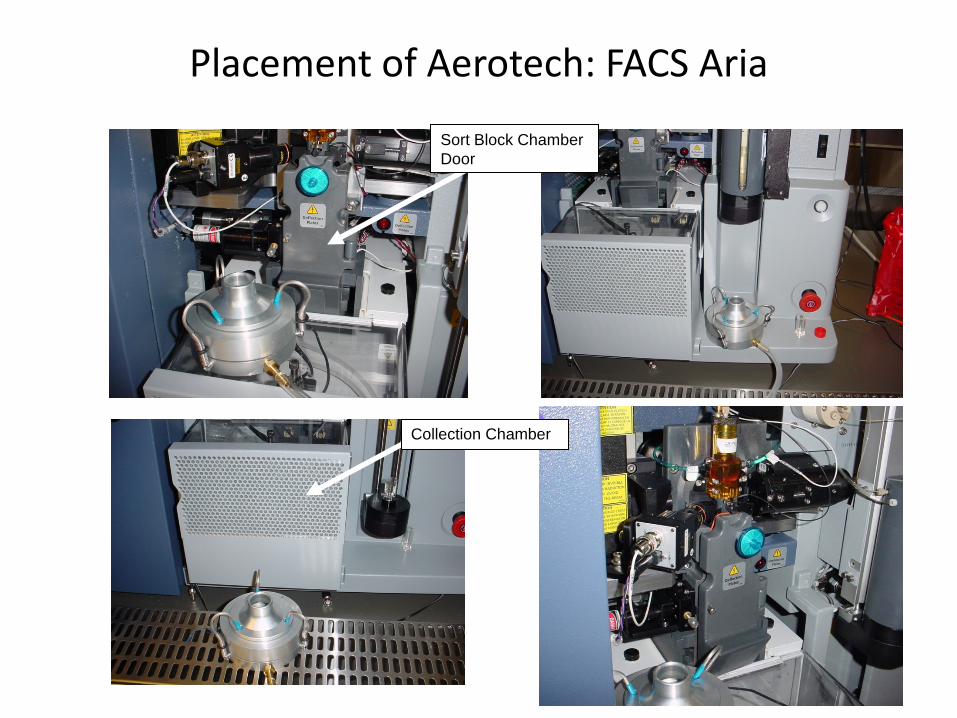
Placement of Aerotech: FACS Aria
Collection Chamber
Sort Block Chamber Door

Critical Tolerances
• Aerosol Management System Tolerance (BD Buffalo filter) = Less than 2.4 inches of H2O. Above this tolerance, replace tubing and HEPA filter
• Particles outside = Zero tolerance, no particles on entire slide. Any positive result must be investigated, resolved and retested before proceeding with infectious sort.
• Positive Control= Greater than 50 per field (10x objective field, result may vary with slide location).

GloGerm Procedures Tips
• Wear a respirator during testing • Have two Aerotech devices: one for positive control and one
for tests • Perform the test condition first, followed by the positive
control • Use positive control slide to adjust microscope for focusing
and observe deposition pattern • Scan slides on both low and high mag (20x & 40x)

Containment testing: When
1. Based upon Risk Assessment, but must be performed: 2. Following instrument service or maintenance involving the
sort chamber and/or AMS hose connections. 3. Following initial instrument installation or relocation. 4. Following change out of the standalone AMS filter. 5. For BSL3/4 labs:
1. Prior to every sort if the frequency of sorting is once/week or less
2. Weekly, if the frequency of sorting is multiple sorts/week

The CyClex-d Disposable bioaerosol sampler (cassette) Environmental Monitoring Systems (EMS)
• A 360 degree impaction chamber, where the aerosol is captured directly onto a slide, allowing us to go almost directly from sample collection to analysis – Suitable collection efficiency for particulates in the respirable
range (d50 is about one micron) – Direct and rapid transition from collection to analysis – Sensitive quantification (over passive settling)

Polysciences YG Beads
• Particle size and shape are known • Particle density is known (close to the density of water) • Hopefully a 1 micron YG sphere behaves like a 1 micron
sphere of water (or saline) • A given diameter YG bead should behave in a more uniform
manner versus Glo Germ, where particles of variable size and density may behave like a gas, a respirable aerosol, or a droplet

TSI Instruments UV-APS
355nm Laser (diode-pumped Nd:YAG freq tripled laser)
From Safe Sorting: Principles and Practices, Kevin a. Holmes, Ph.D., NIAID, NIH
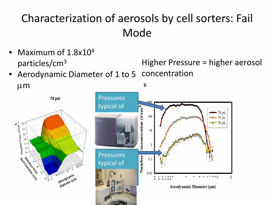
Characterization of aerosols by cell sorters: Fail Mode
Higher Pressure = higher aerosol concentration
• Maximum of 1.8x10⁴ particles/cm3
• Aerodynamic Diameter of 1 to 5 µm
Pressures typical of sorter ca 2000’s
Pressures typical of sorter ca 1990’s

Comparison of 0.75, 1.0 and 2.0µm YG beads 0.75µm: Min=0.9; Mean AD=3.3
1.0µm: Min=1.1; Mean AD=3.4
2.0µm: Min=2.1; Mean AD=3.5
Courtesy Kevin Holmes, NIAID, NIH

CBD (UV dye); no beads. Note correlation of AD and Fluorescence
Courtesy Kevin Holmes, NIAID, NIH
Risk Assessment

5 Steps of Risk Assessment
1. Identify agent hazards 2. Identify laboratory procedure hazards 3. Make final determination of biosafety level 4. Evaluate proficiencies of staff and integrity of safety
equipment 5. Review risk assessment with a biosafety
professional

Risk Assessment Step 1: Agent Hazards
• Classification of microbiological agents into Risk (or Hazard) Groups based upon:
• Ability to infect and cause disease • Virulence (severity of disease) • Availability of preventative measures (such as vaccines) and
effective treatments – Four Risk Groups 1-4 by WHO, Canada, Australia, European
Union and The NIH Recombinant DNA Advisory Committee – See http://www.absa.org/riskgroups/ for more
information – Also: PSDS (Public Health Agency of Canada
http://www.phac-aspc.gc.ca/msds-ftss )

Agent Hazards: recombinant and viral vectors • Agent Hazards
– Genetically modified agent hazards • Samples with recombinant DNA, viruses, bacteria or yeast, and viral vectors:
lentiviral, adenoviral or retroviral vectors • Not classified in NIH/CDC’s BMBL or WHO Laboratory Safety Manual
– NIH Recombinant DNA Advisory Committee • “NIH Guidelines For Research Involving Recombinant DNA Molecules (NIH
Guidelines)” • Risk Group Classification 1-4 • Guidance documents on lentiviruses
– See http://oba.od.nih.gov/rdna_ibc/
– European Union directives on genetically modified micro-organisms (GMM)
• See http://www.biosafety-europe.eu/d20public_300309.pdf for implementation by member countries
• Risk assessment guidance: http://www.hse.gov.uk/biosafety/gmo/acgm/ecrisk.htm

Risk Assessment Step 2: Procedure Hazards
• Laboratory Procedure Hazards – 5 routes of laboratory transmission:
1. Parenteral inoculations: syringe needles/sharps 2. Spills/splashes on skin or mucous membranes 3. Ingestion through mouth pipetting 4. Animal bites & scratches 5. Inhalation exposure to infectious aerosols
• Routes 1-4 account for <20% of LAI’s • Aerosols are serious hazard

Risk Assessment Step 3: Determination of Biosafety Level
• Make final determination of biosafety level and select additional precautions as indicated by risk assessment
• Biosafety or containment levels – NIH/CDC and WHO: BSL1, 2, 3 and 4 (UK: CL1,2,3
and 4). – Risk Groups correlate with but do not equate to
biosafety levels. • Intended use of agent may suggest greater precautions
– Example: aerosol production in cell sorting

Risk Assessment Step 4: Evaluate staff proficiencies and safety equipment
– Proper training and/or certification of personnel is essential
– Equipment must be available and functional • Aerosol management system on cell sorters • Droplet containment systems on analyzers • Personal Protective Equipment (PPE)

Risk Assessment Step 5: Review
• Review Risk Assessment with safety professional, subject matter expert and IBC – Sometimes required by regulatory agency – Cell sorters: not specifically regulated – Safety is a culture: schedule regular reviews with
staff and local safety professionals

Guidelines for Risk Assessment in Cell Sorting
• Takes into account both agent hazards and sample origins for assignment of biosafety containment levels and procedures
• Therefore combines: – Agent Risk Group classifications (i.e. WHO and others) – Standard Precautions (OSHA Blood Borne Pathogen
Standard 1910.1030) – the recognition of cell sorting as a laboratory procedure
hazard

Guidelines for Risk Assessment in Cell Sorting
BSL2 BSL2 w/enhanced precautions
BSL3 BSL4
Risk Assessment Condition
Uninfected non-primate Non-infectious Human /NHP cells Infectious but with low risk assessment
Infectious samples with high risk assessment All samples containing known aerosol pathogens
Extremely Dangerous Pathogens
Example Sample type or Agents
Normal murine cells 3rd gen Lentivirus (non-human cells)
Normal human blood Human cell lines Influenza A 2nd gen Lentivirus or 3rd gen in human cells
Mycobacterium tuberculosis Monkeypox other Class 3 agents
Ebola
Containment system validation
Periodically Periodically
Prior to each sort Prior to each sort
Aerosol containment operating
Required Required Required Required
Respirator Optional N-95 or better PAPR Special suit
Eye Protection Safety Glasses Face Shield or safety goggles
Integral with PAPR Integral with Suit
Lab Coat Front Closure Wrap Around rear closure Coveralls Special suit
Separate room Optional Required or limited access to room2
Required Required

Biosafety levels for cell sorting by Agent (examples)
Agent Recommended Biosafety Level
Restrictions or Comments
Hepatitis C BSL2/3 Influenza A (strain PR8) BSL2/3 Influenza (seasonal) vaccine required Klebsiella pneumonia BSL2/3 LaCrosse virus BSL2/3
LCMV BSL2/3 or BSL3 BSL dependent upon strain; pregnant women excluded from lab during sorting
Leishmania BSL2/3* Malaria BSL2/3* Respiratory Syncytial Virus BSL2/3
Toxoplasma gondii BSL2/3* pregnant women excluded from lab during sorting
Vaccinia BSL2/3 vaccine required Avian influenza BSL3 Influenza (seasonal) vaccine required H1N1 BSL3 H1N1 vaccine required HIV BSL2/3 or BSL3 Monkeypox BSL3 vaccinia vaccine required, every 3 years TB, Mycobacterium tuberculosis BSL3
Here, BSL2/3 means BSL2 with enhanced precautions, as published in Table 2 of the 2014 ISAC Cell Sorter Biosafety Standards

Sorting human cell lines
• “Do we really need to treat the sorting of human cells lines like 293T, Hela, BJAB, U937, K562, KG1a cells as human primary samples?”
• Answer: Human cell lines are sorted at BSL2 w/enhanced precautions • Background:
– Ask the Experts-Biosafety Requirements for Human Cell Lines (Keene, J.H., Applied Biosafety: 8, No.2, 2003)
– Letter from OSHA to ABSA president: interpretation of BPS (29 CFR 1910.1030) for human cell lines
– “Established human cell lines * which are characterized * * to be free of contamination from human hepatitis viruses, human immunodeficiency viruses, and other recognized bloodborne pathogens, are not considered to be OPIM and are not covered by BPS…All primary human cell explants from tissues and subsequent in vitro passages…handled with the BPS.”
– OSHA leaves it to interpretation by local biosafety office
From the CYTO 2013 Workshop “Biosafety Policy Meets Real Life Scenarios”

Standard Operating Procedure (SOP) Development
• Identify hazards and specify practices to minimize hazards
• Process of writing SOP forces critical evaluation of equipment and procedures
• Instrument design specific features determines procedures – How is it designed? What are deficiencies? – Evaluation of containment vs. evacuation

SOP Development: Cell sorting 4 major sections
• Preparation before the sort • PPE requirements • Procedures in the event of a nozzle obstruction • Decontamination procedures
SOP Development: Cell sorting
• Preparation before sort – Check fluids, empty waste – Containment testing – Verification of automated decontamination procedures – Preparation of decontamination reagents – Spare nozzle availability – Filter Samples!

SOP Development: Cell sorting
• PPE required – All Biosafety levels:
• Gloves (single or double) • Lab coat • Safety glasses or face shield
– BSL2 w/enhanced precautions: • N-95 Respirators or PAPR • Wrap around rear-closure gown • Double glove dependent upon risk assessment
– BSL3: • PAPR

SOP Development: Cell sorting
• Failure or nozzle clog – Most dangerous – Automated detection or not
• Turn off stream – Four Goals:
• Stop aerosol formation • Evacuate aerosols • Clear nozzle obstruction • Check aerosol containment system

SOP Development: Cell sorting
• Decontamination – Automated or manual – Type of disinfectants – Surface cleaning

1. Operator training
• Staff proficiency in cell sorter operation and in biosafety procedures – Training essential component of cell sorter lab operation – Amount of training tied to risk assessment
• The higher the risk, the more experience/training required • Inexperienced operator more likely to circumvent safety procedures
• Review of SOP at regular intervals – Review/practice procedures in event of nozzle clog
• Nozzle clogs: Good news and Bad news • Newer sorters may clog less: Data from Aria showed that of 580 samples,
there were 17 nozzle clogs (2.9%). Of these, ~75% were unfiltered. – Newer sorting technology may clog less often: Good because aerosol production is less
probable – Newer sorting technology may clog less often: Bad because the operator is less likely to
remember the proper procedures in the event of a nozzle clog – POST the SOP section for nozzle obstructions PROMINENTLY
From the CYTO 2014 Ask the Experts Tutorial

2. Cell Sorters in BSC’s
• Need for a BSC is dependent upon risk assessment • Sorters cannot just be placed in a BSC: must be certified
– 2014 Standards: “must be manufactured to meet functional certification criteria for personnel and product protection as defined by NSF 49 (US or CSN EN 12469 (Europe) or JIS K 3800: 2009 (Japan) or AS 2252.2 (Australia).”
• Can abrogate requirement for separate room for sorter and requirement for PPE (respirators) for all occupants in the shared lab
• Does not eliminate need for AMS

Resources
• International Society for the Advancement of Cytometry Cell Sorter Biosafety Standards – Cytometry Part A 85A: 434-453, 2014
• ISAC web page (isac-net.org) – Resources for Cytometrists -> Biosafety – Lots of links to references, and the latest revisions to the 2014
Standard
• CYTO U – cytou.peachnewmedia.com – Course: Flow Cytometry Biosafety

CYTO University

ISAC

Where we’re going…
• Develop a repository of instrument specific SOP’s on the ISAC web page
• Develop a new rapid, simple aerosol containment assay (the most important outstanding issue)
• Engage your local biosafety professionals, especially with risk assessment
Acknowledgements
• Kevin Holmes • Ben Fontes • Phil Hogarth • Rich Konz • Simon Monard • Rob Wadley • Ingrid Schmid • Steve Perfetto